Communication

Schiff Base Heterobimetallic Complex as Single-Source Precursor

by
Jocelyn Pradegan
,
Aurélien Crochet
and
Katharina M. Fromm
*
Department of Chemistry, University of Fribourg, Chemin du Musée 9, 1700 Fribourg, Switzerland
*
Author to whom correspondence should be addressed.
Molbank 2025, 2025(4), M2092; https://doi.org/10.3390/M2092
Submission received: 20 October 2025 / Revised: 14 November 2025 / Accepted: 18 November 2025 / Published: 20 November 2025

Abstract

A bimetallic complex based on a salophen-type ligand was synthesized. The compound was characterized by ESI-MS, single-crystal, and powder X-ray diffraction. The heterobimetallic complex was thermally treated to investigate its capacity as a single-source precursor for the formation of mixed metal oxides.

Graphical Abstract

1. Introduction

Salphen (or salophen) is a Schiff base compound formed by a double condensation reaction between o-phenylenediamine and salicylaldehyde [1]. Salphen possesses a tetradentate N2O2 coordination site that can bind one metal ion. Based on salphen’s conjugation, it can coordinate to d0 or d10 metal ions to form complexes and exhibit fluorescence properties generally used for sensing applications [2,3].
With its straightforward synthesis, the starting compounds can be easily replaced by derivatives to generate a panoply of new structures. Shao et al. reported salphen-derived ligands functionalized with glycol chains in a meta position of the salicyaldehyde and its heterobimetallic complexes used for catalytic application [4].
In our study, we report the formation of a heterobimetallic complex based on the same ligand but using different metal ions. The crystal structure of the heterobimetallic complex was determined by single-crystal X-ray diffraction. Furthermore, its ability as a single-source precursor (SSP) for the formation of mixed metal oxide (MMO) was investigated.

2. Results and Discussion

2.1. Synthesis of the LCuCa Complex

The precursor L″ was synthesized via the Williamson ether synthesis by adaptation of the literature procedure [5]. The monometallic LCu complex was formed via a one-pot reaction, where a double condensation reaction between L″ and o-phenylenediamine took place, creating a N2O2 site occupied by the Cu2+ ion upon deprotonation of the phenol group (Scheme 1). The ligand L in the LCu complex adopts an Ω-shape, thereby affording a second O3O3, site formed by the O-atoms of the glycol chains and the phenoxy groups, which can be, e.g., occupied by an oxophilic metal ion, such as a Ca2+ ion. The heterobimetallic complex was characterized by ESI-MS and single-crystal and powder X-ray diffraction. 1H and 13C NMR could not be performed on the complexes as the Cu2+ ion is paramagnetic.

2.2. X-Ray Crystal Structure

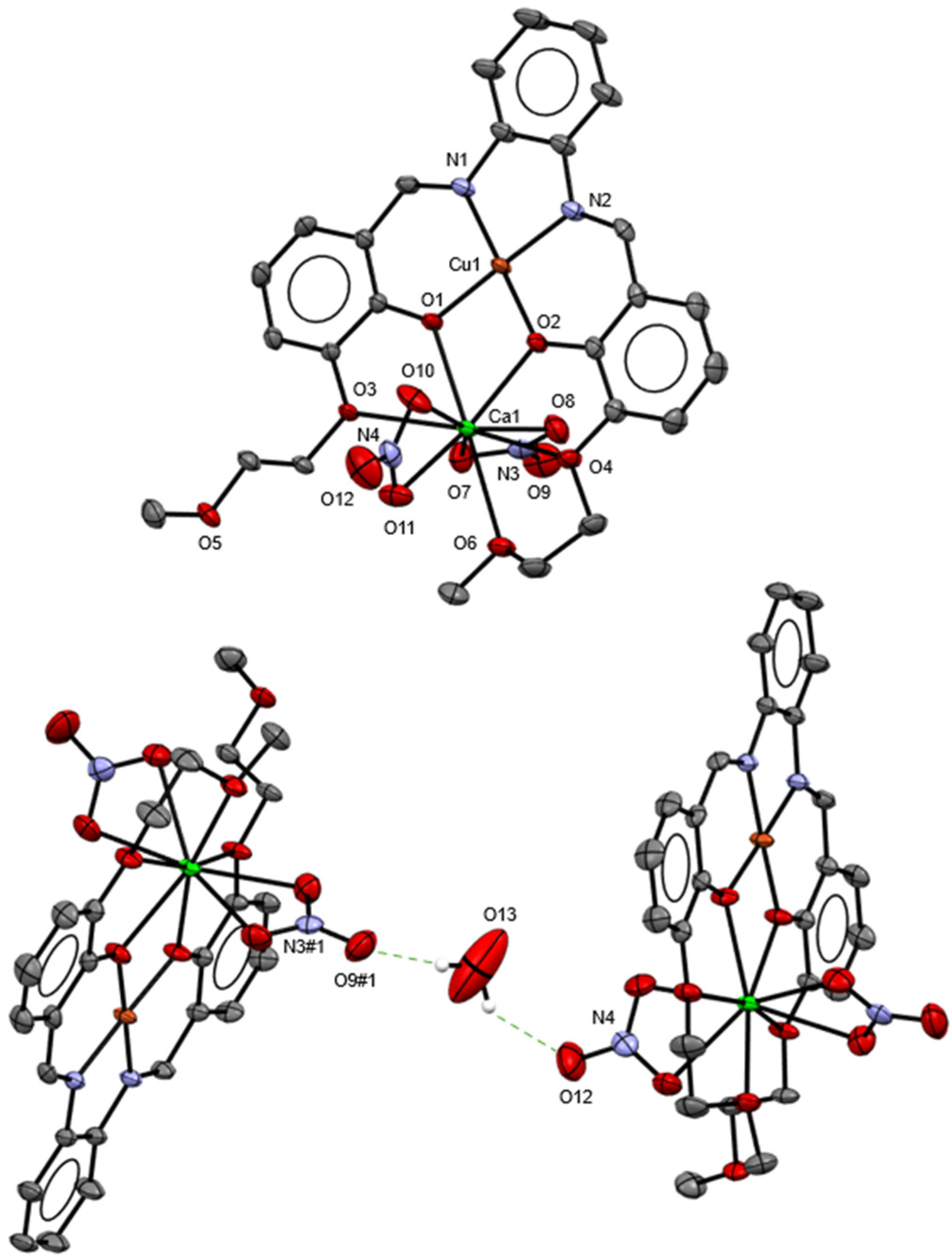
Red-plate-like single crystals of [CuCaL(NO3)2]. (H2O), abbreviated as “LCuCa”, were obtained by slow evaporation of the mother liquor (MeOH). The complex crystallized in the monoclinic P21/n space group (No. 14). Crystal data and structure refinement are available in the Supporting Information (Table S1). The complex has a 1:1:1 (L:M1:M2) stoichiometry. The Cu2+ ion is coordinated by the tetradentate N2O2 chelate site of the ligand in a quasi-perfect square planar geometry with an angle sum close to 360°. The bond valence sum [6] (BVS) of Cu2+ has a value of 2.35, meaning that its coordination sphere is completed. The Ca2+ ion is coordinated by five O-atoms (O1, O2, O3; O4, and O6) of the O3O3 cavity of the ligand. The last O-atoms (O5) points away from the binding cavity. The coordination sphere of Ca2+ is completed by the coordination of two nitrate anions in axial positions, leading to a BVS of 2.16. The asymmetric unit of LCuCa contains one water molecule (O13), which acts as a bridge between two complexes via the formation of H-bonds with the two non-binding O-atoms (O9 and O12) of the nitrate anions with a graph set  C 2 2 (10) [7] (Figure 1).

2.3. Single-Source Precursor

Thermogravimetric analyses (TGA) were conducted on LCuCa from 25 °C until 1000 °C with an increase of 10 °C per minute under air (50 mL/min) (Figure S1). The thermally treated powder was analyzed by X-ray powder diffraction (XRPD). The first loss until (3.3%) between 25 and 250 °C is the evaporation of remaining solvent. The second step (32.7%) at 340 °C corresponds to the partial decomposition of the ligand. It is followed by the first loss of one nitrate moiety (10.3%) around 360 °C. The second part of the ligand is then decomposed (21.3%), followed by the loss of the second nitrate (8.9%). The last step (6.1%) corresponds to the loss of CO2.
The residual powder was analyzed by XRPD and revealed the formation of two mixed metal oxides, Ca2CuO3 and CaCu2O3 (Figure S4). Teske and Müller–Buschbaum synthesized the mixed metal oxide CaCu2O3 using CaO and CuO, with a (1:2) ratio, as starting materials [8]. The compounds were heated at different temperatures (500 °C and 800 °C), and the formation of the Ca2CuO3 oxide by calcinating CaCO3 and CuO at 960 °C for 10 h in air was reported [9].
Compared with the methods reported in the literature, similar calcination temperatures were used in our study. However, our time of calcination (1 h) is shorter compared to that in the literature (10 and 24 h). The obtained MMOs can be used as model systems to study the magnetic interactions in cuprate superconductors by investigating their magnetic [10,11] and superconductivity [12,13] properties.

3. Materials and Methods

3.1. Generalities

All starting materials were purchased commercially and used without any further purification. All the experiments were conducted in air at room temperature (RT). A Bruker® (Billerica, MA, USA) 400 MHz spectrometer was used for 1H and 13C nuclear magnetic resonance (NMR) analyses. Electrospray ionization mass spectrometry (ESI-MS) measurements were carried out in positive ion mode on a Bruker Esquire HCT Ion Trap Mass Spectrometer. High-resolution electrospray ionization mass spectrometry (HR-ESI-MS) was performed under positive ion mode on a Bruker timsTOF Pro. The thermogravimetric analysis was performed using a Mettler Toledo (Greifensee, Switzerland) TGA/DSC 3+ STARe system. Single-crystal X-ray diffraction data were collected on a STOE (Darmstadt, Germany) STADIVARI diffractomer using Cu Kα radiation (λ = 1.54186 Å). A suitable crystal was selected, mounted on an oil-coated loop, and maintained at 250(2) K during data collection. Using Olex2 [14], the structure was solved with a SHELXT [15] structure solution program using Intrinsic Phasing and refined with the SHELXL [16] refinement package using least square minimization. The Crystallographic Information File (CIF) of LCuCa is available in the Cambridge Crystallographic Data Centre (CCDC), CCDC-2490579.

3.2. Synthesis of 2-methoxyethyl 4-methylbenzensulfonate, L′

L′ was synthesized as described in the literature [17]. 2-Methoxyethanol (5.0 g, 1.0 eq., 65.7 mmol) was dissolved in THF (100 mL) and cooled in an ice bath. When the solution reached 0 °C, p-toluenesulfonyl chloride (18.8 g, 98.6 mmol, 1.5 eq.) was added. The mixture was stirred at RT for five minutes. A potassium hydroxide (KOH) (29.5 g, 525.6 mmol, 8.0 eq.) solution (7M) was prepared in distilled water (75 mL). The KOH solution was added dropwise to the reaction mixture at 0 °C. After complete addition, the mixture was stirred overnight at RT. THF was removed under reduced pressure. Ammonium chloride (26.75 g) was dissolved in water (100 mL) to obtain a 5M solution, which was added to the mixture. Then, extraction was performed with dichloromethane (DCM) (5 × 50 mL). After collecting all the organic phases, they were dried with dried magnesium sulfate (MgSO4) and filtered, and DCM was removed under reduced pressure. 2-Methoxyethyl 4-methylbenzenesulfonate, L′, was obtained as a transparent pale green oil. Yield: 96. 1H NMR (400 MHz, DMSO-d6) δ 7.79 (d, J = 7.2 Hz, 2H), 7.48 (d, J = 7.4 Hz, 2H), 4.12 (m, 2H), 3.49 (m, 2H), 3.18 (s, 3H), 2.42 (s, 3H). 13C NMR (101 MHz, DMSO-d6) δ 144.88, 132.40, 130.10, 127.57, 69.69, 69.23, 57.90, 21.03. ESI-MS calculated m/z for C10H14O4S + Na+ ([M + Na]+) = 253.1, found 253.2.

3.3. Synthesis of 2-hydroxy-3-(2-methoxyethoxy)benzaldehyde, L″

The Williamson ether synthesis reaction was carried out by an adaptation of the literature procedure [5]. Sodium hydride (NaH) (3.1 g, 2.1 eq., 76.0 mmol) was placed in a two-neck round-bottom flask equipped with a dropping funnel and then purged with argon. The flask was placed in an ice bath, and dry dimethyl sulfoxide (DMSO) (10 mL) was added. 2,3-dihydroxybenzaldehyde (5.0 g, 1.0 eq., 36.2 mmol) was placed in a round-bottom flask with a septum and then purged with argon before the addition of dry DMSO (10 mL). This solution was transferred to the dropping funnel and added dropwise during 30 min to the NaH solution under a flow of argon. The solution turned dark orange after complete addition and was stirred for 90 min at RT. L′ (8.3 g, 1.0 eq., 36.0 mmol) was added in a single portion, and the reaction mixture was stirred for 150 min at RT. The reaction was quenched by slow addition of water (50 mL), followed by an extraction with DCM (3 × 30 mL). A solution of 1M hydrochloric acid (HCl) (15 mL) was added to acidify the aqueous phase to reach a pH of 2. It was then extracted with DCM (3 × 30 mL). After collecting all the organic phases, they were washed with 1 m HCl (2 × 30 mL), dried with dried MgSO4, and filtered. Evaporation of DCM under reduced pressure afforded an orange–brown oil, which was purified by silica gel column chromatography (pentane: ethyl acetate (7:3)) to yield L″ as a pale green–yellow solid. Yield: 80%. 1H NMR (400 MHz, DMSO-d6) δ 10.23 (s, 1H), 7.31 − 7.23 (m, 2H), 6.91 (t, J = 7.9 Hz, 1H), 4.20 − 4.13 (m, 2H), 3.73 − 3.66 (m, 2H), 3.32 (s, 3H). 13C NMR (101 MHz, DMSO-d6) δ 192.70, 150.88, 147.50, 122.49, 121.05, 119.28, 119.25, 70.26, 68.33, 58.18. ESI-MS calculated m/z for C10H12O4 + H+ ([M + H]+) = 197.1, found 197.4, ESI-MS calculated m/z for C10H12O4 + Na+ ([M + Na]+) = 219.1, found 219.0.

3.4. Synthesis of LCu Complex

L″ (0.5 g, 1.0 eq., 2.6 mmol) was dissolved in ethanol (EtOH) (50 mL). o-Phenylenediamine (0.14 g, 0.5 eq., 1.3 mmol) was dissolved in EtOH (5 mL) and added to the previous solution. The solution turned dark yellow immediately. The solution was stirred overnight at RT. The solution became dark orange. Cu(NO3)2·3H2O (0.32 g, 0.5 eq., 1.3 mmol) was dissolved in EtOH (5 mL) and added to the dark orange solution. A dark green precipitate formed immediately after addition of the metal salt solution. The mixture was stirred for two hours at RT. The precipitate was filtered and dried under a vacuum. Yield: 40%. ESI-MS calculated m/z for C26H26N2O6Cu + Na+ ([M + Na]+) = 548.1, found 548.1.

3.5. Synthesis of LCuCa Complex

LCu (0.13 g, 1.0 eq., 0.25 mmol) was dissolved in MeOH (50 mL), and Ca(NO3)2·4H2O (0.06 g, 1.0 eq. 0.25 mmol) was added to the solution. A brown precipitate formed immediately after addition of the metal salt solution. The mixture was stirred for two hours at RT. The precipitate was filtered and dried under a vacuum. Yield: 52%. ESI-MS calculated m/z for C26H26N3O9CuCa+ ([M]+) = 627.1, found 627.0. HR-ESI-MS calculated m/z for C26H26N2O6CuCa2+ ([M]2+/2) 282.53509, found 282.53536, calculated m/z for C26H26N2O6Cu + Na+ ([M + Na]+) 548.09790, found 548.07922, calculated m/z for C26H26N2O6Cu + K+ ([M + K]+) 564.07184, found 564.07113.

4. Conclusions

A heterobimetallic complex based on a salophen-type ligand was synthesized and characterized by single-crystal X-ray diffraction. The ability of the LCuCa complex as SSPs was investigated. After thermal treatment of the LCuCa complex at 1000 °C for one hour in air, the formation of two mixed metal oxides, Ca2CuO3 and CaCu2O3, was observed.

Supplementary Materials

Table S1. Crystal data and structure refinement; Figure S1. TGA of LCuCa; Figure S2. X-ray powder diffraction pattern of LCu; Figure S3. X-ray powder diffraction pattern of LCuCa; Figure S4. X-ray powder diffraction pattern of thermally treated LCuCa; Figure S5. ESI-MS spectrum of L′; Figure S6. ESI-MS spectrum of L″; Figure S7. ESI-MS spectrum of LCu; Figure S8. ESI-MS spectrum of LCuCa; Figure S9. HR-ESI-MS spectrum of LCuCa; Figure S10. 1H NMR spectrum of L′ in DMSO-d6; Figure S11. 13C NMR spectrum of L′ in DMSO-d6; Figure S12. 1H NMR spectrum of L″ in DMSO-d6; Figure S13. 1C NMR spectrum of L″ in DMSO-d6.

Author Contributions

Conceptualization, J.P. and K.M.F.; methodology, J.P.; validation, K.M.F.; formal analysis, J.P. and A.C.; investigation, J.P.; resources, K.M.F.; data curation, J.P. and A.C.; writing—original draft preparation, J.P.; writing—review and editing, J.P., A.C. and K.M.F.; supervision, K.M.F.; project administration, K.M.F.; funding acquisition, K.M.F. All authors have read and agreed to the published version of the manuscript.

Funding

This research was funded by the Swiss National Science Foundation (Project numbers 2000020_172777 and 200020_204215) and the University of Fribourg.

Data Availability Statement

Supplementary crystallographic data are available in the Cambridge Crystallographic Data Centre under the code CCDC-2490579.

Acknowledgments

The authors would like to thank the synthesis laboratory of the Chemistry Department of the University of Fribourg for their help.

Conflicts of Interest

The authors declare no conflicts of interest.

References

  1. Asatkar, A.K.; Tripathi, M.; Asatkar, D. Stability and Applications of Coordination Compounds; Srivastva, A.N., Ed.; IntechOpen: Rijeka, Croatia, 2020. [Google Scholar]
  2. Khan, T.; Vaidya, S.; Mhatre, D.S.; Datta, A. The Prospect of Salophen in Fluorescence Lifetime Sensing of Al3+. J. Phys. Chem. B 2016, 120, 10319–10326. [Google Scholar] [CrossRef] [PubMed]
  3. Sahudin, M.A.; Su’ait, M.S.; Tan, L.L.; Lee, Y.H.; Karim, N.H.A. Zinc(II) salphen complex-based fluorescence optical sensor for biogenic amine detection. Anal. Bioanal. Chem. 2019, 411, 6449–6461. [Google Scholar] [CrossRef] [PubMed]
  4. Shao, H.; Muduli, S.K.; Tran, P.D.; Soo, H.S. Enhancing electrocatalytic hydrogen evolution by nickel salicylaldimine complexes with alkali metal cations in aqueous media. Chem. Commun. 2016, 52, 2948–2951. [Google Scholar] [CrossRef] [PubMed]
  5. Van Staveren, C.J.; Van Eerden, J.; Van Veggel, F.C.J.M.; Harkema, S.; Reinhoudt, D.N. Cocomplexation of neutral guests and electrophilic metal cations in synthetic macrocyclic hosts. J. Am. Chem. Soc. 1988, 110, 4994–5008. [Google Scholar] [CrossRef]
  6. Brese, N.E.; O’Keeffe, M. Bond-valence parameters for solids. Acta Crystallogr. B Struct. Sci. 1991, 47, 192–197. [Google Scholar] [CrossRef]
  7. Etter, M.C. Encoding and decoding hydrogen-bond patterns of organic compounds. Acc. Chem. Res. 1990, 23, 120–126. [Google Scholar] [CrossRef]
  8. Teske, C.L.; Müller-Buschbaum, H. Über Erdalkalimetall—Oxocuprate. I. Zur Kenntnis von CaCu2 O3. Z. Anorg. Allg. Chem. 1969, 370, 134–143. [Google Scholar] [CrossRef]
  9. Burdina, A.A.; Merkulov, O.V.; Markov, A.A.; Patrakeev, M.V. Evaluation of Ca2CuO3 as an oxygen carrier material. Mater. Lett. 2021, 297, 129968. [Google Scholar] [CrossRef]
  10. Fetisov, A.V.; Slobodin, B.V. Magnetic Properties of Ca2CuO3+β and YBa2Cu3O6+δ. Dokl. Phys. Chem. 2001, 379, 211–214. [Google Scholar] [CrossRef]
  11. Bordas, E.; De Graaf, C.; Caballol, R.; Calzado, C.J. Electronic structure of CaCu2O3: Spin ladder versus one-dimensional spin chain. Phys. Rev. B 2005, 71, 045108. [Google Scholar] [CrossRef]
  12. Liu, H.K.; Dou, S.X.; Li, X.G. Effect of Ca2CuO3 excess on superconducting properties in Bi-Pb-Sr-Ca-Cu-O system. Phys. C Supercond. 1991, 185–189, 2251–2252. [Google Scholar] [CrossRef]
  13. Plakida, N.M.; Vladimirov, A.; Drechsler, S.-L. Superconductivity in the doped “pseudo-ladder” compound CaCu2O3. Phys. C Supercond. 2004, 408–410, 232–233. [Google Scholar] [CrossRef]
  14. Dolomanov, O.V.; Bourhis, L.J.; Gildea, R.J.; Howard, J.A.K.; Puschmann, H. OLEX2: A complete structure solution, refinement and analysis program. J. Appl. Crystallogr. 2009, 42, 339–341. [Google Scholar] [CrossRef]
  15. Sheldrick, G.M. SHELXT—Integrated space-group and crystal-structure determination. Acta Crystallogr. A Found. Adv. 2015, 71, 3–8. [Google Scholar] [CrossRef] [PubMed]
  16. Sheldrick, G.M. Crystal structure refinement with SHELXL. Acta Crystallogr. C Struct. Chem. 2015, 71, 3–8. [Google Scholar] [CrossRef] [PubMed]
  17. Hooper, R.; Lyons, L.J.; Mapes, M.K.; Schumacher, D.; Moline, D.A.; West, R. Highly Conductive Siloxane Polymers. Macromolecules 2001, 34, 931–936. [Google Scholar] [CrossRef]
Scheme 1. General synthesis pathway of the LCuCa complex.
Molbank 2025 m2092 sch001
Figure 1. Asymmetric unit of LCuCa (top) (carbon (C): gray; oxygen (O): red; nitrogen (N): blue; copper (Cu): orange; calcium (Ca): green) (all H-atoms and the water molecule are omitted for clarity.) The packing (bottom) illustrates dimer formation via H-bonds (green dashed line) (#1(1/2 + x, 3/2 − y, 1/2 + z)). (All H-atoms, except H-atoms of the water molecule, are omitted for clarity.)
Molbank 2025 m2092 g001
Disclaimer/Publisher’s Note: The statements, opinions and data contained in all publications are solely those of the individual author(s) and contributor(s) and not of MDPI and/or the editor(s). MDPI and/or the editor(s) disclaim responsibility for any injury to people or property resulting from any ideas, methods, instructions or products referred to in the content.