Short Note

N-(2-Fluoro-2-propen-1-yl)-5-(trifluoromethyl)-2-pyridinecarboxamide

Department of Chemistry and Biochemistry and The Canadian Centre for Advanced Fluorine Technologies, University of Lethbridge, Lethbridge, AB T1K 3M4, Canada
*
Author to whom correspondence should be addressed.
Molbank 2025, 2025(4), M2078; https://doi.org/10.3390/M2078
Submission received: 18 September 2025 / Revised: 14 October 2025 / Accepted: 20 October 2025 / Published: 21 October 2025

Abstract

Herein, the synthesis and crystallization of the unreported compound N-(2-fluoro-2-propen-1-yl)-5-(trifluoromethyl)-2-pyridinecarboxamide is achieved via amide coupling with a (2-fluoroallyl)ammonium salt. The structural properties are analyzed via single-crystal X-ray crystallography. Hydrogen bonding interactions between the amide groups and pyridine nitrogen atoms create a unique linear array of molecules in the crystal packing diagram. Furthermore, to validate the crystallographic data, the structural features of the compound are evaluated and compared to values reported in the literature.

1. Introduction

The small size and high electronegativity of the fluorine atom impart unique chemical and physical properties, making fluorine an essential tool for chemical research [1,2]. For example, it is estimated that fluorinated organic compounds make up 20% of pharmaceuticals and 40% of agrochemicals currently on the market, with these numbers gradually increasing [2,3]. Furthermore, organofluorine compounds have shown applications in materials science and biological imaging [4]. Among the many moieties containing C–F bonds, the trifluoromethylpyridine motif is one that is commonly incorporated in molecules with pharmaceutical and agrochemical applications [5]. For example, a 3-(trifluoromethyl)pyridyl group is present in Apalutamide (1, Figure 1), an FDA-approved androgen receptor inhibitor used to treat prostate cancer [5]. The same motif is seen even more extensively in agrochemicals such as the commercial fungicide Fluopicolide (2) [6]. Incorporation of the (trifluoromethyl)pyridyl group into organic synthesis could therefore assist in the development of new molecules of interest.
The monofluoroalkene represents another structural motif that has seen widespread application in drug design and materials chemistry [7]. Monofluoroalkenes are useful tools in medicinal chemistry as non-hydrolyzable isosteres of peptide bonds and mimics of enols (Figure 2) [8,9,10]. Studies have shown that monofluoroalkene-based peptide analogues can display enhanced hydrophobicity, a key factor that drives protein folding [8].
With both fluorinated motifs showing promise in chemical research, our group has pursued an interest in developing molecules featuring both a monofluoroalkene and a (trifluoromethyl)pyridyl group. These may be achieved via a one-pot amide coupling reaction between 5-(trifluoromethyl)-2-picolinic acid and (2-fluoroallyl)ammonium chloride 3 (Scheme 1). Potential applications of this synthetic route are highlighted by the fact that amide bond coupling is one of the most useful reactions in medicinal chemistry [11]. Due to the unique structure of amide 4, and the lack of similar structures in the published literature, the compound has been characterized exhaustively using X-ray crystallography in addition to standard techniques.

2. Results

2.1. Synthesis and Spectroscopy

5-(Trifluoromethyl)-2-pinolinic acid was converted into the corresponding acyl chloride by treatment with oxalyl chloride and catalytic DMF, then coupled with (2-fluoroallyl)ammonium chloride 3 in the presence of Et3N to give amide 4 in a 36% yield (see the Supplementary Materials for more details). The product was purified by flash chromatography to give an off-white solid. NMR spectroscopy experiments (1H, 13C{1H} and 19F) were conducted to confirm the structure of the product. Furthermore, 13C DEPT-135 and 1H–13C HMBC NMR spectroscopy experiments were performed to help assign carbon resonances. Finally, IR spectroscopy and high-resolution mass spectrometry were used to confirm formation of the desired product.

2.2. Crystal Structure of 4

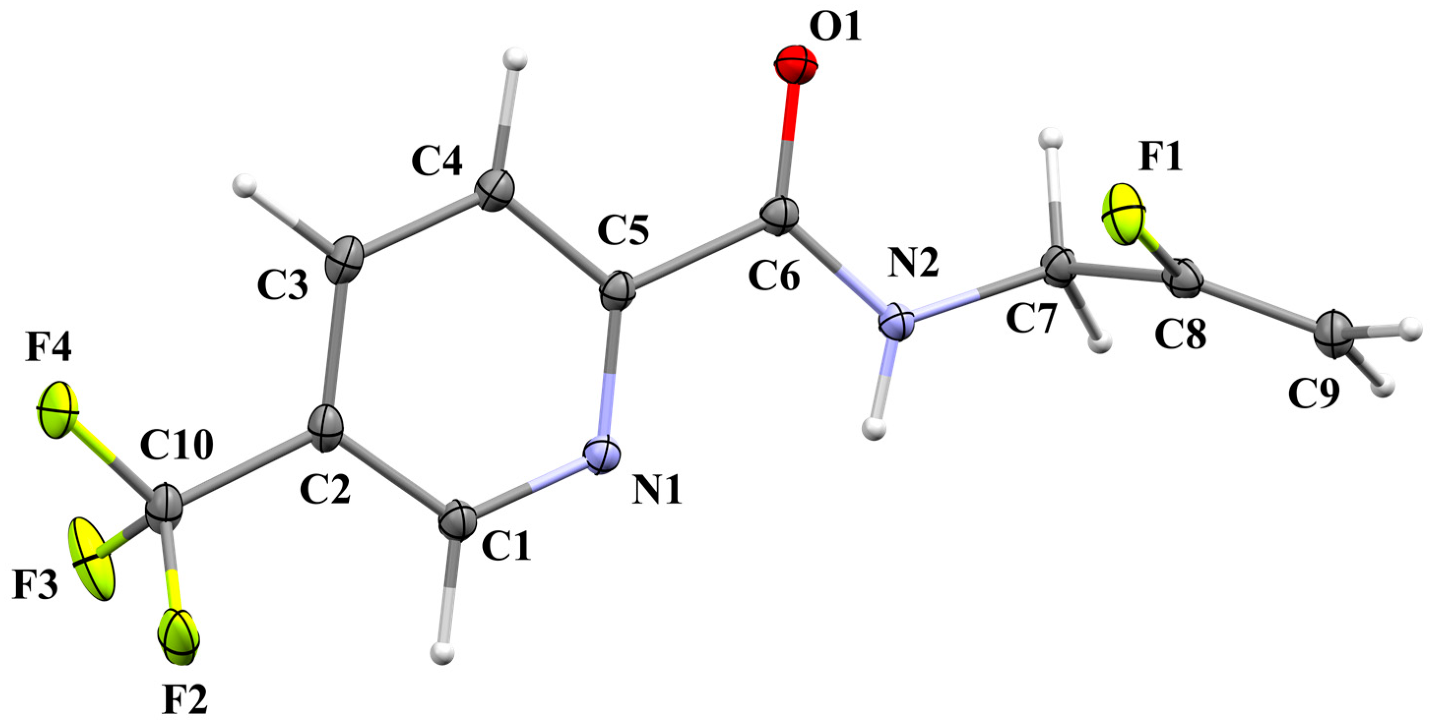
A crystal of 4 suitable for X-ray diffraction was isolated by slow evaporation of dichloromethane from a saturated solution. This resulted in the vertical growth of a colourless plate. The data confirm that the structure of 4 (Figure 3) is in agreement with the spectroscopic data (vide infra).

2.2.1. Structural Commentary

The C=O bond length of 4 is 1.232(2) Å, and the fluoroalkene C–F bond refines to 1.3616(19) Å, which both fit expected values [12]. The methylene group on the N-allyl amide presents an N2–C7 bond length of 1.455(2) Å. The F–C–F bond angles in the trifluoromethyl group show a slight deviation from tetrahedral geometry, with the smallest angle being the F4–C10–F2 angle at 104.79(9)°. As expected, the sp2-hybridized carbon atoms of the alkene and the pyridine core adopt a trigonal planar geometry, and the alkene is perpendicular to the pyridine ring; these systems do not lie in the same plane. A bond angle of 123.83(15)° is seen between N1–C5–C4, which is likely due to the attachment of the amide group on C5 and the extended π-system [13]. As commonly seen with trifluoromethyl groups, the original model had large Fourier peaks between the fluorine atoms. Therefore, a two-part disorder model (87:13) was developed.

2.2.2. Supramolecular Features

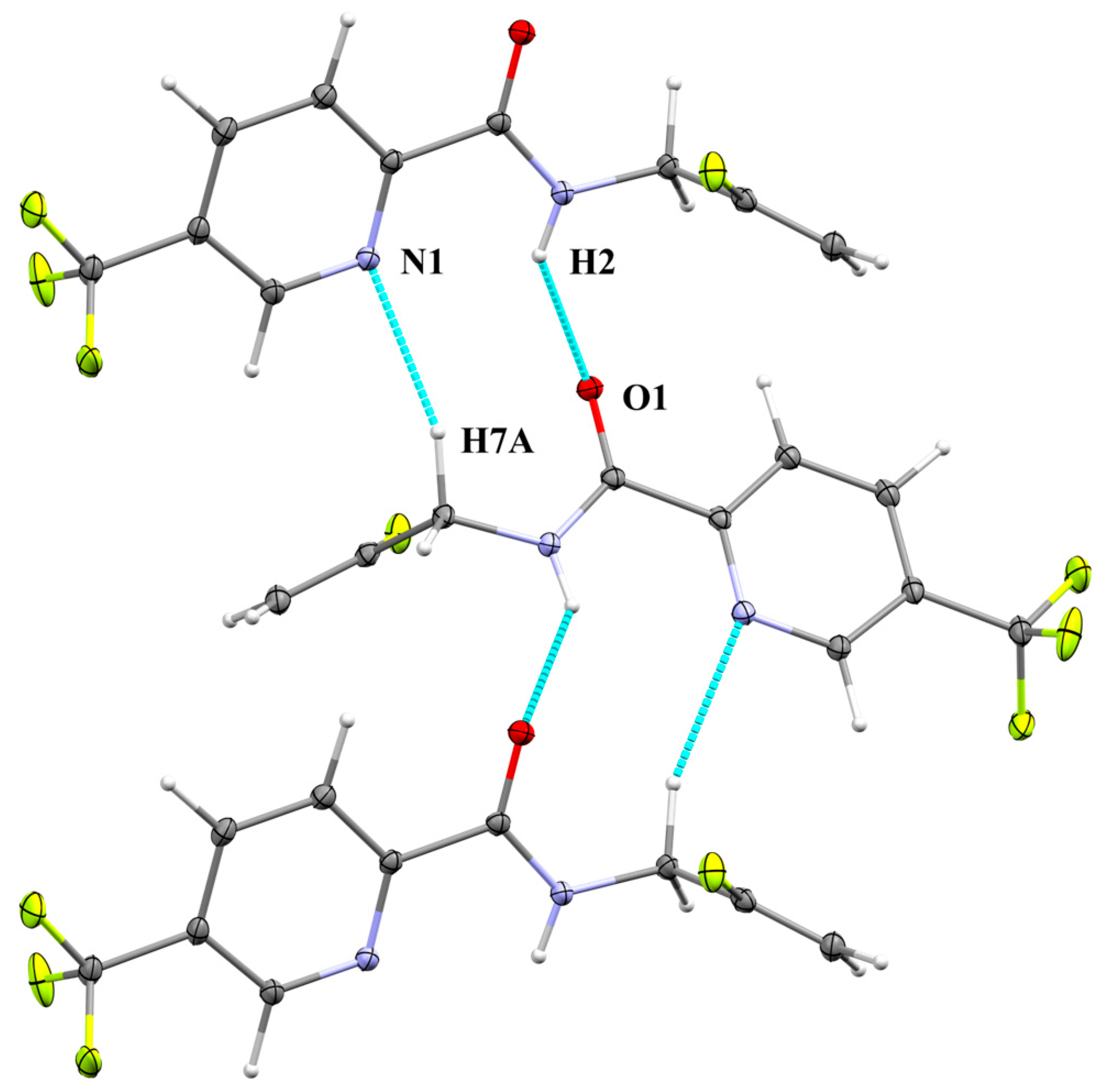
The title compound 4 crystallizes in the monoclinic space group P21/c with Z’ = 1 and a crystal density of 1.575 g cm−3, which is not uncommon for fluorinated compounds. In the crystal, the dominating intermolecular forces are strong H-bonding interactions (N–H···O) between amide groups (Figure 4). The N–H···O distance is 2.123(19) Å, and the intermolecular N···O separation is 2.8483(19) Å (Table 1). This H-bonding interaction helps define the packing structure, which results in a linear array of molecules that alternate by 180°. The N–H···O bond angle that develops from the H-bonding interaction is reported as 138.7(19)°. Longer H-bonding interactions are seen between the nitrogen in the pyridine ring (N1) and one of the hydrogens on C7 (C–H···N = 2.464(14) Å). The fluorine atoms in the trifluoromethyl and fluoroalkene groups do not participate in hydrogen bonding due to the strict packing geometry and because fluorine is a weak hydrogen bond acceptor in organic compounds [14].
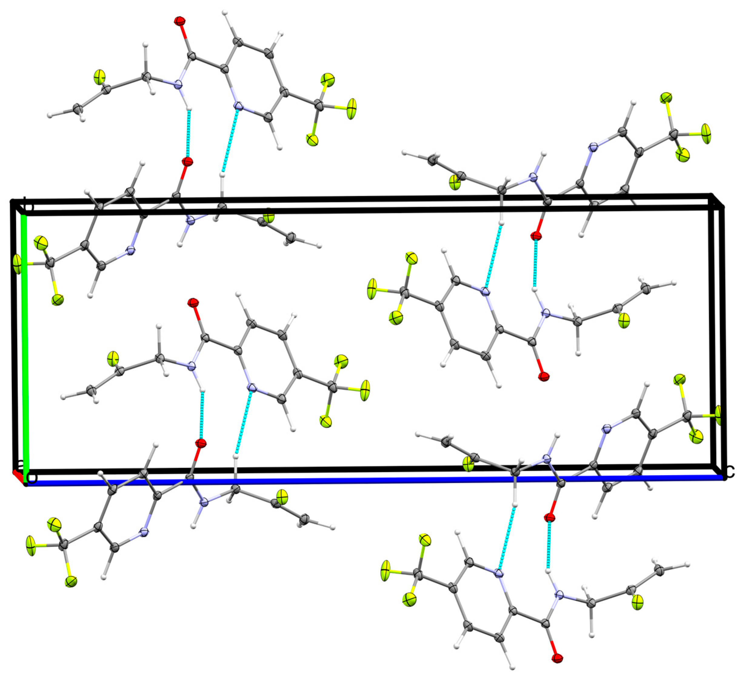
A unit cell diagram of the crystal highlights the lattice structure of 4 (Figure 5), revealing that there are four molecules in the cell. As discussed previously, the hydrogen bonding interactions develop parallel linear arrays of molecules that alternate by 180°. The steric bulk from the CF3 group also benefits from this alternation, which allows for tighter packing. The unit cell possesses inversion centers on the faces of each plane and one in the center of the cell. There are two c-glide planes perpendicular to b at b = ¼ and ¾. Additionally, there are four 21 screw axes parallel to b at a = ¼ and ¾, and at c = ¼, ¾. These symmetry elements and the general positions are consistent with the space group P21/c.

2.2.3. Database Surve

Structures containing both 3-(trifluoromethyl)pyridyl and fluoroalkene motifs are scarce in the crystallographic literature; therefore, the fragments were analyzed separately. Similar (trifluoromethyl)benzamide structures (CSD (The Cambridge Structural Database, version 2025.1 [15]) refcodes PETJIJ [16] and HOGPIE [17]) crystallize in the monoclinic space groups P21/n and P21/c (No. 14), respectively. These structures also have dominating H-bonding interactions between the amide groups. PETJIJ has a N–H···O length of 2.053(18) Å and a N···O separation of 2.869 Å, which is slightly shorter than for 4, likely due to less steric bulk. Substituted pyridine-2-carboxamides such as AMETIX [18] and LESPUW [19] have comparable aromatic C–C bond lengths, and they also possess an enlarged N–C–C bond angle, as is seen in 4 (123.541(10)° and 123.099(9)°, respectively). The fluoroalkene C–F bond length in structures CUMHID [20] (1.367(9) Å) and KARYAG [21] (1.354(10) Å) correlates well with the C8–F1 bond length in 4. The disorder model in the CF3 group compares well to other (3-trifluoromethyl)pyridyl groups. For compound 4, the C–F bond lengths for the primary disorder group were 1.3259(12), 1.3216(12) and 1.3217(12) Å. These values fall within the range of average values for similar structures, such as ERADUZ [22] (1.324(8) Å). The F–C–F bond angles in the primary disorder model of 4 are 106.54(9)°, 108.17(10)° and 104.79(9)° which are all slightly off from purely tetrahedral but consistent with the averages in ERADUZ (105.20(8)°). Overall, analysis of similar compounds in the literature showed agreement with the proposed structure of 4.

3. Experimental Section

3.1. General Considerations

Reactions were carried out under a nitrogen atmosphere. DCM was purified using an MBraun solvent purification system (SPS) and all other commercially available compounds were used as received. Vials (20 mL, Fisher Scientific) used to perform reactions were capped with Qorpak thermoset PTFE caps. Thin-layer chromatography (TLC) analysis of reaction mixtures was performed using Silicycle silica gel 60 Å, 230–400 mesh. 1H, 13C{1H} and 19F NMR spectra were recorded in CDCl3 at room temperature using a Bruker Avance III (700 MHz for 1H, 659 MHz for 19F, 176 MHz for 13C{1H}) NMR spectrometer. 1H and 13C{1H} NMR chemical shifts are referenced to tetramethylsilane (δ = 0.00 ppm) and CDCl3 (δ = 77.16 ppm), respectively. Multiplicities are reported using the following abbreviations: s = singlet, d = doublet, t = triplet, q = quartet, br s = broad signal, m = multiplet. Infrared spectra were recorded using an Agilent Cary 630 FT-IR spectrometer. High-resolution mass spectra were obtained on a Thermo Scientific Orbitrap Fusion mass spectrometer using electrospray ionization.

3.2. Synthesis of N-(2-fluoro-2-propen-1-yl)-5-(trifluoromethyl)-2-pyridinecarboxamide (4)

A 20 mL vial was charged with 5-(trifluoromethyl)-2-picolinic acid (86.0 mg, 0.450 mmol, 1.5 equiv) and DCM (3 mL). The solution was cooled to 0 °C in an ice bath, followed by the addition of oxalyl chloride (77 μL, 0.900 mmol, 3 equiv) and anhydrous DMF (6 μL, 0.078 mmol, 3 mol%). The reaction mixture was stirred for 15 min at 0 °C. The reaction mixture was then brought to room temperature and stirred for an additional 2 h. The solvent was removed under reduced pressure. In a new 20 mL vial, (2-fluoroallyl)ammonium chloride 3 (11.2 mg, 0.100 mmol, 1 equiv) was added, followed by DCM (6 mL) and triethylamine (46 μL, 0.300 mmol, 3 equiv). This new mixture was transferred slowly via syringe to the original reaction vial, which was then capped. The reaction mixture was stirred for 18 h at room temperature. The solvent was then removed under reduced pressure. The desired product (26.7 mg, 36%) was isolated as a white solid by flash chromatography using 20% ethyl acetate in hexanes. IR (ATR, diamond) ν = 3328, 2937, 1666, 1513, 1326, 1226, 1114, 1077, 864, 670 cm−1; 1H NMR (700 MHz, CDCl3) δ 8.84 (s, 1H), 8.35 (d, 1H, J = 8.2 Hz), 8.26 (br s, 1H), 8.13 (dd, 1H, J = 8.2, 1.8 Hz), 4.75 (dd, 1H, J = 16.4, 3.3 Hz), 4.58 (dd, 1H, J = 48.3, 3.3 Hz), 4.23 (dd, 2H, J = 12.2, 6.1 Hz); 19F NMR (659 MHz, CDCl3) δ −62.5 (s, 3F), −103.4 (ddt, 1F, J = 48.2, 16.3, 12.4 Hz); 13C{1H} NMR (176 MHz, CDCl3) δ 163.1 (C=O), 161.7 (d, JC–F = 257 Hz, FC=CH2), 152.3 (C–C=O), 145.5 (q, JC–F = 3.9 Hz, HC=N), 135.0 (q, JC–F = 2.7 Hz, HC=C–CF3), 129.2 (q, JC–F = 33.5 Hz, C–CF3), 123.1 (q, JC–F = 272 Hz, CF3), 122.4 (HC=C–C=O), 92.3 (d, JC–F = 18.1 Hz, H2C=C), 40.0 (d, JC–F = 33.7 Hz, H2C–NH). HRMS calcd for C10H9F4N2O [M + H]+ 249.0646, found 249.0635.
A colorless X-ray quality crystal of 4 was grown by slow evaporation of a solution in DCM kept in a loosely capped 4 mL vial stored in the fridge.

3.3. X-Ray Diffraction Data of 4

X-ray diffraction data were collected at 100 K using a Rigaku Oxford SuperNova Duo/Pilatus 200 K X-ray Diffractometer [CuKα radiation (λ = 1.54184 Å)]. Data were collected and processed using CrysAlisPro (v1.171.42.94a & 109a) [23]. Using Olex2, the structure was solved with the ShelXT (2019) [24,25] structure solution program using intrinsic phasing and refined with olex2.refine.
Crystal data for C10H8F4N2O, M = 248.18 g mol−1, colourless plate, crystal dimensions 0.33 × 0.14 × 0.091 mm, monoclinic, space group P21/n (No. 14), a = 4.96680(10), b = 9.06340(10), c = 23.3515(3) Å, β = 95.2200(10)°, V = 1046.83(3) Å3, Z = 4, Dcalc = 1.575 g cm−3, T = 100 K, 11,643 reflections collected, 2267 independent reflections (Rint = 0.0184). The final R1 [I > 2σ(I)] was 0.0478 and ωR2 [all data] was 0.1258, with a goodness of fit (F2) of 1.098.

3.4. X-Ray Structure Refinement of 4

A variety of refinement parameters were used in olex2.refine to provide a more accurate refinement model. An extinction correction and weights were applied in the refinement. H2, which is involved in H-bonding, was positionally refined but with the N2–H2 bond restrained to 0.88 Å. Large residual peaks around the trifluoromethyl group indicated that positional disorder was present. A two-part disorder model was developed, which resulted in lower residuals but induced large ellipsoids on the disordered CF3 group. Therefore, a weak ISOR restraint was used to make the fluorine atoms approximately isotropic. Two different SADI restraints were used: First, a restraint (su = 0.005) was applied to equalize the F–F distances and keep both CF3 groups approximately tetrahedral. Next, a second stronger SADI restraint (su = 0.001) was applied to the bonds between the fluorine atoms and C10 for C–F bond similarity. A RIGU restraint (su = 0.001) was also applied between the carbon–fluorine bonds of the disorder model to keep displacements in a reasonable direction. By applying these restraints, a suitable disorder model was developed that satisfies the geometric and electronic properties of the molecule and is crystallographically reasonable.

Supplementary Materials

The following supporting information has been attached along with this document. Figure S1: 1H NMR spectrum (700 MHz, CDCl3) of 4. Figure S2: 19F NMR spectrum (659 MHz, CDCl3) of 4. Figure S3: 13C{1H} NMR spectrum (176 MHz, CDCl3) of 4. Figure S4: DEPT-135 13C NMR spectrum (176 MHz, CDCl3) of 4. Figure S5: 1H–13C HMBC NMR spectrum of 4 acquired on a 700 MHz NMR instrument. Figure S6: HRMS data for 4. Table S1: Crystal data and structure refinement for 4. Table S2: Fractional Atomic Coordinates (×104) and Equivalent Isotropic Displacement Parameters (Å2 × 10³) for 4. Table S3: Anisotropic Displacement Parameters (Å2 × 10³) for 4. Table S4: Bond Lengths for 4. Table S5: Bond Angles for 4. Table S6: Hydrogen Bonds for 4. Table S7: Torsion Angles for 4. Table S8: Hydrogen Atom Coordinates (Å × 104) and Isotropic Displacement Parameters (Å2 × 10³) for 4. Table S9: Atomic Occupancy for 4.

Author Contributions

Conceptualization, T.S. and J.-D.H.; methodology, T.S.; data processing, T.S.; resources, J.-D.H.; writing—original draft preparation, T.S.; supervision, J.-D.H.; project administration and funding acquisition, J.-D.H. All authors have read and agreed to the published version of the manuscript.

Funding

This research was undertaken thanks to funding from the Natural Sciences and Engineering Research Council of Canada (NSERC; RGPIN-2022-04290 and DGECR-2022-00009), the Canada Research Chairs Program (CRC; 2020-00156), the Canada Foundation for Innovation (CFI; 40548), and the University of Lethbridge.

Data Availability Statement

Data available for this work is provided in the Supporting Information (experimental details, NMR spectra, and detailed report of the SC-XRD diffraction study). Raw SC-XRD data has been deposited via the joint CCDC/FIZ Karlsruhe deposition service and can be accessed by citing the data acquisition code 2489538.

Acknowledgments

T.S. thanks René T. Boeré for his teaching and mentorship in this study.

Conflicts of Interest

The authors declare no conflicts of interest.

Abbreviations

The following abbreviations are used in this manuscript:
DcalcCalculated density
DCMDichloromethane
DMFDimethylformamide
FDAFood and Drug Administration
H-bondingHydrogen bonding
HRMSHigh-resolution mass spectrometry
IRInfrared spectroscopy
NMRNuclear magnetic resonance
SC-XRDSingle-crystal X-ray diffraction
suStandard uncertainty from least-squares refinement
VdWVan der Waals

References

  1. O’Hagan, D. Understanding organofluorine chemistry. An introduction to the C–F bond. Chem. Soc. Rev. 2008, 37, 308–319. [Google Scholar] [CrossRef]
  2. Champagne, P.A.; Desroches, J.; Hamel, J.-D.; Vandamme, M.; Paquin, J.-F. Monofluorination of Organic Compounds: 10 Years of Innovation. Chem. Rev. 2015, 115, 9073–9174. [Google Scholar] [CrossRef]
  3. Ogawa, Y.; Tokunaga, E.; Kobayashi, O.; Hirai, K.; Shibata, N. Current Contributions of Organofluorine Compounds to the Agrochemical Industry. iScience 2020, 23, 101467. [Google Scholar] [CrossRef]
  4. Le Bars, D. Fluorine-18 and medical imaging: Radiopharmaceuticals for positron emission tomography. J. Fluorine Chem. 2006, 127, 1488–1493. [Google Scholar] [CrossRef]
  5. Nair, A.S.; Singh, A.K.; Kumar, A.; Kumar, S.; Sukumaran, S.; Koyiparambath, V.P.; Pappachen, L.K.; Rangarajan, T.M.; Kim, H.; Mathew, B. FDA-Approved Trifluoromethyl Group-Containing Drugs: A Review of 20 Years. Processes 2022, 10, 2054. [Google Scholar] [CrossRef]
  6. Burriss, A.; Edmunds, A.J.; Emery, D.; Hall, R.G.; Jacob, O.; Schaetzer, J. The importance of trifluoromethyl pyridines in crop protection. Pest Manag. Sci. 2018, 74, 1228–1238. [Google Scholar] [CrossRef]
  7. Lu, M.-Z.; Goh, J.; Maraswami, M.; Jia, Z.; Tian, J.-S.; Loh, T.-P. Recent Advances in Alkenyl sp2 C–H and C–F Bond Functionalizations: Scope, Mechanism, and Applications. Chem. Rev. 2022, 122, 17479–17646. [Google Scholar] [CrossRef] [PubMed]
  8. Drouin, M.; Laxio Arenas, J.; Paquin, J.-F. Incorporating a Monofluoroalkene into the Backbones of Short Peptides: Evaluating the Impact on Local Hydrophobicity. ChemBioChem 2019, 20, 1817–1826. [Google Scholar] [CrossRef]
  9. Semeniuk, T.; Challenger, J.; Dudas, T.; Toporkov, D.; van Hoeve, M.D.; Hamel, J.-D. Photoredox Decarboxylative 3-Fluoroallylation of α-Amino Acids. Eur. J. Org. Chem. 2025, 28, e202500519. [Google Scholar] [CrossRef]
  10. Zhang, X.-J.; Cheng, Y.-M.; Zhao, X.-W.; Cao, Z.-Y.; Xiao, X.; Xu, Y. Catalytic asymmetric synthesis of monofluoroalkenes and gem-difluoroalkenes: Advances and perspectives. Org. Chem. Front. 2021, 8, 2315–2327. [Google Scholar] [CrossRef]
  11. Brown, D.G.; Boström, J. Analysis of Past and Present Synthetic Methodologies on Medicinal Chemistry: Where Have All the New Reactions Gone? J. Med. Chem. 2016, 59, 4443–4458. [Google Scholar] [CrossRef]
  12. Ameram, N.; Adam, F.; Fatihah, N.N.; Al-Juaid, S. Crystal structure of 2-methyl-N-[(4-methylpyridin-2-yl)carbamothioyl]-benzamide. Acta Crystallogr. E. 2015, 71, o356. [Google Scholar] [CrossRef]
  13. Dey, S.; Manogaran, D.; Manogaran, S.; Schaefer, H.F. Substituent effects on the aromaticity of benzene—An approach based on interaction coordinates. J. Chem. Phys. 2019, 150, 214108. [Google Scholar] [CrossRef] [PubMed]
  14. Howard, J.A.K.; Hoy, V.J.; O’Hagan, D.; Smith, G.T. How good is fluorine as a hydrogen bond acceptor? Tetrahedron 1996, 52, 12613. [Google Scholar] [CrossRef]
  15. Groom, C.R.; Bruno, I.J.; Lightfoot, M.P.; Ward, S.C. The Cambridge Structural Database. Acta Crystallogr. B 2016, 72, 171–179. [Google Scholar] [CrossRef] [PubMed]
  16. Panini, P.; Chopra, D. Quantitative insights into energy contributions of intermolecular interactions in fluorine and trifluoromethyl substituted isomeric N-phenylacetamides and N-methylbenzamides. CrystEngComm 2013, 15, 3711–3733. [Google Scholar] [CrossRef]
  17. Zhang, L.; Wang, H.; Yang, B.; Fan, R. Imino exchange reaction in a dearomatization strategy: Synthesis of N-acyl diarylamines and phenothiazines from two anilines. Org. Chem. Front. 2014, 1, 1055–1057. [Google Scholar] [CrossRef]
  18. Light, M.E.; Tesfatsion, B.; Kilburn, J.; Dixon, S. CSD Communication, 2016; CCDC 1476136.
  19. Hernández-Gil, J.; Ferrer, S.; Ballesteros, R.; Castiñeiras, A. N-(5-Amino-1H-1,2,4-triazol-3-yl)-pyridine-2-carboxamide. Acta Crystallogr. E. 2013, 69, o227. [Google Scholar] [CrossRef]
  20. Villiers, E.; Couve-Bonnaire, S.; Cahard, D.; Pannecoucke, X. The fluoroalkene motif as a surrogate of the amide bond: Syntheses of AA-Ψ[(Z) and (E)-CFCH]-Pro pseudodipeptides and an Enalapril analogue. Tetrahedron 2015, 71, 7054–7062. [Google Scholar] [CrossRef]
  21. Nadon, J.-F.; Rochon, K.; Grastilleur, S.; Langlois, G.; Dao, T.T.H.; Blais, V.; Guérin, B.; Gendron, L.; Dory, Y.L. Synthesis of Gly-ψ[(Z)CF=CH]-Phe, a Fluoroalkene Dipeptide Isostere, and Its Incorporation into a Leu-enkephalin Peptidomimetic. ACS Chem. Neurosci. 2017, 8, 40–49. [Google Scholar] [CrossRef]
  22. Chen, Z.; Ren, N.; Ma, X.; Nie, J.; Zhang, F.-G.; Ma, J.-A. Silver-Catalyzed [3 + 3] Dipolar Cycloaddition of Trifluorodiazoethane and Glycine Imines: Access to Highly Functionalized Trifluoromethyl-Substituted Triazines and Pyridines. ACS Catal. 2019, 9, 4600–4608. [Google Scholar] [CrossRef]
  23. CrysAlisPro; v1.171.42.94a & 109a; Rigaku Oxford Diffraction; Rigaku Corporation: Oxford, UK, 2024.
  24. Sheldrick, G.M. SHELXT—Integrated space-group and crystal structure determination. Acta Crystallogr. A 2015, 71, 3–8. [Google Scholar] [CrossRef] [PubMed]
  25. Sheldrick, G.M. Crystal structure refinement with SHELXL. Acta Crystallogr. C 2015, 71, 3–8. [Google Scholar] [CrossRef] [PubMed]
Figure 1. Examples of FDA-approved pharmaceutical (1) and agrochemical (2) compounds containing a 3-(trifluoromethyl)pyridine core.
Molbank 2025 m2078 g001
Figure 2. The monofluoroalkene as a stereodefined peptide-bond isostere and enol mimic.
Molbank 2025 m2078 g002
Scheme 1. Amide coupling between 5-(trifluoromethyl)-2-pinolinic acid and (2-fluoroallyl)ammonium chloride 3.
Molbank 2025 m2078 sch001
Figure 3. The molecular structure of 4. The anisotropic displacement ellipsoids are set at a 50% probability level. The minor-occupancy F atoms of the CF3 two-component disorder model are omitted. The atoms are coloured as follows: carbon = grey; hydrogen = white; nitrogen = blue; oxygen = red; fluorine = yellow.
Molbank 2025 m2078 g003
Figure 4. Diagram showing the hydrogen bonding interactions (blue) in the crystal structure of 4. The atoms are coloured as follows: carbon = grey; hydrogen = white; nitrogen = blue; oxygen = red; fluorine = yellow.
Molbank 2025 m2078 g004
Figure 5. Unit cell diagram for the crystal structure of compound 4, viewed along the a-axis direction. Hydrogen bonding interactions are shown in blue. The atoms are coloured as follows: carbon = grey; hydrogen = white; nitrogen = blue; oxygen = red; fluorine = yellow.
Molbank 2025 m2078 g005
Table 1. Summary of hydrogen bond distances (Å) that are shorter than the sum of the VdW radii.
AtomsLengthLength—VdWSymmetry Operation
N1···H7A2.464−0.396½ − x, −½ + y, ½ − z
H2···O12.123−0.577½ − x, −½ + y, ½ − z
Disclaimer/Publisher’s Note: The statements, opinions and data contained in all publications are solely those of the individual author(s) and contributor(s) and not of MDPI and/or the editor(s). MDPI and/or the editor(s) disclaim responsibility for any injury to people or property resulting from any ideas, methods, instructions or products referred to in the content.