Short Note

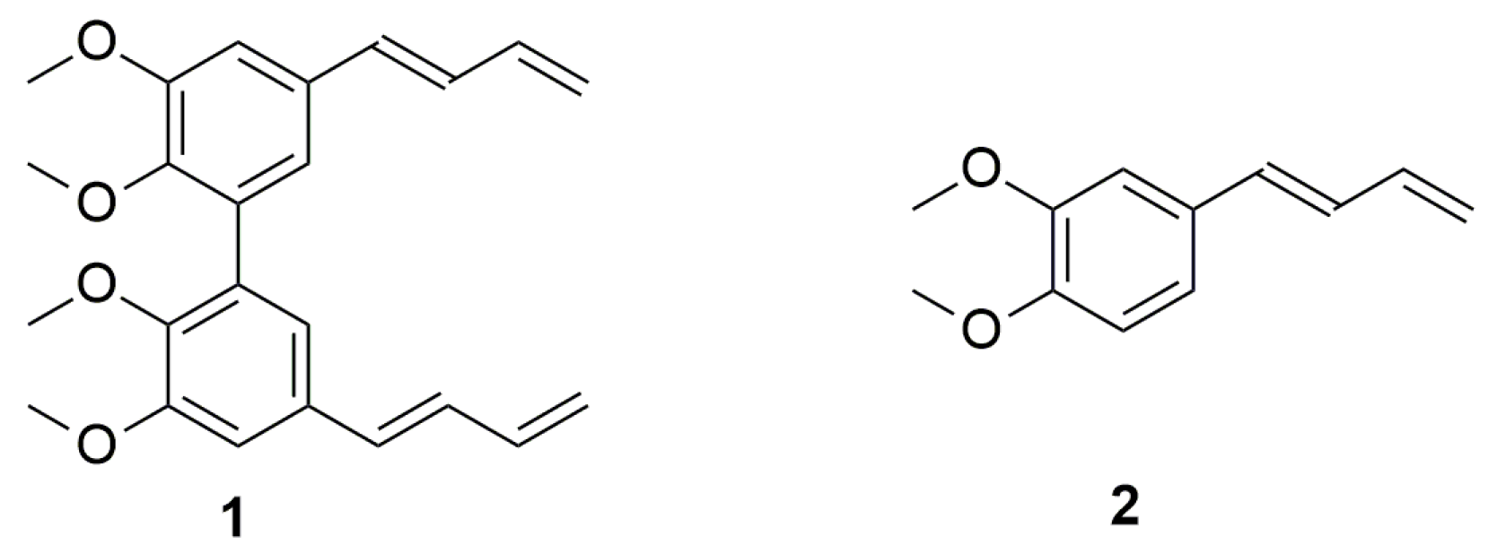
5,5′-Di((E)-buta-1,3-dien-1-yl)-2,2′,3,3′-tetramethoxy-1,1′-biphenyl

Istituto di Chimica Biomolecolare, Consiglio Nazionale delle Ricerche, Traversa La Crucca 3, I–07100 Sassari, Italy
*
Author to whom correspondence should be addressed.
Molbank 2025, 2025(2), M2018; https://doi.org/10.3390/M2018
Submission received: 8 May 2025 / Revised: 3 June 2025 / Accepted: 3 June 2025 / Published: 6 June 2025
(This article belongs to the Section Organic Synthesis and Biosynthesis)

Abstract

Phenylbutanoids, commonly found in various medicinal plants, have attracted significant attention due to their remarkable biological activities, including antioxidant, anti-inflammatory, and neuroprotective effects, as well as for their versatility as starting materials in organic synthesis. Among phenylbutanoids, phenyl-1,3-butadienes represent a unique class of conjugated dienes, characterized by a phenyl (C6H5) group attached to a 1,3-butadiene (-CH=CH-CH=CH2) backbone. In this study, we synthesized the hydroxylated biphenyl 5,5′-di((E)-buta-1,3-dien-1-yl)-2,2′,3,3′-tetramethoxy-1,1′-biphenyl 1, closely related to its corresponding monomer 2, which is known for its broad range of pharmacological activities. The synthesis was carried out using microwave-assisted technologies. The structure of the synthesized compound was confirmed through elemental analysis, 13C-NMR, 1H-NMR, and ESI-MS spectrometry. Furthermore, we computed this novel compound’s conformational energy profile (CEP), evaluating how its energy varies with changes in the dihedral bond angle.

Graphical Abstract

1. Introduction

Phenylbutanoids are a class of naturally occurring secondary metabolites characterized by a phenyl group attached to a four-carbon (butanoid) chain [1]. These compounds share structural similarities with phenylpropanoids but differ by the presence of an additional carbon in their side chain, which can influence their biological activity and physico-chemical properties. Phenylbutanoids are commonly found in various medicinal plants, particularly in the zingiberaceae (ginger family), where they contribute to the plant’s flavor, and pharmacological properties [2]. These compounds have been studied for their antioxidant [3], anti-inflammatory [4], and neuroprotective [5] properties. Moreover, phenylbutanoids serve as versatile starting materials in organic synthesis, particularly for constructing complex natural products and bioactive molecules [6]. Their structural framework provides a synthetically flexible scaffold that can be modified through various chemical transformations. This adaptability makes phenylbutanoids valuable in pharmaceutical and agrochemical applications. Among phenylbutanoids, phenyl-1,3-butadienes are a distinct class of conjugated dienes characterized by a phenyl group attached to a 1,3-butadiene scaffold. The extended π-conjugation enhances their chemical reactivity and optical properties, making them valuable in organic synthesis, polymer chemistry, and materials science [7]. (E)-1-(3,4-dimethoxyphenyl)butadiene 2 is a metabolite isolated from Zingiber cassumunar, a medicinal plant known for its extensive therapeutic properties [8]. This 1,3-butadiene derivative exhibits a wide range of biological activities, including insecticidal [9], anti-inflammatory [10] and antioxidant [11]. The title compound 1, 5,5′-di((E)-buta-1,3-dien-1-yl)-2,2′,3,3′-tetramethoxy-1,1′-biphenyl is formally the C2-symmetric dimer of compound 2 (Figure 1).
Hydroxylated biphenyls are the primary constituents of biologically significant natural substances, such as ellagitannins and lignins. Both ellagitannins and lignins are phenolic-rich compounds that play essential roles in plant defense and structural integrity [12]. Ellagitannins, found in various fruits and plants, are known for their antioxidant and anticancer properties [13], while lignins are crucial for cell wall strength and resistance to pathogens in plants [14]. Hydroxylated biphenyls often exhibit higher biological activity than their corresponding monomeric forms [15]. This class of compounds, along with other phenolic compounds, is well known for their ability to interact with proteins, modulating their activity. NMR-derived binding data reveal that hydroxylated biphenyls exhibit high affinity and specificity toward a broad range of protein targets, making them valuable for therapeutic discovery and drug design [16]. The interaction between hydroxylated biphenyls and proteins can be attributed to conformational isomerism caused by hindered rotation around the aryl–aryl single bond, a phenomenon known as atropisomerism. The configurational stability of atropisomers can be estimated based on their structure, particularly the number, size, and position of substituents near the rotational axis [17]. Furthermore, protein binding is enhanced in hydroxylated biphenyls—due to their greater conformational flexibility—as these structures can better align with key amino acid residues to establish effective interactions, thereby influencing biological activity [18].

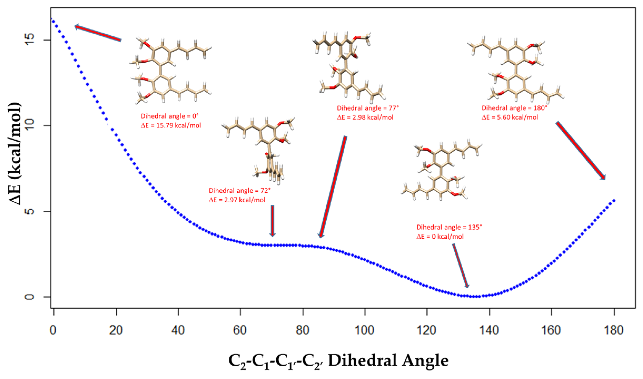
2. Results and Discussion

For many years, our group has focused on synthesizing various substituted, natural-like hydroxylated biphenyls and evaluating their biological activities [19]. Building on these observations, we decided to proceed to synthesize biphenyl 1, the C2-symmetric dimer of compound 2, with the future goal of studying how the symmetric biphenyl system may impact on the biological activities traditionally associated with the monomeric compound 2. Diol 6, the symmetric dimer of a natural compound present in Zingiber cassumunar [20], is a key intermediate in the synthesis of 1 (Scheme 1). Diol 6 was previously synthesized starting from compound 3, the symmetric dimer of natural phenol vanillin, as a mixture of (±) and meso isomers through two distinct steps, each developed and optimized by our group [21,22]. These synthetic pathways efficiently convert vanillin dimer 3 into the desired diol 6 with a total yield of 68%.
A key feature of this synthetic strategy is the use of microwave-assisted methods. Microwave irradiation accelerates reactions and reduces reaction times, making the process more efficient than traditional heating methods [23]. Treatment of 6 with 2.5 equivalents of copper sulfate in dioxane under microwave irradiation at 120 °C for 40 min led to compound 1 in good yield (65%) (Scheme 1).
To enhance the overall yield (44%) in the synthesis of compound 1, we explored an alternative route involving the dehydration of compound 7 (Scheme 2). Compound 7 was obtained as a mixture of (±) and meso isomers from compound 4 through a reaction with zinc and allyl bromide in a saturated ammonium chloride solution. Compound 4 has been previously synthesized by our group from vanillin dimer 3 [24]. The dehydration of compound 7 proceeded smoothly, leading to the formation of compound 1. This alternative pathway notably enhanced the total yield, reaching 68%, compared to 44% obtained via the dehydration of diol 6.
Both dehydration reactions exhibited high stereoselectivity, exclusively producing compound 1 as a single isomer with E, E geometry. This assignment was supported by the coupling constant (J = 15.6 Hz) observed between protons H1 and H2 of the 1,3-butadiene chain, characteristic of a trans olefinic proton-proton coupling (Figure 2). The 1H-NMR (methanol-d4) spectrum of 1 showed characteristic aromatic signals for a 2,2′,3,3′,5,5′ esa-substituted biphenyl system—two meta-aromatic doublets at 6.88 ppm (d, J = 1.6 Hz) and 7.12 ppm (d, J = 1.6 Hz). The 1,3-diene system was confirmed by the following five well-defined olefinic peaks: 5.14 ppm (dd, J = 0.6, 10.2 Hz, H5 and H5), 5.32 ppm (dd, J = 0.6, 16.8 Hz, H4 and H4), 6.47 ppm (m, H3 and H3), 6.54 ppm (d, J = 15.6 Hz, H1 and H1), and 6.80 ppm (dd, J = 10.4, 15.6 Hz, H2 and H2). Remarkably, the chemical shifts in the two methoxy substituents at positions C2 and C3 on each aromatic ring differed significantly, both in 1H-NMR (3.61 vs 3.93 ppm) and in 13C-NMR (55.00 vs. 59.68 ppm). These chemical shift differences were also observed in the 1H-NMR spectra of compounds 4 and 5 [21]. Notably, in all s-cis and s-trans conformations of the biaryl system in compound 1, two distinct perpendicular C2 symmetry axes were present. As a result, the chemical environments of the corresponding protons and carbons on both rings were identical, leading to equivalent signals in the NMR spectra and facilitating a more straightforward analysis [25].
Computational techniques, such as molecular mechanics (MM), semi-empirical methods, and ab initio quantum calculations, are extensively used in drug discovery to investigate the low-energy conformations of molecules [26].
The conformational preferences of biaryl systems result from a delicate interplay of resonance stabilization, steric hindrance, and electrostatic interactions, including lone pair-lone pair repulsions and π-cloud-lone pair interactions [26,27]. To investigate these effects, we computed the conformational energy profile (CEP) of biphenyl 1, analyzing the relative energy variation as a function of its C2-C1-C1′-C2′ dihedral angle (Figure 3).
The energetic barrier between atropisomers in ortho-substituted biphenyls is influenced by the nature and size of the aromatic substituents at the 2,2′ positions, as well as the steric and electronic interactions. These barriers typically range from 10 to 35 kcal/mol. A barrier close to 15 kcal/mol or less suggests rapid interconversion, resulting in atropisomers that lack substantial configurational stability at room temperature [28].
In compound 1, the energy differences among various conformers also provide insight into its conformational preferences and flexibility. The lowest relative energy conformers, s-trans and s-cis, adopted dihedral angles of 135° and 72°, respectively, while the highest-energy conformers (with dihedral angles of 0°, 77°, and 180°) exhibited a greater strain. The relative energy minima at 135° and 72° reflected a balance between the following two factors: resonance stabilization between aromatic rings, which promotes planar conformations, and steric effects between ortho substituents, which oppose planar conformations. Despite these variations, the overall energy differences remained below 16 kcal/mol, indicating that the compound lacked significant conformational rigidity and atropisomeric stability. These relatively low energy barriers suggest that the molecule can undergo conformational interconversion with relative ease, which may impact its reactivity, binding interactions, and stereochemical properties in dynamic environments. In its s-cis conformation (72° dihedral angle), compound 1 exhibited a moderately low local energy minimum (ΔE = 2.97 kcal/mol). In contrast, the s-trans conformer (135° dihedral angle) represented the global minimum with the lowest relative energy (ΔE = 0 kcal/mol). The energy difference between these conformers is likely due to steric interactions between the methoxy groups at the ortho positions of the two phenyl rings. These steric clashes increase the overall strain in the system, making all s-cis conformations less favorable than the corresponding s-trans forms. Examining the two possible planar conformations provides further insight into the steric effects within the molecule. The planar s-cis conformer (dihedral angle = 0°) and the planar s-trans conformer (dihedral angle = 180°) had relative energies of 15.79 kcal/mol and 5.60 kcal/mol, respectively. The markedly higher energy of the s-cis conformations indicated greater steric hindrance between the methoxy-methoxy C2-C2′ phenyl substituents compared to the methoxy-hydrogen C2-C6′ interactions (Figures S5–S9).
In 1,3-butadiene derivatives, the vicinal coupling constant J (H2−H3) provides key insights into their conformational preferences. These molecules can adopt the following two primary conformations: (-CH=CH-CH=CH2) s-cis and (-CH=CH-CH=CH2) s-trans. In the (-CH=CH-CH=CH2) s-trans conformation, the 1,3-butadiene moiety adopts an approximately planar geometry, resulting in a H2-H3 coupling constant of 10–16 Hz, consistent with the Karplus equation’s prediction for a (-CH=CH-CH=CH2) s-trans-vicinal relationship [29]. In contrast, the (-CH=CH-CH=CH2) s-cis conformation exhibits a reduced H2-H3 coupling constant of 6–9 Hz. These variations in vicinal coupling constants enable straightforward differentiation between the two conformers using 1H-NMR spectroscopy. In compound 1, the observed vicinal coupling constant J (H2−H3) was 10.4 Hz, suggesting a predominantly (-CH=CH-CH=CH2) s-trans planar geometry (Figure 4a).
To further investigate the geometric properties of the 1,3-butadiene chain in compound 1, we computed the relative conformational energy of its two possible geometries, namely (-CH=CH-CH=CH2) s-trans (Figure 4a) and (-CH=CH-CH=CH2) s-cis (Figure 4b). As expected, the (-CH=CH-CH=CH2) s-cis conformer, featuring a dihedral angle of 20°, exhibited higher relative energy (ΔE = 7 kcal/mol) than the (-CH=CH-CH=CH2) s-trans conformer, which displayed an almost entirely planar structure. This energy difference was primarily due to increased proton–proton steric interactions in the 1,3-butadiene s-cis conformer, where the distances H1-H5 = 2.37 Å and H2-H3 = 2.41 Å were shorter than those calculated in the 1,3-butadiene s-trans conformer (H1-H3 = 2.57 Å and H2-H5 = 2.52 Å). It is well established that steric repulsions become significant when two hydrogen atoms come closer than their van der Waals distance (approximately 2.4 Å). These interactions can affect molecular stability and influence the preferred geometry of conjugated systems such as 1,3-butadiene, where close non-bonded contacts may arise due to conformational constraints [30].

3. Materials and Methods

Reagents obtained from Sigma Aldrich, Munich, Germany, were used without further purification. 1H-NMR and 13C-NMR spectra were recorded in deuterated methanol at 600 and 150 MHz, respectively, using a 600 MHz Bruker Avance III HD NMR spectrometer (Palo Alto, CA, USA). Full characterization data, including copies of 1H-NMR and 13C-NMR spectra (see Supplementary Material, Figures S1–S4), have been reported for the novel compounds 1 and 7. Compounds 3, 4, 5 and 6 were prepared according to the literature [21,22,24]. Chemical shifts are given in ppm (δ); multiplicities are indicated by s (singlet), d (doublet), m (multiplet), or dd (doublet of doublets). Elemental analysis was performed using an elemental analyzer model 240 C (Perkin Elmer, Waltham, MA, USA). Flash chromatography was performed with silica gel 60 (230–400 mesh) (VWR, Radnor, AF, USA), eluting with an appropriate solution in the stated v:v proportions. Reactions were monitored by 1H-NMR and analytical thin-layer chromatography (TLC) with 0.25 mm thick silica gel plates (60 F 254) (Sigma Aldrich, Munich, Germany). The torsional energy profiles were calculated using Gaussian 16W version in the gas phase. The geometry of compound 1 was optimized and analyzed using the relaxed dihedral scan method on the C2-C1-C1′-C2′ dihedral angle, employing the DFT B3LYP method with the 6–31G basis set, with successive increments of 1˚ in the angle. The rotation was performed from a dihedral angle of 0˚ (s-cis conformation) to a dihedral angle of 180˚ (s-trans conformation). The resulting energies from the 180 optimized structures were plotted as a function of the dihedral angle, and the curve fitting these points provided the conformational energy profile (CEP) using Gaussian View version 6.1 and R version 4.4.2. Angle analysis and 3D images were carried out using Chimera UCSF version 17.3 [31]. The purity of new compounds was judged to be >98% by 1H-NMR spectral determination. ESI-MS spectra were recorded in positive mode on a ThermoScientific LTQ Orbitrap XL Exploris 120 (Thermo Scientific Inc., Milan, Italy).
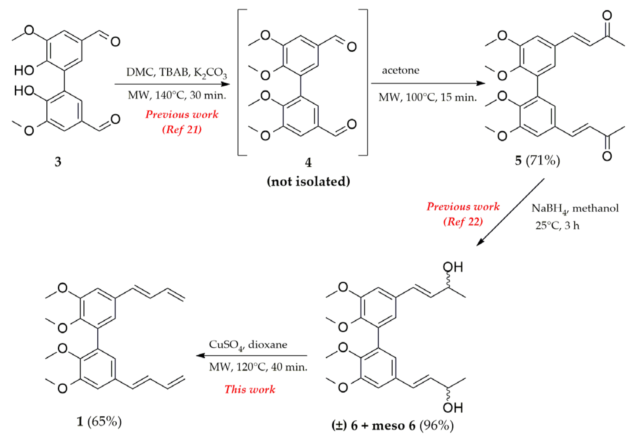
  • Synthesis of 1,1′-(5,5′,6,6′-tetramethoxy-[1,1′-biphenyl]-3,3′-diyl)bis(but-3-en-1-ol) 7.
  • To a solution of 4 (0.5 g, 1.5 mmol) in tetrahydrofuran (5 mL), allyl bromide (0.93 mL, 10.7 mmol) and saturated aqueous ammonium chloride solution (10 mL) were added. The reaction mixture was cooled to 0° C. Zinc powder (1.4 g, 21.4 mmol) was added, and the solution was stirred at 0 °C for 30 min. The resulting precipitate was filtered, and the filtrate was extracted with ethyl acetate. The organic layers were combined and washed with brine, dried over sodium sulfate, filtered, and concentrated in vacuo. The crude compound was purified by flash chromatography using a 1:2 mixture of ethyl acetate/petroleum ether as the eluent to obtain 7 as a colorless oil (0.51 g, 90%). 1H-NMR (methanol-d4) δ 2.25 (m, 4H), 3.58 (s, -OCH3, 6H), 3.91 (s, -OCH3, 6H), 4.66 (t, J = 7.2 Hz, 2H), 5.05 (dd, J = 0.6, 9.0 Hz, Ar, 2H), 5.08 (dd, J = 0.6, 16.8 Hz, 2H), 5.82 (m, 2H), 6.79 (d, J = 2.4 Hz, Ar, 2H), 7.05 (d, J = 2.4 Hz, Ar, 2H); 13C-NMR (methanol-d4) δ 43.93, 54.97, 59.60, 73.29, 109.43, 116.11, 120.39, 132.37, 134.72, 140.02, 145.60, 152.52; Anal. Calcd for C24H30O6: C, 69.54; H, 7.30. Found: C, 69.60; H, 7.35; ESI-MS m/z (ES+) Found: [M+Na]+ 437.1941: C24H30O6 Na, requires [M+Na]+ 437.1939.
  • Synthesis of 5,5′-di((E)-buta-1,3-dien-1-yl)-2,2′,3,3′-tetramethoxy-1,1′-biphenyl 1.
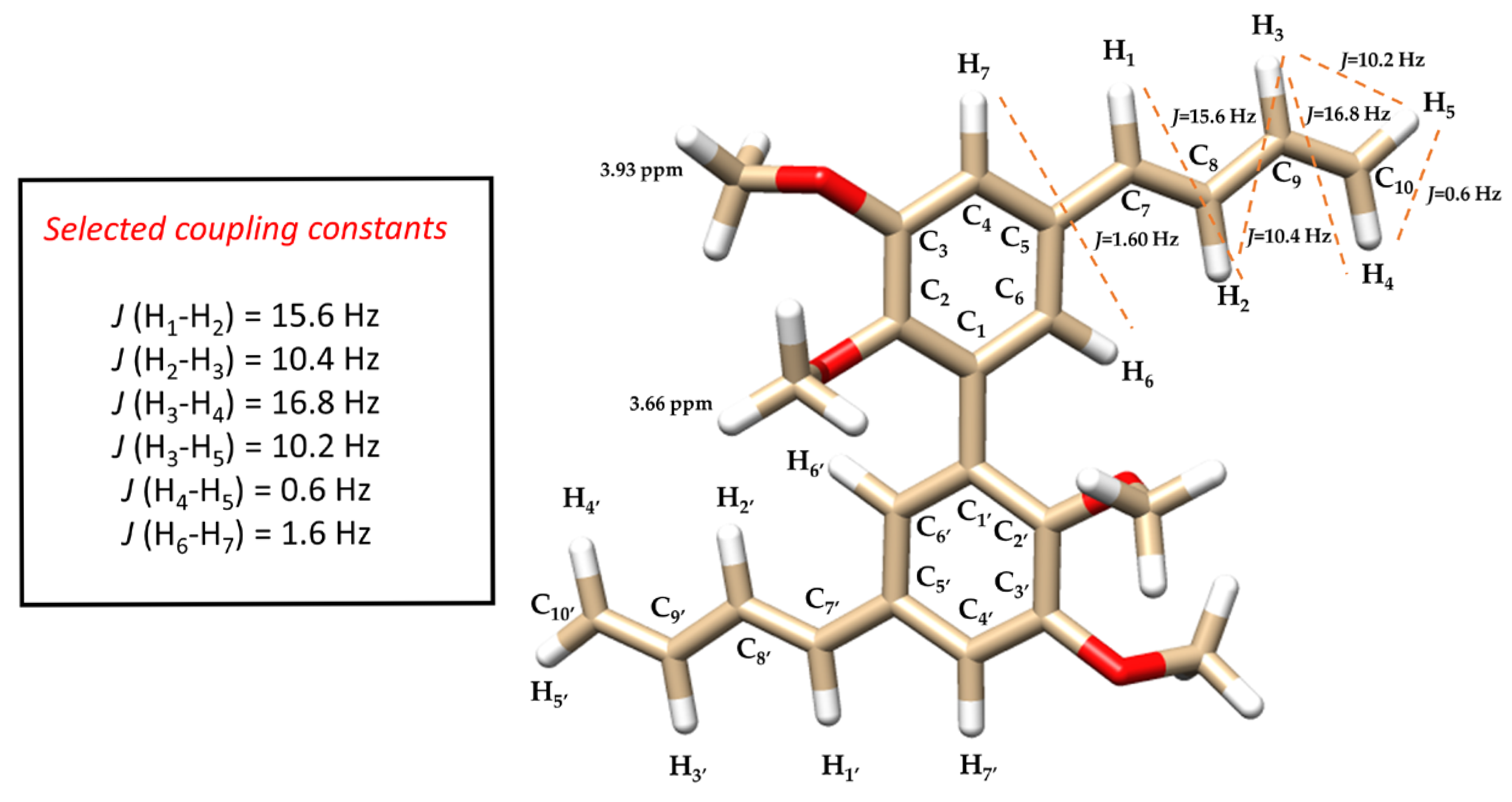
  • To a solution of compound 6 or 7 (0.25 g, 0.6 mmol) in dioxane (10 mL), copper sulfate (0.2 g, 1.2 mmol) was added at room temperature. The solution was then stirred under MW irradiation at 120 °C for 40 min. The solution was cooled at rt and filtered. The filtrate was extracted with ethyl acetate. The organic layers were combined and washed with brine, dried over sodium sulfate, filtered, and concentrated in vacuo. The crude material was purified by flash chromatography using a 1:5 mixture of acetone/petroleum ether as the eluent to obtain 1 as a yellow oil (0.15 g, 65% or 0.22 g, 95% starting from diol 6 or 7 respectively). 1H-NMR (methanol-d4) δ 3.66 (s, -OCH3, 6H), 3.93 (s, -OCH3, 6H), 5.14 (dd, J = 0.6, 10.2 Hz, H5-H5′, 2H), 5.32 (dd, J = 0.6, 16.8 Hz, H4-H4′, 2H), 6.47 (m, H3-H3′, 2H), 6.54 (d, J = 15.6 Hz, H1-H1′, 2H), 6.80 (dd, J = 10.4, 15.6 Hz, H2-H2′, 2H), 6.88 (d, J = 1.6 Hz, Ar, 2H), 7.12 (d, J = 1.6 Hz, Ar, 2H); 13C-NMR (methanol-d4) δ 55.00, 59.68, 109.22, 116.15, 121.33, 129.96, 132.19, 132.63, 132.95, 137.21, 146.36, 152.79; Anal. Calcd for C24H26O4: C, 76.17; H, 6.92. Found: C, 76.23; H, 6.95; ESI-MS m/z (ES+) Found: [M+Na]+ 401.1732: C24H26O4 Na, requires [M+Na]+ 401.1728.

4. Conclusions

In this study, we synthesized the C2-symmetric biphenyl analog of compound 2, 5,5′-di((E)-buta-1,3-dien-1-yl)-2,2′,3,3′-tetramethoxy-1,1′-biphenyl 1, using a microwave-assisted strategy starting from vanillin-derived diols 6 or 7. The reactions proceeded with high stereo selectivity, yielding compound 1 exclusively with E,E geometry. Structural confirmation was achieved through elemental analysis, NMR, and mass spectrometry. Computational studies on the conformational energy profile of compound 1 revealed a non-planar ground state conformation with minimal energy barriers between conformers, indicating low conformational and configurational rigidity. The synthesis of the new compound 1 will allow us to explore whether the increased structural complexity of the biphenyl system influences its biological properties, potentially leading to enhanced biological activities compared to the corresponding monomer 2. Moreover, these insights can aid in developing novel bioactive molecules with potential applications in drug discovery, medicinal chemistry, and other biomedical fields.

Supplementary Materials

The following supporting information can be downloaded online. 2D MDL molfile of compound 1; Figure S1: 1H-NMR spectrum of the title compound 1; Figure S2: 13C-NMR spectrum of the title compound 1; Figure S3: 1H-NMR spectrum of compound 7; Figure S4: 13C-NMR spectrum of compound 7; Figure S5: Output image as calculated with Gaussian 16W (Dihedral Angle: 0°) of compound 1; Figure S6: Output image as calculated with Gaussian 16W (Dihedral Angle: 72°) of compound 1; Figure S7: Output image as calculated with Gaussian 16W (Dihedral Angle: 77°) of compound 1; Figure S8: Output image as calculated with Gaussian 16W (Dihedral Angle: 135°) of compound 1; Figure S9: Output image as calculated with Gaussian 16W (Dihedral Angle: 180°) of compound 1.

Author Contributions

Conceptualization, D.F. and M.A.D.; methodology, P.C., M.A.D. and R.D.; data curation, P.C., D.F. and M.A.D.; writing—original draft preparation, D.F. and M.A.D.; writing—review and editing, P.C., R.D. and M.A.D. All authors have read and agreed to the published version of the manuscript.

Funding

Project “Future raw materials from non-critical, residual, and renewable sources” (FutuRaw), funded through the Ordinary Fund for Research Institutions (FOE 2022) under Ministerial Decree No. 789 issued on June 21, 2023, sub-project DCM.AD005.081.012, CUP B53C23008390005.

Data Availability Statement

Not applicable.

Acknowledgments

The authors gratefully acknowledge Francesco Mugheddu (ICB-CNR, Italy) for providing high-resolution electrospray ionization mass spectrometry (HR-ESI-MS) data for the newly synthesized compounds.

Conflicts of Interest

The authors declare no conflicts of interest.

References

  1. Seaho, B.; Lekwongphaiboon, C.; Tayana, N.; Inthakusol, W.; Kongkiatpaiboon, S.; Mahavorasirikul, W.; Prateeptongkum, S.; Duangdee, N. Absolute Quantification of Phenylbutanoids in Zingiber cassumunar Roxb. Rhizome by Quantitative1 H NMR. Phytochem. Anal. 2024, 36, 876–883. [Google Scholar] [CrossRef] [PubMed]
  2. Nakamura, S.; Iwami, J.; Pongpiriyadacha, Y.; Nakashima, S.; Matsuda, H.; Yoshikawa, M. Chemical Structures of Phenylbutanoids From Rhizomes of Zingiber cassumunar. Nat. Prod. Commun. 2022, 17, 1934578X2210778. [Google Scholar] [CrossRef]
  3. Soberón, J.R.; Sgariglia, M.A.; Sampietro, D.A.; Quiroga, E.N.; Vattuone, M.A. Free Radical Scavenging Activities and Inhibition of Inflammatory Enzymes of Phenolics Isolated from Tripodanthus Acutifolius. J. Ethnopharmacol. 2010, 130, 329–333. [Google Scholar] [CrossRef] [PubMed]
  4. Panichayupakaranant, P.; Kaewchoothong, A. Preparation of Phenylbutanoid-Rich Zingiber Cassumunar Extracts and Simultaneous HPLC Analysis of Phenylbutanoids. Planta Med. 2012, 78, PI157. [Google Scholar] [CrossRef]
  5. Kongsui, R.; Sriraksa, N.; Thongrong, S. The Neuroprotective Effect of Zingiber Cassumunar Roxb. Extract on LPS-Induced Neuronal Cell Loss and Astroglial Activation within the Hippocampus. BioMed Res. Int. 2020, 2020, 4259316. [Google Scholar] [CrossRef]
  6. Sinha, A.K.; Sharma, A.; Joshi, B.P.; Singh, N.P. A Mild Conversion of Phenylpropanoid into Rare Phenylbutanoids: (E)-4-(2′,4′,5′-Trimethoxyphenyl)but-1, 3-Diene and (E)-4-(2′,4′,5′-Trimethoxyphenyl)but-1-Ene Occurring in Zingiber Cassumunar. Nat. Prod. Res. 2005, 19, 771–776. [Google Scholar] [CrossRef]
  7. Devkota, H.P.; Paudel, K.R.; Hassan, M.M.; Dirar, A.I.; Das, N.; Adhikari-Devkota, A.; Echeverría, J.; Logesh, R.; Jha, N.K.; Singh, S.K.; et al. Bioactive Compounds from Zingiber Montanum and Their Pharmacological Activities with Focus on Zerumbone. Appl. Sci. 2021, 11, 10205. [Google Scholar] [CrossRef]
  8. Han, A.-R.; Kim, H.; Piao, D.; Jung, C.-H.; Seo, E.K. Phytochemicals and Bioactivities of Zingiber Cassumunar Roxb. Molecules 2021, 26, 2377. [Google Scholar] [CrossRef]
  9. Li, M.-X.; Ma, Y.-P.; Zhang, H.-X.; Sun, H.-Z.; Su, H.-H.; Pei, S.-J.; Du, Z.-Z. Repellent, Larvicidal and Adulticidal Activities of Essential Oil from Dai Medicinal Plant Zingiber Cassumunar against Aedes Albopictus. Plant Divers. 2021, 43, 317–323. [Google Scholar] [CrossRef]
  10. Jeenapongsa, R.; Yoovathaworn, K.; Sriwatanakul, K.M.; Pongprayoon, U.; Sriwatanakul, K. Anti-Inflammatory Activity of (E)-1-(3,4-Dimethoxyphenyl) Butadiene from Zingiber Cassumunar Roxb. J. Ethnopharmacol. 2003, 87, 143–148. [Google Scholar] [CrossRef]
  11. Truong, V.-L.; Manochai, B.; Pham, T.-T.; Jeong, W.-S. Antioxidant and Anti-Inflammatory Activities of Zingiber Montanum Oil in HepG2 Cells and Lipopolysaccharide-Stimulated RAW 264.7 Macrophages. J. Med. Food 2021, 24, 595–605. [Google Scholar] [CrossRef] [PubMed]
  12. Ward, R.S. Lignans, Neolignans and Related Compounds. Nat. Prod. Rep. 1999, 16, 75–96. [Google Scholar] [CrossRef]
  13. Ismail, T.; Calcabrini, C.; Diaz, A.; Fimognari, C.; Turrini, E.; Catanzaro, E.; Akhtar, S.; Sestili, P. Ellagitannins in Cancer Chemoprevention and Therapy. Toxins 2016, 8, 151. [Google Scholar] [CrossRef] [PubMed]
  14. Saberi Riseh, R.; Fathi, F.; Lagzian, A.; Vatankhah, M.; Kennedy, J.F. Modifying Lignin: A Promising Strategy for Plant Disease Control. Int. J. Biol. Macromol. 2024, 271, 132696. [Google Scholar] [CrossRef]
  15. Paquin, A.; Reyes-Moreno, C.; Bérubé, G. Recent Advances in the Use of the Dimerization Strategy as a Means to Increase the Biological Potential of Natural or Synthetic Molecules. Molecules 2021, 26, 2340. [Google Scholar] [CrossRef]
  16. Hajduk, P.J.; Bures, M.; Praestgaard, J.; Fesik, S.W. Privileged Molecules for Protein Binding Identified from NMR-Based Screening. J. Med. Chem. 2000, 43, 3443–3447. [Google Scholar] [CrossRef]
  17. Heeb, J.-P.; Clayden, J.; Smith, M.D.; Armstrong, R.J. Interrogating the Configurational Stability of Atropisomers. Nat. Protoc. 2023, 18, 2745–2771. [Google Scholar] [CrossRef]
  18. Kostić, K.; Brborić, J.; Delogu, G.; Simić, M.R.; Samardžić, S.; Maksimović, Z.; Dettori, M.A.; Fabbri, D.; Kotur-Stevuljević, J.; Saso, L. Antioxidant Activity of Natural Phenols and Derived Hydroxylated Biphenyls. Molecules 2023, 28, 2646. [Google Scholar] [CrossRef]
  19. Setzu, M.D.; Mocci, I.; Fabbri, D.; Carta, P.; Muroni, P.; Diana, A.; Dettori, M.A.; Casu, M.A. Neuroprotective Effects of the Nutraceutical Dehydrozingerone and Its C2-Symmetric Dimer in a Drosophila Model of Parkinson’s Disease. Biomolecules 2024, 14, 273. [Google Scholar] [CrossRef]
  20. Hodgson, D.M.; Persaud, R.S.D. Convergent Synthesis of Conjugated 1,2-Disubstituted E-Allylic Alcohols from Two Aldehydes and Methylenetriphenylphosphorane. Org. Biomol. Chem. 2012, 10, 7949. [Google Scholar] [CrossRef]
  21. Dettori, M.A.; Carta, P.; Fabbri, D. Environmentally Friendly One-Pot Two-Step Sequential Synthesis of Biological Active Curcumin Analogues. Tetrahedron 2024, 153, 133867. [Google Scholar] [CrossRef]
  22. Sanfilippo, C.; Patti, A.; Dettori, M.A.; Fabbri, D.; Delogu, G. Lipase Behavior in the Stereoselective Transesterification of Zingerol-like Derivatives and Related Biphenyls. J. Mol. Catal. B Enzym. 2013, 90, 107–113. [Google Scholar] [CrossRef]
  23. Kumar, A.; Kuang, Y.; Liang, Z.; Sun, X. Microwave Chemistry, Recent Advancements, and Eco-Friendly Microwave-Assisted Synthesis of Nanoarchitectures and Their Applications: A Review. Mater. Today Nano 2020, 11, 100076. [Google Scholar] [CrossRef]
  24. Pisano, M.; Pagnan, G.; Dettori, M.A.; Cossu, S.; Caffa, I.; Sassu, I.; Emionite, L.; Fabbri, D.; Cilli, M.; Pastorino, F.; et al. Enhanced Anti-Tumor Activity of a New Curcumin-Related Compound against Melanoma and Neuroblastoma Cells. Mol. Cancer 2010, 9, 137. [Google Scholar] [CrossRef]
  25. Woodman, P.R.; Munslow, I.J.; Hitchcock, P.B.; Scott, P. Non-Planar Co-Ordination of C2-Symmetric Biaryl-Bridged Schiff-Base Ligands: Well Expressed Chiral Ligand Environments for Zirconium. J. Chem. Soc. Dalton Trans. 1999, 4069–4076. [Google Scholar] [CrossRef]
  26. Gutiérrez Sanfeliciano, S.M.; Schaus, J.M. Rapid Assessment of Conformational Preferences in Biaryl and Aryl Carbonyl Fragments. PLoS ONE 2018, 13, e0192974. [Google Scholar] [CrossRef]
  27. Dahlgren, M.K.; Schyman, P.; Tirado-Rives, J.; Jorgensen, W.L. Characterization of Biaryl Torsional Energetics and Its Treatment in OPLS All-Atom Force Fields. J. Chem. Inf. Model. 2013, 53, 1191–1199. [Google Scholar] [CrossRef]
  28. Mazzanti, A.; Lunazzi, L.; Minzoni, M.; Anderson, J.E. Rotation in Biphenyls with a Single Ortho-Substituent. J. Org. Chem. 2006, 71, 5474–5481. [Google Scholar] [CrossRef]
  29. Habeck, M.; Rieping, W.; Nilges, M. Bayesian Estimation of Karplus Parameters and Torsion Angles from Three-Bond Scalar Couplings Constants. J. Magn. Reson. 2005, 177, 160–165. [Google Scholar] [CrossRef]
  30. Charry, J.; Tkatchenko, A. Van Der Waals Radii of Free and Bonded Atoms from Hydrogen (Z = 1) to Oganesson (Z = 118). J. Chem. Theory Comput. 2024, 20, 7469–7478. [Google Scholar] [CrossRef]
  31. Pettersen, E.F.; Goddard, T.D.; Huang, C.C.; Couch, G.S.; Greenblatt, D.M.; Meng, E.C.; Ferrin, T.E. UCSF Chimera—A Visualization System for Exploratory Research and Analysis. J Comput. Chem. 2004, 25, 1605–1612. [Google Scholar] [CrossRef]
Figure 1. Chemical structures of compounds 1 and 2.
Molbank 2025 m2018 g001
Scheme 1. Synthesis of title compound 1 by dehydration of diol 6.
Molbank 2025 m2018 sch001
Scheme 2. Synthesis of title compound 1 by dehydration of diol 7.
Molbank 2025 m2018 sch002
Figure 2. 3D structure of compound 1, highlighting selected 1H-NMR (methanol-d4) coupling constants and chemical shift values, presented in an s-trans conformation and axial (aR) configuration with a dihedral angle of 135°, as determined using Chimera UCSF (version 17.3). Double bonds are omitted.
Molbank 2025 m2018 g002
Figure 3. Conformational energy profile (CEP) of compound 1.
Molbank 2025 m2018 g003
Figure 4. Minimized structures of compound 1, including selected H-H distances within the 1,3-butadiene chain, exhibiting: (a): (E) double bond geometry, biphenylic s-trans conformation, axial (aR) configuration, dihedral angle of 135°, and s-trans 1,3-butadiene geometry. The red circles indicate coplanarity between the aromatic ring and the 1,3-butadiene group. (b): (E) double bond geometry, biphenylic s-trans conformation, axial (aR) configuration, dihedral angle of 135°, and s-cis 1,3-butadiene geometry. The red and green circles highlight two distinct planes (C5-C7-H1-C8 and C8-C9-C10-H5) with a dihedral angle of 20°, indicating a slight deviation from coplanarity.
Molbank 2025 m2018 g004
Disclaimer/Publisher’s Note: The statements, opinions and data contained in all publications are solely those of the individual author(s) and contributor(s) and not of MDPI and/or the editor(s). MDPI and/or the editor(s) disclaim responsibility for any injury to people or property resulting from any ideas, methods, instructions or products referred to in the content.

