Short Note

2α-Methyl-5α-androstan-17β-ol-3-one-17β-heptanoate

by
Alexandru Turza
1,
Marieta Muresan-Pop
2,
Maria-Olimpia Miclaus
1 and
Gheorghe Borodi
1,*
1
National Institute For R&D of Isotopic and Molecular Technologies, 67-103 Donat, 400293 Cluj-Napoca, Romania
2
Interdisciplinary Research Institute on Bio-Nano-Sciences, Babes Bolyai University, 42, Treboniu Laurian, 400271 Cluj-Napoca, Romania
*
Author to whom correspondence should be addressed.
Molbank 2024, 2024(4), M1907; https://doi.org/10.3390/M1907
Submission received: 10 October 2024 / Revised: 17 October 2024 / Accepted: 24 October 2024 / Published: 28 October 2024
(This article belongs to the Section Structure Determination)

Abstract

:
Drostanolone is a popular synthetic dihydrotestosterone derivative and an anabolic–androgenic agent which belongs to the steroid class. The crystal structure of a new polymorph of the esterified prodrug of drostanolone, namely drostanolone enanthate, was elucidated using single crystal X-ray diffraction. Furthermore, it was analyzed using the thermal DTA/TGA technique and FT-IR spectroscopy.

1. Introduction

Steroids represent a class of compounds which are synthetic or naturally occurring and are characterized by a backbone with four rings. They are classified as androgens, progestogens, estrogens, mineralcorticoids, and glucocorticoids as well [1]. The title compound is a drug which is included in the class of androgens and testosterone analogues, more specifically, a dihydrotestosterone derivative. Anabolic–androgenic steroids exhibit the stimulation, development, and maintenance of sexual function in men and its related androgenic side effects [2]. The alteration of the testosterone molecule and the development of new analogues were carried out with the aim of synthesizing new agents that present a milder androgenic component but, at the same time, a significant anabolic component [3]. Anabolic steroids are agonists of the androgen receptor sites which promote the synthesis of protein in bones and muscle tissues [4].
The title compound (2α-methyl-5α-androstan-17β-ol-3-one-17β-heptanoate, known as drostanolone enanthate) is a esterified prodrug of its parent hormone (drostanolone, Figure 1a) in which, by its addition in the position of the hydroxyl O-H group of heptanoic acid carboxylic acid ester, the newly obtained drug (drostanolone enanthate, Figure 1b) gained a considerably longer half-life after subcutaneous or intramuscular administration [5].
Drostanolone was developed and marketed in its shorter ester (drostanolone propionate) in the treatment of breast cancer [6]. Being an agent that promotes protein synthesis, muscle recovery, and endurance, another non-medical use is found in sports, but it is banned by the World Anti-Doping Agency.
So far, the literature reports the crystal structures of some drostanolone-related agents: the polymorphs of C17-methylated forms of drostanolone [7], the polymorphs of drostanolone propionate [8], and one drostanolone enanthate polymorph [9].
This paper is designed to structurally characterize a new polymorph of drostanolone enanthate by means of single crystal X-ray diffraction, thermal DTA/TGA, and FT-IR spectroscopy. Polymorphism represents the ability that molecules possess to self-arrange in solid states in multiple patterns, which, as a result, may increase some of their properties such as their solubility and bioavailability [10].

2. Results

2.1. Crystal Structure Analysis of Drostanolone Enanthate

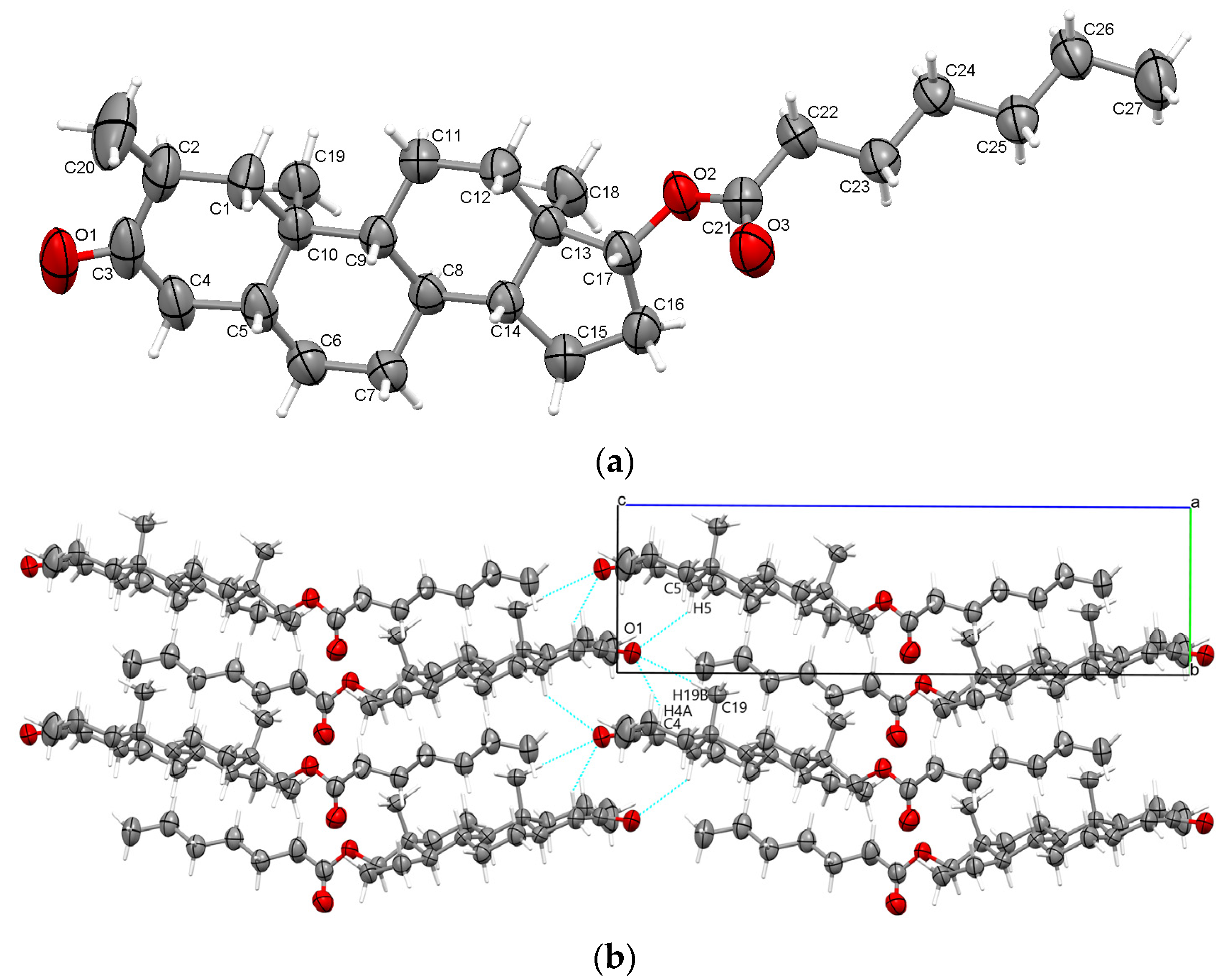
The analysis via a single crystal X-ray diffraction of the new drostanolone enanthate polymorph showed that it crystallized non-centrosymmetrically in the monoclinic crystal system with the P21 space group (Table 1). The asymmetric unit is illustrated in Figure 2a and comprises one steroid molecule. From a configurationally perspective, the six-membered A, B, and C steroid rings depict chair geometry, while the five-membered D ring is an envelope. Such configurations were previously reported in other dihydrotestosterone derivatives and testosterone analogues as well [11,12,13,14,15]. The crystal’s cohesion, stability, and self-assembly formation is driven by trifurcated C-H···O interactions in the direction of the ob-axis (2.690 Å, 2.609 Å, and 2.510 Å) involving O1 ketone oxygen (see Table 2). The supramolecular arrangements seen along the ao-axis are illustrated in Figure 2b. The molecular and packing perspectives were generated using Mercury software [16]. The absolute structure refinement and Flack’s parameter evaluation were conducted according to Parsons et al. [17], and its value of 0.15 (18) suggests that the structure is most likely racemic. The detailed crystal and refinement data are given in Table 1. The atomic coordinates embedded in the CIF file was deposited as Supplementary Material.

2.2. Thermal DTA/TGA

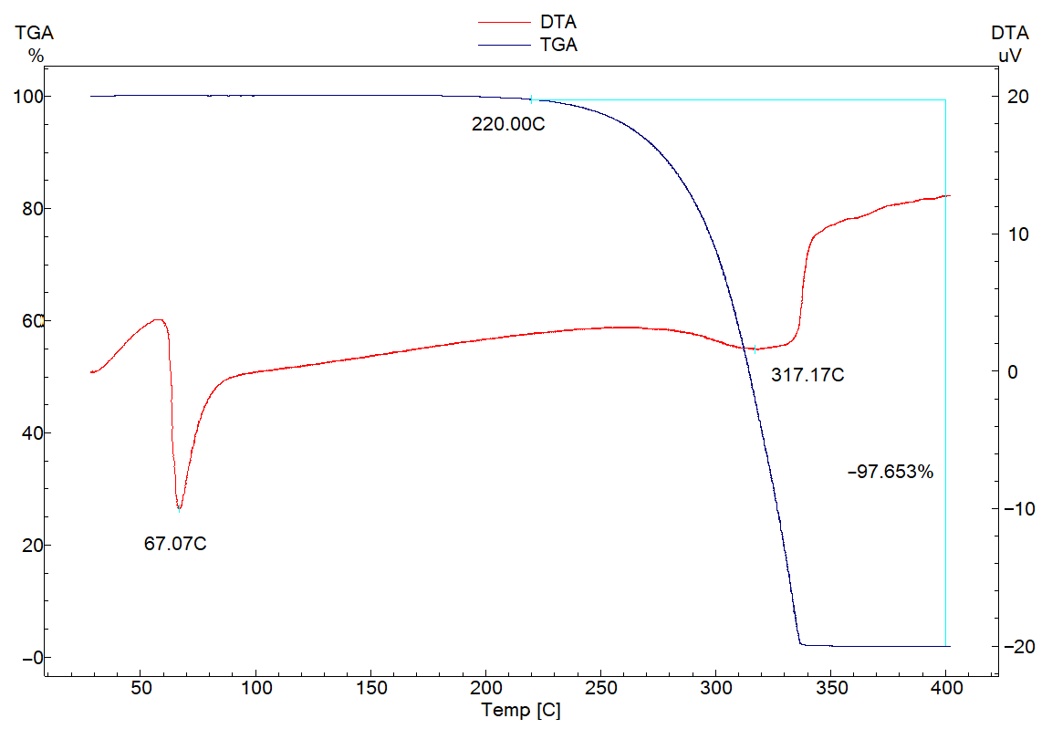
The DTA/TGA are suitable in order to evaluate thermal behaviour, stability, and to determine the melting points of active pharmaceutical ingredients. The sharp endothermic peak manifested at 67 °C (Figure 3) can be attributed to the melting point of the steroid. Furthermore, roughly between 100 and 300 °C, no thermal events were registered, after which, at 317 °C, an endotherm was observed, which can be associated with the decomposition and degradation of the sample, which is accompanied, on the TGA curve, with a mass loss with onset at 220 °C (Figure 3).

2.3. FT-IR Spectroscopy Results

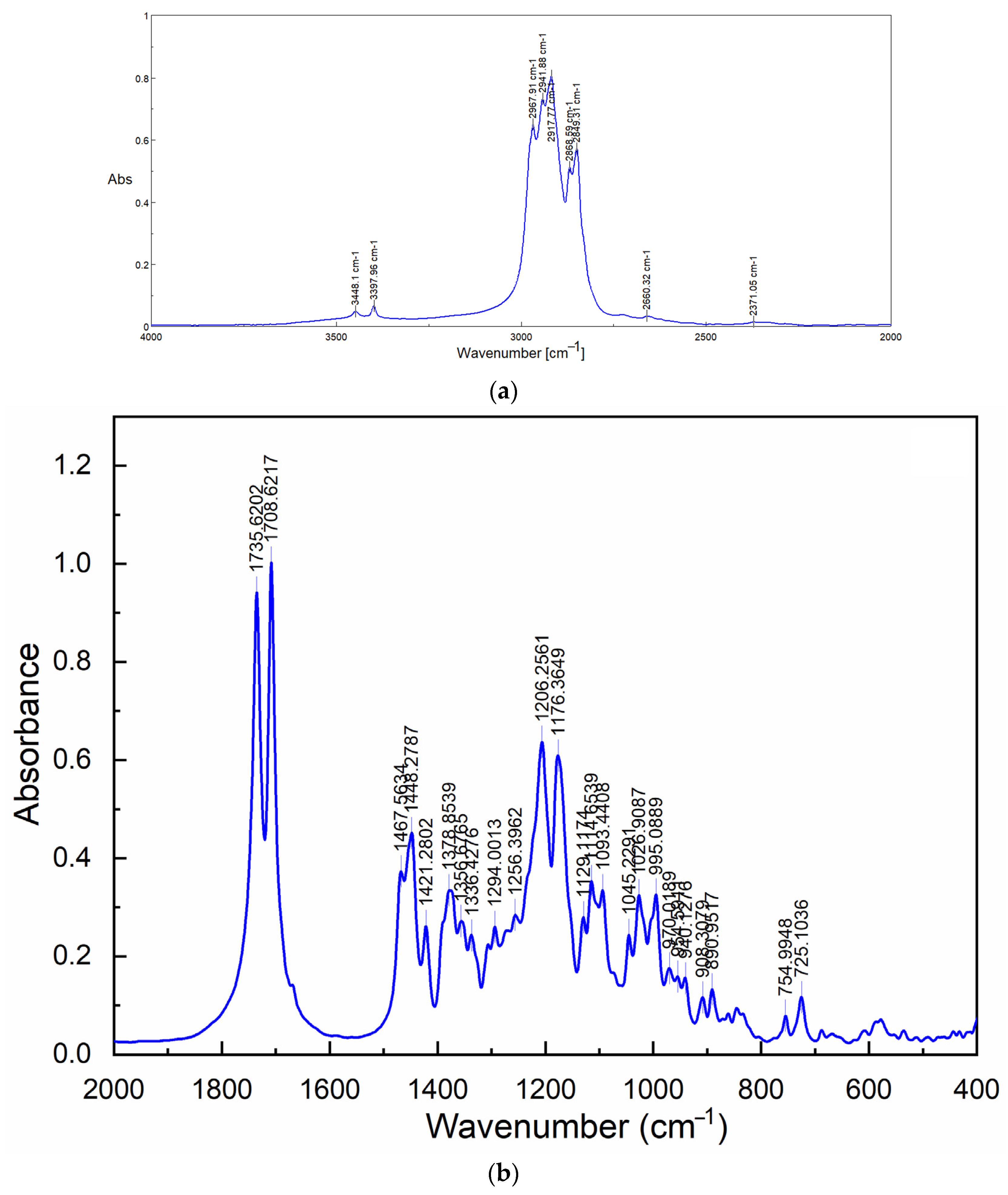
Analysis of active pharmaceutical compounds using FT-IR spectroscopy is a useful tool that can be used in order to explore the presence of functional groups. The spectra (Figure 4) are characterized by two bands at 3448 and 3397 cm−1, which represent the O-H stretching the surface water. The multiple bands between 2967 and 2849 cm−1 are attributed to symmetric and asymmetric C-H stretching. Furthermore, at lower wavenumbers, the two sharp bands at 1735 and 1708 cm−1 can be assigned to the two carbonyl C=O functional groups present in the steroid molecule.

3. Materials and Methods

3.1. General and Sample Preparations

White powder of drostanolone enanthate was received from Wuhan Shu Mai Technology Co., Wuhan, China. The solvents (ethanol and butanol) were received from Sigma Aldrich, Darmstadt, Germany and were used without further purification.
Single crystals of Drost En, which are suitable for X-ray analysis, were successfully obtained using slow evaporation during seven days at a temperature of 4 °C from a saturated mixture of ethanol and butanol in a 1:1 volumetric ratio.

3.2. X-Ray Diffraction and Structure Refinement

The crystallographic data were collected at ambient room temperature using a SuperNova diffractometer which was equipped with dual X-ray sources (Mo and Cu), an Eos CCD detector, and an operating tube at 50 kV and 0.08 mA. The experimental data were collected and then corrected for Lorentz, adsorption, and polarization effects using the CrysAlis PRO package, version 40_64.84a [18]. Using Olex2 software, version 1.5-alpha [19], the crystal structure was solved with the SHELXT solution program [20] with Intrinsic Phasing, and then was refined with the ShelXL [21] package via least squares minimization.
The hydrogen atoms were located and treated as riding, with the isotropic displacement parameter Uiso(H) = 1.2Ueq(C) for ternary CH groups [C-H = 0.93 Å], secondary CH2 groups [C-H = 0.97 Å], and 1.5Ueq(C) for methyl CH3 groups [C-H = 0.96 Å].

3.3. DTA/TGA Thermal Analysis

The differential thermal analysis (DTA) and thermogravimetric analysis (TGA) were recorded simultaneously using a Shimadzu DTG-60H instrument. The sample was placed in an open alumina pan and heated from ambient room temperature up to 400 °C, with a heating rate of 10 °C min−1 under nitrogen gas flow (70 mL/min).

3.4. FT-IR Spectroscopy

The FT-IR diagram of the title compound was obtained with a Jasco 6200 Fourier Transform Infrared Spectrometer using KBr pelleting. It was collected in the spectral range from 400 to 4000 cm−1 with a resolution of 4 cm−1 and 256 scans.

4. Conclusions

The crystal structure of a new drostanolone enanthate polymorph was elucidated and reported. It crystallizes in the monoclinic non-centrosymmetric P21 space group with one steroid in the asymmetric unit. The intermolecular interactions found within shorter distances than the sum of van der Waals radii, which contribute to crystal stability, are represented by C-H···O interactions. The DTA revealed the melting point to be 67 °C, and the FT-IR spectroscopy showed the absorption bands of the functional groups.

Supplementary Materials

CIF file of the drostanolone enanthate polymorph was deposited via the Cambridge Crystallographic Data Centre with the 2389842 deposit number. It can be obtained free of charge on written application to CCDC, 12 Union Road, Cambridge, CB2 1EZ, UK (fax: +44 1223 336033); on request via e-mail to [email protected] or by access to http://www.ccdc.cam.ac.uk (accessed on 9 October 2024).

Author Contributions

Conceptualization: A.T. and G.B.; methodology: M.-O.M.; X-ray crystal structure: A.T.; DTA/TGA: M.M.-P.; investigation: A.T., M.-O.M., G.B. and M.M.-P.; writing—original draft preparation: A.T., M.-O.M., G.B. and M.M.-P.; writing—review and editing; A.T. and G.B. All authors have read and agreed to the published version of the manuscript.

Funding

The authors acknowledge the financial support from the MINISTRY OF RESEARCH, INNOVATION AND DIGITALIZATION-MCID through the “Nucleu” Programme within the National Plan for Research, Development and Innovation 2022–2027, project PN23 24 01 01.

Data Availability Statement

The CIF file of drostanolone enanthate can be obtained on request via e-mail to [email protected].

Conflicts of Interest

The authors declare no conflicts of interest.

References

  1. Lednicer, D. Steroid Chemistry at a Glance, 1st ed.; Wiley: Chichester, UK, 2010. [Google Scholar]
  2. Mooradian, A.D.; Morley, J.E.; Korenman, S.G. Biological actions of androgens. Endocr. Rev. 1987, 8, 1–28. [Google Scholar] [CrossRef] [PubMed]
  3. Shahidi, N.T. A review of the chemistry, biological action, and clinical applications of anabolic-androgenic steroids. Clin. Ther. 2001, 23, 1355–1390. [Google Scholar] [CrossRef] [PubMed]
  4. Kicman, A.T. Pharmacology of anabolic steroids. Br. J. Pharmacol. 2008, 154, 502–521. [Google Scholar] [CrossRef] [PubMed]
  5. Vermeulen, A. Longacting steroid preparations. Acta Clin. Belg. 1975, 30, 48–55. [Google Scholar] [CrossRef] [PubMed]
  6. Chowdhury, M.S.; Banks, A.J.; Bond, W.H.; Jones, W.G.; Ward, H.W. A comparison of drostanolone propionate (Masteril) and nandrolone decanoate (Deca-durabolin) in the treatment of breast carcinoma. Clin. Oncol. 1976, 2, 203–206. [Google Scholar] [PubMed]
  7. Turza, A.; Borodi, G.; Muresan-Pop, M.; Ulici, A. Polymorphism and β-cyclodextrin complexation of methyldrostanolone. J. Mol. Struct. 2022, 1250, 131852. [Google Scholar] [CrossRef]
  8. Borodi, G.; Turza, A.; Bende, A. Exploring the Polymorphism of Drostanolone Propionate. Molecules 2020, 25, 1436. [Google Scholar] [CrossRef] [PubMed]
  9. Turza, A.; Miclaus, M.O.; Borodi, G. Exploring the crystal and molecular structures of methenolone and drostanolone enanthate. Z. Kristallogr. Cryst. Mater. 2024, 239, 129–138. [Google Scholar] [CrossRef]
  10. Chistyakov, D.; Sergeev, G. The polymorphism of Drugs: New Approaches to the Synthesis of Nanostructured Polymorphs. Pharmaceutics 2020, 12, 34. [Google Scholar] [CrossRef] [PubMed]
  11. Rendle, D.F.; Trotter, J. Crystal and molecular structure of 17β-hydroxy-17α-methyl-2-oxa-5α -androstan-3-one. J. Chem. Soc. Perkin II 1975, 1361–1365. [Google Scholar] [CrossRef]
  12. Gaedecki, Z. Structure of 17-α-methyl-testosterone semihydrate C20H30O2·1/2H2O. J. Crystallogr. Spectrosc. Res. 1989, 19, 577–587. [Google Scholar] [CrossRef]
  13. Gaedecki, Z. Structure of 17β-hydroxy-1,4-androstadien-3-one monohydrate C19H26O2·H2O. J. Crystallogr. Spectrosc. Res. 1989, 19, 933–939. [Google Scholar] [CrossRef]
  14. Turza, A.; Miclaus, M.O.; Pop, A.; Borodi, G. Crystal and molecular structures of boldenone and four boldenone steroid esters. Z. Kristallogr. Cryst. Mater. 2019, 234, 671–683. [Google Scholar] [CrossRef]
  15. Precioux, G.; Barrans, Y.; Hospital, M. 17-BETA HYDROXY-4,9,11 ESTRATRIENE-3 ONE,C18H22O2. Cryst. Struct. Commun. 1989, 8, 883–886. [Google Scholar]
  16. Macrae, C.F.; Bruno, I.J.; Chisholm, J.A.; Edigington, P.R.; McCabe, P.; Pidcock, E.; Rodriguez-Monge, L.; Taylor, J.v.d.S.; Wood, P.A. Mercury CSD 2.0-new features for the visualization and investigation of crystal structures. J. Appl. Cryst. 2008, 41, 466–470. [Google Scholar] [CrossRef]
  17. Parsons, S.; Flack, H.D.; Wagner, T. Use of intensity quotients and differences in absolute structure refinement. Acta Cryst. 2013, B69, 249–259. [Google Scholar] [CrossRef] [PubMed]
  18. CrysAlis PRO. Rigaku Oxford Diffraction; CrysAlis PRO: Yarnton, UK, 2015. [Google Scholar]
  19. Dolomanov, O.V.; Bourhis, L.J.; Gildea, R.J.; Howard, J.A.K.; Puschmann, H. OLEX2: A complete structure solution, refinement and analysis program. J. Appl. Cryst. 2009, 42, 339–341. [Google Scholar] [CrossRef]
  20. Sheldrick, G.M. SHELXT-Integrated space-group and crystal-structure determination. Acta Cryst. 2015, A71, 3–8. [Google Scholar] [CrossRef] [PubMed]
  21. Sheldrick, G.M. Crystal structure refinement with SHELXL. Acta Cryst. 2015, C71, 3–8. [Google Scholar] [CrossRef]
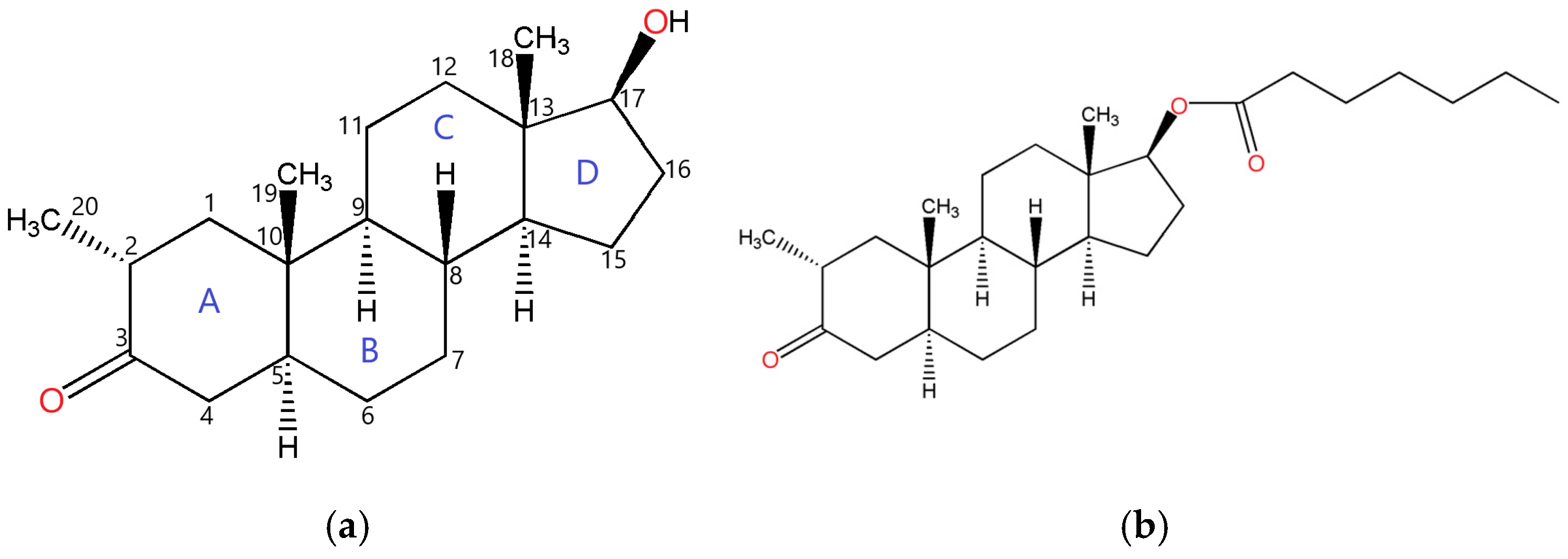
Figure 1. Molecular structure of drostanolone, showing the atom numbering scheme of the steroid backbone (a); drostanolone enanthate (b).
Molbank 2024 m1907 g001
Figure 2. Asymmetric unit presenting the atoms as thermal ellipsoids at a 50% probability level (a); crystal packing and intermolecular interactions in the crystal (b).
Molbank 2024 m1907 g002
Figure 3. DTA/TGA traces of drostanolone enanthate.
Molbank 2024 m1907 g003
Figure 4. FT-IR spectra of drostanolone enanthate in the range of 4000–2000 cm−1 (a) and 2000–400 cm−1 (b).
Molbank 2024 m1907 g004
Table 1. Crystallographic details of investigated crystals.
Identification CodeDrostanolone Enanthate
Empirical formulaC27H44O3
Formula weight416.62
Temperature/K293 (2)
Crystal systemmonoclinic
Space groupP21
a/Å9.0864 (5)
b/Å6.3760 (3)
c/Å21.8006 (8)
α/°90
β/°90.765 (4)
γ/°90
Volume/Å31262.90 (10)
Z2
ρcalc g/cm31.096
μ/mm−10.533
F(000)460.0
RadiationCu Kα (λ = 1.54184)
2Θ range/°8.112 to 142.73
Index ranges−11 ≤ h ≤ 11, −7 ≤ k ≤ 7, −26 ≤ l ≤ 26
Reflections collected16,129
Independent reflections4728 [Rint = 0.0462, Rsigma = 0.0403]
Data/restraints/parameters4728/1/275
Goodness of fit on F21.025
Final R indexes [I ≥ 2σ (I)]R1 = 0.0517, wR2 = 0.1226
Final R indexes [all data]R1 = 0.0758, wR2 = 0.1452
Largest diff. peak/hole/e Å−30.13/−0.21
Flack’s parameter0.15 (18)
Table 2. Hydrogen bond geometry (Å, o).
D-H···AD-HH···AD···A<(D-H···A)
C5-H5···O11.0892.6833.525133.7
C19-H19B···O11.0892.6033.688174.2
C4-H4B···O11.0892.5063.277126.9
Disclaimer/Publisher’s Note: The statements, opinions and data contained in all publications are solely those of the individual author(s) and contributor(s) and not of MDPI and/or the editor(s). MDPI and/or the editor(s) disclaim responsibility for any injury to people or property resulting from any ideas, methods, instructions or products referred to in the content.