Communication

Synthesis and Characterization of 2-(((2,7-Dihydroxynaphthalen-1-yl)methylene)amino)-3′,6′-bis(ethylamino)-2′,7′-dimethylspiro[isoindoline-1,9′-xanthen]-3-one and Colorimetric Detection of Uranium in Water

1
Department of Chemistry and Biochemistry, Norwich University, Northfield, VT 05663, USA
2
Department of Chemistry, University of Vermont, Burlington, VT 05405, USA
*
Author to whom correspondence should be addressed.
Molbank 2023, 2023(3), M1725; https://doi.org/10.3390/M1725
Submission received: 12 August 2023 / Revised: 9 September 2023 / Accepted: 13 September 2023 / Published: 15 September 2023
(This article belongs to the Section Structure Determination)

Abstract

:
2-(((2,7-Dihydroxynaphthalen-1-yl)methylene)amino)-3′,6′-bis(ethylamino)-2′,7′-dimethylspiro[isoindoline-1,9′-xanthen]-3-one was synthesized using Rhodamine 6G hydrazide (prepared using literature methods) and commercially available 2,7-dihydroxynaphthalene-1-carbaldehyde via imine condensation. Structural characterization was performed using FT-IR, 1H-NMR, 13C-NMR, X-ray, and HRMS. This Schiff base shows promise as a ligand for the colorimetric analysis of uranium in water.

1. Introduction

Schiff bases are a class of compounds derived from the reaction of primary amines with carbonyl compounds, resulting in the formation of an imine (C=N) [1]. Structurally, these compounds are well suited for binding transition metals [2,3,4], and Schiff bases have commonly been used as fluorescent or colorimetric probes for metal ions [5,6]. Due to their modular synthesis, these molecules are highly tunable, and varying the structure has been shown to result in high selectivity for a particular metal ion in solution, even in the presence of competing metal ions [6].
Industrially, Schiff bases have been used as catalysts, dyes, or stabilizers [7]. These compounds have also been thoroughly investigated for their high levels of biological activity as metal complexes [8,9,10] or as small-molecule candidates for anticancer activity [11], as well as for antiviral, antibacterial, and anti-inflammatory properties, among others [7].
The following work demonstrates the synthesis and characterization of 2-(((2,7-dihydroxynaphthalen-1-yl)methylene)amino)-3′,6′-bis(ethylamino)-2′,7′-dimethylspiro[isoindoline-1,9′-xanthen]-3-one (PROM1), a Schiff base synthesized from Rhodamine 6G hydrazide and 2,7-dihydroxynaphthalene-1-carbaldehyde. A related Schiff base was previously reported and shown to have high uranium binding affinity [12]. PROM1 was also tested as a ligand for the colorimetric sensing of uranium in water.

2. Results and Discussion

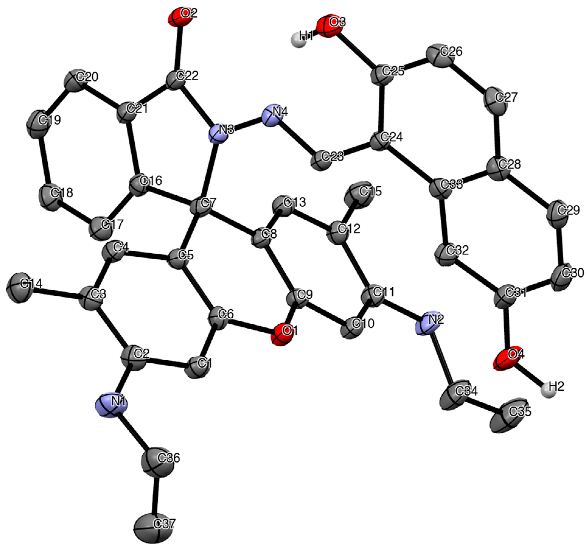
2.1. Synthesis of 2-(((2,7-Dihydroxynaphthalen-1-yl)methylene)amino)-3′,6′-bis(ethylamino)-2′,7′-dimethylspiro[isoindoline-1,9′-xanthen]-3-one (PROM1)

Schiff base PROM1 was synthesized in two steps. First, Rhodamine 6G hydrazide was prepared using literature methods (Scheme 1A) [13]. This starting material was then used for imine condensation with commercially available 2,7-dihydroxynaphthalene-1-carbaldehyde via overnight reflux (Scheme 1B).
PROM1 was recrystallized via slow evaporation in a mixture of acetonitrile and ethanol. The crystalline product was analyzed using X-ray, FT-IR, high-resolution ESI-MS, and 1H- and 13C-NMR (for spectra, see Supplementary Materials, Figures S1–S6). The structural determination of PROM1 was performed via X-ray crystallography (Figure 1), and the structure was confirmed via NMR spectroscopy.

2.2. Colorimetric Detection of Uranium

Uranium is a chemically and radiologically toxic element that occurs naturally in groundwater, and may have increased concentrations due to mining operations, processing for nuclear power, or improper nuclear waste management [14]. A simple colorimetric test for the presence of uranium in drinking water may prevent accidental exposure to uranium, which can result in kidney disease, kidney failure, or cancer [15].
A stock solution of PROM1 was prepared in dimethylsulfoxide (DMSO). When mixed with aqueous solutions of uranyl nitrate, a color change from yellow to pink was observed (Figure 2).

3. Materials and Methods

3.1. General

All starting materials were purchased from commercial suppliers. Rhodamine 6G and 2,7-dihydroxynaphthalene-1-carbaldehyde were purchased from Sigma Aldrich (Burlington, MA, USA), 80% hydrazine from TCI (VWR), and uranyl nitrate hexahydrate from Fisher Scientific (Pittsburgh, PA, USA). Solvents were reagent/ACS-grade and were purchased from VWR, Radnor, PA, USA. Rhodamine 6G hydrazide was synthesized as described previously in the literature [13].
The IR spectrum was recorded on a Perkin Elmer Spectrum 100 FT-IR, UV-Vis spectra were recorded on a Shimadzu UV-2600i, mass spectrometry was run via direct infusion in pos ESI on an Agilent 6530 QToF HRMS, NMR spectra were collected on a Bruker AXR 500 MHz spectrometer in dimethylsulfoxide-d6 solution with reference to residual solvent signals (DMSO-d6, δ = 2.50 ppm for 1H and 39.52 ppm for 13C). X-ray diffraction data were collected on a Bruker APEX 2 CCD platform diffractometer [Mo Kα (λ = 0.71073 Å)].

3.2. 2-(((2,7-Dihydroxynaphthalen-1-yl)methylene)amino)-3′,6′-bis(ethylamino)-2′,7′-dimethylspiro[isoindoline-1,9′-xanthen]-3-one (PROM1)

To Rhodamine 6G hydrazide (0.1915 g, 0.45 mmol), ethanol (15 mL) and acetic acid (5–6 drops) were added. The mixture was stirred to dissolve, and then 2,7-dihydroxynaphthalene-1-carbaldehyde (0.0837 g, 0.44 mmol) was added. The resulting solution was stirred for 24 h under reflux at 72 °C. The yellow solid product was separated via hot vacuum filtration (0.1839 g, 68.8%). A portion of the product was redissolved in warm acetonitrile, layered with ethanol, and recrystallized via slow evaporation at 5 °C. The yellow crystalline product was used directly for X-ray, 1H and 13C NMR, and HRMS. HRMS m/z calcd. for C37H34N4O4: 598.26. Found [M + H]+: 599.2641. 1H-NMR (DMSO-d6): 9.57 (s, 1H, imine-H), 8.00 (d, J = 7.3, 1H, Ar-H), 7.73 (d, J = 8.9, 1H, Ar-H), 7.70–7.63 (m, 3H, Ar-H), 7.13–7.11 (overlapping m, 2H, Ar-H), 6.95 (dd, J = 8.70, 1.96, 1H, Ar-H), 6.82 (d, J = 8.9, 1H, Ar-H), 6.40 (s, 2H, Ar-H), 6.27 (s, 2H, Ar-H), 3.46 (residual water), 3.16 (q, J = 6.58, 4H, N-CH2CH3), 2.54 (DMSO), 2.10 (s, MeCN), 1.87 (s, 6H, Ar-CH3), 1.22 (t, J = 7.09, 6H N-CH2CH3), 1.09 (t, J = 7, EtOH). 13C-NMR (DMSO-d6): 163.5 (quat C, carbonyl), 158.5 (quat Ar), 157.4 (quat Ar), 151.4 (quat Ar), 151.2 (quat Ar), 148.1 (quat Ar), 147.2 (imine CH), 134.1 (Ar CH), 133.7 (quat Ar), 133.1 (Ar CH), 130.8 (Ar CH), 129.0 (quat Ar), 128.6 (Ar CH), 127.0 (Ar CH), 124.0 (Ar CH), 123.1 (Ar CH), 122.2 (quat Ar), 118.7 (quat Ar), 118.1 (CH3CN), 115.6 (Ar CH), 115.2 (Ar CH), 107.0 (quat Ar), 103.9 (quat Ar), 102.7 (Ar CH), 95.7 (Ar CH), 65.6 (quat C), 56.1 (EtOH), 39.5 (q, DMSO), 37.5 (CH2), 18.6 (EtOH), 17.0 (CH3), 14.1 (CH3), 1.22 (CH3CN). FT-IR (ν, cm−1): 3440.96 (νNH), 3300.00 (νOH), 2964.17 (νCH), 2866.53 (νCH), 1694.78 (νCO), 1617.61 (νCH), 1564.92, 1515.18, 1465.34, 1449.02, 1420.64, 1379.85, 1348.29, 1321.63, 1270.01, 1217.86, 1198.74, 1159.27, 1138.86, 1086.14, 1035.71, 1013.46, 964.98, 923.35, 896.72, 876.22, 859.07, 841.39, 830.84, 811.81, 791.70.

3.3. X-ray Data

A suitable yellow crystal, grown from acetonitrile and ethanol, was mounted on a MiTiGen micromount with Paratone-N cryoprotectant oil. The structure was solved via direct methods using SHELXT and refined via full-matrix least-squares methods against F2 by SHELXL-2018/3 [16,17]. All non-hydrogen atoms were refined with anisotropic displacement parameters. All hydrogen atoms were refined with isotropic displacement parameters. H1 and H2 were refined freely. Hydrogen atoms on carbon were included in calculated positions and were refined using a riding model.
Crystallographic data for the structure reported here have been deposited with the Cambridge Crystallographic Data Centre [18]. CCDC 2287758 contain the supplementary crystallographic data for this manuscript. These data can be obtained free of charge from The Cambridge Crystallographic Data Centre via www.ccdc.cam.ac.uk/structures. The final CIF file was generated using FinalCif [19].
Crystal data for C41H40N6O4 (M = 680.79 g/mol): triclinic, space group P–1 (no. 2), a = 11.37(3) Å, b = 11.51(3) Å, c = 14.38(3) Å, α = 80.06(3)°, β = 74.82(3)°, γ = 85.75(3)°, V = 1789(7) Å3, Z = 2, T = 150(2) K, μ(MoΚα) = 0.083 mm−1, Dcalc = 1.264 g/cm3, 14,714 reflections measured (3.59° ≤ 2Θ ≤ 47.48°), 5320 unique (Rint = 0. 0799, Rsigma = 0.0952), which were used in all calculations. The final R1 was 0.0450 (I > 2σ(I)), and wR2 was 0.1066 (all data).

3.4. Colorimetric Analysis of Uranium Binding

Stock solutions of uranyl nitrate in water were prepared by dissolving the appropriate amounts of solid uranyl nitrate hexahydrate in deionized water. The PROM1 stock solution was prepared by dissolving 0.0200 g (0.033 mmol) in 100 mL of DMSO. Equal amounts of the ligand stock solution (in DMSO) and uranyl nitrate hexahydrate solution (in water) were then mixed resulting in the yellow to pink color change upon uranium binding (Figure 2).

4. Conclusions

Novel Schiff base 2-(((2,7-dihydroxynaphthalen-1-yl)methylene)amino)-3′,6′-bis(ethylamino)-2′,7′-dimethylspiro[isoindoline-1,9′-xanthen]-3-one (PROM1) was synthesized from Rhodamine 6G hydrazide and 2,7-dihydroxynaphthalene-1-carbaldehyde. PROM1 was shown to bind uranium in water and is a plausible ligand for the colorimetric analysis of uranium in drinking water.

Supplementary Materials

The Supplementary Materials contain the NMR spectra of PROM1 (Figures S1–S4), IR spectrum (Figure S5), and HRMS (Figure S6), as well as color change resulting from change in pH (Figure S7).

Author Contributions

R.M. and P.O. synthesized and recrystallized the target compound; A.V. performed colorimetric experiments; D.M.S.J. carried out crystallographic experiments and NMR characterization; R.W. assisted with reviewing and editing, as well as compound analysis; S.K.G. conceptualized the project, acquired funding, analyzed data, and wrote the original draft. All authors have read and agreed to the published version of the manuscript.

Funding

This research was supported by an Institutional Development Award (IDeA) from the National Institute of General Medical Sciences of the National Institutes of Health under grant number P20GM103449. Its contents are solely the responsibility of the authors and do not necessarily represent the official views of NIGMS or NIH. Additional support was provided by the U. S. National Science Foundation through CHE-2101766 and CHE-1039436.

Data Availability Statement

The authors confirm that the data supporting the findings of this study are available within the article and/or its Supplemental Materials.

Acknowledgments

S.K.G., R.M., P.O. and A.V. would like to thank the Norwich University Department of Chemistry and Biochemistry, the Norwich University Office of Academic Research, and the Vermont Biomedical Research Network for their support. The authors would also like to thank Bruce O’Rourke for his assistance with HRMS.

Conflicts of Interest

The authors declare no conflict of interest.

References

  1. Raczuk, E.; Dmochowska, B.; Samaszko-Fiertek, J.; Madaj, J. Different Schiff Bases—Structure, Importance and Classification. Molecules 2022, 27, 787. [Google Scholar] [CrossRef] [PubMed]
  2. Hunter, L.; Marriott, J.A. Co-ordinated copper and nickel compounds of salicylidene derivatives. J. Chem. Soc. 1937, 422, 2000–2003. [Google Scholar] [CrossRef]
  3. Sacconi, L.; Ciampolini, M.; Maggio, F.; Cavasino, F.P. Studies in Coordination Chemistry. IX.1 Investigation of the Stereochemistry of Some Complex Compounds of Cobalt(II) with N-Substituted Salicylaldimines. J. Am. Chem. Soc. 1962, 84, 3246–3248. [Google Scholar] [CrossRef]
  4. Holm, R.H.; Swaminathan, K. Studies on Nickel(II) Complexes. III. Bis-(N-arylsalicylaldimine) Complexes. Inorg. Chem. 1962, 1, 599–607. [Google Scholar] [CrossRef]
  5. Wang, Y.; Hao, X.; Liang, L.; Gao, L.; Ren, X.; Wu, Y.; Zhao, H. A coumarin-containing Schiff base fluorescent probe with AIE effect for the copper(ii) ion. RSC Adv. 2020, 10, 6109–6113. [Google Scholar] [CrossRef] [PubMed]
  6. Khan, S.; Chen, X.; Almahri, A.; Allehyani, E.S.; Alhumaydhi, F.A.; Ibrahim, M.M.; Ali, S. Recent developments in fluorescent and colorimetric chemosensors based on schiff bases for metallic cations detection: A review. J. Environ. Chem. Eng. 2021, 9, 106381. [Google Scholar] [CrossRef]
  7. Uddin, M.N.; Ahmed, S.S.; Alam, S.M.R. REVIEW: Biomedical applications of Schiff base metal complexes. J. Coord. Chem. 2020, 73, 3109–3149. [Google Scholar] [CrossRef]
  8. Chaudhary, N.K.; Mishra, P. Metal complexes of a novel Schiff base based on penicillin: Characterization, molecular modeling, and antibacterial activity study. Bioinorg. Chem. Appl. 2017, 2017, 6927675. [Google Scholar] [CrossRef] [PubMed]
  9. Chaudhary, N.K.; Mishra, P. Bioactivity of some divalent M(II) complexes of penicillin based Schiff base ligand: Synthesis, spectroscopic characterization, and thermal study. J. Saudi Chem. Soc. 2018, 22, 601–613. [Google Scholar] [CrossRef]
  10. Sridhar, G.; Bilal, M.; Easwaramoorthy, D.; Rani, K.; Kumar, S.; Manohar, C.S. Synthesis, Characterization and Antimicrobial Activities of Copper, Nickel, Cobalt, Chromium Complexes Derived from (Z)-4-Fluoro-N-(2,7-dimethylhept-6-enylidene) benzenamine. J. Braz. Chem. Soc. 2017, 28, 756–767. [Google Scholar] [CrossRef]
  11. Uddin, N.; Rashid, F.; Ali, S.; Tirmizi, S.A.; Ahmad, I.; Zaib, S.; Zubair, M.; Diaconescu, P.L.; Tahir, M.N.; Iqbal, J.; et al. Synthesis, characterization, and anticancer activity of Schiff bases. J. Biomol. Struct. Dyn. 2019, 38, 3246–3259. [Google Scholar] [CrossRef] [PubMed]
  12. Halali, V.V.; Balakrishna, R.G. An expeditious method for the ultra-level chemosensing of uranyl ions. Anal. Methods 2020, 12, 1070–1076. [Google Scholar] [CrossRef]
  13. Zhang, Z.; Zheng, Y.; Hang, W.; Yan, X.; Zhao, Y. Sensitive and selective off–on rhodamine hydrazide fluorescent chemosensor for hypochlorous acid detection and bioimaging. Talanta 2011, 85, 779–786. [Google Scholar] [CrossRef] [PubMed]
  14. Jalbani, N.; Soylak, M. Spectrophotometric determination of uranium using chromotrope 2R complexes. J. Radioanal. Nucl. Chem. 2014, 301, 263–268. [Google Scholar] [CrossRef]
  15. Srivastava, P.K. Spectrophotometric analysis of underground well water uranium of abandoned coal mines. IOSR J. Environ. Sci. Toxicol. Food Technol. 2016, 10, 101–105. [Google Scholar]
  16. Sheldrick, G.M. SHELXT—Integrated space-group and crystal-structure determination. Acta Cryst. 2015, A71, 3–8. [Google Scholar] [CrossRef] [PubMed]
  17. Sheldrick, G.M. Crystal structure refinement with SHELXL. Acta Cryst. 2015, C71, 3–8. [Google Scholar] [CrossRef]
  18. Groom, C.R.; Bruno, I.J.; Lightfoot, M.P.; Ward, S.C. The Cambridge Structural Database. Acta Cryst. 2016, B72, 171–179. [Google Scholar] [CrossRef] [PubMed]
  19. Kratzert, D. FinalCif, V123. Available online: https://dkratzert.de/finalcif.html (accessed on 5 April 2023).
Scheme 1. (A) Synthesis of Rhodamine 6G hydrazide via literature procedure [13]. (B) Synthesis of PROM1 ligand.
Molbank 2023 m1725 sch001
Figure 1. Molecular structure of PROM1•2 MeCN with thermal ellipsoids drawn at a 30% probability level. The solvent molecules are not shown, and H atoms are omitted, except for on heteroatoms for clarity.
Molbank 2023 m1725 g001
Figure 2. (A) A stock solution of PROM1 in DMSO. (B) PROM1 and 500 μg/L uranyl nitrate in 50/50 DMSO and water. (C) PROM1 and 5000 μg/L uranyl nitrate in 50/50 DMSO and water.
Molbank 2023 m1725 g002
Disclaimer/Publisher’s Note: The statements, opinions and data contained in all publications are solely those of the individual author(s) and contributor(s) and not of MDPI and/or the editor(s). MDPI and/or the editor(s) disclaim responsibility for any injury to people or property resulting from any ideas, methods, instructions or products referred to in the content.

