


Abstract
A new emissive heteroleptic Ag(I) complex formulated as [AgL(POP)][PF6] (L = 4,4′-bis (4-Fluorophenyl)-6,6′-dimethyl-2,2′-bipyridine, POP= bis (2-(diphenylphosphino) phenyl) ether) was synthesized and characterized in both the solid state (X-ray crystallography) and the solution. The compound is a yellow-green phosphor (λem = 528 nm), with moderate quantum efficiency (ΦPL = 25% in deaerated dichloromethane).
1. Introduction
Luminescent mononuclear Ag(I) complexes are a class of compounds which have attracted considerable interest due to their potential use as sensors, photocatalysts, emitters in light-emitting devices, etc. The most studied of these incorporate various chelating diimine-type ligands (N^N) and phosphines (P or P^P), adopting a distorted tetrahedral geometry [1,2,3,4,5,6,7].
Ag(I) complexes often show long-lived phosphorescence originating from ligand-centred excited states (3LC) and rarely exhibit TADF (Thermally Activated Delayed Fluorescence) [8]. In the latter case, a small singlet-triplet energy gap (ΔE(S1 − T1)) is required for an effective reverse intersystem crossing (RISC) to the singlet state and consequently TADF emission [9,10,11]. The nature of the excited state in heteroleptic Ag(N^N)(P^P) type complexes (LL’CT, and/or MLCT) strongly depends on the electron donating properties of both ligands, while the insertion of bulky substituents at positions located near the coordinating atoms (N and/or P) generally increases the photoluminescence quantum yield (ΦPL) [12,13,14,15,16,17].
O. Moudam et al. synthesized the simple cationic complex [Ag(bpy)(POP)]+ (bpy = 2,2′-bipyridine, POP = bis (2-(diphenylphosphino) phenyl) ether). They found that the compound is a promising phosphorescent emitter, with λem =520 nm (yellow-green) and ΦPL = 14% (in PPMA film). It was also the first ionic Ag(I) complex successfully utilized for the construction of a light-emitting electrochemical cell (LEC) [5].
To contribute to this emerging field, we decided to prepare, characterize and investigate the photophysical properties of a novel heteroleptic Ag(I) complex containing the diphosphine POP and the already used in our previous work diimine ligand 4,4′-bis(4-Fluorophenyl)-6,6′-dimethyl-2,2′-bipyridine [17]. We thought that it might be of interest to reveal the effect of bpy core substitutions (at positions 4,4′ and 6,6′) on the emission energy and quantum efficiency.
2. Results and Discussion
2.1. NMR Spectroscopy in Solution
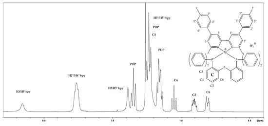
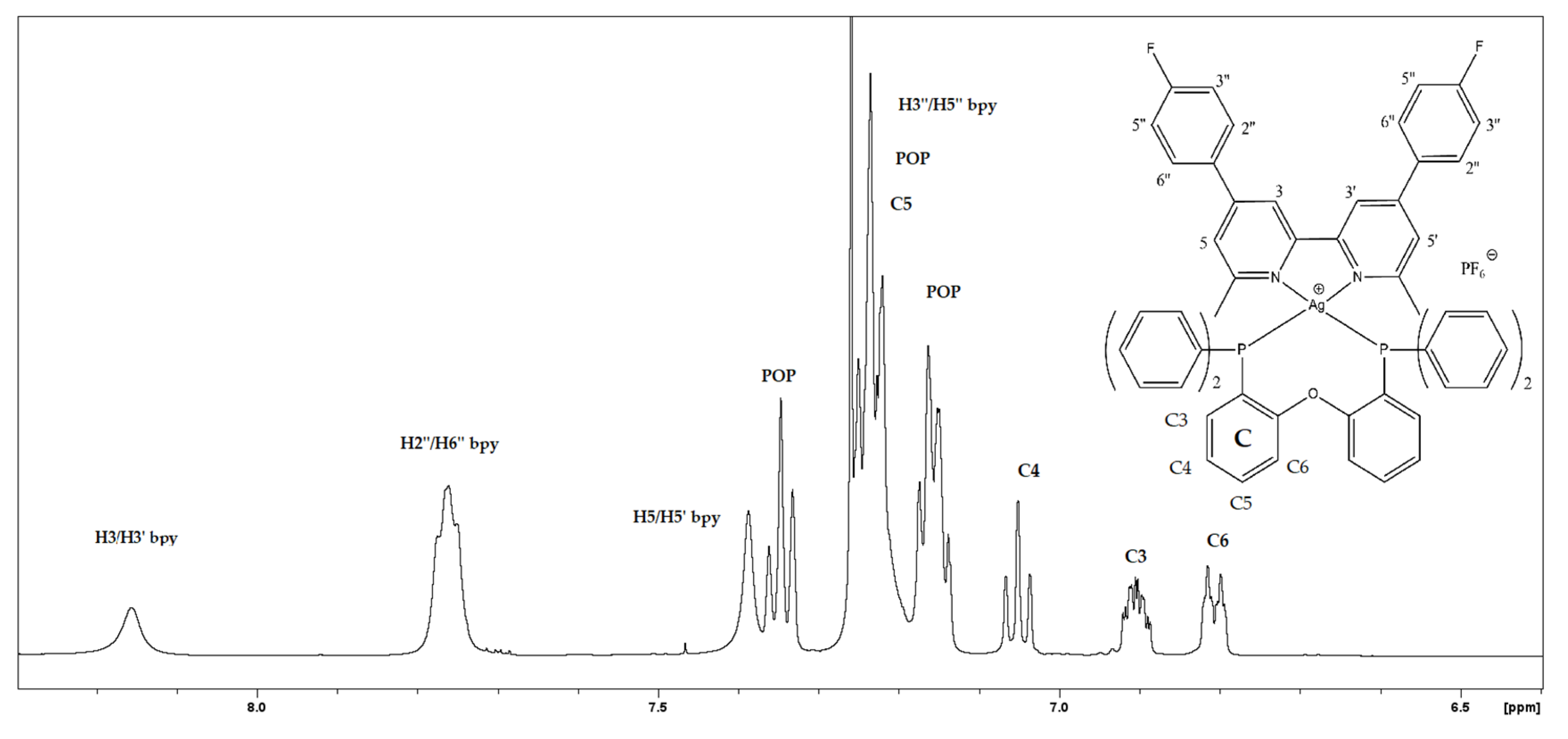
The 1H-NMR spectrum (Figure 1) of [AgL(POP)][PF6] (1) was recorded in CDCl3. 1H assignments and complexation-induced changes (Δδ) are listed in Table 1. The chemical shifts of both ligands are more or less perturbed in the presence of Ag(I), providing evidence of complex formation in the solution [14].
Figure 1.
The 1H NMR spectrum of [Ag(L)(POP)][PF6] in CDCl3 at 500 MHz at 298 K.
Table 1.
The 1H NMR data (δ, ppm) of L/POP and complex 1.
2.2. Photophysical Properties
2.2.1. Electronic Absorption Spectroscopy
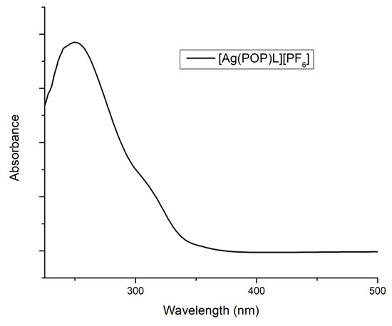
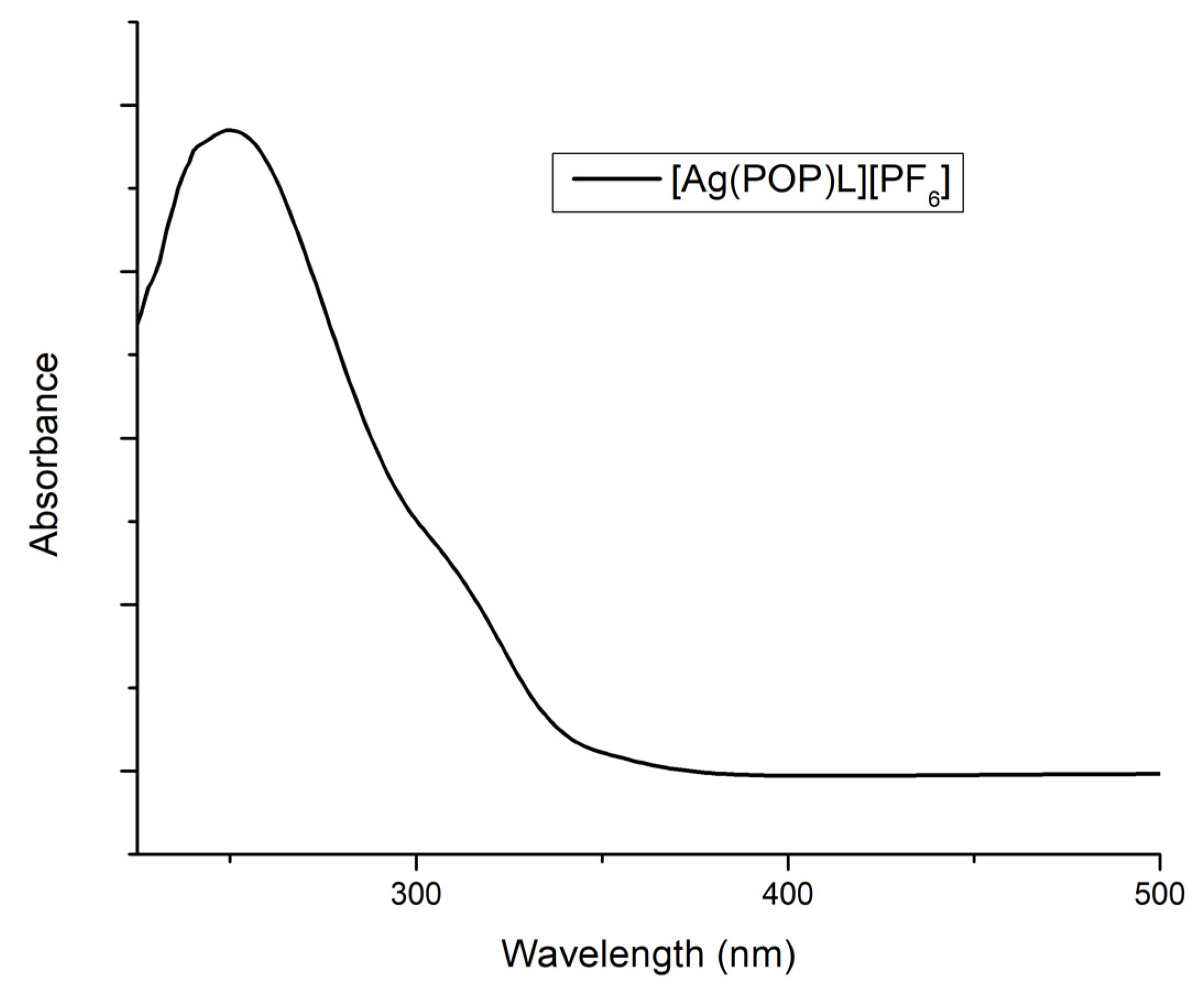
The UV–Vis spectrum of complex 1 in CH2Cl2 registered at room temperature is depicted in Figure 2. The absorption band located at 250 nm could be ascribed to ligand-centred (LC) π → π* transitions, while the shoulder present in the spectral range of 300–350 nm is typical of a composite L’LCT/MLCT character, frequently observed in M[(N^N)(POP)] (M = Ag(I), Cu(I)) type complexes [5,8,12].
Figure 2.
The UV–Vis absorption spectrum of 1 in dichloromethane at 298 K.
2.2.2. Emission Spectrum
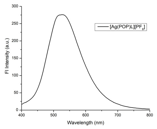
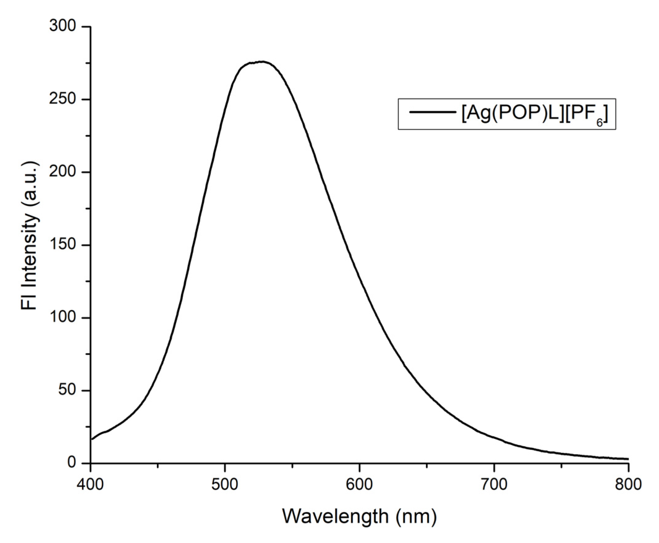
The photoluminescence spectrum of compound 1 (λexc = 350 nm) was acquired in a dichloromethane solution at room temperature (Figure 3). The observed band at λem = 528 nm (yellow-green colour emitter), appears broad and unstructured. This spectral feature and the large Stoke’s shift (~50,000 cm−1) denote the possible presence of a triplet emissive state. The complex was barely emissive in air-equilibrated solution (ΦPL = 2%). This value compares well with the one reported for [Ag(bpy)(POP)]BF4 (reference complex) under the same conditions (~1%) [5]. Remarkably, in deaerated solutions, a 12-fold increase (~25%) in the quantum efficiency of our complex is observed, while [Ag(bpy)(POP)]BF4 reaches only 5%. Summing up, altering the substitution on the bpy core (two methyl and two 4-fluorophenyl groups at the positions 6,6′ and 4,4′, respectively) does not drastically affect the emission maximum (528 for 1 vs. 520 nm for the reference complex). On the contrary, the ΦPL value in deaerated solutions is significantly increased.
Figure 3.
The photoluminescence spectrum of 1 in dichloromethane (λexc = 350 nm).
2.3. Description of the Structure
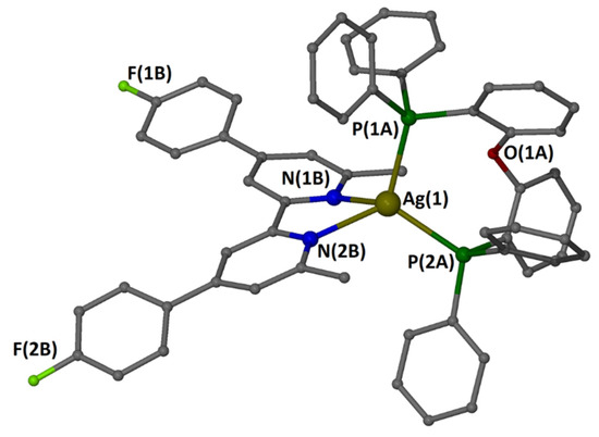
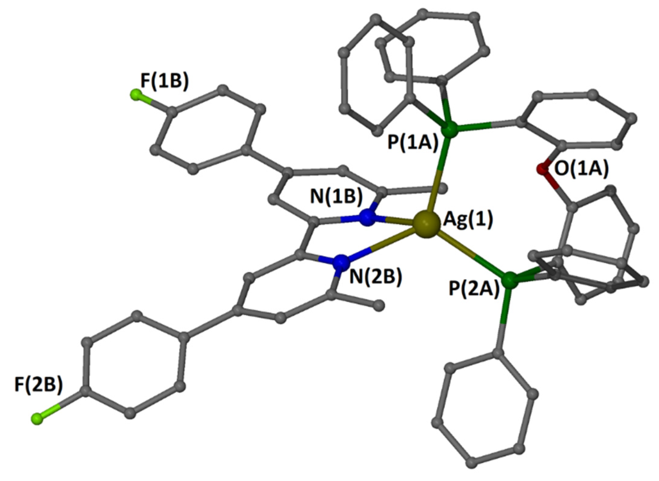
The X-ray crystal structure of complex 1 has been obtained from single crystals grown by slow vapour diffusion of diethyl ether into a concentrated solution of the reaction mixture in dichloromethane/methanol. 1 is formulated as [AgL(POP)]PF6·0.5L, where POP is the bis[(2-diphenylphosphino)phenyl] ether and L is 4,4′-bis(4-fluorophenyl)-6,6′-dimethyl-2,2′-bipyridine, and crystallized in the triclinic space group P. Its asymmetric unit consists of a cation [Ag(POP)L]+, the counter anion PF6− and half fragment of the symmetrical ligand. Selected bond distances (Å) and angles (°) for the coordination sphere of Ag(I) in the cation are presented in Table 2, while the structure of the cation is depicted in Figure 4.
Table 2.
Selected structural characteristics of compound 1.
Figure 4.
A “ball and stick’’ presentation of the cation in compound 1 with a partial labelling scheme. Hydrogen atoms have been omitted for clarity.
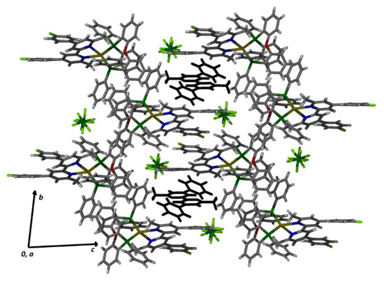
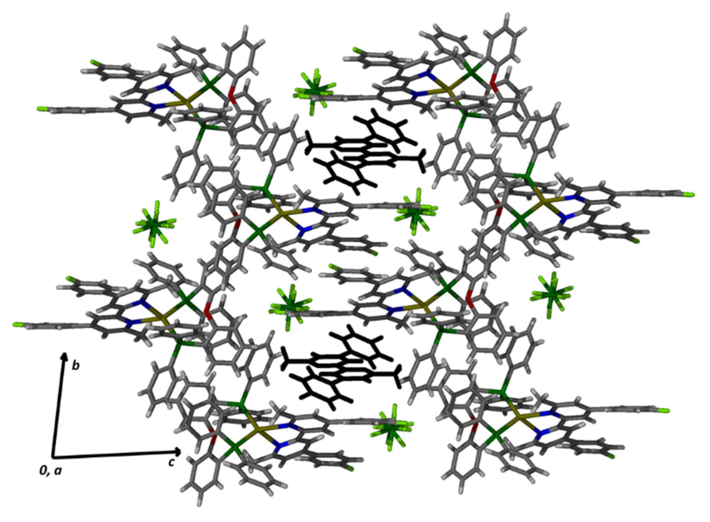
Compound formulated as [Ag(N-N chelate)(P-P chelate)](counter anion) and it is the first example containing the ligand 4,4′-bis(4-fluorophenyl)-6,6′-dimethyl-2,2′-bipyridine. In the cation, Ag(1) sits in the centre of a rather distorted tetrahedron built by two nitrogen atoms [N(1B) and N(2B)] belonging to a chelated diimine ligand and two phosphorus atoms [P(1A) and P(2A)] belonging to POP molecule. The distortions of the tetrahedron can be easily seen from the wide N(1B)–Ag(1)–P(2A) angle [121.68(8)°] due to the large steric hindrance and from the acute N(1B)– Ag(1)–N(2) angle [70.34(10)°] due to the small bite of the chelation. The Ag–P and Ag–N bond distances agree with literature values [5,7]. The bite angle of the POP ligand in the complex is 109.86(3), which is 7 o larger than its natural bite angle (βn) [14]. The two pyridine rings of coordinated L are not coplanar forming a dihedral angle of 18.2° [N(1B)–C(1B)–C(13B)–N(2B)]. The phenyl rings have torsion angles of 23.5° [C(2B)–C(3B)–C(7B)–(8B)] for the N1B core and 25.5° [C(16B)–C(15B)–C(19B)–C(24B)] for the N2B core. The PF6− is held in the crystal with strong intermolecular interactions [C–H··· F H-bonds]. That is probably the reason for the disorder. A packing diagram is shown in Figure 5.
Figure 5.
A packing diagram of 1 down to the α axis of the unit cell. All lattice constituents are shown with the lattice ligand molecule in black to emphasize its position in the crystal structure.
In the crystal lattice, the pyridine rings of uncoordinated ligand are coplanar, as demanded by the inversion centre located in the middle of the py–py bond, while the phenyl rings attached to them are twisted (torsion angle 28.9°) as expected. The fluorine atoms of 4,4′-bis(4-fluorophenyl)-6,6′-dimethyl-2,2′-bipyridine and hexafluorophosphate anion dominate the intramolecular interactions with non-conventional C–H ··· F hydrogen bonding between lattice participants. Intermolecular π ··· π interactions of moderate strength (ca. 4 Å) exist between successive molecules of uncoordinated L forming a 1D stair structure (infinite chain) [3,12].
3. Materials and Methods
3.1. Materials
All solvents were of an analytical grade and were used without further purification. AgPF6 was purchased from Aldrich. POP was purchased from TCI Chemicals (Tokyo, Japan). Diacetyl, 2-bromo-1-p-methyl-ethanone, 4-fluorobenzaldehyde and piperidine were purchased from Alfa Aesar (Ward Hill, MA, USA). The ligand L = 4,4′-bis(4-fluorophenyl)-6,6′-dimethyl-2,2′bipyridine was prepared according to reported procedures [18].
3.2. Methods
Both 1D (1H) and 2D (COSY (Figure S1), TOCSY, NOESY, HSQC (Figure S2), HMBC) spectra were acquired on Bruker Avance spectrometer operating at 500.13 MHz and processed using Topspin 4.07 (Bruker Biospin GmbH, Ettlingen, Germany). C, H and N elemental analysis was performed on a Perkin-Elmer 2400 Series II analyzer (Waltham, MA, USA). The emission study was carried out using a Jasco FP-8300 fluorometer (Tokyo, Japan) equipped with a xenon lamp source and an integrated sphere for solid samples. The relative photoluminescence quantum yield was calculated using the equation Qs = Qr(Ar/As)(Es/Er)(ns/nr)2. ‘A’ stands for the absorbance of the solution, ‘E’ for the integrated fluorescence intensity of the emitted light, ‘n’ is the refractive index of the solvent, while the subscripts ‘r’ and ‘s’ correspond to the reference and the sample, respectively. An air-equilibrated aqueous solution of [Ru(bpy)3]Cl2 was employed as a reference (Qr = 0.04) [19]. The photoluminescence absolute quantum yield of the complex in the solid state was calculated using the equation Q = S2/S0 − S1. S2 denotes the integrated emission intensity of the sample, while S0 and S1 represent the integrated excitation intensities of the standard and the sample, respectively.
3.3. Crystal Structure Determination
A suitable, plate-shaped yellow crystal of compound 1 with approximate dimensions of 0.50 × 0.20 × 0.10 mm3 was glued to a thin glass fibre with a cyanoacrylate (super glue) adhesive and placed on the goniometer head. Diffraction data were collected on a Bruker D8 Quest Eco diffractometer (Ettlingen, Germany) equipped with a Photon II detector (Ettlingen, Germany) and a TRIUMPH (curved graphite) monochromator utilizing Mo Ka radiation (λ = 0.71073 Å), using the APEX 3 software package [20]. The collected frames (ϕ and ω scans) were integrated with the Bruker SAINT software using a wide-frame algorithm. The integration of the data using a triclinic unit cell yielded a total of 215,681 reflections to a maximum θ angle of 25.00° (0.84 Å resolution). The data were corrected for absorption effects using the multi-scan method (SADABS) [21]. The P space group was assigned, and the structure was solved using the Bruker SHELXT Software Package. It was then refined via full-matrix least-squares techniques on F2 (SHELXL 2018/3) [22] via the ShelXle interface [23] A few reflections below theta (min.) were missing, probably because they were affected by the beam stop (usual phenomenon with Mo Ka strategy). The non-H atoms were treated anisotropically, while the organic H atoms were placed in calculated, ideal positions and refined as riding on their respective carbon atoms. PLATON [24] was used for geometric calculations, and X-Seed [25] was used for molecular graphics. The hexafluorophosphate anion was treated as disordered in two positions and modelled with DSR program [26], with partial occupancies of 51% and 49%.
Crystal data for C72H55AgF9N3OP3 (M = 1349.97 g/mol) are as follows: triclinic, space group P (no. 2), a = 11.6053(7) Å, b = 15.0628(8) Å, c = 18.2353(12) Å, α = 80.668(3)°, β = 84.786(3)°, γ = 87.057(3)°, V = 3130.3(3) Å3, Z = 2, T = 296(2) K, μ(MoKα) = 0.474 mm−1, Dcalc = 1.432 g/cm3, 215681 reflections measured (2.632° ≤ θ ≤ 25.000°), 10876 unique (Rint = 0.1069) which were used in all calculations. The final R1 was 0.0633 (I > 2σ(I)) and wR2 was 0.0972 (all data).
Full details on the structure can be found in the CIF file deposited with the CCDC. CCDC 2266193 contains the supplementary crystallographic data for this paper. These data can be obtained free of charge via https://www.ccdc.cam.ac.uk/structures/ (accessed on 30 May 2023).
3.4. Synthesis
Synthesis of [Ag(L)(POP)]PF6 (1)
In a 25 mL round-bottom flask containing 10 mL of 5:2 v/v CH2Cl2/MeOH, 20 mg of POP (0.037 mmol) and 9.4 mg of AgPF6 (0.037 mmol) were added. The solution was stirred for 2 h under Argon at room temperature. Then, 13.8 mg of L (0.037 mmol) was added, and the resulting yellow solution was left to stir for 2 more hours. Evaporation to dryness yields the crude product which was washed subsequently with hot ethanol and diethyl ether. The light-yellow solid was collected and dried under vacuum. Yield: 83%. C60H46N2OP3F8Ag: calc.% C, 61.92; H, 3.98; N, 2.41. Found C, 61.87; H, 3.92; N, 2.44. 1H NMR (500 MHz, CDCl3) δ (ppm) L: 8.15 (s, 2H); 7.76 (m, 4H); 7.38 (s, 2H); 7.25 (m, 4H); 2.32 (s, 6H). POP: 7.34 (t, 4H); 7.23 (m, 10H); 7.15 (m, 8H); 7.05 (t, 2H); 6.90 (m, 2H); 6.80 (m, 2H). UV–Vis (dichloromethane): λmax (nm), ε (M−1 cm−1) 250 (99750); 315sh (27431);
4. Conclusions
To sum up, we synthesized and structurally characterized the novel heteroleptic Ag(I) complex [4,4′-bis(4-Fluorophenyl)-6,6′-dimethyl-2,2′-bipyridine] [bis (2-(diphenylphosphino) phenyl) ether] Silver(I) Hexafluorophosphate. It is a yellow-green colour emitter (λem = 528 nm) with moderate quantum efficiency (ΦPL = 25%). Taking into account that a similar complex, although less efficient, has already been used to construct a LEC [5], our compound might be an excellent candidate as well.
Supplementary Materials
The following supporting information can be downloaded online, Figure S1: 1H-1H-COSY NMR spectrum of the complex; Figure S2: 1H-13C-HSQC NMR spectrum of the complex; File Ag_structure.mol: crystal structure of the compound (*.mol file). File GM20_22_0m.cif (*.cif file).
Author Contributions
Conceptualization: G.M.; Formal analysis: D.G. and J.C.P.; Investigation: D.G.; Writing—original draft: D.G. and J.C.P.; Writing—review and editing: G.M. and J.C.P.; Supervision: G.M.; X-ray crystallography: J.C.P. All authors have read and agreed to the published version of the manuscript.
Funding
This research received no external funding.
Data Availability Statement
CCDC 2266193 contains the supplementary crystallographic data for this paper. These data can be obtained free of charge via https://www.ccdc.cam.ac.uk/structures/. (accessed on 30 May 2023).All other data in this study can be found in Supplementary Materials.
Acknowledgments
The authors would like to thank The Network of Research Supporting Laboratories at the University of Ioannina for providing access to the use of NMR and X-ray diffraction facilities.
Conflicts of Interest
The authors declare no conflict of interest.
Sample Availability
Samples of the compounds are not available from the authors.
References
- Takeda, H.; Kobayashi, A.; Tsuge, K. Recent Developments of Photoactive Cu(I) and Ag(I) Complexes with Diphosphine and Related Ligands. Coord. Chem. Rev. 2022, 470, 214700. [Google Scholar] [CrossRef] [Scilit]
- Zhang, Y.R.; Cui, Y.Z.; Jin, Q.H.; Yang, Y.P.; Liu, M.; Li, Z.F.; Bi, K.L.; Zhang, C.L. Syntheses, Structural Characterizations and Terahertz Spectra of Ag(I)/Cu(I) Complexes with Bis[2-(Diphenylphosphino)Phenyl]Ether and N^N Ligands. Polyhedron 2017, 122, 86–98. [Google Scholar] [CrossRef] [Scilit]
- Wang, Y.; Kuang, X.N.; Cui, Y.Z.; Xin, X.L.; Han, H.L.; Liu, M.; Yang, Y.P.; Jin, Q.H. Synthesis, Structure, Luminescent Properties, and Photocatalytic Behavior of 0D–3D Silver(I) Complexes Bearing Both Diphosphine Ligands and 1,10-Phenanthroline Derivatives. Polyhedron 2018, 155, 135–143. [Google Scholar] [CrossRef] [Scilit]
- Moudam, O.; Bristow, N.; Chang, S.W.; Horie, M.; Kettle, J. Application of UV-Absorbing Silver(I) Luminescent down Shifter for PTB7 Organic Solar Cells for Enhanced Efficiency and Stability. RSC Adv. 2015, 5, 12397–12402. [Google Scholar] [CrossRef] [Scilit]
- Moudam, O.; Tsipis, A.C.; Kommanaboyina, S.; Horton, P.N.; Coles, S.J. First Light-Emitting Electrochemical Cell with [Ag(i)(N^N)(P^P)] Type Complex. RSC Adv. 2015, 5, 95047–95053. [Google Scholar] [CrossRef] [Scilit]
- Medici, S.; Peana, M.; Crisponi, G.; Nurchi, V.M.; Lachowicz, J.I.; Remelli, M.; Zoroddu, M.A. Silver Coordination Compounds: A New Horizon in Medicine. Coord. Chem. Rev. 2016, 327–328, 349–359. [Google Scholar] [CrossRef] [Scilit]
- Kaeser, A.; Delavaux-Nicot, B.; Duhayon, C.; Coppel, Y.; Nierengarten, J.F. Heteroleptic Silver(I) Complexes Prepared from Phenanthroline and Bis-Phosphine Ligands. Inorg. Chem. 2013, 52, 14343–14354. [Google Scholar] [CrossRef] [Scilit]
- Shafikov, M.Z.; Czerwieniec, R.; Yersin, H. Ag(i) Complex Design Affording Intense Phosphorescence with a Landmark Lifetime of over 100 Milliseconds. Dalton Trans. 2019, 48, 2802–2806. [Google Scholar] [CrossRef] [Scilit]
- Shafikov, M.Z.; Suleymanova, A.F.; Czerwieniec, R.; Yersin, H. Thermally Activated Delayed Fluorescence from Ag(I) Complexes: A Route to 100% Quantum Yield at Unprecedentedly Short Decay Time. Inorg. Chem. 2017, 56, 13274–13285. [Google Scholar] [CrossRef] [Scilit]
- Yersin, H.; Czerwieniec, R.; Shafikov, M.Z.; Suleymanova, A.F. TADF Material Design: Photophysical Background and Case Studies Focusing on Cu I and Ag I Complexes. ChemPhysChem 2017, 18, 3508–3535. [Google Scholar] [CrossRef] [Scilit]
- Mahoro, G.U.; Fernandez-Cestau, J.; Renaud, J.; Coto, P.B.; Costa, R.D.; Gaillard, S. Recent Advances in Solid-State Lighting Devices Using Transition Metal Complexes Exhibiting Thermally Activated Delayed Fluorescent Emission Mechanism. Adv. Opt. Mater. 2020, 8, 2000260. [Google Scholar] [CrossRef] [Scilit]
- Sun, L.Z.; Kuang, X.N.; Lin, S.; Zhao, L.; Yu, X.; Li, Z.F.; Liu, M.; Xin, X.L.; Yang, Y.P.; Jin, Q.H. Nine Heteroleptic Copper(I)/Silver(I) Complexes Prepared from Phosphine and Diimine Ligands: Syntheses, Structures and Terahertz Spectra. Polyhedron 2020, 175, 114177. [Google Scholar] [CrossRef] [Scilit]
- Cui, Y.Z.; Yuan, Y.; Li, Z.F.; Liu, M.; Jin, Q.H.; Jiang, N.; Cui, L.N.; Gao, S. From Ring, Chain to Network: Synthesis, Characterization, Luminescent Properties of Silver(I) Complexes Constructed by Diphosphine Ligands and Various N-Donor Ligands. Polyhedron 2016, 112, 118–129. [Google Scholar] [CrossRef] [Scilit]
- Cui, Y.Z.; Yuan, Y.; Han, H.L.; Li, Z.F.; Liu, M.; Jin, Q.H.; Yang, Y.P.; Zhang, Z.W. Synthesis, Characterization, and Luminescent Properties of Silver(I) Complexes Based on Diphosphine Ligands and 2,9-Dimethyl-1,10-Phenanthroline. Z. Anorg. Allg. Chem. 2016, 642, 953–959. [Google Scholar] [CrossRef] [Scilit]
- Brunner, F.; Prescimone, A.; Constable, E.C.; Housecroft, C.E. Positional Isomerism in the N^N Ligand: How Much Difference Does a Methyl Group Make in [Cu(P^P)(N^N)]+ Complexes? Molecules 2020, 25, 2760. [Google Scholar] [CrossRef] [Scilit] [PubMed]
- Keller, S.; Pertegás, A.; Longo, G.; Martínez, L.; Cerdá, J.; Junquera-Hernández, J.M.; Prescimone, A.; Constable, E.C.; Housecroft, C.E.; Ortí, E.; et al. Shine Bright or Live Long: Substituent Effects in [Cu(N^N)(P^P)] + -Based Light-Emitting Electrochemical Cells Where N^N Is a 6-Substituted 2,2′-Bipyridine. J. Mater. Chem. C Mater. 2016, 4, 3857–3871. [Google Scholar] [CrossRef] [Scilit]
- Glykos, D.; Plakatouras, J.C.; Malandrinos, G. Bis(2-Phenylpyridinato,-C2′,N)[4,4′-Bis(4-Fluorophenyl)-6,6′-Dimethyl-2,2′-Bipyridine] Iridium(III) Hexafluorophosphate. Molbank 2023, 2023, M1610. [Google Scholar] [CrossRef] [Scilit]
- Malzner, F.J.; Brauchli, S.Y.; Constable, E.C.; Housecroft, C.E.; Neuburger, M. Halos Show the Path to Perfection: Peripheral Iodo-Substituents Improve the Efficiencies of Bis(Diimine)Copper(I) Dyes in DSCs. RSC Adv. 2014, 4, 48712–48723. [Google Scholar] [CrossRef] [Scilit]
- Ishida, H.; Tobita, S.; Hasegawa, Y.; Katoh, R.; Nozaki, K. Recent Advances in Instrumentation for Absolute Emission Quantum Yield Measurements. Coord. Chem. Rev. 2010, 254, 2449–2458. [Google Scholar] [CrossRef] [Scilit]
- Bruker. APEX 3. In SAINT, SHELXT; Bruker AXS Inc.: Fitchburg, WI, USA, 2016. [Google Scholar]
- Sheldrick, G.M. SADABS; University of Göttingen: Göttingen, Germany, 1996. [Google Scholar]
- Sheldrick, G.M. Crystal Structure Refinement with SHELXL. Acta Crystallogr. C Struct. Chem. 2015, 71, 3–8. [Google Scholar] [CrossRef] [Scilit]
- Hübschle, C.B.; Sheldrick, G.M.; Dittrich, B. ShelXle: A Qt Graphical User Interface for SHELXL. J. Appl. Crystallogr. 2011, 44, 1281–1284. [Google Scholar] [CrossRef] [Scilit] [PubMed]
- Spek, A.L. Structure Validation in Chemical Crystallography. Acta Crystallogr. D Biol. Crystallogr. 2009, 65, 148–155. [Google Scholar] [CrossRef] [Scilit] [PubMed]
- Barbour, L.J. X-Seed—A Software Tool for Supramolecular Crystallography. J. Supramol. Chem. 2001, 1, 189–191. [Google Scholar] [CrossRef] [Scilit]
- Kratzert, D.; Krossing, I. Recent Improvements in DSR. J. Appl. Crystallogr. 2018, 51, 928–934. [Google Scholar] [CrossRef] [Scilit]

Disclaimer/Publisher’s Note: The statements, opinions and data contained in all publications are solely those of the individual author(s) and contributor(s) and not of MDPI and/or the editor(s). MDPI and/or the editor(s) disclaim responsibility for any injury to people or property resulting from any ideas, methods, instructions or products referred to in the content. |
© 2023 by the authors. Licensee MDPI, Basel, Switzerland. This article is an open access article distributed under the terms and conditions of the Creative Commons Attribution (CC BY) license (https://creativecommons.org/licenses/by/4.0/).