Short Note

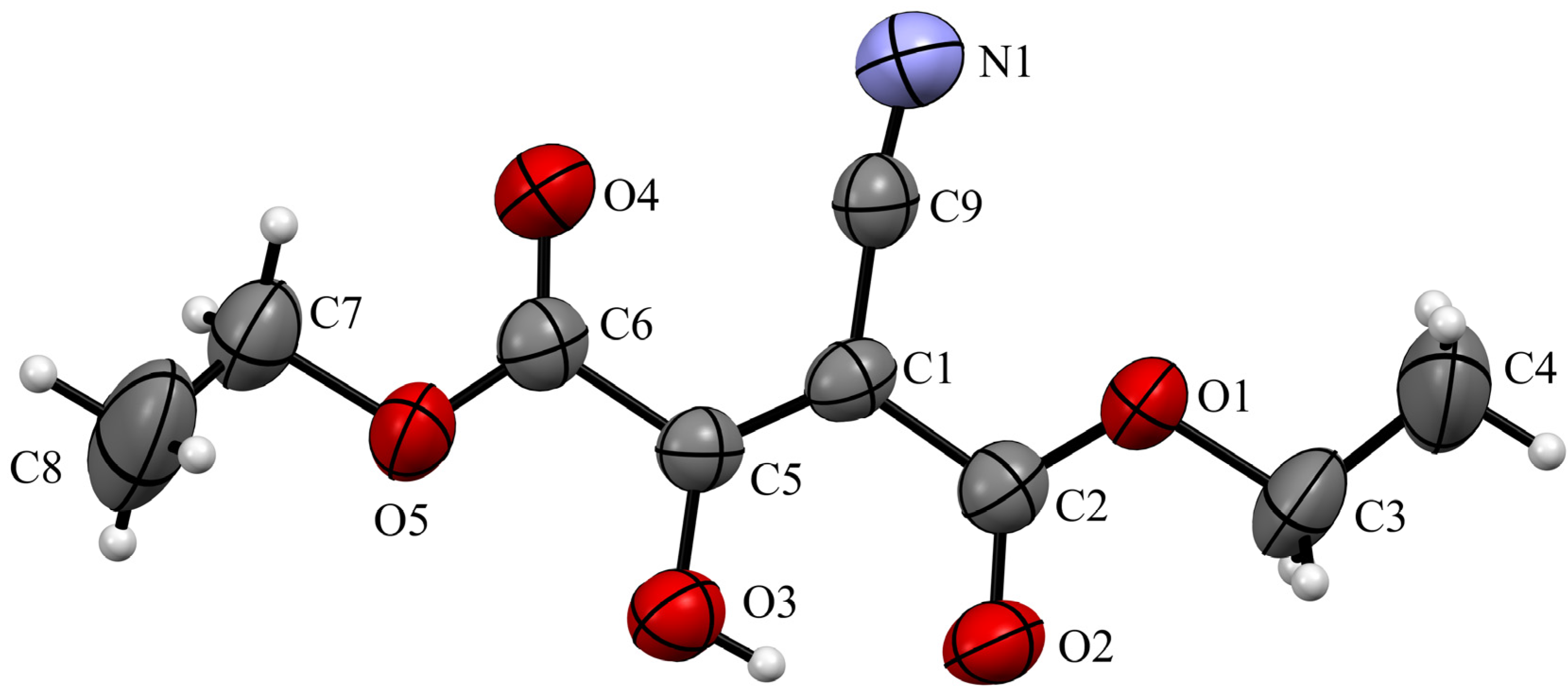
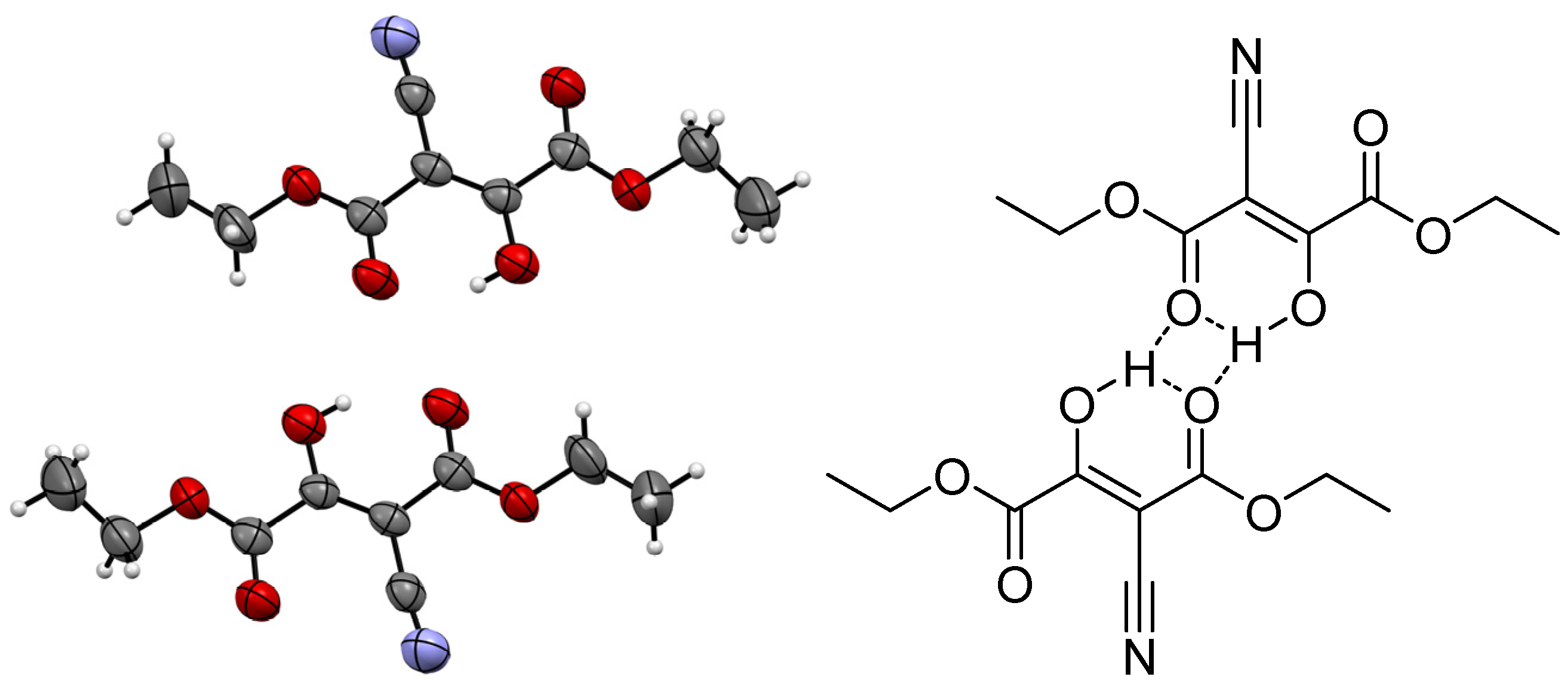
Diethyl 2-Cyano-3-oxosuccinate

by
Oleg N. Markov
1,
Alexander E. Moiseev
1,
Boris N. Tarasevich
1,
Victor A. Tafeenko
1,
Elena K. Beloglazkina
1,
Alexander A. Shtil
1,2,* and
Alexander V. Finko
1,3,*
1
Department of Chemistry, Lomonosov Moscow State University, 119991 Moscow, Russia
2
Institute of Cyber Intelligence Systems, National Research Nuclear University MEPhI, 115409 Moscow, Russia
3
A.V. Topchiev Institute of Petrochemical Synthesis, Russian Academy of Sciences, 119991 Moscow, Russia
*
Authors to whom correspondence should be addressed.
Molbank 2023, 2023(2), M1634; https://doi.org/10.3390/M1634
Submission received: 9 March 2023 / Revised: 19 April 2023 / Accepted: 24 April 2023 / Published: 26 April 2023

Abstract

:
The titular compound was characterized for the first time using a full range of spectroscopic methods, including UV, IR, 1H, and 13C NMR spectra. In solution, all methods showed a keto–enol equilibrium strongly shifted to the enol form. The X-ray structures determined for all simple 2-oxosuccinates showed only the enol form packed as hydrogen-bonded dimer stacks.

1. Introduction

The simple diester diethyl 2-cyano-3-oxosuccinate 1 (Figure 1, R = CN) has been first reported in 1901 [1]. Since then, there have only been two references to 1 [2,3]; accordingly, the experimental data on this compound contain significant gaps. Exploring the synthesis of heterocycles from 2-oxosuccinates, we prepared 1, aiming to convert it into different N- and O-containing heterocycles. Generally, the synthesis of 1 is not new. Herein, we for the first time present the full characterization of 1, including UV, IR, 1H and 13C NMR spectra, as well as X-ray structure.

2. Results

Compound 1 was swiftly obtained via the Claisen condensation of ethyl cyanacetate with diethyl oxalate using sodium ethanolate in ethanol at room temperature (Scheme 1).
The UV spectrum of compound 1 (Figure S1) showed an absorption wavelength at 282 nm, while its IR spectrum (Figures S2 and S3) demonstrated a strong and sharp C≡N absorption at 2227 cm−1 and three strong C=O absorption bands at 1742, 1667, and 1610 cm−1. In addition, there was a broad absorption band at 2500–3300 cm−1 with four narrow low-intensity peaks. Usually, this pattern corresponds to the carboxylic acid OH group; however, in this case, it was the absorption of the acidic enol OH group.
The 1H NMR spectra of 1 (Figure S4) showed only one set of signals containing CH3 and CH2 protons; the latter signal was detected at 13.6 ppm, corresponding to a singlet of the OH proton in the enol form. Similarly, in the 13C NMR spectra (Figure S5), only the enol form of 1 was observed. However, 2-oxosuccinic esters are known to form an equilibrium between keton and enol states; accordingly, the NMR spectra contain two sets of signals [6,7] (Scheme 2).
Next, we determined the acidity constant of 1. The aqueous solutions of this compound were acidic (pH 2.74 of 0.01 mol/L solution), which is not surprising since ethyl 2-cyano-3-oxobutanoate has a pKa = 3.0 [8]. However, we found that it catalyzes autohydrolysis due to its own acidity; thus, 1 is not suitable for titration (see Supplementary materials for the detailed procedure and 1H NMR spectra of 1 in D2O).
The prepared crystals of compound 1 (CCDC 2245592) were directly suitable for X-ray diffraction. The compound’s molecular structure is shown in Figure 2. The molecules of 1 are presented in the enol form as head-to-tail planar dimers stuck in piles (Figure 3).
Strikingly, 1 had a high melting point of 96–97 ℃, whereas most diethyl 2-oxosuccinates are liquids at room temperature [6,7,9,10]. We assume that the high acidity of 1 leads to the prevalence of the enol form, thereby enabling dimerization and stacking. Hydrogen bonding and stacking increased the compound’s melting point.
All known X-ray structures of β-dicarbonyl compounds have been resolved for metal complexes, salts, or substances with a variety of intermolecular interactions [11,12,13,14]. Since other simple 2-oxosuccinate and malonic esters are liquid at room temperature, one cannot obtain their crystal structure. The present study has determined the complex structure of the hydrogen bonds in this chemical class for the first time. These data are relevant to the mechanism behind the unusual properties of 1.

3. Experimental Section

Melting points were determined using an OptiMelt MPA100–Automated melting point system at 1 °C/min and 0.1 °C resolution (Stanford Research Systems, Sunnyvale, CA, USA). IR spectra were recorded using a Thermo Nicolet iS5 FTIR, for which the number of scans was equal to 32 and resolution was equal to 4 cm−1, and by sampling ATR (Thermo Fisher Scientific, Waltham, MA, USA). Electronic spectra in UV and visible regions were recorded using a GENESYS 50 UV–Vis (Thermo Fisher Scientific, Madison, WI, USA) instrument with an operating wavelength range of 190–1100 nm and spectral bandwidth of 2 nm in a quartz cuvette (Agilent Technologies, Santa Clara, CA, USA) with an optical path of 10 mm. NMR spectra were obtained for 1H at 400 MHz and for 13C at 100 MHz using a Bruker Avance 400 Ultra Shield instrument (Bruker BioSpin, Ettlingen, Germany). Spectra were recorded at 25℃ in a DMSO-d6 solution. Chemical shifts δ were measured in ppm. Internal standards used were the residual solvent signals (2.50 ppm for 1H and 39.5 ppm for 13C), while coupling constants (J) were determined in Hz. Data on X-ray diffraction analysis were collected using the STOE diffractometer, the Pilatus100K detector, the focusing mirror collimation using Cu Kα (1.54186 Å) radiation, and in the rotation method mode. Data collection and image processing were performed using X-Area 1.67 (STOE & Cie GmbH, Darmstadt, Germany). Intensity data were scaled with LANA (part of X-Area) to minimize differences of intensities of symmetry-equivalent reflections (a multiscan method). Structures were resolved and refined using the SHELX program [15]. Non-hydrogen atoms were refined using the anisotropic full-matrix least square procedure. Hydrogen atoms were placed in the calculated positions and allowed to ride on their parent atoms. Molecular graphics were prepared using DIAMOND software [16]. The HRMS spectrum was recorded using a quadrupole time-of-flight mass spectrometer TripleTOF 5600+ (AB Sciex, Concord, ON, Canada) equipped with the electrospray ionization source TurboIon Spray and the liquid chromatography system LC-30 «Nexera» (Shimadzu, Tokyo, Japan). Samples were injected directly into the ionization source in 0.2 μL portions and into 0.3 mL/min methanol flow without chromatographic separation. Ionization was performed in positive and negative modes. Overall scanning conditions corresponded to TOFMS. Potentiometric titration was carried out using a CRISON TitroMatic 1S (Barcelona, Spain) autotitrator equipped with «ИT ЭCK-10604» (Moscow, Russia) combining an electrode and CRISON C.A.T. Pt 1000 temperature probe.

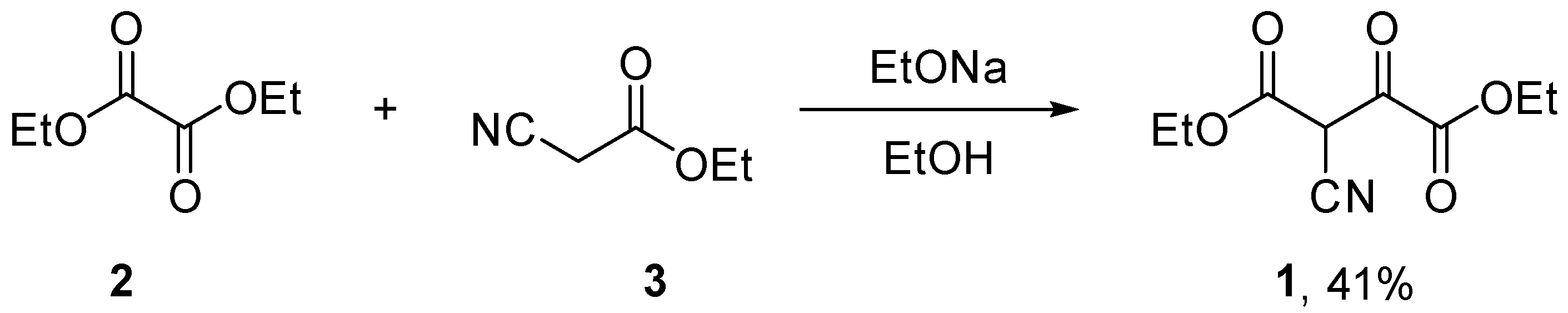
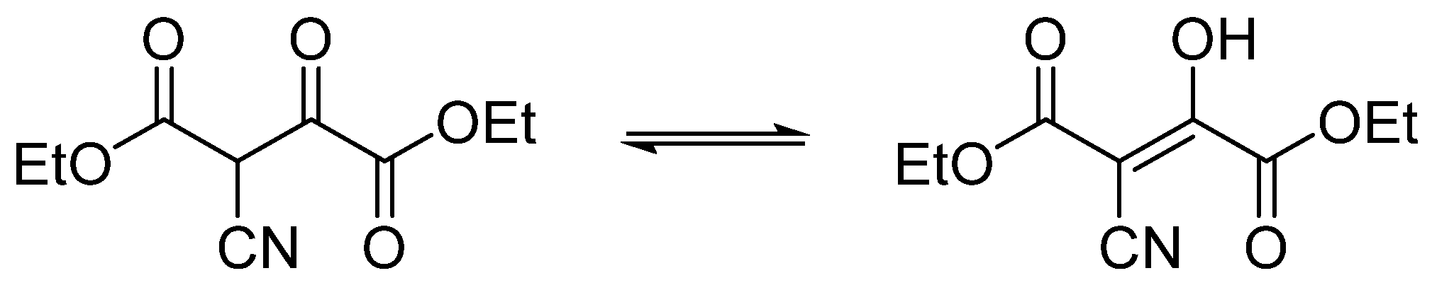
Diethyl 2-cyano-3-oxosuccinate (1)

Metallic sodium (1.7 eq, 12 g, and 521 mmol) was dissolved in a mixture of absolute ethanol (100 mL) and diethyl ether (200 mL), stirred, and cooled to room temperature. After completely dissolving the sodium, diethyl oxalate (1.2 eq, 53.8 g, 50 mL, and 368 mmol) was added. The mixture was stirred at room temperature for 30 min; then, ethyl cyanacetate (1 eq, 34.7 g, 32.8 mL, and 307 mmol) was added. The mixture was kept at room temperature overnight and then diluted with water (20 mL) and 6M HCl (~95 mL, pH ~ 2–3 by pH paper). The organic layer was separated, and the aqueous phase was extracted with chloroform (3 × 200 mL). All organic phases were combined and dried over anhydrous sodium sulfate. Solvents were evaporated. The product was recrystallized from boiling ethanol and obtained as colorless needles. Yield: 27.0 g, 41%.
M.p. 96-97 °C. 1H NMR (400 MHz, DMSO-d6) δ enol (100 mol. %) 1.09 (t, J3H,H = 7.1 Hz, 3H, CH3OEt), 1.17 (t, J3H,H = 7.1 Hz, 3H, CH3OEt), 3.96 (q, J3H,H = 7.1 Hz, 2H, CH2OEt), 4.09 (q, J3H,H = 7.1 Hz, 2H, CH2OEt) and 14.65 (s, 1H, OH). 13C NMR (100 MHz, DMSO-d6) δ 13.7, 14.4, 59.0, 60.6, 73.8, 118.3, 165.5, 168.8, and 179.0. FTIR (Diamond, ν/cm−1): 550, 610, 693, 769, 860, 875, 1027, 1114, 1171, 1240, 1281, 1330, 1356, 1382, 1370, 1417, 1442, 1475, 1610, 1667, 1742, 2227, 2878, 2941, 2987, and 3006. UV–Vis (λ, nm, (ε, L·mol−1·cm−1)): 282 (5600). HRMS (ESI/TOF-MS, m/z): [M+H]+, calcd for C9H12NO5 214,0710; found 214,0706.
Crystal data for C9H12NO5 (M = 213.19 g/mol): monoclinic, space group C2/c, a = 28.234(2) Å, b = 4.4391(6) Å, c = 21.474(2) Å, β = 128.435(7)°, V = 2108.2(4) Å3, Z = 8, T = 295(2) K, μ(CuKα) = 0.952 mm−1, Dcalc = 1.343 g/cm3, and F(000) = 896. CCDC deposition number is 2245592.

Supplementary Materials

The following supporting information can be downloaded online: Figure S1. UV–Vis spectrum of diethyl 2-cyano-3-oxosuccinate (CCl4). Figure S2. IR spectrum of diethyl 2-cyano-3-oxosuccinate (diamond). Figure S3. IR spectrum of diethyl 2-cyano-3-oxosuccinate (CCl4 solution). Figure S4. 1H NMR spectrum of diethyl 2-cyano-3-oxosuccinate. Figure S5. 13C NMR spectrum of diethyl 2-cyano-3-oxosuccinate. Figure S6. HRMS spectrum of diethyl 2-cyano-3-oxosuccinate. Figure S7. Crystal structure of diethyl 2-cyano-3-oxosuccinate. Table S1. Crystal data and structure refinement of compound 1. Table S2. Bond lengths (Å) and angles (°) of compound 1. Table S3. Torsion angles (°) of compound 1. Table S4. Hydrogen bonds (Å and °). Table S5. Benzoic acid titration. Table S6. Titration of compound 1. Table S7. pH values of solutions of 1. Figure S8. 1H NMR spectrum of diethyl 2-cyano-3-oxosuccinate in D2O (immediately after reconstitution). Figure S9. 1H NMR spectrum of diethyl 2-cyano-3-oxosuccinate in D2O (2 days after reconstitution). Reference [17] is cited in the supplementary materials.

Author Contributions

Conceptualization, A.V.F. and O.N.M.; Writing—original draft preparation, O.N.M.; Writing—review and editing, A.V.F. and A.A.S.; Visualization, O.N.M. and V.A.T.; Methodology, O.N.M. and A.E.M.; Formal analysis, A.V.F. and B.N.T.; Supervision, A.V.F. and E.K.B.; Funding, A.V.F. All authors have read and agreed to the published version of the manuscript.

Funding

This study was funded by the Russian Science Foundation grant number 23-23-00207.

Institutional Review Board Statement

Not applicable.

Informed Consent Statement

Not applicable.

Data Availability Statement

The X-ray data were deposited in CCDC 2245592 and included in the Supplementary Material.

Acknowledgments

NMR: FTIR, UV, and X-ray studies were supported by the M. V. Lomonosov Moscow State University Program of Development.

Conflicts of Interest

The authors declare no conflict of interest.

References

  1. Bertini, C. Azione del sodio e dell’idrossilammina sopra i fenilidrazoni dell’etere cianacetico e sintesi dell’etere cianossalacetico. Gazz. Chim. Ital. 1901, 23, 578–588. [Google Scholar]
  2. Trimbach, M. Action des chloroxalates d’ethyle et de methyle sur les ethers cyanacetiques. Bull. Soc. Chim. Fr. 1905, 33, 372–375. [Google Scholar]
  3. Schenone, P.; Fossa, P.; Menozzi, G. Reaction of 2-2-Dimethylaminomethylene-1,3-diones with Dinucleophiles. X. Synthesis of 5-Substituted Ethyl or Methyl 4-Isoxazolecarboxylates and Methyl 4-(2,2-dimethyl-1-propyl)-5-isoxazolescarboxylate. J. Heterocycl. Chem. 1991, 28, 453. [Google Scholar] [CrossRef]
  4. Borodkin, S.; Jonsson, S.; Cocolas, G.H.; McKee, R.L. Orotic acid analogs. 2,5-Disubstituted 6-hydroxy-4-carboxypyrimidines. J. Med. Chem. 1967, 10, 290–291. [Google Scholar] [CrossRef] [PubMed]
  5. Schinz, H.; Hinder, M. Preparation and properties of various α-keto β-lactones. Helv. Chim. Acta. 1947, 30, 1349–1373. [Google Scholar] [CrossRef] [PubMed]
  6. Kolind-Andersen, H.; Dyrnesli, R.; Lawesson, S.-O. Alkylation of some potential 2-oxoesters. Bull. Soc. Chim. Belg. 1975, 84, 341–360. [Google Scholar] [CrossRef]
  7. Jefford, C.W.; Kubota, T.; Zaslona, A. Intramolecular Carbenoid Reactions of Pyrrole Derivatives. A Total Synthesis of (±)-Ipalbidine. Helv. Chim. Acta. 1986, 69, 2048–2061. [Google Scholar] [CrossRef]
  8. Lienhard, G.E.; Jencks, W.P. The Reaction of Carbanions with N,S-Diacetylcysteamine. A Model for Enzymatic Carbon-Carbon Condensation. J. Am. Chem. Soc. 1965, 87, 3863–3874. [Google Scholar] [CrossRef] [PubMed]
  9. Curreli, F.; Belov, D.S.; Do Kwon, Y.; Ramesh, R.; Furimsky, A.M.; O’Loughlin, K.; Byrge, P.C.; Iyer, L.V.; Mirsalis, J.C.; Kurkin, A.V.; et al. Structure-based lead optimization to improve antiviral potency and ADMET properties of phenyl-1H-pyrrole-carboxamide entry inhibitors targeted to HIV-1 gp120. Eur. J. Med. Chem. 2018, 154, 367–391. [Google Scholar] [CrossRef]
  10. Shahak, I.; Bergman, E.D. Organic Fluorine Compounds. XVI. The Preparation and Reactions of Ethyl Ethoxalylfluoroacetate. J. Chem. Soc. 1960, 3225–3229. [Google Scholar] [CrossRef]
  11. Yamazaki, S.; Sugiura, H.; Ohashi, S.; Ishizuka, K.; Saimu, R.; Mikata, Y.; Ogawa, A. Intramolecular [2 + 2] and [4 + 2] Cycloaddition Reactions of Cinnamylamides of Ethenetricarboxylate in Sequential Processes. J. Org. Chem. 2016, 81, 10863–10886. [Google Scholar] [CrossRef] [PubMed]
  12. Pandey, G.; Adate, P.A.; Puranik, V.G. Organocatalytic dynamic kinetic resolution via conjugate addition: Synthesis of chiral trans-2,5-dialkylcyclohexanones. Org. Biomol. Chem. 2012, 10, 8260–8267. [Google Scholar] [CrossRef] [PubMed]
  13. Bailley, N.A.; Coates, E.; Robertson, G.B.; Bonati, F.; Ugo, R. The crystal and molecular structures of acetylacetonatobiscarbonyl-rhodium(I) and of some fluoro-substituted complexes. Chem. Commun. 1967, 1041–1042. [Google Scholar] [CrossRef]
  14. Kashiwabara, T.; Tanaka, M. Arylglyoxylrhodium Complexes, Their Thermolysis, and Attempted Generation by Carbonylation of an Aroylrhodium Complex. Organometallics 2011, 30, 239–244. [Google Scholar] [CrossRef]
  15. Sheldrick, G.M. A short history of SHELX. Acta Crystallogr. A 2008, A64, 112–122. [Google Scholar] [CrossRef] [PubMed]
  16. Brandenburg, K.; Brendt, M. DIAMOND, Release 2.1 d.; Crystal Impact GbR: Bonn, Germany, 2000; pp. 302–303. [Google Scholar]
  17. Albert, A.; Serjeant, E.P. The Determination of Ionization Constants. A Laboratory Manual, 3rd ed.; Chapman and Hall: New York, NY, USA, 1984. [Google Scholar]
Figure 1. Examples of heterocycle construction based on 2-oxosuccinates [4,5].
Molbank 2023 m1634 g001
Scheme 1. Synthesis and structure of 1.
Molbank 2023 m1634 sch001
Scheme 2. Keton and enol forms of 1.
Molbank 2023 m1634 sch002
Figure 2. Molecules of 1 in the crystal structure (thermal ellipsoids at 50% level). Bond lengths: C2–O2 1.234 Å, C5–O3 1.322 Å, C6–O4 1.209 Å, C1–C2 1.472 Å, C1–C5 1.341 Å, and C1–C9 1.429 Å.
Molbank 2023 m1634 g002
Figure 3. Inter- and intramolecular hydrogen bonds in dimers of 1 (crystals). Left—dimers in crystal structure; right—schematic drawing of such dimers.
Molbank 2023 m1634 g003
Disclaimer/Publisher’s Note: The statements, opinions and data contained in all publications are solely those of the individual author(s) and contributor(s) and not of MDPI and/or the editor(s). MDPI and/or the editor(s) disclaim responsibility for any injury to people or property resulting from any ideas, methods, instructions or products referred to in the content.

