Abstract
3-(2-Chloro-5-methylphenoxy)propane-1,2-diol 1 is a possible precursor in the chiral beta blocker Bupranolol synthesis. Both racemic and single-enantiomeric samples of 1 were synthesized and characterized by single crystal XRD. The absolute configuration of an (S)-1 sample was determined by data refinement (the value of the Flack parameter is 0.03(4)). Hydrogen-bonded supramolecular synthons (SMS) were identified for both crystals. In rac-1, as well as in (S)-1 crystals, one of the two main SMS is the homochiral chain (5):21 (⋯O1–C1–C2–O2–H2⋯). The second of the two SMSs changes is the change of chiral environment. For rac-1, this is the heterochiral chain (⋯O2–C2–C1–O1–H1⋯O′2–H′2⋯O″1–H″1⋯), in which molecules with different configurations alternate. In (S)-1 crystals, this is the homochiral chain (2):11 (⋯O1–H1⋯). The results obtained once again confirm the influence of the chiral environment on the crystallization of scalarly identical molecules.
1. Introduction
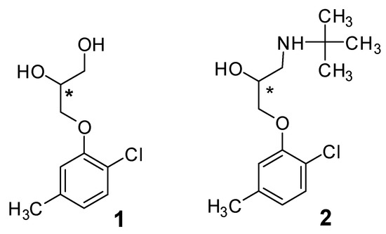
3-(2-Chloro-5-methylphenoxy)propane-1,2-diol 1 is a possible precursor in the synthesis of a non-selective beta blocker Bupranolol 2 (Scheme 1) [1,2,3].
Scheme 1.
3-(2-Chloro-5-methylphenoxy)propane-1,2-diol 1 and bupranolol 2. Chiral center is marked with an asterisk.
The enantiomers of bupranolol are known to possess various cardiostimulatory properties [4,5]. Not only biological activity, but also many other macroscopic properties, including crystal organization, depend on the configuration and enantiomeric composition of a chiral substance. We have studied the crystal structure of bupranolol previously [3]. This work is devoted to the X-ray structure determinations of rac-1 and (S)-1 samples, with the aim of revealing similarities and differences between the racemic and single-enantiomeric crystal packings in the title structures.
2. Results and Discussion
2.1. Crystal Structure of Racemic Diol 1
The synthesis of racemic diol 1 has been described by us previously [3]. The rac-1 compound crystallizes in the P21/n (No. 14) group with one independent molecule (Z’ = 1) in the asymmetric unit. Detailed crystallographic characteristics for this sample are given in Appendix A, Table A1. Figure 1 shows the structure of the independent molecule of rac-1 as well as the partial numbering system used.
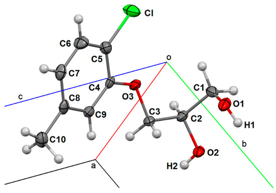
Figure 1.
Molecular structure of rac-1 with anisotropic displacement ellipsoids drawn at the 50% probability level and the partial numbering system used.
In rac-1 crystals, there are no anomalous structural characteristics (bond lengths, bond angles, etc.), while intermolecular hydrogen bonds (HB) O–H⋯O act as the main structure-forming elements. It can be expected that supramolecular motifs in rac-1 crystals will be formed mainly by intermolecular hydrogen bonds between hydrogen atoms H1 and H2 as donors and oxygen atoms O1, O2, and O3 as acceptors. The two-digit symbols 11, 12, 13, 21, 22, and 23 are used to denote potential intermolecular hydrogen bonds in rac-1 crystals. In this case, the first position in each symbol corresponds to the number of the donor hydrogen atoms of one molecule, and the second position corresponds to the number of the acceptor oxygen atoms of another molecule.
To record the sequences of intermolecular hydrogen bonds, we will use the designs C (chain) and R (ring) introduced in [6,7], supplementing each descriptor with a list of intermolecular hydrogen bonds forming an elementary chain link or a ring. For clarity, we will separate successive hydrogen bonds with a slash. Previously, we used a similar notation for the analysis of HB systems in oxycarbonic acids [8], wherein one can find a more detailed description of this method.
A fragment of the rac-1 crystal packing is shown in Figure 2. In principle, the main elements (synthons) that form a general supramolecular motif, which topologically charac-terizes this packing, can be seen directly in the Figure. However, they are illustrated even more clearly in the connectivity scheme, a flat diagram (Scheme 2), in which the chemical bonds in the molecules forming the crystal are indicated by solid lines, and the intermolecular hydrogen bonds are indicated by dotted lines.
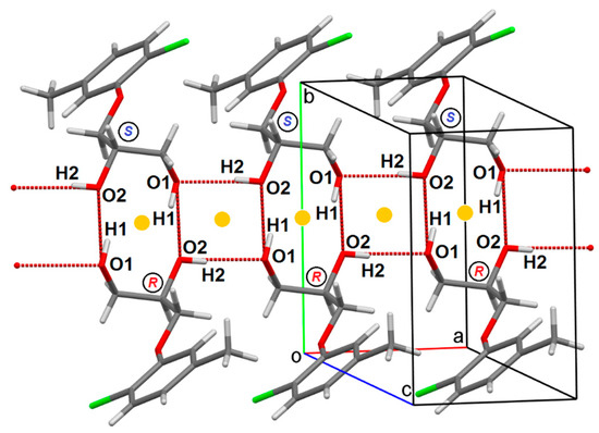
Figure 2.
Fragment of packing in rac-1 crystals. Letters R and S in circles denote the configuration of the C2 chiral centers. Yellow dots indicate inversion centers.
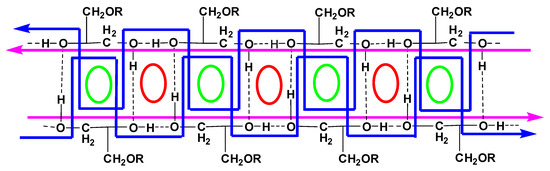
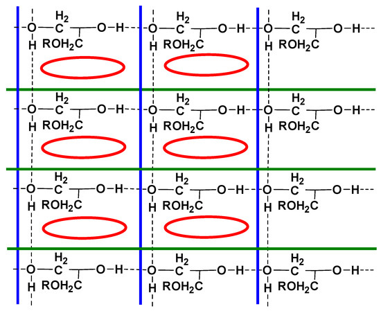
Scheme 2.
Connectivity scheme in rac-1 crystals.
From the connectivity Scheme 2 and Figure 2, it becomes obvious that in this case, the main synthons are the chains (5):21 indicated on the diagram by straight purple lines and the chains (9):{12/12/21} indicated by blue broken lines. According to Table A2 (Appendix A), intermolecular HB 21 possess slightly better geometric characteristics (d = 2.739(3) Å, ∠O2H2O1 = 176(4)°) compared to HB 12 (d = 2.754(3) Å, ∠O1H1O2 = 151(4)°). At the intersections of infinite chains, centrosymmetric unidirectional rings (8):{12/21/12/21} (red ovals) and (10):{12/12} (green ovals) are formed. If we give preference to infinite (in this case, 1D) synthons, namely chains, over finite 0D cyclic synthons, then the supramolecular motif in rac-1 crystals is written as (8):{12/21/12/21}; (10):{12/12}.
2.2. Crystal Structure of Single-Enantiomeric Diol (S)-1
(S)-3-(2-Chloro-5-methylphenoxy)propane-1,2-diol, (S)-1 was obtained from (S)-3-chloropropane-1,2-diol and 2-chloro-5-methylphenol (see Section 3.1). Compound (S)-1 crystallizes in the Sohncke group P212121 (No. 19) with a one symmetry-independent molecule in the asymmetric unit (Z’ = 1). Detailed crystallographic characteristics for this sample are given in Appendix A, Table A1. Molecule 1 contains a “heavy” chlorine atom. This factor allowed us to determine the absolute configuration using the Flack [9,10] parameter. The value 0.03(4) (see Table A1) in combination with the chiroptic characteristics of the compound completely confirms the correctness of the configurational assignment.
The connectivity scheme in (S)-1 crystals is shown in Scheme 3, where infinite chains (2):11 (denoted by blue lines) and (5):21 (denoted by green lines) are easily detected. Intermolecular HBs 11 (d = 2.7412(19) Å, ∠O1H1O1′ = 168(3)°) and 21 (d = 2.866(3) Å, ∠O2H2O1 = 170(3)°) have comparable geometric characteristics.
Scheme 3.
Connectivity scheme in (S)-1 crystals.
The physical implementation of chain motifs in the (S)-1 crystal packing is shown in Figure 3.
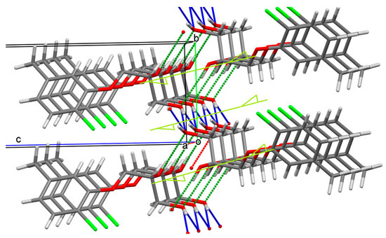
Figure 3.
Infinite chains (2):11 (blue) and (5):21 (green) in (S)-1 crystals.
It can be seen from Figure 3 that both chains lineup round 21 axes parallel to axis 0a. In this case, the first one, in which intermolecular hydrogen bonds are indicated in blue, is organized as an almost flat “herringbone”. The second chain, held together by 21 type hydrogen bonds, is a left-handed M-helix (Figure 4).
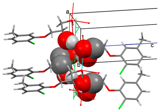
Figure 4.
Spiral organized chain (5):21 in (S)-1 crystals.
Scheme 3 clearly shows the multidirectional cycle (14):{11/21}{21/11} formed by the intersection of the “blue” and “green” chains. In Figure 5, we have illustrated its organization by omitting the aromatic substituents. The figure clearly shows that, in full accordance with Scheme 3, the first and second molecules (the numbers of the molecules in the figure are given in circles) provide both hydroxyl groups present in the molecule for the formation of this ring. In the third molecule, only the primary hydroxyl group is involved in ring formation as a donor and as an acceptor. Finally, the fourth molecule is involved in the hydrogen bond pattern only by the oxygen atom of the primary hydroxyl, which acts as a double intermolecular HB acceptor.
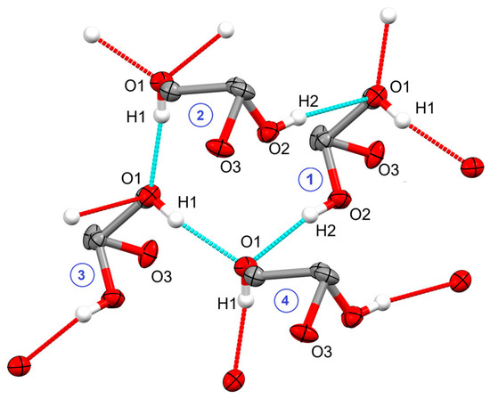
Figure 5.
Multidirectional (14):{11/21}{21/11} ring in (S)-1 crystals. Aromatic substituents are omitted for clarity. The numbers 1–4 in circles represent the molecules discussed in the text.
2.3. Discussion
Both in racemic and single-enantiomeric crystals, due to classical hydrogen bonds and weaker interactions, supramolecular 2D layers are formed. In accordance with the empirical rule established by us earlier in the example of compounds both related to diol 1 and significantly different from them [11], these layers extend along the two smallest unit cell parameters, in this case axes 0a and 0b. Such 2D layers are stacked along the largest parameter 0c in such a way that alternation of predominantly hydrophilic and hydrophobic zones is observed along it. With such stacking, a sufficiently high molecular packing index is achieved for both rac-1 (70.7%) and (S)-1 (70.8%) crystals.
As expected, the main crystal-forming elements of both racemic and enantiopure crystals of diol 1 are O–H⋯O intermolecular hydrogen bonds. Both hydroxyl groups present in molecule 1, O1–H1, and O2–H2 (Figure 1) act as HB donors, whereas, out of four potential acceptors (O1, O2, O3, and Cl), only the first pair is involved. The intermolecular hydrogen bond O2–H2⋯O2′ (HB 22 in the accepted notation) does not occur in any of the two crystals studied.
In both crystals, one of the two main supramolecular synthons is the chain (5):21 (⋯O1–C1–C2–O2–H2⋯). Let us draw the reader’s attention to the fact that each such chain is built from the same enantiomers of diol 1 (Figure 2), that is, it is a homochiral construct, which, for this very reason, can be realized both in racemic and single-enantiomeric crystals. However, the second of two infinite supramolecular synthons changes with a change in the chiral environment. In the case of a racemate, this is a heterochiral chain (⋯O2–C2–C1–O1–H1⋯O′2–H′2⋯O″1–H″1⋯) in which molecules with different configurations alternate. Such a construction is impossible in the case of a single-enantiomeric crystal, and in (S)-1 crystals, this chain turns out to be replaced by the homochiral (2):11 (⋯O1–H1⋯) chain.
It is clear that a comparison of two crystal packings does not give rise to broad generalizations. However, the significant effect of the chiral environment on the crystal packing of scalarly identical molecules, which we noted earlier [8,12], is also confirmed in the case under study.
3. Materials and Methods
3.1. Materials
Racemic 3-chloropropane-1,2-diol (99%) and 2-chloro-5-methylphenol (99%) were purchased from Acros Organics (Geel, Belgium). (S)-3-Chloropropane-1,2-diol (98%, 98% ee) was purchased from Alfa Aesar (Ward Hill, MA, USA). Racemic 3-(2-chloro-5-methylphenoxy)propane-1,2-diol, rac-1 was prepared from racemic 3-chloropropane-1,2-diol and 2-chloro-5-methylphenol according to published procedure [3]: mp 73.5–75.5 °C. (S)-3-(2-Chloro-5-methylphenoxy)propane-1,2-diol, (S)-1 was obtained by analogy, as described for racemic diol. To a solution of 2-chloro-5-methylphenol (1.07 g, 7.5 mmol) in ethanol (5 mL), a solution of NaOH (0.38 g, 9.4 mmol) in water (3 mL) was added, and the resulting mixture was stirred and heated under reflux for 3 h. A solution of (S)-3-chloropropane-1,2-diol (1 g, 9.1 mmol) in ethanol (5 mL) was then added dropwise, and the mixture was further stirred and heated at reflux for 15 h. After cooling, the volume of the resulting mixture was reduced to about one third followed by the addition of water (5 mL) and extraction with EtOAc (4 × 40 mL). The combined organic layers were dried over MgSO4, and the solvent was removed. The crude diol (S)-1 was purified via recrystallization from light petroleum ether/EtOAc (5:1). The following properties were also attained: yield 1.19 g, 73%; mp 72–74 °C (light petroleum ether/EtOAc); −4.2 (c 1.0, MeOH), −8.4 (c 1.0, MTBE); 99% ee (chiral HPLC analysis, column temperature 21 °C; flow rate 0.7 mL·min−1; eluent: hexane/2-propanol (9:1); tR 17.7 min (minor), tR 18.8 min (major)). Cf. lit. [3] for (R)-diol: mp 73–74 °C; +2.7 (c 1.0, MeOH), +9.3 (c 1.0, MTBE).
3.2. Instruments
Optical rotations were measured on a Perkin–Elmer model 341 polarimeter (PerkinElmer, Waltham, MA, USA), and concentration c was given as g/100 mL. Melting points were determined using a Boëtius apparatus. HPLC analyses were performed on a Shimadzu LC-20AD system controller (SHIMADZU CORPORATION, Kyoto, Japan), and a UV monitor 275 nm was used as a detector. The column used, from Daicel Inc. (Osaka, Japan), was Chiralpak AD (0.46 × 25 cm).
3.3. Single Crystal X-ray Diffraction
The single crystals of the compounds rac-1 and (S)-1 first investigated by X-ray diffraction in this paper were prepared via slow evaporation of solutions of the corresponding samples in a mixture containing cyclohexane and ethyl acetate. The X-ray diffraction data of the investigated crystals were collected at 173(2) K using a Bruker AXS Smart Apex II CCD diffractometer (Bruker AXS GmbH, Karlsruhe, Germany) in the ω-scan and φ-scan modes using graphite monochromated Mo Kα (λ 0.71073 Å) radiation. Data were corrected for the absorption effect using the SADABS program [13]. The structures were solved with a direct method using SHELXS [14] and refined with the full matrix least-squares using SHELXL-2014 [15] and WinGX [16] programs. All non-hydrogen atoms were refined anisotropically. All hydrogen atoms were inserted at calculated positions and refined as riding atoms, except the hydrogen atoms of the OH groups, which were located from the difference maps and refined isotropically. During data collection, the images were indexed, integrated, and scaled using the APEX2 data reduction package [17]. Details for single crystal structure determination experiments are summarized in Appendix A, Table A1 (see Supplementary Materials for cif and check-cif files for rac- and (S)-1). Deposition numbers 2,224,985 (rac-1) and 2,224,986 ((S)-1) contain the supplementary crystallographic data for this paper. These data are provided free of charge by the joint Cambridge Crystallographic Data Centre at https://www.ccdc.cam.ac.uk/structures/ (accessed on 13 March 2023).
Supplementary Materials
The following supporting information can be downloaded online: cif and check-cif files for rac- and (S)-1.
Author Contributions
Conceptualization, writing–review & editing, A.A.B.; methodology, samples preparation, proofreading of the manuscript, Z.A.B.; SC-XRD experiments, A.I.S.; methodology, SC-XRD experiments, proofreading of the manuscript, A.T.G. All authors have read and agreed to the published version of the manuscript.
Funding
This research received no external funding.
Data Availability Statement
Not applicable.
Acknowledgments
X-ray diffraction data were registered on the equipment of the Assigned Spectral-Analytical Center of FRC Kazan Scientific Center of RAS. The authors thank A.V. Kurenkov for valuable technical assistance.
Conflicts of Interest
The authors declare no conflict of interest.
Appendix A. Crystallographic Data

Table A1.
Experimental crystallographic data and structure refinement details for rac-1 and (S)-1 crystals.
Table A2.
Main geometrical parameters of classical hydrogen bonds O–H∙∙∙O′ in the crystals.
References
- Merck and Co. The Merck Index, 14th ed.; O’Neil, M.J., Ed.; Merck and Co.: Whitehouse Station, NJ, USA, 2006; p. 245, Entry 1496. [Google Scholar]
- Ding, H.; Liu, S.; Zhao, K.-X.; Pu, J.; Xie, Y.-F.; Zhang, X.-W. Comparative efficacy of antihypertensive agents in flow-mediated vasodilation of patients with hypertension: Network meta-analysis of randomized controlled trial. Int. J. Hypertens. 2022, 2022, 2432567. [Google Scholar] [CrossRef] [PubMed]
- Bredikhin, A.A.; Bredikhina, Z.A.; Kurenkov, A.V.; Zakharychev, D.V.; Gubaidullin, A.T. Synthesis, phase behavior and absolute configuration of β-adrenoblocker bupranolol and related compounds. J. Mol. Struct. 2018, 1173, 157–165. [Google Scholar] [CrossRef]
- Malinowska, B.; Kieć-Kononowicz, K.; Flau, K.; Godlewski, G.; Kozłowska, H.; Kathmann, M.; Schlicker, E. Atypical cardiostimulant β-adrenoceptor in the rat heart: Stereoselective antagonism by bupranolol but lack of effect by some bupranolol analogues. Br. J. Pharmacol. 2003, 139, 1548–1554. [Google Scholar] [CrossRef] [PubMed]
- Martin, L.J.; Piltonen, M.H.; Gauthier, J.; Convertino, M.; Acland, E.L.; Dokholyan, N.V.; Mogil, J.S.; Diatchenko, L.; Maixner, W. Differences in the antinociceptive effects and cinding properties of propranolol and bupranolol enantiomers. J. Pain 2015, 16, 1321–1333. [Google Scholar] [CrossRef] [PubMed]
- Etter, M.C. Encoding and decoding hydrogen-bond patterns of organic-compounds. Acc. Chem. Res. 1990, 23, 120–126. [Google Scholar] [CrossRef]
- Bernstein, J.; Davis, R.E.; Shimoni, L.; Chang, N.L. Patterns in hydrogen bonding—Functionality and graph set analysis in crystals. Angew. Chem. Int. Ed. 1995, 34, 1555–1573. [Google Scholar] [CrossRef]
- Bredikhin, A.A.; Fayzullin, R.R.; Gubaidullin, A.T.; Bredikhina, Z.A. Intermolecular hydrogen bonding in alpha-hydroxy carboxylic acids crystals: Connectivity, synthons, supramolecular motifs. Crystals 2022, 12, 1479. [Google Scholar] [CrossRef]
- Flack, H.D.; Bernardinelli, G. Reporting end evaluating absolute structure and absolute configuration determination. J. Appl. Crystallogr. 2000, 33, 1143–1148. [Google Scholar] [CrossRef]
- Parsons, S.; Flack, H.D.; Wagner, T. Use of intensity quotients and differences in absolute structure refinement. Acta Crystallogr. 2013, B69, 249–259. [Google Scholar] [CrossRef] [PubMed]
- Gubaidullin, A.T.; Samigullina, A.I.; Bredikhina, Z.A.; Bredikhin, A.A. Crystal structure of chiral ortho-alkyl phenyl ethers of glycerol: True racemic compound, normal, false and anomalous conglomerates within the single five-membered family. CrystEngComm 2014, 16, 6716–6729. [Google Scholar] [CrossRef]
- Bredikhin, A.A.; Bredikhina, Z.A.; Gubaidullin, A.T. Chirality-dependent supramolecular synthons based on the 1,3-oxazolidin-2-one framework: Chiral drugs mephenoxalone, metaxalone and other 114 examples. CrystEngComm 2020, 22, 7252–7261. [Google Scholar] [CrossRef]
- Sheldrick, G.M. SADABS, Program for Empirical X-ray Absorption Correction; Bruker-Nonius: Delft, The Netherlands, 2004. [Google Scholar]
- Sheldrick, G.M. A short history of SHELX. Acta Crystallogr. A 2008, 64, 112–122. [Google Scholar] [CrossRef] [PubMed]
- Sheldrick, G.M. Crystal structure refinement with SHELXL. Acta Crystallogr. Sect. C Struct. Chem. 2015, 71, 3–8. [Google Scholar] [CrossRef] [PubMed]
- Farrugia, L.J. WinGX and ORTEP for Windows: An update. J. Appl. Crystalogr. 2012, 45, 849–854. [Google Scholar] [CrossRef]
- BrukerAXS. APEX2 (Version 2.1), SAINTPlus. Data Reduction and Correction Program (Version 7.31A), Bruker Advanced X-ray Solutions; BrukerAXS: Madison, WI, USA, 2006. [Google Scholar]


Disclaimer/Publisher’s Note: The statements, opinions and data contained in all publications are solely those of the individual author(s) and contributor(s) and not of MDPI and/or the editor(s). MDPI and/or the editor(s) disclaim responsibility for any injury to people or property resulting from any ideas, methods, instructions or products referred to in the content. |
© 2023 by the authors. Licensee MDPI, Basel, Switzerland. This article is an open access article distributed under the terms and conditions of the Creative Commons Attribution (CC BY) license (https://creativecommons.org/licenses/by/4.0/).