Short Note

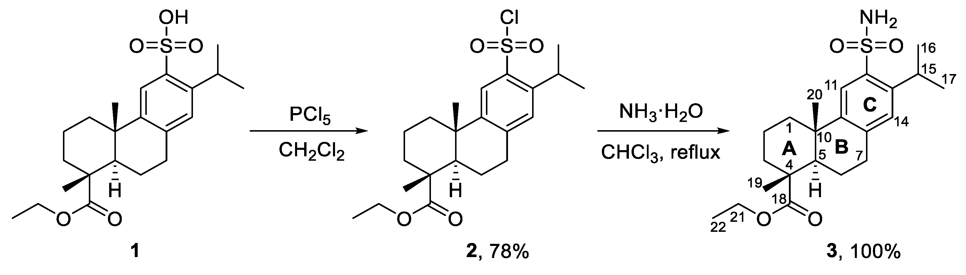
Ethyl 12-Sulfamoyl-abieta-8,11,13-trien-18-oate

1
Institute of Chemistry, Federal Research Center, Komi Science Center, Ural Branch of the Russian Academy of Sciences, 48 Pervomaiskaya St., 167000 Syktyvkar, Russia
2
Arbuzov Institute of Organic and Physical Chemistry, FRC Kazan Scientific Center, Russian Academy of Sciences, 8 Arbuzov St., 420029 Kazan, Russia
3
Biologically Active Terpenoids Laboratory, Kazan Federal University, 18 Kremlevskaya St., 420008 Kazan, Russia
4
General and Organic Chemistry Department, Kazan State Medical University, 49 Butlerov St., 420012 Kazan, Russia
5
Institute of Fundamental Medicine and Biology, Kazan Federal University, 18 Kremlevskaya St., 420008 Kazan, Russia
*
Author to whom correspondence should be addressed.
Molbank 2023, 2023(1), M1584; https://doi.org/10.3390/M1584
Submission received: 19 January 2023 / Revised: 8 February 2023 / Accepted: 11 February 2023 / Published: 13 February 2023
(This article belongs to the Section Structure Determination)

Abstract

:
We synthesized the novel compound ethyl 12-sulfamoyl-abieta-8,11,13-trien-18-oate in good yield from ethyl 12-sulfo-abieta-8,11,13-trien-18-oate via a two-step protocol. The product was comprehensively characterized by one- and two-dimensional NMR methods, single-crystal X-ray diffraction, IR spectroscopy, and high-resolution mass spectrometry.

1. Introduction

Dehydroabietane derivatives, in general, are promising in the synthesis of compounds possessing antiulcer, antiviral, antibacterial and antifungal activity [1]. However, despite the great potential, the chemistry of sulfonamides currently remains poorly understood. The only dehydroabietane derivatives that contain a sulfonamide group at the C-18 [2] and C-12 [3] position are described in the literature. Sulfonamides with amino acid fragments at the C-12 position have been found to be able to inhibit matrix proteinases that promote the migration and proliferation of cancer cells [4]. Some sulfonamide derivatives bearing a glycosidic moiety have antipepsinogenic activity [5], but it is not too high compared to 12-sulfodehydroabietic acid used under the trade name Sodium Ecabet [6]. To understand the biological action mechanisms and processes of binding of sulfonamides to enzymes, it is necessary to thoroughly analyze their spatial structure by means of X-ray diffraction. However, the majority of dehydroabietane-derived sulfonamides do not crystallize well, and those with several polar functional groups are poorly soluble in organic solvents and precipitate instead, not forming crystals suitable for analysis. The aim of this work was the synthesis of a dehydroabietane-derived sulfonamide with a free -SO2NH2 group at position C-12 (3) proceeding from 12-sulfodehydroabietic acid ethyl ester 1 and a comprehensive study of its structure by NMR spectroscopy and X-ray diffraction methods.

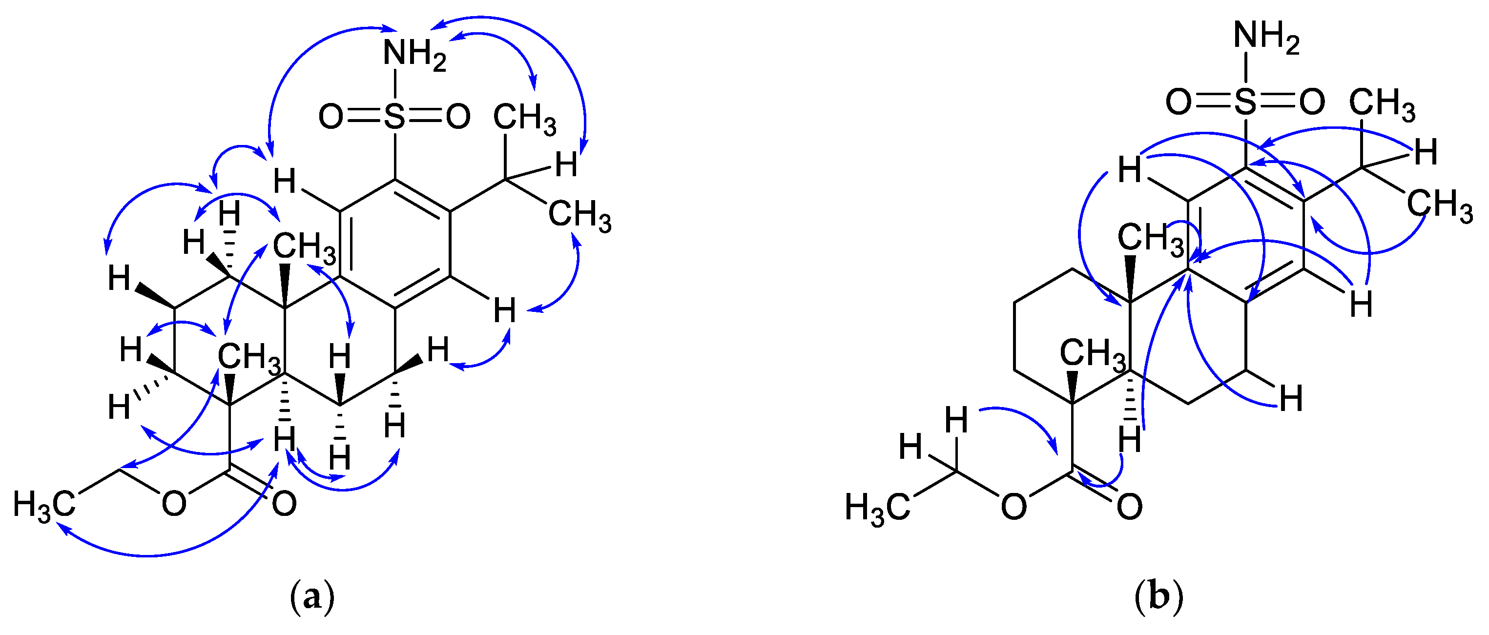
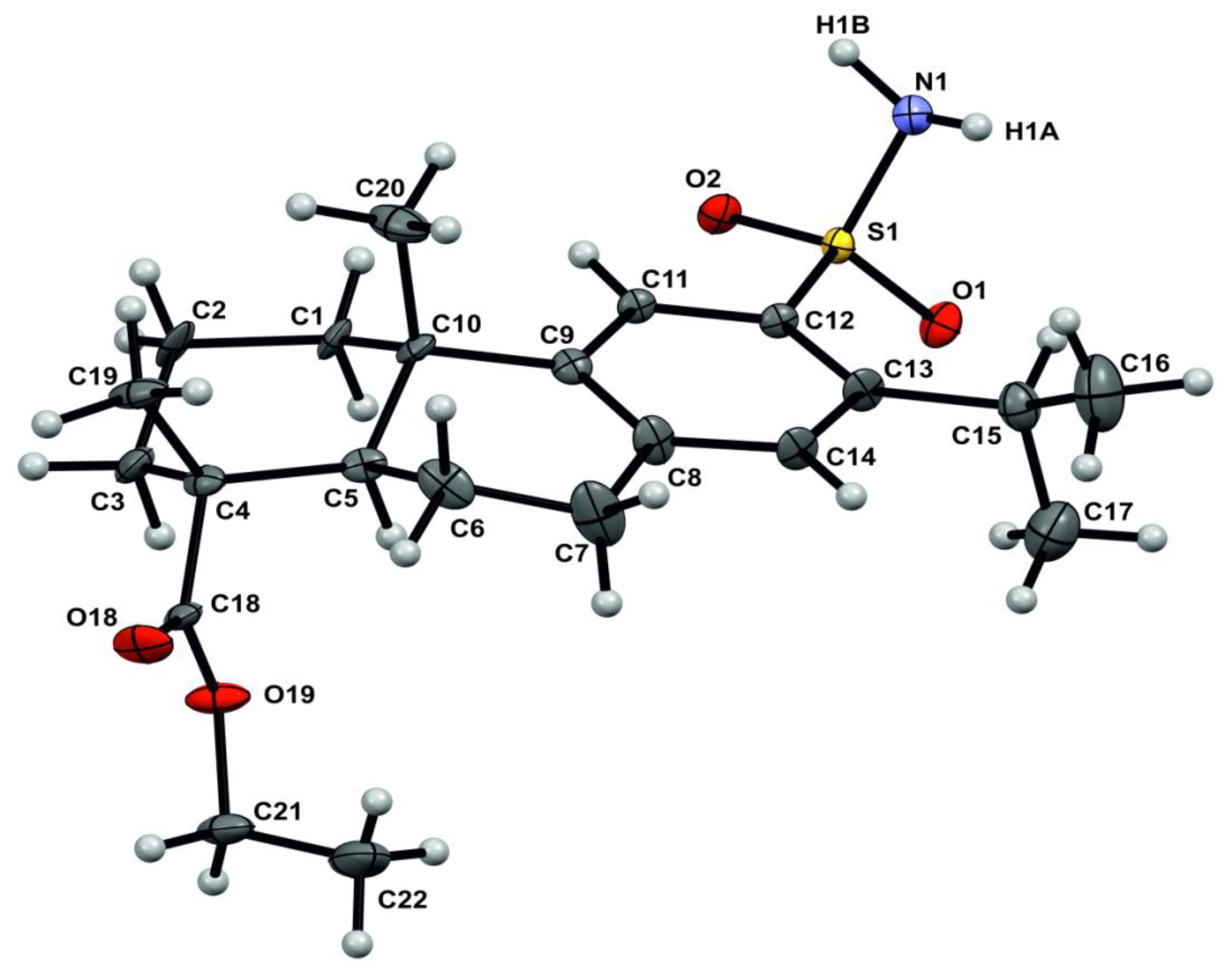
2. Results and Discussion

Sulfonamide 3 was obtained in quantitative yield by the reaction of sulfochloride 2 with an aqueous solution of ammonia. The synthesis of sulfochloride 2, in turn, was carried out from sulfonic acid 1 and PCl5 (Scheme 1). The physicochemical properties and spectral characteristics of acid 1 and sulfochloride 2, as well as the methods for their preparation, were described earlier in [3].
The 1H and 13C-NMR spectra of compound 3 are in accordance with its structure. In the 1H-NMR spectrum of sulfonamide 3, there exists a signal of NH2 group as a singlet at 5.04 ppm and two singlets of the aromatic protons H-11 and H-14 at 7.87 and 7.12 ppm, respectively. Complete assignment of the signals in the 1H and 13C-NMR spectra was implemented using two-dimensional homo- (COSY, NOESY) and heteronuclear (HSQC, HMBC) experiments. The mutual spatial arrangement of protons, established using nuclear Overhauser effect spectroscopy (NOESY), is consistent with the X-ray diffraction data. The general scheme of NOE coupling between protons in the sulfonamide 3 is shown in Figure 1a. The presence of cross peaks formed by the NH2 protons with both H-11 and H-15 indicates unhindered rotation of the sulfonamide group, while the rotation of the isopropyl group hardly takes place, as evidenced by the absence of a cross peak between H-15 and H-14 in the NOE spectrum. Similarly, the ester group is also able to rotate about the C(4)–C(18) bond, which is proven by cross peaks between methyl H-22 protons and the oppositely directed H-5 and H-19.
The signals of quaternary carbons were assigned by the Heteronuclear Multiple Bond Correlation (HMBC) method. According to it, the C-12 atom has the corresponding correlations with H-15 and H-14 protons. The C-9 atom can be easily recognized by its cross-peak correlations with the protons H-5, H-7, H-14, and H-20. On the contrary, for the neighboring C-8 atom, only one significant correlation peak between it and H-11 was identified. The proton H-11, along with methyl H-16/17 protons, also has a cross peak with C-13 in the HMBC spectrum. The carbonyl C-18 atom, whose signal is in the downfield at 178.26 ppm in the 13C-NMR spectrum, does not need to be interpreted using the HMBC method, since its downfield shift is obvious due to the presence of two electron-withdrawing oxygen atoms; nevertheless, a couple of correlations can be found between it and protons H-5 and H-22. The signal of C-4, which is bounded to an electron-withdrawing group, also evidently shifts downfield more than the C-10 signal. In general, it is difficult to distinguish these atoms according to the HMBC data because of the close position of the C-1, C-3 and C-10 signals in the 13C-NMR spectrum. The main observed HMBC correlations are provided in Figure 1b.
The presence of the main functional groups, ester and sulfonamide, was confirmed by IR spectroscopy data. A strong band of stretching vibrations of the C=O group is observed at 1721 cm−1, as well as two bands at 1250 and 1177 cm−1, characteristic of asymmetric stretching vibrations of CO–O and O–C–C fragments. Two strong absorption bands of stretching vibrations of the -SO2- group are present at 1327 cm−1 (as) and 1152 cm−1 (sy). In addition, there is an intense absorption band of the NH2 group at 3267 cm−1 (st) and bending vibrations (δ) of the N–H group at 1553 cm−1.
Finally, comprehensive information on the crystal structure of ethyl 12-sulfamoyl-abieta-8,11,13-trien-18-oate 3 was obtained by X-ray diffraction.
Crystals of compound 3 are orthorhombic, C22H33NO4S; at 293(2) K: a 6.1196(19), b 15.078(4), c 23.215(7) Å, V 2142.1(11) Å3, Z 4, dcalc 1.264 g cm−3, space group P212121. The intensity of 26,807 reflections was measured, for 3772 of which I ≥ 2σ. The final values of R factors are R 0.1055, Rw 0.2439. The absolute configuration was established precisely on the basis of the value of Flack parameter 0.04(8).
The conformation of the six-membered cycles of molecule 3 in the crystal is as follows: ring A—«chair», ring B—«envelope» with an offset from the plane of the C-5 atom (Figure 2). Steric repulsion of bulk substituents in adjacent positions C-12 and C-13 leads to a redistribution of the valence angles of these atoms (∠C11C12S1 = 114.2(6)°, ∠C13C12S1 = 123.9(6)°, ∠C12C13C15 = 125.2(8)°, ∠C14C13C15 = 119.1(7)°). The nitrogen atom has a pyramidal geometry; the sum of the valence angles with its participation is 329.8(2)°. N–H···O hydrogen bonds are realized in the crystal (Figure 3, Table 1) with the participation of both hydrogen atoms of the amino group on the one hand, and on the other, one of the oxygen atoms of the sulfonyl group O2 and the oxygen atom of the carbonyl group as part of the carboxyl function O18. Through these interactions, a three-dimensional system of hydrogen bonds is formed in the crystal.

3. Materials and Methods

3.1. Instrumental Methods

An IR spectrum was recorded on a Shimadzu IR Prestige 21 spectrometer with Fourier transform (Kyoto, Japan). The melting point was measured on a Gallenkamp-Sanyo MPD350BM3.5 (Osaka, Japan) instrument and uncorrected. Nuclear magnetic resonance spectra (1H, 13C) were registered on a Bruker Avance-300 spectrometer (Karlsruhe, Germany) at 300.17 and 75.48 MHz, respectively, using CDCl3 as a solvent and reference. The complete assignment of signals in 1H and 13C-NMR spectra was performed using two-dimensional homo- (1H–1H COSY, 1H–1H NOESY) and heteronuclear experiments (1H–13C HSQC, HMBC). Optical rotation was measured on an automated digital polarimeter PolAAr 3001 (Ramsey, Great Britain).
The X-ray diffraction studies of the crystal were carried out on a Bruker D8Quest diffractometer (Karlsruhe, Germany) (graphite monochromator, λMoKα 0.71073 Å, 293 K). Data collection and indexing, determination, and refinement of unit cell parameters were carried out using the APEX2 [7] software package, APEX2 programs. A semi-empirical account of extinction was performed using SADABS software [8]. The structure was solved by the direct method using SHELXT software [9]. The non-hydrogen atoms were refined in isotropic and then in anisotropic approximations by SHELXL software [10]. The hydrogen atoms bonded to the carbon atoms were placed into the calculated positions and refined by a rider model. The H(N) atom was refined in isotropic approximation at the final stage of refinement.
The figures were made using Mercury software [11]. Crystallographic data of structure 3 were deposited at the Cambridge Crystallographic Data Center under accession number 2236513.
A mass spectrum was obtained on an Impact II (Bruker Daltonik GmbH, Bremen, Germany) mass spectrometer with an Elute UHPLC (Bruker Daltonik GmbH, Germany) LC system. The column used was a YMC-Triart C18 (50 × 2.0 mm; 3 μm). The temperature of the column thermostat was set at 40 °C, and the temperature of the autosampler at 12 °C. Elution solvents used were Milli-Q water + 0.1% FA (A) and HPLC-grade acetonitrile + 0.1% FA (B), and the elution gradient was the following: 0 min at 50% B, 2 min at 5% B, 4 min at 5% B, 4.1 min at 50% B, and 6 min at 50% B with a flow rate of 0.3 mL/min. Analytes were ionized by electrospray in positive polarity. ESI conditions were set with the capillary temperature at 220 °C, capillary voltage at 4.5 kV, and a sheath gas flow rate of 6 L/min. Measurements were made in the range m/z 50–1900. The solution of sodium iodide in Milli-Q water (200 g/L) was used as a calibrant. The relative error in determining the masses was no more than 1.0 ppm. The m/z values of monoisotopic ions are given in the descriptions. For instrument control and data acquiring, the otofControl software (Bruker Daltonik GmbH, Version 5.2) was used. Data processing was performed by DataAnalysis software (Bruker Daltonik GmbH, Version 5.3).
All the data obtained and described in this work (one- and two-dimensional NMR, FT-IR, HR-MS, CIF-file for XRD) are provided in a graphical form in Supplementary Materials.

3.2. Synthetic Procedures

Commercially available starting materials and solvents of at least reagent grade were used without further purification. The chromatographic separation was performed on SiO2 (0.06–0.2 mm, Alfa Aesar, Tewksbury, MA, USA) using the same solvent system (CHCl3:MeOH, 40:1) as in TLC. Thin-layer chromatography was performed on Sorbfil plates with visualization by a solution of phosphomolybdic acid in EtOH.
Ethyl 12-sulfamoyl-abieta-8,11,13-trien-18-oate (3).
Ethyl 12-chlorosulfo-abieta-8,11,13-trien-18-oate 2 (0.427 g, 1 mmol) (prepared from sulfonic acid 1 according to the previously described procedure [3]) was dissolved in 10 mL of CHCl3. The resulting solution was boiled for 5 h with gradual addition of 25 mL of aqueous ammonia solution (25%). When the reaction ceased, the organic layer was separated and then evaporated under reduced pressure, and the residue obtained was purified by column chromatography by passing it through a 15 cm layer of SiO2 (column inner diameter 17 mm). Yield 100%, white powder, m.p. 163–164 °C, [ α ] D 26 = +44.2 (c 0.24, CHCl3). 1H-NMR (300.17 MHz, CDCl3, δH, ppm, J, Hz): 1.15–1.33 (m, 15H, H-16, H-17, H-19, H-20, H-22), 1.40–1.54 (m, 2H, H-1a, H-6a), 1.59–1.90 (m, 5H, H-2, H-3, H-6b), 2.18 (d, 1H, J = 11.8, H-5), 2.34 (d, 1H, J = 12.4, H-1b), 2.85–2.97 (m, 2H, H-7), 3.71 (quin, 1H, J = 6.7, H-15), 4.02–4.23 (m, 2H, H-21), 5.04 (s, 2H, NH2), 7.12 (s, 1H, H-14), 7.87 (s, 1H, H-11). 13C-NMR (JMOD) (75.48 MHz, CDCl3, δC, ppm): 14.18 (C-22), 16.40 (C-20), 18.31 (C-2), 21.17 (C-6), 24.01 (C-16), 24.07 (C-17), 24.92 (C-19), 29.02 (C-15), 29.89 (C-7), 36.44 (C-3), 37.11 (C-10), 37.74 (C-1), 44.49 (C-5), 47.25 (C-4), 60.53 (C-21), 124.36 (C-11), 128.39 (C-14), 136.36 (C-12), 140.83 (C-8), 144.34 (C-13), 147.39 (C-9), 178.26 (C-18). FT-IR (KBr): νmax = 3267 (NH2, st), 2936, 1721 (C=O, st), 1552 (N–H, δ), 1462, 1391, 1327 (SO2, st as), 1152 (SO2, st sy), 1250 (C–O), 1043, 895, 756, 569 cm−1. HR-MS (ESI) (C22H33NO4S), m/z: [M + H]+ calcd for C22H34NO4S 408.2203, found 408.2199; [M + NH4]+ calcd for C22H37N2O4S 425.2469, found 425.2465; [M + Na]+ calcd for C22H33NO4SNa 430.2023, found 430.2019; [2M + H]+ calcd for C44H67N2O8S2 815.4333, found 815.4336; [2M + NH4]+ calcd for C44H70N3O8S2 832.4599, found 832.4599; [2M + Na]+ calcd for C44H66N2O8S2Na 837.4153, found 837.4155; [2M + K]+ calcd for C44H66N2O8S2K 853.3892, found 853.3886.

4. Conclusions

Thus, for the first time, we synthesized a dehydroabietane-derived sulfonamide, the structure of which was exhaustively established by a complex of physicochemical methods of analysis. The information obtained supplements the data about the structure of diterpene compounds and can be used in practice when performing spatial modeling methods in the development of biologically active compounds based on the described compound and its derivatives.

Supplementary Materials

The following supporting information can be downloaded online. NMR spectra (1H, 13C/JMOD, HSQC, COSY, NOESY, HMBC), FT-IR spectrum, HR-MS spectra, X-ray data in CIF format.

Author Contributions

Conceptualization, investigation, writing—original draft preparation, E.S.I. and S.V.P.; investigation, D.P.G. and O.B.B.; methodology and data curation L.E.N. and S.A.R.; visualization, O.A.L.; supervision, A.R.K. All authors have read and agreed to the published version of the manuscript.

Funding

The study was financially supported by the Ministry of Science and Higher Education of the Russian Federation (State Assignment No. 122040600073-3) and assisted by the World-class Scientific and Educational Center “Russian Arctic: New Materials, Technologies and research Methods”.

Data Availability Statement

Data are available by request to the corresponding author.

Acknowledgments

XRD and HR-MS data were obtained in the Collective Spectro-Analytical Center for the study of the structure, properties, and composition of substances and materials of FRC Kazan Scientific Center of RAS by support of the State Assignment of the Federal Research Center “Kazan Scientific Center”, Russian Academy of Sciences. Spectral characteristics (IR, NMR) and optical rotation were obtained in the Shared-Use Equipment Center “Khimia” (Institute of Chemistry, Komi Science Center, Ural Branch, Russian Academy of Sciences).

Conflicts of Interest

The authors declare no conflict of interest.

References

  1. Gonzalez, M.A. Aromatic abietane diterpenoids: Their biological activity and synthesis. Nat. Prod. Rep. 2015, 32, 684–704. [Google Scholar] [CrossRef] [PubMed]
  2. Izmest’ev, Y.S.; Pestova, S.V.; Lezina, O.M.; Rubtsova, S.A.; Kutchin, A.V. Synthesis of novel chiral 18-sulfanyl and sulfonyl dehydroabietane derivatives. ChemistrySelect 2019, 4, 11034–11037. [Google Scholar] [CrossRef]
  3. Pestova, S.V.; Petukhov, D.V.; Izmest’ev, E.S.; Rubtsova, S.A. Synthesis of dehydroabietane-derived sulfonamides with a lysine fragment. Russ. J. Org. Chem. 2022, 58, 1170–1177. [Google Scholar] [CrossRef]
  4. Huang, R.Z.; Liang, G.B.; Huang, X.C.; Zhang, B.; Zhou, M.M.; Liao, Z.X.; Wang, H.S. Discovery of dehydroabietic acid sulfonamide based derivatives as selective matrix metalloproteinases inactivators that inhibit cell migration and proliferation. Eur. J. Med. Chem. 2017, 138, 979–992. [Google Scholar] [CrossRef] [PubMed]
  5. Wada, H.; Kodato, S.; Kawamori, M.; Morikawa, T.; Nakai, H.; Takeda, M.; Saito, S.; Onoda, Y.; Tamaki, H. Antiulcer activity of dehydroabietic acid derivatives. Chem. Pharm. Bull. 1985, 33, 1472–1487. [Google Scholar] [CrossRef] [PubMed]
  6. Ito, Y.; Shibat, K.; Hongo, A.; Kinoshita, M. Ecabet sodium, a locally acting antiulcer drug, inhibits urease activity of Helicobacter pylori. Eur. J. Pharmacol. 1998, 345, 193–198. [Google Scholar] [CrossRef]
  7. APEX (Version 2.1), SAINTPlus, Data Reduction and Correction Program, Version 7.31A, Bruker Advanced X-ray Solutions; BrukerXS Inc.: Madison, WI, USA, 2006.
  8. Sheldrick, G.M. SADABS; University of Göttingen: Gottingen, Germany, 2004. [Google Scholar]
  9. Sheldrick, G.M. SHELXT: Integrating space group determination and structure solution. Acta Crystallogr. A Found. Adv. 2014, 70, 1437–1442. [Google Scholar] [CrossRef]
  10. Sheldrick, G.M. Crystal Structure Refinement with SHELXL. Acta Crystallogr. C Struct. Chem. 2015, 71, 3–8. [Google Scholar] [CrossRef] [PubMed]
  11. Edgington, P.R.; McCabe, P.; Macrae, C.F.; Pidcock, E.; Shields, G.P.; Taylor, R.; Towler, M.; Van De Streek, J. Mercury: Visualization and analysis of crystal structures. J. Appl. Crystallogr. 2006, 39, 453–457. [Google Scholar] [CrossRef] [Green Version]
Scheme 1. Synthesis of ethyl 12-sulfamoyl-abieta-8,11,13-trien-18-oate (3).
Molbank 2023 m1584 sch001
Figure 1. (a) Selected nuclear Overhauser effect (NOE) correlations for ethyl 12-sulfamoyl-abieta-8,11,13-trien-18-oate (3); (b) Selected heteronuclear multiple bond correlation (HMBC) for ethyl 12-sulfamoyl-abieta-8,11,13-trien-18-oate (3).
Molbank 2023 m1584 g001
Figure 2. Geometry of molecule 3 in a crystal by X-ray data.
Molbank 2023 m1584 g002
Figure 3. The system of hydrogen bonds in a crystal of 3.
Molbank 2023 m1584 g003
Table 1. H-bond parameters by X-ray data.
H-BondN-H, ÅH···O, ÅN···O, Å∠N-H···O, °Symmetric
Transformation
N1-H1A···O180.83(10) 2.26(10)3.046(11)159(8)1/2 − x, 1 − y, 1/2 + z
N1-H1B···O20.95(11)2.02(11)2.963(11)179(12)−1/2 + x, 3/2 − y, 1 − z
Disclaimer/Publisher’s Note: The statements, opinions and data contained in all publications are solely those of the individual author(s) and contributor(s) and not of MDPI and/or the editor(s). MDPI and/or the editor(s) disclaim responsibility for any injury to people or property resulting from any ideas, methods, instructions or products referred to in the content.

