
X-ray Structures of 3-Acetyloxazolidin-2-one, 3-Acetyloxazolin-2-one and Oxazolin-2(3H)-one


Abstract
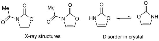
1. Introduction
2. Results
3. Experimental
Supplementary Materials
Author Contributions
Funding
Data Availability Statement
Conflicts of Interest
References
- Ager, D.J.; Prakash, I.; Schaad, D.R. 1,2-Amino alcohols and their heterocyclic derivatives as chiral auxiliaries in asymmetric synthesis. Chem. Rev. 1996, 96, 835–875. [Google Scholar] [CrossRef]
- Murakata, M.; Tsutsui, H.; Hoshino, O. Unprecedented effect of achiral oxazolidinones on enantioselective radical-mediated conjugate additions using a chiral zinc triflate. Org. Lett. 2001, 3, 299–302. [Google Scholar] [CrossRef] [PubMed]
- Turley, J.W. The crystal structure of 2-oxazolidinone. Acta Crystallogr. Sect. B 1972, 28, 140–143. [Google Scholar] [CrossRef]
- Wouters, J.; Ooms, F.; Durant, F. 2-Oxazolidinone. Acta Crystallogr. Sect. C 1997, 53, 895–897. [Google Scholar] [CrossRef]
- Scholz, K.-H.; Heine, H.-G.; Hartmann, W. Eine einfach Synthese von 4-Oxazolin-2-on. Liebigs Ann. Chem. 1976, 1319–1322. [Google Scholar] [CrossRef]
- Scholz, K.-H.; Heine, H.-G.; Hartmann, W. Synthesis and Diels-Alder reactions of 3-acetyl-2(3H)-oxazolone: 6-Amino-3,4-dimethyl-cis-3-cyclohexen-1-ol. Org. Synth. 1984, 62, 149–153. [Google Scholar] [CrossRef]
- Gaenzler, F.C.; Smith, M.B. A dichlorination-reductive-dechlorination route to N-acetyl-2-oxazolone. Synlett 2007, 1299–1301. [Google Scholar] [CrossRef]
- Ishizuka, T.; Ishibuchi, S.; Kunieda, K. New camphor-derived auxiliaries in methoxyselenation and methoxybromination with opposite diastereofacial selectivity. Preparation of β-amino alcohol chiral synthons. Tetrahedron Lett. 1989, 30, 3449–3452. [Google Scholar] [CrossRef]
- Scholz, K.-H.; Hartmann, W.; Heine, H.-G. Δ4-Oxazolin-2. German Patent 2610676, 15 September 1977. [Chem. Abstr. 1978, 88, 6860]. [Google Scholar]
- Tavernier, D.; Van Damme, S.; Ricquier, P.; Anteunis, M.J.O. A Convenient Preparation of 3H-1,3-oxazol-2-one and its N-Formyl Derivative. Bull. Soc. Chim. Belg. 1988, 97, 859–865. [Google Scholar] [CrossRef]
- Allen, F.H.; Kennard, O.; Watson, D.G.; Brammer, L.; Orpen, A.G.; Taylor, R. Tables of bond lengths determined by X-ray and neutron diffraction. Part 1. Bond lengths in organic compounds. J. Chem. Soc., Perkin Trans. 2 1987, S1–S19. [Google Scholar] [CrossRef]
- Cheng, J.-L.; Tan, C.-X.; Zhu, G.-N. 3-Benzoyl-1,3-oxazolidin-2-one. Acta Crystallogr. Sect. E 2005, 61, o3194–o3195. [Google Scholar] [CrossRef]
- Soloshonok, V.A.; Cai, C.; Hruby, V.J.; Van Meervelt, L.; Yamazaki, T. Rational design of highly diastereoselective, organic base-catalyzed, room-temperature Michael addition reactions. J. Org. Chem. 2000, 65, 6688–6696. [Google Scholar] [CrossRef]
- Shen, Y.-D.; Wang, Y.; Xiao, Z.-L.; Lei, H.-T.; Sun, Y.-M. (Z)-3-(2-Oxo-1,3-oxazolidin-3-ylcarbonyl)prop-2-enoic acid. Acta Crystallogr. Sect. E 2007, 63, o710–o711. [Google Scholar] [CrossRef]
- Skelton, B.W.; Pyne, S.G. CCDC 1543018: Experimental Crystal Structure Determination; The Cambridge Crystallographic Data Centre (CCDC): Cambridge, UK, 2017. [Google Scholar] [CrossRef]
- Yamada, S.; Misono, T.; Tsuzuki, S. Cation–π interactions of a thiocarbonyl group and a carbonyl group with a pyridinium nucleus. J. Am. Chem. Soc. 2004, 126, 9862–9872. [Google Scholar] [CrossRef]
- Marsh, R.E.; Clemente, D.A. A survey of crystal structures published in the Journal of the American Chemical Society. Inorg. Chim. Acta 2007, 360, 4017–4024. [Google Scholar] [CrossRef]
- Shen, Y.; Chai, J.; Yang, G.; Chen, W.; Chai, Z. Stereocontrolled synthesis of trans/cis-2,3-disubstituted cyclopropane-1,1-diesters and applications in the synthesis of furanolignans. J. Org. Chem. 2018, 83, 12549–12558. [Google Scholar] [CrossRef]
- Li, L.; Guo, J.-Y.; Liu, X.-G.; Chen, S.; Wang, Y.; Tan, B.; Liu, X.-Y. Amide groups switch selectivity: CH trifluoromethylation of α,β-unsaturated amides and subsequent asymmetric transformation. Org. Lett. 2014, 16, 6032–6035. [Google Scholar] [CrossRef]
- Etter, M.C.; MacDonald, J.C.; Bernstein, J. Graph-set analysis of hydrogen-bond patterns in organic crystals. Acta Crystallogr. Sect. B 1990, 46, 256–262. [Google Scholar] [CrossRef]
- Görgen née Boersch, C.; Lutsenko, K.; Merkul, E.; Frank, W.; Müller, T.J.J. Catalytic one-pot synthesis of 4-(hetero)aryl substituted 5-(2-oxoethyl)oxazol-2(3H)-ones by coupling–isomerization–elimination (CIE) sequence. Org. Chem. Front. 2016, 3, 887–896. [Google Scholar] [CrossRef]
- Sheldrick, G.M. A short history of SHELXL. Acta Crystallogr. Sect. A 2008, 64, 112–122. [Google Scholar] [CrossRef] [PubMed]


| Bond Length (Å) | 1 | 3 | Angle (°) | 1 | 3 |
|---|---|---|---|---|---|
| O(1)–C(2) | 1.329(4) | 1.358(1) | C(2)–O(1)–C(5) | 111.1(3) | 108.0(1) |
| C(2)–O(2) | 1.192(5) | 1.201(1) | O(1)–C(2)–N(3) | 109.2(3) | 106.62(9) |
| C(2)–N(3) | 1.374(4) | 1.381(1) | O(1)–C(2)–O(2) | 122.7(3) | 123.1(1) |
| N(3)–C(4) | 1.458(5) | 1.409(1) | O(2)–C(2)–N(3) | 128.1(3) | 130.3(1) |
| C(4)–C(5) | 1.518(4) | 1.313(1) | C(2)–N(3)–C(4) | 111.6(3) | 108.60(9) |
| C(5)–O(1) | 1.433(5) | 1.390(2) | C(2)–N(3)–C(6) | 129.2(3) | 128.0(1) |
| N(3)–C(6) | 1.380(4) | 1.408(2) | C(4)–N(3)–C(6) | 119.1(3) | 122.99(9) |
| C(6)–O(6) | 1.204(4) | 1.204(2) | N(3)–C(4)–C(5) | 101.1(2) | 106.4(1) |
| C(6)–C(7) | 1.499(5) | 1.491(2) | C(4)–C(5)–O(1) | 105.6(3) | 110.4(1) |
| N(3)–C(6)–O(6) | 119.2(3) | 117.7(1) | |||
| N(3)–C(6)–C(7) | 118.3(3) | 117.8(1) | |||
| O(6)–C(6)–C(7) | 122.5(3) | 124.6(1) |
| Compound | R | CCDC Ref Code | Angle Sum at N (°) | Torsion Angle (°) O=C–N–C(=O)O | Ref. |
|---|---|---|---|---|---|
| 1 | — | — | 359.9(3) | 177.5(3) | This work |
| 5a | Ph | JAXRAC | 357.4(1) | 153.9(1) | [12] |
| 5b | (E)-PhCH=CH | ECAVOT | 360.0(2) | 179.3(2) | [13] |
| 5c | (E)-HO2C-CH=CH | JEVNEE | 360.0(1) | 177.4(1) | [14] |
| 5d | MeC≡C | LAWWEO | 359.6(3) | 174.0(3) | [15] |
| 5e a | 3-Pyridyl | SAGDUA | 356.5(2) | 149.1(3) | [16] |
| 357.1(2) | 152.3(3) | ||||
| 356.9(2) | 152.0(3) | ||||
| 356.3(2) | 149.1(3) | ||||
| 5f b | 3-Pyridyl | SAGDUA01 | 356.4 | 149.1 | [17] |
| 357.0 | 152.1 | ||||
| 5g | (Z)-PhCH=C(Br) | WIQNUI | 355.7(4) | 150.7(5) | [18] |
| 5h | (E)-F3C-CH=CH(Me) | YOXQIN | 359.2(5) | 163.5(5) | [19] |
| 3 | — | — | 359.6(1) | 179.8(1) | This work |
| 6 | — | KATVUW | 360.0(4) | 178.3(5) | [8] |
| Compound | D—H…A | D—H | H…A | D…A | D—H…A | Ref. |
|---|---|---|---|---|---|---|
| 4 | N(3)–H(3)…O(2) | 0.98(5) | 1.90(5) | 2.858(8) | 166(7) | this work |
| 4 | N(13)–H(13)…O(7) | 0.98(6) | 1.93(6) | 2.904(8) | 176(8) | this work |
| 7 | N(1)–H(1)…O(7) | 0.856(16) | 1.971(16) | 2.7998(18) | 162.6(16) | [21] |
| 7 | N(2)–H(2)…O(2) | 0.914(15) | 1.941(16) | 2.8356(17) | 165.8(14) | [21] |
Publisher’s Note: MDPI stays neutral with regard to jurisdictional claims in published maps and institutional affiliations. |
© 2022 by the authors. Licensee MDPI, Basel, Switzerland. This article is an open access article distributed under the terms and conditions of the Creative Commons Attribution (CC BY) license (https://creativecommons.org/licenses/by/4.0/).
Share and Cite
Aitken, R.A.; Logan, J.S.; Slawin, A.M.Z. X-ray Structures of 3-Acetyloxazolidin-2-one, 3-Acetyloxazolin-2-one and Oxazolin-2(3H)-one. Molbank 2022, 2022, M1445. https://doi.org/10.3390/M1445
Aitken RA, Logan JS, Slawin AMZ. X-ray Structures of 3-Acetyloxazolidin-2-one, 3-Acetyloxazolin-2-one and Oxazolin-2(3H)-one. Molbank. 2022; 2022(3):M1445. https://doi.org/10.3390/M1445
Chicago/Turabian StyleAitken, R. Alan, Joe S. Logan, and Alexandra M. Z. Slawin. 2022. "X-ray Structures of 3-Acetyloxazolidin-2-one, 3-Acetyloxazolin-2-one and Oxazolin-2(3H)-one" Molbank 2022, no. 3: M1445. https://doi.org/10.3390/M1445
APA StyleAitken, R. A., Logan, J. S., & Slawin, A. M. Z. (2022). X-ray Structures of 3-Acetyloxazolidin-2-one, 3-Acetyloxazolin-2-one and Oxazolin-2(3H)-one. Molbank, 2022(3), M1445. https://doi.org/10.3390/M1445