Review

Analysis of Mechanisms for Electron Uptake by Methanothrix harundinacea 6Ac During Direct Interspecies Electron Transfer

1
School of Life Science and Technology, Inner Mongolia University of Science and Technology, Baotou 014010, China
2
School of Mining and Coal, Inner Mongolia University of Science and Technology, Baotou 014010, China
*
Authors to whom correspondence should be addressed.
These authors contributed equally to this work.
Int. J. Mol. Sci. 2025, 26(9), 4195; https://doi.org/10.3390/ijms26094195
Submission received: 23 March 2025 / Revised: 24 April 2025 / Accepted: 25 April 2025 / Published: 28 April 2025
(This article belongs to the Section Physical Chemistry and Chemical Physics)

Abstract

:
Direct interspecies electron transfer (DIET) is a syntrophic metabolism wherein free electrons are directly transferred between microorganisms without the mediation of intermediates such as molecular hydrogen or formate. Previous research has demonstrated that Methanothrix harundinacea 6Ac is capable of reducing carbon dioxide through DIET. However, the mechanisms underlying electron uptake in M. harundinacea 6Ac during DIET remain poorly understood. This study aims to elucidate the electron and proton flux in M. harundinacea 6Ac during DIET and to propose a model for electron uptake in this organism, primarily based on the analysis of gene transcript levels, genomic characteristics of M. harundinacea 6Ac, and the pathways generating fully reduced ferridoxin (Fdred2−), reduced coenzyme F420 (F420H2), coenzyme M (CoM-SH), and coenzyme B (CoB-SH) during DIET. The findings suggest that membrane-bound heterodisulfide reductase (HdrED), F420H2-dehydrogenase lacking subunit F (Fpo), and cytoplasmic heterodisulfide reductase (HdrABC)-subunit B of F420-reducing hydrogenase (FrhB) complex play critical roles in electron uptake in M. harundinacea 6Ac during DIET. Specifically, Fpo is responsible for generating Fdred2− with reduced methanophenazine (MPH2), driven by a proton motive force, while HdrED facilitates the reduction of heterodisulfide of coenzyme M and coenzyme B (CoM-S-S-CoB) to CoM-SH and CoB-SH using MPH2. Additionally, cytoplasmic heterodisulfide reductase HdrABC and subunit B of coenzyme F420-hydrogenase complex (HdrABC-FrhB complex) catalyzes the reduction of oxidized coenzyme F420 (F420) to F420H2, utilizing CoM-SH, CoB-SH, and Fdred2−. This study represents the first genetics-based functional characterization of electron and proton flux in M. harundinacea 6Ac during DIET, providing a model for further investigation of electron uptake in Methanosaeta species. Furthermore, it deepens our understanding of the mechanisms underlying electron uptake in methanogens during DIET.

1. Introduction

For years, interspecies H2 and formate transfer were regarded as the dominant mechanisms for microbial electron exchange [1,2,3]. During syntrophic interspecies hydrogen (H2) and formate transfer, acetogens metabolize primary fermentation products (e.g., propionate, butyrate, and ethanol) to produce acetate, formate, and H2. These metabolic byproducts are subsequently utilized by partner microorganisms, including methanogens [3,4,5], sulfate-reducing bacteria [6], or nitrate/nitrite-reducing bacteria [7,8,9,10]. The efficiency of this syntrophic degradation of fermentation intermediates is critically dependent on maintaining low H2 concentrations, as this thermodynamic constraint ensures the favorable directionality of the reaction. Such interspecies H2 and formate transfer mechanisms have been extensively documented in diverse anaerobic co-culture systems [11,12,13,14,15].
However, emerging evidence suggests that direct interspecies electron transfer (DIET) may serve as a viable alternative to indirect or mediated interspecies electron transfer pathways. DIET enables direct electron flow between microbial species through bioelectrical connections or conductive materials, bypassing the need for soluble electron shuttles. This phenomenon was first demonstrated in a defined co-culture system where Geobacter metallireducens GS-15 and Geobacter sulfurreducens PCA collaboratively reduced fumarate to succinate using ethanol as an electron donor substrate (Figure 1) [16]. Subsequent studies have identified numerous pure co-cultures capable of DIET, with most documented cases involving syntrophic partnerships between Geobacter species and methanogenic archaea [17,18,19,20,21,22,23,24,25].
Since the discovery of DIET, significant research efforts have focused on elucidating its key components, including: (i) electron-donating microorganisms [22,24,25,27,28,29,30], (ii) electron-accepting microorganisms [17,18,19,21,22,23,30,31,32,33], (iii) the extracellular electron transfer mechanisms employed by electron-donating microorganisms [23,34], and (iv) the extracellular electron uptake processes of electron-accepting microorganisms [23,28,31,35,36], and effect of (semi)conductive material on DIET.
While previous studies have demonstrated that certain microorganisms can directly accept electrons from syntrophic partners through direct cell contact or via (semi)conductive materials, the molecular mechanisms governing extracellular electron uptake remain poorly understood. To address this knowledge gap, transcriptomic analysis was employed to elucidate the key enzymes and proteins involving in extracellular electron uptake, along with electron and proton flux during CO2 reduction to methane in Methanosarcina barkeri 800 [28,35,36], Methanosarcina acetivorans strain WWM1(Δhpt) [31], and Methanothrix thermoacetophila [23] during DIET.
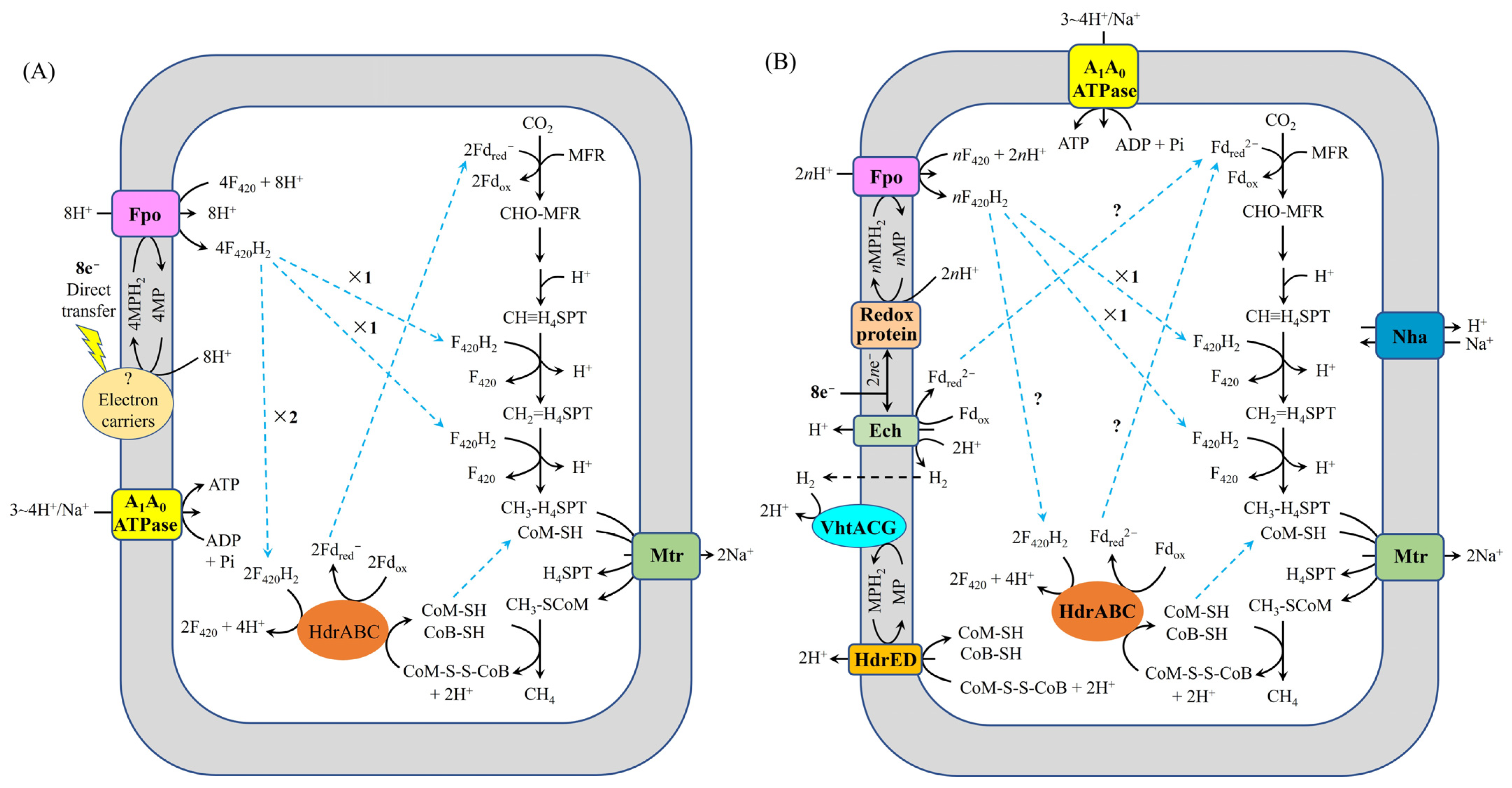
M. barkeri 800 represents the first microorganism for which the molecular mechanism underlying the reduction of carbon dioxide to methane via DIET has been comprehensively elucidated. Holmes et al. [35] proposed a metabolic model for CO₂-reductive methanogenesis in M. barkeri 800 during DIET by conducting comparative transcriptomic analysis between two syntrophic growth modes (Figure 2A): (i) DIET-based coculture with Geobacter metallireducens GS-15 and (ii) H2-mediated interspecies transfer (HIT) with Pelobacter carbinolicus. Their transcriptomic data revealed that F420H2 dehydrogenase (Fpo) and cytoplasmic heterodisulfide reductase HdrABC (HdrABC) were significantly upregulated during DIET, suggesting their crucial involvement in extracellular electron uptake in M. barkeri 800 during DIET. Fpo catalyzes the reduction of oxidized coenzyme F420 (F420) to reduced coenzyme F420 (F420H2) using reduced methanophenazine (MPH2) driven by proton motive force, while HdrABC utilizes a portion of the generated F420H2 to simultaneously reduce both heterodisulfide of coenzyme M and coenzyme B (CoM-S-S-CoB) and oxidized ferredoxin (Fdox), yielding coenzyme M (CoM-SH), coenzyme B (CoB-SH), and one-electron reduced ferredoxin (Fdred), respectively. The proposed model suggests that the membrane-bound heterodisulfide reductase HdrED was not involved in CoM-S-S-CoB reduction in M. barkeri 800 during DIET. Huang et al. [28] established an alternative model for CO2-reductive methanogenesis in M. barkeri 800 during DIET with Rhodopseudomonas palustris, based on transcriptomic profiling of DIET-grown cocultures (Figure 2B). Their analysis revealed significant upregulation of genes encoding key respiratory complexes, including: the energy-converting hydrogenase (Ech), the membrane-bound F420H2 dehydrogenase (Fpo), the methanophenazine-reducing hydrogenase (VhtACG), and both heterodisulfide reductase systems (HdrED and HdrABC). These findings suggest that these enzyme complexes collectively facilitate extracellular electron uptake during DIET in M. barkeri 800. Notably, the model proposed by Holmes et al. [35] suggests that HdrED does not participate in either CoM-S-S-CoB reduction or extracellular electron transfer, contrasting with Huang et al.′s findings [28]. In addition, under physiological conditions (pH 7.0; F420/F420H2 ratio ≤ 0.1; 37 °C), thermodynamic analysis reveals that the free energy change (ΔG′) for F420 reduction by MPH2 coupled to proton motive force ranges from + 0.41 to +13.91 kJ/mol, while CoM-S-S-CoB reduction by MPH2 exhibits a more favorable ΔG′ range of −9.55 to + 3.95 kJ/mol. This significant energetic difference indicates that CoM-S-S-CoB reduction is thermodynamically preferred over F420 reduction under these conditions. Consequently, these calculations support the hypothesis that in M. barkeri 800 during DIET, HdrED likely utilizes MPH2 as the immediate electron donor for CoM-S-S-CoB reduction [28]. These apparent discrepancy highlights the need for additional mechanistic studies. Furthermore, Wang et al. [36] identified a functional role for the F420-reducing hydrogenase (FrhABG) in electron transfer processes in M. barkeri 800 during DIET. The finding suggests that FrhABG catalyzes the oxidation of a portion of the generated F420H2 to produce H2, consistent with the observed upregulation of frhABG gene expression under DIET conditions. These findings collectively demonstrate that in Type I Methanosarcina species (e.g., M. barkeri 800), both the extracellular electron uptake mechanisms and intracellular electron transfer pathways exhibit greater complexity than previously anticipated, warranting further investigation.
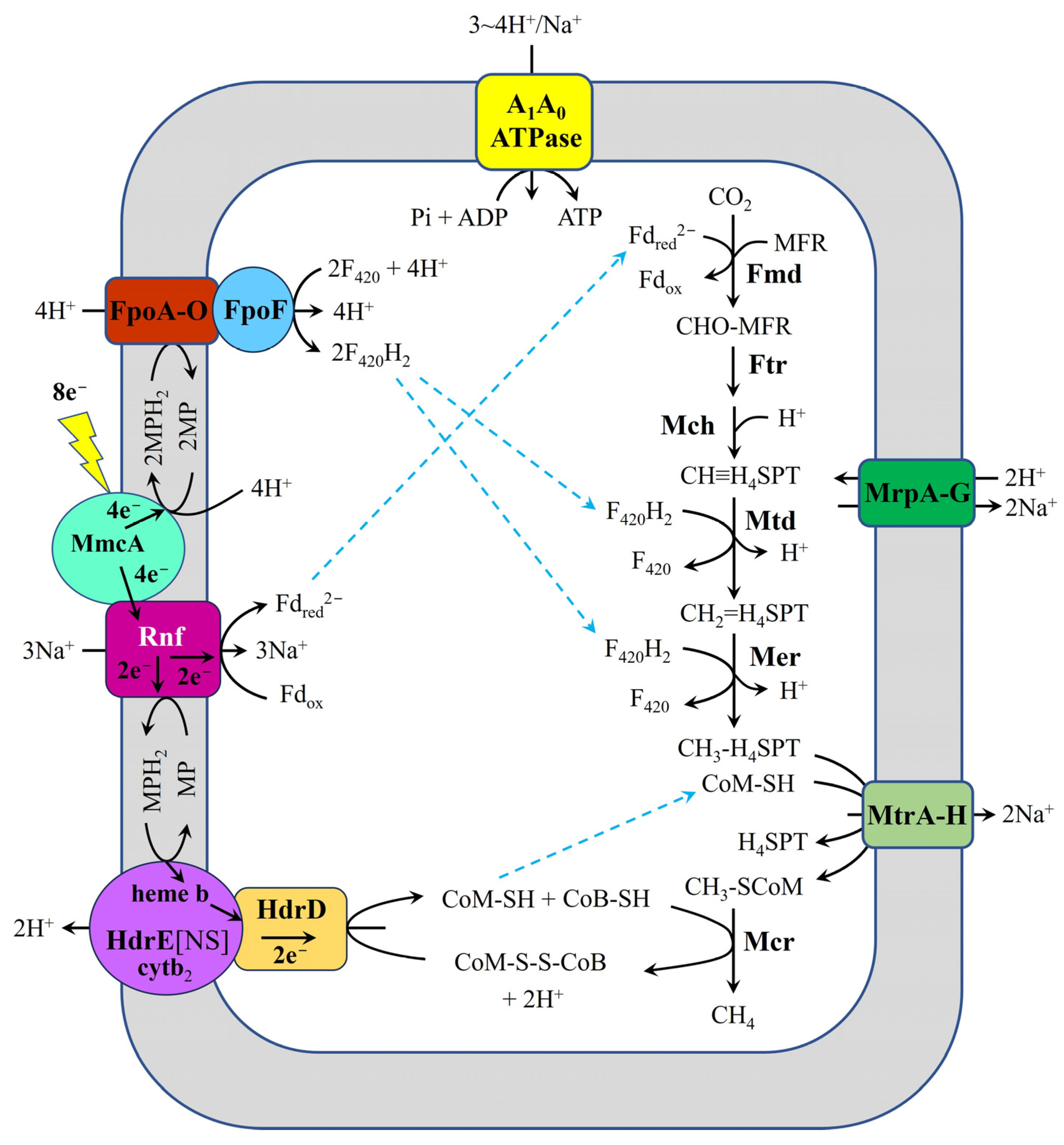
Methanosarcina acetivorans strain WWM1(Δhpt) represents another Methanosarcina species for which potential extracellular electron uptake mechanisms during DIET with G. metallireducens GS-15 have been investigated. Holmes et al. [31] systematically characterized the electron and proton transfer pathways in M. acetivorans WWM1(Δhpt) during DIET-based growth with G. metallireducens GS-15 (using ethanol as the sole electron donor) through integrated comparative transcriptomic approaches (Figure 3). Their analysis revealed significant upregulation of key respiratory components in DIET-grown cocultures compared to acetate-grown controls, including: (i) the outer surface multiheme c-type cytochrome MmcA, (ii) the Na+-translocating ferredoxin: methanophenazine reductase complex (Rnf complex), (iii) Fpo, and (iv) HdrED. These findings strongly suggest that these molecular components collectively mediate extracellular electron uptake in M. acetivorans WWM1(Δhpt). Notably, the proposed electron and proton transfer pathway in this strain exhibits fundamental mechanistic differences from the system previously characterized in M. barkeri 800 [28,35], highlighting distinct DIET adaptation strategies among methanogens.
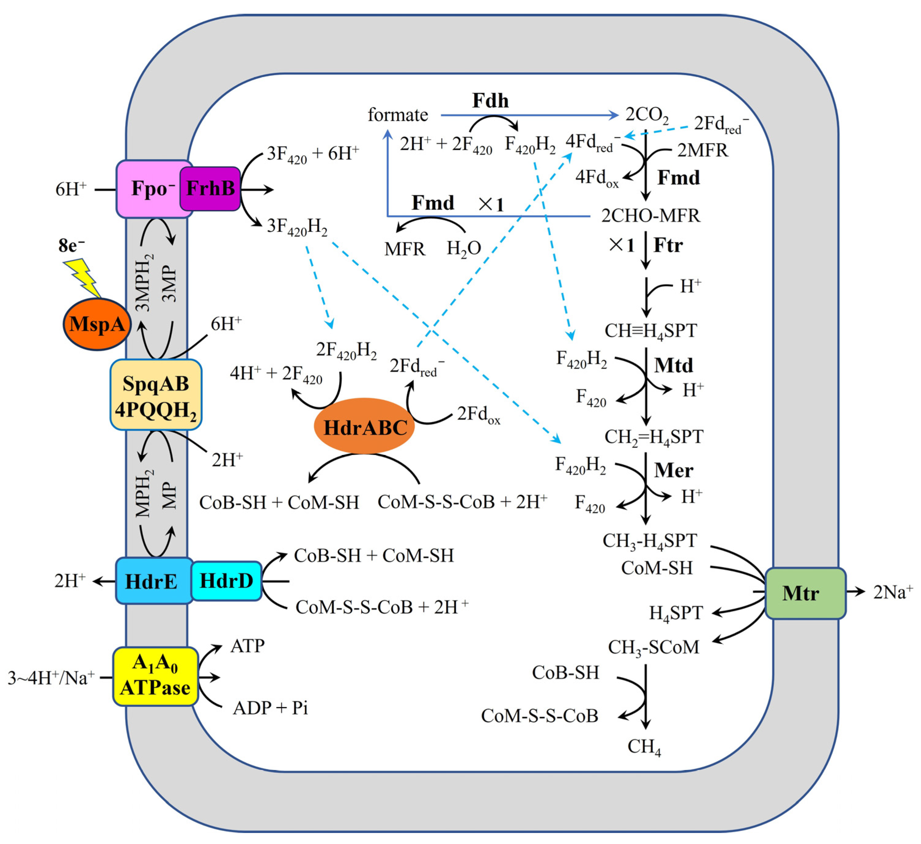
Recent work by Zhou et al. [23] has elucidated a novel mechanism for DIET-mediated methanogenesis in Methanothrix thermoacetophila through comparative transcriptomic analysis of DIET-grown cocultures (with G. metallireducens GS-15) versus acetoclastic growth conditions (Figure 4). The study revealed significant upregulation of genes encoding several key components: (i) the surface-associated primary sheath fiber protein (MspA), (ii) the surface quinoprotein complex (SqpAB), (iii) heterodisulfide reductases (HdrED and HdrABC), (iv) formylmethanofuran dehydrogenase (Fmd), (v) formate dehydrogenase (Fdh), (vi) Fpo, and (vii) subunit B of FrhABG (FrhB). These findings suggest that this unique ensemble of surface proteins and redox enzymes collectively facilitates extracellular electron uptake in M. thermoacetophila, representing a distinct DIET adaptation mechanism among methanogens. This proposed model postulates that partial F420H2 production requires coupling of Fmd and Fdh with two moles of one-electron reduced ferredoxin (Fdred) generated from acetate oxidation in M. thermoacetophila. However, this hypothesis warrants critical examination, as several methanogens—including Methanosarcina harundinacea 6Ac [37] and M. barkeri 800 [36]—demonstrate the capability for DIET-based CO2 reduction to methane in the absence of acetate. This apparent discrepancy could be resolved experimentally by determining whether G. metallireducens GS-15/M. thermoacetophila cocultures can couple methane production with the oxidation of alternative electron donors such as propanol or butanol. The proposed mechanism whereby the Fpo-FrhB complex mediates F420 reduction to F420H2 using MPH2 driven by proton motive force also requires careful consideration. This hypothesis is challenged by experimental evidence demonstrating that a subunit F of Fpo (FpoF)-deficient strain of Methanosarcina mazei Gö1, despite retaining both Fpo and FrhB components, was incapable of direct F420H2 oxidation via membrane-bound electron transport [38]. These findings not only question the functional capacity of Fpo-FrhB in F420H2 generation but also necessitate re-examination of the proposed coupled system where HdrABC generates Fdred through F420H2 oxidation. These findings collectively highlight the need for more comprehensive investigations into both the extracellular electron uptake mechanisms and intracellular electron transfer pathways during DIET-mediated methanogenesis in M. thermoacetophila.
Although several unresolved questions remain regarding extracellular electron uptake mechanisms and intracellular electron transfer pathways during DIET-mediated methanogenesis in the studied methanogens, these findings nevertheless provide valuable insights for investigating similar processes in other methanogenic species. Furthermore, these findings motivate and direct further research in this emerging field. Methanothrix harundinacea 6Ac (formerly named Methanosaeta harundinacea 6Ac) (JCM-13211, https://jcm.brc.riken.jp/) has also been reported to perform DIET-dependent methanogenesis [19,37], however, the extracellular electron uptake and intracellular electron transfer pathways in this archaeon remains unresolved. This study aims to elucidate these mechanisms by: (i) analyzing transcriptomic profiles of M. harundinacea 6Ac during DIET-based growth, (ii) examining genomic features associated with electron uptake, and (iii) determining the pathways for generating fully reduced ferredoxin (Fdred2−), F420H2, CoM-SH, and CoB-SH. Based on these analyses, we propose a model for extracellular electron uptake in M. harundinacea 6Ac via DIET. This model will serve as a foundational framework for future investigations into electron uptake mechanisms in Methanosaeta spp. and broaden our understanding of DIET-based methanogenesis in anaerobic microbial communities.

2. Possible Mechanisms for Extracellular Electron Uptake by M. harundinacea 6Ac

As a member of the Methanosarcinales, M. harundinacea 6Ac possesses cytochromes [39], which are typically localized between the cytoplasmic membrane and S-layer in this order [40]. Genomic analysis reveals that M. harundinacea 6Ac encodes key redox-active components, including a class I c-type cytochrome (Mhar_1883), cytochrome c biogenesis proteins (Mhar_0020, Mhar_1541, Mhar_1552), and multiple S-layer proteins (Mhar_0562, Mhar_0637, Mhar_0867, Mhar_1021, Mhar_1810) [19]. Notably, the gene encoding the class I cytochrome (Mhar_1883) was highly expressed during DIET-based syntrophic growth with G. metallireducens GS-15, where ethanol was stoichiometrically converted to methane [19]. Given that c-type cytochromes in Methanosarcina species exhibit midpoint potentials of −230 ± 10 mV and −140 ± 10 mV (vs. SHE) [41], we propose a thermodynamically feasible electron transfer pathway in M. harundinacea 6Ac: Electrons may flow from cytochrome c (−230 ± 10 mV) to oxidized methanophenazine (MP) (′ = −165 ± 6 mV [42]), yielding a favorable Gibbs free energy change (ΔG′ = −9.33 kJ/mol at 37 °C, pH 7, assuming equal substrate and product concentrations). Collectively, these findings support a model where c-type cytochromes in M. harundinacea 6Ac may directly accept extracellular electrons and transfer them to MP, generating MPH2 to drive methanogenesis. This mechanism appears analogous to that observed in M. acetivorans strain WWM1(Δhpt), where the outer surface multiheme c-type cytochrome MmcA was proposed to mediate direct electron uptake from syntrophic partners during DIET [31]. Nevertheless, to comprehensively establish the mechanistic details of these proposed electron transfer pathways, molecular-level validation is essential.

3. Possible Mechanisms for Generation of Fdred2− in M. harundinacea 6Ac During DIET

In cytochrome-containing methanogens, membrane-bound energy conserving hydrogenase (Ech), Rnf complex, Fpo, and HdrABC are typically associated with Fdred2− oxidation or generation. However, genomic analysis reveals that M. harundinacea 6Ac lacks both ech and rnf genes [43], excluding their involvement in membrane-bound electron transport during DIET with G. metallireducens GS-15. Instead, this organism possesses a complete fpo gene cluster (fpoABCDEHIJKLMNO) [43], which was highly expressed during DIET-based growth [19]. Notably, Fpo cannot oxidize F420H2, as demonstrated by studies with Methanosarcina mazei Gö1 ΔfpoF mutants (an FpoF-deficient strain of M. mazei Gö1) [38]. This demonstrates that F420H2 cannot be generated by Fpo with MPH2 driven by a proton motive force in M. harundinacea 6Ac during DIET. Consequently, it rules out the possibility of F420H2-dependent Fdred2− generation via cytoplasmic heterodisulfide reductase HdrA2B2C2 in M. harundinacea 6Ac during DIET (Figure 5). Nevertheless, accumulating evidence indicates that Fpo can oxidize Fdred2− in both Methanosaeta spp. [44,45] and M. mazei Δech mutants [46]. Cumulatively, the evidence suggests that, in M. harundinacea 6Ac, Fpo likely catalyzes Fdred2− generation using MPH2 driven by proton motive force during DIET, which is consistent with the high expression of its fpo gene cluster [19]. The function of Fpo in M. harundinacea 6Ac during DIET could be investigated using the Fpo-specific inhibitor p-chloromercuriphenyl sulfonate or sodium azide [41]. Since the generation of one mole of Fdred2− consumes three moles of protons [44,45], during DIET in M. harundinacea 6Ac, eight moles of protons from its syntrophic partner can only contribute to the generation of two to three moles of Fdred2−. The generation of two to three moles of Fdred2− requires four to six moles of electrons (in the form of MPH₂), indicating that the remaining two to four moles of electrons are transferred intracellularly via another pathway. In Methanosaeta spp., only membrane-bound Fpo and HdrED are involved in electron transfer. Thus, it is hypothesized that HdrED is responsible for mediating the remaining extracellular electrons into the intracellular environment.

4. Possible Mechanisms Underlying the Generation of CoM-SH and CoB-SH in M. harundinacea 6Ac During DIET

In methanogens, heterodisulfide CoM-S-S-CoB reduction can occur through four distinct pathways: (i) the cytoplasmic H2:heterodisulfide oxidoreductase complex (MvhADG-HdrABC complex) utilizing H2 [39,49,50], (ii) membrane-bound HdrED with MPH2 [48,51], (iii) cytoplasmic heterodisulfide reductase HdrA1B1C1 with Fdred2− [52], or (iv) cytoplasmic heterodisulfide reductase HdrA2B2C2 with F420H2 [47]. As an obligate acetoclastic methanogen [53], M. harundinacea 6Ac lacks the MvhADG-HdrABC complex, eliminating the first pathway. Genomic analysis reveals only hdrED and hdrABC clusters in M. harundinacea 6Ac [43], and the absence of F420H2 during initial DIET stages precludes HdrA2B2C2 involvement. While HdrA1B1C1 represents another potential candidate, neither its encoding genes [43] nor transcriptional expression [19] have been detected in M. harundinacea 6Ac. Notably, hdrED genes (Mhar_0792-0793) were highly expressed in M. harundinacea 6Ac during DIET [19], suggesting HdrED mediates CoM-S-S-CoB reduction using MPH2. This conclusion aligns with prior reports that HdrED is responsible for transferring parts of extracellular electrons intracellularly in methanogens during DIET-based growth [23,28,31,36]. Functional validation could employ the HdrED-specific inhibitor pyridine [41]. The generation of Fdred2− consumes only two to three moles of MPH2. This indicates that at least one, but no more than two, moles of MPH2 remain available for the reduction of CoM-S-S-CoB, which is consistent with the proposed HdrED activity.

5. Possible Mechanisms for F420H2 Generation in M. harundinacea 6Ac During DIET

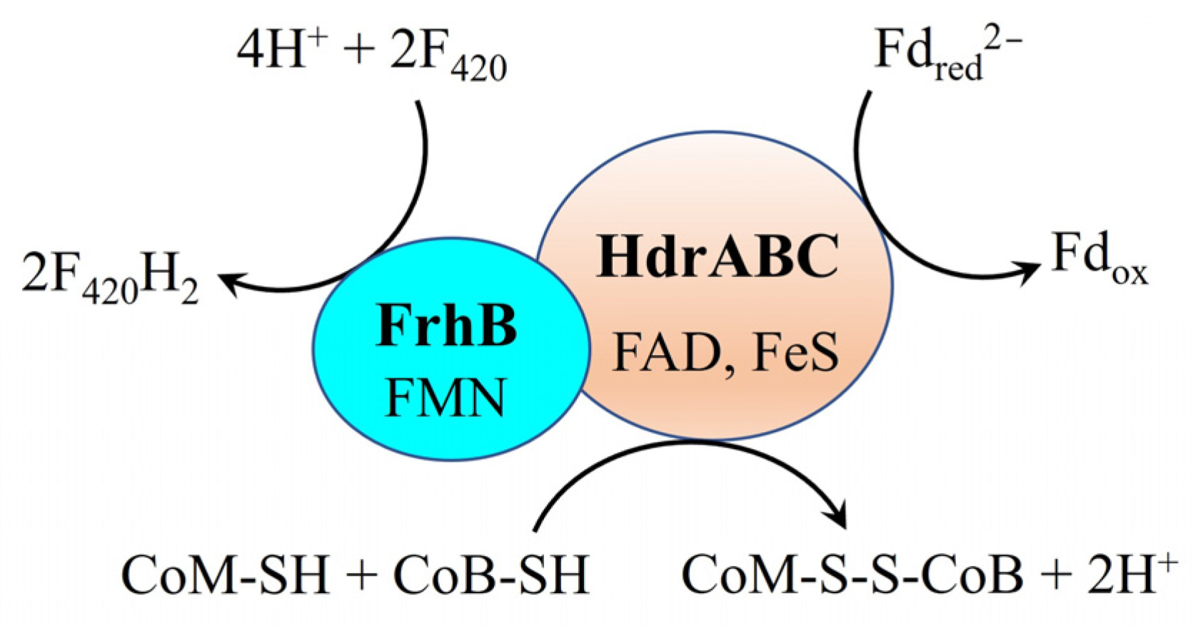
F420H2, an essential electron and proton carrier for CO2 reduction to methane, can be produced through multiple pathways in methanogens: (i) FrhABG, fructose-inducible hydrogenase (Fru hydrogenase), or fructose-repressible hydrogenases (Frc hydrogenases) using H2 [39,49,54], (ii) Fdh with formate [54], (iii) HdrA1B1C1 or HdrA2B2C2 with Fdred2− [47,52], or (iv) the HdrABC and FrhB complex (HdrABC-FrhB complex) via electron bifurcation (Figure 3) [55,56,57]. In M. harundinacea 6Ac, no genes for Fru/Frc hydrogenases or other subunits of FrhABG were identified, except for the gene encoding FrhB (Mhar_2358) [43]. This implies that F420H2 cannot be generated by FrhABG, Fru hydrogenase, or Frc hydrogenase during DIET in M. harundinacea 6Ac. As analyzed in Section 3, Fdred2− can potentially be generated by Fpo with MPH2 in M. harundinacea 6Ac during DIET. Additionally, CoM-S-S-CoB is present in M. harundinacea 6Ac. Although F420H2 could potentially be produced by HdrA1B1C1 or HdrA2B2C2 with Fdred2− and CoM-S-S-CoB, genes for HdrA1B1C1 and HdrA2B2C2 are absent from the M. harundinacea 6Ac genome [43], ruling out this production route. Notably, highly expressed hdrABC (Mhar_0604-0607) and frhB (Mhar_2358) genes in M. harundinacea 6Ac during DIET [19] suggest the HdrABC-FrhB complex mediates F420H2 generation via electron bifurcation (Figure 6). Collectively, these findings suggest that the HdrABC-FrhB complex may facilitate the generation of F420H2 in M. harundinacea 6Ac during DIET. If this electron bifurcation reaction occurs during DIET in M. harundinacea 6Ac, it can account for the generation of F420H2 required for carbon dioxide reduction to methane. The role of the HdrABC-FrhB complex in M. harundinacea 6Ac during DIET-based growth can be further validated by examining the growth of the ΔhdrABC/ΔfrhB mutant of M. harundinacea 6Ac on DIET. If the mutant can grow on DIET, it suggests that the HdrABC-FrhB complex does not contribute to the generation of F420H2 during DIET-based growth in M. harundinacea 6Ac. Otherwise, it indicates the complex′s involvement in F420H2 generation during DIET-based growth.
Furthermore, an alternative pathway for partial F420H2 production involves the interaction of Fmd and Fdh utilizing Fdred2− generated by the Fpo with MPH2. The significant expression of genes encoding Fmd and subunit A of Fdh in M. harundinacea 6Ac during DIET [19] supports this hypothesis. Moreover, this proposed pathway aligns with previous research findings [23], except for the source of Fdred2−. In the model proposed by Zhou et al. [23], the Fdred2− utilized for partial F420H2 production through the interaction of Fmd and Fdh was derived from acetate oxidation, rather than being generated by the Fpo with MPH2, as discussed in this study. However, additional evidence casts doubt on the origin of Fdred2− in this manner. For example, the co-cultures of G. metallireducens GS-15/M. barkeri 800 [36] and G. metallireducens GS-15/M. harundinacea 6Ac [37] can facilitate the syntrophic conversion of propanol and butanol to their corresponding fatty acids (propionate and butyrate) and methane via DIET. Given that M. harundinacea 6Ac lacks the capacity to metabolize propionate or butyrate [37,43], the Fdred2− required for partial F420H2 production—mediated by the interaction of Fmd and Fdh—must have been generated by Fpo with MPH2, rather than originating from acetate oxidation. Notably, this pathway only yields limited F420H2 because the Fdred2− consumed in this process reduces the availability of electrons for HdrED-mediated CoM-S-S-CoB reduction. This subsequently diminishes the transmembrane proton gradient, ultimately impairing ATP synthesis and negatively affecting methanogen growth. However, the necessity of this bypass route remains uncertain. Stoichiometric analyses indicate that the HdrABC-FrhB system alone can generate sufficient F420H2 to satisfy the F420H2 demand for reduction of CO2 to methane.

6. Pathway Proposed for Reduction of Carbon Dioxide in M. harundinacea 6Ac During DIET

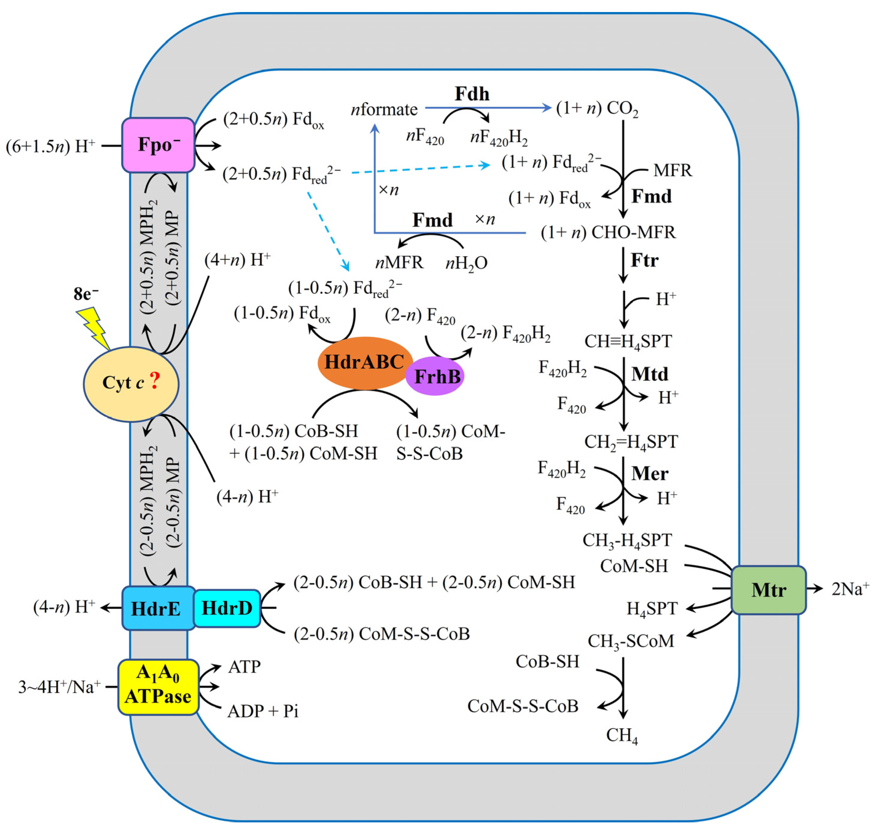
Building on the preceding analysis, we propose a pathway for the electron uptake in M. harundinacea 6Ac during growth via DIET (Figure 7). In this proposed model, eight moles of extracellular electrons are shuttled to MP through cytochrome c, generating four moles of MPH2. Subsequently, HdrED reduces a portion of CoM-S-S-CoB (more than one but less than two moles) using MPH2. Concurrently, Fpo reduces Fdox (more than two but less than three moles) with MPH2 driven by the proton motive force. Finally, F420H2 is synthesized by two distinct routes: one involving the HdrABC-FrhB complex, and the other via the combined action of Fmd and Fdh.
In M. harundinacea 6Ac, the mechanisms for generating reducing equivalents (MPH2, Fdred2−, F420H2, CoM-SH, and CoB-SH) required for CO2 reduction to methane during DIET differ significantly from those in traditional CO2-reducing methanogenesis. In cytochrome-lacking methanogens growing on CO2 and H2, Fdred2− is produced by the energy-converting hydrogenase (Eha and/or Ehb), as well as the cytoplasmic MvhADG–HdrABC complex with H2. The MvhADG–HdrABC complex catalyzes the generation of CoM-SH, CoB-SH, and Fdred2− via an electron bifurcation reaction using H2. F420H2 is generated by FrhABG with H2. In cytochrome-containing methanogens growing on CO2 and H2, Fdred2− is produced by the Ech hydrogenase with H2, driven by proton motive force. The formation of CoM-SH and CoB-SH involves the interaction of VhtACG and HdrED with H2, while F420H2 is synthesized by FrhABG with H2. However, in M. harundinacea 6Ac during DIET-based growth, Fdred2− is generated by the Fpo using MPH2 (derived from the reduction of MP with extracellular electrons) as the electron donor, driven by proton motive force. CoM-SH and CoB-SH are produced by HdrED using MPH2 (derived from the reduction of MP with extracellular electrons). F420H2 is generated through two distinct pathways: (i) the cytoplasmic HdrABC-FrhB complex utilizing CoM-SH, CoB-SH, and Fdred2−, and (ii) the combined activity of Fmd and Fdh with Fdred2−. This comparative analysis highlights the unique metabolic adaptations of M. harundinacea 6Ac for DIET-dependent methanogenesis, differing fundamentally from conventional H2-dependent pathways.
To better understand the biochemical mechanisms underlying extracellular electron uptake and the generation of reducing equivalents (MPH2, Fdred2−, F420H2, CoM-SH, and CoB-SH) in M. harundinacea 6Ac during DIET-based growth, we performed comparative genomic analyses of core metabolic networks across model methanogens including M. barkeri 800, M. acetivorans WWM1(Δhpt), M. thermoacetophila, and M. harundinacea 6Ac (Table 1). Two critical knowledge gaps persist: (i) the molecular basis of transmembrane electron transfer in electroactive methanogens (e.g., how extracellular electrons are transferred to MP), and (ii) species-specific flux partitioning mechanisms for metabolic product reduction. Notably, both extracellular electron uptake systems and MP reduction modules exhibit significant phylogenetic divergence among methanogens.
Despite the lack of clarity regarding the initial step of methanogens accepting extracellular electrons and the mechanism by which the MP is reduced with extracellular electrons, based on the diverse energy-conservation strategies employed by methanogens, there are approximately three distinct mechanisms for the reduction of carbon dioxide to methane in methanogens undergoing DIET-dependent growth:
  • Methanogens capable of conserving energy via Fpo, such as type I Methanosarcina spp., including M. bakeri 800, may utilize the mechanism for carbon dioxide reduction to methane illustrated in Figure 2. During DIET, Fpo, HdrED, FrhABG, HdrABC, VhtACG, and Ech in these methanogens are likely to contribute to extracellular electron uptake.
  • For methanogens that conserve energy through Rnf, like type II Methanosarcina spp., such as M. acetivorans WWM1(Δhpt) and Methanosarcina horonobensis HB-1, the mechanism for carbon dioxide reduction to methane is depicted in Figure 3. In these organisms, Fpo, Rnf, and HdrED are likely involved in extracellular electron uptake during DIET. Additionally, HdrABC may also facilitate the acquisition of extracellular electrons.
  • Methanogens that conserve energy through Fpo, like Methanothrix spp. (formerly named Methanosaeta spp.), including M. harundinacea 6Ac and M. thermoacetophila, may follow the mechanism for carbon dioxide reduction to methane that is shown in Figure 7. During DIET, the Fpo, HdrED, and HdrABC-FrhB complexes are potentially responsible for extracellular electron uptake in these methanogens.
Although the proposed model outlines the reduction of carbon dioxide to methane and electron uptake processes in M. harundinacea 6Ac during growth via DIET, the exact mechanism of electron uptake in M. harundinacea 6Ac during DIET remains poorly understood. Similar to all previously reported models on extracellular electron uptake in methanogens during DIET, the present proposed model has not yet been validated at the molecular level. Therefore, further experimental investigations are necessary to validate the proposed model.

7. Conclusions

The discovery of DIET has revealed a more efficient microbial energy-sharing mechanism than previously recognized. While significant progress has been made in DIET research, the molecular mechanisms of extracellular electron uptake in electron-accepting microbes, particularly methanogens, remain poorly understood. Here, we propose a comprehensive model for reduction of CO2 to methane and extracellular electron uptake in M. harundinacea 6Ac during DIET, integrating transcriptomic data, genomic analysis, and analysis on the mechanisms for generation of key redox components (Fdred2−, F420H2, CoM-SH, and CoB-SH). Our model identifies three critical players for electron uptake: (i) Fpo mediates Fdred2− generation using MPH2 driven by proton motive force; (ii) HdrED reduces CoM-S-S-CoB to CoM-SH and CoB-SH with MPH2; and (ⅲ) the HdrABC-FrhB complex catalyzes F420 reduction to F420H2 via electron bifurcation using CoM-SH, CoB-SH, and Fdred2−.
It is worth emphasizing that, although this model is primarily constructed upon transcriptomic analyses and genomic features of M. harundinacea 6Ac, along with the established functions of several key enzymes (such as the Fpo, HdrED, and HdrABC-FrhB complex), the molecular-level functions of the majority of enzymes engaged in electron transfer in M. harundinacea 6Ac during DIET-mediated growth remain uncharacterized. Future research could employ mutant strains of related enzymes or inhibitors of related enzymes to validate their functions in electron transfer during DIET. Moreover, the initial step of extracellular electron uptake by M. harundinacea 6Ac or how extracellular electron is transferred to MP of M. harundinacea 6Ac during DIET remains unclear and warrants further exploration. This exploration is essential for understanding the mechanisms of extracellular electron uptake by methanogens. This framework not only advances our understanding of Methanothrix spp. physiology during DIET, but it also provides new insights into DIET-based methanogenesis across diverse archaeal species.

Funding

This study was financially supported by the National Natural Science Foundation of China (Grant No. 31860011, Grant No. 51864037), the Key Technology Research Program Project of Inner Mongolia Autonomous Region (Grant No. 2020GG0018), the Key R&D and Achievement Transformation Plan Project of Inner Mongolia Autonomous Region (Grant No. 2022YFHH0026), the foundation of central governmental guidance for local scientific and technological development (Grant No. 2022ZY0213), the National Natural Science Foundation of Inner Mongolia (Grant No. 2021MS05066), and the fundamental research funds for the directly affiliated universities in Inner Mongolia (Grant No. 2023QNJS154).

Conflicts of Interest

The authors declare no conflict of interest.

References

  1. Bryant, M.P.; Wolin, E.A.; Wolin, M.J.; Wolfe, R.S. Methanobacillus omelianskii, a symbiotic association of two species of bacteria. Arch. Mikrobiol. 1967, 59, 20–31. [Google Scholar] [CrossRef] [PubMed]
  2. de-Bok, F.A.; Luijten, M.L.; Stams, A.J. Biochemical evidence for formate transfer in syntrophic propionate-oxidizing cocultures of Syntrophobacter fumaroxidans and Methanospirillum hungatei. Appl. Environ. Microbiol. 2002, 68, 4247–4252. [Google Scholar] [CrossRef] [PubMed]
  3. Stams, A.J.M.; Plugge, C.M. Electron transfer in syntrophic communities of anaerobic bacteria and archaea. Nat. Rev. Microbiol. 2009, 7, 568–577. [Google Scholar] [CrossRef]
  4. Schink, B.; Stams, A.J.M. Syntrophism among prokaryotes. In The Prokaryotes; Rosenberg, E., DeLong, E.F., Lory, S., Stackebrandt, E., Thompson, F., Eds.; Springer: Berlin/Heidelberg, Germany, 2013; pp. 309–335. [Google Scholar]
  5. Mcinerney, M.J.; Struchtemeyer, C.G.; Sieber, J.; Mouttaki, H.; Stams, A.J.; Schink, B.; Rohlin, L.; Gunsalus, R.P. Physiology, ecology, phylogeny, and genomics of microorganisms capable of syntrophic metabolism. Ann. N. Y. Acad. Sci. 2008, 1125, 58–72. [Google Scholar] [CrossRef] [PubMed]
  6. Muyzer, G.; Stams, A.J. The ecology and biotechnology of sulphate reducing bacteria. Nat. Rev. Microbiol. 2008, 6, 441–454. [Google Scholar] [CrossRef]
  7. Smith, R.L.; Ceazan, M.L.; Brooks, M.H. Autotrophic, hydrogen-oxidizing, denitrifying bacteria in groundwater, potential agents for bioremediation of nitrate contamination. Appl. Environ. Microbiol. 1998, 60, 1949–1955. [Google Scholar] [CrossRef]
  8. Strohm, T.O.; Griffin, B.; Zumft, W.G.; Schink, B. Growth yields in bacterial denitrification and nitrate ammonification. Appl. Environ. Microbiol. 2007, 73, 1420–1424. [Google Scholar] [CrossRef]
  9. Smith, R.L.; Miller, D.N.; Brooks, M.H.; Widdowson, M.A.; Killingstad, M.W. In situ stimulation of groundwater denitrification with formate to remediate nitrate contamination. Environ. Sci. Technol. 2001, 35, 196–203. [Google Scholar] [CrossRef]
  10. Ginige, M.P.; Keller, J.; Blackall, L.L. Investigation of an acetate-fed denitrifying microbial community by stable isotope probing, full-cycle rRNA analysis, and fluorescent in situ hybridization-microautoradiography. Appl. Environ. Microbiol. 2005, 71, 8683–8691. [Google Scholar] [CrossRef]
  11. McInerney, M.; Bryant, M. Anaerobic degradation of lactate by syntrophic associations of Methanosarcina barkeri and Desulfovibrio species and effect of H2 on acetate degradation. Appl. Environ. Microbiol. 1981, 41, 346–354. [Google Scholar] [CrossRef]
  12. Doloman, A.; Boeren, S.; Miller, C.D.; Sousa, D.Z. Stimulating effect of trichococcus flocculiformis on a coculture of Syntrophomonas wolfei and Methanospirillum hungatei. Appl. Environ. Microbiol. 2022, 88, 14. [Google Scholar] [CrossRef] [PubMed]
  13. Xiao, Z.; Awata, T.; Zhang, D.; Katayama, A. Denitrification by Pseudomonas stutzeri coupled with CO2 reduction by Sporomusa ovata with hydrogen as an electron donor assisted by solid-phase humin. J. Biosci. Bioeng. 2016, 122, 307–313. [Google Scholar] [CrossRef]
  14. Ozuolmez, D.; Na, H.; Lever, M.A.; Kjeldsen, K.U.; Jørgensen, B.B.; Plugge, C.M. Methanogenic archaea and sulfate reducing bacteria co-cultured on acetate: Teamwork or coexistence? Front. Microbiol. 2015, 6, 492. [Google Scholar] [CrossRef]
  15. Heijthuijsen, J.H.F.G.; Hansen, T.A. Interspecies hydrogen transfer in co-cultures of methanol-utilizing acidogens and sulfate-reducing or methanogenic bacteria. FEMS Microbiol. Lett. 1986, 38, 57–64. [Google Scholar] [CrossRef]
  16. Summers, Z.M.; Fogarty, H.E.; Leang, C.; Franks, A.E.; Malvankar, N.S.; Lovley, D.R. Direct exchange of electrons within aggregates of an evolved syntrophic coculture of anaerobic bacteria. Science 2010, 330, 1413–1415. [Google Scholar] [CrossRef] [PubMed]
  17. Kato, S.; Hashimoto, K.; Watanabe, K. Microbial interspecies electron transfer via electric currents through conductive minerals. Proc. Natl. Acad. Sci. USA 2012, 109, 10042–10046. [Google Scholar] [CrossRef] [PubMed]
  18. Rotaru, A.E.; Shrestha, P.M.; Liu, F.; Markovaite, B.; Chen, S.; Nevin, K.P.; Lovley, D.R. Direct interspecies electron transfer between Geobacter metallireducens and Methanosarcina barkeri. Appl. Environ. Microbiol. 2014, 80, 4599–4605. [Google Scholar] [CrossRef]
  19. Rotaru, A.E.; Shrestha, P.M.; Liu, F.; Shrestha, M.; Shrestha, D.; Embree, M.; Zengler, K.; Wardman, C.; Nevin, K.P.; Lovley, D.R. A new model for electron flow during anaerobic digestion: Direct interspecies electron transfer to Methanosaeta for the reduction of carbon dioxide to methane. Energy Environ. Sci. 2014, 7, 408–415. [Google Scholar] [CrossRef]
  20. Wegener, G.; Krukenberg, V.; Riedel, D.; Tegetmeyer, H.E.; Boetius, A. Intercellular wiring enables electron transfer between methanotrophic archaea and bacteria. Nature 2015, 526, 587–590. [Google Scholar] [CrossRef]
  21. Ha, P.T.; Lindemann, S.R.; Shi, L.; Dohnalkova, A.C.; Fredrickson, J.K.; Madigan, M.T.; Beyenal, H. Syntrophic anaerobic photosynthesis via direct interspecies electron transfer. Nat. Commun. 2017, 8, 13924. [Google Scholar] [CrossRef]
  22. Yee, M.O.; Rotaru, A.E. Extracellular electron uptake in Methanosarcinales is independent of multiheme c-type cytochromes. Sci. Rep. 2020, 10, 372. [Google Scholar] [CrossRef] [PubMed]
  23. Zhou, J.J.; Smith, J.A.; Li, M.; Holmes, D.E. Methane production by Methanothrix thermoacetophila via direct interspecies electron transfer with Geobacter metallireducens. mBio 2023, 14, e0036023. [Google Scholar] [CrossRef] [PubMed]
  24. Deng, P.; Wang, L.; Li, X.; Zhang, J.; Jiang, H. Geobacter grbiciae—A new electron donor in the formation of co-cultures via direct interspecies electron transfer. Microbiol. Res. 2023, 14, 1774–1787. [Google Scholar] [CrossRef]
  25. Kalathil, S.; Rahaman, M.; Lam, E.; Augustin, T.L.; Greer, H.F.; Reisner, E. Solar-Driven Methanogenesis through Microbial Ecosystem Engineering on Carbon Nitride. Angew. Chem. Int. Ed. 2024, 63, e202409192. [Google Scholar] [CrossRef]
  26. Lovley, D.R. Live wires: Direct extracellular electron exchange for bioenergy and the bioremediation of energy-related contamination. Energy Environ. Sci. 2011, 4, 4896–4906. [Google Scholar] [CrossRef]
  27. Rotaru, A.E.; Woodard, T.L.; Nevin, K.P.; Lovley, D.R. Link between capacity for current production and syntrophic growth in Geobacter species. Front. Microbiol. 2015, 6, 744. [Google Scholar] [CrossRef]
  28. Huang, L.Y.; Liu, X.; Zhang, Z.S.; Ye, J.; Rensing, C.; Zhou, S.G.; Nealson, K.H. Light-driven carbon dioxide reduction to methane by Methanosarcina barkeri in an electric syntrophic coculture. ISME J. 2022, 16, 370–377. [Google Scholar] [CrossRef]
  29. Walker, D.J.F.; Nevin, K.P.; Holmes, D.E.; Rotaru, A.E.; Ward, J.E.; Woodard, T.L.; Zhu, J.X.; Ueki, T.; Nonnenmann, S.S.; McInerney, M.J.; et al. Syntrophus conductive pili demonstrate that common hydrogen-donating syntrophs can have a direct electron transfer option. ISME J. 2020, 14, 837–846. [Google Scholar] [CrossRef]
  30. Zhao, Y.C.; Sha, C.; Zhao, X.M.; Du, J.X.; Zou, L.; Yong, Y.C. Unnatural Direct Interspecies Electron Transfer Enabled by Living Cell-Cell Click Chemistry. Angew. Chem. Int. Ed. 2024, 63, e202402318. [Google Scholar] [CrossRef]
  31. Holmes, D.E.; Zhou, J.J.; Ueki, T.; Woodard, T.; Lovley, D.R. Mechanisms for Electron Uptake by Methanosarcina acetivorans during Direct Interspecies Electron Transfer. mBio 2021, 12, e0234421. [Google Scholar] [CrossRef]
  32. Liu, X.; Huang, L.Y.; Rensing, C.; Ye, J.; Nealson, K.H.; Zhou, S.G. Syntrophic interspecies electron transfer drives carbon fixation and growth by Rhodopseudomonas palustris under dark, anoxic conditions. Sci. Adv. 2021, 7, eabh1852. [Google Scholar] [CrossRef]
  33. Yee, M.O.; Snoeyenbos-West, O.L.; Thamdrup, B.; Ottosen, L.D.M.; Rotaru, A.E. Extracellular Electron Uptake by Two Methanosarcina Species. Front. Microbiol. 2019, 7, 29. [Google Scholar] [CrossRef]
  34. McGlynn, S.E.; Chadwick, G.L.; Kempes, C.P.; Orphan, V.J. Single cell activity reveals direct electron transfer in methanotrophic consortia. Nature 2015, 526, 531–535. [Google Scholar] [CrossRef]
  35. Holmes, D.E.; Rotaru, A.E.; Ueki, T.; Shrestha, P.M.; Ferry, J.G.; Lovley, D.R. Electron and Proton Flux for Carbon Dioxide Reduction in Methanosarcina barkeri During Direct Interspecies Electron Transfer. Front. Microbiol. 2018, 9, 3109. [Google Scholar] [CrossRef] [PubMed]
  36. Wang, L. Research on Stoichiometrical Production of Methane by Microbes via Direct Interspecies Electron Transfer. Master’s Thesis, Inner Mongolia University of Science and Technology, Baotou, China, 2020. [Google Scholar]
  37. Wang, L.; Nevin, K.P.; Woodard, T.L.; Mu, B.Z.; Lovley, D.R. Expanding the Diet for DIET: Electron Donors Supporting Direct Interspecies Electron Transfer (DIET) in Defined Co-Cultures. Front. Microbiol. 2016, 7, 236. [Google Scholar] [CrossRef] [PubMed]
  38. Welte, C.; Deppenmeier, U. Re-evaluation of the function of the F420 dehydrogenase in electron transport of Methanosarcina mazei. FEBS J. 2011, 278, 1277–1287. [Google Scholar] [CrossRef]
  39. Thauer, R.K.; Kaster, A.K.; Seedorf, H.; Buckel, W.; Hedderich, R. Methanogenic archaea: Ecologically relevant differences in energy conservation. Nat. Rev. Microbiol. 2008, 6, 579–591. [Google Scholar] [CrossRef]
  40. Arnulf, K.; Thomas, H.; Jennifer, F.; Laura, V.N.; Reinhard, R. Cytochromes c in Archaea-distribution, maturation, cell architecture, and the special case of Ignicoccus hospitalis. Front. Microbiol. 2015, 6, 439. [Google Scholar]
  41. Kamlage, B.; Blaut, M. Characterization of Cytochromes from Methanosarcina Strain Göl and Their Involvement in Electron Transport during Growth on Methanol. J. Bacteriol. 1992, 174, 3921–3927. [Google Scholar] [CrossRef]
  42. Tietze, M.; Beuchle, A.; Lamla, I.; Orth, N.; Dehler, M.; Greiner, G.; Beifuss, U. Redox Potentials of Methanophenazine and CoB-S-S-CoM, Factors Involved in Electron Transport in Methanogenic Archaea. Chembiochem 2003, 4, 333–335. [Google Scholar] [CrossRef]
  43. Zhu, J.X.; Zheng, H.J.; Ai, G.M.; Zhang, G.S.; Liu, D.; Liu, X.L.; Dong, X.Z. The Genome Characteristics and Predicted Function of Methyl-Group Oxidation Pathway in the Obligate Aceticlastic Methanogens, Methanosaeta spp. PLoS ONE 2012, 7, e36756. [Google Scholar] [CrossRef] [PubMed]
  44. Welte, C.; Deppenmeier, U. Membrane-bound electron transport in Methanosaeta thermophila. J. Bacteriol. 2011, 193, 2868–2870. [Google Scholar] [CrossRef]
  45. Welte, C.; Deppenmeier, U. Bioenergetics and anaerobic respiratory chains of aceticlastic methanogens. BBA 2014, 1837, 1130–1147. [Google Scholar] [CrossRef]
  46. Welte, C.; Kallnik, V.; Grapp, M.; Bender, G.; Ragsdale, S.; Deppenmeier, U. Function of Ech Hydrogenase in Ferredoxin-Dependent, Membrane-Bound Electron Transport in Methanosarcina mazei. J. Bacteriol. 2010, 192, 674–678. [Google Scholar] [CrossRef] [PubMed]
  47. Yan, Z.; Wang, M.; Ferry, J.G. A Ferredoxin- and F420H2-dependent, electron-bifurcating, heterodisulfide reductase with homologs in the domains Bacteria and Archaea. mBio 2017, 8, e0228516. [Google Scholar] [CrossRef] [PubMed]
  48. Yan, Z.; Ferry, J.G. Electron Bifurcation and Confurcation in Methanogenesis and Reverse Methanogenesis. Front. Microbiol. 2018, 9, 1322. [Google Scholar] [CrossRef]
  49. Thauer, R.K.; Kaster, A.K.; Goenrich, M.; Schick, M.; Hiromoto, T.; Shima, S. Hydrogenases from Methanogenic Archaea, Nickel, a Novel Cofactor, and H2 Storage. Annu. Rev. Biochem. 2010, 79, 507–536. [Google Scholar] [CrossRef]
  50. Zhang, Y.D.; Liu, Y. Harnessing Carbonaceous materials’ multifaceted roles for enhanced anaerobic digestion performance. Chem. Eng. J. 2023, 477, 146931. [Google Scholar] [CrossRef]
  51. Deppenmeier, U. Redox-driven proton translocation in methanogenic Archaea. Cell. Mol. Life Sci. 2002, 59, 1513–1533. [Google Scholar] [CrossRef]
  52. Buan, N.R.; Metcalf, W.W. Methanogenesis by Methanosarcina acetivorans involves two structurally and functionally distinct classes of heterodisulfide reductase. Mol. Microbiol. 2010, 75, 843–853. [Google Scholar] [CrossRef]
  53. Ma, K.; Liu, X.; Dong, X. Methanosaeta harundinacea sp. nov., a novel acetate-scavenging methanogen isolated from a UASB reactor. Int. J. Syst. Evol. Microbiol. 2006, 56, 127–131. [Google Scholar] [CrossRef] [PubMed]
  54. Lie, T.J.; Costa, K.C.; Lupa, B.; Korpole, S.; Whitman, W.B.; Leigh, J.A. Essential anaplerotic role for the energy-converting hydrogenase Eha in hydrogenotrophic methanogenesis. Proc. Natl. Acad. Sci. USA 2012, 109, 15473–15478. [Google Scholar] [CrossRef] [PubMed]
  55. Arshad, A.; Speth, D.R.; Graaf RMd Camp, H.J.O.; Jetten, M.S.M.; Welte, C.U. A metagenomics-based metabolic model of nitrate-dependent anaerobic oxidation of methane by Methanoperedens-like archaea. Front. Microbiol. 2015, 6, 1423. [Google Scholar] [CrossRef] [PubMed]
  56. Cai, C.; Leu, A.O.; Xie, G.J.; Guo, J.H.; Feng, Y.X.; Zhao, J.X.; Tyson, G.W.; Yuan, Z.G.; Hu, S.H. A methanotrophic archaeon couples anaerobic oxidation of methane to Fe(III) reduction. ISME J. 2018, 12, 1929–1939. [Google Scholar] [CrossRef]
  57. Leu, A.O.; Cai, C.; McIlroy, S.J.; Southam, G.; Orphan, V.J.; Yuan, Z.; Hu, S.; Tyson, G.W. Anaerobic methane oxidation coupled to manganese reduction by members of the Methanoperedenaceae. ISME J. 2020, 14, 1030–1041. [Google Scholar] [CrossRef]
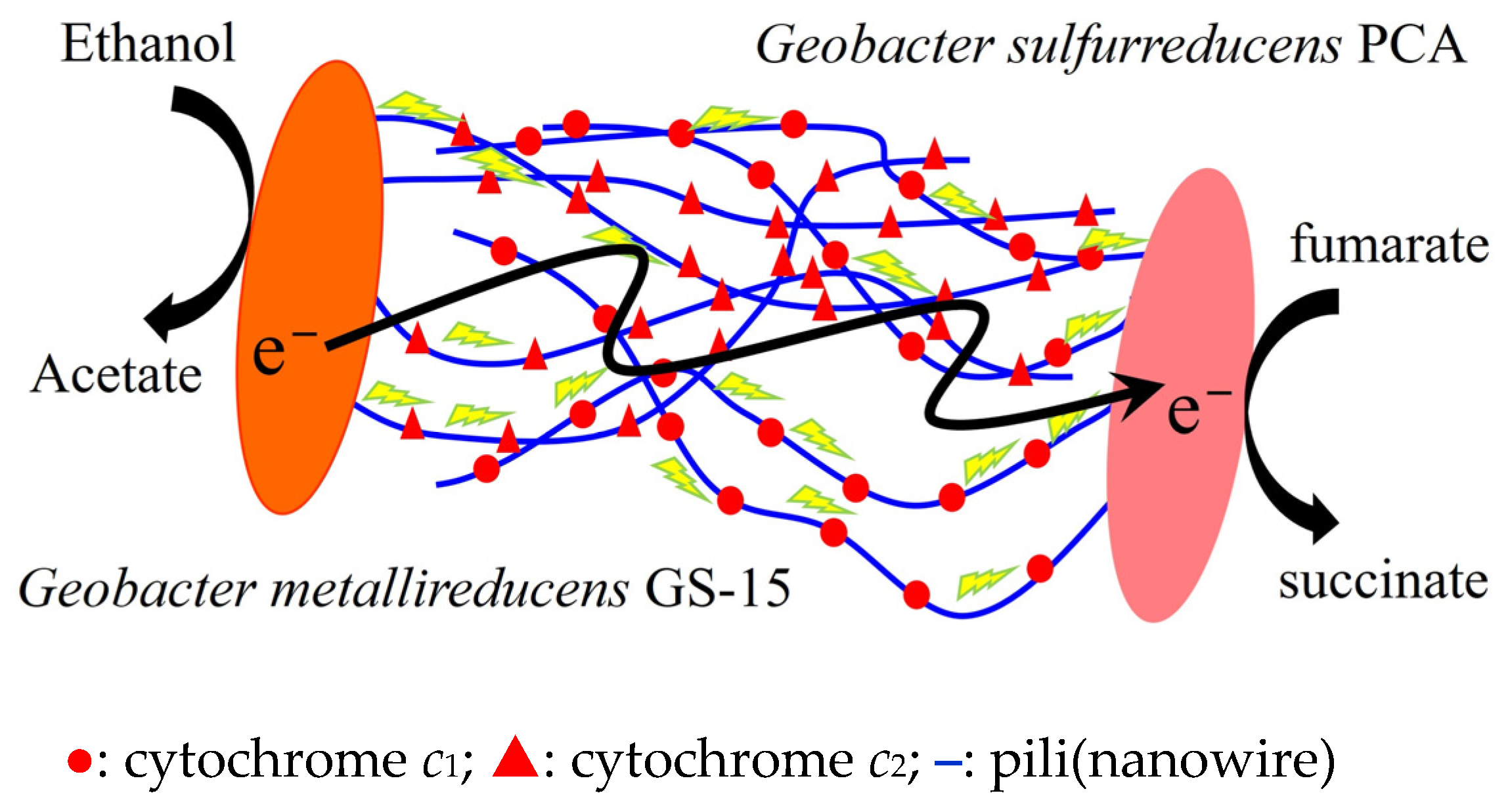
Figure 1. Model for direct interspecies electron transfer between G. metallireducens GS-15 and G. sulfurreducens PCA adapted from Lovley [26].
Ijms 26 04195 g001
Figure 2. Proposed model for electron and proton flux for carbon dioxide reduction to methane in M. barkeri 800 during DIET-based growth. (A) adapted from Holmes et al. [35]. (B) adapted from Huang et al. [28]. H4MPT, tetrahydromethanopterin; MFR, methanofuran; Mtr, Na+-translocating methyl-H4MPT:coenzyme M methyltransferase; CoM-S-S-CoB, heterodisulfide of coenzyme M and coenzyme B; CoB-SH, coenzyme B; CoM-SH, coenzyme M; Fdred, one-electron reduced ferredoxin; Fdred2−, fully reduced ferredoxin; Fdox, oxidized ferredoxin; F420, oxidized coenzyme F420; F420H2, reduced coenzyme F420; MP, oxidized methanophenazine; MPH2, reduced methanophenazine; HdrABC, cytoplasmic heterodisulfide reductase; Mcr, methyl-coenzyme M reductase; Fpo, F420H2 dehydrogenase; Ech, ferredoxin-dependent hydrogenase; VhtACG, methanophenazine-reducing hydrogenases; A1A0ATPase, ATP synthase; Nha, Na+/H+ antiporter; n, the amount of F420H2 generated by Fpo (2 < n < 3); “?”, it indicates uncertainty.
Ijms 26 04195 g002
Figure 3. Proposed model for electron and proton flux in M. acetivorans strain WWM1(Δhpt) during DIET with G. metallireducens GS-15, adapted from Holmes et al. [31]. Ethanol is provided as the source of electrons. H4MPT, tetrahydromethanopterin; MFR, methanofuran; Mtr, Na+-translocating methyl-H4MPT:coenzyme M methyltransferase; CoM-S-S-CoB, heterodisulfide of coenzyme M and coenzyme B; CoB-SH, coenzyme B; CoM-SH, coenzyme M; Fdred2−, fully reduced ferredoxin; Fdox, oxidized ferredoxin; F420, oxidized coenzyme F420; F420H2, reduced coenzyme F420; MP, oxidized methanophenazine; MPH2, reduced methanophenazine; HdrED, heterodisulfide reductase; Mcr, methyl-coenzyme M reductase; Fpo, F420H2 dehydrogenase; A1A0ATPase, ATP synthase; MmcA, outer-surface multiheme c-type cytochrome; MrpA-G, H+/Na+ antiporter complex; Rnf, Na+-translocating ferredoxin: methanophenazine reductase complex.
Ijms 26 04195 g003
Figure 4. Proposed pathway for carbon dioxide reduction in M. thermoacetophila during DIET-based growth, adapted from Zhou et al. [23]. MspA, main sheath fiber protein; SpqAB, surface quinoprotein complex; PQQH2, reduced pyrroloquinoline quinone;H4MPT, tetrahydromethanopterin; MFR, methanofuran; Mtr, Na+-translocating methyl-H4MPT:coenzyme M methyltransferase; CoM-S-S-CoB, heterodisulfide of coenzyme M and coenzyme B; CoB-SH, coenzyme B; CoM-SH, coenzyme M; Fdred, one electron reduced ferredoxin; Fdox, oxidized ferredoxin; F420, oxidized coenzyme F420; F420H2, reduced coenzyme F420; MP, oxidized methanophenazine; MPH2, reduced methanophenazine; HdrABC, heterodisulfide reductase subunits A, B, C; HdrED, heterodisulfide reductase subunits E and D; FrhB, subunit B of F420-reducing hydrogenase; Mcr, methyl-coenzyme M reductase; Fpo, F420H2 dehydrogenase lacking subunit F; A1A0ATPase, ATP synthase; Fdh, formate-dehydrogenase complex; Fmd, formylmethanofuran dehydrogenase complex.
Ijms 26 04195 g004
Figure 5. Electron transfer reactions catalyzed by HdrA2B2C2 [47,48]. HdrA2B2C2, cytoplasmic heterodisulfide reductase subunits A2, B2, C2; CoM-S-S-CoB, heterodisulfide of coenzyme M and coenzyme B; CoB-SH, coenzyme B; CoM-SH, coenzyme M; Fdred2−, fully reduced ferredoxin; Fdox, oxidized ferredoxin; F420, oxidized coenzyme F420; F420H2, reduced coenzyme F420.
Ijms 26 04195 g005
Figure 6. Proposed scheme for the reduction of Fdox and CoM-S-S-CoM with F420H2 through cytoplasmic HdrABC-FrhB complex [55]. Fdred2−, fully reduced ferredoxin; Fdox, oxidized ferredoxin; F420, oxidized coenzyme F420; F420H2, reduced coenzyme F420; FrhB, subunit B of F420-reducing hydrogenase; CoM-S-S-CoB, heterodisulfide of coenzyme M and coenzyme B; CoM-SH, coenzyme M; CoB-SH, coenzyme B; HdrABC, heterodisulfide reductase subunits A, B, C; FAD, flavin adenine dinucleotide; FeS, iron-sulfur cluster.
Ijms 26 04195 g006
Figure 7. Proposed model for electron and proton flux for carbon dioxide reduction to methane in M. harundinacea 6Ac during DIET with G. metallireducens GS-15 based on the results of previous studies [19,23,38,43,44,45,53,55,56,57]. Cyt c, c-type cytochrome; H4MPT, tetrahydromethanopterin; MFR, methanofuran; Mtr, Na+-translocating methyl-H4MPT:coenzyme M methyltransferase; CoM-S-S-CoB, heterodisulfide of coenzyme M and coenzyme B; CoB-SH, coenzyme B; CoM-SH, coenzyme M; Fdred2−, fully reduced ferredoxin; Fdox, oxidized ferredoxin; F420, oxidized coenzyme F420; F420H2, reduced coenzyme F420; MP, oxidized methanophenazine; MPH2, reduced methanophenazine; HdrABC, heterodisulfide reductase subunits A, B, C; HdrED, heterodisulfide reductase subunits E and D; FrhB, subunit B of F420-reducing hydrogenase; Mcr, methyl-coenzyme M reductase; Fpo, F420H2 dehydrogenase (Fpo) lack of subunit F; A1A0ATPase, ATP synthase; Nha, Na+/H+ antiporter; Fdh, formate-dehydrogenase complex; Fmd, formylmethanofuran dehydrogenase complex; n, the amount of F420H2 generated by combining Fmd and Fdh with Fdred2− (0 ≤ n < 2); “?”, it indicates uncertainty.
Ijms 26 04195 g007
Table 1. The key differences between the multiple mechanisms of extracellular electron uptake in reported methanogens during DIET-based growth.
MicrobesExtracellular Electron UptakeMP
Reduction
F420 ReductionCoM-S-S-CoB
Reduction
Fdox ReductionReference
M. barkeri 800UnknowUnknowFpo with MPH2HdrABC with F420H2, HdrED with MPH2,
interaction of VhtACG and HdrED with H2
HdrABC with F420H2,
Ech direct with
extracellular electron,
Ech with H2 from
oxidation of F420H2 by FrhABG
[28,35,36]
M. acetivoransMmcAMmcA, Rnf with electrons from MmcAFpo with MPH2HdrED with MPH2Rnf with electrons from MmcA[31]
M. thermoacetophilaMspASpqAB with PQQH2Fpo-FrhB complex with MPH2, interaction of Fmd and Fdh with Fdred2−HdrABC with F420H2 and HdrED with MPH2HdrABC with F420H2 and oxidation of
acetate
[23]
M.
harundinacea 6Ac
UnknowCyt c?HdrABC-FrhB complex with Fdred2−, CoM-SH and CoB-SHHdrED with MPH2Fpo with MPH2This article
Disclaimer/Publisher’s Note: The statements, opinions and data contained in all publications are solely those of the individual author(s) and contributor(s) and not of MDPI and/or the editor(s). MDPI and/or the editor(s) disclaim responsibility for any injury to people or property resulting from any ideas, methods, instructions or products referred to in the content.