Review

The Role of Tetrahydrocurcumin in Tumor and Neurodegenerative Diseases Through Anti-Inflammatory Effects

1
Translational Chinese Medicine Key Laboratory of Sichuan Province, Sichuan Institute for Translational Chinese Medicine, Sichuan Academy of Chinese Medicine Sciences, Chengdu 610041, China
2
Sichuan Institute for Translational Chinese Medicine, Chengdu 610041, China
3
West China Hospital, Sichuan University, Chengdu 610041, China
4
School of Medical and Life Sciences, Chengdu University of Traditional Chinese Medicine, Chengdu 611137, China
*
Authors to whom correspondence should be addressed.
Int. J. Mol. Sci. 2025, 26(8), 3561; https://doi.org/10.3390/ijms26083561
Submission received: 24 February 2025 / Revised: 24 March 2025 / Accepted: 24 March 2025 / Published: 10 April 2025
(This article belongs to the Section Molecular Neurobiology)

Abstract

Tetrahydrocurcumin (THC), a curcumin derivative, shows potential in oncology and neurology. It regulates NF-κB, reduces inflammation, promotes cancer cell apoptosis, inhibits tumor angiogenesis, and enhances antioxidants, aiding in treating inflammation-related cancers. In neurology, THC’s anti-inflammatory and antioxidant properties protect neurons, reduce neuroinflammation, and support autophagy for cellular debris clearance, with its blood–brain barrier penetration offering a neuroprotective edge. Research on THC’s therapeutic application must focus on improving delivery and bioavailability and confirming its clinical safety and efficacy.

1. Introduction

Tetrahydrocurcumin (THC), a principal metabolite of curcumin (CUR), exhibits superior bioavailability and a spectrum of pharmacological activities that surpass those of its parent compound [1]. The principal metabolic pathways of THC within the organism encompass antioxidant activity, hypoglycemic and hypolipidemic effects, along with other therapeutic functions. Furthermore, THC demonstrates neuroprotective potential, exemplified by its capacity to modulate the Ras/ERK signaling pathway, thereby alleviating cell cycle arrest and inhibiting apoptosis of microglia in Alzheimer’s disease models [2,3,4,5]. Regarding the bioavailability of THC, studies indicate that its solid dispersion formulation significantly enhances bioavailability in mice compared to the compound in its isolated form [6,7]. This enhancement is attributed to a modest extension in mean residence time (MRT) and elimination half-life (t1/2), coupled with a marked reduction in the time to reach peak concentration (tmax), thereby augmenting plasma concentrations of THC [8]. This finding holds profound implications for elevating the clinical utility of THC, as it may amplify its efficacy while minimizing the requisite dosage. However, certain studies have highlighted that THC may exhibit a “hormesis effect”, wherein low doses may foster the proliferation of tumor cells, while higher doses can suppress tumor growth and modulate the tumor microenvironment. As a metabolite of curcumin, THC retains the diverse biological activities of its precursor and further potentiates its pharmacological effects by enhancing bioavailability [9,10,11]. Despite potential adverse effects, the therapeutic potential of THC can be optimized through precise dosage control and the application of suitable administration modalities. Future research endeavors should delve deeper into the safety and efficacy of THC to harness the full potential of this promising therapeutic agent.
Inflammation assumes a multifaceted role in the genesis of tumors and neurodegenerative diseases, implicating cytokines, immune cells, and the intricate network that interacts with these cellular and molecular elements [12,13,14]. Persistent inflammatory responses, stemming from chronic inflammation or tissue injury, foster cellular transformation via genetic damage or pro-inflammatory agents, thereby precipitating chronic inflammation and tumorigenesis [15,16]. Tumor-extrinsic inflammation arises from a variety of factors, including bacterial and viral infections, autoimmune disorders, obesity, smoking, asbestos exposure, and excessive alcohol consumption, all of which elevate the risk of cancer and spur malignant progression [17,18,19,20]. Inflammation is not only correlated with the emergence of tumors but also exerts a significant influence on their progression. For instance, inflammatory cells within the tumor microenvironment, such as macrophages and microglia, promote the proliferation, invasion, and metastasis of tumor cells by secreting inflammatory mediators like interleukins and tumor necrosis factors [21,22,23,24,25]. Furthermore, inflammation impacts immune surveillance and responsiveness to therapy. Tumors associated with inflammation typically exhibit enhanced angiogenic potential, facilitating the survival and dissemination of tumor cells. For example, mast cells promote angiogenesis in squamous epithelial carcinogenesis by activating specific enzymes. Moreover, inflammation can also foster drug resistance in tumor cells by altering the tumor microenvironment [26,27,28,29].
In the context of neurodegenerative afflictions, pivotal inflammatory mediators, including IL-1β and IL-18, are instrumental in the subsequent release post-neural injury [30]. Such inflammatory mediators are capable of instigating an immune response within the nervous system, culminating in neuronal demise and impaired functionality [31]. The NLRP3 inflammasome constitutes a vital element of the innate immune apparatus, with its activation precipitating neuroinflammation—a critical factor in the etiology and advancement of neurodegenerative pathologies such as Alzheimer’s, Parkinson’s, amyotrophic lateral sclerosis, and multiple sclerosis [32,33]. Empirical research indicates that persistent infections of bacterial, viral, and fungal origins may be contributory risk factors for the onset of neurodegenerative diseases. Such infections are capable of inducing chronic neuroinflammation, which can precipitate metabolic disturbances and a spectrum of other pathological conditions [34,35,36]. Furthermore, inflammation can intensify the trajectory of neurodegenerative diseases by modulating the immune response and the mechanisms governing cell death within the nervous system.
To encapsulate, inflammation exerts a complex and pivotal influence on the evolution of both oncological and neurodegenerative conditions. It partakes not solely in the incipient phases of these pathologies but also significantly contributes to their progression and exacerbation [37,38]. Consequently, therapeutic interventions that target inflammatory mechanisms, including the employment of anti-inflammatory pharmaceuticals or alternative modalities, may offer innovative strategies for the prophylaxis and amelioration of such afflictions. As a burgeoning anti-inflammatory and neuroprotective entity, the prospective applications of THC in the mitigation of inflammation-driven oncogenesis and neurodegenerative disorders warrant additional investigation. This treatise elucidates the role of THC in addressing inflammation-induced carcinogenesis and neurodegeneration, with the ambition of furnishing direction for the more efficacious development and application of this compound.

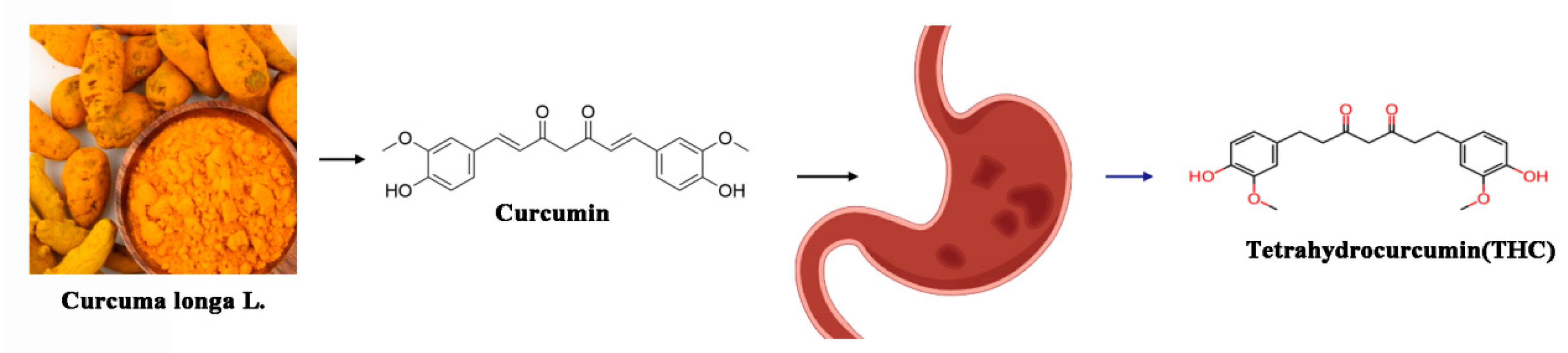
2. Chemical and Biological Properties of THC

2.1. Chemical Structure of THC Compared with Curcumin

THC serves as the principal metabolite of curcumin (CUR) in the human body, exhibiting superior antioxidant efficacy and bioavailability through gastrointestinal absorption compared to its precursor. Curcumin, a lipophilic bioactive polyphenolic compound with the molecular formula C21H20O6 (depicted in Figure 1), is derived from the rhizomes of Curcuma longa, a revered medicinal plant in traditional Chinese medicine. In contrast to curcumin, the chemical architecture of THC arises from the saturation of methylene bridges within the β-diketone core of curcumin through hydrogenation, yielding the THC molecule [39,40].
The synthetic routes to THC commence with vanillin and acetone, employing Claisen condensation to synthesize curcumin, which is subsequently subjected to a palladium-catalyzed hydrogenation process to yield THC. Furthermore, microbial transformation techniques are harnessed to alter the molecular scaffold of curcumin, efficiently transforming it into an array of modified derivatives, among which THC is notably included [41].
In contrast to curcumin, THC demonstrates enhanced pharmacological potency, particularly in its antioxidant, hypoglycemic, and hypolipidemic capabilities [39,42]. This enhanced activity can be attributed to the improved stability and bioavailability of THC within the physiological milieu, thereby conferring more robust efficacy in these domains [11]. Furthermore, THC manifests neuroprotective properties, adept at mitigating G1/S cell cycle arrest in Alzheimer’s disease paradigms and modulating microglial cell cycling. It also downregulates the expression of tumor necrosis factor α, upregulates TGF-β1 levels, and synergistically impedes both cell cycle stagnation and apoptosis [43,44,45]. To encapsulate, as the principal metabolite of curcumin, THC maintains the diverse biological activities of its precursor while demonstrating superior performance in specific aspects, including its heightened antioxidant potential and enhanced bioavailability. These attributes render THC an asset in the realms of medical and health product development.

2.2. Solubility, Stability, and Bioavailability of THC

As the principal metabolite of curcumin within the body, THC possesses more robust antioxidant activity and gastrointestinal absorption than curcumin, along with an array of biological activities that surpass those of its precursor. Nonetheless, the solubility, stability, and bioavailability of curcumin and its derivatives, including THC, represent pivotal factors that constrain their clinical utility [39]. Curcumin, a naturally occurring pigment characterized by low solubility and instability, can undergo enhancement of its aqueous solubility and stability through techniques such as solid dispersion and microencapsulation [46]. The bioavailability of THC can be augmented via a spectrum of pharmaceutical delivery systems, encompassing nanoparticles, liposomes, micelles, self-microemulsions, solid dispersions, and phospholipids. Empirical studies have demonstrated that solid dispersions of THC can markedly enhance the bioavailability in mice when administered via gavage, as opposed to the administration of THC in its unadulterated form. Furthermore, refining the biotransformation process to optimize THC yield can substantially augment production, thereby indirectly enhancing bioavailability. The solubility, stability, and bioavailability of THC can be ameliorated through a multitude of approaches. Such enhancements not only bolster its in vivo efficacy but also broaden its applicability within the domains of food and medicinal sciences. Future investigative endeavors should delve into diverse solubilization and stabilization methodologies to harness its utmost potential for clinical deployment [11,47].

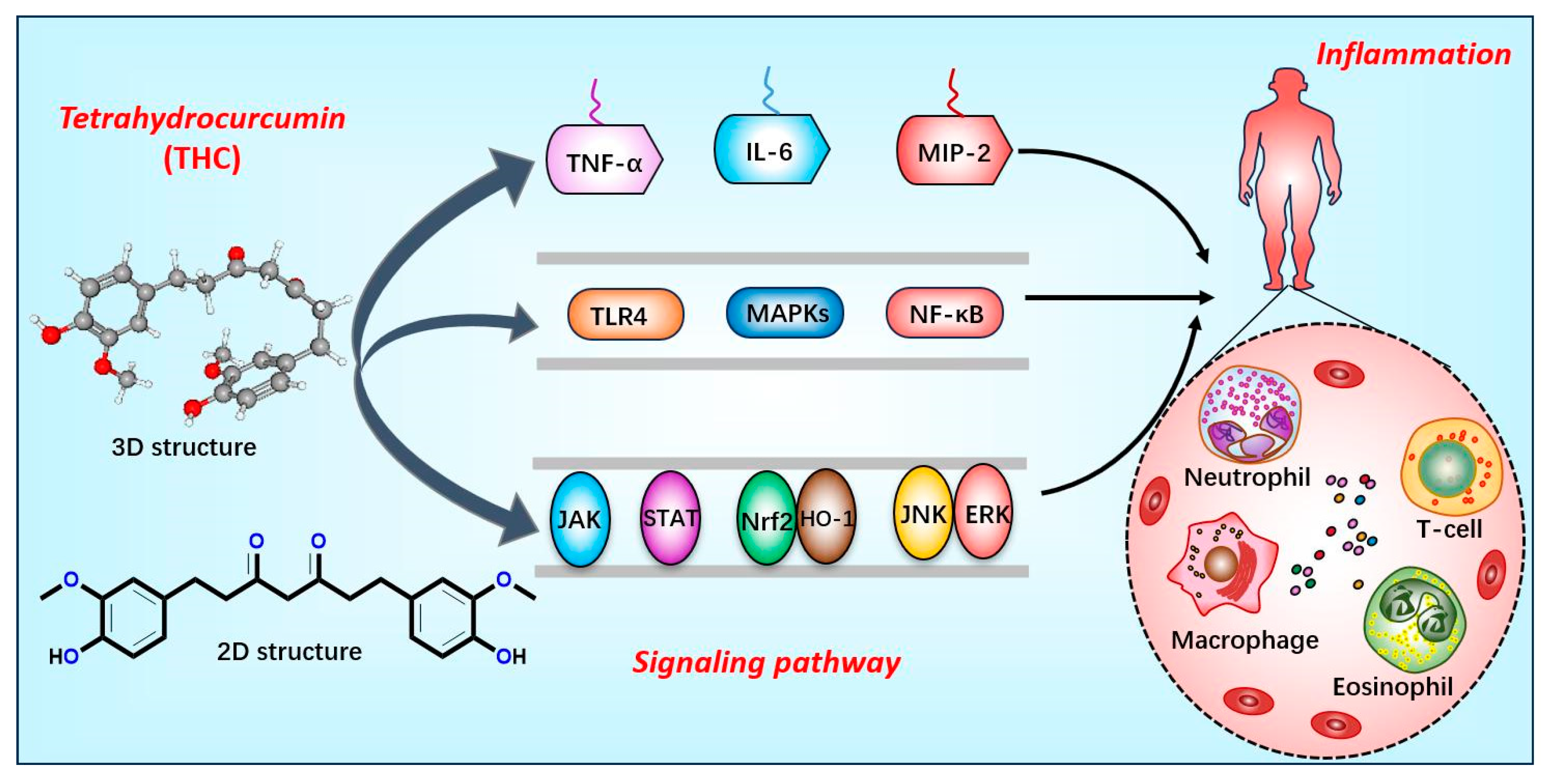
2.3. Anti-Inflammatory Mechanisms of THC

2.3.1. Inhibiting Inflammatory Factors and Signaling Pathways

THC potently curbs the genesis of a spectrum of inflammatory cytokines, primarily through the modulation of the expression of a plethora of inflammatory mediators and signaling cascades, including TNF-α, IL-6, and MIP-2. Furthermore, THC is capable of attenuating inflammatory responses via a multitude of distinct mechanisms [11,48,49]. Additionally, THC efficaciously mitigates the excessive secretion of MCP-1 and IL-6 in vascular smooth muscle cells incited by lipopolysaccharide (LPS), in addition to dampening the heightened expression of TLR4 and decreasing the phosphorylation levels within the MAPKs signaling pathway [50]. Collectively, these observations suggest that THC modulates the expression of inflammatory factors by impacting particular signaling pathways, namely TLR4, MAPKs, and NF-κB. Moreover, THC can diminish inflammation by impeding the activation of the JAK/STAT, Nrf2/HO-1, and JNK/ERK signaling pathways [50,51,52]. Within specific experimental paradigms, THC has also manifested inhibitory actions on the NF-κB/VEGF/MMP-9 signaling axis, thus ameliorating cerebral edema and inflammation induced by acute hypobaric hypoxia [53]. These revelations lay a theoretical foundation for the continued exploration of the therapeutic application of THC in the context of inflammation-associated diseases (Figure 2).

2.3.2. Improved Vascular Function and Structure

THC ameliorates hypertension, arteriosclerosis, and vascular remodeling induced by cadmium, conditions frequently linked to a proliferation of smooth muscle cells, excessive collagen deposition, and heightened matrix metalloproteinase (MMP)-2 and MMP-9 levels. THC exerts a protective effect by augmenting the bioavailability of nitric oxide (NO), mitigating oxidative stress, and facilitating the amelioration of vascular remodeling processes [54,55].

2.3.3. Antioxidant Effect

When inflammation occurs, the production of free radicals such as reactive oxygen species (ROS) and active nitrogen (RNS) in the body increases, and the antioxidant defense system may not be able to effectively neutralize these free radicals, leading to oxidative stress. Oxidative stress damages cellular components such as DNA, proteins, and lipids, which in turn triggers and maintains an inflammatory response [56]. Antioxidants are able to neutralize free radicals and reduce oxidative stress, thereby regulating the inflammatory response to some extent. Antioxidants such as vitamin C, vitamin E, beta-carotene, and selenium, for example, can reduce the damage to cells caused by oxidative stress, which in turn relives inflammatory symptoms [56].
THC functions as a robust antioxidant, curtailing the generation of reactive oxygen species (ROS) and mitigating the oxidative stress and fibrosis engendered by elevated glucose levels through the activation of the SIRT1 signaling pathway. Furthermore, THC augments the capacity to withstand hypoxia by bolstering superoxide dismutase (SOD) activity [42,53,57].

2.3.4. Modulating Immune Response

THC modulates allergic responses in asthmatic mice through the regulation of the gut microbiome. Moreover, THC diminishes the synthesis of platelet thromboxane A2 (TxA2) by impeding the MAPKs/cPLA2 signaling pathway, consequently mitigating the occurrence of thrombosis [58].

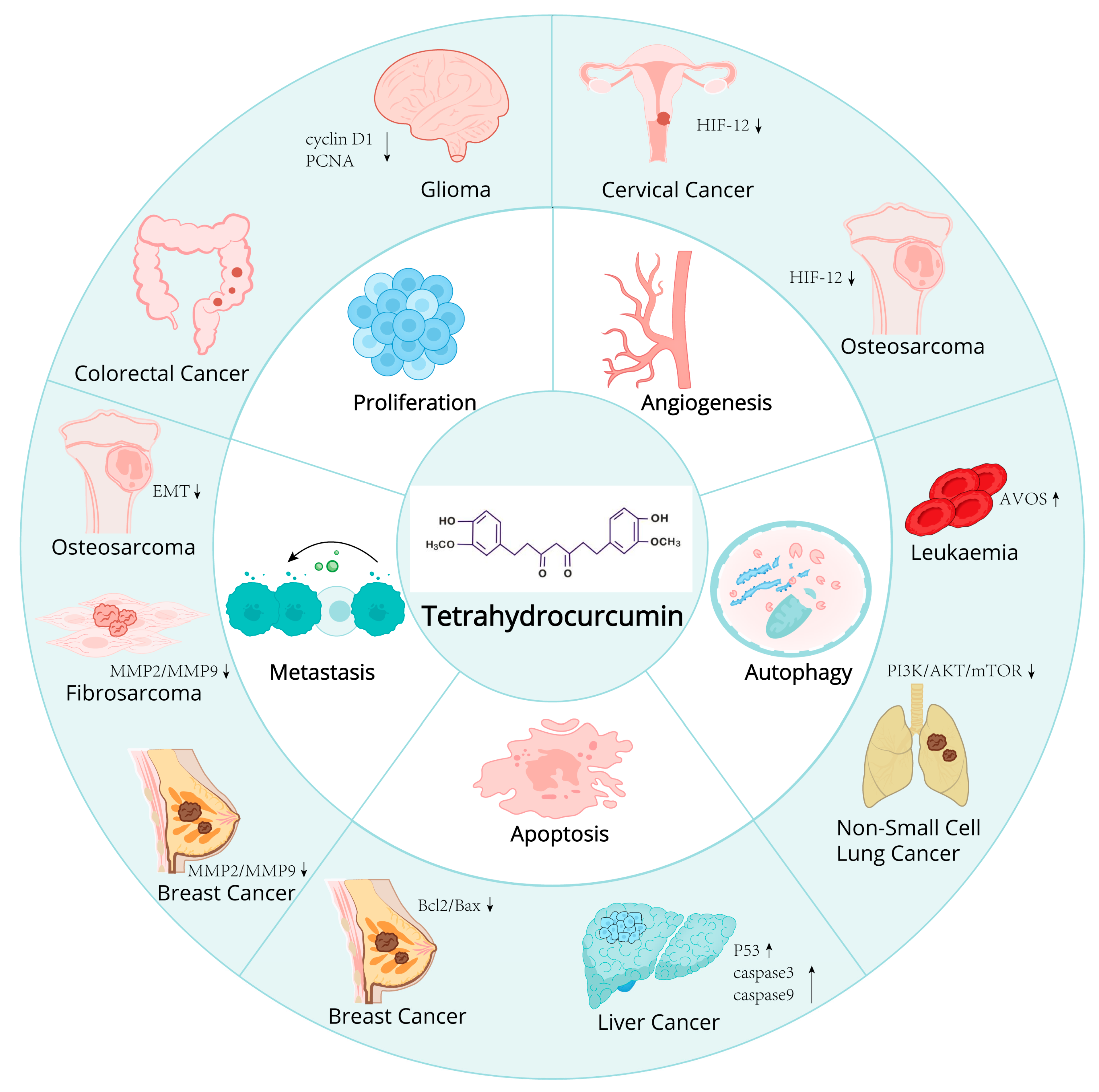
2.4. Potential of THC in Cancer Prevention and Therapy

2.4.1. Mechanism of Action of THC on Cancer Cells

Anti-Proliferative Effect
THC possesses the capacity to inhibit the proliferation of tumor cells. Studies have demonstrated that THC exerts a discernible inhibitory effect on breast cancer cells MCF-7; its impact on the proliferative capacity of MCF-7 cells, as ascertained by CCK8 assays and clonal formation tests, suggests a potential anti-proliferative action. Furthermore, investigations into CT26 colorectal cancer cells have illustrated that THC can markedly suppress both the quantity and dimensions of tumor cell clonal formations in a concentration-dependent fashion [59,60].
Proapoptotic Effect
THC facilitates the apoptosis of tumor cells via multiple mechanisms. A study has reported that THC can diminish the expression of Bcl-2/Bax proteins, thereby triggering the apoptosis of tumor cells. Subsequent research indicated that treatment with THC induced morphological alterations in CT26 colorectal cancer cells, culminating in cellular apoptosis. This treatment also notably increased the expression of BAX and activated caspase 3, while decreasing the expression of BCL-2 protein. These findings imply that THC advances the apoptotic process in tumor cells by modulating the expression of pivotal proteins associated with apoptosis [9,59].
Anti-Angiogenic Effect
THC also exerts anti-angiogenic effects, playing a pivotal role in the suppression of tumor growth and metastasis. Bhornprom Yoysungnoe et al. have demonstrated that the combinatorial treatment with THC and celecoxib leads to the inhibition of tumor growth and angiogenesis through the downregulation of VEGF, COX-2, and EGFR expression. Nonetheless, this combined therapeutic approach did not manifest a synergistic effect on curbing tumor growth and angiogenesis within the cervical cancer (CaSki)-implanted nude mice model [61]. Pornprom Yoysungnoen et al. investigated the anti-cancer and anti-angiogenic properties of THC in a model of implanted hepatocellular carcinoma in nude mice. Their findings indicate the presence of pathological angiogenic characteristics, such as microvascular dilation, tortuosity, and hyperpermeability. Both curcumin (CUR) and THC were found to mitigate these pathological features. In the HepG2 groups, the chorioallantoic membrane (CAM) vascularity index (CV) was significantly elevated on days 7 (52.43%), 14 (69.17%), and 21 (74.08%) compared to the control group (33.04%, p < 0.001). Administration of CUR and THC led to a substantial reduction in the CV, with respective p-values of <0.005 and <0.001. Notably, the anti-angiogenic effects of both CUR and THC exhibited a dose-dependent response. Nonetheless, a more pronounced beneficial effect of THC over CUR treatment was discernible, particularly in the CV measurements on day 21 (44.96% and 52.86%, respectively, p < 0.0575) [62] (Figure 3 and Table 1).

2.4.2. Research Progress of THC in Different Cancer Models

Cervical Cancer
Cervical cancer represents the most prevalent malignant gynecological tumor. Investigations have revealed that THC possesses the capacity to inhibit tumor angiogenesis [63,64]. In vivo experimentation involved administering oral doses of 100, 300, and 500 mg/kg of THC to mice daily for 30 days. The findings indicated that THC notably suppressed tumor angiogenesis, as well as tumor volume and growth rate, in the treatment cohort. This inhibitory effect is likely mediated through the downregulation of Hypoxia-Inducible Factor-1 alpha (HIF-1-α).
Breast Cancer
Breast cancer, a malignancy arising in the epithelial tissues of the breast, is witnessing a rising incidence and affecting increasingly younger demographics annually. Breast cancer cells readily diminish in their intercellular adhesion capabilities, proliferate and disseminate with ease, swiftly metastasize to organs including bone marrow and lymph nodes, and ultimately pose a significant threat to patients’ lives [65]. Researchers discovered that THC exerted a discernible inhibitory effect on the in vitro proliferation of MCF-7 cells, and that THC administration could facilitate the apoptosis of these cells and diminish their metastatic potential. The underlying mechanism is likely the downregulation of Bcl-2/Bax protein expression to trigger tumor cell apoptosis, coupled with the suppression of matrix metalloproteinases MMP-2 and MMP-9 expression to impede cell metastasis [66,67]. Concurrently, Ning Kang et al. also determined that THC significantly restrained cell growth by instigating mitochondrial-mediated apoptosis and inducing G2/M cell cycle arrest in MCF-7 cells. Concurrently, they also determined that THC significantly restrained cell growth by instigating mitochondrial-mediated apoptosis and inducing G2/M cell cycle arrest in MCF-7 cells [67].
Liver Cancer
Hepatocellular carcinoma (HCC) poses a significant global health challenge, characterized by a high incidence and propensity for progression. It is also the predominant cause of mortality among individuals with cirrhosis. The researchers have demonstrated that THC markedly enhances the survival rate of mice bearing hepatocellular carcinoma in in vivo assays, suppresses the proliferation of HCC cells, and diminishes the ascites volume and abdominal girth associated with liver cancer [68]. Mechanistic studies have revealed that THC significantly activates and induces the cleavage of caspase-3 and caspase-9, elevates the expression of the p53 gene, facilitates the apoptosis of HT22 liver cancer cells, and exerts antitumor effects. Additionally, Pornprom Yoysungnoen et al. discovered that THC impedes the proliferation of liver cancer cells by exerting anti-angiogenic properties [62].
Acute Myeloid Leukemia
Acute leukemia represents the predominant form of cancer among children, comprising 26% of total cancer diagnoses and 20% of cancer-related fatalities in the pediatric population. The majority of anti-cancer agents are designed to induce cancer cell death, yet, the emergence of multidrug resistance frequently culminates in chemotherapy failure. Studies have revealed that THC primarily elicits the demise of drug-resistant HL60 cells via the induction of apoptosis and autophagy [69]. Concurrently, THC triggers autophagic cell death in HL-60 promyelocytic leukemia cells by augmenting the development of acidic vascular organelles (AVOs), which serve as markers of autophagy [70]. These findings suggest that THC harbors potential therapeutic value in the treatment of acute myeloid leukemia.
Fibrosarcoma
Fibrosarcoma is a malignant tumor that originates from fibroblasts and is characterized by the production of collagen fibers. It is commonly observed in young adults, exhibiting rapid growth and a propensity for hematogenous metastasis. Research has demonstrated that an escalation in THC treatment concentration correlates with a significant diminution in the invasive and migratory capabilities of HT1080 human fibrosarcoma cells [71]. Additionally, THC diminished cellular adhesion to both matrix and lamin-coated surfaces. Enzymatic assessments revealed that THC treatment led to a reduction in the levels of matrix metalloproteinases 2 and 9 (MMP-2, MMP-9), as well as urokinase plasminogen activator (uPA). THC exerts an inhibitory effect on the expression of matrix metalloproteinase1 (MT1-MMP) and MMPs and is a tissue inhibitor of metalloproteinases 2 (TIMP-2) proteins. The findings collectively indicate that THC can markedly suppress the metastatic potential of fibrosarcoma cells in vitro.
Colon Cancer
Colon cancer is a prevalent malignant neoplasm of the digestive tract, specifically arising in the colon, and is the third most frequently occurring gastrointestinal tumor. Research has determined that the in vitro IC50 value of THC for human colorectal adenocarcinoma cells (HT-29) is 28.67 ± 1.01 μg/mL, and the inhibitory selectivity index of THC for HT-29 cells is fourfold higher compared to that for normal colorectal epithelial cells [72]. Concurrently, THC demonstrates a significant chemopreventive efficacy against azoxymethane (AOM)-induced colon carcinogenesis in murine models [60]. Collectively, these findings substantiate that THC exerts a favorable chemopreventive influence on the genesis of colon tumors.
Osteosarcoma
Osteosarcoma represents the most prevalent primary malignant bone tumor among children and adolescents. Nonetheless, the absence of early biomarkers renders it challenging to detect, resulting in the majority of patients being diagnosed with high-grade malignancies. Human osteosarcoma is classified as a malignant tumor with a poor prognosis and a propensity for metastasis. Research has identified that THC can markedly diminish the proliferation of osteosarcoma cells and curb their migration and invasion within a nude mouse model of lung metastasis [73]. Furthermore, THC fosters the mesenchymal-to-epithelial transition (MET) process. Studies have illuminated that hypoxia-inducible factor-1 alpha (HIF-1α) is pivotal in the anti-metastatic effects of THC. Notably, THC downregulates HIF-1α expression by impeding the Akt/mTOR and p38 MAPK signaling pathways. Moreover, THC substantially suppressed HIF-1α expression and angiogenesis under hypoxic conditions. THC activates autophagy, induces MET, and curbs angiogenesis, mechanisms that are interrelated with HIF-1α regulation.
Non-Small Cell Lung Cancer
Lung cancer is the predominant cause of cancer-related mortality globally. The most prevalent variant of lung cancer is non-small cell lung cancer (NSCLC). Presently, therapeutic options for non-small cell lung cancer are rather constrained, encompassing chemotherapy, radiation therapy, surgical resection, and targeted therapies. The study has discovered that THC can suppress the growth and proliferation of A549 cells. THC treatment significantly augments autophagy [74]. Reverse transcription quantitative polymerase chain reaction (RT-qPCR) analysis indicated an elevation in Beclin-1 expression following THC treatment. The ratio of microtubule-associated protein 1A/1B-light chain 3 (LC3)-II to LC3-I was diminished. Cellular protein expression levels of diverse autophagy markers, including p62, phosphorylated mammalian target of rapamycin (p-mTOR), phosphatidylinositol-3 kinase (PI3K), phosphorylated PI3K (P-PI3K), protein kinase B (Akt), and phosphorylated Akt (P-Akt), were substantially decreased. These findings suggest that THC induces autophagy by impeding the PI3K/Akt/mTOR signaling pathway, thereby suppressing the progression of NSCLC.
Glioma
Glioma is among the most prevalent and malignant brain tumors within the central nervous system. Currently, radiotherapy remains the most efficacious treatment modality for glioma. Regrettably, the low radiosensitivity of tumor cells often leads to unsatisfactory radiotherapy outcomes, with gliomas having dismal relative survival rates [76]. Utilization of radiosensitizers can elevate the rate of glioma cell death, thereby enhancing the therapeutic efficacy of radiotherapy. Studies have revealed that, in comparison to the radiotherapy-only group, there was a marked reduction in tumor cell viability and an increase in the rate of apoptosis within the group administered THC in conjunction with radiotherapy. Cells treated with combined THC and radiation demonstrated lower cell viability and a higher apoptosis rate compared to the radiation group. Moreover, the intracellular GSH was also decreased in the THC co-treated C6 cells. More importantly, the combinatorial treatment group significantly induced G0/G1 cell cycle arrest and a decrease in the S phase cell through the downregulation of cyclin D1 and PCNA. The in vivo therapeutic efficacy assay indicated that the growth of the tumor was greatly inhibited in the combinatorial group [76]. Furthermore, the intracellular glutathione (GSH) levels in C6 cells treated with THC were also observed to diminish. Most importantly, the combination therapy group significantly induced G0/G1 cell cycle arrest and a reduction in the S-phase cell population through the downregulation of Cyclin D1 and proliferating cell nuclear antigen (PCNA) expression. These findings suggest that THC can act synergistically to augment the radiosensitivity of glioma cells.

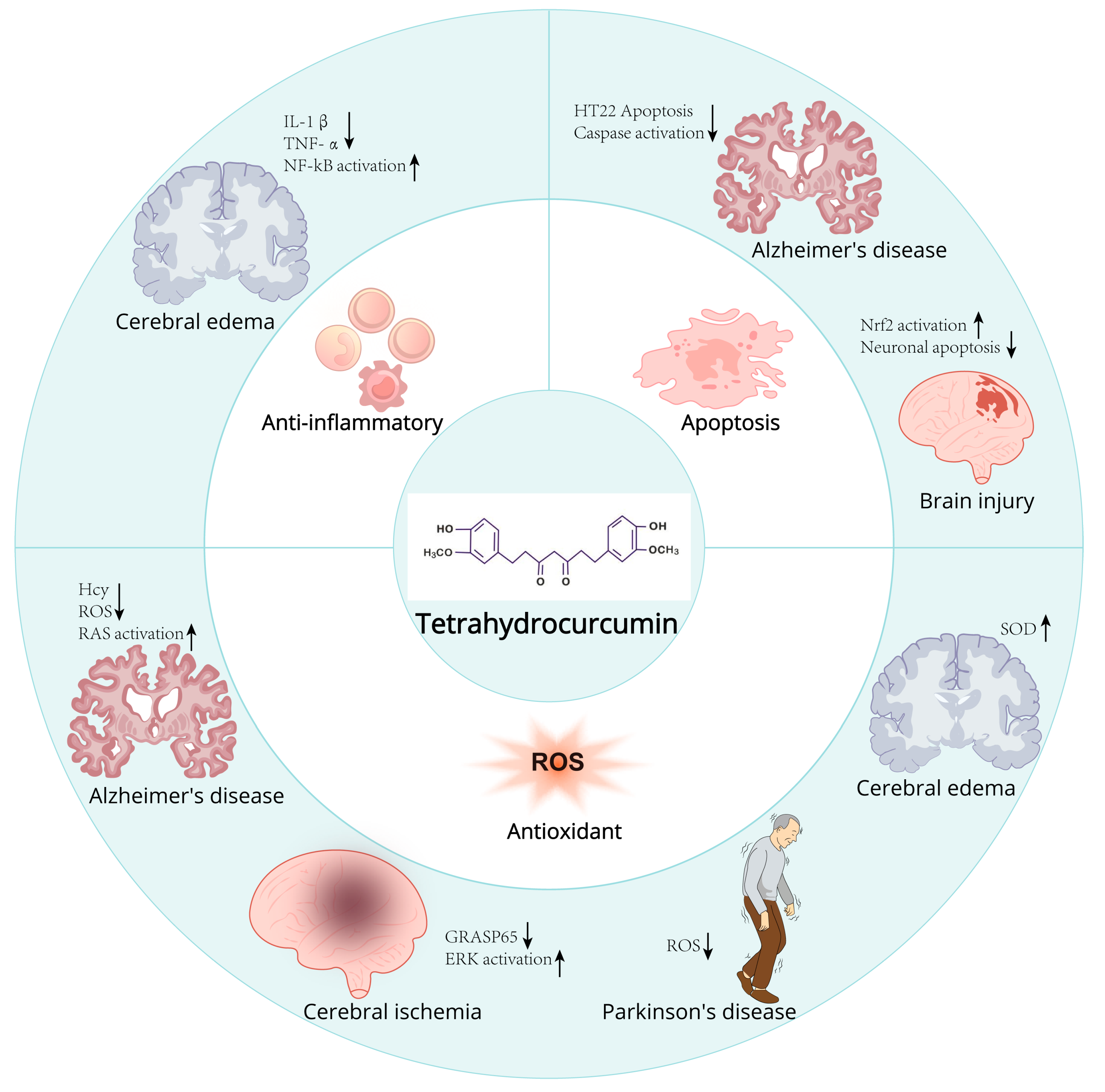
2.5. Protective Effects of THC in Neurodegenerative Diseases

2.5.1. Potential Mechanisms of THC on Neuroprotection and Repair

Antioxidation
THC expedites the clearance of free radicals within the body by activating a suite of antioxidant enzymes, including superoxide dismutase (SOD), glutathione peroxidase (GSH-Px), catalase (CAT), and glutathione S-transferase (GST), thereby reinforcing the stability of the brain’s antioxidant enzyme system [5]. Furthermore, THC diminishes the concentration of reactive oxygen species (ROS) and shields hippocampal cells against oxidative injury. These salutary effects contribute to the amelioration of neuronal damage instigated by free radicals and enhance the viability of neuronal cells [77,78,79].
Anti-Inflammatory Effect
THC can modulate the TLR-4/P38/MAPK signaling pathway and attenuate the levels of IL-6, TNF-α, and other inflammatory mediators, thereby dampening the inflammatory response. Moreover, THC can downregulate the expression of tumor necrosis factor α (TNF-α) and upregulate the expression of transforming growth factor β1 (TGF-β1), synergistically inhibiting cell cycle arrest and apoptosis. These multifaceted effects contribute to the enhancement of neuronal function and the reduction in neuronal mortality [49,50].
Inhibition of Apoptosis
THC can diminish the activity of caspase 3 by activating the mitogen-activated protein kinase (MAPK) signaling pathway, suppress the expression of pro-apoptotic proteins, and ultimately curb apoptosis triggered by cerebral ischemia–reperfusion injury. Furthermore, THC can ameliorate autophagy by reducing the release of cytochrome c and homocysteinylation, thereby conferring neuroprotective effects [70,79].
Other Potential Mechanisms
THC may also modulate the PI3K/Akt/mTOR signal transduction pathway, curb the accumulation of edema in the spinal cord, and mitigate inflammatory mediators, thereby effectively downregulating the gene expression of matrix metalloproteinases 3 and 13 and cyclooxygenase-2 (COX-2), facilitating the phosphorylation of Akt, augmenting the expression of forkhead box (FOX) O4 protein, and thereby achieving spinal cord protection [57]. In summary, THC can efficaciously shield neuronal cells from a spectrum of injurious stimuli through its anti-inflammatory and antioxidant properties. This neuroprotection is mediated by the activation of antioxidant enzyme systems, attenuation of inflammatory mediator levels, suppression of apoptotic pathways, enhancement of autophagy, and other related mechanisms (Figure 4 and Table 2).

2.5.2. Potential Use of THC in Neurodegenerative Diseases

Brain Injury
Traumatic brain injury (TBI) represents a significant health issue, characterized by high rates of morbidity and mortality. The neuroprotective effects of THC in experimental models of traumatic brain injury (TBI) have been established, primarily through the inhibition of oxidative stress, mitigation of mitochondrial dysfunction, and preservation against cellular apoptosis [80]. In 2018, Guan Wei et al. [81] investigated the neuroprotective efficacy of THC in rats subjected to traumatic brain injury, along with its underlying mechanisms. Post-THC administration, the researchers assessed neurological scores, cerebral water content, and neuronal degeneration in the cerebral cortex. Cerebral tissue samples were harvested subsequent to the neurological assessments for further analytical examination. The findings indicated that THC treatment could mitigate cerebral edema, reduce TBI-induced neuronal apoptosis, and enhance neurobehavioral performance. Following THC treatment, the expression of Nrf2 was found to be upregulated in response to TBI. These findings propose that THC may ameliorate neuronal function following traumatic brain injury through the activation of the Nrf2 signaling pathway.
Cerebral Edema
High-altitude cerebral edema (HACE), frequently regarded as an advanced manifestation of acute mountain sickness (AMS), affects individuals subjected to acute hypobaric hypoxia (AHH) in the absence of acclimatization [82]. HACE constitutes a grave and potentially lethal high-altitude affliction. Yang Pan et al. [53] determined that the prophylactic administration of THC (40 mg/kg) over a three-day period could markedly reduce the cerebral water content (BWC) induced by acute hypobaric hypoxia (AHH) and diminish the concentrations of interleukin-1β (IL-1β) and tumor necrosis factor α (TNF-α). Elevate the levels of superoxide dismutase (SOD) and augment the body’s hypoxia resistance. Histological and ultrastructural assessments of brain tissue revealed that THC mitigated AHH-induced pericellular edema and diminished perivascular space, thereby concurrently reducing edema and safeguarding brain mitochondria. In vitro experimentation indicated an upregulation of IL-1β expression 24 h post-implantation, succeeded by an escalation in vascular endothelial growth factor (VEGF) levels. Furthermore, THC substantially downregulated the expression of VEGF, matrix metallopeptidase-9 (MMP-9), and nuclear factor-kappa B (NF-κB) in astrocytes subjected to hypoxic conditions (4% O2). Collectively, these findings imply that THC possesses potential preventative efficacy against HACE, which is associated with the suppression of the NF-κB/VEGF/MMP-9 signaling pathway.
Cerebral Ischemia
Cerebral ischemia is a pathological phenomenon that induces damage to localized brain tissue and its functional integrity. The severity of the damage correlates with the duration of ischemia and the extent of residual blood flow. Short-term, incomplete ischemia typically results in reversible injury, whereas prolonged, complete, or severe ischemia can lead to infarction. Cerebral ischemia followed by reperfusion can lead to significant impairment of brain function. During cerebral ischemia, alterations in the bioelectric properties of neuronal cells occur, accompanied by the emergence of pathological slow waves. Upon reperfusion after a period of ischemia, these slow waves persist and intensify [83]. In a study conducted by Nandan K. Mondal [84], eight 10-week-old male C57BL/6 wild-type mice received a daily intraperitoneal injection of THC (25 mg/kg) for three consecutive days, commencing after a 40 min reperfusion period following middle cerebral artery occlusion (MCAO), and continued for 72 h thereafter. Post-THC treatment, significant enhancements in cerebral function and motor coordination were observed in the ischemic mice, with reductions in neurological deficit scores, infarct volume, cerebral edema, and microvascular leakage in the parenchyma. Following treatment, there were significant alterations in total homocysteine (tHcy) levels, homocysteine metabolic enzymes, and mitochondrial oxidative stress. Matrix metalloproteinase-9 (MMP-9) activity was elevated, and the expression of tight junction proteins was reduced. Additionally, notable variations in autophagy markers, as well as proteins involved in fusion and fission events, were identified. Experimental evidence has confirmed that THC ameliorates mitochondrial dysfunction in cerebral vasculature during ischemic stroke via epigenetic mechanisms [85]. The experimental findings of Neetu Tyagi et al. also demonstrated that THC exerts neuroprotection against homocysteine-induced neurotoxicity and enhances autophagy in ischemia/reperfusion injury by mitigating the homocysteinylation of cellular components under hyperhomocysteinemic pathological conditions [85]. Bin Lin et al. also discovered that THC attenuated the activation of the ERK signaling pathway triggered by ischemia/reperfusion (I/R) injury and diminished the phosphorylation of GRASP65. This can induce changes in ERK and GRASP65 phosphorylation levels. THC demonstrated a protective effect on cerebral ischemia/reperfusion injury [86].
Parkinson’s Disease
Parkinson’s disease (PD) is a prevalent neurodegenerative disorder with a higher incidence in the elderly, typically manifesting around the age of 60 years. In China, the prevalence of PD among individuals over the age of 65 is approximately 1.7%. The principal pathological hallmark of Parkinson’s disease is the degeneration and demise of dopaminergic neurons within the substantia nigra of the midbrain. This results in a marked decrease in dopamine (DA) levels in the striatum, thereby precipitating the disease [87]. In a study utilizing the 1-methyl-4-phenyl-1,2,3,6-tetrahydropyridine (MPTP)-induced mouse model of Parkinson’s disease, A. Radjeswari et al. observed that the administration of THC (60 mg/kg) could notably counteract the MPTP-induced depletion of dopamine (DA) and dihydroxyphenylacetic acid (DOPAC). The findings indicated that THC exerts neuroprotective properties against MPTP-induced neurotoxicity and confers a measurable ameliorative effect on Parkinson’s disease [88].
Alzheimer’s Disease
  • Improve the Neurotoxicity Caused by Aβ
Alzheimer’s disease (AD) is a progressively debilitating neurodegenerative disorder with a subtle onset. AD is a neurodegenerative condition influenced by a complex interplay of genetic and non-genetic factors. AD is typified by the presence of amyloid-beta (Aβ) plaques and neurofibrillary tangles. The hallmark pathologies of AD include the substantial deposition of amyloid-beta (Aβ) plaques, the formation of tau protein neurofibrillary tangles, and neuroinflammation. These features are widely acknowledged and are ultimately responsible for synaptic dysfunction, neuronal demise, and the cognitive decline observed in AD patients [89]. Shilpa Mishra et al. discovered that THC can mitigate the Aβ-induced escalation in reactive oxygen species, attenuate the reduction in mitochondrial membrane potential, and inhibit caspase activation [90]. THC also confers protection to human neurons against Aβ-induced toxicity. THC exerts a protective effect against Aβ-induced neurotoxicity. Yu Xiao et al. further corroborated that THC can ameliorate the apoptosis of glial cells provoked by Aβ in in vitro experiments. In in vivo experiments, THC was shown to enhance the learning and memory capabilities of APP/PS1 transgenic mice and to diminish the hippocampal Aβ content. Proteomic analysis of the mouse hippocampus indicates that the impact of THC on neuronal apoptosis is predominantly associated with the “Ras signaling pathway”. These studies offer novel insights and research directions regarding the potential of THC to mitigate the progression of AD [91].
  • Neuroprotective Effect
Elevated levels of homocysteine (Hcy), termed hyperhomocysteinemia (HHcy), represent a significant risk factor for a multitude of neurological disorders. HHcy is also recognized as a contributing risk factor for neurodegenerative diseases [92]. This is attributed to the susceptibility of the mercaptan (SH) group present in Hcy to oxidation, thereby generating reactive oxygen species (ROS) that precipitate oxidative stress. A hallmark of numerous neurodegenerative diseases is the presence of mitochondrial dysfunction and localized neuronal cell death within the nervous system [76]. Mitochondria play an essential role in neuronal survival and function, with their dysfunction being a pivotal factor in the pathogenesis of neurological diseases. Mitochondria, as morphologically dynamic organelles, fulfill diverse roles in cellular processes related to survival and demise, including metabolite synthesis, apoptosis, and energy production [93]. Jonathan C. et al. discovered that THC could ameliorate mitochondrial remodeling in rat brain endothelial cells (bEnd3) induced by homocysteine (Hcy). Moreover, bEnd3 cells were subjected to Hcy treatment, both with and without the presence of THC [93]. The pretreatment of bEnd3 cells with THC (15 μM) enhanced the cells’ resilience to Hcy-induced oxidative stress, mitochondrial dynamics, and the process of mitophagy. Chang-Hyun Park et al. determined that THC also exerted a protective effect on HT22 hippocampal neuronal cells in response to glutamate excitotoxicity, mitigating HT22 cell death and demonstrating robust antioxidant properties [94]. Furthermore, THC significantly attenuated the intracellular calcium ion influx heightened by glutamate. Additionally, THC markedly decreased the intracellular buildup of oxidative stress instigated by glutamate. Moreover, THC substantially inhibited the apoptotic processes in HT22 cells. The experimental outcomes indicate that THC is a potent neuroprotective agent, safeguarding neuronal cells from glutamate-induced injury.

2.6. Clinical Applications and Safety Considerations

Dose, Mode of Administration, and Potential Side Effects of THC

As the principal metabolite of curcumin within biological systems, THC exhibits a plethora of biological activities, encompassing antioxidant, neuroprotective, oncostatic, hypoglycemic, and lipid-lowering effects. The dosing regimen, mode of administration, and potential side effects of THC constitute a focal point of investigative efforts. The therapeutic spectrum of THC dosages is notably broad. Varied dosing paradigms have been employed across studies, spanning a low to high dosage continuum. Illustratively, within a study aimed at mitigating doxorubicin-induced cardiotoxicity, a dosage of 100 mg/kg was administered daily for a quintuple period via gavage. Within the purview of research investigating cognitive and mnemonic deficits engendered by the amalgamation of D-galactose and aluminum trichloride in murine models, different groups were given different concentrations of drugs (20 mg/kg, 60 mg/kg, and 200 mg/kg) during a 35-day basis. This infers that the efficacious dosage of THC may be contingent upon the objectives and the experimental paradigm under scrutiny. The modalities for THC administration are predominantly gavage and intraperitoneal injection. The intragastric route is frequently employed for drug delivery, exemplified in a study involving db/db mice exhibiting spontaneous type 2 diabetes, wherein micro-powdered THC was intragastrically administered over a six-week duration. In the realm of dermatological research focusing on the reparative effects on photoaged murine skin, daily gavage was similarly utilized. Furthermore, intraperitoneal injection represents another prevalent administration method, as evidenced in breast cancer research where varying concentrations of THC were intraperitoneally dispensed [59]. While THC may bestow a multitude of potential health benefits, vigilance regarding its possible side effects is imperative and cannot be overlooked. Within the context of breast cancer research, it was observed that the tumor volume in the low-dose cohort of tumor-bearing mice exhibited an increment relative to the control group. Conversely, in the high-dose cohort, a reduction in tumor volume post-administration was noted, indicating a dose-dependent therapeutic response and underscoring the inherent risk of side effects [59]. Moreover, curcumin and its derivatives are characterized by their exceedingly low aqueous solubility and pronounced lipophilicity, which collectively restrict their oral bioavailability, culminating in suboptimal gastrointestinal absorption. Consequently, despite the favorable biological activity of THC, prudence in dose regulation and vigilance for potential side effects are essential in the clinical realm [95,96,97]. The dosage, mode of administration, and potential side effects of THC exhibit a spectrum of variability, necessitating the delineation of the most apt utilization strategy predicated upon the specific scientific aims and experimental models. Concurrently, a rigorous assessment of its potential side effects is imperative to guarantee its safety and efficacy within the clinical therapeutic spectrum.

2.7. Drug Delivery Systems and Pharmacokinetics

Nanoparticle

Vandita Kakkar and associates have delved into the anti-inflammatory potential of a THC gel by encapsulating THC within a nanocarrier system, ultimately administering it in the form of a hydrogel. The THC lipid nanoparticles (THC-SLNs), fabricated through microemulsification, assumes an elliptical morphology, as evidenced by transmission electron microscopy, with a mean particle size of 96.6 nm and a zeta potential of −22 millivolts. The THC-SLNs exhibited a drug content of 94.51% ± 2.15% and an encapsulation efficiency of 69.56% ± 1.35%. Thermal analysis via differential scanning calorimetry and X-ray diffraction confirmed the successful formation of THC-SLNs. In vitro drug release kinetics revealed that the THC-SLNs gels adhered to the Higuchi model, indicative of non-Fickian diffusion. Transdermal permeation studies demonstrated that the THC-SLNs gels facilitated approximately 17-fold enhanced skin penetration compared to gels containing free THC. Assessments of skin irritation, occlusivity, and stability indicated that the formulation is non-irritating, stable, and possesses desirable occlusive attributes. Pharmacodynamic evaluation utilizing an excised wound mouse model categorically illustrated the augmented anti-inflammatory efficacy of the THC-SLN gel, a finding corroborated by biochemical and histopathological analyses. Remarkably, the THC-SLN gels exhibited significantly superior activity (p ≤ 0.001) when contrasted with gels containing unencapsulated THC. Given that inflammation is a fundamental component of myriad skin pathologies, this innovative product development heralds novel therapeutic pathways for the treatment of a spectrum of dermatological disorders [98].

2.8. Future Research Directions and Challenges

2.8.1. Development of New Dosage Forms

In recognition of the pharmacological potency of THC, as well as its inherent challenges such as tenuous stability and limited bioavailability, there has been an impetus to develop advanced drug delivery systems like liposomes and nanoparticles. These systems are specifically designed to enhance the in vivo concentration and bioavailability of THC. Furthermore, the pursuit of THC-based Mannich base derivatives, which have demonstrated superior antioxidant and anti-cancer activities due to their structurally optimized nature, represents a promising trajectory for future investigation.

2.8.2. Applications of Synthetic Biology

The synthesis of THC has been optimized through the auspices of synthetic biology techniques and microbial engineering, resulting in enhanced production efficiency and reduced costs. These advancements not only furnish a more robust supply of raw materials to support clinical applications but also facilitate the expedited development of THC and its derivatives, thereby broadening the scope of their potential therapeutic utility.
Utilizing Escherichia coli BL21 (DE3) as a host cell, researchers have successfully expressed NADPH-dependent curcumin convertase (CurA) to facilitate the efficient conversion of curcumin to THC via an in vitro cascade catalytic process. This approach leverages a method for the effective synthesis of THC through a cascade enzyme system based on the NADPH cofactor cycle. Furthermore, the metabolic pathway for the production of curcumin and THC was established, with optimizations in fermentation conditions and enhancements in NADPH supply. This has enabled the synthesis of curcumin and THC from ferulic acid, thereby expanding the capabilities of biotechnological production of these compounds.
In separate investigations, the utilization of a specialized microorganism, Rhodococcus sp., was instrumental in the biotransformation of curcumin into hexahydrocurcumin and octahydrocurcumin. This demonstrates that the biosynthesis of curcumin and its derivatives can be successfully accomplished through the auspices of microbial engineering, thereby showcasing the versatility and potential of biotechnological approaches in the production of bioactive compounds.
In summation, the trajectory of future THC research should be directed towards the identification of novel biological targets, the enhancement of clinical trial design, the innovation of new dosage forms, and the integration of synthetic biology. Through these avenues of inquiry, we can gain a deeper understanding of the pharmacological mechanisms of THC, enhance its clinical utility, and devise fresh strategies and techniques for the management of associated pathologies.

2.8.3. Challenge

Low Bioavailability
Bioavailability is a critical pharmacokinetic parameter that refers to the rate and extent to which a drug reaches the systemic circulation from its site of administration, and it is a pivotal determinant of therapeutic efficacy. Research has indicated that THC, including its solid dispersion form, exhibits enhanced bioavailability in mice when compared to curcumin; however, there are still limitations that need to be addressed [99,100,101,102]. Curcumin and its derivatives are typically characterized by poor solubility and structural instability, which significantly contribute to their low bioavailability. To overcome these challenges and improve the bioavailability of THC, investigators have explored various strategies, including the development of alternative administration routes and the formulation of different dosage forms. Additionally, microbial transformation was employed to modify the structure of curcumin, thereby enhancing its bioavailability and optimizing its potential for therapeutic application [95,97,103].
Long-Term Toxicity
Despite the prowess of THC in displaying favorable pharmacological activity, investigations into its long-term toxicity remain relatively scarce. The assessment of a drug’s long-term safety profile necessitates prolonged clinical trials, which are frequently prohibitively expensive and time-intensive. Moreover, the latent toxicity of a pharmaceutical agent may be attributed to its metabolites, underscoring the critical importance of conducting comprehensive analyses of THC and its metabolic pathways to thoroughly evaluate its long-term safety [104].
Individual Difference
Individual differences encompass the variability in response to a drug among different individuals, which can be attributed to a multitude of factors including genetics, age, gender, and the status of liver and kidney function [105]. Such disparities in response to THC may significantly influence its therapeutic efficacy and safety, necessitating their consideration in clinical practice. For instance, variations in the absorption, distribution, metabolism, and excretion (ADME) of THC across individuals can result in divergent outcomes in terms of treatment effectiveness or the incidence of adverse reactions [106]. In synthesis, while THC has exhibited promising pharmacological activity, the existing body of research confronts challenges related to its limited bioavailability, the uncertainty of its long-term toxicity, and the pronounced individual differences in its response. Future investigations should focus on devising strategies to enhance THC’s bioavailability, conducting comprehensive long-term toxicity studies, and gaining a more profound understanding of its mechanisms of action across diverse populations to support its clinical application.

3. Conclusions

THC, as the primary metabolite of CUR, has garnered substantial attention for its notable pharmacological activities and therapeutic promise in recent years. Derived from the rhizome of turmeric, curcumin is a naturally occurring compound that has been traditionally employed to modulate a plethora of conditions, including disorders of glucose and lipid metabolism. However, the intrinsic low bioavailability of curcumin has posed a significant impediment to its clinical utility. Structural modification of curcumin into THC not only enhances its stability but also amplifies its biological potencies, such as antioxidant, anti-inflammatory, and anti-cancer properties [95,107]. Research findings indicate that THC surpasses curcumin in terms of antioxidant activity and gastrointestinal absorption, demonstrating a spectrum of biological activities that are superior to those of its parent compound [39]. Notably, THC was implicated in neuroprotection by mitigating the G1/S block in Alzheimer’s disease models and inhibiting the cell cycle of microglia. Additionally, THC was shown to augment the inhibitory effects of curcumin on breast cancer cells, highlighting its potential in the realm of anti-cancer therapeutics [59,78,108].
The exploration of THC extends beyond its direct pharmacological effects to encompass the design and synthesis of its derivatives. Through structural modifications, researchers have engineered a series of novel THC derivatives that exhibit enhanced anti-cancer and antioxidant activities compared to the parent molecule. These investigations chart new territories and methodologies for the continued development of THC and its derivatives [11,78,108].
In essence, THC, as a naturally occurring bioactive molecule, introduces novel horizons in the treatment of inflammation-related and neurodegenerative diseases. Its multifaceted mechanism of action, particularly its role in modulating pivotal inflammatory pathways and bolstering the intrinsic antioxidant defense system, has demonstrated efficacy in cancer prevention and neuroprotection. Future research endeavors and clinical trials should focus on refining the administration of THC to address its hydrophilicity challenges and limited bioavailability, potentially through the deployment of nanotechnology or carrier systems to ensure effective concentrations at the site of action. Concurrently, a thorough evaluation of the safety profile of THC for long-term use and its potential interactions with other pharmaceuticals is crucial for its transition from the laboratory setting to clinical application. With the resolution of these challenges, THC is poised to emerge as an innovative therapeutic agent, expanding the arsenal of options available for clinical interventions.

Author Contributions

Conceptualization: L.S., Y.D. and J.Z.; writing—original draft preparation: A.Z.; writing—review and editing: A.Z., Y.Q. and H.T.; visualization: A.Z and Y.Q. All authors have read and agreed to the published version of the manuscript.

Funding

The work is supported by the Foundation of National Natural Science Foundation of China (82404939), the National Natural Science Foundation of Sichuan (2024NSFSC56101834). the Science and Technology Development Fund (005/2023/SKL) and the State Key Laboratory of Quality Research in Chinese Medicine, University of Macau (No.SKL-QRCM-OP24014).

Informed Consent Statement

Not applicable.

Data Availability Statement

There are no data associated with this study as it is a review article.

Conflicts of Interest

The authors declare no conflicts of interest directly related to this paper.

References

  1. Kothaplly, S.; Alukapally, S.; Nagula, N.; Maddela, R. Superior Bioavailability of a Novel Curcumin Formulation in Healthy Humans Under Fasting Conditions. Adv. Ther. 2022, 39, 2128–2138. [Google Scholar] [CrossRef]
  2. Yuan, T.; Yin, Z.; Yan, Z.; Hao, Q.; Zeng, J.; Li, L.; Zhao, J. Tetrahydrocurcumin ameliorates diabetes profiles of db/db mice by altering the composition of gut microbiota and up-regulating the expression of GLP-1 in the pancreas. Fitoterapia 2020, 146, 104665. [Google Scholar] [CrossRef]
  3. Ramirez, B.G.; Blazquez, C.; del Pulgar, T.G.; Guzman, M.; de Ceballos, M.L. Prevention of Alzheimer’s disease pathology by cannabinoids: Neuroprotection mediated by blockade of microglial activation. J. Neurosci. 2005, 25, 1904–1913. [Google Scholar] [CrossRef]
  4. Iuvone, T.; Esposito, G.; Esposito, R.; Santamaria, R.; Di Rosa, M.; Izzo, A.A. Neuroprotective effect of cannabidiol, a non-psychoactive component from Cannabis sativa, on beta-amyloid-induced toxicity in PC12 cells. J. Neurochem. 2004, 89, 134–141. [Google Scholar] [CrossRef] [PubMed]
  5. Hampson, A.J.; Grimaldi, M.; Axelrod, J.; Wink, D. Cannabidiol and (-)Delta9-tetrahydrocannabinol are neuroprotective antioxidants. Proc. Natl. Acad. Sci. USA 1998, 95, 8268–8273. [Google Scholar] [CrossRef] [PubMed]
  6. Zimmer, A.; Zimmer, A.M.; Hohmann, A.G.; Herkenham, M.; Bonner, T.I. Increased mortality, hypoactivity, and hypoalgesia in cannabinoid CB1 receptor knockout mice. Proc. Natl. Acad. Sci. USA 1999, 96, 5780–5785. [Google Scholar] [CrossRef] [PubMed]
  7. Watanabe, K.; Yamamoto, I.; Oguri, K.; Yoshimura, H. Metabolic disposition of delta 8-tetrahydrocannabinol and its active metabolites, 11-hydroxy-delta 8-tetrahydrocannabinol and 11-oxo-delta 8-tetrahydrocannabinol, in mice. Drug Metab. Dispos 1981, 9, 261–264. [Google Scholar] [CrossRef]
  8. Johansson, E.; Agurell, S.; Hollister, L.E.; Halldin, M.M. Prolonged apparent half-life of delta 1-tetrahydrocannabinol in plasma of chronic marijuana users. J. Pharm. Pharmacol 1988, 40, 374–375. [Google Scholar] [CrossRef]
  9. Liu, W.; Zhang, Z.; Lin, G.; Luo, D.; Chen, H.; Yang, H.; Liang, J.; Liu, Y.; Xie, J.; Su, Z.; et al. Tetrahydrocurcumin is more effective than curcumin in inducing the apoptosis of H22 cells via regulation of a mitochondrial apoptosis pathway in ascites tumor-bearing mice. Food Funct. 2017, 8, 3120–3129. [Google Scholar] [CrossRef]
  10. Chen, B.L.; Chen, Y.Q.; Ma, B.H.; Yu, S.F.; Li, L.Y.; Zeng, Q.X.; Zhou, Y.T.; Wu, Y.F.; Liu, W.L.; Wan, J.B.; et al. Tetrahydrocurcumin, a major metabolite of curcumin, ameliorates allergic airway inflammation by attenuating Th2 response and suppressing the IL-4Ralpha-Jak1-STAT6 and Jagged1/Jagged2 -Notch1/Notch2 pathways in asthmatic mice. Clin. Exp. Allergy 2018, 48, 1494–1508. [Google Scholar] [CrossRef]
  11. Lai, C.S.; Ho, C.T.; Pan, M.H. The Cancer Chemopreventive and Therapeutic Potential of Tetrahydrocurcumin. Biomolecules 2020, 10, 831. [Google Scholar] [CrossRef] [PubMed]
  12. Lu, H.; Ouyang, W.; Huang, C. Inflammation, a key event in cancer development. Mol. Cancer Res. 2006, 4, 221–233. [Google Scholar] [CrossRef] [PubMed]
  13. Amor, S.; Peferoen, L.A.; Vogel, D.Y.; Breur, M.; van der Valk, P.; Baker, D.; van Noort, J.M. Inflammation in neurodegenerative diseases—An update. Immunology 2014, 142, 151–166. [Google Scholar] [CrossRef] [PubMed]
  14. Whitney, N.P.; Eidem, T.M.; Peng, H.; Huang, Y.; Zheng, J.C. Inflammation mediates varying effects in neurogenesis: Relevance to the pathogenesis of brain injury and neurodegenerative disorders. J. Neurochem. 2009, 108, 1343–1359. [Google Scholar] [CrossRef]
  15. Federico, A.; Morgillo, F.; Tuccillo, C.; Ciardiello, F.; Loguercio, C. Chronic inflammation and oxidative stress in human carcinogenesis. Int. J. Cancer 2007, 121, 2381–2386. [Google Scholar] [CrossRef]
  16. Schottenfeld, D.; Beebe-Dimmer, J. Chronic inflammation: A common and important factor in the pathogenesis of neoplasia. CA Cancer J. Clin. 2006, 56, 69–83. [Google Scholar] [CrossRef]
  17. Trinchieri, G. Cancer and inflammation: An old intuition with rapidly evolving new concepts. Annu. Rev. Immunol. 2012, 30, 677–706. [Google Scholar] [CrossRef]
  18. Sgambato, A.; Cittadini, A. Inflammation and cancer: A multifaceted link. Eur. Rev. Med. Pharmacol. Sci. 2010, 14, 263–268. [Google Scholar]
  19. Coussens, L.M.; Werb, Z. Inflammation and cancer. Nature 2002, 420, 860–867. [Google Scholar] [CrossRef]
  20. Singh, N.; Baby, D.; Rajguru, J.P.; Patil, P.B.; Thakkannavar, S.S.; Pujari, V.B. Inflammation and cancer. Ann. Afr. Med. 2019, 18, 121–126. [Google Scholar] [CrossRef]
  21. Lewis, C.E.; Pollard, J.W. Distinct role of macrophages in different tumor microenvironments. Cancer Res. 2006, 66, 605–612. [Google Scholar] [CrossRef]
  22. Hinshaw, D.C.; Shevde, L.A. The Tumor Microenvironment Innately Modulates Cancer Progression. Cancer Res. 2019, 79, 4557–4566. [Google Scholar] [CrossRef] [PubMed]
  23. Chanmee, T.; Ontong, P.; Konno, K.; Itano, N. Tumor-associated macrophages as major players in the tumor microenvironment. Cancers 2014, 6, 1670–1690. [Google Scholar] [CrossRef]
  24. Landskron, G.; De la Fuente, M.; Thuwajit, P.; Thuwajit, C.; Hermoso, M.A. Chronic inflammation and cytokines in the tumor microenvironment. J. Immunol. Res. 2014, 2014, 149185. [Google Scholar] [CrossRef] [PubMed]
  25. Simpson, K.D.; Templeton, D.J.; Cross, J.V. Macrophage migration inhibitory factor promotes tumor growth and metastasis by inducing myeloid-derived suppressor cells in the tumor microenvironment. J. Immunol. 2012, 189, 5533–5540. [Google Scholar] [CrossRef] [PubMed]
  26. Coussens, L.M.; Raymond, W.W.; Bergers, G.; Laig-Webster, M.; Behrendtsen, O.; Werb, Z.; Caughey, G.H.; Hanahan, D. Inflammatory mast cells up-regulate angiogenesis during squamous epithelial carcinogenesis. Genes. Dev 1999, 13, 1382–1397. [Google Scholar] [CrossRef]
  27. Aguilar-Cazares, D.; Chavez-Dominguez, R.; Carlos-Reyes, A.; Lopez-Camarillo, C.O.N. Hernadez de la Cruz and J. S. Lopez-Gonzalez, Contribution of Angiogenesis to Inflammation and Cancer. Front. Oncol. 2019, 9, 1399. [Google Scholar] [CrossRef]
  28. Ono, M. Molecular links between tumor angiogenesis and inflammation: Inflammatory stimuli of macrophages and cancer cells as targets for therapeutic strategy. Cancer Sci. 2008, 99, 1501–1506. [Google Scholar] [CrossRef]
  29. Kimura, Y.N.; Watari, K.; Fotovati, A.; Hosoi, F.; Yasumoto, K.; Izumi, H.; Kohno, K.; Umezawa, K.; Iguchi, H.; Shirouzu, K.; et al. Inflammatory stimuli from macrophages and cancer cells synergistically promote tumor growth and angiogenesis. Cancer Sci. 2007, 98, 2009–2018. [Google Scholar] [CrossRef]
  30. Piancone, F.; La Rosa, F.; Marventano, I.; Saresella, M.; Clerici, M. The Role of the Inflammasome in Neurodegenerative Diseases. Molecules 2021, 26, 953. [Google Scholar] [CrossRef]
  31. Li, T.; Lu, L.; Pember, E.; Li, X.; Zhang, B.; Zhu, Z. New Insights into Neuroinflammation Involved in Pathogenic Mechanism of Alzheimer’s Disease and Its Potential for Therapeutic Intervention. Cells 2022, 11, 1925. [Google Scholar] [CrossRef] [PubMed]
  32. Kelley, N.; Jeltema, D.; Duan, Y.; He, Y. The NLRP3 Inflammasome: An Overview of Mechanisms of Activation and Regulation. Int. J. Mol. Sci. 2019, 20, 3328. [Google Scholar] [CrossRef] [PubMed]
  33. Jha, S.; Srivastava, S.Y.; Brickey, W.J.; Iocca, H.; Toews, A.; Morrison, J.P.; Chen, V.S.; Gris, D.; Matsushima, G.K.; Ting, J.P. The inflammasome sensor; NLRP3, regulates CNS inflammation and demyelination via caspase-1 and interleukin-18. J. Neurosci. 2010, 30, 15811–15820. [Google Scholar] [CrossRef]
  34. Stephenson, J.; Nutma, E.; van der Valk, P.; Amor, S. Inflammation in CNS neurodegenerative diseases. Immunology 2018, 154, 204–219. [Google Scholar] [CrossRef]
  35. Deleidi, M.; Isacson, O. Viral and inflammatory triggers of neurodegenerative diseases. Sci. Transl. Med. 2012, 4, 121ps3. [Google Scholar] [CrossRef]
  36. Patrick, K.L.; Bell, S.L.; Weindel, C.G.; Watson, R.O. Exploring the “Multiple-Hit Hypothesis” of Neurodegenerative Disease: Bacterial Infection Comes Up to Bat. Front. Cell Infect. Microbiol. 2019, 9, 138. [Google Scholar] [CrossRef] [PubMed]
  37. Akiyama, H.; Barger, S.; Barnum, S.; Bradt, B.; Bauer, J.; Cole, G.M.; Cooper, N.R.; Eikelenboom, P.; Emmerling, M.; Fiebich, B.L.; et al. Inflammation and Alzheimer’s disease. Neurobiol. Aging 2000, 21, 383–421. [Google Scholar] [CrossRef]
  38. Chen, Y.; Yu, Y. Tau and neuroinflammation in Alzheimer’s disease: Interplay mechanisms and clinical translation. J. Neuroinflammation 2023, 20, 165. [Google Scholar] [CrossRef]
  39. Aggarwal, B.B.; Deb, L.; Prasad, S. Curcumin differs from tetrahydrocurcumin for molecular targets, signaling pathways and cellular responses. Molecules 2014, 20, 185–205. [Google Scholar] [CrossRef]
  40. Pari, L.; Amali, D.R. Protective role of tetrahydrocurcumin (THC) an active principle of turmeric on chloroquine induced hepatotoxicity in rats. J. Pharm. Pharm. Sci. 2005, 8, 115–123. [Google Scholar]
  41. Qiu, Y.; Wang, Y.; Lu, J.; Zhu, Q.; Jia, L.; Lei, F.; Shen, L.; Jiang, L.; Wu, A. Synthesis, spectroscopic analysis, DFT, docking, MD and antioxidant activity of tetrahydrocurcumin. J. Biomol. Struct. Dyn. 2023, 42, 13447–13459. [Google Scholar] [CrossRef] [PubMed]
  42. Li, K.; Zhai, M.; Jiang, L.; Song, F.; Zhang, B.; Li, J.; Li, H.; Li, B.; Xia, L.; Xu, L.; et al. Tetrahydrocurcumin Ameliorates Diabetic Cardiomyopathy by Attenuating High Glucose-Induced Oxidative Stress and Fibrosis via Activating the SIRT1 Pathway. Oxid. Med. Cell Longev. 2019, 2019, 6746907. [Google Scholar] [CrossRef] [PubMed]
  43. Lim, G.P.; Chu, T.; Yang, F.; Beech, W.; Frautschy, S.A.; Cole, G.M. The curry spice curcumin reduces oxidative damage and amyloid pathology in an Alzheimer transgenic mouse. J. Neurosci. 2001, 21, 8370–8377. [Google Scholar] [CrossRef]
  44. Yang, F.; Lim, G.P.; Begum, A.N.; Ubeda, O.J.; Simmons, M.R.; Ambegaokar, S.S.; Chen, P.P.; Kayed, R.; Glabe, C.G.; Frautschy, S.A.; et al. Curcumin inhibits formation of amyloid beta oligomers and fibrils, binds plaques, and reduces amyloid in vivo. J. Biol. Chem. 2005, 280, 5892–5901. [Google Scholar] [CrossRef]
  45. He, W.; Yuan, K.; Ji, B.; Han, Y.; Li, J. Protective effects of curcumin against neuroin fl ammation induced by Abeta25-35 in primary rat microglia: Modulation of high-mobility group box 1, toll-like receptor 4 and receptor for advanced glycation end products expression. Ann. Transl. Med. 2020, 8, 88. [Google Scholar] [CrossRef]
  46. Kharat, M.; Du, Z.; Zhang, G.; McClements, D.J. Physical and Chemical Stability of Curcumin in Aqueous Solutions and Emulsions: Impact of pH, Temperature, and Molecular Environment. J. Agric. Food Chem. 2017, 65, 1525–1532. [Google Scholar] [CrossRef]
  47. Stohs, S.J.; Chen, O.; Ray, S.D.; Ji, J.; Bucci, L.R.; Preuss, H.G. Highly Bioavailable Forms of Curcumin and Promising Avenues for Curcumin-Based Research and Application: A Review. Molecules 2020, 25, 1397. [Google Scholar] [CrossRef]
  48. Sandur, S.K.; Pandey, M.K.; Sung, B.; Ahn, K.S.; Murakami, A.; Sethi, G.; Limtrakul, P.; Badmaev, V.; Aggarwal, B.B. Curcumin, demethoxycurcumin, bisdemethoxycurcumin, tetrahydrocurcumin and turmerones differentially regulate anti-inflammatory and anti-proliferative responses through a ROS-independent mechanism. Carcinogenesis 2007, 28, 1765–1773. [Google Scholar] [CrossRef] [PubMed]
  49. Lin, H.W.; Chen, T.C.; Yeh, J.H.; Tsou, S.C.; Wang, I.; Shen, T.J.; Chuang, C.J.; Chang, Y.Y. Suppressive Effect of Tetrahydrocurcumin on Pseudomonas aeruginosa Lipopolysaccharide-Induced Inflammation by Suppressing JAK/STAT and Nrf2/HO-1 Pathways in Microglial Cells. Oxid. Med. Cell Longev. 2022, 2022, 4978556. [Google Scholar] [CrossRef]
  50. Gonzalez, Y.; Mojica-Flores, R.; Moreno-Labrador, D.; Pecchio, M.; Rao, K.S.J.; Ahumedo-Monterrosa, M.; Fernandez, P.L.; Larionov, O.V.; Lakey-Beitia, J. Tetrahydrocurcumin Derivatives Enhanced the Anti-Inflammatory Activity of Curcumin: Synthesis, Biological Evaluation, and Structure-Activity Relationship Analysis. Molecules 2023, 28, 7787. [Google Scholar] [CrossRef]
  51. Wu, J.C.; Tsai, M.L.; Lai, C.S.; Wang, Y.J.; Ho, C.T.; Pan, M.H. Chemopreventative effects of tetrahydrocurcumin on human diseases. Food Funct. 2014, 5, 12–17. [Google Scholar] [CrossRef] [PubMed]
  52. Zhu, H.; Zhang, L.; Jia, H.; Xu, L.; Cao, Y.; Zhai, M.; Li, K.; Xia, L.; Jiang, L.; Li, X.; et al. Tetrahydrocurcumin improves lipopolysaccharide-induced myocardial dysfunction by inhibiting oxidative stress and inflammation via JNK/ERK signaling pathway regulation. Phytomedicine 2022, 104, 154283. [Google Scholar] [CrossRef]
  53. Pan, Y.; Zhang, Y.; Yuan, J.; Ma, X.; Zhao, Y.; Li, Y.; Li, F.; Gong, X.; Zhao, J.; Tang, H.; et al. Tetrahydrocurcumin mitigates acute hypobaric hypoxia-induced cerebral oedema and inflammation through the NF-kappaB/VEGF/MMP-9 pathway. Phytother. Res. 2020, 34, 2963–2977. [Google Scholar] [CrossRef] [PubMed]
  54. Sangartit, W.; Kukongviriyapan, U.; Donpunha, W.; Pakdeechote, P.; Kukongviriyapan, V.; Surawattanawan, P.; Greenwald, S.E. Tetrahydrocurcumin protects against cadmium-induced hypertension, raised arterial stiffness and vascular remodeling in mice. PLoS ONE 2014, 9, e114908. [Google Scholar] [CrossRef]
  55. Nakmareong, S.; Kukongviriyapan, U.; Pakdeechote, P.; Donpunha, W.; Kukongviriyapan, V.; Kongyingyoes, B.; Sompamit, K.; Phisalaphong, C. Antioxidant and vascular protective effects of curcumin and tetrahydrocurcumin in rats with L-NAME-induced hypertension. Naunyn Schmiedebergs Arch. Pharmacol. 2011, 383, 519–529. [Google Scholar] [CrossRef]
  56. Xu, P.; Huang, Z.; Xu, Y.; Liu, H.; Liu, Y.; Wang, L. Editorial: Antioxidants and inflammatory immune-related diseases. Front. Immunol. 2024, 15, 1476887. [Google Scholar] [CrossRef] [PubMed]
  57. Xi, J.; Luo, X.; Wang, Y.; Li, J.; Guo, L.; Wu, G.; Li, Q. Tetrahydrocurcumin protects against spinal cord injury and inhibits the oxidative stress response by regulating FOXO4 in model rats. Exp. Ther. Med. 2019, 18, 3681–3687. [Google Scholar] [CrossRef]
  58. Wu, Y.; Chen, Y.; Li, Q.; Ye, X.; Guo, X.; Sun, L.; Zou, J.; Shen, Y.; Mao, Y.; Li, C.; et al. Tetrahydrocurcumin alleviates allergic airway inflammation in asthmatic mice by modulating the gut microbiota. Food Funct. 2021, 12, 6830–6840. [Google Scholar] [CrossRef]
  59. Zeng, A.; Yu, X.; Chen, B.; Hao, L.; Chen, P.; Chen, X.; Tian, Y.; Zeng, J.; Hua, H.; Dai, Y.; et al. Tetrahydrocurcumin regulates the tumor immune microenvironment to inhibit breast cancer proliferation and metastasis via the CYP1A1/NF-kappaB signaling pathway. Cancer Cell Int. 2023, 23, 12. [Google Scholar] [CrossRef]
  60. Lai, C.S.; Wu, J.C.; Yu, S.F.; Badmaev, V.; Nagabhushanam, K.; Ho, C.T.; Pan, M.H. Tetrahydrocurcumin is more effective than curcumin in preventing azoxymethane-induced colon carcinogenesis. Mol. Nutr. Food Res. 2011, 55, 1819–1828. [Google Scholar] [CrossRef]
  61. Yoysungnoen, B.; Bhattarakosol, O.; Changtam, C.; Patumraj, S. Combinational Treatment Effect of Tetrahydrocurcumin and Celecoxib on Cervical Cancer Cell-Induced Tumor Growth and Tumor Angiogenesis in Nude Mice. J. Med. Assoc. Thai 2016, 99 (Suppl. S4), S23–S31. [Google Scholar]
  62. Yoysungnoen, P.; Wirachwong, P.; Changtam, C.; Suksamrarn, A.; Patumraj, S. Anti-cancer and anti-angiogenic effects of curcumin and tetrahydrocurcumin on implanted hepatocellular carcinoma in nude mice. World J. Gastroenterol. 2008, 14, 2003–2009. [Google Scholar] [CrossRef]
  63. Yoysungnoen, B.; Bhattarakosol, P.; Changtam, C.; Patumraj, S. Effects of Tetrahydrocurcumin on Tumor Growth and Cellular Signaling in Cervical Cancer Xenografts in Nude Mice. Biomed. Res. Int. 2016, 2016, 1781208. [Google Scholar] [CrossRef] [PubMed]
  64. Yoysungnoen, B.; Bhattarakosol, P.; Patumraj, S.; Changtam, C. Effects of tetrahydrocurcumin on hypoxia-inducible factor-1alpha and vascular endothelial growth factor expression in cervical cancer cell-induced angiogenesis in nude mice. Biomed. Res. Int. 2015, 2015, 391748. [Google Scholar] [CrossRef] [PubMed]
  65. Zeng, A.; Liang, X.; Zhu, S.; Liu, C.; Wang, S.; Zhang, Q.; Zhao, J.; Song, L. Chlorogenic acid induces apoptosis, inhibits metastasis and improves antitumor immunity in breast cancer via the NF-kappaB signaling pathway. Oncol. Rep. 2021, 45, 717–727. [Google Scholar] [CrossRef] [PubMed]
  66. Han, X.; Deng, S.; Wang, N.; Liu, Y.; Yang, X. Inhibitory effects and molecular mechanisms of tetrahydrocurcumin against human breast cancer MCF-7 cells. Food Nutr. Res. 2016, 60, 30616. [Google Scholar] [CrossRef]
  67. Kang, N.; Wang, M.M.; Wang, Y.H.; Zhang, Z.N.; Cao, H.R.; Lv, Y.H.; Yang, Y.; Fan, P.H.; Qiu, F.; Gao, X.M. Tetrahydrocurcumin induces G2/M cell cycle arrest and apoptosis involving p38 MAPK activation in human breast cancer cells. Food Chem. Toxicol. 2014, 67, 193–200. [Google Scholar] [CrossRef]
  68. Qian, K.; Zhang, F.; Allison, S.K.; Zheng, C.; Yang, X. Image-guided locoregional non-intravascular interventional treatments for hepatocellular carcinoma: Current status. J. Interv. Med. 2021, 4, 1–7. [Google Scholar] [CrossRef]
  69. Tseng, Y.H.; Chiou, S.S.; Weng, J.P.; Lin, P.C. Curcumin and tetrahydrocurcumin induce cell death in Ara-C-resistant acute myeloid leukemia. Phytother. Res. 2019, 33, 1199–1207. [Google Scholar] [CrossRef]
  70. Wu, J.C.; Lai, C.S.; Badmaev, V.; Nagabhushanam, K.; Ho, C.T.; Pan, M.H. Tetrahydrocurcumin, a major metabolite of curcumin, induced autophagic cell death through coordinative modulation of PI3K/Akt-mTOR and MAPK signaling pathways in human leukemia HL-60 cells. Mol. Nutr. Food Res. 2011, 55, 1646–1654. [Google Scholar] [CrossRef]
  71. Yodkeeree, S.; Garbisa, S.; Limtrakul, P. Tetrahydrocurcumin inhibits HT1080 cell migration and invasion via downregulation of MMPs and Upa. Acta Pharmacol. Sin. 2008, 29, 853–860. [Google Scholar] [CrossRef] [PubMed]
  72. Plyduang, T.; Lomlim, L.; Yuenyongsawad, S.; Wiwattanapatapee, R. Carboxymethylcellulose-tetrahydrocurcumin conjugates for colon-specific delivery of a novel anti-cancer agent, 4-amino tetrahydrocurcumin. Eur. J. Pharm. Biopharm. 2014, 88, 351–360. [Google Scholar] [CrossRef]
  73. Zhang, Y.; Liu, Y.; Zou, J.; Yan, L.; Du, W.; Zhang, Y.; Sun, H.; Lu, P.; Geng, S.; Gu, R.; et al. Tetrahydrocurcumin induces mesenchymal-epithelial transition and suppresses angiogenesis by targeting HIF-1alpha and autophagy in human osteosarcoma. Oncotarget 2017, 8, 91134–91149. [Google Scholar] [CrossRef] [PubMed]
  74. Song, G.; Lu, H.; Chen, F.; Wang, Y.; Fan, W.; Shao, W.; Lu, H.; Lin, B. Tetrahydrocurcumin-induced autophagy via suppression of PI3K/Akt/mTOR in non-small cell lung carcinoma cells. Mol. Med. Rep. 2018, 17, 5964–5969. [Google Scholar]
  75. Zhang, X.; Peng, L.; Liu, A.; Ji, J.; Zhao, L.; Zhai, G. The enhanced effect of tetrahydrocurcumin on radiosensitivity of glioma cells. J. Pharm. Pharmacol. 2018, 70, 749–759. [Google Scholar] [CrossRef]
  76. Perez, A.; Huse, J.T. The Evolving Classification of Diffuse Gliomas: World Health Organization Updates for 2021. Curr. Neurol. Neurosci. Rep. 2021, 21, 67. [Google Scholar] [CrossRef] [PubMed]
  77. Wang, Q.; Sun, A.Y.; Simonyi, A.; Jensen, M.D.; Shelat, P.B.; Rottinghaus, G.E.; MacDonald, R.S.; Miller, D.K.; Lubahn, D.E.; Weisman, G.A.; et al. Neuroprotective mechanisms of curcumin against cerebral ischemia-induced neuronal apoptosis and behavioral deficits. J. Neurosci. Res. 2005, 82, 138–148. [Google Scholar] [CrossRef]
  78. Park, C.H.; Song, J.H.; Kim, S.N.; Lee, J.H.; Lee, H.J.; Kang, K.S.; Lim, H.H. Neuroprotective Effects of Tetrahydrocurcumin against Glutamate-Induced Oxidative Stress in Hippocampal HT22 Cells. Molecules 2019, 25, 144. [Google Scholar] [CrossRef]
  79. Gao, Y.; Zhuang, Z.; Gao, S.; Li, X.; Zhang, Z.; Ye, Z.; Li, L.; Tang, C.; Zhou, M.; Han, X.; et al. Tetrahydrocurcumin reduces oxidative stress-induced apoptosis via the mitochondrial apoptotic pathway by modulating autophagy in rats after traumatic brain injury. Am. J. Transl. Res. 2017, 9, 887–899. [Google Scholar]
  80. Bateman, E.A.; VanderEnde, J.; Sequeira, K.; MacKenzie, H.M. Postural neurologic deficits after decompressive craniectomy: A case series of sinking skin flap syndrome in traumatic brain injury. NeuroRehabilitation 2021, 49, 663–672. [Google Scholar] [CrossRef]
  81. Wei, G.; Chen, B.; Lin, Q.; Li, Y.; Luo, L.; He, H.; Fu, H. Tetrahydrocurcumin Provides Neuroprotection in Experimental Traumatic Brain Injury and the Nrf2 Signaling Pathway as a Potential Mechanism. Neuroimmunomodulation 2017, 24, 348–355. [Google Scholar] [CrossRef] [PubMed]
  82. Xu, J.; Lv, L.; He, B.; Wang, G.; Bianbazhuoma; Kong, D. Characteristics of High Altitude Pulmonary Edema in Naqu at the Altitude of 4500 m. Am. J. Med. Sci. 2021, 362, 154–160. [Google Scholar] [CrossRef] [PubMed]
  83. Nathal, E.; Serrano-Rubio, A.; Maciel, E.; Arauz, A. Moyamoya disease in Mexico: Our experience. Neurologia (Engl. Ed.) 2021, 36, 603–610. [Google Scholar] [CrossRef]
  84. Mondal, N.K.; Behera, J.; Kelly, K.E.; George, A.K.; Tyagi, P.K.; Tyagi, N. Tetrahydrocurcumin epigenetically mitigates mitochondrial dysfunction in brain vasculature during ischemic stroke. Neurochem. Int. 2019, 122, 120–138. [Google Scholar] [CrossRef] [PubMed]
  85. Tyagi, N.; Qipshidze, N.; Munjal, C.; Vacek, J.C.; Metreveli, N.; Givvimani, S.; Tyagi, S.C. Tetrahydrocurcumin ameliorates homocysteinylated cytochrome-c mediated autophagy in hyperhomocysteinemia mice after cerebral ischemia. J. Mol. Neurosci. 2012, 47, 128–138. [Google Scholar] [CrossRef]
  86. Lin, B.; Yu, H.; Lin, Y.; Cai, C.; Lu, H.; Zhu, X. Suppression of GRASP65 phosphorylation by tetrahydrocurcumin protects against cerebral ischemia/reperfusion injury via ERK signaling. Mol. Med. Rep. 2016, 14, 4775–4780. [Google Scholar] [CrossRef]
  87. Islam, A.; Alcock, L.; Nazarpour, K.; Rochester, L.; Pantall, A. Effect of Parkinson’s disease and two therapeutic interventions on muscle activity during walking: A systematic review. NPJ Parkinsons Dis. 2020, 6, 22. [Google Scholar] [CrossRef]
  88. Rajeswari, A.; Sabesan, M. Inhibition of monoamine oxidase-B by the polyphenolic compound, curcumin and its metabolite tetrahydrocurcumin, in a model of Parkinson’s disease induced by MPTP neurodegeneration in mice. Inflammopharmacology 2008, 16, 96–99. [Google Scholar] [CrossRef]
  89. Altuna-Azkargorta, M.; Mendioroz-Iriarte, M. Blood biomarkers in Alzheimer’s disease. Neurologia (Engl. Ed.) 2021, 36, 704–710. [Google Scholar] [CrossRef]
  90. Mishra, S.; Mishra, M.; Seth, P.; Sharma, S.K. Tetrahydrocurcumin confers protection against amyloid beta-induced toxicity. Neuroreport 2011, 22, 23–27. [Google Scholar] [CrossRef]
  91. Xiao, Y.; Dai, Y.; Li, L.; Geng, F.; Xu, Y.; Wang, J.; Wang, S.; Zhao, J. Tetrahydrocurcumin ameliorates Alzheimer’s pathological phenotypes by inhibition of microglial cell cycle arrest and apoptosis via Ras/ERK signaling. Biomed. Pharmacother. 2021, 139, 111651. [Google Scholar] [CrossRef] [PubMed]
  92. Tawfik, A.; Samra, Y.A.; Elsherbiny, N.M.; Al-Shabrawey, M. Implication of Hyperhomocysteinemia in Blood Retinal Barrier (BRB) Dysfunction. Biomolecules 2020, 10, 1119. [Google Scholar] [CrossRef]
  93. De la Fuente-Munoz, C.E.; Arias, C. The therapeutic potential of mitochondrial transplantation for the treatment of neurodegenerative disorders. Rev. Neurosci. 2021, 32, 203–217. [Google Scholar] [CrossRef] [PubMed]
  94. Alaghehbandan, R.; Montiel, D.P.; Luis, A.S.; Hes, O. Molecular Genetics of Renal Cell Tumors: A Practical Diagnostic Approach. Cancers 2019, 12, 85. [Google Scholar] [CrossRef]
  95. Anand, P.; Kunnumakkara, A.B.; Newman, R.A.; Aggarwal, B.B. Bioavailability of curcumin: Problems and promises. Mol. Pharm. 2007, 4, 807–818. [Google Scholar] [CrossRef] [PubMed]
  96. Cas, M.D.; Ghidoni, R. Dietary Curcumin: Correlation between Bioavailability and Health Potential. Nutrients 2019, 11, 2147. [Google Scholar] [CrossRef]
  97. Liu, W.; Zhai, Y.; Heng, X.; Che, F.Y.; Chen, W.; Sun, D.; Zhai, G. Oral bioavailability of curcumin: Problems and advancements. J. Drug Target. 2016, 24, 694–702. [Google Scholar] [CrossRef]
  98. Kakkar, V.; Kaur, I.P.; Kaur, A.P.; Saini, K.; Singh, K.K. Topical delivery of tetrahydrocurcumin lipid nanoparticles effectively inhibits skin inflammation: In vitro and in vivo study. Drug Dev. Ind. Pharm. 2018, 44, 1701–1712. [Google Scholar] [CrossRef]
  99. Stielow, M.; Witczynska, A.; Kubryn, N.; Fijalkowski, L.; Nowaczyk, J.; Nowaczyk, A. The Bioavailability of Drugs-The Current State of Knowledge. Molecules 2023, 28, 8038. [Google Scholar] [CrossRef]
  100. Kao, Y.W.; Hsu, S.K.; Chen, J.Y.; Lin, I.L.; Chen, K.J.; Lee, P.Y.; Ng, H.S.; Chiu, C.C.; Cheng, K.C. Curcumin Metabolite Tetrahydrocurcumin in the Treatment of Eye Diseases. Int. J. Mol. Sci. 2020, 22, 212. [Google Scholar] [CrossRef]
  101. Maiti, P.; Manna, J.; Thammathong, J.; Evans, B.; Dubey, K.D.; Banerjee, S.; Dunbar, G.L. Tetrahydrocurcumin Has Similar Anti-Amyloid Properties as Curcumin: In Vitro Comparative Structure-Activity Studies. Antioxidants 2021, 10, 1592. [Google Scholar] [CrossRef] [PubMed]
  102. Lau, W.L.; Khazaeli, M.; Savoj, J.; Manekia, K.; Bangash, M.; Thakurta, R.G.; Dang, A.; Vaziri, N.D.; Singh, B. Dietary tetrahydrocurcumin reduces renal fibrosis and cardiac hypertrophy in 5/6 nephrectomized rats. Pharmacol. Res. Perspect. 2018, 6, e00385. [Google Scholar] [CrossRef]
  103. Pandey, A.; Chaturvedi, M.; Mishra, S.; Kumar, P.; Somvanshi, P.; Chaturvedi, R. Reductive metabolites of curcumin and their therapeutic effects. Heliyon 2020, 6, e05469. [Google Scholar] [CrossRef]
  104. Majeed, M.; Natarajan, S.; Pandey, A.; Bani, S.; Mundkur, L. Subchronic and Reproductive/Developmental Toxicity Studies of Tetrahydrocurcumin in Rats. Toxicol. Res. 2019, 35, 65–74. [Google Scholar] [CrossRef]
  105. Kitdumrongthum, S.; Trachootham, D. An Individuality of Response to Cannabinoids: Challenges in Safety and Efficacy of Cannabis Products. Molecules 2023, 28, 2791. [Google Scholar] [CrossRef] [PubMed]
  106. Brown, J.D. Potential Adverse Drug Events with Tetrahydrocannabinol (THC) Due to Drug-Drug Interactions. J. Clin. Med. 2020, 9, 919. [Google Scholar] [CrossRef] [PubMed]
  107. Esatbeyoglu, T.; Huebbe, P.; Ernst, I.M.; Chin, D.; Wagner, A.E.; Rimbach, G. Curcumin--from molecule to biological function. Angew. Chem. Int. Ed. Engl. 2012, 51, 5308–5332. [Google Scholar] [CrossRef]
  108. Cao, C.; Li, Y.; Liu, H.; Bai, G.; Mayl, J.; Lin, X.; Sutherland, K.; Nabar, N.; Cai, J. The potential therapeutic effects of THC on Alzheimer’s disease. J. Alzheimers Dis. 2014, 42, 973–984. [Google Scholar] [CrossRef]
Figure 1. Source of tetrahydrocurcumin.
Ijms 26 03561 g001
Figure 2. Relationship between tetrahydrocurcumin and inflammation. Tetrahydrocurcumin influences disease progression by regulating inflammatory cytokines (TNF-α, IL-6, MIP-2), key proteins (TLR4, MAPKs, and NF-κB), and signaling pathways (JAK/STAT, Nrf2/HO-1, and JNK/ERK).
Ijms 26 03561 g002
Figure 3. The antitumor mechanism of tetrahydrocurcumin and its role in various types of tumors.
Ijms 26 03561 g003
Figure 4. Mechanism and role of tetrahydrocurcumin in neurodegenerative diseases.
Ijms 26 03561 g004
Table 1. Therapeutic impacts and molecular mechanisms of THC in cancer treatment.
Disease NameTargets and Signaling PathwaysEffectReferences
Cervical CancerTumor angigenesis
HIF-1-α
Suppressed tumor angiogenesis, volume, and growth rate[63,64,65,66,67,68,69,70,71,72,73,74]
Breast CancerBcl-2/Bax protein
MMP-2
MMP-9
Apoptosis
G2/M cell cycle arrest
Inhibited proliferation, facilitated apoptosis
Diminished metastatic potential
[65,66,67]
Liver Cancer (HCC)Caspase-3
Caspase-9
p53 gene
Anti-angiogenic properties
Enhanced survival rate
Suppressed proliferation
Diminished ascites volume and abdominal girth
[63,68]
Acute Myeloid LeukemiaApoptosis
Autophagy
AVOs
Elicited cell death in drug-resistant cells[69,70]
FibrosarcomaMMP-2
MMP-9
Upa
MT1-MMP
TIMP-2
Reduced invasive and migratory capabilities
Diminished cellular adhesion
[71]
Colon CancerBCL-2/BAX
Caspase3
MMP-2
MMP-9
E-Cadherin
N-Cadherin
Vimentin
Inhibitted cell proliferation and transfer
Promote apoptosis
Inhibition of EMT transformation
[59,72]
OsteosarcomaHIF-1α
Akt/mTOR
p38 MAPK signaling
Autophagy
MET process
Diminished proliferation, migration, and invasion
Fostered MET
Curbed angiogenesis
[73]
Non-Small Cell Lung Cancer (NSCLC)Autophagy
Beclin-1
LC3-II/LC3-I
PI3K/Akt/mTOR signaling
Induced autophagy
Suppressed growth and proliferation
[74]
GliomaGSH levels
Cyclin D1
PCNA expression
G0/G1 cell cycle arrest
Augmented radiosensitivity
Reduced tumor cell viability
Increased apoptosis rate
[7,75]
Table 2. Therapeutic landscape of tetrahydrocurcumin in neurological conditions.
Disease NameTargets and Signaling PathwaysEffectReferences
Traumatic Brain InjuryOxidative stress
Mitochondrial dysfunction
Nrf2 signaling pathway
Mitigates cerebral edema
Reduces neuronal apoptosis
[80,81]
High-Altitude Cerebral EdemaIL-1β
TNF-α
SOD
VEGF
MMP-9
NF-κB
Reduces cerebral water content
Diminishes IL-1β and TNF-α
[53,82]
Cerebral IschemiaTHcy levels
Mitochondrial oxidative stress
MMP-9
Tight junction proteins
Autophagy markers
ERK pathway
Enhances cerebral function
Reduces infarct volume
[83,84,85,86]
Parkinson’s DiseaseDopaminergic neurons
DA and DOPAC levels
Counteracts MPTP-induced depletion of DA and DOPAC[87,88]
Alzheimer’s DiseaseAβ plaques
Tau protein neurofibrillary tangles
Ras signaling pathway
Mitochondrial remodeling
Oxidative stress
Calcium ion influx
Glutamate-induced injury
Mitigates Aβ-induced neurotoxicity
Enhances learning/memory
Enhances resilience to oxidative stress
Reduces cell death
[72,89,90,91,92,93,94]
Disclaimer/Publisher’s Note: The statements, opinions and data contained in all publications are solely those of the individual author(s) and contributor(s) and not of MDPI and/or the editor(s). MDPI and/or the editor(s) disclaim responsibility for any injury to people or property resulting from any ideas, methods, instructions or products referred to in the content.

