Article

Dynamics of IgM and IgA Antibody Response Profile Against Vibrio cholerae Toxins A, B, and P

by
Salvatore Giovanni De-Simone
1,2,3,4,*,
Paloma Napoleão-Pêgo
1,2,
Guilherme Curty Lechuga
1,2,
Joao Pedro Rangel Silva Carvalho
1,3,
Sergian Vianna Cardozo
5,
Alexandre Oliveira Saisse
1,2,
Carlos Medicis Morel
1,
David William Provance, Jr.
1 and
Flavio Rocha da Silva
1
1
Center for Technological Development in Health (CDTS)/National Institute of Science and Technology for Innovation in Neglected Population Diseases (INCT-IDPN), Oswaldo Cruz Foundation (FIOCRUZ), Rio de Janeiro 21040-900, Brazil
2
Epidemiology and Molecular Systematics Laboratory (LEMS), Oswaldo Cruz Institute, Oswaldo Cruz Foundation (FIOCRUZ), Rio de Janeiro 21040-900, Brazil
3
Program of Post-Graduation on Science and Biotechnology, Department of Molecular and Cellular Biology, Biology Institute, Federal Fluminense University, Niteroi 22040-036, Brazil
4
Program of Post-Graduation on Parasitic Biology, Oswaldo Cruz Institute, Oswaldo Cruz Foundation, Rio de Janeiro 21040-900, Brazil
5
Department of Health, Graduate Program in Translational Biomedicine (BIOTRANS), University of Grande Rio (UNIGRANRIO), Caxias 25071-202, Brazil
*
Author to whom correspondence should be addressed.
Int. J. Mol. Sci. 2025, 26(8), 3507; https://doi.org/10.3390/ijms26083507
Submission received: 8 December 2024 / Revised: 14 January 2025 / Accepted: 16 January 2025 / Published: 9 April 2025
(This article belongs to the Section Molecular Biophysics)

Abstract

The first immune response controls many bacterial and viral inflammatory diseases. Oral immunization with cholera toxin (CT) elicits antibodies and can prevent cholerae in endemic environments. While the IgG immune response to the toxin is well-documented, the IgA and IgM epitopes responsible for the initial immune reaction to the toxin remained uncharted. In this study, our objective was to identify and characterize immunologically and structurally these IgA and IgM epitopes. We conducted SPOT synthesis to create two libraries, each containing one hundred twenty-two 15-mer peptides, encompassing the entire sequence of the three chains of the CT protein. We could map continuous IgA and IgM epitopes by testing these membrane-bound peptides with sera from mice immunized with an oral vaccine (Schankol™). Our approach involved topological studies, peptide synthesis, and the development of an ELISA. We successfully identified seven IgA epitopes, two in CTA, two in CTB, and three in protein P. Additionally, we discovered eleven IgM epitopes, all situated within CTA. Three IgA-specific and three IgM-specific epitopes were synthesized as MAP4 and validated using ELISA. We then used two chimeric 45-mer peptides, which included these six epitopes, to coat ELISA plates and screened them with sera from immunized mice. This yielded sensitivities and specificities of 100%. Our findings have unveiled a significant collection of IgA and IgM-specific peptide epitopes from cholera toxins A, B, and P. These epitopes, along with those IgG previously identified by our group, reflect the immunoreactivity associated with the dynamic of the immunoglobulins switching associated with the cholera toxin vaccination.

1. Introduction

The Gram-negative bacterium Vibrio cholerae is the causative agent of cholera, a life-threatening diarrheal disease primarily transmitted through contaminated food or water. This pathogen continues to pose a global threat, predominantly affecting developing nations with inadequate sewage treatment and clean water infrastructure, notably in Southeast Asia, Latin America, and parts of Africa. Annually, cholera leads to 1.3–4 million cases and 20,000–140,000 cholera-related fatalities worldwide, with nearly half of the victims being children under the age of five [1,2]. Furthermore, the expanding geographical reach of V. cholerae due to climate change and rising temperatures underscores the pressing need for further research to enhance our understanding of this pathogen [3,4].
V. cholerae strains responsible for disease outbreaks and pandemics fall under the O1 or O139 serogroups, while most V. cholerae are considered “non-O1/O139” environmental strains, which may or may not cause gastroenteritis [5,6]. Cholerae is highly contagious and is characterized by severe watery diarrhea, often leading to extreme dehydration, shock, and even death [6]. According to World Health Organization (WHO) reports, more than 80% of cholerae patients can be effectively treated with oral rehydration therapy [7].
The cholerae toxin (CT) is an 85 kDa protein in the AB class of bacterial toxins. It comprises the A subunit (CTA), a 28 kDa heterodimer, and the B subunit (CTB5), with a molecular weight of 55 kDa. CT is transported via cell receptors and converts ATP into cyclic AMP while transferring ADP-ribose from Nicotinamide Adenine Dinucleotide (NAD) to a specific arginine residue in the target protein (Gsa) [8]. This sequential process initiates adenyl cyclase activation, ultimately leading to increased intracellular cAMP levels, which, in turn, triggers the activation of cAMP-dependent protein kinase. This cascade results in protein phosphorylation, altered ion transport, and diarrhea with significant fluid loss [9]. Consequently, public health interventions are of utmost importance to limit the spread of cholera, particularly in regions lacking proper sanitation and clean water infrastructure, where outbreaks can easily occur.
Immunization through cholera vaccines is pivotal in controlling cholerae epidemics and pandemics. Key virulence factors of V. cholerae, including CT [10,11], toxin-coregulated pili [12], lipopolysaccharide (LPS) [13], and outer membrane proteins (Omps) [14,15,16], are considered ideal candidates for the development of cholera vaccines. Currently, two types of oral vaccines are in use: attenuated oral cholera vaccines (aOCVs) and inactivated oral cholera vaccines (kOCVs). aOCVs are composed of attenuated whole V. cholerae cells and have demonstrated direct effectiveness in protecting populations in endemic regions [17,18]. They have also been deployed in ‘reactive’ vaccination campaigns during outbreaks in non-endemic areas to contain the spread of cholerae [19,20]. In contrast, kOCVs provide herd protection for at least three years and offer short-term immunity with a single dose, which is valuable for outbreak management. However, their optimal efficacy necessitates two doses administered 14 days apart, and these vaccines require refrigeration [21]. These characteristics may limit the ability of kOCVs to swiftly control ongoing outbreaks in unstable or resource-limited settings. Nevertheless, single-dose live aOCVs have demonstrated satisfactory efficacy in challenge studies [22] and early-phase clinical trials in endemic regions [23]. Utilizing live aOCVs in reactive vaccination initiatives may be the most effective approach for reducing cholerae incidence during outbreaks [24,25].
While available oral-killed cholera vaccines are a valuable addition to control efforts, they may offer limited long-term protection, particularly in individuals with no prior exposure to the pathogen, such as children under five years of age, who bear a significant proportion of the global cholerae burden [26]. In contrast, cholerae survivors, including young children, develop high-level protective immunity that lasts three to four years [7,27].
Oral cholera vaccination effectively prevents outbreaks in high-risk areas and manages cholerae in endemic regions [18,28]. However, these vaccines may provide only limited and temporary protection, especially in individuals lacking prior exposure to the pathogen, such as children under five, who represent a significant portion of the global cholerae-affected population [29]. In contrast, cholerae survivors, including young children, develop high-level protective immunity that persists for years [30].
As the supply and utilization of aOCVs expand, crucial questions must be addressed regarding coverage and the vaccination schedule required to establish herd immunity [31]. Additionally, mucosal vaccines, such as nasal vaccines, can activate immune cells located in the mucosal tissues of the upper and lower respiratory tract. This dual immune system stimulation, combined with needle-free administration, supports the development of novel nasal vaccines designed to provide a strong intestinal IgA antibody response and long-lasting immunological memory [32].
Previously, we characterized the secondary immune response following oral cholera vaccination, identifying eighteen IgG B linear epitopes, with six cross-reacting with several bacteria [33]. This study focuses on the primary IgA and IgM immune response epitopes induced by aOCVs with CT. Serological diagnostic assays can be used in large population studies to determine the duration of the immune response, which can potentially be enhanced by identifying CT epitopes recognized by vaccine-induced antibodies. This advancement could improve our understanding of the duration of the immune response and herd protection.
Efficient high-content screening for epitope mapping involves screening peptide libraries representing target coding sequences [34,35]. By synthesizing peptides directly onto cellulose membranes, it is possible to create refined maps of the entire epitome and numerous epitopes within a large protein [36]. SPOT synthesis analysis was employed in this study to identify the IgA and IgM epitopes in three toxins from V. cholerae for the development of fast new specific diagnostic assays. Eighteen epitopes (seven IgA and 11 IgM) distinct from the previously identified IgG [33] were identified. ELISA selected and evaluated three of these as multiepitope peptides using sera from mice orally vaccinated.

2. Results

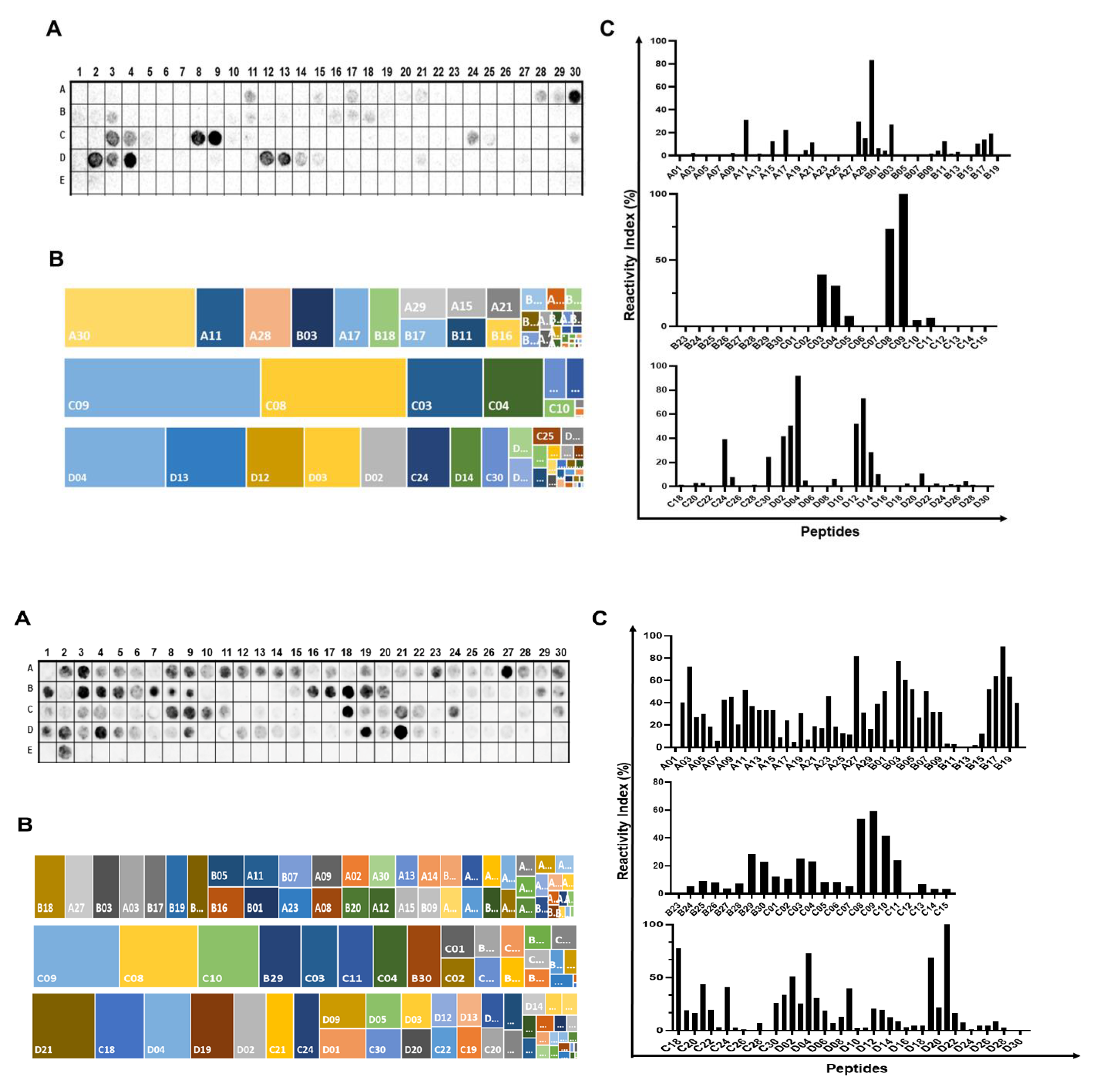
2.1. Identification of the Immunodominant IgA and IgM Epitopes in Cholera Toxin Subunits

In this section, we aimed to identify the immunodominant epitopes within the three components of the CT, namely CTA (258 aa), CTB (124 aa), and CTP (221 aa). To achieve this, we synthesized two libraries of peptides and assessed their recognition by antibodies from mice immunized with an oral V. cholerae vaccine (15 days post-vaccination). Figure 1A,C present the positions of these peptides and their respective measured intensities, as detected through chemiluminescent methods for mouse IgA and IgM antibodies present in the sera of mice that received the aOCV vaccination. Additionally, Figure 1B shows each peptide–epitope’s hierarchical position in terms of its reactivity, arranged from the top left to the bottom right position. Epitopes were defined based on the overlap of spots with signal strengths exceeding 30%, establishing the minimum sequence for these epitopes. The comprehensive list of the synthetic peptides and their positions on the membrane is in Figure S1. This analysis identified 18 IgG epitopes, with sizes ranging from 4 to 14 amino acids, which were generated because of vaccination with aOCV (Table 1).

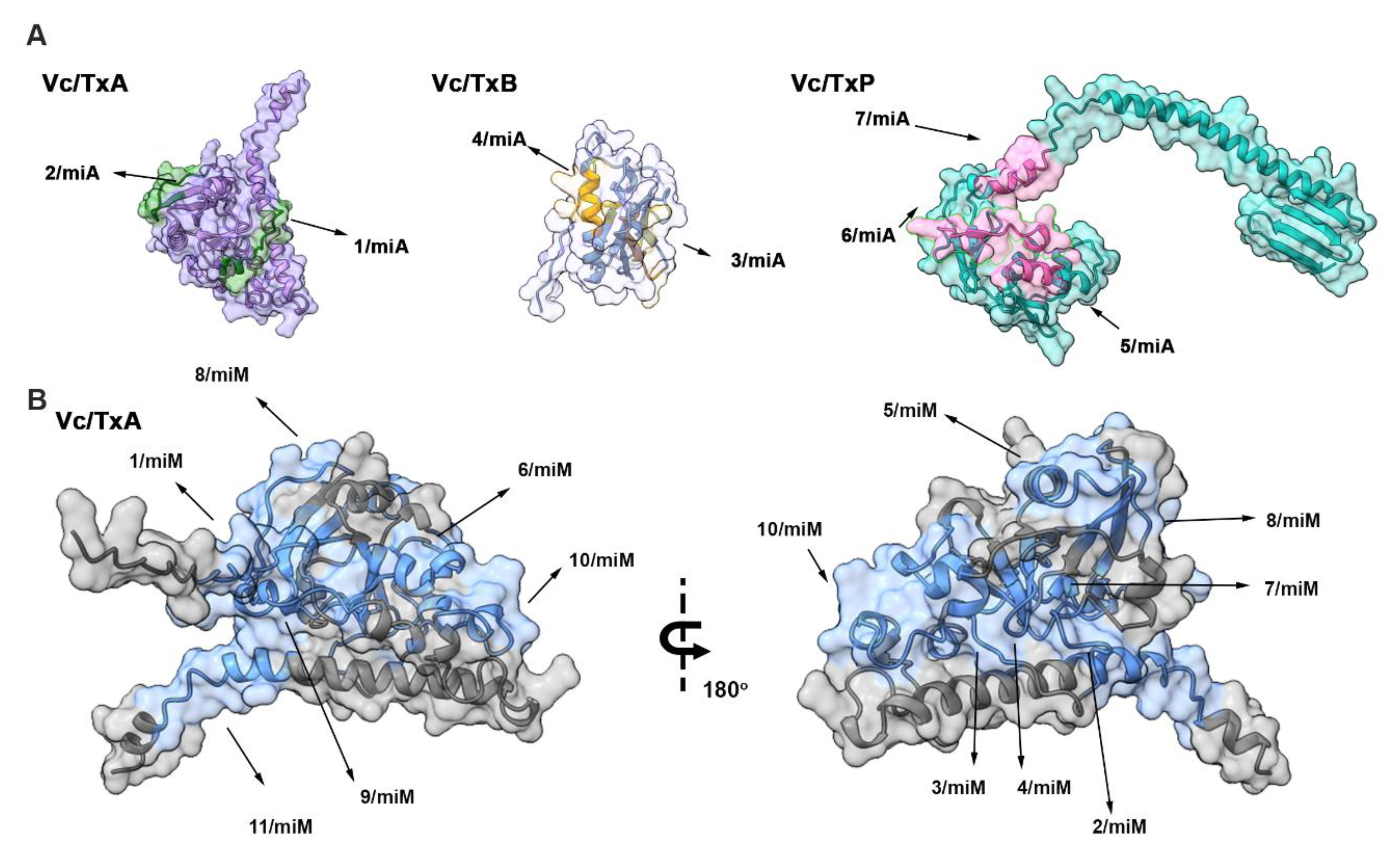
2.2. Spatial Localization of the Major IgA and IgM Epitopes Within the Three Chain of the Toxin

The seven IgA and eleven IgM linear epitopes identified by SPOT synthesis analysis were distributed throughout TxA, TxB, and TxP (Table 1). The enterotoxin chain A contains two IgA epitopes (TxA-1A and TxA-2A) and all 11 IgM epitopes (TxA-1M-TxA-11. Enterotoxin B houses two IgA epitopes (TxA-3A and TxA-4A) and no IgM epitopes. Enterotoxin P has three IgA epitopes (TxB-5A to TxB-7A) and no IgM epitopes (Table 1).

2.3. Spatial Distribution of the Reactive Epitopes of Enterotoxin A, B, and P

The analysis of the seven IgA and eleven IgM epitopes, as determined by the SPOT synthesis approach, revealed their distribution throughout the V. cholera toxin proteins (Figure 2). The V. cholera protein gene can be divided into three distinct segments: a short signal peptide in the N-terminal extension (aa 1–18), the A1 subunit chain segment (aa 19–212), the A2 subunit chain segment (aa 213–258), the B subunit (aa 1–124), and the P protein (aa 1–221).
To pinpoint the location of these epitopes, we accessed the enterotoxin A, B, and P structures from the Protein Data Bank (PDB) or utilized AlphaFold DB predictions. Alternatively, we deduced the tertiary structure using the I-TASSER server (http://zhanggroup.org/I-TASSER/, accessed on 11 May 2023). Figure 2 showcases the resulting predicted structural models for the enterotoxin A, B, and P proteins. These models offer insights into the spatial localization of the eight reactive epitopes in CTA, three epitopes in CTB, and seven epitopes in CTP. Notably, most of the identified epitopes were situated within loop/coil structures, making them accessible to the solvent and located on the protein’s surface (Table 1 and Figure 2).

2.4. Specific and Cross-Immune IgA and IgM Epitopes

In exploring potential cross-immunity conferred by CT proteins, we employed CTA, CTB, and CTP sequences as templates in a multi-peptide matching search within the UniProtKB database. The criteria for this search involved the identification of peptides with four or more identical amino acids. Our analysis revealed that ten epitopes from V. cholera were specific (Table 1), while eight epitopes (TxA-1A, TxB-4A, TxP-5A, TxP-7A, TxA-1M, TxA-4M, TxA-5M, TxA-7M) exhibited cross-reactivity with E. coli or various bacteria. Specifically, the V. cholera-specific epitopes in CTA included TxA-2A, TxB-6A, TxA-2M, TxA-3M, TxA-6M, and TxA-8M to TxA-11M. Within CTP, only the epitope TxP-6A was identified as specific; in CTB, the specific epitope was TxB-3A. The epitopes TxP-5A and TxP-6A were determined to be exact within CTP (Table 1).

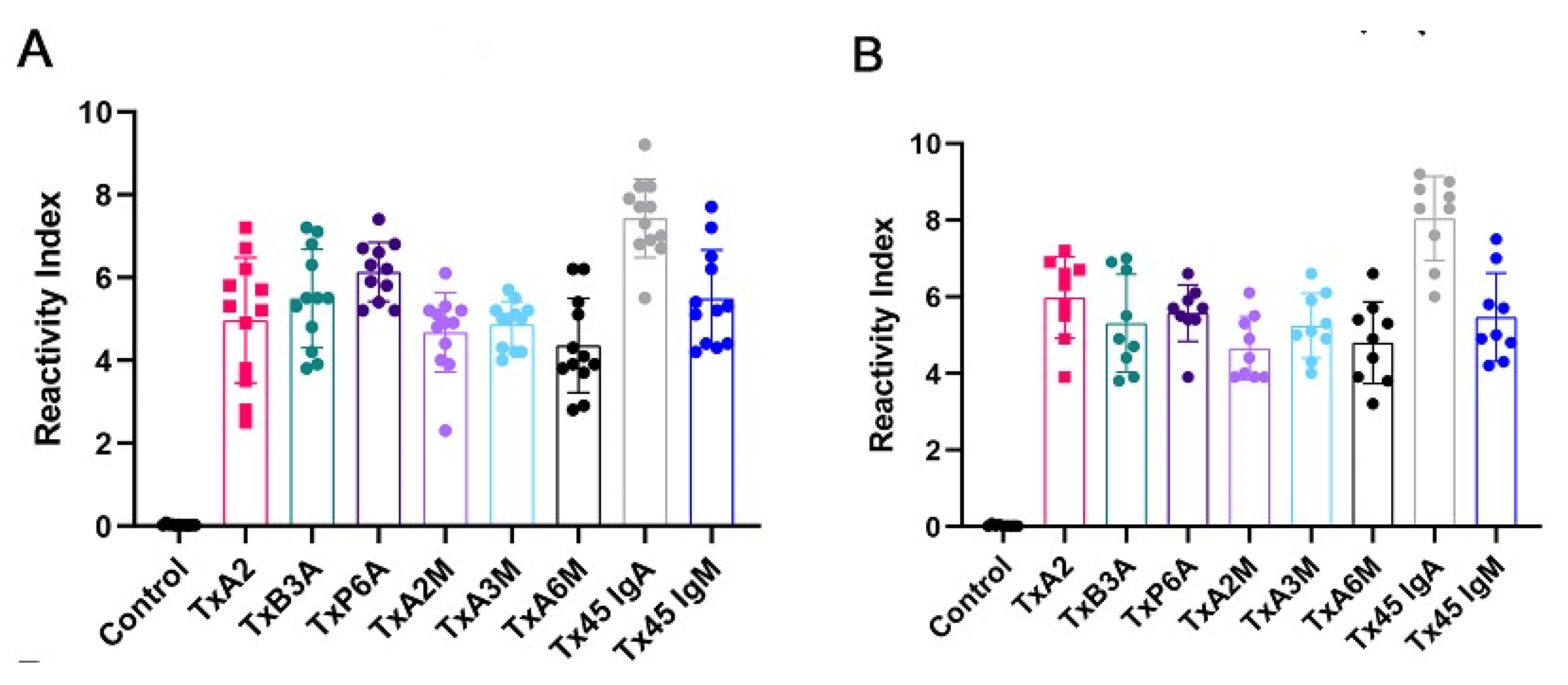
2.5. Reactivity of MAP4 and Chimeric Peptides via ELISA

It is well-known that serological assays based on bacterial proteins may exhibit cross-reactivity, underscoring the need to identify individual linear epitopes in the CT involved in the vaccination process. By restricting the antigens employed in an assay to capture reactive antibodies definitively specific to V. cholera through sequence analysis, developing a diagnostic ELISA that eliminates the potential for false positive results becomes feasible. To achieve this, three IgA and three IgM epitopes were carefully selected, synthesized, and subjected to ELISA analysis, both as individual peptides (Figure S3) and within multi-antigen peptides (MAP4) (Figure 3). A single IgA 45-mer and an IgM 45-mer chimeric peptide (Figure 3) encompassing the three chosen peptides of each subclass were synthesized. The sequences of the synthetic peptides are provided in Figure S2.
Upon examination, the sera from each immunized mouse showed reactivity to either the three synthetic peptides derived from IgA epitopes or the IgM epitopes, with a reassuring lack of response to the negative control peptide QEVRKYFCV (Vaccinia virus; Figure 1A, G9, and G17). This reactivity was detected 15 days after vaccination (Figure 3A) and 30 days later (Figure 3B), with no significant difference in antibody levels observed when using single peptides versus MAPs. Though the performance of the chimeric peptide IgA was maintained, considering the analysis of the sera collected 15 days after immunization against the 30 days after immunization, the performance of the chimeric IgM was slightly diminished. These results, as shown in Figure 3 and Figure S3, were found to be statistically significant (p < 0.001).
The choice of serum collection times was based on a clear understanding of the timeline of antibody development after infection. We know that IgM and IgA antibodies appear in the first week after infection or immunization, followed in the following weeks by IgG antibodies [37]. Infection occurs from a few hours to 5 days, and in most cases, it lasts 2 to 3 days. The period of transmissibility lasts as long as the vibrio is eliminated in the feces, which occurs, in most cases, until a few days after recovery. For surveillance purposes, the standard period accepted is 20 days. Therefore, in this study, we chose to collect serum from animals on the 15th and 30th days when vibriocidal, agglutinating, and protective antibodies are present [38,39,40].
Based on ROC curve analysis, the Area Under the Curve (AUC) for the epitope Tx45-IgA and Tx45-IgM was 0.9987 and 0.9896 (p < 0.0001), as detected by ELISA with a confidence interval of 95%, demonstrating high diagnostic accuracy for both the peptides. The Tx45-IgM peptide presented a lower reactivity than the Tx45-IgA, but both demonstrated 100% sensitivity and specificity.

3. Discussion

In this study, we identified linear B epitopes in CT recognized by IgA and IgM antibodies from mice vaccinated with an inactivated vaccine using peptide microarray analysis. CT serves as the primary virulence factor of V. cholerae, capable of inducing cholera symptoms with as little as 5 µg ingestion [41]. The serum reactivity against specific regions in CT demonstrated the vaccine’s high antigenic potential with a single dose. We identified two IgA and eleven IgM linear epitopes in CTA, two in CTB, and three in protein CTP, all accessible to the immune system due to their exposure on the molecular surface.
CTA is initially synthesized as a single polypeptide with a signal peptide (aa 1–18). The enzymatic activity begins after proteolytic cleavage, “nicking” at residue Arg-192, resulting in two functional domains: A1 (~21.8 kDa, aa 19–204) and A2 (~5.4 kDa, aa 206–258), covalently linked by a disulfide bond. The B subunit comprises five chains forming a pentameric ring around a central pore structure. CTA1 is responsible for ADP-ribosyltransferase activity, while the α-helical CTA2 anchors CTA1 and CTB5 together [42,43].
CTB, a crucial component of koCVs, is known to induce a robust protective response capable of neutralizing CT in the gut at low concentrations [9]. This response, mainly of the IgG type [9], with IgA and IgM responses [44] also playing important blocking roles, is acquired against CT during an infection or oral vaccination. Importantly, this protection is both T-dependent and crucial for developing a robust immune response against CT [44].
Our study demonstrated that the CTB subunit possesses only two major IgA epitopes among the seven detected. A previous study showed that when administered orally, two synthetic peptides of the TxB (aa 30–50; aa 50–75) induced a protective secretory IgA response in the gut [45]. Other authors demonstrated similar results in rats [46,47]. However, only the TxB- (aa 66–75) epitope identified in our study showed a structural correlation with the peptide (aa-50–75) described previously [45]. The aa 30–50 peptide from the previous study is possibly a minority epitope and was not identified in our studies. However, none of these circulating IgA epitopes shared IgM or IgG [33] sequences, underscoring the unique and distinct nature of the immunological response to TxB.
During the immune response, isotype change is a protective property that has the function of the organism producing antibodies with greater affinity to neutralize the antigen. After antigen recognition, recombination occurs between DNA segments of the genes that encode the constant regions of the heavy chains, determining the isotype [48]. The production time of these isotypes is variable and appears to depend on the antigen and the host. Consequently, some types of antibodies have a short half-life while others take a long time to disappear from circulation, which may explain the identification and not persistence of some isotypes of immunoglobulins/epitopes in the case of cholerae. Therefore, it is important to know which epitopes induce antibodies of longer duration and neutralizing capacity [48].
The IgA presents biological functions particularly dependent on its tertiary structure [49] and is given as a monomer (IgA1 circulating) or homodimer (IgA2 mucosal) after its synthesis [50] and different glycosylation profiles with IgA1 possessing more sialic acid than IgA2 [51]. The serum IgA can induce pro-inflammatory responses, such as releasing cytokines and chemokines, phagocytosis, degranulation, and formation of neutrophil extracellular traps (NETs) [52]. While IgA2 is composed of two IgA molecules (homodimer IgA), a joining protein (J chain), and a secretory component exert different biological functions in the mucosal tissues such as microbiota homeostasis, pro-inflammatory on neutrophils and macrophages, and opsonization of the bacteria [53] and even converts anti-inflammatory intestinal CD103+ DCs to a pro-inflammatory phenotype, which protects against invading pathogens, but might also result in chronic inflammation [54]. Independent of its biological functions, the heavy chains of the IgA immunoglobulins are produced in response to encountered antigen/specific epitopes independent of its subclasses [55,56,57,58]. Therefore, the analysis of circulating IgA, which is in most cases IgA1 [54], is sufficient to answer our question in advancing the qualitative identification of the major cholera epitope repertoire for diagnosis purposes and, in general, immune response.
In mouse models, it has been demonstrated that the primary immune response to cholerae is dominated by IgG against CTB, which is protective [58,59]. However, our study indicates that CTB also elicits IgA and IgM responses. Still, these responses appear weak compared to the number of identified IgG epitopes, despite the IgM being involved in opsonophagocytosis of the bacteria [34,60]. Importantly, it is a known fact that constant stimulation of the immune response leads to the production of long-lasting secretory IgA [61].
Each isotype is designed to tackle specific challenges, especially involving affinity. Normally, the IgM is the first antibody to emerge during embryonic development, and the humoral immune response is followed by IgA and IgG [62,63]. However, some IgM is also produced in secondary and subsequent responses, and after somatic hypermutation, although other isotypes dominate the later phases of the antibody response. The IgM can exist under distinct forms, including monomeric, membrane-bound IgM within the B cell receptor (BCR) complex, pentameric and hexameric IgM in serum can provide effective anti-microbial immunity [64,65], and secretory IgM on the mucosal surface. However, the regulation of humoral immune responses and B cell tolerance by the IgM Fc receptor (FcμR) is poorly understood [44,66].
Each isotype might prefer certain epitopes or residues of amino acids, which may be better suited to deal with specific types of invaders [67]. However, these epitopes possess different residues (right or left) but shared residues indicating distinct response preferences and evolution of the immune response, providing immunoglobulins with a higher binding or neutralization ability. Therefore, changes in the affinity will allow the immune system to be highly adaptable and effective in fine tuning, ready to handle diverse threats or situations [68].
Several IgG rapid antigen detection-based tests are available for cholerae diagnosis, such as immunochromatographic lateral flow devices like Crystal VC and Cholkit, which detect the presence of the O1 or O139 antigen in watery diarrheal stools and others [69,70,71]. However, limitations exist, and these tests may only sometimes offer high specificity [59]. Molecular testing using polymerase chain reaction (PCR) has also been used for diagnosis but primarily for research and surveillance [72,73]. Darkfield microscopy is another diagnostic approach for detecting V. cholerae, but it needs the required sensitivity for reliable diagnosis.
Thus, more precise, rapid diagnostic assays are needed. The IgA and IgM epitopes identified in this study can be applied in an ELISA assay to identify the primary immune response and provide insights into serological immunity. These unique IgA and IgM epitopes hold the potential for developing chimeric polyproteins for diagnostic purposes.
Vaccine production is typically a labor-intensive and costly experimental process. Nonetheless, these immunobiological molecules/agents are the most potent defenses against infectious diseases.
Presently, there are at least three pre-qualified oral cholera vaccines (OCVs): Dukoral® Crucell, The Netherlands), Shanchol™ (Shantha, Biotechnics-Sanofi Pasteur, India), and Euvichol-Plus® (EuBiologics Co., Seoul, Republic of Korea). While both categories have demonstrated protective effects against cholerae [4,74,75,76], the immunological mechanisms involving B-cell and/or T-cell immunity remain unclear [77,78]. It is well established that the key stimulator of innate immunity and subsequent adaptive immune responses is the LPS O antigen [6,10], which promotes long-term mucosal protection [8,13,79,80,81]. Hence, the pressing question is how to create more effective cholera vaccines, as current vaccine efficacies hover around 60% [1,27,82].
The findings on mice receiving a combination vaccination demonstrates a substantial increase in vibrocidal, IgM OSP-specific serum responses, and OSP-specific IgM memory B-cell responses. However, the research also indicates that enhanced opsonophagocytosis or antibody-dependent cytotoxic activity in the intestinal lumen does not seem to play a role in mediating protection against cholera, however, may present neutralizing ability. Even though cell-free antibody-based killing via complement lysis might be a possibility in the intestinal lumen, viability studies in animals have shown that bactericidal activity is not necessary for protection from disease [83,84,85,86]. These novel findings provide a deeper understanding of cholera protection mechanisms and vaccination responses, sparking curiosity in the field of microbiology and immunology.
Therefore, identifying immunodominant epitopes that interact with antibodies generated in response to vaccination can contribute to selecting better antigens and improve our understanding either for diagnostics or vaccination.

4. Materials and Methods

4.1. Immunization of Mice

We orally immunized thirty female BalbC mice, each weighing between 15 and 21 g, with 20 µL of the SchancolTM cholerae vaccine (lot SCN021A15; Shantha Biotechnics Ltd. in Muppiriddipalli, Telangana, India). Normally the infection occurs within a few hours to 5 days, with most cases lasting 2 to 3 days. The period of transmission lasts if the vibrio is eliminated in the feces, which occurs in most cases up to a few days after recovery. For surveillance purposes, the standard period accepted is 20 days. Fifteen days following vaccination, we collected blood samples and performed a complete bleed at the 30-day mark [34]. In parallel, blood samples were collected from 30 healthy, unvaccinated mice to serve as controls. The serum from each group was separately collected, divided into 0.5 mL aliquots in Eppendorf tubes, and stored at −20 °C. The study received ethical approval from the Ethics Commission of UNIGRANRIO (no 052/2021) study center ethics committee.

4.2. Synthesis of the Cellulose Membrane-Bound Peptide Array

Two peptide libraries comprising 122 peptides were synthesized to cover the full coding sequences of cholera enterotoxin A (P01555, 258 amino acids), enterotoxin B (P01556, 124 amino acids), and Toxin P (P29485, 221 amino acids) of serotype O1. The synthesis was performed on amino-PEG500-UC540 cellulose membranes (Intavis Bioanalytical Instruments, Köln, Germany) using standard SPOT synthesis protocols [87].
Each peptide was 15 amino acids long with a 10-amino-acid overlap, created with an Auto-Spot Robot ASP-222 (Intavis Bioanalytical Instruments AG, Köln, Germany). To enhance the libraries, a GSGSG spacer sequence was added at the amino and carboxy termini of each protein (positions A1, D5, D6, C4, C5, and F2). The libraries were constructed and programmed using the MultiPep 1 software (Intavis, Bioanalytical Instruments, Köln, Germany). Negative and positive control peptides were included: a negative control peptide (QEVRKYFCV from Vaccinia virus, located at spots G9 and G17) and positive controls such as KEVPALTAVETGATN (Poliovirus, at spots G3 and G11), GYPKDGNAFNNLDRI (Clostridium tetani, at spots G5 and G13), and YDYDVPDYAGYPYDV (Influenza hemagglutinin, at spots G7, G15, and G24).
The coupling reactions were followed by a blocking step using 4% acetic anhydride in N, N-dimethylformamide (DMF). The Fmoc protecting group was removed from the N-terminus of each peptide using 20% piperidine in DMF. This cycle of coupling, blocking, and deprotection was repeated for each amino acid addition. After the final amino acid was coupled, sidechain deprotection was performed with a solution of dichloromethane, trifluoroacetic acid, and triisopropylsilane (1:1:0.05, v/v/v), followed by ethanol washing. The synthetic peptide membranes were immediately prepared for probing

4.3. Screening of SPOT Membranes

The SPOT membranes were washed with TBS-T (50 mM Tris, 136 mM NaCl, 2 mM KCl, and 0.05% Tween® 20, pH 7.4) for 10 min, then blocked in TBS-T containing 1.5% BSA for 90 min at 8 °C with agitation. Following additional washes with TBS-T, the membranes were incubated for 12 h with pooled sera from ten vaccinated mice, diluted 1:150 (for IgA or IgM detection) in TBS-T with 0.75% BSA. After thorough washing with TBS-T, the membranes were incubated for 1 h with goat anti-mouse IgM and IgA alkaline phosphatase-conjugated antibodies (Sigma Chemical Co., Saint Louis, MO, USA; diluted 1:5000) prepared in TBS-T with 0.75% BSA. Further washes with TBS-T and CBS (50 mM citrate-buffered saline) were performed. The chemiluminescent substrate Nitro-Block II Enhance (Applied Biosystems, Waltham, MA, USA) was added to finalize the reaction. The integrity of the library synthesis was validated by the reactivity of control peptides with human sera.

4.4. Scanning and Measurement of Spot Signal Intensities

Chemiluminescent signals were captured using an Odyssey FC imaging system (LI-COR Bioscience, Lincoln, NE, USA) with slight modifications to previously reported conditions [88]. A digital image was generated at 5 MP resolution, and signal intensities (SI) were analyzed using TotalLab TL100 software (v 2009, Nonlinear Dynamics, Newcastle-Upon-Tyne, UK). Background signals, determined from the negative control, were subtracted from the SI values, which were then normalized as a percentage of the highest signal detected. Epitopes were identified as sequences comprising two or three contiguous spots with normalized SI values of 30% or more. For epitopes involving three or more contiguous spots with SI values of 30% or greater, identification relied on visual evaluation.
A comparative analysis of the reactivity index of the normalized SPOTs was conducted using a dimensional hierarchical approach as previously described [89].

4.5. Preparation of Single and Multi-Antigen Peptides (MAPs)

The single and multi-antigen peptides were synthesized using a standard solid-phase synthesis protocol [90], employing Wang and tetrameric Fmoc2-Lys-B-Ala Wang resins (Figure S2). The constructs were generated using an automated peptide synthesizer (MultiPep-1, CEM Corp., Charlotte, NC, USA). Side chains of tetrafunctional Fmoc-protected amino acids were safeguarded with TFA-labile protecting groups where necessary. After assembling the peptide sequences, Fmoc groups were removed, and the peptide resin was cleaved and fully deprotected using a TFA/H2O/EDT/TIS mixture (94/2.5/2.5/1.0, v/v) for 90 min.
The peptides were precipitated by adding chilled diethyl ether, followed by centrifugation at 30,000× g for 10 min at 4 °C. The resulting pellet was dissolved in 10% aqueous AcOH (v/v), lyophilized, and stored as a dry powder. When required, MAPs were dissolved in water, centrifuged at 10,000× g for 60 min at 15 °C, and the supernatant was filtered through a Centricon™ filter (Merck Millipore, Burlington, MA, USA). Purification was conducted using an XBridge BEH C18 column (2.7 μm, 5 cm × 4.6 mm) integrated with a Waters AutoPurify HPLC system (Waters Corp., Newcastle, Australia). The flow rate was set at 1.2 mL/min, with mobile phase A consisting of 0.05% formic acid in water (18 MΩ × cm) and mobile phase B comprising 0.05% formic acid in acetonitrile. A gradient of 0–97% phase B over 40 min was employed, with detection at 200–300 nm using a diode array detector.
For ESI-TOF analysis, peptides were dissolved in deionized water to a final concentration of 10 µg/mL, and formic acid was added to achieve a 0.1% concentration. Mass spectrometry was performed using a Waters UPLC Acquity-I Class system (Waters Corp., Newcastle, Australia), with 1 µL/min sample injections. Ion detection spanned a range of 1000 to 11,500 m/z.

4.6. Preparation of 45-mer Chimeric Peptides

Two chimeric multiepitope peptides, Tx45-IgA and Tx45-IgM, were synthesized in sequence, incorporating two to six glycine spacers (Figure S3). The synthesis was performed using the Fmoc protocol on TentaGel-S-NH2 resin [91]. An automated peptide synthesizer (MultiPep-1, CEM Corp., Charlotte, NC, USA) was utilized, along with Fmoc-protected amino acids and appropriate protecting groups. Following synthesis, the peptides were cleaved from the resin, deprotected, and precipitated. Their identities were verified using mass spectrometry techniques, including MALDI-TOF or electrospray ionization.

4.7. In-House ELISA

Peptide-based ELISA (pELISA) assays were performed as described previously [91]. 4HB NUNC plates were coated with 80 ng of each peptide in a Na2CO3–NaHCO3 coating buffer (pH 9.6) overnight at 4 °C. After three PBS-T washes, the plates were blocked with PBS-T containing 2.5% BSA for 2 h at 37 °C. Diluted mouse sera (1:100) were applied and incubated for 2 h at 37 °C. The plates were washed and incubated with goat anti-mouse IgA-HRP or IgM-HRP antibodies for 2 h at 37 °C. Following further washing, pNPP substrate was added, and absorbance was measured at 405 nm. The serum dilution was optimized using a titration series and ROC curve analysis.

4.8. Structural Localization of IgG Epitopes and Bioinformatics Tools

To identify the locations of epitopes within the 3D molecular structures of the enterotoxin A, B, and P proteins from V. cholerae, in silico protein models were generated using the I-TASSER server (http://zhanglab.ccmb, accessed on 10 April 2023). The models were selected based on the highest C-score and TM-score (topological evaluation value) [92]. Additionally, the resulting 3D structural models were cross verified using the AlphaFold v3 database [93].
Sequence homologies for V. cholerae were analyzed by comparing the identified sequences with annotated proteins from other organisms available in the UniProt database (http://www.uniprot.org/, accessed on 12 April 2023) and through multiple peptide matching using the tool at https://research.bioinformatics.udel.edu/peptide match/index.jsp (accessed on 10 May 2023).

4.9. Statistical Analysis

The data were analyzed using the R program (version 3.6.0) and R Studio. To assess the statistical significance between the two samples, a paired t-test was employed, with significance considered at p ≤ 0.05. GraphPad Prism version 5.0 was used to analyze the receiver operating characteristic curve.

5. Conclusions

A resolutive assessment of the immune response was conducted using a peptide array that was directly generated on a cellulose membrane. This approach facilitated the identification of the major antigenic determinants in enterotoxins A, B, and P, which were recognized by antibodies from mice that had been orally vaccinated with a single dose of the aOCV. In total, we identified seven IgA and eleven IgM epitopes that were distributed throughout the bacterial CT protein. Specifically, six epitopes were in the A1 chain of the enterotoxin, two in the A2 chain, three in enterotoxin B, and seven in enterotoxin P. Notably, six of these epitopes exhibited a degree of similarity to proteins in other pathogens, indicating a high potential for cross-reactivity. Furthermore, we defined ten V. cholera-specific epitopes (three IgA and seven IgM) and elucidated their spatial locations within protein structural models. The efficacy of three of these IgA epitopes (TxA-2A, TxB-3A, and TxP-6A) and three IgM epitopes (TxA-2M, TxA-3M, and TxA-6M) in detecting antibodies produced in response to vaccinations in mice was confirmed using an indirect ELISA. This comprehensive molecular characterization of linear IgA and IgM epitopes in CT holds significant promise for developing poly epitope chimeric proteins as produced by our group for Chagas disease [87], which could be employed in next-generation rapid diagnostic tests. Additionally, our findings underscore the value of epitope mapping in enhancing our understanding of the IgM and IgA immune response to current and future vaccines, ultimately leading to improved production of specific neutralizing antibodies.

Supplementary Materials

The following supporting information can be downloaded at https://www.mdpi.com/article/10.3390/ijms26083507/s1.

Author Contributions

Conceptualization, S.G.D.-S. and F.R.d.S.; methodology, P.N.-P., F.R.d.S., J.P.R.S.C., S.V.C., J.P.R.S.C., A.O.S. and G.C.L.; validation, D.W.P.J. and S.G.D.-S.; writing—original draft, S.G.D.-S.; writing—review and editing, S.G.D.-S.; funding acquisition, C.M.M. and S.G.D.-S. All authors have read and agreed to the published version of the manuscript.

Funding

This work was supported by the Brazilian Council for Scientific Research (CNPq #30515-2020-5) and the Carlos Chagas Filho Foundation of Research Support of the State of Rio de Janeiro (FAPERJ #200.960-2022). FAPERJ (#210.003/2018) also provided funding through the 518 National Institutes of Science and Technology Program (INCT) to Carlos M. Morel (INCT-IDPN).

Institutional Review Board Statement

The study was approved by the UNIGRANRIO (CEUA 052/2021) study center ethics committee and conducted under good clinical practice and all applicable regulatory requirements, including the declaration of Helsinki.

Informed Consent Statement

Not applicable.

Data Availability Statement

The data presented in this study are available on request from the corresponding author.

Acknowledgments

We thank the National Institute of Quality Control (INCQs) from FIOCRUZ for their support with the vaccine and with animal immunization, and Ulisses C. Araujo from the Multiuser Research Laboratory in Mass Spectrometry applied to Biomolecules (MultiMasses/IBRAG/UERJ). P.N.-P. and G.C.L. are postdoc fellows from the CAPES-FIOCRUZ program. J.P.R.S.C., is a CAPES fellow from the Post-Graduation Program on Science and Biotechnology at Federal Fluminense University.

Conflicts of Interest

The authors declare no conflict of interest.

References

  1. Ali, M.; Nelson, A.R.; Lopez, A.L.; Sack, D.A. Updated global burden of cholera in endemic countries. PLoS Negl. Trop. Dis. 2015, 9, e0003832. [Google Scholar] [CrossRef] [PubMed]
  2. Yuki, Y.; Nojima, M.; Hosono, O.; Tanaka, H.; Kimura, Y.; Satoh, T.; Imoto, S.; Uematsu, S.; Kurokawa, S.; Kashima, K.; et al. Oral MucoRice-CTB vaccine for safety and microbiota-dependent immunogenicity in humans: A phase 1 randomized trial. Lancet Microbe 2021, 2, e429–e440. [Google Scholar] [CrossRef] [PubMed]
  3. Vezzulli, L.; Grande, C.; Reid, P.C.; Hélaouët, P.; Edwards, M.; Höfle, M.G.; Brettar, I.; Colwell, R.R.; Pruzzo, C. Climate influence on vibrio and associated human diseases during the past half-century in the coastal North Atlantic. Proc. Natl. Acad. Sci. USA 2016, 113, E5062–E5071. [Google Scholar] [CrossRef] [PubMed]
  4. Deeb, R.; Tufford, D.; Scott, G.I.; Moore, J.G.; Dow, K. Impact of climate change on Vibrio vulnificus abundance and exposure risk. Estuaries Coasts 2018, 41, 2289–2303. [Google Scholar] [CrossRef] [PubMed]
  5. Baker-Austin, C.; Oliver, J.D.; Alam, M.; Ali, A.; Waldor, M.K.; Qadri, F.; Martinez-Urtaza, J. Vibrio spp. Infections. Nat. Rev. Dis. Prim. 2018, 4, 1–19. [Google Scholar] [CrossRef]
  6. Legros, D.; Partners of the Global Task Force on Cholerae Control. Global cholerae epidemiology: Opportunities to reduce the burden of cholera by 2030. J. Infect. Dis. 2018, 218, S137–S140, Erratum in J. Infect. Dis. 2019, 219, 509. [Google Scholar] [CrossRef]
  7. WHO (World Health Organization). Cholerae; World Health Organization: Geneva, Switzerland, 2022; Available online: https://www.who.int/news-room/fact-sheets/detail/cholera?gadsource=1&gclid=EAIaIQobChMIqYXdhPW1hgMVgUVIAB0qbwipEAAYASAAEgJ1CfD_BwE (accessed on 21 March 2023).
  8. Banerjee, T.; Grabon, A.; Taylor, M.; Teter, K. cAMP-Independent activation of the unfolded protein response by cholera toxin. Infect. Immun. 2021, 89, e00447-20. [Google Scholar] [CrossRef]
  9. Clemens, J.D.; Nair, G.B.; Ahmed, T.; Qadri, F.; Holmgren, J. Cholerae. Lancet 2017, 390, 1539–1549. [Google Scholar] [CrossRef]
  10. Bourque, D.L.; Bhuiyan, T.R.; Genereux, D.P.; Rashu, R.; Ellis, C.N.; Chowdhury, F.; Khan, A.I.; Alam, N.H.; Paul, A.; Hossain, L.; et al. Analysis of the human mucosal response to cholerae reveals sustained activation of innate immune signaling pathways. Infect. Immun. 2018, 86, e00594-17. [Google Scholar] [CrossRef]
  11. Sánchez, J.; Holmgren, J. Cholera toxin-a foe & a friend. Indian J Med Res. 2011, 133, 153–163. [Google Scholar]
  12. Taylor, R.K.; Miller, V.L.; Furlong, D.B.; Mekalanos, J.J. Use of phoA gene fusions to identify a pilus colonization factor coordinately regulated with cholera toxin. Proc. Natl. Acad. Sci. USA 1987, 84, 2833–2837. [Google Scholar] [CrossRef] [PubMed]
  13. Waldor, M.K.; Colwell, R.; Mekalanos, J.J. The Vibrio cholerae O139 serogroup antigen includes an O-antigen capsule and lipopolysaccharide virulence determinants. Proc. Natl. Acad. Sci. USA 1994, 91, 11388–11392. [Google Scholar] [CrossRef] [PubMed]
  14. Sperandio, V.; Giron, J.A.; Silveira, W.D.; Kaper, J.B. The OmpU outer membrane protein, a potential adherence factor of Vibrio cholerae. Infect. Immun. 1995, 63, 4433–4438. [Google Scholar] [CrossRef] [PubMed]
  15. Sengupta, D.K.; Sengupta, T.K.; Ghose, A.C. Major outer membrane proteins of Vibrio cholerae and their role in induction of protective immunity through inhibition of intestinal colonization. Infect. Immun. 1992, 60, 4848–4855. [Google Scholar] [CrossRef] [PubMed]
  16. Nandi, B.; Nandy, R.K.; Sarkar, A.; Ghose, A.C. Structural features, properties, and regulation of the outer-membrane protein W (OmpW) of Vibrio cholerae. Microbiology 2005, 151, 2975–2986. [Google Scholar] [CrossRef]
  17. Qadri, F.; Ali, M.; Chowdhury, F.; Khan, A.I.; Saha, A.; Khan, I.A.; Begum, Y.A.; Bhuiyan, T.R.; Chowdhury, M.I.; Uddin, M.J.; et al. Feasibility and effectiveness of oral cholerae vaccine in an urban endemic setting in Bangladesh: A cluster randomized open-label trial. Lancet 2015, 386, 1362–1371. [Google Scholar] [CrossRef]
  18. Bi, Q.; Ferreras, E.; Pezzoli, L.; Legros, D.; Ivers, L.C.; Date, K.; Qadri, F.; Digilio, L.; Sack, D.A.; Ali, M.; et al. Protection against cholera from killed whole-cell oral cholerae vaccines: A systematic review and meta-analysis. Oral cholerae vaccine working group of the global task force on cholerae control. Lancet Infect. Dis. 2017, 17, 1080–1088. [Google Scholar] [CrossRef]
  19. Peak, C.M.; Reilly, A.L.; Azman, A.S.; Buckee, C.O. Prolonging herd immunity to cholerae via vaccination: Accounting for human mobility and waning vaccine effects. PLoS Negl. Trop. Dis. 2018, 12, e0006257. [Google Scholar] [CrossRef]
  20. Royal, J.M.; Reeves, M.A.; Matoba, N. Repeated oral administration of a KDEL-tagged recombinant cholerae toxin B subunit effectively mitigates DSS colitis despite a robust immunogenic response. Toxins 2019, 11, 678. [Google Scholar] [CrossRef]
  21. Kabir, S. Critical analysis of compositions and protective efficacies of oral killed cholera vaccines. Clin. Vaccine Immunol. 2014, 21, 1195–1205. [Google Scholar] [CrossRef]
  22. Chen, W.H.; Cohen, M.B.; Kirkpatrick, B.D.; Brady, R.C.; Galloway, D.; Gurwith, M.; Hall, R.H.; Kessler, R.A.; Lock, M.; Haney, D.; et al. Single-dose live oral cholera vaccine CVD 103-HgR protects against human experimental infection with Vibrio cholerae El Tor. Clin. Infect. Dis. 2016, 62, 1329–1335. [Google Scholar] [CrossRef] [PubMed]
  23. Holmgren, J.J. Modern history of cholera vaccines and the pivotal role of ICDDR. Infect. Dis. 2021, 224, S742–S748. [Google Scholar] [CrossRef] [PubMed]
  24. Wierzba, T.F. Oral cholera vaccines and their impact on the global burden of disease. Hum. Vaccines Immunother. 2019, 15, 1294–1301. [Google Scholar] [CrossRef] [PubMed]
  25. Qadri, F.; Chowdhury, M.I.; Faruque, S.M.; Salam, M.A.; Ahmed, T.; Begum, Y.A.; Saha, A.; Al Tarique, A.; Seidlein, L.V.; Park, E.; et al. PXV Study Group, Peru-15, a live attenuated oral cholera vaccine, is safe and immunogenic in Bangladeshi toddlers and infants. Vaccine 2007, 25, 231–238. [Google Scholar] [CrossRef]
  26. Khan, A.I.; Islam, M.T.; Khan, Z.H.; Tanvir, N.A.; Amin, M.A.; Khan, I.I.; Bhuiyan, A.T.M.R.H.; Hasan, A.S.M.M.; Islam, M.S.; Bari, T.I.A.; et al. Implementation and delivery of oral cholera vaccination campaigns in humanitarian crisis settings among Rohingya Myanmar nationals in Cox’s Bazar, Bangladesh. Vaccines 2023, 11, 843. [Google Scholar] [CrossRef]
  27. Song, K.R.; Lim, J.K.; Park, S.E.; Saluja, T.; Cho, S.I.; Wartel, T.A.; Lynch, J. Oral cholera vaccine efficacy and effectiveness. Vaccines 2021, 9, 1482. [Google Scholar] [CrossRef]
  28. Ali, M.; Qadri, F.; Kim, D.R.; Islam, M.T.; Im, J.; Ahmmed, F.; Khan, A.I.; Zaman, K.; Marks, F.; Kim, J.H.; et al. Effectiveness of a killed whole-cell oral cholera vaccine in Bangladesh: Further follow-up of a cluster-randomised trial. Lancet Infect. Dis. 2021, 21, 1407–1414. [Google Scholar] [CrossRef]
  29. Koelle, K.; Rodo, X.; Pascual, M.; Yunus, M.; Mostafa, G. Refractory periods and climate forcing in cholerae dynamics. Nature 2005, 436, 696–700. [Google Scholar] [CrossRef]
  30. Khatib, A.M.; Ali, M.; von Seidlein, L.; Kim, D.R.; Hashim, R.; Reyburn, R.; Ley, B.; Thriemer, K.; Enwere, G.; Hutubessy, R.; et al. Effectiveness of an oral cholerae vaccine in Zanzibar: Findings from a mass vaccination campaign and observational cohort study. Lancet Infect. Dis. 2012, 1, 837–844. [Google Scholar] [CrossRef]
  31. Ali, M.; Sur, D.; You, Y.A.; Kanungo, S.; Sah, B.; Manna, B.; Puri, M.; Wierzba, T.F.; Donner, A.; Nair, G.B.; et al. Herd protection by a bivalent killed whole-cell oral cholerae vaccine in the slums of Kolkata, India. Clin. Infect. Dis. 2013, 56, 1123–1131. [Google Scholar] [CrossRef]
  32. Holmgren, J. An update on cholerae immunity and current and future cholerae vaccines. Trop Med Infect Dis. 2021, 6, 64. [Google Scholar] [CrossRef]
  33. De-Simone, S.G.; Napoleão-Pêgo, P.; Gonçalves, P.S.; Provance-Jr, D.W.; Morel, C.M.; Silva, F.R. B cell epitope mapping of the Vibrio cholerae toxins A, B, and P. Int. J. Mol. Sci. 2023, 24, 531. [Google Scholar] [CrossRef]
  34. Pandey, S.; Malviya, G.; Chottova Dvorakova, M. Role of peptides in diagnostics. Int. J. Mol. Sci. 2021, 22, 8828. [Google Scholar] [CrossRef] [PubMed]
  35. Andresen, H.; Bier, F.F. Peptide microarrays for serum antibody diagnostics. Methods Mol. Biol. 2009, 509, 123–134. [Google Scholar] [CrossRef]
  36. Winkler, D.F.; Campbell, W.D. The spot technique: Synthesis and screening of peptide macroarrays on cellulose membranes. Methods Mol. Biol. 2008, 494, 47–70. [Google Scholar] [CrossRef]
  37. Naidu, A.; Lulu, S.S. Mucosal and systemic immune responses to Vibrio cholerae infection and oral cholerae vaccines (OCVs) in humans: A systematic review. Expert. Rev. Clin. Immunol. 2022, 18, 1307–1318. [Google Scholar] [CrossRef]
  38. Kanungo, S.; Azman, A.S.; Ramamurthy, T.; Deen, J.; Dutta, S. Cholerae. Lancet 2022, 399, 1429–1440. [Google Scholar] [CrossRef]
  39. Rask, C.; Fredriksson, M.; Lindblad, M.; Czerkinsky, C.; Holmgren, J. Mucosal and systemic antibody responses after peroral or intranasal immunization: Effects of conjugation to enterotoxin B subunits and/or of co-administration with free toxin as adjuvant. APMIS 2000, 108, 178–186. [Google Scholar] [CrossRef]
  40. Akter, A.; Kelly, M.; Charles, R.C.; Harris, J.B.; Calderwood, S.B.; Bhuiyan, T.R.; Biswas, R.; Xu, P.; Kováč, P.; Qadri, F.; et al. Parenteral vaccination with a cholera conjugate vaccine boosts vibriocidal and anti-OSP responses in mice previously immunized with an oral cholera vaccine. Am. J. Trop. Med. Hyg. 2021, 104, 2024–2030. [Google Scholar] [CrossRef]
  41. Montero, D.A.; Vidal, R.M.; Velasco, J.; George, S.; Lucero, Y.; Gómez, L.A.; Carreño, L.J.; García-Betancourt, R.; O’Ryan, M. Vibrio cholerae, classification, pathogenesis, immune response, and trends in vaccine development. Front Med (Lausanne) 2023, 10, 1155751. [Google Scholar] [CrossRef]
  42. Sánchez, J.; Holmgren, J. Cholerae toxin structure, gene regulation, and pathophysiological and immunological aspects. Cell. Mol. Life Sci. 2008, 65, 1347–1360. [Google Scholar] [CrossRef] [PubMed]
  43. Baldauf, K.J.; Royal, J.M.; Hamorsky, K.T.; Matoba, N. Cholera toxin B: One subunit with many pharmaceutical applications. Toxins 2015, 7, 974–996. [Google Scholar] [CrossRef] [PubMed]
  44. Lycke, N.; Eriksen, L.; Holmgren, J. Protection against cholera toxin after oral immunization is thymus-dependent and associated with intestinal production of neutralizing IgA antitoxin. Scand. J. Immunol. 1987, 25, 413–419. [Google Scholar] [CrossRef] [PubMed]
  45. Pedoussaut, S.; Delmas, A.; Milhaud, G.; Rivaille, P.; Gruaz-Guyon, A. Oral immunization with a free peptide from cholera toxin: Local protection and IgA production. Mol. Immunol. 1989, 26, 115–119. [Google Scholar] [CrossRef]
  46. Jacob, C.O.; Vaerman, J.P. Induction of rat secretory IgA antibodies against cholera toxin by a synthetic peptide. Immunology 1986, 59, 129–133. [Google Scholar]
  47. Delmas, A.; Gruaz-Guyon, A.; Pedoussaut, S.; Pierre, P.; Rivaille, P.; Vaerman, J.P. Neutralization of cholera toxin by rat IgA secretory antibodies induced by a free synthetic peptide. Biochem. Biophys. Res. Commun. 1989, 159, 707–712. [Google Scholar] [CrossRef]
  48. Sikora, A.E. Proteins secreted via the type II secretion system: Smart strategies of Vibrio cholerae to maintain fitness in different ecological niches. PLoS Pathog. 2013, 9, e1003126. [Google Scholar] [CrossRef]
  49. Schroeder, H.W., Jr.; Cavacini, L. Structure and function of immunoglobulins. J. Allergy Clin. Immunol. 2010, 125, S41–S52. [Google Scholar] [CrossRef]
  50. Kerr, M.A. The structure and function of human IgA. Biochem. J. 1990, 271, 285–296. [Google Scholar] [CrossRef]
  51. Steffen, U.; Koeleman, C.A.; Sokolova, M.V.; Bang, H.; Kleyer, A.; Rech, J.; Unterweger, H.; Schicht, M.; Garreis, F.; Hahn, J.; et al. IgA subclasses have different effector functions associated with distinct glycosylation profiles. Nat. Commun. 2020, 11, 120. [Google Scholar] [CrossRef]
  52. Hansen, I.S.; Baeten, D.L.P.; den Dunnen, J. The inflammatory function of human IgA. Cell Mol. Life Sci. 2019, 76, 1041–1055. [Google Scholar] [CrossRef] [PubMed]
  53. Bunker, J.J.; Bendelac, A. IgA Responses to Microbiota. Immunity 2018, 49, 211–224. [Google Scholar] [CrossRef] [PubMed]
  54. Hansen, I.S.; Krabbendam, L.; Bernink, J.H.; Loayza-Puch, F.; Hoepel, W.; van Burgsteden, J.A.; Kuijper, E.C.; Buskens, C.J.; Bemelman, W.A.; Zaat, S.A.J.; et al. FcαRI co-stimulation converts human intestinal CD103+ dendritic cells into pro-inflammatory cells through glycolytic reprogramming. Nat. Commun. 2018, 28, 863. [Google Scholar] [CrossRef] [PubMed]
  55. Casadevall, A.; Janda, A. Immunoglobulin isotype influences affinity and specificity. Proc. Natl. Acad. Sci. USA. 2012, 109, 12272–12273. [Google Scholar] [CrossRef]
  56. Janda, A.; Bowen, A.; Greenspan, N.S.; Casadevall, A. Ig Constant region effects on variable region structure and function. Front. Microbiol. 2016, 7, 22. [Google Scholar] [CrossRef]
  57. Garzón-Ospina, D.; Buitrago, S.P. Immunoglobulin heavy constant gamma gene evolution is modulated by both the divergent and birth-and-death evolutionary models. Primates 2022, 63, 611–625. [Google Scholar] [CrossRef]
  58. Mekalanos, J.; Collier, R.; Romig, W. Enzymic activity of cholerae toxin. II. Relationships to proteolytic processing, disulfide bond reduction, and subunit composition. J. Biol. Chem. 1979, 254, 5855–5861. [Google Scholar] [CrossRef]
  59. Mayo, S.; Royo, F.; Hau, J. Correlation between adjuvanticity and immunogenicity of cholerae toxin B subunit in orally immunized young chickens. APMIS 2005, 113, 284–287. [Google Scholar] [CrossRef]
  60. Price, G.A.; Holmes, R.K. Evaluation of TcpF-A2-CTB chimera and evidence of additive protective efficacy of immunizing with TcpF and CTB in the suckling mouse model of cholerae. PLoS ONE 2012, 7, e42434. [Google Scholar] [CrossRef]
  61. Maślanka, T.; Clapp, B.; Hoffman, C.; Robison, A.; Gregorczyk, I.; Pascual, D.W. Nasal vaccination of β7 integrin-deficient mice retains elevated IgA immunity. Immunol. Cell Biol. 2020, 98, 667–681. [Google Scholar] [CrossRef]
  62. Adachi, M.; Kurihara, Y.; Nojima, H.; Takeda-Shitaka, M.; Kamiya, K.; Umeyama, H. Interaction between the antigen and antibody is controlled by the constant domains: Normal mode dynamics of the HEL-HyHEL-10 complex. Protein Sci. 2003, 12, 2125–2131. [Google Scholar] [CrossRef] [PubMed]
  63. Liu, J.; Wang, Y.; Min, Q.; Xiong, E.; Heyman, B.; Wang, J.Y. Regulation of humoral immune responses and B cell tolerance by the IgM Fc receptor (FcμR). Adv. Exp. Med. Biol. 2020, 1254, 75–86. [Google Scholar] [CrossRef] [PubMed]
  64. Yang, J.S.; An, S.J.; Jang, M.S.; Song, M.; Han, S.H. IgM specific to lipopolysaccharide of Vibrio cholerae is a surrogate antibody isotype responsible for serum vibriocidal activity. PLoS ONE 2019, 14, e0213507. [Google Scholar] [CrossRef] [PubMed]
  65. Freytag, L.C.; Clements, J.D. Mucosal adjuvants. Vaccine. 2005, 23, 1804–1813. [Google Scholar] [CrossRef]
  66. Keyt, B.A.; Baliga, R.; Sinclair, A.M.; Carroll, S.F.; Peterson, M.S. Structure, function, and therapeutic use of IgM antibodies. Antibodies 2020, 9, 53. [Google Scholar] [CrossRef]
  67. Li, Y.; Shen, H.; Zhang, R.; Ji, C.; Wang, Y.; Su, C.; Xiao, J. Immunoglobulin M perception by FcμR. Nature 2023, 615, 907–912. [Google Scholar] [CrossRef]
  68. Hiramoto, E.; Tsutsumi, A.; Suzuki, R.; Matsuoka, S.; Arai, S.; Kikkawa, M.; Miyazaki, T. The IgM pentamer is an asymmetric pentagon with an open groove that binds the AIM protein. Sci. Adv. 2018, 4, eaau1199. [Google Scholar] [CrossRef]
  69. Wang, Y.; Su, C.; Ji, C.; Xiao, J. CD5L associates with IgM via the J chain. Nat. Commun. 2024, 15, 8397. [Google Scholar] [CrossRef]
  70. Muzembo, B.A.; Kitahara, K.; Ohno, A.; Debnath, A.; Okamoto, K.; Miyoshi, S.I. Cholera rapid diagnostic tests for the detection of Vibrio cholerae O1: An updated meta-analysis. Diagnostics 2021, 11, 2095. [Google Scholar] [CrossRef]
  71. Debes, A.K.; Murt, K.N.; Waswa, E.; Githinji, G.; Umuro, M.; Mbogori, C.; Roskosky, M.; Ram, M.; Shaffer, A.; Sack, D.A. Laboratory and field evaluation of the Crystal VC-O1 cholera rapid diagnostic test. Am. J. Trop. Med. Hyg. 2021, 104, 2017–2023. [Google Scholar] [CrossRef]
  72. Sayeed, M.A.; Islam, K.; Hossain, M.; Akter, N.J.; Alam, M.N.; Sultana, N.; Khanam, F.; Kelly, M.; Charles, R.C.; Kováč, P.; et al. Development of a new dipstick (Cholkit) for rapid detection of Vibrio cholerae O1 in acute watery diarrheal stools. PLoS Negl. Trop. Dis. 2018, 12, e0006286. [Google Scholar] [CrossRef] [PubMed]
  73. Heyman, B.; Shulman, M.J. Encyclopedia of Immunobiology; DeFranco, A.L., Ratcliffe, M.J.H., Eds.; Academic Press: Cambridge, MA, USA, 2016; pp. 1–14. ISBN 012374282X/9780123742827. [Google Scholar]
  74. McGettigan, S.E.; Aira, L.E.; Kumar, G.; Ballet, R.; Butcher, E.C.; Baumgarth, N.; Debes, G.F. Secreted IgM modulates IL-10 expression in B cells. Nat Commun 2024, 15, 324. [Google Scholar] [CrossRef] [PubMed]
  75. De-Simone, S.G.; Gomes, L.R.; Napoleão-Pêgo, P.; Lechuga, G.C.; Pina, J.C.; Silva, F.R. Identification of linear B epitopes liable for the protective immunity of diphtheria toxin. Vaccines 2021, 9, 313. [Google Scholar] [CrossRef]
  76. Muzembo, B.A.; Kitahara, K.; Debnath, A.; Okamoto, K.; Miyoshi, S.I. Accuracy of cholera rapid diagnostic tests: A systematic review and meta-analysis. Clin. Microbiol. Infect. 2022, 28, 155–162. [Google Scholar] [CrossRef] [PubMed]
  77. Nelson, E.J.; Grembi, J.A.; Chao, D.L.; Andrews, J.R.; Alexandrova, L.; Rodriguez, P.H.; Ramachandran, V.V.; Sayeed, M.A.; Wamala, J.F.; Debes, A.K.; et al. Gold standard cholera diagnostics are tarnished by lytic bacteriophage and antibiotics. J. Clin. Microbiol. 2020, 58, e00412-20. [Google Scholar] [CrossRef]
  78. Ramamurthy, T.; Das, B.; Chakraborty, S.; Mukhopadhyay, A.K.; Sack, D.A. Diagnostic techniques for rapidly detecting Vibrio cholerae O1/O139. Vaccine 2020, 38, A73–A82. [Google Scholar] [CrossRef]
  79. Chakraborty, S.; Velagic, M.; Connor, S. Development of a simple, rapid, and sensitive molecular diagnostic assay for cholera. PLoS Negl. Trop. Dis. 2023, 17, e0011113. [Google Scholar] [CrossRef]
  80. Zareitaher, T.; Sadat, T.; Seyed, A.S.; Gargari, L.M. Immunogenic efficacy of DNA and protein-based vaccine from a chimeric gene consisting of OmpW, TcpA, and CtxB, of Vibrio cholerae. Immunobiology 2022, 227, 152190. [Google Scholar] [CrossRef]
  81. Zereen, F.; Akter, S.; Sobur, M.A.; Hossain, M.T.; Rahman, M.T. Molecular detection of Vibrio cholerae from human stool collected from SK Hospital, Mymensingh, and their antibiogram. J. Adv. Vet. Anim. Res. 2019, 6, 451–455. [Google Scholar] [CrossRef]
  82. Pezzoli, L. Oral cholera vaccine working group of the global task force on cholera control. Global oral cholera vaccine use, 2013–2018. Vaccine 2020, 38, A132–A140. [Google Scholar] [CrossRef]
  83. Saif-Ur-Rahman, K.M.; Mamun, R.; Hasan, M.; Meiring, J.E.; Khan, M.A. Oral killed cholera vaccines for preventing cholera. Cochrane Database Syst. Rev. 2024, 1, CD014573. [Google Scholar] [CrossRef]
  84. Bishop, A.L.; Schild, S.; Patimalla, B.; Klein, B.; Camilli, A. Mucosal immunization with Vibrio cholerae outer membrane vesicles provides maternal protection mediated by antilipopolysaccharide antibodies that inhibit bacterial motility. Infect. Immun. 2010, 78, 4402–4420. [Google Scholar] [CrossRef] [PubMed]
  85. Charles, R.C.; Kelly, M.; Tam, J.M.; Akter, A.; Hossain, M.; Islam, K.; Biswas, R.; Kamruzzaman, M.; Chowdhury, F.; Khan, A.I.; et al. Humans surviving cholera develop antibodies against Vibrio cholerae O-specific polysaccharide that inhibit pathogen motility. mBio 2020, 11, e02847-20. [Google Scholar] [CrossRef] [PubMed]
  86. Kauffman, R.C.; Adekunle, O.; Yu, H.; Cho, A.; Nyhoff, L.E.; Kelly, M.; Harris, J.B.; Bhuiyan, T.R.; Qadri, F.; Calderwood, S.B.; et al. Impact of immunoglobulin isotype and epitope on the functional properties of Vibrio cholerae O-specific polysaccharide-specific monoclonal antibodies. mBio 2021, 12, e03679-20. [Google Scholar] [CrossRef]
  87. Dias, E.R.; Durans, A.M.; Succar, B.B.; Pinto, L.A.L.T.; Lechuga, G.C.; Miguez, M.G.; Figueira-Mansur, J.; Argondizzo, A.P.C.; Bernardo, A.R.; Diniz, R.L.; et al. A multi-epitope protein for high-performance serodiagnosis of chronic Chagas Disease in ELISA and lateral flow platforms. Int. J. Mol. Sci. 2024, 25, 9811. [Google Scholar] [CrossRef]
  88. Silva, F.R.; Napoleão-Pêgo, P.; De-Simone, S.G. Identification of linear B epitopes of pertactin of Bordetella pertussis induced by immunization with whole and acellular vaccine. Vaccine 2014, 32, 6251–6258. [Google Scholar] [CrossRef]
  89. Moutsinas, G.; Shuaib, C.; Guo, W.; Jarvis, S. Graph hierarchy: A novel framework to analyze hierarchical structures in complex networks. Sci. Rep. 2021, 11, 13943. [Google Scholar] [CrossRef]
  90. De-Simone, S.G.; Napoleão-Pêgo, P.; Lechuga, G.C.; Carvalho, J.P.R.S.; Gomes, L.R.; Cardoso, S.V.; Morel, C.M.; Provance-Jr, D.W.; Silva, F.R.S. High-throughput IgG epitope mapping of tetanus neurotoxin: Implications for immunotherapy and vaccine design. Toxins 2023, 15, 239. [Google Scholar] [CrossRef]
  91. De-Simone, S.; Souza, A.L.A.; Melgarejo, A.R.; Aguiar, A.S.; Provance, D.W., Jr. Development of elisa assay to detect specific human IgE anti-therapeutic horse sera. Toxicon 2017, 138, 37–42. [Google Scholar] [CrossRef]
  92. Roy, A.; Kucukural, A.; Zhang, Y. I-TASSER: A unified platform for automated protein structure and function prediction. Nat. Protoc. 2010, 5, 725–738. [Google Scholar] [CrossRef]
  93. Jumper, J.; Evansm, R.; Pritzel, A.; Green, T.; Figurnov, M.; Ronneberger, O.; Tunyasuvunakool, K.; Bates, R.; Žídek, A.; Potapenko, A.; et al. Highly accurate protein structure prediction with AlphaFold. Nature 2021, 596, 583–589. [Google Scholar] [CrossRef]
Figure 1. IgA (upper) and IgM (low) epitope mapping of V. cholerae toxin A (P01555), B (P01556), and P (P29485). A membrane-bound peptide library representing the three chains of the toxin was probed with a pool of vaccinated mouse sera (n = 15), and reactivity was detected using goat anti-mouse IgA (I) and IgM (II) alkaline phosphatase labeled secondary antibody and chemiluminescence substrate. Panels (A) present an image of the peptide array showing reactivity as dark circles. The panels in (B) show the hierarchy position of each positive peptide and, in (C), the percentage signal after normalizing the signals to the positive and negative controls. The sequences of peptides in each position are listed in Figure S1.
Ijms 26 03507 g001
Figure 2. Epitope localization in three-dimensional structures of V. cholerae toxins. (A) IgA epitopes in V. choleraee toxins A (TxA), B (TxB), and P (TxP) epitopes are presented and colored within models constructed using the structure of toxin A, B, and P predicted by AlphaFold V3 (B) IgM epitope mapping within V. choleraee toxin A. Images were created using AlphaFold with ChimeraX (https://elearning.vib.be/courses/alphafold/lessons/vib-training-session-alphafold/topic/chimerax-display-alphafold-predictions-and-error-estimates-using-chimerax/).
Ijms 26 03507 g002
Figure 3. Reactivity of vaccinated mice (n = 12) sera 15 (A) and 30 (B) days post vaccination against the MAP4 peptides (TxA-2A, TxB-3A, TxP-6A, TxA-2M, TxA-3M, and TxA-6M) and chimeric tripeptides (Tx45-IgA and Tx45-IgM) by ELISA. As controls were used, sera of unvaccinated mice (n = 30). The relative index was calculated using the cut-off of each MAP to normalize the measured value from the sera of 12 vaccinated mice. The ROC curve was employed to establish the peptide cut-off values (Tx45-IgA and Tx45-IgM; 0.053 = 0.056), evaluate reactivity (99–100%), assess specificity (100%), and calculate the Area Under the Curve (AUC) values (ranging from 0.9987 to 0.9896).
Ijms 26 03507 g003
Table 1. A list of B-cell linear IgA and IgM epitopes identified in TxA, TxB, and P was deduced from the overlap of consecutive positive peptides with signal intensities greater than 30%.
Protein CodeCodeaaSequenceSecond Structure *Peptide Search **
P01555TxA-1A51RGTQMNINLYDHARGCE. coli
TxA-2A146–160YRVHFGVLDEQLHRNCSp
P01556TxB-3A66–75REMAIITFKNC + HSp
TxB-4A81–90SQKKAIERMKHE. coli
P29485TxP-5A31–45KPERLIGTPSIIQTC + HSp
TxP-6A81–90AIKRTRDFLNC + HSp
TxP-7A126–135QKKSVKERIKC + HVarious bacteria
P01555TxA-1M11–25FLSSFSYANDDKLYRCVarious bacteria
TxA-2M41–50MPRGQSEYFDCSp
TxA-3M56–64NINLYDHARC + HSp
TxA-4M71–80VRHDDGYVSTCE. coli
TxA-5M91–105GQTILSGHSTYYIYVC + HVarious bacteria
TxA-6M111–125NMFNVNDVLGAYSPHCSp
TxA-7M131–145VSALGGIPYSQIYGWCVarious bacteria
TxA-8M151–160GVLDEQLHRNCSp
TxA-9M171–185RGYRDRYYSNLDIAPCSp
TxA-10M191–205GLAGFPPEHRAWREECSp
TxA-11M236–250VKRQIFSGYQSDIDTC + HSp
Sp, specific epitopes; C, coil; H, helix; * based on an I-TASSER analysis; ** UNIPROT.
Disclaimer/Publisher’s Note: The statements, opinions and data contained in all publications are solely those of the individual author(s) and contributor(s) and not of MDPI and/or the editor(s). MDPI and/or the editor(s) disclaim responsibility for any injury to people or property resulting from any ideas, methods, instructions or products referred to in the content.

