Article

Genome-Wide Identification and Characterization of the Growth-Regulating Factor Gene Family Responsive to Abiotic Stresses and Phytohormone Treatments in Populus ussuriensis

1
State Key Laboratory of Tree Genetics and Breeding, Northeast Forestry University, Harbin 150040, China
2
School of Forestry, Northeast Forestry University, Harbin 150040, China
*
Authors to whom correspondence should be addressed.
Int. J. Mol. Sci. 2025, 26(7), 3288; https://doi.org/10.3390/ijms26073288
Submission received: 17 February 2025 / Revised: 30 March 2025 / Accepted: 30 March 2025 / Published: 1 April 2025

Abstract

:
As a unique class of plant-specific transcription factors, the GROWTH-REGULATING FACTORs (GRFs) play pivotal roles in regulating plant growth, development, and stress responses. In this study, the woody plant Populus ussuriensis was taken as the research object. Nineteen PuGRFs were identified and classified into six clades, and their potential evolutionary relationships were analyzed. The possible biological functions of PuGRFs were speculated through bioinformatics analysis. Combining real-time fluorescence quantitative PCR, PuGRFs were determined to be actively expressed in young tissues, and there are distinct tissue-specific expressions in the mature tissues of woody plants. We also conducted RT-qPCR of PuGRFs under different abiotic stresses and phytohormone treatments, most of the family members were induced under the treatments of methyl jasmonate (MEJA) and salicylic acid (SA), and we also found that 4 of 19 PuGRFs might participate in abscisic acid (ABA)-mediated osmotic stress in roots. Protein–protein interaction prediction analysis showed that six PuGRFs can interact with two types of growth-regulating interaction factors (GIFs). Further prediction and verification revealed that PuGRF1/2c and PuGRF1/2d, which belong to the same clade and have highly similar sequences, exhibited divergent interaction capabilities with GIFs, indicating evolutionary fine-tuning and functional redundancy within the GRF family. These findings lay a foundation for studying the molecular mechanisms of PuGRFs in P. ussuriensis, suggest that PuGRFs play important roles in responding to hormones and environmental changes, and the potential interaction relationships are worthy of exploration.

1. Introduction

The growth and development of plants is an exceedingly complex process delicately regulated by multiple genes [1]. GROWTH-REGULATING FACTOR (GRF) belongs to a category of transcription factors unique to plants, and plays an extremely crucial role in the process of plant growth and development. OsGRF1 was initially discovered and named in rice (Oryza sativa) [2]. Shortly after that, more GRFs were successively identified in various plants. In herbaceous plants like Arabidopsis thaliana, O. sativa, and Zea mays, a total of 9, 12, and 14 GRF family members have been successfully identified respectively. Similarly, in woody species such as Populus trichocarpa [3], Cajanus cajan [4], Jatropha curcas [5], and Citrus sinensis [6], 19, 10, 10, and 19 GRF members have been detected. The identification of members of the GRF gene family mainly relies on unique structural characteristics [7]. The N-terminal region harbors two highly conserved domains: QLQ (glutamine, leucine, and glutamine) and WRC (tryptophan, arginine, and cysteine). The QLQ domain interacts with the SNH domain of growth-regulating interaction factors (GIFs). GIFs function by forming GRF–GIF complexes, which play a regulatory role in processes like cell proliferation, differentiation, and organ formation [8,9,10]. The WRC domain exhibits even greater sequence conservation, encompassing a nuclear localization signal and a C3H-type zinc finger motif. This domain can be tightly bound to the cis-acting elements of downstream genes to regulate the temporal and spatial expression of genes. In contrast, the C-terminal sequence of the GRF protein has a wide variation range. Some motifs with relatively low conservation, such as TQL, GGPL, and FFD, typically appear at the C-terminal [11,12,13]. The conserved and diverse characteristics of the plant GRF domains collectively endow GRFs with the ability to play crucial functions in all aspects of plant growth and development.
In plants, GRFs were shown significantly to contribute to the early growth and development of plants and play crucial regulatory roles in the formation of plant tissues and organs. The GRF gene family in A. thaliana is widely involved in the growth process, such as AtGRF1, which regulates leaf size and cell proliferation; AtGRF2, involved in controlling root growth and plant height [7]; and AtGRF5, which promotes leaf growth by influencing cell division in leaf primordia [14]. Previous studies have shown that OsGRF4 regulates grain size and yield by modulating cell division and expansion in developing seeds [15]. OsGRF7 and OsGRF8, together with GA, were shown to determine rice leaf length by regulating cell division zone size [16]. In addition, OsGRF1 is shown to influence rice leaf growth [2].
Plants often face multiple stressors in nature, and transcription factors (TFs) play a crucial role in regulating stress-responsive genes to help plants adapt to adverse environmental conditions. Previous studies have shown that the AtGRF7-targeted cis-element (TGTCAGG) negatively regulates multiple downstream responses to osmotic stress in A. thaliana [17]. PdbGRF1 banded to the drought response element (DRE, ‘A/GCCGAC’) to regulate the expression of PdbPOD17 and PdbAKT1 and activate the related physiological pathways, showing a positive regulatory effect on salt stress in Populus davidiana × P. bolleana [18]. In addition to directly regulating other genes as transcription factors, GRFs can also interact with other proteins. For example, the A. thaliana AtGRF–AtGIF complex binds to promoters of cell cycle genes, like CYCLIN family members, during early leaf development. Multiple AtGRFs interacted with DELLA and regulated plant responses to cold stress [19]. It has been reported in studies that all 19 GRFs in poplar are predicted to be regulated by miRNA396 [20,21,22], and they are mainly recognized in the region encoding the last few amino acids of the WRC domain. Although there are multiple reports in the literature on the functions of GRFs in improving the early growth and development of woody plants, the relevant research on woody plants, to a certain degree, trailed behind that in herbaceous plants.
P. ussuriensis, one of the main fast-growing tree species in the mountainous forest regeneration of eastern Northeast China, is early maturity, high-yield, and has high economic value. In this research, we leveraged P. ussuriensis materials to conduct a more in-depth exploration of the evolutionary dynamics of GRFs in woody plants via bioinformatics analysis. We detected the expression profiles of PuGRFs across various tissues during the seedling and rapid-growth stages. Our findings revealed that, as the plant transitioned through different growth and development phases, the tissue-specificity of PuGRFs underwent a notable shift. By simulating the environmental challenges that trees may encounter through abiotic stress and phytohormone treatments, we observed that the majority of PuGRFs displayed similar expression patterns in leaves in response to phytohormone treatments. Moreover, a subset of PuGRFs potentially responded to drought or salt stress mediated by ABA. Concurrently, we employed predictive models to forecast the potential interaction relationships among PuGRFs. Subsequently, we selected two highly representative genes to conduct preliminary verification of their interaction with PuGIFs. The outcomes of this study not only present a comprehensive panorama of the PuGRF gene family, but also open up novel perspectives for the further exploration and validation of the functions of PuGRF genes in the woody plant P. ussuriensis.

2. Results

2.1. Genome-Wide Discovery of GRFs in P. ussuriensis

To identify the GRF gene family in the P. ussuriensis genome, we used transcriptome sequencing data of P. ussuriensis obtained from the previous work of our laboratory. Since the genomes of P. ussuriensis and P. trichocarpa are very similar [23,24,25] (Figure S1), we screened 19 genes of the GRF gene family in P. ussuriensis based on that of P. trichocarpa. We successfully pinpointed 19 members of the GRF gene family in P. ussuriensis. These genes were designated PuGRF1/2a toPuGRF12b, primarily based on their homology with PtGRFs.
Subsequently, basic information on these identified GRFs was compiled (Table 1). Nineteen PtGRFs were unevenly distributed across eleven chromosomes. The open-reading frame (ORF) of the 19 PuGRFs represents CDS ranging in length from the shortest 606 bp (PuGRF12b) to the longest 1836 bp (PuGRF1/2d), which encode proteins with lengths ranging from 201 to 611 amino acids (aa), molecular weight (Mw) ranging from 22.32 kDa to 66.53 kDa, and isoelectric points (pI) ranging between 6.44 and 10.06. Except for PuGRF12b, which may be localized in the mitochondrion and nucleus, the predicted localization of other PuGRFs is in the nucleus. Predicting microRNAs shows that all PuGRFs could be recognized and cleaved by ptc-miR396a to ptc-miR396g.

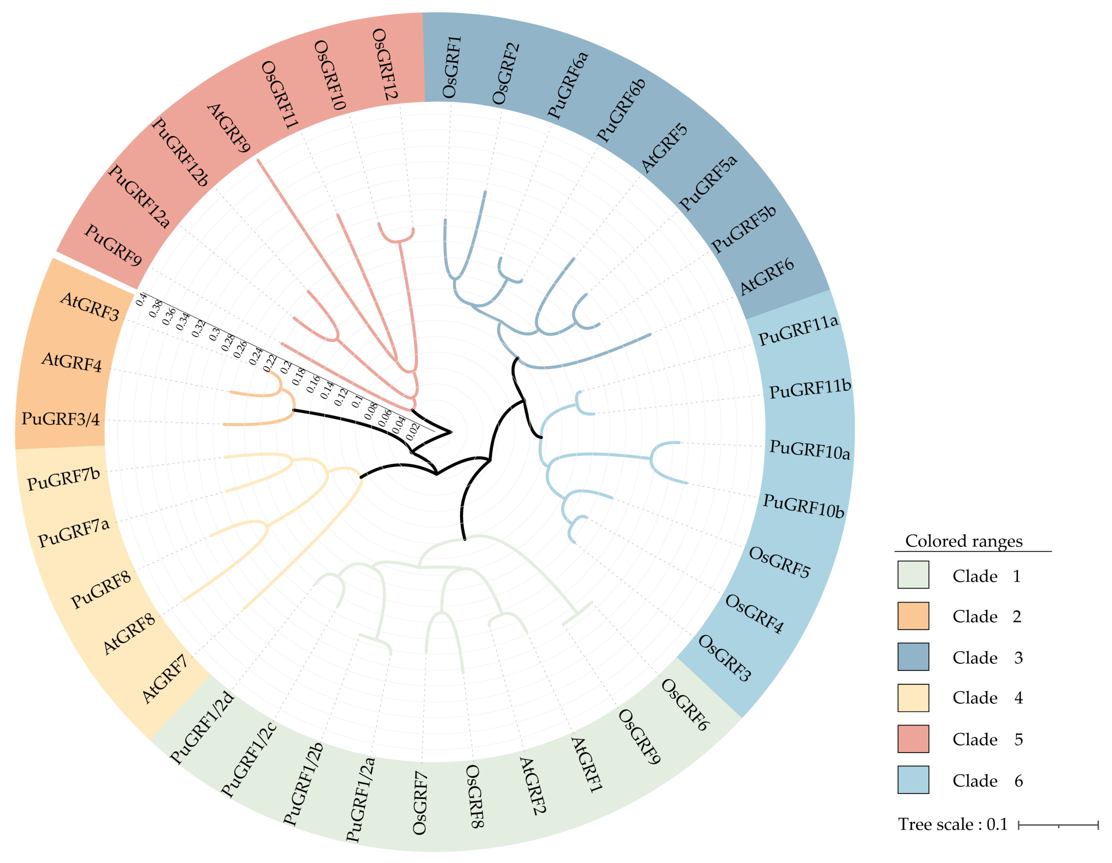
2.2. Protein Evolution of PuGRFs

To uncover the phylogenetic relationship, an interspecific phylogenetic tree was constructed using 40 GRFs from P. ussuriensis (19), O. sativa (12), and A. thaliana (9) (Figure 1). The results demonstrated that the PuGRFs were divided into six clades. Meanwhile, the distribution of GRFs in poplar, arabidopsis, and rice was different. OsGRFs were only distributed in clades I, III, V, and VI. AtGRFs were not distributed in clade VI. The obvious differences in the interspecific phylogenetic tree indicate that the evolutionary history of GRFs is rich and complex, potentially reflecting functional diversification and adaptation to different ecological niches.
Phylogenetic analysis was performed using MEGA7.0 software with the adjacency (NJ) method and 1000 iterations were carried out for bootstrap testing. The modules are color-coded to represent the six clades of GRFs. The 9 members in A. thaliana and 12 members in O. sativa are as follows: AT2G22840.1, AT4G37740.1, AT2G36400.1, AT3G52910.1, AT3G13960.1, AT2G06200.1, AT5G53660.1, AT4G24150.1, AT4G24150.1, LOC_Os02g53690.1, LOC_Os06g10310.1, LOC_Os04g51190.1, LOC_Os02g47280.1, LOC_Os06g02560.3, LOC_Os03g51970.1, LOC_Os12g29980.1, LOC_Os11g35030.1, LOC_Os03g47140.1, LOC_Os02g45570.1, LOC_Os07g28430.1, and LOC_Os04g48510.1.

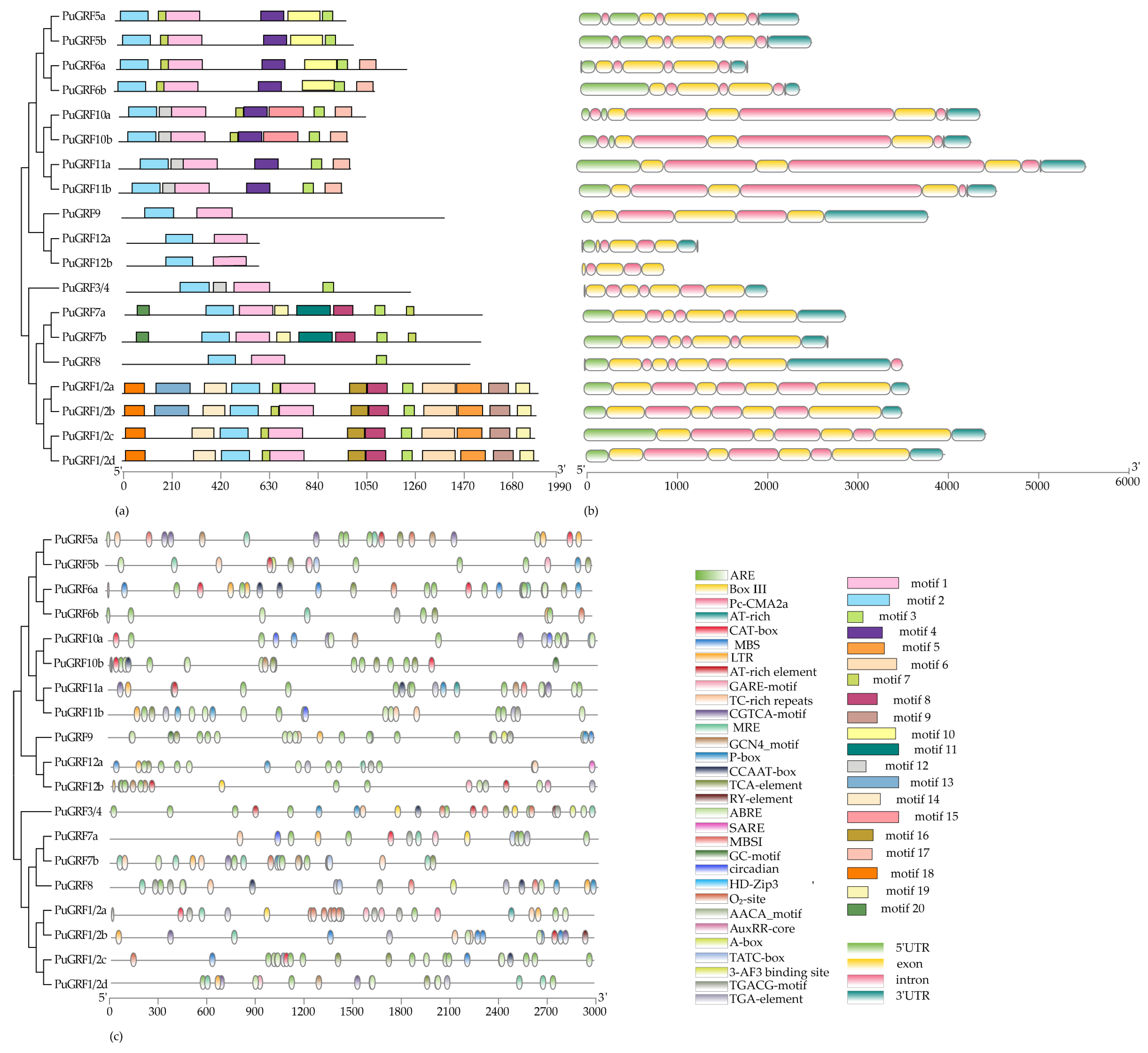
2.3. The Protein Motif, Structure, and Cis-Element Analysis of PuGRFs

To understand the unification and diversification of the PuGRFs, analyses of conserved motifs, gene structure, and cis-elements were conducted. Protein motifs are not only key sites for the mutual recognition and binding between proteins, but also capable of mediating the interactions between proteins and nucleic acids. Regarding protein motifs, motif 1 and motif 2, annotated as the GRF domain, were identified in all clades (Table S4). Except for clade II, where only PuGRF3/4 was present, and clade IV, where there was a relatively large difference among PuGRF7a, PuGRF7b, and PuGRF8, the PuGRFs belonging to the same clade showed high agreement in motifs (Figure 2a).
The gene structure reflects the evolutionary history of genes to a certain extent. During the evolutionary process, the structures of some key genes often show a high degree of conservation. The 19 PuGRFs identified in this study range in length from 914 to 5642 base pairs, each containing intron structures and either 3 or 4 exons (Figure 2b). Although there is little difference in the number of structures, PuGRF10a-PuGRF11b, which belong to clade VI, encode medium-length CDS with a longer intron. Notably, PuGRF12b lacks the 5′ UTR and 3′ UTR regions, while the remaining genes possess the typical 5′ UTR and 3′ UTR structures.
The 3.0 kb promoter region of all PuGRFs was utilized to accurately predict the promoter cis-elements and explore the possible biological roles of PuGRFs (Figure 2c). In addition to TATA-box, CAAT-box, and GC-box, which are essential for initiating transcription, a total of 31 cis-regulatory elements were obtained and classified into the following categories according to their functions (Table S5): (1) Binding sites for proteins or specific transcription factors, such as CCAAT-boc (MYBHv1 binding site) and HD zip 3 (proteins binding site); (2) Photoreactive elements. Each GRF promoter region contains a large number of photoreactive elements, such as MRE and Pc-CMA2a. (3) Growth and development response elements. These include CAT (meristematic tissue), RY-element (seed specificity), GCN4-motif (endosperm expression), and circadian; (4) Hormone response elements. These include TGA (methyl jasmonate), ABRE (abscisic acid), GARE (gibberellin), and TCA-element (salicylic acid); and (5) Environmental stress response elements, such as TC-rich (defense and stress response), LTR (cold), ARE (anaerobic induction), and MBS (drought).
The results indicate that the number of promoter elements in the entire family ranges from 7 to 15, with little interspecies variation. For instance, with the exception of PuGRF7a, all GRFs possess elements that respond to abscisic acid. There are 12 GRFs with cis-acting elements involved in defense and stress responses, while the other 7 GRFs lack this element, but have elements that respond to low temperatures or droughts. Collectively, these findings strongly suggest that GRF genes may indeed be involved in biological functions related to these hormone and stress response pathways [26].

2.4. Expression Pattern of PuGRFs

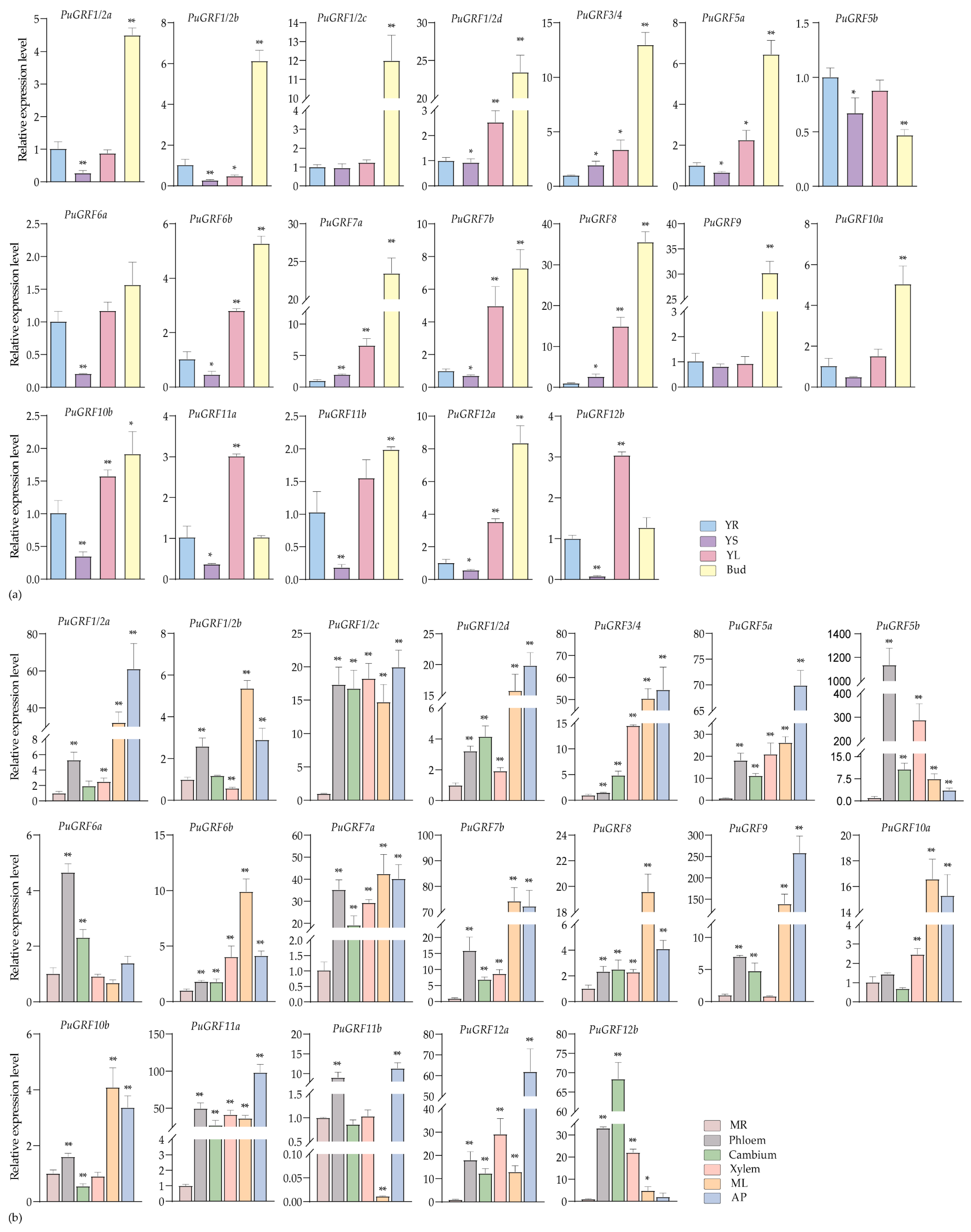
2.4.1. Expression Analysis of PuGRFs During Growth and Development of P. ussuriensis

We selected various tissues from two growth stages of P. ussuriensis: the seedling stage (young root (YR), young stem (YS), young leaves (YLs), and bud)), and the rapid growth stage (mature root (MR), phloem, cambium, xylem, mature leaves (MLs), and apical bud (AP)) to examine the expression of PuGRFs. By analyzing expression changes in various tissues of woody plants across different growth stages, this study suggests that the functions of PuGRFs exhibit potential conservation and divergence compared with those in herbaceous plants.
The results showed that, in the materials at the seedling stage, most of the PuGRFs were regularly highly expressed in young leaves (YL) and bud tissues, with the lowest expression levels in YS, except for PuGRF5b (Figure 3a). It should be particularly noted that the expression levels of PuGRF1/2d, PuGRF7a, PuGRF8, and PuGRF9 in buds were more than 20 times higher than those in YR, YS, and YL during the same period. During the rapid growth stage, PuGRFs exhibit diverse tissue specificity. Twelve PuGRFs were specifically highly expressed in ML and AP. Furthermore, relatively high expression levels of PuGRF1/2c, PuGRF7a, PuGRF11a, PuGRF12a, and PuGRF12b were observed in phloem, cambium, and xylem tissues. This result is likely due to the involvement of these PuGRFs in stem differentiation changes associated with woody plant growth cycle transitions, hinting at their potential roles in radial development, particularly for the paralogous pair PuGRF12a and PuGRF12b. Among them, PuGRF1/2c and PuGRF11a have different expression patterns from their homologous gene pairs PuGRF1/2d and PuGRF11b, respectively. PuGRF1/2d is significantly expressed in ML and AP, and PuGRF11b is significantly expressed in phloem and AP, suggesting functional differences among homologous genes (Figure 3b).

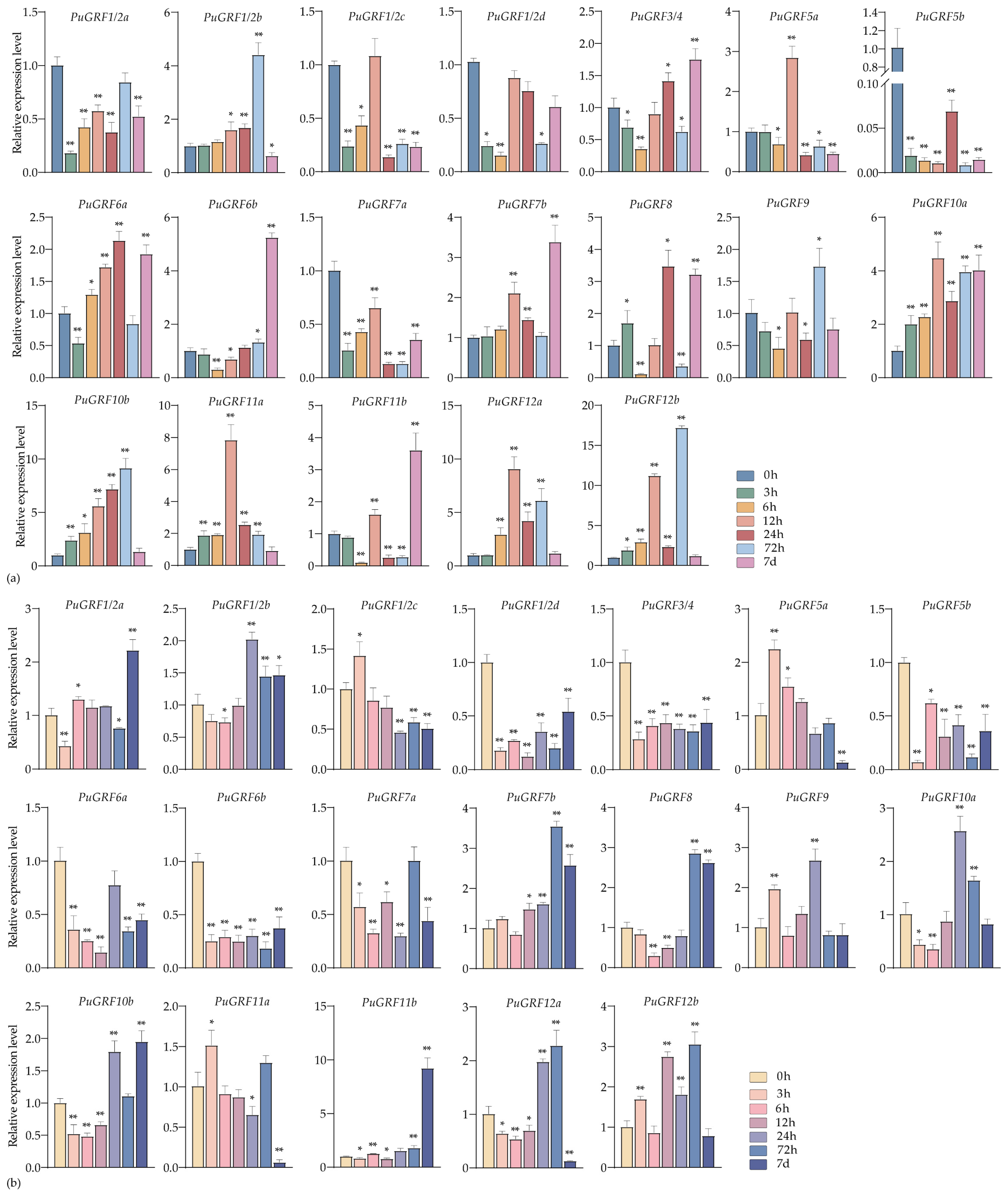
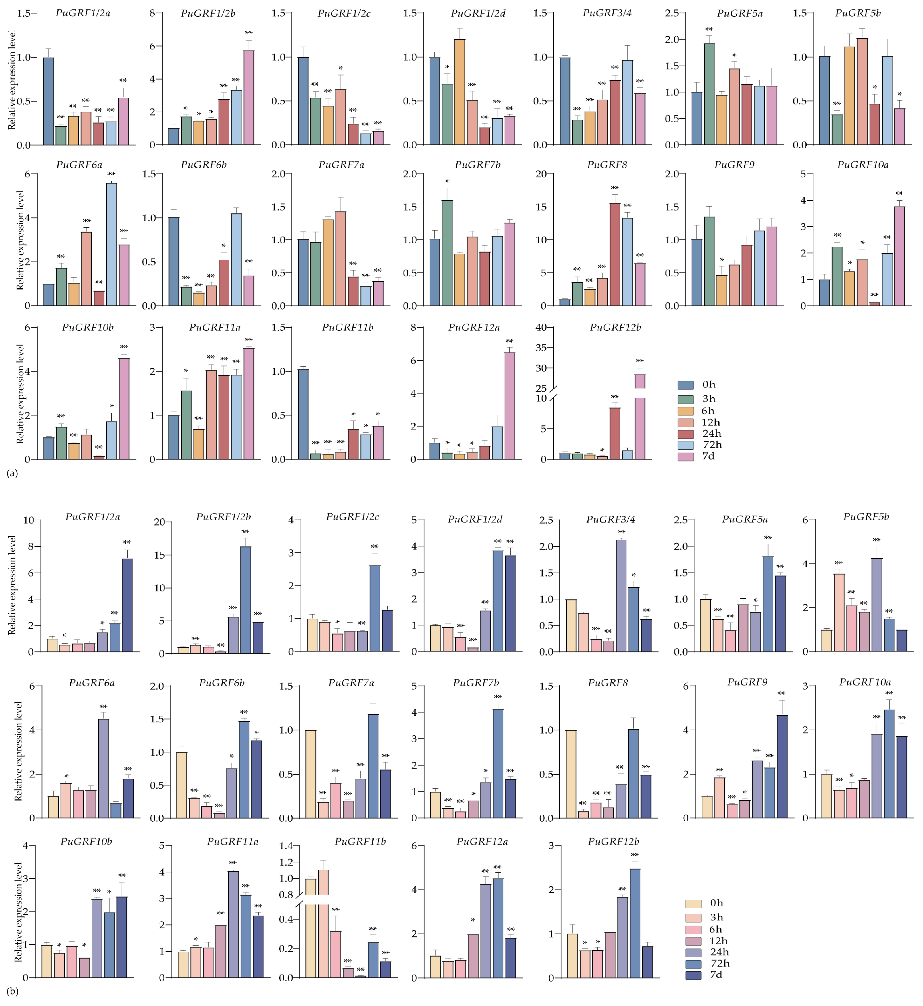
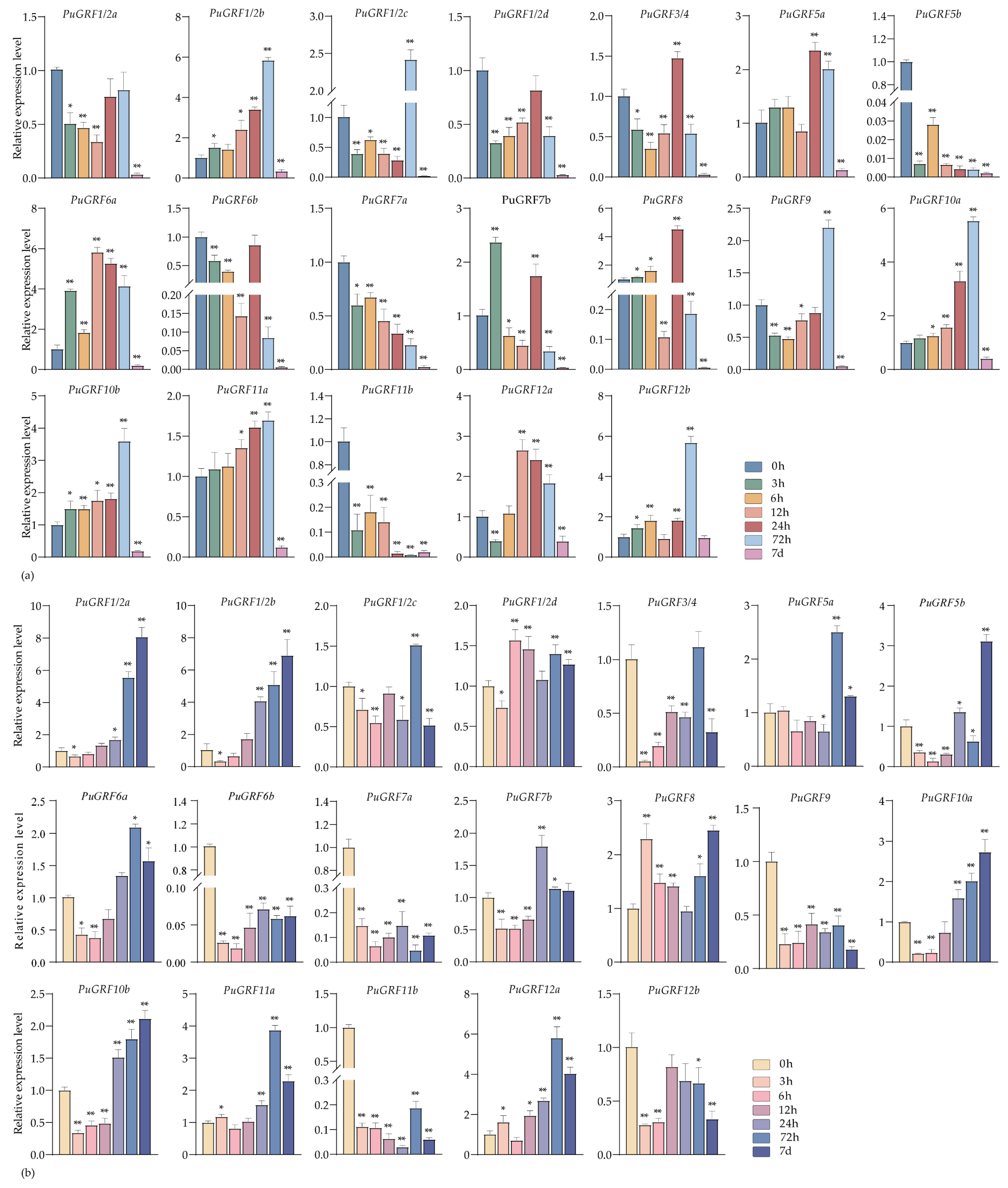
2.4.2. Expression Analysis of PuGRFs Under Salt and Osmotic Stresses

We analyzed the expression patterns of PuGRFs in P. ussuriensis under salt and osmotic stress during the seedling stage at different time points (0 h, 3 h, 6 h, 12 h, 24 h, 48 h, 72 h, and 7 days).
Under salt stress of 200 mM NaCl, in leaves, eight PuGRFs showed a rapid up-regulation response and continued to be induced (Figure 4a). In roots, although a few PuGRFs showed rapid up-regulation in response to the salt stress, six PuGRFs could function under salt stress at 7 days (Figure 4b). The results showed that PuGRF7b, PuGRF8, and PuGRF11b were induced in both leaves and roots under salt stress at 7 days. We speculate that these genes are helpful for P. ussuriensis to adapt to the salt stress environment.
Under the osmotic stress induced by 7% PEG6000, the expression of nine PuGRFs was up-regulated in both leaves and roots at 7 days (Figure 5a,b). We found that PuGRF1/2b and PuGRF8 were consistently up-regulated in leaves, maintaining an active response throughout the stress process at various time points. In roots, most PuGRFs exhibited an expression pattern of being first inhibited by stress and then induced, except for PuGRF1/2b, PuGRF5b, PuGRF6a, PuGRF9, PuGRF11a, PuGRF11b. Among them, PuGRF1/2c, PuGRF1/2d, PuGRF5a, PuGRF6b, PuGRF7b, PuGRF10a, PuGRF12a, and PuGRF12b all reached their highest expression levels at 72 h. These research findings may establish a potential link between PuGRFs and complex drought-response mechanisms.

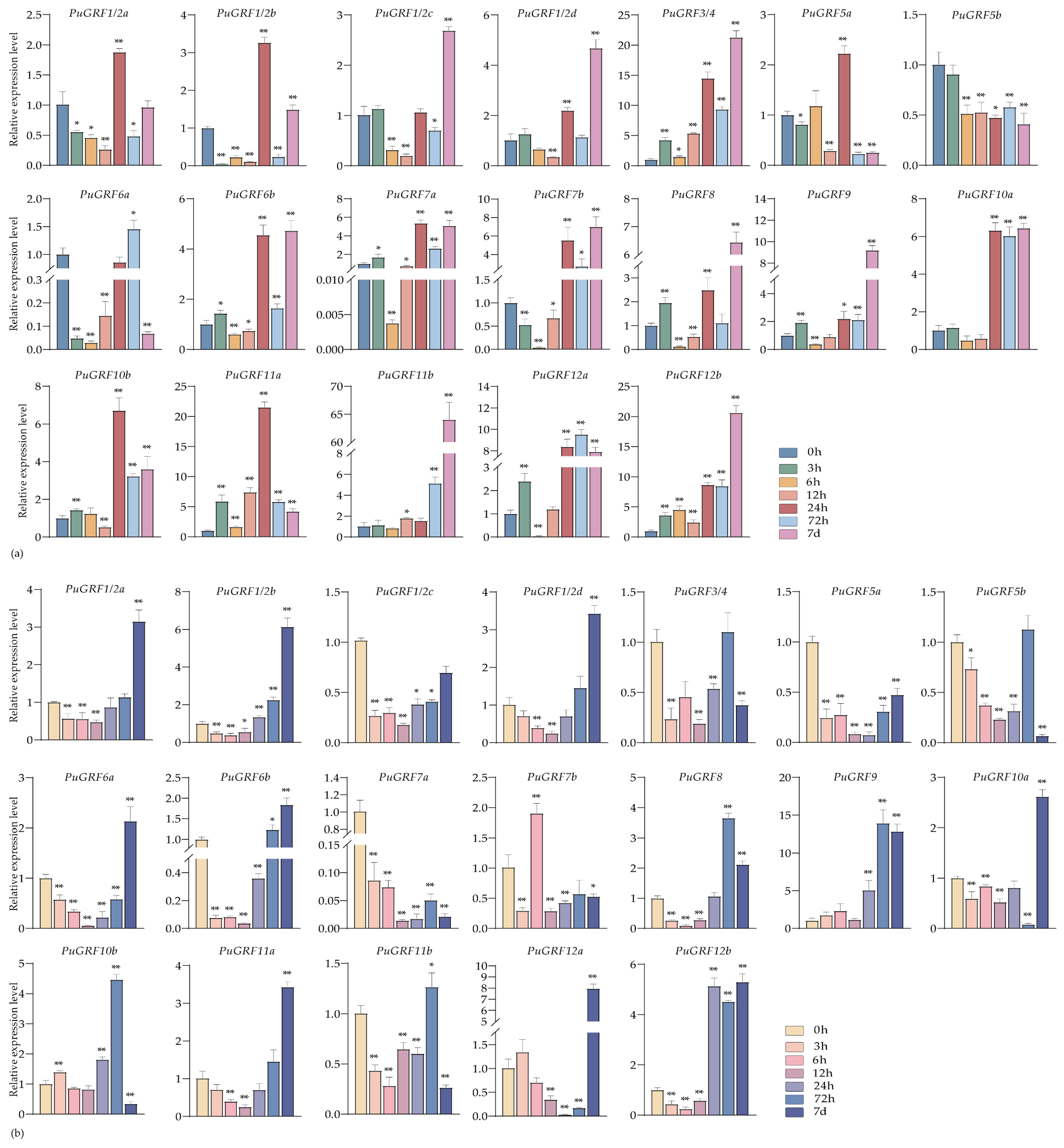
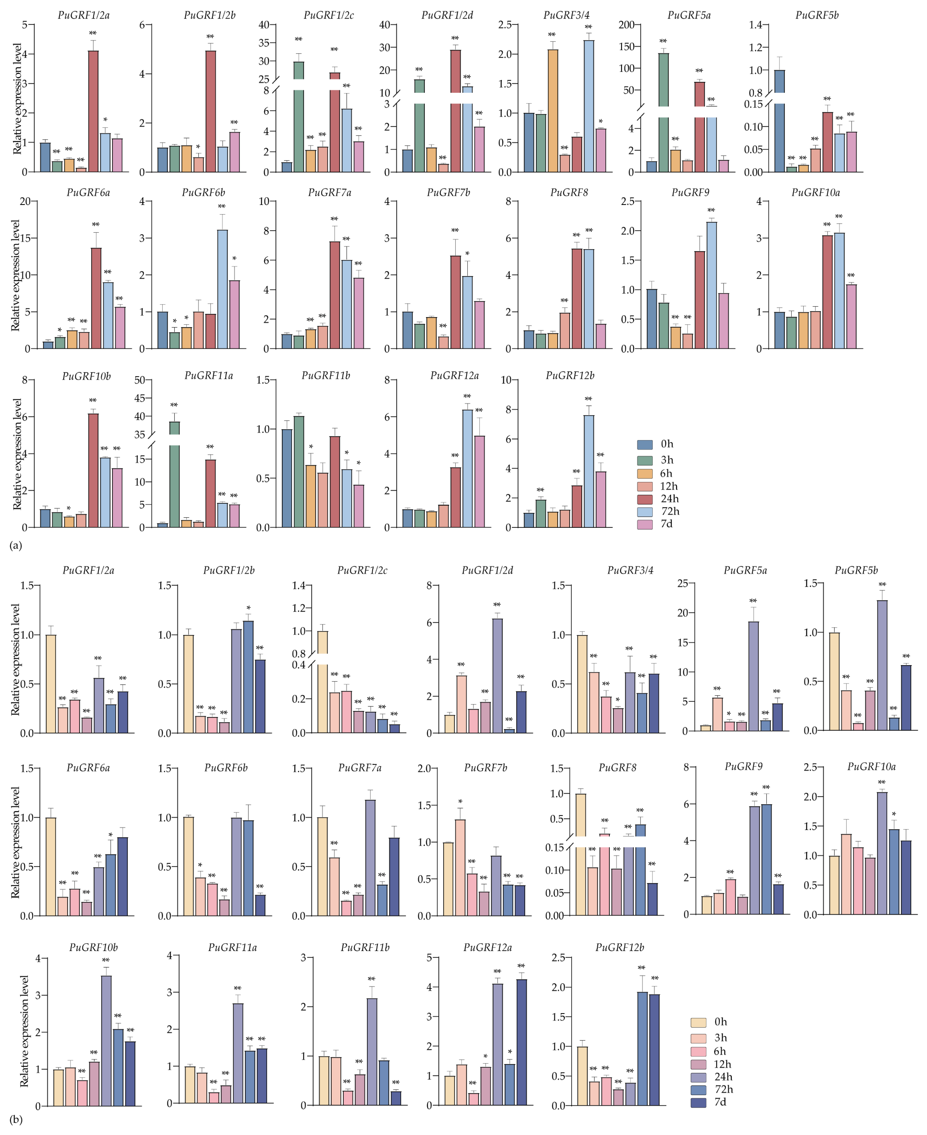
2.4.3. Expression Analysis of PuGRFs Under ABA, MeJA and SA Treatment

We also analyzed the expression patterns of PuGRFs under ABA, MeJA, and SA treatment during the seedling stage at different time points (0 h, 3 h, 6 h, 12 h, 24 h, 48 h, 72 h, and 7 days).
Under the treatment of 200 μM ABA, in leaves, upon reaching a treatment duration of 72 h, the expression levels of seven genes attained their peak values. Under the treatment of ABA at 7 days, the expression levels of all PuGRFs diminished to the lowest point (Figure 6a). In roots, the expression of 12 PuGRFs was induced and up-regulated by 7 days (Figure 6b). Meanwhile, the homologous genes PuGRF1/2a and PuGRF1/2b had a consistent expression pattern in roots and exhibited an almost opposite expression pattern in leaves. This phenomenon prompts us to postulate the existence of functional redundancy and tissue-specificity under the treatment of ABA.
Under the treatment of 100 μM MeJA, the expression of 10 PuGRFs was induced in both leaves and roots at 7 days (Figure 7a,b). As the results indicate, PuGRFs without MeJA-binding elements, such as PuGRF5b, are sensitive to MeJA. However, PuGRFs like PuGRF12b show continuous and significant induced expression in leaves and are also induced in roots during the treatment period from 24 h to 7 days. These results confirm that MeJA treatment can indeed induce the expression of PuGRFs.
Under the treatment of 100 μM SA, in leaves, only PuGRF5b and PuGRF11b showed obvious sensitivity, and the remaining PuGRFs responded to varying degrees, especially with obvious time-specificity at 24 h and 7 days (Figure 8a). In roots, 11 PuGRFs exhibited an expression pattern of being first inhibited and then induced. Among them, except for PuGRF1/2c and PuGRF5a, all PuGRFs started to resume responding and were up-regulated at 24 h (Figure 8b). We speculate that the key time for PuGRFs to respond to SA may start from 24 h. All these results indicate that PuGRFs may be involved in the complex processes of hormone signal transduction and regulation, helping plants to better adapt to environmental stresses.

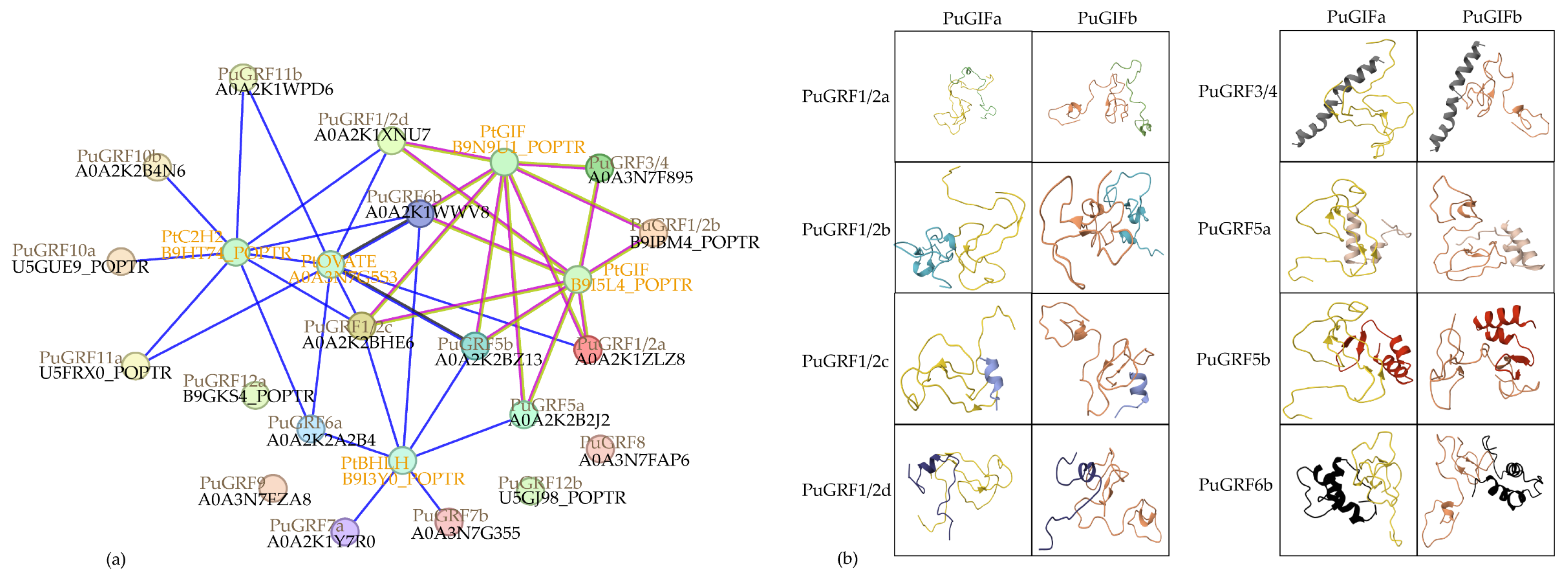
2.5. Predict and Analyze the Interacting Proteins of PuGRFs

Utilizing the STRING12.0 online tool, we found that the possible interaction relationships with PuGRFs can be divided into two categories. One category is the interaction with the GRF’s partner protein GIF. Specifically, PuGRF1/2a, PuGRF1/2b, PuGRF1/2c, PuGRF1/2d, PuGRF3/4, PuGRF5a, PuGRF5b, and PuGRF6b all have interactions with GIFs (B9I5L4 and B9N9U1). The other category is gene co-occurrence with genes possessing the ZnF-C2H2 domain (B9HT74), OVATE domain (A0A3N7G5S3), and BHLH domain (B9I3Y0) (Figure 9a). Through sequence alignment, we obtained the two most homologous PuGIFs in P. ussuriensis, which were named PuGIFa and PuGIFb respectively. The HDOCK server was used to simulate the protein–protein interaction models between the above-mentioned eight PuGRFs and PuGIFa/PuGIFb (Figure 9b). The scoring results showed that, except for the interaction between PuGRF1/2c and the two PuGIFs being unlikely, interactions between the other seven PuGRFs and PuGIFs were possible. Specifically, the likelihood of interaction was moderate between PuGRF1/2a, PuGRF1/2d, PuGRF5b, and the two PuGIFs, between PuGRF3/4 and PuGIFb, and between PuGRF5a and PuGIFa. Interaction was highly likely between PuGRF1/2b, PuGRF6b, and the two PuGIFs, between PuGRF3/4 and PuGIFa, and between PuGRF5a and PuGIFb (Table 2).

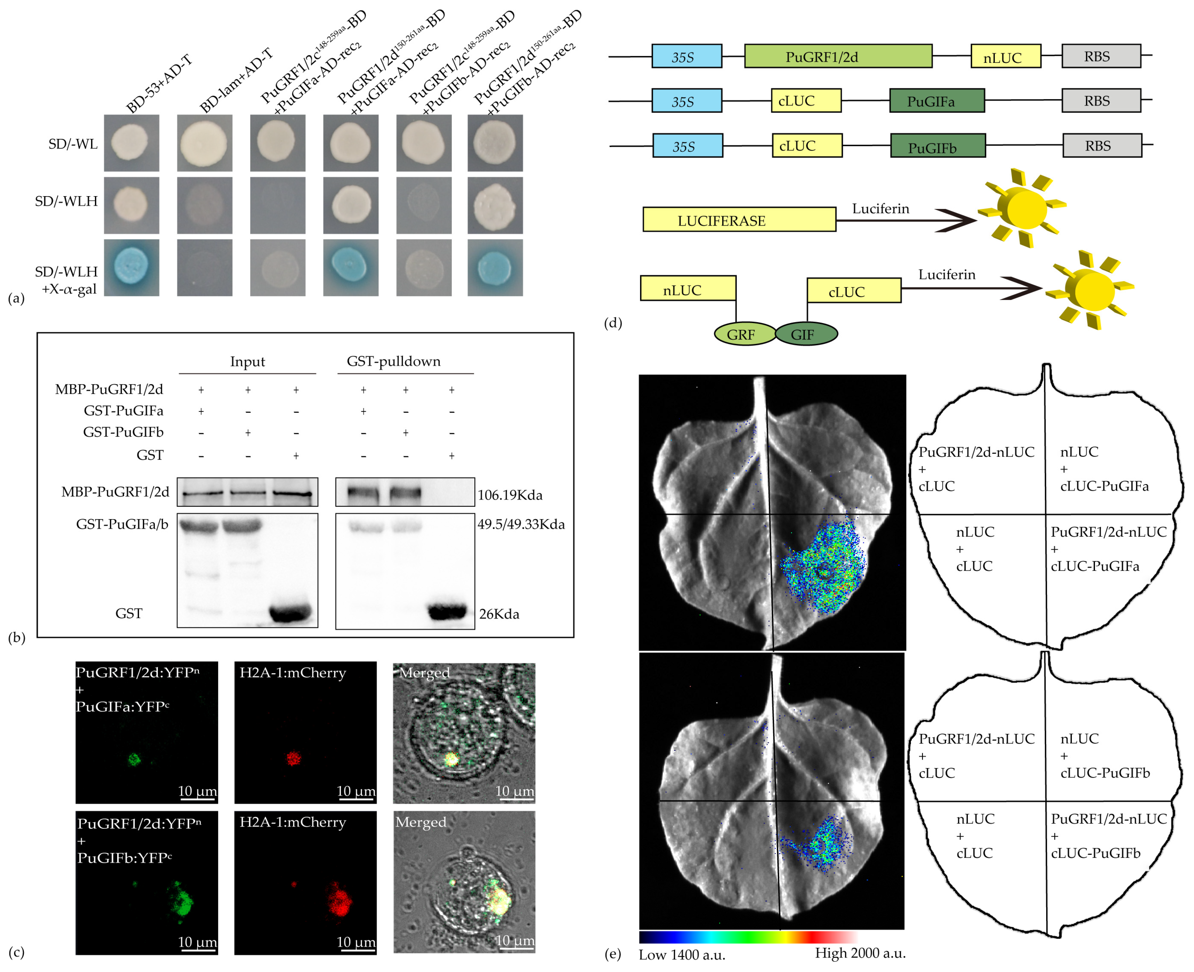
2.6. Verification of the Interaction Relationship Between PuGRFs and PuGIFa/b

Based on the predicted protein interaction results, we selected PuGRF1/2c, PuGRF1/2d, PuGIFa, and PuGIFb for experimental validation. Domain mapping experiments conducted through yeast two-hybrid (Y2H) assays indicated that the QLQ and WRC domains of PuGRF1/2c and PuGRF1/2d were involved in the interaction between PuGRFs and PuGIFs (Figure S2). Consistent with the prediction results of the HDOCK SERVER, PuGRF1/2d interacted with PuGIFa/b, while PuGRF1/2c failed to interact with PuGIFa/b (Figure 10a). Next, in order to determine the accuracy of the interaction between PuGRF1/2d and PuGIFa/b, we further demonstrated, through in vitro pull-down assays, that PuGIFa/b tagged with glutathione S-transferase (GST) (GST-PuGIFa/b), rather than the GST protein itself, could pull down PuGRF1/2d tagged with maltose-binding protein (MBP) (MBP-PuGRF1/2d) (Figure 10b), thus confirming that PuGRF1/2d and PuGIFa/b interact with each other in vitro. Bimolecular fluorescent complementation (BiFC) also showed that yellow fluorescence could only be detected when PuGRF1/2d and PuGIFa/b were fused to the N-terminal and C-terminal of yellow fluorescent protein (YFP), respectively, and were present simultaneously (Figure 10c and Figure S3). This further confirmed that PuGRF1/2d and PuGIFa or PuGIFb interact with each other in vitro.
Subsequently, we utilized the well-established firefly luciferase (LUC) complementation imaging (LCI) assay in Nicotiana benthamiana leaves to determine whether PuGRF1/2d and PuGIFa/b interact with each other in planta. A strong fluorescence signal was observed in N. benthamiana cells co-expressing PuGRF1/2d-nLUC and cLUC-PuGIFa/b, whereas no signal was detected in those co-expressing PuGRF1/2d-nLUC and cLUC, nLUC, and cLUC-GIFs or nLUC and cLUC (Figure 10d,e). These results indicate that PuGRF1/2d and PuGIFa/b interact with each other in vivo.

3. Discussion

As key players in the regulatory network of plant growth and development, GRFs play a crucial role throughout the entire life cycle of plants. GRFs precisely regulate cell proliferation and differentiation, and actively participate in the process of plant responses to various environmental stresses, thereby helping plants to adjust their growth strategies and enhance their tolerance to adverse environments. Although certain progress has been made in the research on the GRF gene family in most herbaceous plants, studies on higher woody plants still lag behind. Therefore, we conducted a genome-wide identification and analysis of the GRF gene family in P. ussuriensis, one of the tree species with strong regional growth, and deeply explored its potential functions and action mechanisms.
We referred to the basic information of GRFs in P. trichocarpa and identified 19 PuGRFs to study their evolutionary relationships, gene structures, and conserved motifs. A phylogenetic tree was constructed with the GRF family members of A. thaliana and O. sativa. The results showed that PuGRFs were unevenly classified into six clades. PuGRFs belonging to the same clade had more similar gene structures and protein motifs. Consistent with this, numerous studies have found that genes within the same clade have similar functions. For instance, OsGRF6, OsGRF7, OsGRF8, and OsGRF9 exhibit a strong response to Magnaporthe oryzae [27]. AtABF2, AtABF3, AtABF4, and TaABF are all key regulatory factors in the ABA signal transduction pathway [28,29]. In our study, different from the gene structures of A. thaliana, O. sativa, and C. sinensis [6,7,30], which have 2–4 exons, all PuGRFs have 4 exons, except for PuGRF9, PuGRF12a, and PuGRF12b in the clade V, which have only 3 exons. Compared with other genes in the same clade, the 4 PuGRFs in clade I have more than 10 protein motifs, especially showing significant diversity at the C-terminus. In addition, the evolutionary branches where most PuGRFs are located have experienced multiple evolutionary nodes. Overall, we speculate that these results are due to the diversification and adaptive changes that PuGRFs have undergone during the evolutionary process.
Plant morphogenesis and every growth stage rely on the tissue-specific expression of various genes. In this study, we selected various tissues from two critical growth stages of P. ussuriensis to analyze the tissue specificity of PuGRFs. The results showed that most PuGRFs were specifically expressed in the buds and YL at the seedling stage, as well as in the ML and AP at the rapid growth stage. This result is consistent with previous studies [11,16,31,32]. Furthermore, some GRFs are expressed in the root cap or meristem and have an important impact on root growth and branching, such as MtGRF5 in Medicago truncatula and PtGRF1/2d [33,34]. This tissue specificity often also represents a degree of functional specificity; for example, the specific expression of AtARF7 and AtARF19 can regulate the formation of lateral roots and primary roots in A. thaliana [35]. The specific expression of MtARF2, MtARF3, and MtARF4 in M. truncatula [36] is a crucial morphogenetic program that controls root system development and the formation of nitrogen-fixing nodules. Compared with herbaceous plants with a relatively short growth cycle, the functions of GRFs in woody plants are perhaps more differentiated in different tissues. For example, PuGRF5b was expressed at a higher level in the roots at the seedling stage, while at the rapid growth stage, it was predominantly expressed in the phloem. We speculated that the function of PuGRF5b in poplar may have evolved based on the potential regulatory effect of its homologous OsGRF1 in response to gibberellin and on stem growth [30]. In addition, PuGRF9 was specifically and highly expressed in the buds at the seedling stage and in the ML and AP at the rapid growth stage. Perhaps it has a similar function to its homologous AtGRF9 [37], that is, negatively regulating leaf growth. We also noticed that PuGRF12a and PuGRF12b, which belong to the same clade as PuGRF9, have different expression patterns and obvious tissue specificity. Wang et al. identified that the overexpression of PagGRF12a and PagGRF12b promoted leaf development [3], while PagGRF12a interacted with PagGIF1b to upregulate PagXND1a expression, thereby inhibiting xylem development in Populus alba × P. glandulosa [38]. Therefore, we infer that this paralogous pair, PuGRF12a and PuGRF12b, may compensate for the negative regulatory function similar to AtGRF9 or represent novel functions evolved in woody plants to influence stem development. Additionally, the overexpression of PagGRF11 resulted in stem development and dwarfing [39]. The complex mechanisms underlying GRF involvement in woody plant growth and development require further dissection.
The key genes of plants exist in intricate regulatory networks based on their adaptability and evolutionary characteristics. These key genes interact with the environment, generating complex molecular and cellular activity patterns, which in turn induce various plant phenotypes, enabling plants to adapt to variable environments. ABA is an important signaling molecule in plants that regulates physiological processes and helps plants to cope with various stress conditions. It can interact and crosstalk with MeJA, SA, etc., to form a complex signaling network, which regulates the development of plant leaves and roots, affects stomatal movement, and stabilizes the cell’s osmotic capacity to adapt to salt or drought stress [40,41]. Our study preliminarily analyzed the expression patterns of PuGRFs under the stresses of NaCl and 7% PEG6000, as well as under the treatments of ABA, MeJA, and SA. The results indicated that PuGRF1/2a, PuGRF1/2b, PuGRF7b, and PuGRF10b were induced in roots under the stresses of NaCl, 7% PEG6000, and ABA treatment, respectively, suggesting that they may be involved in root development-related responses in the ABA-mediated drought-resistance or salt-tolerance pathways. In leaves treated with ABA, MeJA, and SA respectively, 13 genes could be upregulated. Although the expression levels of these genes in leaves under ABA treatment reached their peaks at 24 h or 72 h, most of these genes showed a consistent expression pattern under MeJA and SA treatments. Notably, among these 13 genes, 7 genes (PuGRF6a, PuGRF7b, PuGRF8, PuGRF10a, PuGRF10b, PuGRF12a, and PuGRF12b) were induced in leaves under the stresses of NaCl and 7% PEG6000. These results suggest that these genes are crucial for P. ussuriensis to cope with environmental changes and are likely to respond to drought or salt stress by regulating stomatal changes. Consistent with this, similar findings have also been observed in studies of other plants. For example, ABA upregulates the expression of AtMYB41, AtMYB74, and AtMYB102, and mediates the AtMYB41-BRM module to regulate the stomatal movement of A. thaliana, thereby enhancing its drought tolerance [42]. In the regulatory pathway where AtERF114/115/109 targets non-canonical JAZ proteins to regulate JA signaling, JA induces the transcription of AtERF114. The accumulated AtERF114/115/109 inhibits the activity of JAZ8 by forming a positive feedback loop, further promoting JA-induced root hair elongation [43]. In the AtDELLA-GRF module that regulates growth under cold stress, GA regulates the upregulation of GRF genes following cold stress. Moreover, the cold stress-induced accumulation of DELLA positively regulates their expression, and GRF proteins interact with DELLA [19]. These analytical results establish potential connections among woody plants, ABA signal transduction, the drought-stress response, and the MeJA response, which contribute to coordinating the growth, development, and stress resistance of woody plants.
The miR396-GRF module, characterized by relatively conserved functions, has been demonstrated to play pivotal roles across multiple species. It is intricately involved in research related to plant morphogenesis and stress resistance, as detailed in references [22,44]. Moreover, the more complex integrated module miR396-GRF/GIF has been shown to effectively promote plant regeneration and enhance transgenic efficiency, as reported in [45,46,47]. Evolutionary analysis of GRF and GIF gene families across multiple species reveals that GRF and GIF proteins share similar biological functions, while their interactions are highly conserved [48,49,50]. Given the prediction that all PtGRFs are susceptible to recognition and cleavage by miR396, our initial step was to predict the interacting proteins of PuGRFs. This was aimed at comprehensively exploring and enriching the potential functions of PuGRFs. Through a meticulous analysis of the network interaction relationships among the 19 PuGRFs, we determined that there was no interaction among all members of the PuGRF family. Specifically, eight PuGRFs exhibited a potential connection with PuGIFa/b, while the remaining 11 PuGRFs were more likely to cooccur in specific environmental contexts. Subsequently, we harnessed the HDOCK server to simulate the interaction model between PuGRFs and PuGIFs in P. ussuriensis. Intriguingly, we discovered that PuGRF1/2c and PuGRF1/2d, which belong to the same clade and possess highly similar sequences, might have divergent interaction relationships with PuGIFa/b. This finding was corroborated through a series of well-designed experiments. The distinct interaction effects not only reflect the remarkable adaptability of plants to their surroundings, but also underscore the conservation of GRF evolution. On one hand, plants are capable of leveraging existing protein structures and functional modules and can achieve novel functions through subtle modifications. For instance, under particular environmental conditions, such as periods of stress or specific growth stages, PuGRF1/2d, which interacts with PuGIFa/b, exerts its function. Conversely, under other conditions, PuGRF1/2c, which does not interact with PuGIFa/b, becomes active, enabling plants to respond nimbly to diverse environmental challenges. On the other hand, these two PuGRFs with strikingly similar sequences may exhibit functional redundancy, implying that they might still perform their functions, even in the absence of PuGIFa/b. In any event, this divergence merits our close attention. Whether it represents functional specificity or redundancy ultimately necessitates further experimental validation. These findings collectively indicate that the functional roles of GRF proteins are intricately linked to their multifaceted interaction networks, thereby establishing a molecular framework that connects GRF-mediated biological processes with their dynamically evolving protein partnership landscapes.
In summary, our research represents the first in-depth exploration of the GRF gene family in P. ussuriensis. Through a comprehensive step-by-step analysis of the evolutionary trends and functional predictions, we found a substantial amount of reliable evidence that indicates that PuGRFs are mainly expressed in young tissues during the seedling stage, and there is a significant differentiation in tissue specificity during the rapid growth stage. Additionally, PuGRFs mainly respond to hormonal changes in leaves, and they may be mediated by ABA and involved in root development and stomatal movement under drought or salt stress. Further predictive exploration suggested potential interaction relationships between PuGRFs and genes of other domain types, among which eight PuGRFs might interact with PuGIFa/b. Through experiments, we also found that the homologous genes PuGRF1/2c and PuGRF1/2d, despite their highly identical sequences, showed significant differences in their interactions with PuGIFa/b.

4. Materials and Methods

4.1. Identifcation of PuGRFs in P. ussuriensis

The genomic information of P. ussuriensis was derived from a large amount of transcriptional sequencing data within our research group. We downloaded the GRF family proteins of P. trichocarpa, A. thaliana, and O. sativa from Phytozome (https://phytozome-next.jgi.doe.gov/, accessed on 15 April 2024), TAIR (https://www.arabidopsis.org/, accessed on 15 April 2024), and RiceData (http://www.ricedata.cn/gene/, accessed on 15 April 2024). Based on the GRF proteins of known species, we used BLASTP (BLAST: Basic Local Alignment Search Tool, accessed on 15 April 2024) to search for potential GRF transcription factors (TFs) in P. ussuriensis. Potential GRF transcription factors were identified from the protein sequences of P. ussuriensis by HMMER3 (http://www.hmmer.org/, accessed on 15 April 2024). The results of BLASTP and HMMER3 were comprehensively analyzed to select potential GRFs. The protein domains of the originally identified members were analyzed using the online software SMART (http://smart.embl.de/, accessed on 15 April 2024) and NCBI-CDD (https://www.ncbi.nlm.nih.gov/cdd/, accessed on 15 April 2024). The physical and chemical properties of PuGRFs were analyzed using the online tool ExPASy (SIB Swiss Institute of Bioinformatics | Expasy, https://www.expasy.org/, accessed on 15 April 2024). The subcellular location was predicted by Plant-mPLoc (http://www.csbio.sjtu.edu.cn/bioinf/plant/, accessed on 15 April 2024). The microRNAs were predicted by psRNATarget (psRNATarget: A Plant Small RNA Target Analysis Server (2017 Update), https://www.zhaolab.org/psRNATarget/, accessed on 27 April 2024).

4.2. Phylogenetic Investigation of the PuGRFs

The ClustalW method [51] was employed to analyze P. ussuriensis, A. thaliana, and O. sativa. The phylogenetic tree of the GRF family was constructed by the neighbor-joining (NJ) method. In MEGA7.0 software [52], 1000 iterations were performed for bootstrap testing, and then the phylogenetic tree was beautified using the online software Evolview (https://evolgenius.info/evolview, accessed on 6 May 2024).

4.3. Analysis of Conserved Motifs, Gene Structure, and Promoter Regions in PuGRFs

The conserved motifs for PuGRF proteins were identified using MEME (https://meme-suite.org/meme/tools/meme, accessed on 20 May 2024) [53]. The parameters were set to 20 motifs while keeping other parameters at their default values. Detailed information about 20 conserved motifs is listed in the Supplementary Table S4. The gene structure was visualized using TBtools (version 2.202, South China Agricultural University, China) [54]. The promoters of the PuGRFs were obtained from the genome of P. ussuriensis. The promoter region, specifically the 3 kb sequences upstream of the translation initiation site (ATG), was analyzed by the online software PlantCARE (http://bioinformatics.psb.ugent.be/webtools/plantcare/html/, accessed on 17 April 2024) [55] to identify the cis-acting elements. The conserved motifs and cis-elements of the PuGRFs were visualized with TBtools.

4.4. Plant Materials and Treatments

The plant material was sourced from the wild-type P. ussuriensis at Northeast Forestry University. It was sterilized with 0.1% HgCl2 solution and then cultured in woody plant medium (WPM) supplemented with 20 g/L sucrose and 6 g of agar using tissue culture technology [56,57]. The tissue culture chamber was maintained at 25 °C with a photoperiod of 16 h of light and 8 h of darkness and a light intensity of 46 µmol m−2 s−1. Uniformly grown, healthy three-week-old in vitro plants (seedling stage) obtained from the above culture protocols were used for abiotic stress treatments (21 plant samples per treatment, with 3 plants per time point), phytohormone treatment (21 plant samples per treatment, with 3 plants per time point), and tissue-specific analysis (different tissues from 40 plant samples were pooled separately). Additionally, uniformly grown, healthy two-month-old soil-grown seedlings (rapid growth stage) were used for tissue-specific analysis (different tissues from 12 plant samples were pooled separately). For abiotic stress and phytohormone treatments, plants were exposed to a WPM medium containing 200 mM NaCl, 7% PEG6000, 200 µM ABA, 100 µM MeJA, and 100 µM SA. The concentrations of NaCl, PEG6000, ABA, SA, and MeJA were based on repeated laboratory trials and slightly adjusted [58,59,60,61]. Each treatment lasted for 0 h, 3 h, 6 h, 12 h, 24 h, 72 h, and 7 d. Three biological replicates were conducted for each treatment, and roots and leaves at different time points need to be quickly frozen with liquid nitrogen at −80 °C for later use.

4.5. RNA Isolation and RT-qPCR Analysis

Highly purified RNA was extracted by employing the Universal Plant Total RNA Isolation Kit (Spin-Column, BioTeke, Beijing China). The MonScript™ RTIII All-in-One Mix with dsDNase (monadbiotech, Suzhou, China) was utilized to obtain cDNA, with the following reaction system: 1000 ng of total RNA, 4 µL of MonScript™ 5×RTIII All-in-One Mix, 1 µL of MonScript™ dsDNase, and an appropriate amount of nuclease-free water to bring the total volume to 20 µL. The cDNA was diluted 10-fold (cDNA: nuclease-free water = 1:10) for further RT-qPCR analysis. The RT-qPCR specific primers were designed by the NCBI Primer Blast tool (https://www.ncbi.nlm.nih.gov/tools/primer-blast/index.cgi, accessed on 31 May 2024), and the β-Actin gene (EF418792.1) was selected as an internal reference. TransStart® Top Green qPCR Super Mix (TransGen-AQ131, Beijing, China) was used to conduct RT-qPCR, with the following reaction system: 10 µL of 2 × TransStart® Top Green qPCR Super Mix, 6 µL of double-distilled H2O, 2 µL of diluted template, and 1 µL each of forward primer and reverse primer. The qTOWER 3G Cycler and qPCR software (version 3.2, Analytik Jena, Jena, Germany) were used as a work program, and the 2−∆∆CT method [62] was employed to perform the relative gene expression level analysis. Triplicate independent technical and biological replications were used in the analysis. The primers for RT-qPCR are listed in Supplementary Table S1. SPSS software (version 20, IBM, Chicago, IL, USA) was used to analyze the data by performing one-way analysis of variance (ANOVA) to assess significant differences between the control and each treatment. Significance was defined as * p < 0.05 and ** p < 0.01.

4.6. Protein-Protein Interaction Studies of PuGRFs

To analyze the protein-protein interactions (PPIs), the STRING 12.0 database [63] (https://string-db.org/, accessed on 8 May 2024) was employed, utilizing the default parameters and Populus trichocarpa as the reference organism. HDOCK SERVER [20,64,65,66,67,68] (http://hdock.phys.hust.edu.cn/, accessed on 8 May 2024) was used to simulate Protein-protein docking.

4.7. Y2H Assays

The Y2H assay was carried out using the Matchmaker Gold Yeast Two-Hybrid System (Coolaber-YH2011, Beijing, China). To detect the interactions between PuGRF1/2c, PuGRF1/2d, and two PuGIF proteins (PuGIFa and PuGIFb), the coding sequences (CDS) of PuGRF1/2c and PuGRF1/2d were amplified from complementary DNA (cDNA) and cloned into the pGBKT7 vector. Meanwhile, the full-length coding sequences of PuGIFa and PuGIFb were cloned into the pGADT7-rec2 vector. To identify the domains of PuGRF1/2c and PuGRF1/2d involved in the interaction, the coding sequences of PuGRF1/2c and PuGRF1/2d and their derivative fragments were cloned into the pGBKT7 vector. The primers used for plasmid construction are listed in Supplementary Tables S2 and S3. Then, they were transformed into the yeast (Saccharomyces cerevisiae) strain Y2H Gold respectively. The transformed yeast cell suspensions were spread onto the plates of SD/-Trp, SD/-Trp/-His/-Ade, and SD/-Trp + X-a-gal medium. PuGRF1/2c148−259aa, PuGRF1/2d150−261aa, and the two PuGIFs were co-transformed into the yeast strain Y2H Gold respectively. Positive (BD-53 + AD-T) and negative (BD-lam + AD-T) controls were included. The transformed yeast cell suspensions were spread onto the plates of SD/-Trp/-Leu (-WL), SD/-Trp/-Leu/-His(-WLH), and SD/-Trp/-Leu/-His+ X-a-ga (-WLH+ X-a-gal) medium to detect protein–protein interactions. All the plates were incubated at 30 °C for 72 h and experiments were repeated three times.

4.8. BiFC

For BiFC assays, the full-length coding sequences of PuGRF1/2d, PuGIFa, and PuGIFb were inserted into the pENTER/D-TOPO vector respectively. Subsequently, they were recombined with nEYFP/pUGW2 and cEYFP/pUGW2 using Gateway recombination technology, respectively. According to the established protocol [69], each pair of constructs was co-transfected into poplar protoplasts together with the nuclear localization marker H2A–1:mCherry. The empty vectors of yellow fluorescent protein C-terminus (YFPC) and yellow fluorescent protein N-terminus (YFPN) were used as a negative control. A confocal laser-scanning microscope (LSM 800; Zeiss, Oberkochen, Germany)was employed to detect the fluorescence signals. Three biological replicates were tested for both the interaction groups and the negative control groups. The primers used for plasmid construction are listed in the Supplementary Tables S2 and S3.

4.9. LCI Assays

The LCI assays were performed in N. benthamiana leaves according to the method described previously [70]. The full-length PuGRF1/2d was cloned into the pCAMBIA1300-nLUC vector, while the full-length PuGIFa and PuGIFb were cloned into the pCAMBIA1300-cLUC vector. The primers used for plasmid construction are listed in Supplementary Tables S2 and S3. The resulting constructs were separately introduced into Agrobacterium tumefaciens strain GV3101(p19). The transformed cells were cultured in LB liquid medium containing 2-(N-morpholino) ethanesulfonic acid (Sigma, Darmstadt, Germany) and acetosyringone (Sigma, Germany) at 28 °C. After culturing at 28 °C for 16 h, the bacterial cells were harvested and resuspended in sterile water containing MgCl2 and acetosyringone to an optical density (OD600) of 1.5. Equal volumes of the two bacterial suspensions carrying the specific constructs were mixed, incubated at room temperature for 3 h, and then injected into the leaves of N. benthamiana. The plants with infiltrated leaves were cultured at 23 °C under a 16 h light/8 h dark photoperiod for 3–4 days. Then, 1 mM luciferin was sprayed onto the infiltrated leaves, which were left in the dark at 37 degrees Celsius for 10 min, and a CCD imaging device was used to capture LUC images.

4.10. In Vitro Pull-Down Assays

The in vitro pull-down assay was carried out with finetuning according to the previously described method [71]. An amount of 200 μL of MBP-PuGRF1/2d was mixed with equal amounts of GST-PuGIFa and GST-PuGIFb and then added to 50 μL of glutathione magnetic beads (PureCube Glutathione MagBeads32201, Cube Biotech, Monheim, Germany) equilibrated with BW buffer (50 mM NaH2PO4, 150 mM NaCl) in a 2 mL centrifuge tube. The mixture was incubated at 4 °C for two hours. Beads were washed four times, and proteins were eluted with 40 μL elution buffer (125 mM Tris base, 150 mM NaCl, 0.1% (v/v) Triton X-100, 50 mM Reduced glutathione, and 1 mM DTT). Eluates were resolved by SDS-PAGE and immunoblotted using anti-GST (1:3000, Abclonal-AE006, ABclonal Technology, Wuhan, China) and anti-MBP (1:3000, Abclonal-AE016, ABclonal Technology, Wuhan, China) antibodies. The primers used for plasmid construction are listed in Supplementary Tables S2 and S3.

5. Conclusions

In this study, we identified 19 PuGRFs in P. ussuriensis and characterized their physicochemical properties, evolutionary relationships, and dynamic expression profiles. Phylogenetic analysis classified these genes into six distinct clades, revealing lineage-specific structural conservation and diversification. Notably, PuGRFs exhibited pronounced tissue-specific expression patterns, predominantly in young tissues during seedling stages and diversified expression across mature tissues during rapid growth phases. Furthermore, these genes demonstrated robust responsiveness to abiotic stresses (salt and osmotic) and phytohormones (ABA, MeJA, and SA), with distinct temporal and spatial regulatory dynamics. Protein interaction predictions and experimental validations revealed functional divergence among closely related PuGRFs, suggesting evolutionary fine-tuning and potential redundancy. This duality highlights the adaptability of GRFs in balancing conserved roles and lineage-specific innovations. Our findings not only provide the first genome-wide framework for the GRF family in P. ussuriensis, but also uncover critical insights into their roles in coordinating growth, stress responses, and hormonal signaling.

Supplementary Materials

The following supporting information can be downloaded at: https://www.mdpi.com/article/10.3390/ijms26073288/s1.

Author Contributions

The study was conceived and directed by M.W. and C.L. Y.Z. performed experiments and wrote the original draft, and M.W. and C.L. edited the manuscript. Y.L., Y.C. and H.Z. performed the data analysis. All authors have read and agreed to the published version of the manuscript.

Funding

This research was funded by the National Natural Science Foundation of China (32371899 and 32201577).

Institutional Review Board Statement

Not applicable.

Informed Consent Statement

Not applicable.

Data Availability Statement

I have shared the code to my data in the manuscript.

Conflicts of Interest

The authors declare that they have no known competing financial interests or personal relationships that could have appeared to influence the work reported in this paper.

References

  1. Kooke, R.; Keurentjes, J.J.B. Multi-dimensional regulation of metabolic networks shaping plant development and performance. J. Exp. Bot. 2011, 63, 3353–3365. [Google Scholar] [CrossRef] [PubMed]
  2. Van der Knapp, E.; Kim, J.H.; Kende, H. A novel gibberellin-induced gene from rice and its potential regulatory role in stem growth. Plant Physiol. 2000, 122, 695–704. [Google Scholar]
  3. Wang, J.; Zhou, H.; Zhao, Y.; Sun, P.; Tang, F.; Song, X.; Lu, M.-Z. Characterization of poplar growth-regulating factors and analysis of their function in leaf size control. BMC Plant Biol. 2020, 20, 509. [Google Scholar] [CrossRef]
  4. Khisti, M.; Avuthu, T.; Yogendra, K.; Valluri, V.K.; Kudapa, H.; Reddy, P.S.; Tyagi, W. Genome-wide identification and expression profiling of growth-regulating factor (GRF) and GRF-interacting factor (GIF) gene families in chickpea and pigeonpea. Sci. Rep. 2024, 14, 17178. [Google Scholar] [CrossRef] [PubMed]
  5. Tang, Y.; Cheng, W.; Li, S.; Li, Y.; Wang, X.; Xie, J.; He, Y.; Wang, Y.; Niu, Y.; Bao, X.; et al. Genome-wide identification and expression analysis of the growth regulating factor (GRF) family in Jatropha curcas. PLoS ONE 2021, 16, e0254711. [Google Scholar]
  6. Fu, M.-K.; He, Y.-N.; Yang, X.-Y.; Tang, X.; Wang, M.; Dai, W.-S. Genome-wide identification of the GRF family in sweet orange (Citrus sinensis) and functional analysis of the CsGRF04 in response to multiple abiotic stresses. BMC Genom. 2024, 25, 37. [Google Scholar] [CrossRef]
  7. Kim, J.H.; Choi, D.; Kende, H. The AtGRF family of putative transcription factors is involved in leaf and cotyledon growth in Arabidopsis. Plant J. 2003, 36, 94–104. [Google Scholar] [CrossRef]
  8. Kim, J.H. Biological roles and an evolutionary sketch of the GRF-GIF transcriptional complex in plants. BMB Rep. 2019, 52, 227–238. [Google Scholar] [CrossRef]
  9. Lee, B.H.; Ko, J.-H.; Lee, S.; Lee, Y.; Pak, J.-H.; Kim, J.H. The Arabidopsis GRF-INTERACTING FACTOR Gene Family Performs an Overlapping Function in Determining Organ Size as Well as Multiple Developmental Properties. Plant Physiol. 2009, 151, 655–668. [Google Scholar] [CrossRef]
  10. Hoe, J.K.; Tsukaya, H. Regulation of plant growth and development by the GROWTH-REGULATING FACTOR and GRF-INTERACTING FACTOR duo. J. Exp. Bot. 2015, 66, 6093–6107. [Google Scholar]
  11. Cheng, Z.; Wen, S.; Wu, Y.; Shang, L.; Wu, L.; Lyu, D.; Yu, H.; Wang, J.; Jian, H. Comparatively Evolution and Expression Analysis of GRF Transcription Factor Genes in Seven Plant Species. Plants 2023, 12, 2790. [Google Scholar] [CrossRef] [PubMed]
  12. Li, Z.; Xie, Q.; Yan, J.; Chen, J.; Chen, Q. Genome-Wide Identification and Characterization of the Abiotic-Stress-Responsive GRF Gene Family in Diploid Woodland Strawberry (Fragaria vesca). Plants 2021, 10, 1916. [Google Scholar] [CrossRef] [PubMed]
  13. Cao, J.-F.; Huang, J.-Q.; Liu, X.; Huang, C.-C.; Zheng, Z.-S.; Zhang, X.-F.; Shangguan, X.-X.; Wang, L.-J.; Zhang, Y.-G.; Wendel, J.F.; et al. Genome-wide characterization of the GRF family and their roles in response to salt stress in Gossypium. BMC Genom. 2020, 21, 575. [Google Scholar] [CrossRef]
  14. Horiguchi, G.; Kim, G.; Tsukaya, H. The transcription factor AtGRF5 and the transcription coactivator AN3 regulate cell proliferation in leaf primordia of Arabidopsis thaliana. Plant J. 2005, 43, 68–78. [Google Scholar] [CrossRef]
  15. Duan, P.; Ni, S.; Wang, J.; Zhang, B.; Xu, R.; Wang, Y.; Chen, H.; Zhu, X.; Li, Y. Regulation of OsGRF4 by OsmiR396 controls grain size and yield in rice. Nat. Plants 2015, 2, 15203. [Google Scholar] [CrossRef]
  16. Jathar, V.; Saini, K.; Chauhan, A.; Rani, R.; Ichihashi, Y.; Ranjan, A. Spatial control of cell division by GA-OsGRF7/8 module in a leaf explaining the leaf length variation between cultivated and wild rice. New Phytol. 2022, 234, 867–883. [Google Scholar]
  17. Kim, J.S.; Mizoi, J.; Kidokoro, S.; Maruyama, K.; Nakajima, J.; Nakashima, K.; Mitsuda, N.; Takiguchi, Y.; Ohme-Takagi, M.; Kondou, Y.; et al. Arabidopsis GROWTH-REGULATING FACTOR7 functions as a transcriptional repressor of abscisic acid-and osmotic stress–responsive genes, including DREB2A. Plant Cell 2012, 24, 3393–3405. [Google Scholar]
  18. Liu, Z.-Y.; Han, Y.-T.; Wang, C.-Y.; Lei, X.-J.; Wang, Y.-Y.; Dong, W.-F.; Xie, Q.-J.; Fu, Y.-J.; Gao, C.-Q. The growth-regulating factor PdbGRF1 positively regulates the salt stress response in Populus davidiana × P. bolleana. Plant Sci. 2022, 326, 111502. [Google Scholar] [CrossRef] [PubMed]
  19. Lantzouni, O.; Alkofer, A.; Falter-Braun, P.; Schwechheimer, C. GROWTH-REGULATING FACTORS Interact with DELLAs and Regulate Growth in Cold Stress. Plant Cell 2020, 32, 1018–1034. [Google Scholar] [CrossRef]
  20. Jones-Rhoades, M.W.; Bartel, D.P. Computational Identification of Plant MicroRNAs and Their Targets, Including a Stress-Induced miRNA. Mol. Cell 2004, 14, 787–799. [Google Scholar] [CrossRef]
  21. Debernardi, J.M.; Rodriguez, E.R.; Mecchia, M.A.; Palatnik, J.F. Functional specialization of the plant miR396 regulatory network through distinct microRNA-target interactions. PLoS Genet. 2012, 8, e1002419. [Google Scholar] [CrossRef]
  22. Liebsch, D.; Palatnik, J.F. MicroRNA miR396, GRF transcription factors and GIF co-regulators: A conserved plant growth regulatory module with potential for breeding and biotechnology. Curr. Opin. Plant Biol. 2020, 53, 31–42. [Google Scholar] [CrossRef] [PubMed]
  23. Zhang, H.; Yang, J.; Li, W.; Chen, Y.; Lu, H.; Zhao, S.; Li, D.; Wei, M.; Li, C. PuHSFA4a Enhances Tolerance To Excess Zinc by Regulating Reactive Oxygen Species Production and Root Development in Populus. Plant Physiol. 2019, 180, 2254–2271. [Google Scholar] [CrossRef] [PubMed]
  24. Wei, M.; Liu, Q.; Wang, Z.; Yang, J.; Li, W.; Chen, Y.; Lu, H.; Nie, J.; Liu, B.; Lv, K.; et al. PuHox52-mediated hierarchical multilayered gene regulatory network promotes adventitious root formation in Populus ussuriensis. New Phytol. 2020, 228, 1369–1385. [Google Scholar] [CrossRef] [PubMed]
  25. Wei, M.; Zhang, M.; Sun, J.; Zhao, Y.; Pak, S.; Ma, M.; Chen, Y.; Lu, H.; Yang, J.; Wei, H.; et al. PuHox52 promotes coordinated uptake of nitrate, phosphate, and iron under nitrogen deficiency in Populus ussuriensis. J. Integr. Plant Biol. 2022, 65, 791–809. [Google Scholar]
  26. Schmitz, R.J.; Grotewold, E.; Stam, M. Cis-regulatory sequences in plants: Their importance, discovery, and future challenges. Plant Cell 2021, 34, 718–741. [Google Scholar] [CrossRef]
  27. Chandran, V.; Wang, H.; Gao, F.; Cao, X.-L.; Chen, Y.-P.; Li, G.-B.; Zhu, Y.; Yang, X.-M.; Zhang, L.-L.; Zhao, Z.-X.; et al. miR396-OsGRFs Module Balances Growth and Rice Blast Disease-Resistance. Front. Plant Sci. 2019, 9, 1999. [Google Scholar] [CrossRef]
  28. Rikiishi, K.; Matsuura, T.; Maekawa, M. TaABF1, ABA response element binding factor 1, is related to seed dormancy and ABA sensitivity in wheat (Triticum aestivum L.) seeds. J. Cereal Sci. 2010, 52, 236–238. [Google Scholar] [CrossRef]
  29. Gao, S.; Gao, J.; Zhu, X.; Song, Y.; Li, Z.; Ren, G.; Zhou, X.; Kuai, B. ABF2, ABF3, and ABF4 Promote ABA-Mediated Chlorophyll Degradation and Leaf Senescence by Transcriptional Activation of Chlorophyll Catabolic Genes and Senescence-Associated Genes in Arabidopsis. Mol. Plant 2016, 9, 1272–1285. [Google Scholar] [CrossRef]
  30. Choi, D.; Kim, J.H.; Kende, H. Whole Genome Analysis of the OsGRF Gene Family Encoding Plant-Specific Putative Transcription Activators in Rice (Oryza sativa L.). Plant Cell Physiol. 2004, 45, 897–904. [Google Scholar] [CrossRef]
  31. Wu, W.; Li, J.; Wang, Q.; Lv, K.; Du, K.; Zhang, W.; Li, Q.; Kang, X.; Wei, H. Growth-regulating factor 5 (GRF5)-mediated gene regulatory network promotes leaf growth and expansion in poplar. New Phytol. 2021, 230, 612–628. [Google Scholar] [CrossRef]
  32. Beltramino, M.; Ercoli, M.F.; Debernardi, J.M.; Goldy, C.; Rojas, A.M.L.; Nota, F.; Alvarez, M.E.; Vercruyssen, L.; Inzé, D.; Palatnik, J.F.; et al. Robust increase of leaf size by Arabidopsis thaliana GRF3-like transcription factors under different growth conditions. Sci. Rep. 2018, 8, 13447. [Google Scholar] [CrossRef]
  33. Zhou, H.; Wei, K.; Jiang, C.; Zhao, J.Q.; Song, X.Q.; Liu, M.Z. Negative negulation of GRF1/2d on the formation and development of adventitious roots in Populus alba × P. glandulosa “84K”. Sci. Silvae Sin. 2017, 53, 33–39. [Google Scholar]
  34. Bazin, J.; Khan, G.A.; Combier, J.P.; Bustos-Sanmamed, P.; Manuel Debernardi, J.; Rodriguez, R.; Sorin, C.; Palatnik, J.; Hartmann, C.; Crespi, M.; et al. miR396 affects mycorrhization and root meristem activity in the legume Medicago truncatula. Plant J. 2013, 74, 920–934. [Google Scholar] [CrossRef] [PubMed]
  35. Lee, H.W.; Cho, C.; Pandey, S.K.; Park, Y.; Kim, M.-J.; Kim, J. LBD16 and LBD18 acting downstream of ARF7 and ARF19 are involved in adventitious root formation in Arabidopsis. BMC Plant Biol. 2019, 19, 46. [Google Scholar] [CrossRef]
  36. Kirolinko, C.; Hobecker, K.; Wen, J.; Mysore, K.S.; Niebel, A.; Blanco, F.A.; Zanetti, M.E. Auxin Response Factor 2 (ARF2), ARF3, and ARF4 Mediate Both Lateral Root and Nitrogen Fixing Nodule Development in Medicago truncatula. Front. Plant Sci. 2021, 12, 659061. [Google Scholar] [CrossRef]
  37. Poethig, R.S.; Omidbakhshfard, M.A.; Olas, J.J.; Xue, G.-P.; Balazadeh, S.; Mueller-Roeber, B. GROWTH-REGULATING FACTOR 9 negatively regulates arabidopsis leaf growth by controlling ORG3 and restricting cell proliferation in leaf primordia. PLoS Genet. 2018, 14, e1007484. [Google Scholar]
  38. Wang, J.; Zhou, H.; Zhao, Y.; Jiang, C.; Li, J.; Tang, F.; Liu, Y.; Zhao, S.; Hu, J.; Song, X.; et al. PagGRF12a interacts with PagGIF1b to regulate secondary xylem development through modulating PagXND1a expression in Populus alba × P. glandulosa. J. Integr. Plant Biol. 2021, 63, 1683–1694. [Google Scholar]
  39. Tian, Y.; Zhao, Y.; Sun, Y.; El-Kassaby, Y.A.; Song, G.; Mi, Y.; Han, J.; Li, Y. PagGRF11 Overexpression Promotes Stem Development and Dwarfing in Populus. Int. J. Mol. Sci. 2022, 23, 7858. [Google Scholar] [CrossRef]
  40. Iqbal, S.; Wang, X.; Mubeen, I.; Kamran, M.; Kanwal, I.; Díaz, G.A.; Abbas, A.; Parveen, A.; Atiq, M.N.; Alshaya, H.; et al. Phytohormones Trigger Drought Tolerance in Crop Plants: Outlook and Future Perspectives. Front. Plant Sci. 2022, 12, 799318. [Google Scholar] [CrossRef]
  41. Kim, G.; Ryu, H.; Sung, J. Hormonal Crosstalk and Root Suberization for Drought Stress Tolerance in Plants. Biomolecules 2022, 12, 811. [Google Scholar] [CrossRef] [PubMed]
  42. Gao, L.; Lv, Q.; Wang, L.; Han, S.; Wang, J.; Chen, Y.; Zhu, W.; Zhang, X.; Bao, F.; Hu, Y.; et al. Abscisic acid–mediated autoregulation of the MYB41-BRAHMA module enhances drought tolerance in Arabidopsis. Plant Physiol. 2024, 196, 1608–1626. [Google Scholar] [CrossRef] [PubMed]
  43. Sui, J.; Yin, Q.; Chen, Y.; Sun, M.; Yuan, X.; Ding, Z.; Kong, X. ERF114/115/109 are essential for jasmonate-repressed non-canonical JAZ8 activity in JA signaling. Cell Rep. 2025, 44, 115222. [Google Scholar] [CrossRef]
  44. Kim, Y.; Takahashi, S.; Miyao, M. Relationship between reduction in rice (Nipponbare) leaf blade size under elevated CO2 and miR396-GRF module. Plant Signal. Behav. 2022, 17, 2041280. [Google Scholar]
  45. Pan, W.; Cheng, Z.; Han, Z.; Yang, H.; Zhang, W.; Zhang, H. Efficient transformation and genome editing of watermelon assisted by genes that enco dedevelopmental regulators. bioRxiv 2021. [Google Scholar] [CrossRef]
  46. Lu, Y.; Zeng, J.; Liu, Q. The Rice miR396-GRF-GIF-SWI/SNF Module: A Player in GA Signaling. Front. Plant Sci. 2022, 12, 786641. [Google Scholar] [CrossRef]
  47. Yang, Z.; Zhao, M.; Zhang, X.; Gu, L.; Li, J.; Ming, F.; Wang, M.; Wang, Z. MIR396-GRF/GIF enhances in planta shoot regeneration of Dendrobium catenatum. BMC Genom. 2024, 25, 543. [Google Scholar] [CrossRef]
  48. Chen, X.; Zhang, J.; Wang, S.; Cai, H.; Yang, M.; Dong, Y. Genome-wide molecular evolution analysis of the GRF and GIF gene families in Plantae (Archaeplastida). BMC Genom. 2024, 25, 74. [Google Scholar] [CrossRef]
  49. Bao, M.; Bian, H.; Zha, Y.; Li, F.; Sun, Y.; Bai, B.; Chen, Z.; Wang, J.; Zhu, M.; Han, N. miR396a-Mediated Basic Helix–Loop–Helix Transcription Factor bHLH74 Repression Acts as a Regulator for Root Growth in Arabidopsis Seedlings. Plant Cell Physiol. 2014, 55, 1343–1353. [Google Scholar] [CrossRef]
  50. Zhang, D.-F.; Li, B.; Jia, G.-Q.; Zhang, T.-F.; Dai, J.-R.; Li, J.-S.; Wang, S.-C. Isolation and characterization of genes encoding GRF transcription factors and GIF transcriptional coactivators in Maize (Zea mays L.). Plant Sci. 2008, 175, 809–817. [Google Scholar] [CrossRef]
  51. Larkin, M.A.; Blackshields, G.; Brown, N.P.; Chenna, R.; McGettigan, P.A.; McWilliam, H.; Valentin, F.; Wallace, I.M.; Wilm, A.; Lopez, R.; et al. Clustal W and Clustal X version 2.0. Bioinformatics 2007, 23, 2947–2948. [Google Scholar] [CrossRef] [PubMed]
  52. Kumar, S.; Stecher, G.; Li, M.; Knyaz, C.; Tamura, K. MEGA X: Molecular Evolutionary Genetics Analysis across Computing Platforms. Mol. Biol. Evol. 2018, 35, 1547–1549. [Google Scholar] [CrossRef] [PubMed]
  53. Bailey, T.L.; Johnson, J.; Grant, C.E.; Noble, W.S. The MEME suite. Nucleic Acids Res. 2015, 43, 39–49. [Google Scholar]
  54. Chen, C.J.; Chen, H.; Zhang, Y.; Thomas, H.R.; Frank, M.H.; He, Y.H.; Xia, R. TBtools: An Integrative Toolkit Developed for Interactive Analyses of Big Biological Data. Mol. Plant 2020, 13, 1194–1202. [Google Scholar] [CrossRef]
  55. Lescot, M.; Déhais, P.; Thijs, G.; Marchal, K.; Moreau, Y.; Van de Peer, Y.; Rouzé, P.; Rombauts, S. PlantCARE, a database of plant cis-acting regulatory elements and a portal to tools for in silico analysis of promoter sequences. Nucleic Acids Res. 2002, 30, 325–327. [Google Scholar]
  56. Watanabe, S.; Kojima, K.; Ide, Y.; Sasaki, S. Establishment of a tissue culture system of Populus euphratica Oliv. Bull. Tokyo Univ. For. 1999, 107, 87–92. [Google Scholar]
  57. Gaur, A.; Kumar, P.; Thakur, A.K.; Srivastava, D.K. REVIEW: In vitro plant regeneration studies and their potential applications in Populus spp.: A review. Isr. J. Plant Sci. 2016, 63, 77–84. [Google Scholar] [CrossRef]
  58. Wang, H.; Leng, X.; Xu, X.; Li, C. Comprehensive Analysis of the TIFY Gene Family and Its Expression Profiles under Phytohormone Treatment and Abiotic Stresses in Roots of Populus trichocarpa. Forests 2020, 11, 315. [Google Scholar] [CrossRef]
  59. Wang, H.; Leng, X.; Yang, J.; Zhang, M.; Zeng, M.; Xu, X.; Wang, F.; Li, C. Comprehensive analysis of AHL gene family and their expression under drought stress and ABA treatment in Populus trichocarpa. PeerJ 2021, 9, e10932. [Google Scholar] [CrossRef]
  60. Wang, Z.; Liu, Q.; Wang, H.; Zhang, H.; Xu, X.; Li, C.; Yang, C. Comprehensive analysis of trihelix genes and their expression under biotic and abiotic stresses in Populus trichocarpa. Sci. Rep. 2016, 6, 36274. [Google Scholar] [CrossRef]
  61. Yang, J.; Wu, Y.; Li, L.; Li, C. Comprehensive analysis of the BES1 gene family and its expression under abiotic stress and hormone treatment in Populus trichocarpa. Plant Physiol. Biochem. 2022, 173, 1–13. [Google Scholar] [CrossRef] [PubMed]
  62. Livak, K.J.; Schmittgen, T.D. Analysis of relative gene expression data using real-time quantitative PCR and the 2−ΔΔCT Method. Methods 2001, 25, 402–408. [Google Scholar] [CrossRef] [PubMed]
  63. Szklarczyk, D.; Kirsch, R.; Koutrouli, M.; Nastou, K.; Mehryary, F.; Hachilif, R.; Gable, A.L.; Fang, T.; Doncheva, N.T.; Pyysalo, S.; et al. The STRING database in 2023: Protein–protein association networks and functional enrichment analyses for any sequenced genome of interest. Nucleic Acids Res. 2022, 51, D638–D646. [Google Scholar] [CrossRef]
  64. Pearson, W.R.; Lipman, D.J. Improved tools for biological sequence comparison. Proc. Natl. Acad. Sci. USA 1988, 85, 2444–2448. [Google Scholar] [CrossRef]
  65. Berman, H.M.; Westbrook, J.; Feng, Z.; Gilliland, G.; Bhat, T.N.; Weissig, H.; Shindyalov, I.N.; Bourne, P.E. The protein data bank. Nucleic Acids Res. 2000, 28, 235–242. [Google Scholar]
  66. Martí-Renom, M.A.; Stuart, A.C.; Fiser, A.; Sánchez, R.; Melo, F.; Šali, A. Comparative Protein Structure Modeling of Genes and Genomes. Annu. Rev. Biophys. 2000, 29, 291–325. [Google Scholar] [CrossRef]
  67. Remmert, M.; Biegert, A.; Hauser, A.; Söding, J. HHblits: Lightning-fast iterative protein sequence searching by HMM-HMM alignment. Nat. Methods 2011, 9, 173–175. [Google Scholar] [CrossRef]
  68. Sievers, F.; Wilm, A.; Dineen, D.; Gibson, T.J.; Karplus, K.; Li, W.; Lopez, R.; McWilliam, H.; Remmert, M.; Söding, J.; et al. Fast, scalable generation of high-quality protein multiple sequence alignments using Clustal Omega. Mol. Syst. Biol. 2011, 7, 539. [Google Scholar] [CrossRef]
  69. Lin, Y.-C.; Li, W.; Chen, H.; Li, Q.; Sun, Y.-H.; Shi, R.; Lin, C.-Y.; Wang, J.P.; Chen, H.-C.; Chuang, L.; et al. A simple improved-throughput xylem protoplast system for studying wood formation. Nat. Protoc. 2014, 9, 2194–2205. [Google Scholar] [CrossRef]
  70. Chen, H.; Zou, Y.; Shang, Y.; Lin, H.; Wang, Y.; Cai, R.; Tang, X.; Zhou, J.-M. Firefly Luciferase Complementation Imaging Assay for Protein-Protein Interactions in Plants. Plant Physiol. 2007, 146, 323–324. [Google Scholar] [CrossRef]
  71. Zhang, D.; Tan, W.; Yang, F.; Han, Q.; Deng, X.; Guo, H.; Liu, B.; Yin, Y.; Lin, H. A BIN2-GLK1 Signaling Module Integrates Brassinosteroid and Light Signaling to Repress Chloroplast Development in the Dark. Dev. Cell 2021, 56, 310–324.e7. [Google Scholar] [CrossRef] [PubMed]
Figure 1. Phylogenetic analysis of GRF proteins across P. ussuriensis, O. sativa, and A. thaliana.
Ijms 26 03288 g001
Figure 2. Phylogenetic tree and structure of PuGRFs. (a) Conserved motifs identified in PuGRF proteins using MEME. Twenty motifs (1–20) are color-coded, with the QLQ domain (blue) and WRC domain (pink) universally conserved. (b) The gene structure of the PuGRF genes. Yellow and pink boxes represent exons and introns, respectively; blue and green boxes denote 3′UTR and 5′UTR regions. Gene lengths (base pairs) are scaled proportionally. (c) Distribution of cis-regulatory elements in the 3.0 kb promoter regions of PuGRFs. The boxes in different colors respectively represent 31 types of cis-elements.
Ijms 26 03288 g002
Figure 3. Tissue-specific expression profiles of PuGRFs during seedling and rapid growth stages. (a) The expression of PuGRFs at the seedling stage. The different tissues are respectively YR (young root), YS (young stem), YLs (young leaves), and Bud. (b) The expression of PuGRFs at the rapid growth stage. The different tissues are respectively MR (mature root), phloem, cambium, xylem, MLs (mature leaves), and AP (apical bud). The different color blocks represent the different tissues. The standard deviation is shown at the top of the bar graph. Data represent the mean ± SD (n = 3). Asterisks denote significant differences (* p < 0.05, ** p < 0.01, Student’s t-test).
Ijms 26 03288 g003
Figure 4. Expression patterns of PuGRFs under 200 mM NaCl stress. Different colored blocks represent the samples collected at 0 h, 3 h, 6 h, 12 h, 24 h, 48 h, 72 h, and 7 days after the stress. (a,b) respectively show the expressions of PuGRFs in the leaves and roots of P. ussuriensis. Standard deviation is shown at the top of the bar graph. Data represent the mean ± SD (n = 3). Asterisks denote significant differences (* p < 0.05, ** p < 0.01, Student’s t-test).
Ijms 26 03288 g004
Figure 5. Expression patterns of PuGRFs under osmotic stress. Different colored blocks represent the samples collected at 0 h, 3 h, 6 h, 12 h, 24 h, 48 h, 72 h, and 7 days after the stress. (a,b) respectively show the expressions of PuGRFs in the leaves and roots of P. ussuriensis. Standard deviation is shown at the top of the bar graph. Data represent the mean ± SD (n = 3). Asterisks denote significant differences (* p < 0.05, ** p < 0.01, Student’s t-test).
Ijms 26 03288 g005
Figure 6. Expression patterns of PuGRFs under the treatment of 200 μM ABA. Different color blocks represent the samples collected at 0 h, 3 h, 6 h, 12 h, 24 h, 48 h, 72 h, and 7 d after the stress. (a,b) respectively show the expressions of PuGRFs in leaves and roots of P. ussuriensis. The standard deviation is shown at the top of the bar graph. Data represent the mean ± SD (n = 3). Asterisks denote significant differences (* p < 0.05, ** p < 0.01, Student’s t-test).
Ijms 26 03288 g006
Figure 7. Expression patterns of PuGRFs under the treatment of 100 μM MeJA. Different color blocks represent the samples collected at 0 h, 3 h, 6 h, 12 h, 24 h, 48 h, 72 h, and 7 days after the stress. (a,b) respectively show the expressions of PuGRFs in the leaves and roots of P. ussuriensis. The standard deviation is shown at the top of the bar graph. Data represent the mean ± SD (n = 3). Asterisks denote significant differences (* p < 0.05, ** p < 0.01, Student’s t-test).
Ijms 26 03288 g007
Figure 8. Expression patterns of PuGRFs under the treatment of 100 μM SA. Different color blocks represent the samples collected at 0 h, 3 h, 6 h, 12 h, 24 h, 48 h, 72 h, and 7 days after the stress. (a,b) respectively show the expressions of PuGRFs in leaves and roots of P. ussuriensis. The standard deviation is shown at the top of the bar graph. Data represent the mean ± SD (n = 3). Asterisks denote significant differences (* p < 0.05, ** p < 0.01, Student’s t-test).
Ijms 26 03288 g008
Figure 9. Predicted protein interaction networks and structural models of PuGRFs. (a) STRING-based PPI network of PtGRFs (black), their P. ussuriensis homologs (brown), and predicted interacting proteins (orange). Interaction lines indicate co-expression (black), gene fusion (red), text mining (yellow), and co-occurrence (blue). (b) HDOCK-predicted binding models between PuGRFs (green, blue, purple, etc.) and PuGIFs (yellow: PuGIFa; brown: PuGIFb). Docking scores and confidence values (Table 2) reflect interaction likelihood.
Ijms 26 03288 g009
Figure 10. Experimental validation of PuGRF1/2d interactions with PuGIFa/b. (a) Y2H assays.t the interaction relationships between PuGRF1/2c, PuGRF1/2d, and PuGIFa or PuGIFb. PuGRF1/2c148−259aa and PuGRF1/2d150−261aa respectively represent the regions without activation activity of PuGRF1/2c and PuGRF1/2d (Figure S2). SD/-WL stands for SD/-Trp/-Leu. SD/-WLH stands for SD/-Trp/-Leu/-His. SD/-WLH + X-α-gal stands for SD/-Trp/-Leu/-His + X-α-gal. Controls: BD-53 + AD-T (positive), BD-lam + AD-T (negative). (b) In vitro pull-down assays. MBP-PuGRF1/2d interacted with GST-PuGIFa/b. The input served as the control group. Immunoblotting analysis was carried out using anti-GST and anti-MBP antibodies. (c) BiFC assays. PuGRF1/2d interacted with PuGIFa/b. PuGRF1/2d and PuGIFa or PuGIFb were respectively fused to the N-terminal and C-terminal of yellow fluorescent protein (YFP) and then co-transfected into protoplast cells. The nYFP and cYFP were set as controls. Scale bar = 10 µm (Figure S3). (d,e) LCI assays. PuGRF1/2d interacted with PuGIFa/b. Various constructs were co-infiltrated into the leaves of N. benthamiana, and the luciferase (LUC) activity was captured (as shown in the upper panel). The four squares in the lower part showed different combinations of constructs co-infiltrated into the leaves of N. benthamiana. The pseudocolor bar shows the range of luminescence intensity.
Ijms 26 03288 g010
Table 1. Physicochemical properties of GRFs in Populus ussuriensis.
Gene NameGene ID of the Homologous Genes in P. trichocarpaFull (bp)Protein Length (aa)Theoretical
(pI)
Molecular Weight (Da)Predicted Location(s)Predicted microRNAs
(ptc-miR)
PuGRF1/2aPotri.007G00710018336117.2866,518.92Nucleus396a-396g, 7822, 159c, 169y, 6439a, 6454, 7835
PuGRF1/2bPotri.014G00720018246076.4465,962.14Nucleus396a-396g, 6421, 7829
PuGRF1/2cPotri.002G11510018186057.2265,496.67Nucleus396a-396g, 169b, 164a-164f, 399d, 6426a-6426b
PuGRF1/2dPotri.014G01280018366118.6166,179.14Nucleus396a-396g, 164a-164f, 397a, 6444, 6466, 6473
PuGRF3/4Potri.006G11520012034007.3343,882.40Nucleus396a-396g, 390a-d, 482d, 7813, 7824
PuGRF5aPotri.003G06500010233407.2537,883.77Nucleus396a-396g, 2111a-2111b, 399a-399d, 399j, 478e, 395b-395k, 475d
PuGRF5bPotri.001G16910010473487.7439,163.48Nucleus6426a-6426b, 396a-396g, 399f-399i, 399d, 6444, 159c, 169n, 393a-363c, 481d
PuGRF6aPotri.006G14320012874288.8848,392.25Nucleus396a-396g, 476b, 7814, 156k-156l, 395a, 169s, 172a-172f, 390a-390d, 476a, 6442
PuGRF6bPotri.018G06540011553848.6443,491.38Nucleus396a-396g, 156a-156l, 390a-390d, 7814, 156a-156f, 474a-474b, 7817b
PuGRF7aPotri.012G02260016085357.7158,256.64Nucleus396a-396g, 172b, 172d-172e, 172g-172i, 475a-475b, 475d, 1444a, 6461, 6475, 7835, 7841
PuGRF7bPotri.015G00620016145378.3768,981.47Nucleus396a-396g, 1444a-1444c, 172a-172i, 6425a-6425e, 6447
PuGRF8Potri.001G08270015725236.9656,896.02Nucleus396a-396g, 472b, 482d, 390a-390d, 6471
PuGRF9Potri.014G07180013774589.1150,349.58Nucleus396a-396g, 6470, 6427
PuGRF10aPotri.001G13260010863619.340,379.25Nucleus396a-396g, 172b, 172g, 6445a-6445b, 6472, 7828
PuGRF10bPotri.003G10080010113369.137,297.52Nucleus396a-396g, 6445a-6445b, 171l, 7815
PuGRF11aPotri.013G07750010233407.7237,402.25Nucleus396a-396g, 164a-164f, 475d, 7822
PuGRF11bPotri.019G0423009873288.9636,233.07Nucleus396a-396g, 390a-390d, 408, 164f, 6423, 7813
PuGRF12aPotri.001G11400060920210.0622,477.85Nucleus396a-396g
PuGRF12bPotri.003G1181006062019.9422,313.44Mitochondrion;
Nucleus
396a-396g, 6480
Table 2. The scores of the HDOCK SERVER prediction model.
NameModel 1Model 2Model 3Model 4Model 5
DockConfidenceDockConfidenceDockConfidenceDockConfidenceDockConfidence
PuGRF1/2aPuGIFa−171.370.6053−161.520.5573−160.810.5538−1570.5349−154.840.5242
PuGIFb−185.910.6722−164.850.5737−160.970.5546−160.280.5512−154.920.5246
PuGRF1/2bPuGIFa−235.840.8477−234.640.8446−209.290.766−193.590.7051−190.970.6941
PuGIFb−203.770.7456−195.50.713−190.320.6913−183.810.6629−181.40.652
PuGRF1/2cPuGIFa−144.310.4716−138.130.4409−136.260.4317−129.220.3976−127.870.3911
PuGIFb−145.950.4798−134.190.4216−131.630.4092−130.460.4035−128.240.3929
PuGRF1/2dPuGIFa−190.730.6931−177.090.6322−171.510.6059−170.980.6034−170.940.6032
PuGIFb−184.810.6673−164.60.5725−159.50.5474−157.810.539−156.010.53
PuGRF3/4PuGIFa−196.470.717−194.870.7104−187.220.678−185.670.6712−184.510.666
PuGIFb−191.070.6945−185.840.6719−180.970.6501−179.170.6418−166.010.5794
PuGRF5aPuGIFa−189.60.6886−185.40.67−173.420.615−170.670.6019−170.670.6019
PuGIFb−202.750.7417−189.250.6868−179.490.6433−178.570.6391−177.390.6336
PuGRF5bPuGIFa−176.720.6305−174.260.619−167.510.5867−165.870.5787−165.760.5782
PuGIFb−190.40.6917−177.210.6328−176.90.6313−176.570.6298−176.510.6295
PuGRF6bPuGIFa−248.480.8776−218.490.7973−212.270.7765−209.910.7682−204.960.7501
PuGIFb−203.150.7433−202.320.7401−200.220.7319−196.610.7175−195.060.7112
Note: The docking scores are calculated by the knowledge-based iterative scoring function ITScorePP or ITScorePR. A more negative docking score means a more possible binding model. Confidence score = 1.0/[1.0 + e0.02∗(Docking_Score+150)]. The docking score-dependent confidence score to indicate the binding likeliness of two molecules is as follows: confidence score > 0.7, the two molecules would be very likely to bind; 0.5 < confidence score < 0.7, the two molecules would be possible to bind; confidence score < 0.5, the two molecules would be unlikely to bind.
Disclaimer/Publisher’s Note: The statements, opinions and data contained in all publications are solely those of the individual author(s) and contributor(s) and not of MDPI and/or the editor(s). MDPI and/or the editor(s) disclaim responsibility for any injury to people or property resulting from any ideas, methods, instructions or products referred to in the content.

