Review

Reactive Oxygen Species as a Common Pathological Link Between Alcohol Use Disorder and Alzheimer’s Disease with Therapeutic Implications

Department of Clinical Pharmacology, College of Medicine, Soonchunhyang University, 31, Soonchunhyang 6-gil, Dongnam-gu, Cheonan-si 31151, Chungcheongnam-do, Republic of Korea
*
Author to whom correspondence should be addressed.
Int. J. Mol. Sci. 2025, 26(7), 3272; https://doi.org/10.3390/ijms26073272
Submission received: 13 February 2025 / Revised: 29 March 2025 / Accepted: 31 March 2025 / Published: 1 April 2025
(This article belongs to the Special Issue Latest Review Papers in Molecular Neurobiology 2025)

Abstract

Chronic alcohol consumption leads to excessive production of reactive oxygen species (ROS), driving oxidative stress that contributes to both alcohol use disorder (AUD) and Alzheimer’s disease (AD). This review explores how ROS-mediated mitochondrial dysfunction and neuroinflammation serve as shared pathological mechanisms linking these conditions. We highlight the role of alcohol-induced oxidative damage in exacerbating neurodegeneration and compare ROS-related pathways in AUD and AD. Finally, we discuss emerging therapeutic strategies, including mitochondrial antioxidants and inflammasome inhibitors, that target oxidative stress to mitigate neurodegeneration. Understanding these overlapping mechanisms may provide new insights for preventing and treating ROS-driven neurodegenerative disorders.

1. Introduction

Alcohol use disorder (AUD) and Alzheimer’s disease (AD) are two major neurological disorders that share oxidative stress as a key pathological mechanism [1,2]. Chronic alcohol consumption induces excessive production of reactive oxygen species (ROS), leading to mitochondrial dysfunction, neuroinflammation, and neuronal apoptosis—processes that also contribute to AD pathology [3,4]. Despite accumulating evidence linking oxidative stress to both conditions, the precise mechanisms through which alcohol-induced ROS accelerates neurodegeneration remain poorly understood. Recent studies suggest that ROS-driven mitochondrial dysfunction not only exacerbates AUD-related neuronal damage but may also accelerate AD progression by promoting amyloid-beta (Aβ) aggregation and tau hyperphosphorylation [5,6]. Moreover, oxidative stress-induced neuroinflammation appears to be a shared feature of both disorders, implicating immune dysregulation as a potential link between chronic alcohol exposure and neurodegenerative decline [7,8]. Activation of the Toll-like receptor 4 (TLR4) pathway by alcohol metabolites and gut-derived endotoxins, such as lipopolysaccharide (LPS), has been shown to trigger inflammatory cascades that contribute to both AUD-related neurotoxicity and AD pathology (Figure 1) [9,10]. Epidemiological data further support the link between AUD and an increased risk of dementia. Studies indicate that individuals with chronic alcohol abuse have a significantly higher incidence of cognitive impairment and AD-related pathology compared to non-drinkers [11,12]. However, some reports suggest that low-to-moderate alcohol consumption may have neuroprotective effects, underscoring the complexity of alcohol’s impact on brain health [13]. These conflicting findings highlight the need for a deeper investigation into the molecular pathways by which alcohol-induced ROS influences neurodegeneration. This review explores the dual role of ROS in AUD and AD, highlighting mitochondrial dysfunction and neuroinflammation as key shared pathways. We first examine the impact of alcohol-induced ROS on neuronal integrity and immune activation, followed by a comparative analysis of oxidative stress-mediated mechanisms in AUD and AD. Finally, we discuss emerging therapeutic strategies, including mitochondria-targeted antioxidants and inflammasome inhibitors, that hold promise for reducing oxidative damage and slowing neurodegeneration. By bridging insights from both disorders, this review aims to provide a comprehensive perspective on ROS-driven brain pathology and potential therapeutic avenues.

2. Reactive Oxygen Species (ROS)

ROS are chemically reactive molecules containing oxygen, primarily including superoxide anions (O2), hydrogen peroxide (H2O2), and hydroxyl radical (•OH). These species form when molecular oxygen interacts with free radicals [19,20,21]. At low levels, ROS function as essential signaling molecules that support cell survival, facilitate cell cycle progression, and regulate processes such as proliferation, differentiation, migration, and programmed cell death. They modulate key pathways—including PI3K/Akt, MAPK, Nrf2/Keap1, NF-κB, and p53—to balance immune responses and maintain redox homeostasis [22,23,24,25,26]. However, excessive ROS can oxidatively damage cellular components—proteins, lipids, and nucleic acids—thereby inducing inflammation and cell death [26]. ROS are generated both endogenously and exogenously. Endogenously, the mitochondrial electron transport chain (ETC) is the principal source, where electron leakage during oxidative phosphorylation (OXPHOS) results in ROS production. Mitochondria contribute approximately 90% of cellular ROS, and mitochondrial dysfunction can amplify ROS levels, promoting oxidative stress and neurodegeneration—a process implicated in AD [4,26,27,28,29,30,31,32]. In addition to mitochondrial electron leakage, exogenous sources such as chronic alcohol consumption also contribute to ROS accumulation. Ethanol metabolism primarily occurs in the liver via alcohol dehydrogenase (ADH), catalase (CAT), and cytochrome P450 2E1 (CYP2E1). Notably, CYP2E1 is highly inducible by alcohol and generates significant amounts of ROS during ethanol oxidation, especially in chronic drinking conditions [33,34,35]. Within the mitochondrial ETC, Complexes I and III are the major sites of ROS production. Complex I, responsible for NADH oxidation and electron entry, is critical for ATP production; its dysfunction is linked to neurodegeneration, amyloid aggregation, and brain atrophy in AD [36,37]. Complex III, due to its high expression, generates ROS that can affect intracellular signaling over longer distances. Deficiency of its subunit RISP has been associated with increased oxidative stress, Aβ accumulation, and other neurodegenerative features, including tauopathy and neuroinflammation [38,39,40]. Exogenously, ROS arise from environmental stressors, such as alcohol, drugs, industrial solvents, certain nutrients, and radiation [41]. For instance, alcohol consumption produces superoxide anions and hydrogen peroxide (H2O2) in the gastrointestinal tract, contributing to mucosal damage and the development of alcoholic liver disease and pancreatitis [42,43,44]. Moreover, sleep deprivation has been shown to exacerbate oxidative stress—particularly in the hippocampus—by increasing ROS production. This can lead to enhanced Aβ accumulation, abnormal tau phosphorylation, and neuroinflammation, further linking disrupted sleep patterns to AD pathology (Figure 1) [45,46].

3. Alcohol, ROS, and Mitochondria

The brain is the most metabolically active organ, consuming large amounts of oxygen primarily for energy metabolism [47,48]. Neurons and astrocytes, which are rich in mitochondria and have high energy demands, are particularly vulnerable to increased production of ROS. Alcohol-induced ROS are generated through several mechanisms. For example, disruption of mitochondrial function during OXPHOS results in excess ROS production [3,4,27]. In addition, although several ethanol-metabolizing enzymes including ADH are expressed in the central nervous system, current evidence suggests that CYP2E1 plays a more prominent role in brain alcohol metabolism, particularly under chronic alcohol exposure, due to its higher inducibility and ROS-generating capacity [49]. With elevated alcohol concentrations or chronic consumption, a greater fraction of alcohol is processed via the microsomal ethanol oxidizing system (MEOS), where CYP2E1 plays a major role in converting alcohol to acetaldehyde while generating ROS—such as superoxide anions and hydrogen peroxide [10,50]. Furthermore, acetylcholine-mediated activation of NADPH oxidases and xanthine oxidase can also contribute to ROS production [51]. Under normal physiological conditions, cells counteract ROS through a complex antioxidant defense system that includes enzymatic antioxidants, such as superoxide dismutase (SOD), glutathione peroxidase (GPx), and catalase (CAT), as well as non-enzymatic antioxidants like glutathione (GSH), which directly neutralizes reactive species and helps maintain redox homeostasis [26]. However, the brain’s defenses are relatively limited due to its high oxygen consumption, the presence of redox-active metals (e.g., iron and copper), its susceptibility to lipid peroxidation owing to a high polyunsaturated fatty acid content, and low levels of endogenous antioxidants [52]. Consequently, chronic alcohol consumption depletes essential antioxidants in the brain, thereby exacerbating oxidative damage. Studies have shown that alcohol-induced ROS can lead to excitotoxicity, mitochondrial impairment, neuronal dysfunction, and ultimately cell death—all of which contribute to the development of AUD [48,53]. In both the liver and brain, alcohol can also be metabolized through the microsomal ethanol oxidizing system (MEOS), which consists of cytochrome P450 enzymes, primarily CYP2E1, along with NADPH and oxygen, generating significant amounts of ROS in the process [49,54]. Elevated levels of CYP2E1 not only increase ROS production but also trigger endoplasmic reticulum stress and the unfolded protein response. For instance, acute alcohol consumption has been shown to cause significant dissolution of hepatic mitochondrial DNA via CYP2E1-induced oxidative stress—a phenomenon that can be mitigated by inhibitors such as 4 methylpyrazole [55]. Moreover, acute ethanol exposure disrupts mitochondrial integrity, calcium homeostasis, and synaptic vesicle activity in hippocampal neurons [56]. Mitochondria, the “powerhouse” of cellular energy metabolism, serve as both major sources and targets of oxidative stress. Alcohol-induced oxidative stress damages proteins and DNA and impairs mitochondrial function [49,57]. A critical regulator of mitochondrial biogenesis and energy metabolism is peroxisome proliferator-activated receptor gamma coactivator 1 alpha (PGC 1α), which collaborates with transcription factors such as Nrf 1/2 and TFAM to promote the expression of mitochondrial genes and the formation of new mitochondria [58,59]. However, under inflammatory conditions, pro-inflammatory cytokines like TNF α and IL 1β, via NF κB activation, suppress PGC 1α expression, thereby further exacerbating ROS accumulation and oxidative stress [60]. Chronic alcohol exposure not only compromises mitochondrial function but also leads to structural alterations. Animal studies have shown that chronic intermittent alcohol exposure suppresses mitochondrial aerobic respiration by affecting both the ETC and OXPHOS [61]. Alcohol also reduces the expression of mitochondrial fusion proteins while increasing the levels of Dynamin-related protein 1 (Drp1), which promotes excessive mitochondrial fission. Sustained high levels of ROS can further alter mitochondrial permeability by triggering the opening of the mitochondrial permeability transition pore (mPTP) [57]. This event results in the rupture of the outer mitochondrial membrane, the release of cytochrome c, and the activation of caspase-3, ultimately leading to apoptosis [62]. The subsequent loss of mitochondrial membrane potential and ATP depletion creates a vicious cycle of ROS accumulation and further mitochondrial damage [63]. Clinical studies and meta-analyses comparing antioxidant levels in AUD patients with healthy controls have revealed that antioxidant enzyme activities—such as those of SOD and glutathione—are significantly reduced, while markers of lipid peroxidation like malondialdehyde (MDA) are markedly elevated [64,65,66]. These findings underscore the critical role of alcohol-induced oxidative stress in impairing mitochondrial function and contributing to neurodegeneration (Figure 1).

4. ROS and AUD

Chronic heavy drinking induces cellular apoptosis and cognitive impairment that lead to white matter atrophy, axonal loss, and demyelination, thereby significantly increasing the risk of developing Alzheimer’s disease, vascular dementia, and other neurodegenerative disorders [48,67,68,69,70,71]. One key mechanism underlying these changes is oxidative stress, which initiates neurodegeneration through a cascade of inflammatory responses. For instance, binge alcohol consumption disrupts the intestinal microbiota and increases the permeability of the intestinal mucosa—a condition commonly referred to as “leaky gut”. This disruption enables pathogen-associated molecular patterns (PAMP), such as bacterial LPS, to translocate into the extraintestinal space and enter systemic circulation [72,73]. Once in circulation, LPS can migrate to organs like the liver and brain, where it activates the TLR4 signaling pathway, an essential component of the innate immune response [7,8,9,10]. Experimental studies have further demonstrated the deleterious effects of LPS on the brain. In one study, intracerebroventricular injection of LPS in mice resulted in a significant decline in neuronal activity, accompanied by cell loss and microglial activation in the hippocampus [74]. Behavioral assessments, including the Morris Water Maze and passive avoidance tests, confirmed that LPS administration impairs cognitive function and memory [75]. Upon binding to TLR4, LPS facilitates the translocation of NF-κB transcription factors into the nucleus, thereby increasing the expression of innate immune genes and allowing for the resultant inflammatory cytokines to cross the blood–brain barrier (BBB) [76,77]. Furthermore, TLR4 activation—either directly by LPS or via cytokine receptor signaling—upregulates the mRNA expression of pyrin domain-containing protein 3 (NLRP3) and pro-IL-1β, setting the stage for the assembly of the NLRP3 inflammasome [78]. Once activated, the NLRP3 inflammasome dissociates from its autoinhibited state, activates caspase-1, and promotes the release of pro-inflammatory cytokines, particularly IL-1β and IL-18, thereby initiating an inflammatory cascade. In addition, there exists a bidirectional feedback mechanism between NLRP3 activation and mitochondrial dysfunction in microglia. Excessive alcohol consumption impairs OXPHOS and alters mitochondrial permeability, leading to increased levels of cytosolic mitochondrial DNA. Owing to its prokaryotic origin, mitochondrial DNA acts as a PAMP that further stimulates inflammasome assembly, while the released cytokines exacerbate mitochondrial damage [48]. Although cytokines and toxic metabolites of alcohol are primarily generated in peripheral organs, such as the liver, heart, lungs, and reproductive organs, they can readily cross the BBB—especially after alcohol consumption—thereby triggering neuroinflammatory and neurodegenerative processes in the brain [10,79]. Supporting this, several studies have demonstrated microglial activation in various brain regions of human AUD patients, including the putamen, external globus pallidus, and ventral pallidum, as evidenced by the upregulation of the microglial activation marker Iba-1. Further research on microglial activation in these regions will deepen our understanding of the pathological mechanisms underlying AUD, not only in the context of alcohol-induced oxidative stress but also concerning alcohol-seeking behavior, relapse, and compulsion (Figure 1) [80].

5. ROS and AD

In AD, certain brain regions are particularly vulnerable to damage induced by ROS due to their high metabolic activity. Neurons in the cornu ammonis 1 (CA1) region of the hippocampus, the entorhinal cortex, the temporal lobe, the frontal cortex, and the amygdala require substantial energy and are, therefore, more susceptible to oxidative stress. For instance, the CA1 region not only plays a crucial role in processing sensory and motor signals but also in storing and integrating spatial, contextual, and emotional information before relaying it to other brain regions [81]. As a result, excessive ROS in this region can lead to neurodegeneration, impairing learning and memory [82]. The entorhinal cortex, known as one of the earliest sites of degeneration in AD, exhibits pathological changes, such as nerve fiber tangling and cell death. Given its extensive connections with the hippocampus, damage in the entorhinal cortex can disrupt cognitive processes and memory formation, thereby contributing to the progression of AD [83,84]. Similarly, atrophy in the temporal lobe is among the first structural changes observed in AD and is associated with deficits in memory, language, and vision along with early signs of nerve fiber tangling [85]. The frontal cortex, essential for higher cognitive functions, is also notably vulnerable; its thinning is a characteristic marker of AD-related neurodegeneration [86]. Moreover, the amygdala, which plays a key role in emotional regulation, often shows progressive atrophy in AD. Although initial volume loss may be subtle, ongoing degeneration is associated with olfactory deficits, emotional dysfunction, and neuropsychiatric symptoms, such as depression and hypersensitivity [87,88]. Additionally, the NADPH oxidase (NOX) family contributes significantly to ROS production in various tissues. In particular, NOX2-mediated post-synaptic peroxide production in the CA1 synapses has been implicated in the long-term inhibition of synaptic transmission, further linking oxidative stress to impaired neuronal communication [89,90]. It is also noteworthy that alcohol consumption exacerbates hippocampal damage, leading to rapid aging and atrophy of the CA1 region. This alcohol-induced neurodegeneration contributes to spatial learning impairments and altered emotional behaviors, which may eventually progress toward an AD phenotype (Figure 1) [91,92,93,94].

6. Mechanisms of ROS Dysregulation in AD

Oxidative stress is defined as an imbalance between oxidation promoters and antioxidants, which leads to the disruption of the redox circuit and subsequent macromolecular damage [95]. In normal cells, a delicate balance between ROS production and antioxidant defenses is essential, but the brain—requiring large amounts of oxygen and glucose—is particularly susceptible to oxidative damage [96]. In AD, oxidative stress affects nucleic acids, proteins, and lipids within the central nervous system, and as the activity of antioxidant enzymes fluctuates, oxidative damage continues to accumulate. When this balance is disrupted, increased ROS production overwhelms the antioxidant defenses, leading to mitochondrial damage that can occur even in the early stages of AD, prior to the overt pathology of Aβ deposition [97]. Excessive ROS not only induce chronic neuroinflammation but also contribute to mitochondrial dysfunction, nerve cell loss, and protein misfolding [98,99]. Furthermore, heightened ROS levels trigger the toxic processing of amyloid beta precursor protein (APP), which in turn increases the production of Aβ peptides, a hallmark of AD pathology [6,100]. Amyloid-beta-binding alcohol dehydrogenase (ABAD), a mitochondrial enzyme structurally similar to the ADH family, plays a neuroprotective role under physiological stress conditions. However, when ABAD binds to Aβ peptides, its active tetramer form converts into an inactive dimer, leading to the accumulation of toxic aldehydes and ROS. This interaction contributes to mitochondrial dysfunction and promotes further Aβ deposition in Alzheimer’s disease. However, when the ADH binds to Aβ, its active tetramer form converts into an inactive dimer, leading to the accumulation of toxic aldehydes and ROS. This interaction disrupts OXPHOS, further increasing the spontaneous production of H2O2 and ROS, thereby promoting the aggregation of Aβ in AD patients [5,101]. Given that ABAD is named for its structural similarities to ADH family members, a potential relationship between alcohol consumption and ADH function is plausible, although it has not yet been fully explored. ADH genes, including ADH3 and ADH4, exhibit significant genetic polymorphisms across populations. These variants can influence the rate of ethanol metabolism and the accumulation of acetaldehyde, thereby potentially modulating susceptibility to alcohol-related brain damage and neurodegenerative processes [102]. Recent studies have demonstrated that Aβ interacts not only with ABAD but also with ADH3 and ADH4 [5], suggesting that the involvement of ADH and related ADH family members in alcohol-induced AD pathology remains an important subject for future investigation (Figure 1).

7. ROS Play Distinct Roles in AUD and AD

Previous studies have suggested that abnormal alcohol consumption in AUD may negatively impact neurodegenerative processes, increasing the risk for conditions such as AD and dementia. For example, a large cohort study in Sweden reported that approximately 10 million discharged AUD patients were readmitted with alcoholic dementia, implying that alcohol use contributes to neurodegeneration via oxidative stress and other mechanisms [11,12]. Although chronic, high levels of alcohol consumption are well known to promote AD development, recent findings also indicate that low-to-moderate alcohol intake might confer neuroprotective effects during aging, underscoring the need to consider consumption patterns when evaluating its impact [13]. From the perspective of oxidative stress, ROS serve as key drivers of neurodegeneration in both AUD and AD, yet the underlying mechanisms differ between the two. In AUD, chronic alcohol exposure induces oxidative stress primarily through CYP2E1-mediated ethanol metabolism [49], leading to excessive ROS accumulation in neurons and glial cells [1]. This accumulation initiates mitochondrial dysfunction, disrupts mitophagy, and exacerbates excitotoxicity by altering glutamate homeostasis. Furthermore, ROS-induced dysregulation of tight junction proteins in the gut epithelium increases intestinal permeability, allowing for endotoxins such as LPS to enter the systemic circulation. The resulting gut-derived inflammation further amplifies central nervous system oxidative stress via the gut–brain axis, thereby compromising neuronal function [103]. In contrast, AD pathology is largely characterized by Aβ accumulation and tau hyperphosphorylation. The oxidative environment in AD promotes lipid and protein oxidation as well as DNA damage, which accelerates neuronal death [5,104]. Moreover, mitochondrial ROS contribute to the activation of the NLRP3 inflammasome and subsequent neuroimmune responses that lead to neurodegeneration. Emerging evidence suggests that alterations in the gut microbiome and increased permeability also exacerbate oxidative stress and promote systemic inflammation in AD models, paralleling mechanisms observed in AUD [105,106,107]. Another shared mechanism involves glutamate-induced excitotoxicity. Increased extracellular glutamate levels and impaired reuptake have been implicated in tissue damage across various neurodegenerative diseases, including AD, Parkinson’s disease, amyotrophic lateral sclerosis, and Huntington’s disease [108]. In this context, glutamate transporters such as EAAT1 and EAAT2—predominantly expressed in microglia and astrocytes—play a critical role in maintaining glutamate homeostasis. In AUD, post-mortem human brain samples have revealed increased EAAT1 expression, possibly representing a compensatory response to elevated extracellular glutamate, while studies in alcohol-fed rats have shown decreased EAAT2 expression, which may exacerbate excitotoxicity and oxidative stress [109]. Conversely, in AD, both EAAT1 and EAAT2 levels are reduced, potentially contributing to increased Aβ accumulation [110,111]. In addition, chronic alcohol intake has been shown to suppress the expression of glutamate transporter-1 (GLT-1) in various reward-associated brain regions, leading to abnormal extracellular glutamate levels that further contribute to excitotoxicity [108,112,113]. These observations suggest a novel intersection between AUD and AD, where mitochondrial dysfunction, excitotoxicity, and gut–brain interactions converge to drive oxidative stress-related neurodegeneration. In AUD, ethanol directly damages mitochondrial DNA, creating a vicious cycle of increased oxidative stress and diminished cellular defenses [57]. While aging remains the primary risk factor for AD [114], non-aging factors such as elevated Aβ levels can also induce mitochondrial dysfunction and increased ROS production. This leads to abnormal energy metabolism, synaptic dysfunction, and tau phosphorylation, further exacerbating neurodegeneration and ultimately resulting in neuronal death [115]. Understanding these shared and distinct mechanisms of ROS dysregulation in AUD versus AD may provide valuable insights for developing targeted interventions aimed at mitigating the complex pathophysiology of oxidative stress-related neurodegeneration (Figure 1).

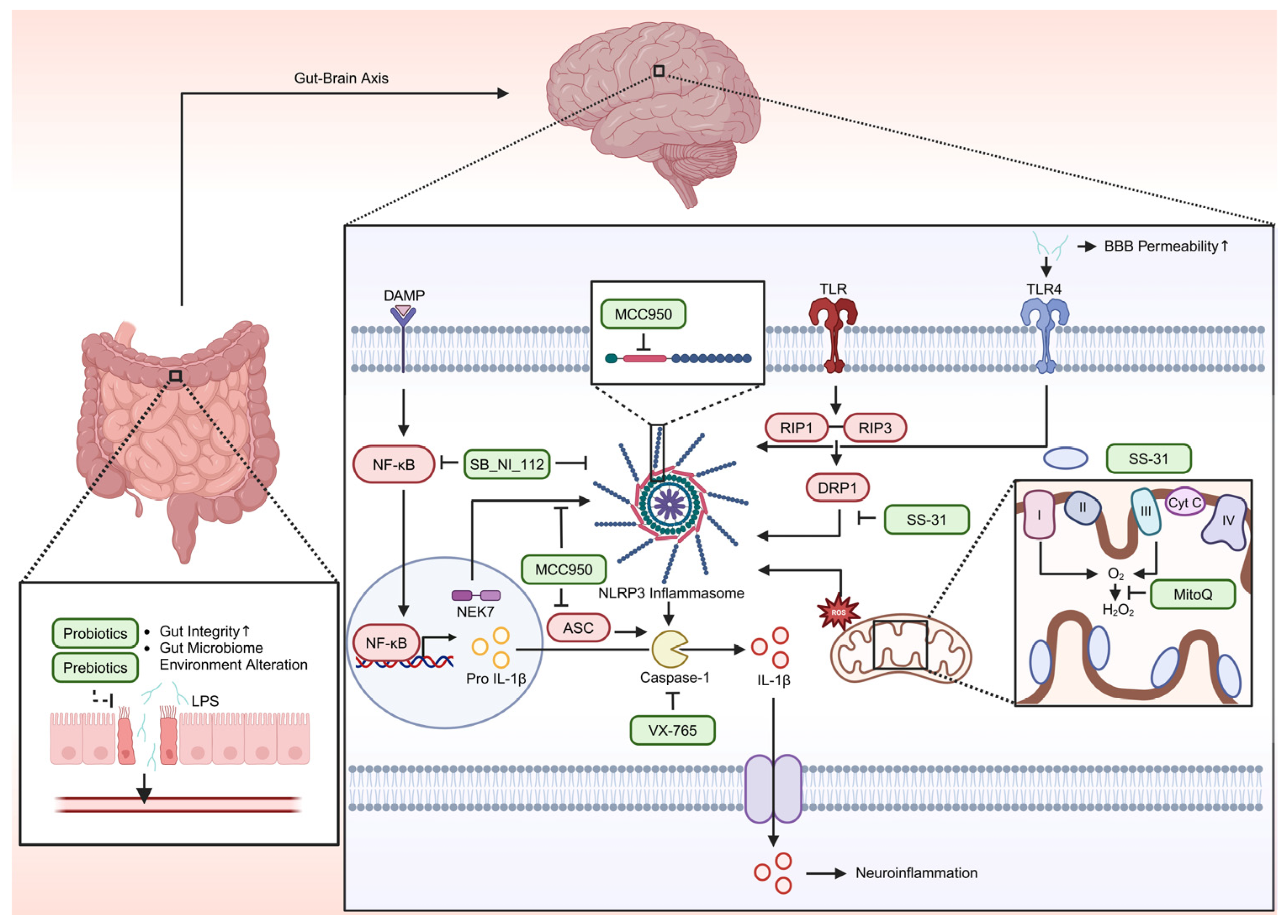
8. Therapeutic Target

Various studies suggest that mitochondrial-targeted strategies aimed at reducing ROS production hold promise for mitigating diseases such as AUD and AD. One approach involves activation of the antioxidant transcription factor Nrf2. Nrf2 is expressed in most tissues and regulates the expression of cell-protective genes [116]. Nrf2 protein comprises several evolutionarily conserved domains (Neh1–Neh6) [117]; notably, its N-terminal Neh2 domain contains the DLG (Asp-Leu-Gly) and ETGE (Glu-Thr-Gly-Glu) motifs that directly interact with the negative regulator Keap1, facilitating degradation of excess Nrf2 and thereby maintaining cellular homeostasis [118]. Under conditions of oxidative stress, specific cysteine residues within Keap1 become modified, altering its conformation and allowing for Nrf2 to escape degradation; in the brains of AD patients, Nrf2 is predominantly localized in the cytoplasm of hippocampal neurons, where it drives the expression of antioxidant genes to reduce ROS accumulation caused by Aβ peptides [119,120]. Pharmacological upregulation of Nrf2 has shown potential as a therapeutic target for AD, contributing to the alleviation of associated symptoms [121]. Mitochondria-targeted antioxidants represent another therapeutic strategy. MitoQ, a derivative of ubiquinol, accumulates in the inner mitochondrial membrane via its triphenylphosphonium ion moiety and is converted to its active antioxidant form, ubiquinol [122]. MitoQ inhibits lipid peroxidation and the accumulation of ROS primarily produced by Complexes I and III of the electron transport chain; its oxidized form, ubiquinone, is subsequently recycled to its active form by Complex II [123]. In an in vivo study of chronic alcohol abuse, lung tissue from mice treated with MitoQ showed reduced ROS levels compared to those from alcohol-exposed mice without MitoQ, as evidenced by dihydroethidium staining [124]. Moreover, MitoQ suppressed NLRP3 inflammasome activity and IL-1β secretion in these models [124], and similar antioxidant and anti-inflammatory effects were observed in human prostate cells [125]. MitoQ also crosses the BBB, decreases superoxide production (thereby reducing nitrotyrosine, an oxidative stress biomarker of AD) [126], and promotes neuroblastoma cell proliferation by suppressing mitochondrial fission gene expression and enhancing antioxidant enzyme levels [127]. Although some studies have reported potential nephrotoxicity and gastrointestinal side effects—such as increased brachial artery flow-mediated dilation with mild-to-moderate gastrointestinal discomfort during 6-week chronic supplementation and adverse effects (nausea and vomiting) in Parkinson’s patients administered 80 mg MitoQ [128]—a recent study in healthy adults found that acute, high-dose MitoQ (100–160 mg, adjusted by body weight) did not adversely affect kidney functions [129]. It is also worth noting that the effect of MitoQ may vary by biological sex; for instance, a study in rats demonstrated lower superoxide levels in female offspring compared to males after MitoQ administration during fetal hypoxia [130], although other studies, such as those using Sacs gene knockout mice, did not observe gender differences [131]. Another potential antioxidant is SS-31, a synthetic peptide that specifically targets the inner mitochondrial membrane by binding to cardiolipin, thereby stabilizing mitochondrial structure and preventing the formation of the mPTP [132,133]. SS-31 also scavenges ROS through its dimethyl tyrosine residue, forming di tyrosine radicals that reduce LDL oxidation [134]. Additionally, SS-31 has been shown to inhibit NLRP3 inflammasome activity in hippocampal microglial cells, potentially by modulating DRP1, a protein involved in mitochondrial fission and ROS production [135]. Preclinical studies demonstrate that SS-31 is distributed across major organs—including the heart, lungs, liver, skeletal muscles, and kidneys, with the highest concentrations in the kidneys—and is completely cleared through urination [134]. In a phase IIa clinical trial involving patients with atherosclerotic renal artery stenosis, SS-31 administration did not result in significant adverse effects, such as fever, headache, vomiting, hematuria, or allergic reactions, and no changes in serum creatinine or urine cytology were observed within 24 h [136]. Despite favorable safety profiles in early-phase trials, a phase III study in patients with mitochondrial myopathy did not show significant improvements in measures such as walking distance or fatigue scores [137]; however, SS-31 was well tolerated overall. Dosage optimization remains a subject of ongoing research, as one study in patients with heart failure with reduced ejection fraction (HFrEF) reported that 24% of patients receiving a 40 mg injection experienced treatment-emergent adverse events (TEAEs), such as nausea and fatigue, compared to 4.5% at a 4 mg dose [138]. Targeting the NLRP3 inflammasome is another emerging therapeutic strategy, given its activation by ROS and its involvement in both AD and AUD. MCC950 is a potent NLRP3 inflammasome inhibitor that blocks the interaction between NEK7 and NLRP3 by binding to the Walker B motif within the NACHT domain [139,140]. MCC950 not only reduces NLRP3 activity but also lowers ASC protein levels, thereby inhibiting caspase-1 activation and subsequent release of pro-inflammatory cytokines such as IL-1β and IL-18 [141]. In rat models treated with streptozotocin, MCC950 reduced NLRP3 inflammasome complex levels and caspase-1 activation in the dentate gyrus, CA1, and CA3 regions of the hippocampus, with concomitant improvements in spatial learning, episodic memory, and anxiety [142]. However, while several animal studies support the safety of MCC950, some reports indicate potential adverse effects—including elevated expression of the pro-oxidant genes NOX2 and NOX4, increased glomerular nitrotyrosine content, and markers of renal fibrosis [143], as well as conflicting data regarding hepatotoxicity [144,145]—necessitating further investigation. Following MCC950, SB_NI_112 has emerged as another candidate for inhibiting the NLRP3 inflammasome. SB_NI_112 crosses the BBB and acts in multiple brain regions, including the hippocampus, cortex, cerebellum, and brainstem [146]. It functions by inhibiting NF-κB—an upstream activator of NLRP3—and directly suppressing NLRP3 inflammasome activity, thereby reducing pro-inflammatory cytokines (IL-2, IL-1α, and IL-1β) implicated in neuroinflammation [146]. Notably, SB_NI_112 appears effective even in older animals, in contrast to other inhibitors such as OLT1177 [147], and has demonstrated a favorable safety profile across various administration routes (intravenous, subcutaneous, intraperitoneal, and intranasal) without significant pathological changes in kidney, blood, or electrolyte levels [148]. Nonetheless, comprehensive human safety assessments remain necessary. Caspase inhibitors also offer a potential therapeutic approach. VX-765, for example, acts as a potent inhibitor of caspase-1 by modifying the enzyme’s catalytic cysteine residue. Caspase-1, activated by the NLRP3 inflammasome, cleaves pro IL-1β into its active form, IL-1β, thereby triggering neuroinflammation and further ROS production [149]. In vitro studies have shown that VX-765 reduces levels of NLRP3, caspase-1, and IL-1β while promoting cell survival under H2O2-induced stress [150]. Additionally, animal studies have demonstrated that VX-765 normalizes IL-1β levels, improves spatial learning and memory in the Barnes maze, and alleviates episodic memory impairments in mutant amyloid protein mice across various age groups [151]. In terms of safety, VX-765 has been evaluated in phase IIa clinical trials for epilepsy in a double-blind, randomized, placebo-controlled design. In these trials, 72.9% of participants in the treatment group experienced TEAEs compared to 83.3% in the placebo group; notably, the treatment group’s TEAEs rate was 6%, while the placebo group’s rate was 0% [152]. However, VX-765 was also associated with elevated liver enzyme levels, highlighting the need for further safety evaluation [152]. As our understanding of ROS in AUD and AD expands, several potential avenues for future investigation have emerged. One promising area involves the gut microbiome. Recent studies suggest that disruptions in the gut microbiota—caused by aging, alcohol consumption, or other factors—lead to reduced levels of anti-inflammatory molecules and compromised intestinal integrity [153]. This disruption facilitates the leakage of LPS into the bloodstream, allowing for its transport to the brain via the gut–brain axis. Upon reaching the brain, LPS increases BBB permeability and activates the NLRP3 inflammasome, potentially accelerating AD progression [153,154]. Furthermore, gut dysbiosis may contribute to increased oxidative stress and alcohol dependence by altering the composition of microbiomes that favor ethanol metabolism, ultimately inducing memory loss and neuropsychiatric behaviors [155]. Future research into the development of prebiotics and probiotics targeting ROS, AUD, and AD may offer novel therapeutic opportunities by modulating the gut microbiome, enhancing gut integrity, and preventing LPS translocation, thereby mitigating neuroinflammation and reducing BBB permeability [156]. In summary, therapeutic strategies aimed at reducing ROS—whether by enhancing Nrf2 activity, employing mitochondria targeted antioxidants such as MitoQ and SS-31, inhibiting the NLRP3 inflammasome via agents including MCC950 or SB_NI_112, or using caspase inhibitors such as VX-765—represent promising approaches to mitigating the neurodegenerative processes associated with both AUD and AD. Continued research into these mechanisms along with the exploration of gut–brain interactions are critical for the development of effective, targeted interventions against oxidative stress-related neurodegeneration (Figure 2).

9. Conclusions

ROS play a pivotal role in the pathogenesis of both AUD and AD, acting as central mediators of oxidative stress. Excessive ROS production leads to mitochondrial dysfunction, a decline in endogenous antioxidant defenses, and the dysregulation of neuroinflammatory pathways—such as those mediated by TLR4 and the NLRP3 inflammasome—which further promote neurodegeneration. Although AD and AUD exhibit distinct hallmark features (e.g., Aβ plaques and tau tangles in AD versus alcohol-induced neurotoxicity in AUD), they share common underlying mechanisms driven by the overproduction of ROS. Given the critical role of ROS in both conditions, therapeutic strategies that aim to reduce oxidative stress show considerable promise. Mitochondria-targeted antioxidants, such as Mito-Q and SS-31, have demonstrated the capacity to preserve mitochondrial integrity and lower ROS levels. In addition, agents that inhibit the NLRP3 inflammasome (e.g., MCC950) and caspase-1 (e.g., VX-765) offer further potential by mitigating neuroinflammation. Complementary interventions that target glutamate transport dysfunction and gut–brain axis dysregulation also provide additional therapeutic avenues, particularly in the context of AUD-related neurodegeneration. Future research should prioritize the identification of reliable ROS biomarkers for early diagnosis as well as the development of combination therapies that concurrently address oxidative stress and inflammation. Moreover, a deeper investigation into the interplay of genetic, environmental, and lifestyle factors in ROS-related diseases is essential. The advancement of accessible technologies capable of measuring ROS in vivo and in real time would be of significant therapeutic importance, as such tools would enable the early identification and management of elevated ROS levels, thereby preventing the progression of neurodegenerative lesions (Table 1). In summary, this perspective not only deepens our understanding of the common oxidative mechanisms linking AUD and AD but also highlights promising directions for future clinical trials and targeted interventions against ROS-induced neurodegeneration.

Funding

This research was supported by the Soonchunhyang University (2025-007).

Acknowledgments

We thank all the laboratory members for their helpful discussions and comments.

Conflicts of Interest

The authors have declared no conflicts of interest.

Abbreviations

The following abbreviations are used in this manuscript:
ROSReactive oxygen species
AUDAlcohol use disorder
ADAlzheimer’s disease
Amyloid-beta
TLR4Toll-like receptor 4
LPSLipopolysaccharide
ETCElectron transport chain
CYP2E1Cytochrome P450 2E1
ADHAlcohol dehydrogenase
MEOSMicrosomal ethanol oxidizing system
SODSuperoxide dismutase
GPxGlutathione peroxidase
CATCatalase
PGC-1αPeroxisome proliferator-activated receptor gamma coactivator 1-alpha
NOXNADPH oxidase
Drp1Dynamin-related protein 1
APPAmyloid beta precursor protein
HFrEFHeart failure with reduced ejection fraction
TEAEsTreatment-emergent adverse events
mPTPMitochondrial permeability transition pore
MDAMalondialdehyde
GLT-1Glutamate transporter-1
UPRUnfolded protein response
ABADAβ-binding alcohol dehydrogenase
OXPHOSOxidative phosphorylation
NLRP3Pyrin domain-containing protein 3
BBBBlood–brain barrier
PI3KPhosphoinositide 3-kinase
MAPKMitogen-activated protein kinase
Nrf2Nuclear factor erythroid 2-related factor 2
Keap1Kelch like-ECH-associated protein 1
NF-κBNuclear factor kappa B
PAMPPathogen-associated molecular patterns
miRNAsmicroRNAs
EAAT1Excitatory amino acid transporter 1
EAAT2Excitatory amino acid transporter 2

References

  1. Bhatt, S.; Puli, L.; Patil, C.R. Role of reactive oxygen species in the progression of Alzheimer’s disease. Drug Discov. Today 2021, 26, 794–803. [Google Scholar] [PubMed]
  2. Simpson, D.S.A.; Oliver, P.L. ROS Generation in Microglia: Understanding Oxidative Stress and Inflammation in Neurodegenerative Disease. Antioxidants 2020, 9, 743. [Google Scholar] [CrossRef] [PubMed]
  3. Balaban, R.S.; Nemoto, S.; Finkel, T. Mitochondria, oxidants, and aging. Cell 2005, 120, 483–495. [Google Scholar]
  4. Pritchard, K.A., Jr.; Ackerman, A.W.; Gross, E.R.; Stepp, D.W.; Shi, Y.; Fontana, J.T.; Baker, J.E.; Sessa, W.C. Heat shock protein 90 mediates the balance of nitric oxide and superoxide anion from endothelial nitric-oxide synthase. J. Biol. Chem. 2001, 276, 17621–17624. [Google Scholar] [PubMed]
  5. Ye, Z.; Liu, Y.; Jin, X.; Wu, Y.; Zhao, H.; Gao, T.; Deng, Q.; Cheng, J.; Lin, J.; Tong, Z. Abeta-binding with alcohol dehydrogenase drives Alzheimer’s disease pathogenesis: A review. Int. J. Biol. Macromol. 2024, 264 Pt 2, 130580. [Google Scholar]
  6. Tamagno, E.; Parola, M.; Bardini, P.; Piccini, A.; Borghi, R.; Guglielmotto, M.; Santoro, G.; Davit, A.; Danni, O.; Smith, M.A.; et al. Beta-site APP cleaving enzyme up-regulation induced by 4-hydroxynonenal is mediated by stress-activated protein kinases pathways. J. Neurochem. 2005, 92, 628–636. [Google Scholar]
  7. Kany, S.; Janicova, A.; Relja, B. Innate Immunity and Alcohol. J. Clin. Med. 2019, 8, 1981. [Google Scholar] [CrossRef]
  8. Subramaniyan, V.; Chakravarthi, S.; Jegasothy, R.; Seng, W.Y.; Fuloria, N.K.; Fuloria, S.; Hazarika, I.; Das, A. Alcohol-associated liver disease: A review on its pathophysiology, diagnosis and drug therapy. Toxicol. Rep. 2021, 8, 376–385. [Google Scholar] [CrossRef]
  9. Kuzmich, N.N.; Sivak, K.V.; Chubarev, V.N.; Porozov, Y.B.; Savateeva-Lyubimova, T.N.; Peri, F. TLR4 Signaling Pathway Modulators as Potential Therapeutics in Inflammation and Sepsis. Vaccines 2017, 5, 34. [Google Scholar] [CrossRef]
  10. Kong, E.Q.Z.; Subramaniyan, V.; Lubau, N.S.A. Uncovering the impact of alcohol on internal organs and reproductive health: Exploring TLR4/NF-kB and CYP2E1/ROS/Nrf2 pathways. Anim. Model. Exp. Med. 2024, 7, 444–459. [Google Scholar]
  11. Schwarzinger, M.; Pollock, B.G.; Hasan, O.S.M.; Dufouil, C.; Rehm, J.; QalyDays Study Group. Contribution of alcohol use disorders to the burden of dementia in France 2008-13: A nationwide retrospective cohort study. Lancet Public Health 2018, 3, e124–e132. [Google Scholar]
  12. Zhang, P.; Edenberg, H.J.; Nurnberger, J.; Lai, D.; Cheng, F.; Liu, Y. Alcohol use disorder is associated with higher risks of Alzheimer’s and Parkinson’s diseases: A study of US insurance claims data. Alzheimer’s Dement. 2022, 14, e12370. [Google Scholar]
  13. Kang, S.; Lee, J.; Ali, D.N.; Choi, S.; Nesbitt, J.; Min, P.H.; Trushina, E.; Choi, D.S. Low to moderate ethanol exposure reduces astrocyte-induced neuroinflammatory signaling and cognitive decline in presymptomatic APP/PS1 mice. Sci. Rep. 2024, 14, 23989. [Google Scholar]
  14. Ising, C.; Venegas, C.; Zhang, S.; Scheiblich, H.; Schmidt, S.V.; Vieira-Saecker, A.; Schwartz, S.; Albasset, S.; McManus, R.M.; Tejera, D.; et al. NLRP3 inflammasome activation drives tau pathology. Nature 2019, 575, 669–673. [Google Scholar] [PubMed]
  15. Walter, S.; Letiembre, M.; Liu, Y.; Heine, H.; Penke, B.; Hao, W.; Bode, B.; Manietta, N.; Walter, J.; Schulz-Schuffer, W.; et al. Role of the toll-like receptor 4 in neuroinflammation in Alzheimer’s disease. Cell. Physiol. Biochem. 2007, 20, 947–956. [Google Scholar] [CrossRef]
  16. Balu, D.; Valencia-Olvera, A.C.; Nguyen, A.; Patnam, M.; York, J.; Peri, F.; Neumann, F.; LaDu, M.J.; Tai, L.M. A small-molecule TLR4 antagonist reduced neuroinflammation in female E4FAD mice. Alzheimer’s Res. Ther. 2023, 15, 181. [Google Scholar] [CrossRef]
  17. Zhang, X.; Wang, R.; Hu, D.; Sun, X.; Fujioka, H.; Lundberg, K.; Chan, E.R.; Wang, Q.; Xu, R.; Flanagan, M.E.; et al. Oligodendroglial glycolytic stress triggers inflammasome activation and neuropathology in Alzheimer’s disease. Sci. Adv. 2020, 6, eabb8680. [Google Scholar] [CrossRef]
  18. Heneka, M.T.; Kummer, M.P.; Stutz, A.; Delekate, A.; Schwartz, S.; Vieira-Saecker, A.; Griep, A.; Axt, D.; Remus, A.; Tzeng, T.C.; et al. NLRP3 is activated in Alzheimer’s disease and contributes to pathology in APP/PS1 mice. Nature 2013, 493, 674–678. [Google Scholar]
  19. Li, R.; Jia, Z.; Trush, M.A. Defining ROS in Biology and Medicine. React. Oxyg. Species 2016, 1, 9–21. [Google Scholar]
  20. D’Autreaux, B.; Toledano, M.B. ROS as signalling molecules: Mechanisms that generate specificity in ROS homeostasis. Nat. Rev. Mol. Cell Biol. 2007, 8, 813–824. [Google Scholar]
  21. Koopman, W.J.; Nijtmans, L.G.; Dieteren, C.E.; Roestenberg, P.; Valsecchi, F.; Smeitink, J.A.; Willems, P.H. Mammalian mitochondrial complex I: Biogenesis, regulation, and reactive oxygen species generation. Antioxid. Redox Signal 2010, 12, 1431–1470. [Google Scholar] [PubMed]
  22. Bae, Y.S.; Oh, H.; Rhee, S.G.; Yoo, Y.D. Regulation of reactive oxygen species generation in cell signaling. Mol. Cells 2011, 32, 491–509. [Google Scholar]
  23. Kaminskyy, V.O.; Zhivotovsky, B. Free radicals in cross talk between autophagy and apoptosis. Antioxid. Redox Signal. 2014, 21, 86–102. [Google Scholar]
  24. Benhar, M. Oxidants, Antioxidants and Thiol Redox Switches in the Control of Regulated Cell Death Pathways. Antioxidants 2020, 9, 309. [Google Scholar] [CrossRef]
  25. Covarrubias, L.; Hernandez-Garcia, D.; Schnabel, D.; Salas-Vidal, E.; Castro-Obregon, S. Function of reactive oxygen species during animal development: Passive or active? Dev. Biol. 2008, 320, 1–11. [Google Scholar]
  26. Zhang, J.; Wang, X.; Vikash, V.; Ye, Q.; Wu, D.; Liu, Y.; Dong, W. ROS and ROS-Mediated Cellular Signaling. Oxid. Med. Cell. Longev. 2016, 2016, 4350965. [Google Scholar]
  27. Poyton, R.O.; Castello, P.R.; Ball, K.A.; Woo, D.K.; Pan, N. Mitochondria and hypoxic signaling: A new view. Ann. N. Y. Acad. Sci. 2009, 1177, 48–56. [Google Scholar] [PubMed]
  28. Bhattacharyya, A.; Chattopadhyay, R.; Mitra, S.; Crowe, S.E. Oxidative stress: An essential factor in the pathogenesis of gastrointestinal mucosal diseases. Physiol. Rev. 2014, 94, 329–354. [Google Scholar] [PubMed]
  29. Angelova, P.R.; Abramov, A.Y. Role of mitochondrial ROS in the brain: From physiology to neurodegeneration. FEBS Lett. 2018, 592, 692–702. [Google Scholar]
  30. Castellani, R.; Hirai, K.; Aliev, G.; Drew, K.L.; Nunomura, A.; Takeda, A.; Cash, A.D.; Obrenovich, M.E.; Perry, G.; Smith, M.A. Role of mitochondrial dysfunction in Alzheimer’s disease. J. Neurosci. Res. 2002, 70, 357–360. [Google Scholar]
  31. Gibson, G.E.; Sheu, K.F.; Blass, J.P. Abnormalities of mitochondrial enzymes in Alzheimer disease. J. Neural Transm. 1998, 105, 855–870. [Google Scholar] [PubMed]
  32. Wang, X.; Su, B.; Zheng, L.; Perry, G.; Smith, M.A.; Zhu, X. The role of abnormal mitochondrial dynamics in the pathogenesis of Alzheimer’s disease. J. Neurochem. 2009, 109 (Suppl. S1), 153–159. [Google Scholar]
  33. Koop, D.R.; Coon, M.J. Ethanol oxidation and toxicity: Role of alcohol P-450 oxygenase. Alcohol. Clin. Exp. Res. 1986, 10 (Suppl. S6), 44S–49S. [Google Scholar] [PubMed]
  34. Lieber, C.S. Alcoholic fatty liver: Its pathogenesis and mechanism of progression to inflammation and fibrosis. Alcohol 2004, 34, 9–19. [Google Scholar] [PubMed]
  35. Koop, D.R. Alcohol metabolism’s damaging effects on the cell: A focus on reactive oxygen generation by the enzyme cytochrome P450 2E1. Alcohol Res. Health 2006, 29, 274–280. [Google Scholar]
  36. Okoye, C.N.; Koren, S.A.; Wojtovich, A.P. Mitochondrial complex I ROS production and redox signaling in hypoxia. Redox Biol. 2023, 67, 102926. [Google Scholar]
  37. Terada, T.; Therriault, J.; Kang, M.S.; Savard, M.; Pascoal, T.A.; Lussier, F.; Tissot, C.; Wang, Y.T.; Benedet, A.; Poltronetti, N.M.; et al. Mitochondrial complex I abnormalities underlie neurodegeneration and cognitive decline in Alzheimer’s disease. Eur. J. Neurol. 2022, 29, 1324–1334. [Google Scholar]
  38. Brand, M.D. The sites and topology of mitochondrial superoxide production. Exp. Gerontol. 2010, 45, 466–472. [Google Scholar]
  39. Brand, M.D. Riding the tiger—Physiological and pathological effects of superoxide and hydrogen peroxide generated in the mitochondrial matrix. Crit. Rev. Biochem. Mol. Biol. 2020, 55, 592–661. [Google Scholar]
  40. Diaz, F.; Garcia, S.; Padgett, K.R.; Moraes, C.T. A defect in the mitochondrial complex III, but not complex IV, triggers early ROS-dependent damage in defined brain regions. Hum. Mol. Genet. 2012, 21, 5066–5077. [Google Scholar]
  41. Bayo Jimenez, M.T.; Frenis, K.; Hahad, O.; Steven, S.; Cohen, G.; Cuadrado, A.; Munzel, T.; Daiber, A. Protective actions of nuclear factor erythroid 2-related factor 2 (NRF2) and downstream pathways against environmental stressors. Free Radic. Biol. Med. 2022, 187, 72–91. [Google Scholar]
  42. Cross, C.E.; Halliwell, B.; Allen, A. Antioxidant protection: A function of tracheobronchial and gastrointestinal mucus. Lancet 1984, 1, 1328–1330. [Google Scholar]
  43. Wu, D.; Cederbaum, A.I. Oxidative stress and alcoholic liver disease. Semin. Liver Dis. 2009, 29, 141–154. [Google Scholar] [PubMed]
  44. Palmieri, V.O.; Grattagliano, I.; Palasciano, G. Ethanol induces secretion of oxidized proteins by pancreatic acinar cells. Cell Biol. Toxicol. 2007, 23, 459–464. [Google Scholar] [CrossRef] [PubMed]
  45. Wang, W.; Yang, L.; Liu, T.; Wang, J.; Wen, A.; Ding, Y. Ellagic acid protects mice against sleep deprivation-induced memory impairment and anxiety by inhibiting TLR4 and activating Nrf2. Aging 2020, 12, 10457–10472. [Google Scholar] [PubMed]
  46. Lv, Y.N.; Cui, Y.; Zhang, B.; Huang, S.M. Sleep deficiency promotes Alzheimer’s disease development and progression. Front. Neurol. 2022, 13, 1053942. [Google Scholar] [CrossRef] [PubMed]
  47. Tavares, W.M.; Araujo de Franca, S.; Paiva, W.S.; Teixeira, M.J. Early tracheostomy versus late tracheostomy in severe traumatic brain injury or stroke: A systematic review and meta-analysis. Aust. Crit. Care 2023, 36, 1110–1116. [Google Scholar] [CrossRef]
  48. Leon, B.E.; Kang, S.; Franca-Solomon, G.; Shang, P.; Choi, D.S. Alcohol-Induced Neuroinflammatory Response and Mitochondrial Dysfunction on Aging and Alzheimer’s Disease. Front. Behav. Neurosci. 2021, 15, 778456. [Google Scholar] [CrossRef]
  49. Waddell, J.; McKenna, M.C.; Kristian, T. Brain ethanol metabolism and mitochondria. Curr. Top. Biochem. Res. 2022, 23, 1–13. [Google Scholar]
  50. Jin, M.; Ande, A.; Kumar, A.; Kumar, S. Regulation of cytochrome P450 2e1 expression by ethanol: Role of oxidative stress-mediated pkc/jnk/sp1 pathway. Cell Death Dis. 2013, 4, e554. [Google Scholar]
  51. Haorah, J.; Ramirez, S.H.; Floreani, N.; Gorantla, S.; Morsey, B.; Persidsky, Y. Mechanism of alcohol-induced oxidative stress and neuronal injury. Free Radic. Biol. Med. 2008, 45, 1542–1550. [Google Scholar] [CrossRef]
  52. Feng, J.; Zheng, Y.; Guo, M.; Ares, I.; Martinez, M.; Lopez-Torres, B.; Martinez-Larranaga, M.R.; Wang, X.; Anadon, A.; Martinez, M.A. Oxidative stress, the blood-brain barrier and neurodegenerative diseases: The critical beneficial role of dietary antioxidants. Acta Pharm. Sin. B 2023, 13, 3988–4024. [Google Scholar] [CrossRef]
  53. Tsermpini, E.E.; Plemenitas Iljes, A.; Dolzan, V. Alcohol-Induced Oxidative Stress and the Role of Antioxidants in Alcohol Use Disorder: A Systematic Review. Antioxidants 2022, 11, 1374. [Google Scholar] [CrossRef] [PubMed]
  54. Manzo-Avalos, S.; Saavedra-Molina, A. Cellular and mitochondrial effects of alcohol consumption. Int. J. Environ. Res. Public Health 2010, 7, 4281–4304. [Google Scholar] [CrossRef]
  55. Mansouri, A.; Gaou, I.; De Kerguenec, C.; Amsellem, S.; Haouzi, D.; Berson, A.; Moreau, A.; Feldmann, G.; Letteron, P.; Pessayre, D.; et al. An alcoholic binge causes massive degradation of hepatic mitochondrial DNA in mice. Gastroenterology 1999, 117, 181–190. [Google Scholar] [CrossRef] [PubMed]
  56. Perez, M.J.; Loyola, R.; Canelo, F.; Aranguiz, A.; Tapia-Monsalves, C.; Osorio-Fuentealba, C.; Quintanilla, R.A. NADPH oxidase contributes to oxidative damage and mitochondrial impairment induced by acute ethanol treatment in rat hippocampal neurons. Neuropharmacology 2020, 171, 108100. [Google Scholar] [CrossRef] [PubMed]
  57. Hoek, J.B.; Cahill, A.; Pastorino, J.G. Alcohol and mitochondria: A dysfunctional relationship. Gastroenterology 2002, 122, 2049–2063. [Google Scholar] [CrossRef]
  58. Abu Shelbayeh, O.; Arroum, T.; Morris, S.; Busch, K.B. PGC-1alpha Is a Master Regulator of Mitochondrial Lifecycle and ROS Stress Response. Antioxidants 2023, 12, 1075. [Google Scholar] [CrossRef]
  59. Chen, L.; Qin, Y.; Liu, B.; Gao, M.; Li, A.; Li, X.; Gong, G. PGC-1alpha-Mediated Mitochondrial Quality Control: Molecular Mechanisms and Implications for Heart Failure. Front. Cell Dev. Biol. 2022, 10, 871357. [Google Scholar]
  60. Rius-Perez, S.; Torres-Cuevas, I.; Millan, I.; Ortega, A.L.; Perez, S. PGC-1alpha, Inflammation, and Oxidative Stress: An Integrative View in Metabolism. Oxid. Med. Cell Longev. 2020, 2020, 1452696. [Google Scholar] [CrossRef]
  61. Shang, P.; Lindberg, D.; Starski, P.; Peyton, L.; Hong, S.I.; Choi, S.; Choi, D.S. Chronic Alcohol Exposure Induces Aberrant Mitochondrial Morphology and Inhibits Respiratory Capacity in the Medial Prefrontal Cortex of Mice. Front. Neurosci. 2020, 14, 561173. [Google Scholar]
  62. Oberdoerster, J.; Rabin, R.A. Enhanced caspase activity during ethanol-induced apoptosis in rat cerebellar granule cells. Eur. J. Pharmacol. 1999, 385, 273–282. [Google Scholar] [PubMed]
  63. De Nicolo, B.; Cataldi-Stagetti, E.; Diquigiovanni, C.; Bonora, E. Calcium and Reactive Oxygen Species Signaling Interplays in Cardiac Physiology and Pathologies. Antioxidants 2023, 12, 353. [Google Scholar] [CrossRef] [PubMed]
  64. Chen, C.H.; Pan, C.H.; Chen, C.C.; Huang, M.C. Increased oxidative DNA damage in patients with alcohol dependence and its correlation with alcohol withdrawal severity. Alcohol. Clin. Exp. Res. 2011, 35, 338–344. [Google Scholar]
  65. Yang, M.; Zhou, X.; Tan, X.; Huang, X.; Yuan, L.; Zhang, Z.; Yang, Y.; Xu, M.; Wan, Y.; Li, Z. The Status of Oxidative Stress in Patients with Alcohol Dependence: A Meta-Analysis. Antioxidants 2022, 11, 1919. [Google Scholar] [CrossRef]
  66. Pemberton, P.W.; Smith, A.; Warnes, T.W. Non-invasive monitoring of oxidant stress in alcoholic liver disease. Scand. J. Gastroenterol. 2005, 40, 1102–1108. [Google Scholar] [CrossRef]
  67. Kitagaki, H.; Araki, Y.; Funato, K.; Shimoi, H. Ethanol-induced death in yeast exhibits features of apoptosis mediated by mitochondrial fission pathway. FEBS Lett. 2007, 581, 2935–2942. [Google Scholar]
  68. Breido, I.S. On the 120th anniversary of the antiseptic method of Joseph Lister (1867–1987). Vestn. Khir Im. I I Grek. 1988, 140, 123–125. [Google Scholar]
  69. Kieron, M.; Zekanowski, C.; Falk, A.; Wezyk, M. Oxidative DNA Damage Signalling in Neural Stem Cells in Alzheimer’s Disease. Oxid. Med. Cell Longev. 2019, 2019, 2149812. [Google Scholar] [CrossRef]
  70. Rehm, J.; Hasan, O.S.M.; Black, S.E.; Shield, K.D.; Schwarzinger, M. Alcohol use and dementia: A systematic scoping review. Alzheimer’s Res. Ther. 2019, 11, 1. [Google Scholar]
  71. Sullivan, E.V.; Pfefferbaum, A. Brain-behavior relations and effects of aging and common comorbidities in alcohol use disorder: A review. Neuropsychology 2019, 33, 760–780. [Google Scholar] [PubMed]
  72. Lopez-Valencia, L.; Moya, M.; Escudero, B.; Garcia-Bueno, B.; Orio, L. Bacterial lipopolysaccharide forms aggregates with apolipoproteins in male and female rat brains after ethanol binges. J. Lipid Res. 2024, 65, 100509. [Google Scholar]
  73. Thakur, V.; Pritchard, M.T.; McMullen, M.R.; Wang, Q.; Nagy, L.E. Chronic ethanol feeding increases activation of NADPH oxidase by lipopolysaccharide in rat Kupffer cells: Role of increased reactive oxygen in LPS-stimulated ERK1/2 activation and TNF-alpha production. J. Leukoc. Biol. 2006, 79, 1348–1356. [Google Scholar]
  74. Zhao, J.; Bi, W.; Xiao, S.; Lan, X.; Cheng, X.; Zhang, J.; Lu, D.; Wei, W.; Wang, Y.; Li, H.; et al. Neuroinflammation induced by lipopolysaccharide causes cognitive impairment in mice. Sci. Rep. 2019, 9, 5790. [Google Scholar]
  75. Vorhees, C.V.; Williams, M.T. Assessing spatial learning and memory in rodents. ILAR J. 2014, 55, 310–332. [Google Scholar]
  76. Di Lorenzo, F.; De Castro, C.; Silipo, A.; Molinaro, A. Lipopolysaccharide structures of Gram-negative populations in the gut microbiota and effects on host interactions. FEMS Microbiol. Rev. 2019, 43, 257–272. [Google Scholar] [PubMed]
  77. Crews, F.T.; Sarkar, D.K.; Qin, L.; Zou, J.; Boyadjieva, N.; Vetreno, R.P. Neuroimmune Function and the Consequences of Alcohol Exposure. Alcohol Res. 2015, 37, 331–341, 344–351. [Google Scholar]
  78. McManus, R.M.; Latz, E. NLRP3 inflammasome signalling in Alzheimer’s disease. Neuropharmacology 2024, 252, 109941. [Google Scholar]
  79. Ramos, A.; Joshi, R.S.; Szabo, G. Innate immune activation: Parallels in alcohol use disorder and Alzheimer’s disease. Front. Mol. Neurosci. 2022, 15, 910298. [Google Scholar]
  80. Rasool, A.E.; Furlong, T.; Prasad, A.A. Microglia activity in the human basal ganglia is altered in alcohol use disorder and reversed with remission from alcohol. Addict. Biol. 2024, 29, e13374. [Google Scholar]
  81. Graves, A.R.; Moore, S.J.; Bloss, E.B.; Mensh, B.D.; Kath, W.L.; Spruston, N. Hippocampal pyramidal neurons comprise two distinct cell types that are countermodulated by metabotropic receptors. Neuron 2012, 76, 776–789. [Google Scholar] [PubMed]
  82. Fukui, K.; Takatsu, H.; Shinkai, T.; Suzuki, S.; Abe, K.; Urano, S. Appearance of amyloid beta-like substances and delayed-type apoptosis in rat hippocampus CA1 region through aging and oxidative stress. J. Alzheimer’s Dis. 2005, 8, 299–309. [Google Scholar]
  83. Igarashi, K.M. Entorhinal cortex dysfunction in Alzheimer’s disease. Trends Neurosci. 2023, 46, 124–136. [Google Scholar]
  84. Olajide, O.J.; Suvanto, M.E.; Chapman, C.A. Molecular mechanisms of neurodegeneration in the entorhinal cortex that underlie its selective vulnerability during the pathogenesis of Alzheimer’s disease. Biol. Open 2021, 10, bio056796. [Google Scholar] [CrossRef]
  85. de Flores, R.; Das, S.R.; Xie, L.; Wisse, L.E.M.; Lyu, X.; Shah, P.; Yushkevich, P.A.; Wolk, D.A. Medial Temporal Lobe Networks in Alzheimer’s Disease: Structural and Molecular Vulnerabilities. J. Neurosci. 2022, 42, 2131–2141. [Google Scholar] [PubMed]
  86. Du, A.T.; Schuff, N.; Kramer, J.H.; Rosen, H.J.; Gorno-Tempini, M.L.; Rankin, K.; Miller, B.L.; Weiner, M.W. Different regional patterns of cortical thinning in Alzheimer’s disease and frontotemporal dementia. Brain 2007, 130, 1159–1166. [Google Scholar]
  87. Gonzalez-Rodriguez, M.; Villar-Conde, S.; Astillero-Lopez, V.; Villanueva-Anguita, P.; Ubeda-Banon, I.; Flores-Cuadrado, A.; Martinez-Marcos, A.; Saiz-Sanchez, D. Human amygdala involvement in Alzheimer’s disease revealed by stereological and dia-PASEF analysis. Brain Pathol. 2023, 33, e13180. [Google Scholar] [PubMed]
  88. Sakato, Y.; Shima, A.; Terada, Y.; Takeda, K.; Sakamaki-Tsukita, H.; Nishida, A.; Yoshimura, K.; Wada, I.; Furukawa, K.; Kambe, D.; et al. Delineating three distinct spatiotemporal patterns of brain atrophy in Parkinson’s disease. Brain 2024, 147, 3702–3713. [Google Scholar]
  89. Choi, D.H.; Lee, K.H.; Kim, J.H.; Seo, J.H.; Kim, H.Y.; Shin, C.Y.; Han, J.S.; Han, S.H.; Kim, Y.S.; Lee, J. NADPH oxidase 1, a novel molecular source of ROS in hippocampal neuronal death in vascular dementia. Antioxid. Redox Signal. 2014, 21, 533–550. [Google Scholar]
  90. Yi, J.H.; Kim, D.H.; Piers, T.M.; Kim, S.C.; Whitcomb, D.J.; Regan, P.; Cho, K. Postsynaptic p47phox regulates long-term depression in the hippocampus. Cell Discov. 2018, 4, 44. [Google Scholar]
  91. Davolio, C.; Greenamyre, J.T. Selective vulnerability of the CA1 region of hippocampus to the indirect excitotoxic effects of malonic acid. Neurosci. Lett. 1995, 192, 29–32. [Google Scholar] [CrossRef] [PubMed]
  92. Ghosh, A.; Muthuraju, S.; Badal, S.; Wooden, J.; Leasure, J.L.; Roman, G.; Das, J. Differential Expression of Presynaptic Munc13-1 and Munc13-2 in Mouse Hippocampus Following Ethanol Drinking. Neuroscience 2022, 487, 166–183. [Google Scholar] [CrossRef] [PubMed]
  93. Sawyer, K.S.; Adra, N.; Salz, D.M.; Kemppainen, M.I.; Ruiz, S.M.; Harris, G.J.; Oscar-Berman, M. Hippocampal subfield volumes in abstinent men and women with a history of alcohol use disorder. PLoS ONE 2020, 15, e0236641. [Google Scholar]
  94. Zahr, N.M.; Pohl, K.M.; Saranathan, M.; Sullivan, E.V.; Pfefferbaum, A. Hippocampal subfield CA2+3 exhibits accelerated aging in Alcohol Use Disorder: A preliminary study. Neuroimage Clin. 2019, 22, 101764. [Google Scholar] [PubMed]
  95. Jones, D.P. Redefining oxidative stress. Antioxid. Redox Signal. 2006, 8, 1865–1879. [Google Scholar]
  96. Floyd, R.A.; Carney, J.M. Free radical damage to protein and DNA: Mechanisms involved and relevant observations on brain undergoing oxidative stress. Ann. Neurol. 1992, 32, S22–S27. [Google Scholar] [CrossRef]
  97. Uttara, B.; Singh, A.V.; Zamboni, P.; Mahajan, R.T. Oxidative stress and neurodegenerative diseases: A review of upstream and downstream antioxidant therapeutic options. Curr. Neuropharmacol. 2009, 7, 65–74. [Google Scholar]
  98. Butterfield, D.A.; Drake, J.; Pocernich, C.; Castegna, A. Evidence of oxidative damage in Alzheimer’s disease brain: Central role for amyloid beta-peptide. Trends Mol. Med. 2001, 7, 548–554. [Google Scholar] [CrossRef]
  99. Tabner, B.J.; El-Agnaf, O.M.; German, M.J.; Fullwood, N.J.; Allsop, D. Protein aggregation, metals and oxidative stress in neurodegenerative diseases. Biochem. Soc. Trans. 2005, 33 Pt 5, 1082–1086. [Google Scholar]
  100. Tamagno, E.; Bardini, P.; Obbili, A.; Vitali, A.; Borghi, R.; Zaccheo, D.; Pronzato, M.A.; Danni, O.; Smith, M.A.; Perry, G.; et al. Oxidative stress increases expression and activity of BACE in NT2 neurons. Neurobiol. Dis. 2002, 10, 279–288. [Google Scholar] [CrossRef]
  101. Takuma, K.; Yao, J.; Huang, J.; Xu, H.; Chen, X.; Luddy, J.; Trillat, A.C.; Stern, D.M.; Arancio, O.; Yan, S.S. ABAD enhances Abeta-induced cell stress via mitochondrial dysfunction. FASEB J. 2005, 19, 597–598. [Google Scholar]
  102. Hurley, T.D.; Edenberg, H.J. Genes encoding enzymes involved in ethanol metabolism. Alcohol Res. 2012, 34, 339–344. [Google Scholar]
  103. Smith, K.R.; Lank, K.M.; Dismukes, W.E.; Cobbs, C.G. In vitro comparison of cilofungin alone and in combination with other antifungal agents against clinical isolates of Candida species. Eur. J. Clin. Microbiol. Infect. Dis. 1991, 10, 588–592. [Google Scholar] [PubMed]
  104. Pellerin, L.; Magistretti, P.J. Ampakine CX546 bolsters energetic response of astrocytes: A novel target for cognitive-enhancing drugs acting as alpha-amino-3-hydroxy-5-methyl-4-isoxazolepropionic acid (AMPA) receptor modulators. J. Neurochem. 2005, 92, 668–677. [Google Scholar]
  105. Frausto, D.M.; Engen, P.A.; Naqib, A.; Jackson, A.; Tran, L.; Green, S.J.; Shaikh, M.; Forsyth, C.B.; Keshavarzian, A.; Voigt, R.M. Impact of alcohol-induced intestinal microbiota dysbiosis in a rodent model of Alzheimer’s disease. Front. Aging 2022, 3, 916336. [Google Scholar]
  106. Frausto, D.M.; Forsyth, C.B.; Keshavarzian, A.; Voigt, R.M. Dietary Regulation of Gut-Brain Axis in Alzheimer’s Disease: Importance of Microbiota Metabolites. Front. Neurosci. 2021, 15, 736814. [Google Scholar]
  107. Peng, B.; Yang, Q.; B Joshi, R.; Liu, Y.; Akbar, M.; Song, B.J.; Zhou, S.; Wang, X. Role of Alcohol Drinking in Alzheimer’s Disease, Parkinson’s Disease, and Amyotrophic Lateral Sclerosis. Int. J. Mol. Sci. 2020, 21, 2316. [Google Scholar] [CrossRef]
  108. Rao, P.S.; Bell, R.L.; Engleman, E.A.; Sari, Y. Targeting glutamate uptake to treat alcohol use disorders. Front. Neurosci. 2015, 9, 144. [Google Scholar]
  109. Rao, P.S.; Sari, Y. Glutamate transporter 1: Target for the treatment of alcohol dependence. Curr. Med. Chem. 2012, 19, 5148–5156. [Google Scholar]
  110. Kim, J.; Yoo, I.D.; Lim, J.; Moon, J.S. Pathological phenotypes of astrocytes in Alzheimer’s disease. Exp. Mol. Med. 2024, 56, 95–99. [Google Scholar]
  111. Zoia, C.; Cogliati, T.; Tagliabue, E.; Cavaletti, G.; Sala, G.; Galimberti, G.; Rivolta, I.; Rossi, V.; Frattola, L.; Ferrarese, C. Glutamate transporters in platelets: EAAT1 decrease in aging and in Alzheimer’s disease. Neurobiol. Aging 2004, 25, 149–157. [Google Scholar] [PubMed]
  112. Alotaibi, A.; Travaglianti, S.; Wong, W.; Abou-Gharbia, M.; Childers, W.; Sari, Y. Effects of MC-100093 on Ethanol Drinking and the Expression of Astrocytic Glutamate Transporters in the Mesocorticolimbic Brain Regions of Male and Female Alcohol-Preferring Rats. Neuroscience 2024, 552, 89–99. [Google Scholar] [PubMed]
  113. Sari, Y.; Sreemantula, S.N. Neuroimmunophilin GPI-1046 reduces ethanol consumption in part through activation of GLT1 in alcohol-preferring rats. Neuroscience 2012, 227, 327–335. [Google Scholar] [PubMed]
  114. Liu, Y.; Tan, Y.; Zhang, Z.; Yi, M.; Zhu, L.; Peng, W. The interaction between ageing and Alzheimer’s disease: Insights from the hallmarks of ageing. Transl. Neurodegener. 2024, 13, 7. [Google Scholar]
  115. Tonnies, E.; Trushina, E. Oxidative Stress, Synaptic Dysfunction, and Alzheimer’s Disease. J. Alzheimer’s Dis. 2017, 57, 1105–1121. [Google Scholar]
  116. Marzec, J.M.; Christie, J.D.; Reddy, S.P.; Jedlicka, A.E.; Vuong, H.; Lanken, P.N.; Aplenc, R.; Yamamoto, T.; Yamamoto, M.; Cho, H.Y.; et al. Functional polymorphisms in the transcription factor NRF2 in humans increase the risk of acute lung injury. FASEB J. 2007, 21, 2237–2246. [Google Scholar]
  117. Nioi, P.; Nguyen, T.; Sherratt, P.J.; Pickett, C.B. The carboxy-terminal Neh3 domain of Nrf2 is required for transcriptional activation. Mol. Cell. Biol. 2005, 25, 10895–10906. [Google Scholar]
  118. McMahon, M.; Thomas, N.; Itoh, K.; Yamamoto, M.; Hayes, J.D. Redox-regulated turnover of Nrf2 is determined by at least two separate protein domains, the redox-sensitive Neh2 degron and the redox-insensitive Neh6 degron. J. Biol. Chem. 2004, 279, 31556–31567. [Google Scholar]
  119. Eftekharzadeh, B.; Maghsoudi, N.; Khodagholi, F. Stabilization of transcription factor Nrf2 by tBHQ prevents oxidative stress-induced amyloid beta formation in NT2N neurons. Biochimie 2010, 92, 245–253. [Google Scholar] [CrossRef]
  120. Ramsey, C.P.; Glass, C.A.; Montgomery, M.B.; Lindl, K.A.; Ritson, G.P.; Chia, L.A.; Hamilton, R.L.; Chu, C.T.; Jordan-Sciutto, K.L. Expression of Nrf2 in neurodegenerative diseases. J. Neuropathol. Exp. Neurol. 2007, 66, 75–85. [Google Scholar]
  121. Deshmukh, P.; Unni, S.; Krishnappa, G.; Padmanabhan, B. The Keap1-Nrf2 pathway: Promising therapeutic target to counteract ROS-mediated damage in cancers and neurodegenerative diseases. Biophys. Rev. 2017, 9, 41–56. [Google Scholar] [CrossRef] [PubMed]
  122. Mendez, D.; Arauna, D.; Fuentes, F.; Araya-Maturana, R.; Palomo, I.; Alarcon, M.; Sebastian, D.; Zorzano, A.; Fuentes, E. Mitoquinone (MitoQ) Inhibits Platelet Activation Steps by Reducing ROS Levels. Int. J. Mol. Sci. 2020, 21, 6192. [Google Scholar] [CrossRef]
  123. Kirkman, D.L.; Robinson, A.T.; Rossman, M.J.; Seals, D.R.; Edwards, D.G. Mitochondrial contributions to vascular endothelial dysfunction, arterial stiffness, and cardiovascular diseases. Am. J. Physiol. Heart Circ. Physiol. 2021, 320, H2080–H2100. [Google Scholar] [PubMed]
  124. Sang, W.; Chen, S.; Lin, L.; Wang, N.; Kong, X.; Ye, J. Antioxidant mitoquinone ameliorates EtOH-LPS induced lung injury by inhibiting mitophagy and NLRP3 inflammasome activation. Front. Immunol. 2022, 13, 973108. [Google Scholar]
  125. Jin, B.R.; Lim, C.Y.; Kim, H.J.; Lee, M.; An, H.J. Antioxidant mitoquinone suppresses benign prostatic hyperplasia by regulating the AR-NLRP3 pathway. Redox Biol. 2023, 65, 102816. [Google Scholar]
  126. Young, M.L.; Franklin, J.L. The mitochondria-targeted antioxidant MitoQ inhibits memory loss, neuropathology, and extends lifespan in aged 3xTg-AD mice. Mol. Cell. Neurosci. 2019, 101, 103409. [Google Scholar] [PubMed]
  127. Manczak, M.; Mao, P.; Calkins, M.J.; Cornea, A.; Reddy, A.P.; Murphy, M.P.; Szeto, H.H.; Park, B.; Reddy, P.H. Mitochondria-targeted antioxidants protect against amyloid-beta toxicity in Alzheimer’s disease neurons. J. Alzheimer’s Dis. 2010, 20 (Suppl. S2), S609–S631. [Google Scholar] [CrossRef]
  128. Ciocca, M.; Pizzamiglio, C. Clinical Benefits of Therapeutic Interventions Targeting Mitochondria in Parkinson’s Disease Patients. CNS Neurol. Disord. Drug Targets 2024, 23, 554–561. [Google Scholar]
  129. Linder, B.A.; Stute, N.L.; Hutchison, Z.J.; Barnett, A.M.; Tharpe, M.A.; Kavazis, A.N.; Kirkman, D.L.; Gutierrez, O.M.; Robinson, A.T. Acute high-dose MitoQ does not increase urinary kidney injury markers in healthy adults: A randomized crossover trial. Am. J. Physiol. Renal Physiol. 2024, 326, F135–F142. [Google Scholar] [CrossRef]
  130. Ganguly, E.; Aljunaidy, M.M.; Kirschenman, R.; Spaans, F.; Morton, J.S.; Phillips, T.E.J.; Case, C.P.; Cooke, C.M.; Davidge, S.T. Sex-Specific Effects of Nanoparticle-Encapsulated MitoQ (nMitoQ) Delivery to the Placenta in a Rat Model of Fetal Hypoxia. Front. Physiol. 2019, 10, 562. [Google Scholar] [CrossRef]
  131. Marquez, B.T.; Leung, T.C.S.; Hui, J.; Charron, F.; McKinney, R.A.; Watt, A.J. A mitochondrial-targeted antioxidant (MitoQ) improves motor coordination and reduces Purkinje cell death in a mouse model of ARSACS. Neurobiol. Dis. 2023, 183, 106157. [Google Scholar]
  132. Chavez, J.D.; Tang, X.; Campbell, M.D.; Reyes, G.; Kramer, P.A.; Stuppard, R.; Keller, A.; Zhang, H.; Rabinovitch, P.S.; Marcinek, D.J.; et al. Mitochondrial protein interaction landscape of SS-31. Proc. Natl. Acad. Sci. USA 2020, 117, 15363–15373. [Google Scholar]
  133. Li, M.; Kong, D.; Meng, L.; Wang, Z.; Bai, Z.; Wu, G. Discovery of novel SS-31 (d-Arg-dimethylTyr-Lys-Phe-NH(2)) derivatives as potent agents to ameliorate inflammation and increase mitochondrial ATP synthesis. RSC Adv. 2024, 14, 29789–29799. [Google Scholar]
  134. Zhu, Y.; Luo, M.; Bai, X.; Li, J.; Nie, P.; Li, B.; Luo, P. SS-31, a Mitochondria-Targeting Peptide, Ameliorates Kidney Disease. Oxid. Med. Cell. Longev. 2022, 2022, 1295509. [Google Scholar] [PubMed]
  135. Zhong, L.; Ren, X.; Ai, Y.; Liu, Z. SS-31 Improves Cognitive Function in Sepsis-Associated Encephalopathy by Inhibiting the Drp1-NLRP3 Inflammasome Activation. Neuromol. Med. 2023, 25, 230–241. [Google Scholar]
  136. Saad, A.; Herrmann, S.M.S.; Eirin, A.; Ferguson, C.M.; Glockner, J.F.; Bjarnason, H.; McKusick, M.A.; Misra, S.; Lerman, L.O.; Textor, S.C. Phase 2a Clinical Trial of Mitochondrial Protection (Elamipretide) During Stent Revascularization in Patients with Atherosclerotic Renal Artery Stenosis. Circ. Cardiovasc. Interv. 2017, 10, 944. [Google Scholar]
  137. Karaa, A.; Bertini, E.; Carelli, V.; Cohen, B.H.; Enns, G.M.; Falk, M.J.; Goldstein, A.; Gorman, G.S.; Haas, R.; Hirano, M.; et al. Efficacy and Safety of Elamipretide in Individuals with Primary Mitochondrial Myopathy: The MMPOWER-3 Randomized Clinical Trial. Neurology 2023, 101, e238–e252. [Google Scholar] [PubMed]
  138. Butler, J.; Khan, M.S.; Anker, S.D.; Fonarow, G.C.; Kim, R.J.; Nodari, S.; O’Connor, C.M.; Pieske, B.; Pieske-Kraigher, E.; Sabbah, H.N.; et al. Effects of Elamipretide on Left Ventricular Function in Patients with Heart Failure with Reduced Ejection Fraction: The PROGRESS-HF Phase 2 Trial. J. Card. Fail. 2020, 26, 429–437. [Google Scholar]
  139. Brinkschulte, R.; Fussholler, D.M.; Hoss, F.; Rodriguez-Alcazar, J.F.; Lauterbach, M.A.; Kolbe, C.C.; Rauen, M.; Ince, S.; Herrmann, C.; Latz, E.; et al. ATP-binding and hydrolysis of human NLRP3. Commun. Biol. 2022, 5, 1176. [Google Scholar]
  140. Coll, R.C.; Hill, J.R.; Day, C.J.; Zamoshnikova, A.; Boucher, D.; Massey, N.L.; Chitty, J.L.; Fraser, J.A.; Jennings, M.P.; Robertson, A.A.B.; et al. MCC950 directly targets the NLRP3 ATP-hydrolysis motif for inflammasome inhibition. Nat. Chem. Biol. 2019, 15, 556–559. [Google Scholar]
  141. Liu, S.Q.; Xie, S.Y.; Zhang, T.; Zhang, H.; Chen, M.Y.; Xing, Y.; Zhao, N.; Li, L.; Chen, S.; Wang, S.S.; et al. Impeding Nucleotide-Binding Oligomerization Domain-Like Receptor 3 Inflammasome Ameliorates Cardiac Remodeling and Dysfunction in Obesity-Associated Cardiomyopathy. J. Am. Heart Assoc. 2024, 13, e035234. [Google Scholar]
  142. Naeem, A.; Prakash, R.; Kumari, N.; Ali Khan, M.; Quaiyoom Khan, A.; Uddin, S.; Verma, S.; Ab Robertson, A.; Boltze, J.; Shadab Raza, S. MCC950 reduces autophagy and improves cognitive function by inhibiting NLRP3-dependent neuroinflammation in a rat model of Alzheimer’s disease. Brain Behav. Immun. 2024, 116, 70–84. [Google Scholar]
  143. Ostergaard, J.A.; Jha, J.C.; Sharma, A.; Dai, A.; Choi, J.S.Y.; de Haan, J.B.; Cooper, M.E.; Jandeleit-Dahm, K. Adverse renal effects of NLRP3 inflammasome inhibition by MCC950 in an interventional model of diabetic kidney disease. Clin. Sci. 2022, 136, 167–180. [Google Scholar]
  144. Li, H.; Guan, Y.; Liang, B.; Ding, P.; Hou, X.; Wei, W.; Ma, Y. Therapeutic potential of MCC950, a specific inhibitor of NLRP3 inflammasome. Eur. J. Pharmacol. 2022, 928, 175091. [Google Scholar]
  145. Yan, W.; Shen, Y.; Huang, J.; Lu, L.; Zhang, Q. MCC950 Ameliorates Acute Liver Injury Through Modulating Macrophage Polarization and Myeloid-Derived Suppressor Cells Function. Front. Med. 2021, 8, 752223. [Google Scholar]
  146. Risen, S.J.; Boland, S.W.; Sharma, S.; Weisman, G.M.; Shirley, P.M.; Latham, A.S.; Hay, A.J.D.; Gilberto, V.S.; Hines, A.D.; Brindley, S.; et al. Targeting Neuroinflammation by Pharmacologic Downregulation of Inflammatory Pathways Is Neuroprotective in Protein Misfolding Disorders. ACS Chem. Neurosci. 2024, 15, 1533–1547. [Google Scholar] [PubMed]
  147. Anton, P.E.; Nagpal, P.; Moreno, J.; Burchill, M.A.; Chatterjee, A.; Busquet, N.; Mesches, M.; Kovacs, E.J.; McCullough, R.L. NF-kappaB/NLRP3 Translational Inhibition by Nanoligomer Therapy Mitigates Ethanol and Advanced Age-Related Neuroinflammation. bioRxiv 2024. [Google Scholar] [CrossRef]
  148. Risen, S.; Sharma, S.; Gilberto, V.S.; Brindley, S.; Aguilar, M.; Brown, J.M.; Chatterjee, A.; Moreno, J.A.; Nagpal, P. Large- and Small-Animal Studies of Safety, Pharmacokinetics, and Biodistribution of Inflammasome-Targeting Nanoligomer in the Brain and Other Target Organs. ACS Pharmacol. Transl. Sci. 2024, 7, 3439–3451. [Google Scholar] [PubMed]
  149. Liang, Y.B.; Luo, R.X.; Lu, Z.; Mao, Y.; Song, P.P.; Li, Q.W.; Peng, Z.Q.; Zhang, Y.S. VX-765 attenuates secondary damage and beta-amyloid accumulation in ipsilateral thalamus after experimental stroke in rats. Exp. Neurol. 2025, 385, 115097. [Google Scholar]
  150. Feng, X.; Chen, Z.; Cheng, W.; Liu, C.; Liu, Q. Role for NLRP3 inflammasome-mediated, Caspase1-dependent response in glaucomatous trabecular meshwork cell death and regulation of aqueous humor outflow. Heliyon 2024, 10, e38258. [Google Scholar]
  151. Mbareche, H.; Dumont-Leblond, N.; Bilodeau, G.J.; Duchaine, C. An Overview of Bioinformatics Tools for DNA Meta-Barcoding Analysis of Microbial Communities of Bioaerosols: Digest for Microbiologists. Life 2020, 10, 185. [Google Scholar] [CrossRef] [PubMed]
  152. Dhani, S.; Zhao, Y.; Zhivotovsky, B. A long way to go: Caspase inhibitors in clinical use. Cell Death Dis. 2021, 12, 949. [Google Scholar]
  153. Shandilya, S.; Kumar, S.; Kumar Jha, N.; Kumar Kesari, K.; Ruokolainen, J. Interplay of gut microbiota and oxidative stress: Perspective on neurodegeneration and neuroprotection. J. Adv. Res. 2022, 38, 223–244. [Google Scholar]
  154. Yang, D.; Wang, Z.; Chen, Y.; Guo, Q.; Dong, Y. Interactions between gut microbes and NLRP3 inflammasome in the gut-brain axis. Comput. Struct. Biotechnol. J. 2023, 21, 2215–2227. [Google Scholar] [PubMed]
  155. Koutromanos, I.; Legaki, E.; Gazouli, M.; Vasilopoulos, E.; Kouzoupis, A.; Tzavellas, E. Gut microbiome in alcohol use disorder: Implications for health outcomes and therapeutic strategies-a literature review. World J. Methodol. 2024, 14, 88519. [Google Scholar]
  156. Aleman, R.S.; Moncada, M.; Aryana, K.J. Leaky Gut and the Ingredients That Help Treat It: A Review. Molecules 2023, 28, 619. [Google Scholar] [CrossRef] [PubMed]
  157. Escudero, B.; Moya, M.; Lopez-Valencia, L.; Arias, F.; Orio, L. Reelin Plasma Levels Identify Cognitive Decline in Alcohol Use Disorder Patients During Early Abstinence: The Influence of APOE4 Expression. Int. J. Neuropsychopharmacol. 2023, 26, 545–556. [Google Scholar]
  158. Hammond, T.C.; Xing, X.; Yanckello, L.M.; Stromberg, A.; Chang, Y.H.; Nelson, P.T.; Lin, A.L. Human Gray and White Matter Metabolomics to Differentiate APOE and Stage Dependent Changes in Alzheimer’s Disease. J. Cell Immunol. 2021, 3, 397–412. [Google Scholar]
Figure 1. Oxidative stress as a central mechanism linking AUD and AD. The schematic illustrates how oxidative stress and mitochondrial dysfunction contribute to neuroinflammation and neuronal damage in AUD and AD. On the left side, alcohol consumption disrupts the gut barrier, increasing intestinal permeability and allowing for LPS to enter the bloodstream. LPS activates TLR4, triggering NLRP3 inflammasome assembly and promoting the release of pro-inflammatory cytokines and microRNAs(miRNAs). Alcohol-induced mitochondrial damage leads to ATP depletion, mitochondrial DNA (mtDNA) release, and oxidative stress, amplifying neuroinflammation. Chronic alcohol exposure also activates the MEOS in brain tissues, where CYP2E1 catalyzes ethanol metabolism, producing excessive ROS and acetaldehyde, which contributes to unfolded protein response (UPR) and neuronal toxicity. On the right side, Aβ and tau protein accumulation, key hallmarks of AD, activate TLR4 signaling in microglia, leading to NLRP3 inflammasome assembly and inflammatory responses. The interaction between Aβ and ABAD increases ROS accumulation and mitochondrial dysfunction by inactivating ABAD, which usually exists in an active tetramer form. Inflammatory responses in both AUD and AD create a vicious cycle of ROS production and mitochondrial dysfunction, reinforcing oxidative damage through a bidirectional feedback mechanism. Mitochondrial impairment leads to ETC disruption, ATP depletion, and mPTP activation, increasing oxidative stress. As the cycle progresses, neuronal cell death through pyroptosis, brain structural changes, and Alzheimer’s pathology progression accelerate. The entorhinal cortex, known as one of the earliest sites of degeneration in AD, exhibits pathological changes, such as nerve fiber tangling and cell death [14,15,16,17,18].
Ijms 26 03272 g001
Figure 2. Therapeutic strategies targeting ROS in AUD and AD. The interplay between the gut–brain axis, mitochondrial dysfunction, and neuroinflammation highlights potential therapeutic targets in AD and AUD. In the gut, probiotics and prebiotics help maintain intestinal integrity, regulate the gut microbiota, and prevent LPS translocation. Increased intestinal permeability allows for LPS to enter the bloodstream, activating TLR4 and triggering NLRP3 inflammasome activation, which leads to BBB dysfunction. At the mitochondrial level, MitoQ reduces ROS by targeting the inner mitochondrial membrane, preventing lipid peroxidation and ROS production at Complexes I and III. SS-31 binds to cardiolipin to stabilize mitochondrial dynamics, prevent mPTP formation, and modulate Dynamin-related protein 1 (DRP1). Both compounds inhibit NLRP3 inflammasome activation, thereby mitigating neuroinflammation. Inflammasome inhibitors provide additional therapeutic strategies. MCC950 blocks NEK7/NLRP3 interaction by binding the Walker B motif in the NACHT domain, suppressing caspase-1 activation and downstream inflammation. SB_NI_112 inhibits NF-κB signaling to reduce NLRP3 activation and the production of inflammatory cytokines. VX-765 selectively inhibits caspase-1, preventing IL-1β release and attenuating neuroinflammation.
Ijms 26 03272 g002
Table 1. Comparison of AUD and AD.
CategoryAlcohol Use Disorder (AUD)Alzheimer’s Disease (AD)References
Primary CausesChronic heavy drinking and alcohol dependenceAging, heredity (APOE4), environmental factors[114,157]
Key Pathological Mechanisms-Increased ROS production via alcohol metabolism
-Mitochondrial dysfunction and ATP depletion
-Neuroinflammation (TLR4, NLRP3 activation)
-Aβ plaque and tau protein aggregation
-Oxidative stress and mitochondrial dysfunction
-Neuroinflammation (TLR4, NLRP3 activation)
[2,10,57,59,115]
Neuronal Damage Mechanisms-Cytotoxic effects from alcohol metabolism
-Mitochondrial dysfunction-induced neuronal loss
-Activation of inflammasome pathways
-Aβ-mediated cytotoxicity
-Mitochondrial dysfunction-induced neuronal loss
-Chronic neuroinflammation and synaptic loss
[7,8,12,14]
Affected Brain RegionsFrontal cortex, Hippocampus, Basal gangliaFrontal cortex, Hippocampus, Temporal lobe, Amygdala[1,81,87]
Cognitive ImpairmentsMemory loss, impaired impulse control, reduced attention spanMemory loss, spatial disorientation, impaired judgment[85,86]
Psychiatric EffectsDepression, anxiety, increased impulsivityDepression, social withdrawal, personality changes[88,158]
Therapeutic Strategies-Antioxidants (MitoQ, SS-31)
-Inflammation modulators (MCC950, SB_NI_112)
-Neuroprotective treatments under investigation
-Antioxidants (MitoQ, SS-31)
-Research on Aβ clearance therapies
[132,142,149]
Disclaimer/Publisher’s Note: The statements, opinions and data contained in all publications are solely those of the individual author(s) and contributor(s) and not of MDPI and/or the editor(s). MDPI and/or the editor(s) disclaim responsibility for any injury to people or property resulting from any ideas, methods, instructions or products referred to in the content.

