Article

Integrated Analysis of mRNA and miRNA Associated with Reproduction in Female and Male Gonads in Abalone (Haliotis discus hannai)

1
Fujian Key Laboratory on Conservation and Sustainable Utilization of Marine Biodiversity, Fuzhou Institute of Oceanography, College of Geography and Oceanography, Minjiang University, Fuzhou 350108, China
2
College of Ocean and Earth Sciences, Xiamen University, Xiamen 361102, China
*
Authors to whom correspondence should be addressed.
Int. J. Mol. Sci. 2025, 26(7), 3235; https://doi.org/10.3390/ijms26073235
Submission received: 13 February 2025 / Revised: 28 March 2025 / Accepted: 29 March 2025 / Published: 31 March 2025
(This article belongs to the Section Molecular Genetics and Genomics)

Abstract

Reproduction and breeding are crucial to maintaining abalone aquaculture. Understanding the molecular underpinnings of sexual maturation is essential for advancing knowledge in reproductive biology. However, the molecular mechanisms of gonadal development in abalones remain poorly understood, particularly in microRNA (miRNA)-mediated regulation. Thus, this study conducted a comprehensive transcriptomic analysis of abalone Haliotis discus hannai (H. discus hannai) to identify genes and miRNAs associated with ovarian and testicular discovery. This study identified 685 differentially expressed (DE) genes between the H. discus hannai ovary (DD_ovary) and testis (DD_testis) groups, comprising 479 upregulated and 206 downregulated genes in the DD_ovary. Moreover, 137 miRNAs, including 83 novel and 54 known miRNAs, were detected, with 30 upregulated and 27 downregulated in the DD_ovary compared to the DD_testis. Bioinformatics analysis revealed that these miRNAs regulate key processes such as carbohydrate metabolic processes, kinase and hydrolase activity, and starch and sucrose metabolism, all potentially associated with reproductive traits. Further, key mRNA candidates, including Vitelline envelope sperm lysin receptor (Verl) and Testis-specific serine/threonine-protein kinase (Tssk) 1, and miRNAs such as novel_90 and novel_120, were identified as components of a functional miRNA-mRNA network associated with sexual maturity and sex determination. These key genes were verified using qRT-PCR and fluorescence in situ hybridization (FISH). These transcriptomic and miRNA datasets provide valuable resources for understanding abalone reproductive biology and may support molecular breeding strategies.

1. Introduction

Gonadal development is a crucial process of an organism’s life cycle and serves as the basis for reproductive function. In the shellfish aquaculture industry, stable and sustainable growth relies heavily on a consistent supply of high-quality and abundant seedlings [1], necessitating an in-depth understanding of the mechanisms regulating gonadal development. This process is affected by both endogenous and exogenous factors, with endogenous factors including mRNAs and microRNAs (miRNAs) [2,3]. These miRNAs, small non-coding RNAs (18 to 22 nt), control post-transcriptional gene expression via modulation of the 3′untranslated regions (UTRs) of transcripts [4,5]. They play critical roles in development [6], sex identification, differentiation [7], and apoptosis [8]. For instance, miR-125c has been shown to suppress the maturation of immature females by repressing or degradation of vitellogenins and yolk protein expression [9]. However, despite these advances, the role of miRNAs in regulating shellfish reproduction remains poorly understood and requires further research.
Abalones are marine gastropods [10], holding significant economic value in Europe, the United States, and across Asia. In China, abalone constitutes a major mariculture species, with annual production steadily increasing, contributing substantial social and economic benefits. Among these, abalone Haliotis discus hannai (H. discus hannai) is the dominant species cultivated in Chinese aquaculture [11]. According to the “2023 China Fishery Statistical Yearbook”, the national aquaculture yield of abalone reached an impressive 228,190 tons [12]. Considering its economic and social importance, understanding the regulatory mechanisms of sexual maturation and sex determination in abalone is vital for advancing aquaculture practices. Currently, research on reproductive biology in abalone, including gonadal development, maturation, and gametogenesis, is limited, with the genetic and molecular basis of sexual differentiation and sex-dependent traits remaining poorly characterized. Therefore, more understanding in these areas is essential for optimizing breeding strategies and improving production efficiency.
Recent advancements in modern molecular technologies, particularly high-throughput sequencing (HTS) and bioinformatics, have provided a transformative platform for studying miRNAs. These technologies can identify multiple mature miRNAs and precursor small RNAs, deepening the understanding of gene expression regulation. Moreover, HTS has become a widely adopted approach for exploring gene expression and regulatory mechanisms in various aquaculture species [13,14,15], including studies on reproduction and sex-related genes through RNA-seq technology [16,17,18,19]. However, there remains a significant gap in the genetic information of mRNAs and miRNAs associated with sexual maturation and determination in abalone. In this study, HTS was employed for screening of ovarian and testicular differentially expressed genes (DEGs) as well as miRNAs (DEMs) of H. discus hannai. A comprehensive analysis was conducted to integrate these DEGs and DEMs, elucidating their expression profiles and uncovering miRNA–target gene networks associated with sexual maturity and sex determination. These findings offer key insights into the molecular regulation of reproductive traits in H. discus hannai and contribute valuable knowledge to an understanding of abalone reproductive biology.

2. Results

2.1. Overview of RNA-Seq Data

Using the gonads samples of the six H. discus hannai, we produced 268,041,282 raw RNA-seq reads. Table 1 displays the RNA quality results. Approximately 260,167,186 clean reads remained after removing reduced-quality, adaptor, and poly-N sequences. We were able to map between 70.86% and 82.94% of the clean reads in each library to the H. discus hannai reference genome. Among them, an average of 59.32%, 7.07%, and 33.6% of the mapped reads aligned with exons, introns, and intergenics, respectively.
Furthermore, HTS yielded 11.71 million raw reads, ranging from 11.15 to 12.60 million reads (Table 2). After quality control measures, including the removal of reads with N% > 10%, reduced-quality reads, adaptor sequences, and poly A/T/G/C reads, 11.41 million clean reads (97.43% of the total) were retained for analysis. Among these, 5.02 million sRNAs were annotated, with 89.64% successfully mapped to the H. discus hannai reference genome. Moreover, 22 nt sRNAs were the most abundant, underscoring the reliability of our small RNA sequencing methodology. To screen for both reported and new miRNAs in H. discus hannai, the small RNA sequences were aligned against mature Haliotis rufescens miRNAs from the miRBase database. This analysis identified 54 previously reported and 83 new miRNAs (Table S1).
The raw reads produced in this study were deposited in the NCBI database Sequence Read Archive under the accession number PRJNA1243016.

2.2. DEGs and Functional Enrichment Analyses

Following annotation, 19,499 ensemble genes were identified in all samples (Table S2). Overall, 685 DEGs were found and expressed in the DD_ovary_group compared with the DD_testis_group (Table S3). Among them, 479 genes were upregulated, and 206 genes were downregulated. Among these upregulated DEGs, the top 50 genes (Table S4) included Verl, Brain tumor protein (brat), Papilin (Ppn), Protein lev-9 (lev-9), Galactoside alpha-(1,2)-fucosyltransferase 2 (Fut2), Tubulin alpha-1A chain (Tuba1A), G2/mitotic-specific cyclin-A (Fragment), Fatty acid synthase (Fasn), Indolethylamine N-methyltransferase (Inmt), Thyroid peroxidase (Tpo), Protocadherin Fat 3 (hFat3), Peroxidasin, Complement Clr-like EGF-like, Lectin C-type domain, Integrase core domain, Zona pellucida-like domain (Zp-like), and so on. Among these downregulated DEGs, the top 50 genes (Table S4) included Tubulin alpha-2/alpha-4 chain, Transcription factor SOX-30 (SOX30), Tssk, Testis, prostate and placenta-expressed protein (Tepp), Kelch-like protein, Heat shock factor protein 1 (HSF1), Egg lysin (Sperm-lysin), and so on. The current volcano plots and heatmaps indicated significant differences in mRNA expression (p < 0.05) between the groups (Figure 1).
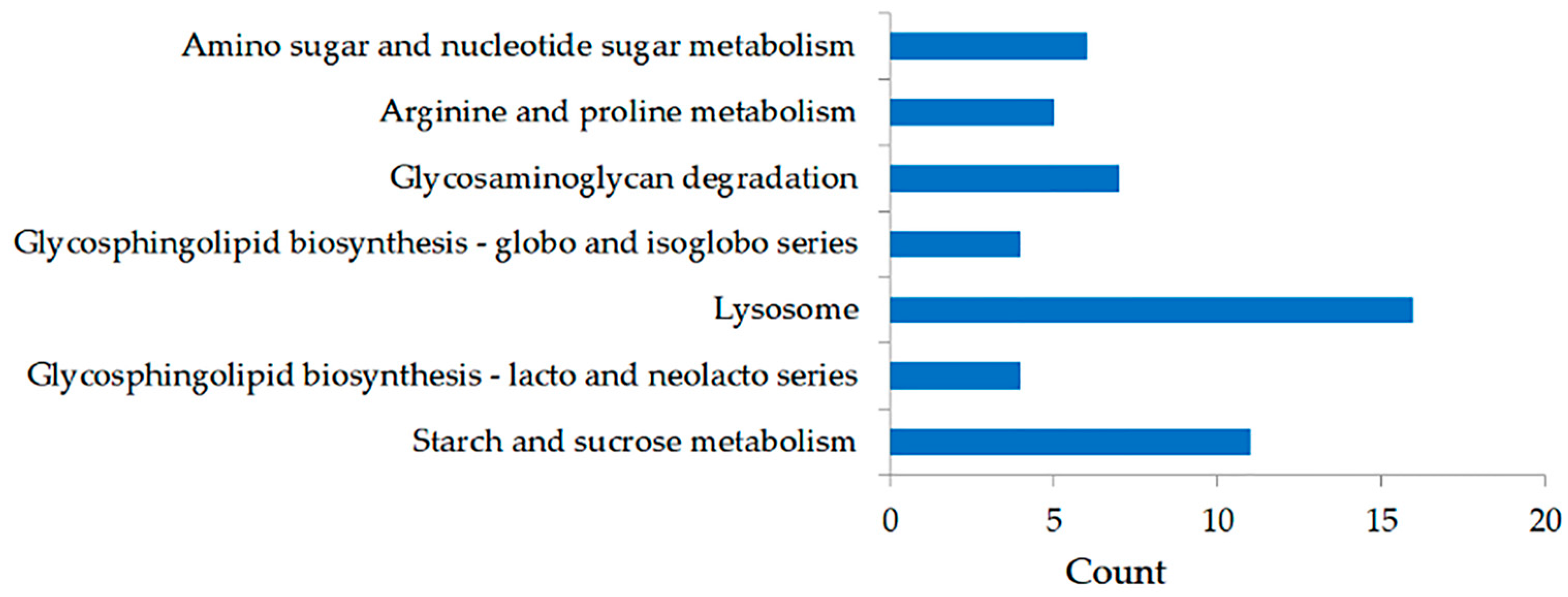
Both GO and KEGG network enrichment assessments showed the functional significance of the 685 DEGs. These DEGs were considerably associated with 46 GO BPs, 16 GO MFs, and 1 GO CC (Table S5). The BPs included pathways such as carbohydrate metabolic process, protein phosphorylation, phosphorylation, DNA integration, cell–matrix adhesion, etc. Certain DEGs were classified under the CC category, specifically the extracellular region, while others were related to key MFs, including kinase activity, hydrolase activity acting on glycosyl bonds, protein kinase activity, etc. The KEGG pathway analysis identified seven enriched pathways (p < 0.05), which included starch and sucrose metabolism, glycosphingolipid biosynthesis (lacto and neolacto series), lysosome, glycosphingolipid biosynthesis (globo and isoglobo series), glycosaminoglycan degradation, arginine and proline metabolism, as well as amino sugar and nucleotide sugar metabolism (Figure 2).

2.3. DEMs and Potential miRNA–mRNA Interaction Networks

Approximately 57 miRNAs were examined as significantly DEMs between the two abalone groups (Table S6). Of these, 30 miRNAs were upregulated and 27 miRNAs were downregulated. The differential expression of these miRNAs was further validated through volcano plots and heatmap analyses, which highlighted significant expression difference (p < 0.05) between both groups (Figure 3).
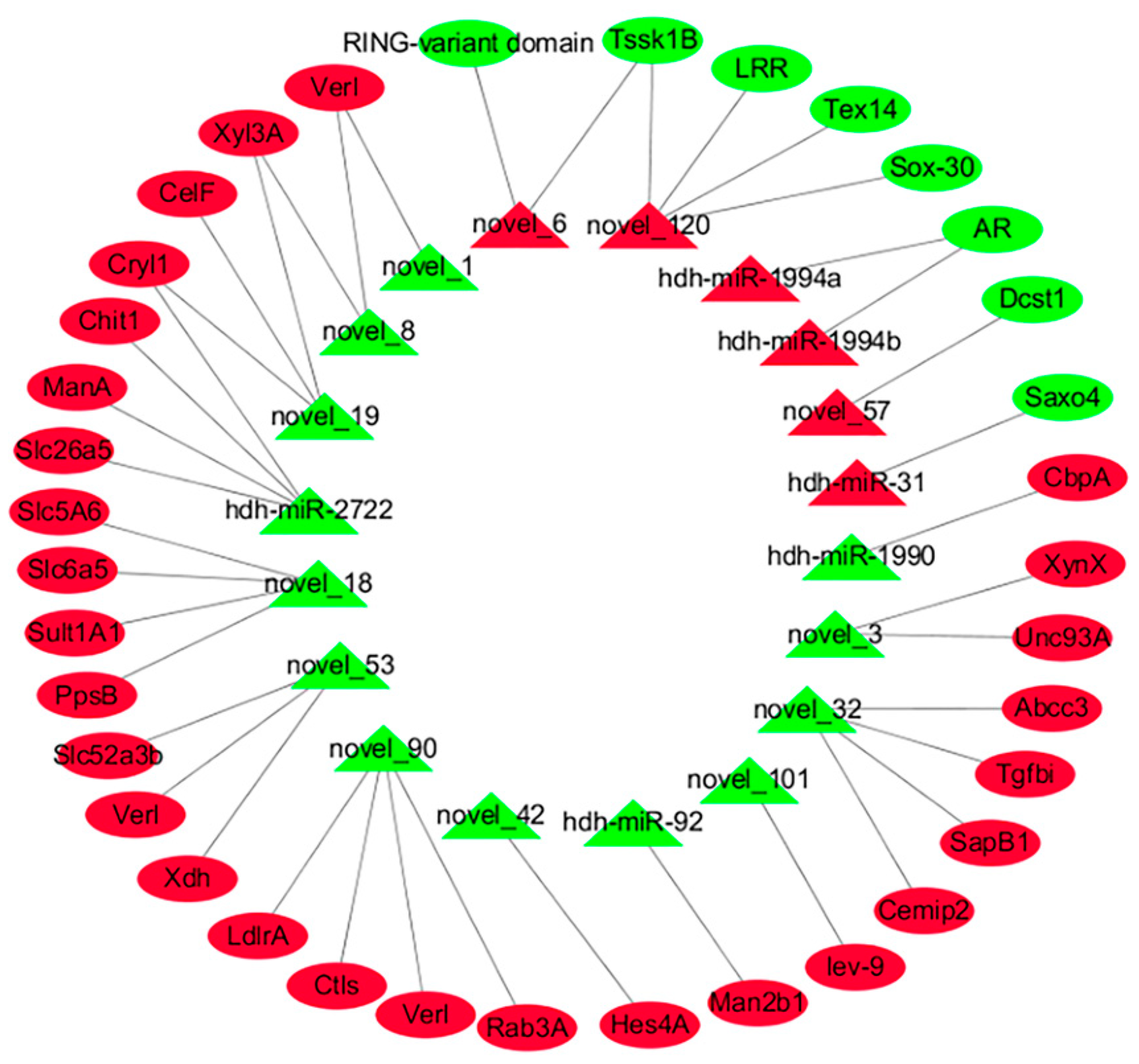
To clarify the MFs of the DEMs in both groups, the target genes of 57 DEMs were analyzed. The analysis identified 13 significantly DE target genes (target-DEGs) associated with 7 upregulated miRNAs and 49 target-DEGs related to 16 downregulated miRNAs (Table S7). Interestingly, certain DE-miRNAs were found to target genes related to reproductive processes. For instance, Verl, Inactive cell surface hyaluronidase CEMIP2 (Cemip2), and Tssk1B were targeted by novel_90, novel_32, and novel_120, respectively, suggesting that these miRNAs may affect reproductive regulation by modulating these genes (Table 3). Figure 4 illustrates a complex interaction network involving the DE-miRNAs and target-DEGs, highlighting their potential significances in H. discus hannai reproductive biology. Moreover, the GO distribution of the predicted target DEGs is depicted in Figure 5. These assessments showed multiple BPs between both groups. Some targets were categorized as BPs, including carbohydrate metabolic process, protein phosphorylation, cell–matrix adhesion, and cell–substrate adhesion. All other targets were related to important MFs, including hydrolase activity acting on glycosyl bonds, kinase activity, protein kinase activity, hydrolase activity hydrolyzing O-glycosyl compounds, and phosphotransferase activity alcohol group as acceptor. A KEGG pathway analysis showed starch and sucrose metabolism significant pathways (p < 0.05).

2.4. Verification Analysis of Identified DE-mRNA and DE-miRNA

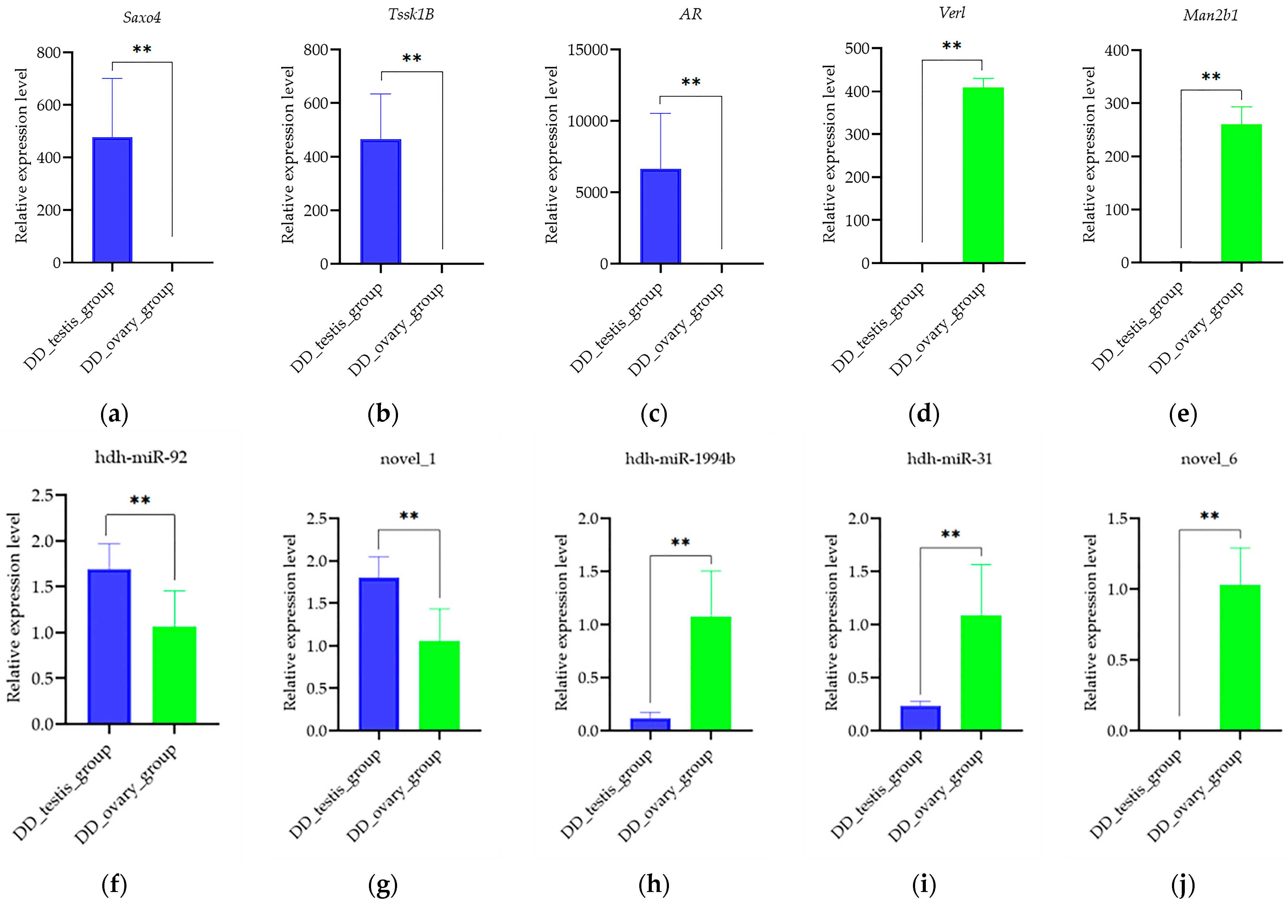
The expression levels of the DEGs (Saxo4, Tssk1B, AR, Verl, and Man2b1) and DEMs (hdh-miR-92, novel_1, hdh-miR-1994b, hdh-miR-31, and novel_6) were verified using qRT-PCR. The expression patterns of these DEGs and DEMs were consistent with the results of the RNA sequencing (Figure 6), suggesting high reliability of the RNA sequencing analysis.

2.5. Localization of Verl and Tssk1B in the Gonad of H. discus hannai

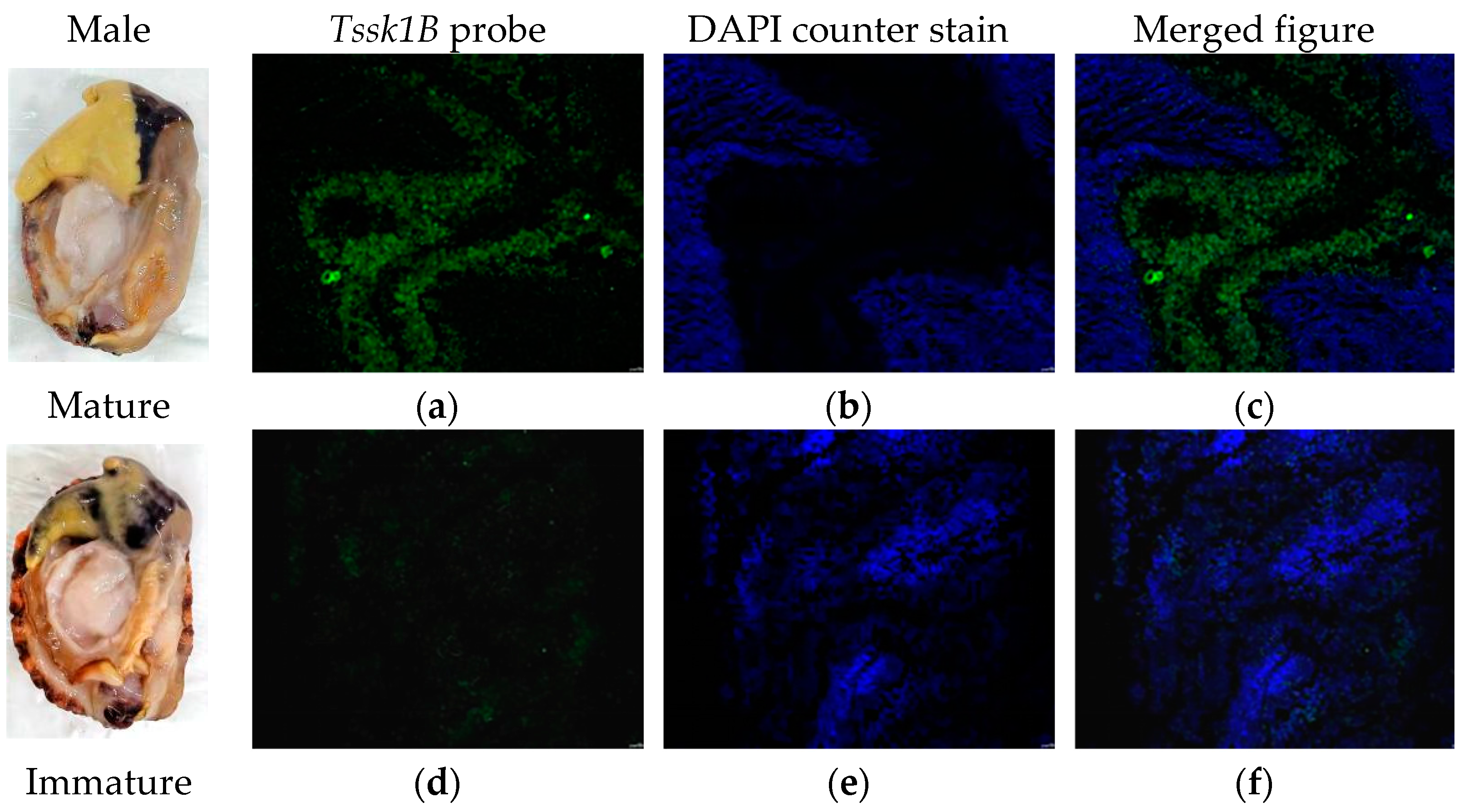
FISH was performed to detect the expression of Verl and Tssk1B mRNAs in the gonadal tissues of H. discus hannai. Fluorescence micrographs revealed that the Verl antisense probe produced strong positive signals (red; Figure 7a,c) in the gonads of mature females, while only weak positive signals were observed in immature female gonads (Figure 7d,f). Similarly, the Tssk1B antisense probe displayed positive signals (green; Figure 8a,c) in the gonads of mature males, but weak signals were detected in the gonads of immature males (Figure 8d,f). Blue fluorescence (DAPI-stained) indicating counterstaining of the positive signal of Verl (Figure 7b,e) and Tssk1B (Figure 8b,e).

3. Discussion

Reproduction is a biological process essential for the survival and propagation of all living organisms. It ensures the continuity of species across successive generations by promoting gamete production and offspring development. Understanding the mechanisms and regulatory pathways involved in reproductive processes is pivotal for advancing animal breeding practices. Enhancing reproductive efficiency in both genders is a crucial strategy in the production process, as it minimizes the number of animals required for breeding, thereby decreasing production costs and optimizing resource consumption. The stable and sustainable growth of the shellfish aquaculture industry relies heavily on a consistent supply of high-quality and abundant seedlings [1]. A comprehensive examination of shellfish reproductive traits is essential to ensure the consistent advancement of seedling production. Currently, several genes and proteins associated with the gonads and reproduction of abalone have been identified using HTS techniques. For example, Mendoza-Porras et al. [20,21] identified genes and proteins associated with sexual maturity, spawning, and reproduction in H.laevigata. Moreover, there are also some reports about the genes related to sexual maturity, sex determination, and reproduction in H. discus hannai and H. diversicolor [22,23,24,25,26]. MicroRNAs (miRNAs) are small non-coding RNAs (18–22 nt) that are crucial in regulating reproductive traits [3]. However, to date, no studies have been conducted on miRNAs related to reproduction in abalone. Thus, herein, we aimed to recognize miRNAs and miRNA–gene interactions in the ovaries and testes of abalones through an integrated analysis of miRNA and mRNA transcriptomes. This analysis elucidates the mechanistic role underlying reproduction and identifies possible molecular markers associated with shellfish reproductive processes, including sexual maturity and sex determination.
In this study, both mRNA and miRNA transcriptomes of the ovary and testis were sequenced from six sexually mature H. discus hannai. A total of 685 DEGs were found between both groups. These top 50 upregulated DEMs included Verl, lev-9, Zp-like, etc. Verl is associated with a species-specific mechanism to promote fertilization in the marine mollusk abalone [27]. Zp-like is expressed in the extracellular matrix that surrounds all mammalian oocytes, eggs, and early embryos. It is essential for oogenesis, fertilization, and preimplantation development [28]. These genes are highly expressed in the female H. discus hannai gonad and rarely or minimally expressed in the male H. discus hannai gonad. Among these downregulated DEGs, the top 50 genes included Sox-30, Tssk, Tepp, Sperm-lysin, etc. Tssk critically modulates reproductive cell differentiation and sperm activity [29]. Tepp displayed differential expression in sperm across various seasons [30]. Abalone sperm-lysin, a rapidly evolving reproductive protein, is a 16-kDa protein with a high positive charge. It plays a key role in dissolving the VE surrounding abalone oocytes [27]. These genes are highly expressed in the male H. discus hannai gonad and rarely or minimally expressed in the female H. discus hannai gonad. These findings depicted that these genes may be used as molecular markers to distinguish between male and female H. discus hannai.
Next, the biological functions of these DEGs were explored to determine their potential relevance in regulating reproductive traits. The analysis revealed that these DEGs were significantly correlated with various biological processes, including carbohydrate metabolic processes, starch and sucrose metabolism, glycosphingolipid biosynthesis, glycosaminoglycan degradation, amino and nucleotide sugar metabolism, etc. Importantly, energy metabolism plays a critical role in ATP production, which is essential for gonadal development, spermatogenesis, and oogenesis [24,31]. In this study, Endoglucanase A, Fut1, XynX, Chit1, xyl3A, Alpha-amylase, celF, and Man2b1 were differently expressed between both groups of abalones, revealing that these genes are highly promising candidates for regulating oogenesis, spermatogenesis, and gonad development via energy metabolism. Further verification of the function of these genes in both genders of abalone will be undertaken in the future.
Multiple classes of sRNAs, such as miRNAs, regulate the processes of sex identification and differentiation, gonadal differentiation, and maturation [32,33]. Mature sequences of miRNAs are approximately 20–25 nt in length and participate in post-transcriptional gene repression [34]. The association between miRNA and RNA is complex, since one miRNA can potentially affect several genes, and one gene can potentially be affected by several miRNAs [35]. The investigation of associations between miRNA profiles and established target mRNAs, as well as the identification of possible correlation between novel miRNA and mRNA interactions, can be simplified by paired expression profiling of mRNA and miRNA [36,37]. The function of miRNAs and the identification of the miRNA-mRNA pairs involved in physiology could be understood through the profile analysis of combined miRNA and corresponding target mRNA [38]. Several key miRNAs, namely, let-7, miR-8, miR-100, miR-8*, miR-9*, and miR-315, have been implicated in sex identification, reproduction, differentiation, and growth. Remarkably, miR-8 and miR-8* may synergistically modulate the fruitless gene during female sexual differentiation and gonadal development in Scylla paramamosain [39]. To date, there have been no reports of integrated miRNA and target mRNA analyses in studies of sexual maturity and sex determination in H. discus hannai. The current study detected 57 DEMs between the DD_ovary_group and DD_testis_group abalones, of which there were 73 pairs (Table S7) with adversely associated miRNA-mRNA pairs, depicting that these specific miRNA-mRNA pairs participate in abalone reproduction traits. For example, novel_90 targeted Verl, novel_32 targeted Cemip2, novel_120 targeted Tssk1B, and so on. These miRNAs potentially target these genes to modulate reproduction, as they were reported to be associated with reproduction. Further, the enrichment assessment of these target DEGs demonstrated that these miRNA-mRNA pairs regulate the sexual maturity and sex determination of H. discus hannai by regulating carbohydrate metabolic processes, protein phosphorylation, kinase activity, and starch and sucrose metabolisms. However, additional research is necessary to determine the functional significance of the miRNA-mRNA pairs that were identified in the ovary and testis of H. discus hannai.
Finally, qRT-PCR analysis verified the accuracy of transcriptome sequencing. The findings showed that the relative candidate mRNA and miRNA expression trends strongly corroborated with the transcriptomic analysis, illustrating the reliability of the transcriptomic profile. Previous studies [40,41] have used FISH technology to examine the expression patterns of genes in abalone. Herein, FISH was also used to evaluate the gene expression localization within the gonads of H. discus hannai. The findings depicted that Verl was highly present in the ovaries of mature H. discus hannai, with weak positive signals observed in the ovaries of immature abalones. Similarly, Tssk1B depicted a high level in the testes of mature H. discus hannai, while weak positive signals were detected in the testes of immature abalones. These findings suggest that these genes, along with their role in sex determination, may serve as markers for assessing the sexual maturity of H. discus hannai.

4. Materials and Methods

4.1. Abalones and RNA Preparation

Sexually mature H. discus hannai (body weight 108.2 ± 9.7 g; 36 months old; three males and three females) with a visual gonad index (VGI) of 3 [42] were procured from Fuda Aquaculture (Jinjiang, Fujian Province, China). The VGI consists of four categories (0–3) that relate to changes in the size and shape of the gonad, wherein VGI 3 refers to abalones with a swollen gonad with rounded tip and are classified as sexually mature. Gonadal tissues were collected from three individuals of each sex, with each sample processed independently. To ensure sample integrity, the outer membrane of the gonads was carefully excised without disturbing the underlying digestive gland. Approximately 500 mg of gonadal tissue (eggs or sperm) was snap-frozen in liquid nitrogen and kept at −80 °C for storage. Total RNA was isolated from the gonadal samples using TRIzol reagent (Invitrogen, Waltham, MA, USA). The isolated RNA purity and integrity were then evaluated to ensure suitability for downstream analyses.

4.2. Transcriptome Sequencing and Data Processing

Approximately 3 µg of RNA per sample was processed to synthesize cDNA libraries. Index-coded samples underwent clustering via the TruSeq PE Cluster Kit v3-cBot-HS and the cBot Cluster Generation System (Illumina, San Diego, CA, USA). Sequencing employed the Illumina HiSeqX platform and produced 150 bp paired-end reads. Raw sequencing data underwent quality control using custom in-house Perl scripts, which eliminated reduced-quality reads, adapter sequences, and poly-N-harboring sequences to produce clean reads. Quality metrics, namely, Q20, Q30, and GC levels, were computed for the processed data.
Clean reads underwent alignment to the reference genome of H. discus hannai using TopHat v2.0.9 [43] under default parameters. The reference genome and gene model annotation files were acquired from Dr. Weiwei You (Xiamen University, Xiamen, China). Following assembly of mapped reads via Scripture (beta2) [44] and Cufflinks v2.1.1 [45,46], gene profiles were computed as fragments per kilobase (kb) of transcript per million mapped reads (FPKM) with Cuffdiff v2.1.1 [47], and gene counts were analyzed with HTSeq [48]. Furthermore, DEGs identification utilized the edgeR package [49], with selection criteria as follows: corrected p-value < 0.05 and |log2 fold change| ≥ 10. To interpret DEG functions, enrichment analyses were conducted using the Kyoto Encyclopedia of Genes and Genomes (KEGG) networks [50] and Gene Ontology (GO) [51], namely, molecular functions (MF), biological processes (BP), and cellular components (CC). These analyses were carried out via the database for Annotation, Visualization, and Integrated Discovery (DAVID) [52], with GO terms and networks considered significant at p < 0.05. Further, protein–protein interaction (PPI) axes were developed via STRING v10 [53].

4.3. sRNA Sequencing and Analysis

In all, 3 µg of total RNA/sample was employed to prepare the small RNA library. Following the manufacturer’s recommendations, single-end sequencing (50 bp) was performed on an Illumina HiSeq2500 at Novogene (Tianjin, China). Post-sequencing, clean reads were generated by eliminating sequences with poly-N, poly-A/T/G/C, adapter contamination, and reduced-quality reads from the raw information. The Q20, Q30, and GC levels of the raw data were computed to assess data quality. Next, we conducted downstream analyses on clean reads selected within a specified length range [54].
Small RNA tags underwent mapping to a reference sequence using Bowtie 1.0.1 [55], prior to search against the known miRNA database miRBase 20.0. To elucidate potential miRNAs, the modified software mirdeep2 [56] and srna-tools-cli (http://srna-tools.cm p.uea.ac.uk/ (accessed on 13 February 2025)) were employed, enabling secondary structure prediction. Novel miRNAs were predicted using an integrated approach combining miREvo 1.1 [57] and mirdeep2 [56] software. Differential expression between both groups was evaluated via the DESeq R package (1.8.3) [58] with p-values corrected via the Benjamini–Hochberg method [59]. An adjusted p < 0.05 threshold was considered significant for identifying DE.

4.4. miRNA Target Prediction and Functional Analyses

The miRNA’s target gene was identified via psRobot_v1.2 in miRanda-3.3a [60]. The roles of the estimated target genes were annotated using GOSeq [61] and KOBAS 2.0 [62] software. Further, Cytoscape (http://www.cytoscape.org) was employed for construction of the interaction networks between DE-miRNAs and their respective target genes.

4.5. Quantitative Real-Time PCR

We conducted gene expression assessment via qRT-PCR to further confirm the selected gene and miRNA expressions. Gene-specific primers were designed for the target genes, with β-actin as the internal control (Table 4). Specific and universal reverse primers were employed for miRNAs, with U6 employed as the reference gene for normalization [63]. Next, qRT-PCR assays were conducted using a CFX96 Real-Time System (Bio-Rad, Hercules, CA, USA). Amplification reactions (25 μL) comprised 12.5 μL of 2× M5 HiPer SYBR Premix EsTaq (withTli RNaseH), 0.5 μL of each forward and reverse primer (10 μM), 2 μL of cDNA (diluted 1:100), and 9.5 μL of nuclease-free water. The reaction conditions were as follows: a 30 s denaturation at 95 °C, with subsequent 40 cycles of 5 s denaturation at 95 °C, then a 30 s annealing/extension at 60 °C. Fluorescence intensities were recorded after each cycle. Relative target gene and miRNA expressions were estimated via the 2−∆∆CT formula [64]. All experiments were repeated in triplicate, with three independent biological replicates.

4.6. Fluorescence In Situ Hybridization (FISH)

Fluorescein-labeled specific oligonucleotides were used as probes to target specific binding sequences. These probes hybridized with their complementary sequences, allowing for the fluorescent labeling of the target gene–probe hybrids. This enabled both the localization and quantification of the hybrids. Based on the Verl and Tssk1B gene sequence characteristics, FISH probes were designed, with their sequences detailed in Table 5. These probes were synthesized by Wuhan Servicebio Technology Co., Ltd. (Wuhan, China).
Gonadal tissues (ovary and testis) of H. discus hannai were fixed in an in situ hybridization fixative for >12 h and kept at 4 °C. These fixed tissues were sectioned into 3 mm thick blocks, dehydrated in an ethanol gradient, then cleared in xylene, prior to paraffin embedding. Sections were sliced to a 4 μm thickness, spread, and dried in a 62 °C oven for 2 h. The sections were immersed in dewaxing transparent liquid I for 15 min, followed by dewaxing transparent liquid II for 15 min, anhydrous ethanol I for 5 min, anhydrous ethanol II for 5 min, 85% alcohol for 5 min, 75% alcohol for 5 min, and DEPC water. Depending on the tissue type, the length of the fixed time, and the positioning of the index, different repair fluids were selected for repair, and the specific repair conditions are shown in Table 5. After natural cooling, the stroke circle was organized. Tissue-specific antigen retrieval was conducted as detailed in Table 5, and sections were digested with proteinase K (20 μg/mL) at 40 °C for different durations, also specified in Table 5. Following rinsing with DEPC water and PBS (thrice, 5 min each), pre-hybridization solution was introduced (dropwise) prior to a 1 h incubation at 40 °C. Subsequently, the solution was replaced with a probe-containing hybridization solution and allowed to react overnight in the incubator. All hybridization conditions are shown in Table 5. Post-hybridization washes included 10 min 2× SSC at 40 °C, 5 min 1× SSC at 40 °C, and a 10 min 0.5× SSC at room temperature. Sections can further be washed with formamide when nonspecific hybridization can be observed. Finally, an 8 min DAPI staining was performed without light, and an anti-fluorescence quenching sealing agent was added after washing.
Tissue sections were examined with a Nikon upright fluorescence microscope, and all images were captured for analysis. The ultraviolet (UV) excitation wavelength (λ) ranged from 330 to 380 nm with an emission λ of 420 nm, producing blue fluorescence. The green fluorescence emitted by FAM (488) was observed under an excitation λ of 465–495 nm and an emission λ of 515–555 nm, while CY3 red fluorescence was visualized with an excitation λ of 510–560 nm and an emission λ of 590 nm. Under UV excitation, nuclei stained with DAPI emitted blue fluorescence, and positive expressions were identified based on the respective fluorescein-labeled fluorescence. FAM (488) emitted green light, whereas CY3 produced red light, with the fluorescence intensity indicating the level of expression.

4.7. Statistics

All of the qRT-PCR data were expressed as mean ± standard deviation (SD). Statistical significance was evaluated using SPSS 19.0 (IBM Corp., Armonk, NY, USA). Significant differences were analyzed by two-tailed t-tests, with p < 0.05 considered statistically significant.

5. Conclusions

This study demonstrated the transcript and miRNA expressions of abalone gonads from DD_ovary_group and DD_testis_group abalones using HTS. This study revealed the identity of 137 miRNAs associated with the ovary and testis of H. discus hannai for the first time. Several key candidate mRNAs have detected and outlined a crucial functional miRNA–mRNA network related to abalone sexual maturity and sex determination. These key mRNAs and miRNAs may regulate sexual maturation and sex determination of abalone via carbohydrate metabolic processes, phosphotransferase activity, and starch and sucrose metabolisms. Multiple mRNAs and miRNAs were also selected for further analysis, and the expression was analyzed using RT-qPCR and FISH analyses. Overall, these findings enrich the genetic information resources of abalone, suggest a new direction of the mechanistic role of reproductive traits of shellfish, and offer a theoretical basis for improving the reproductive efficiency of abalone.

Supplementary Materials

The following supporting information can be downloaded at: https://www.mdpi.com/article/10.3390/ijms26073235/s1.

Author Contributions

Methodology, validation, and investigation, J.H., M.Z. and Z.S.; formal analysis, data curation, visualization, writing—original draft preparation, writing—review and editing, J.H.; writing—review and editing, supervision, project administration, J.C. and C.K.; funding acquisition, J.H. and C.K. All authors have read and agreed to the published version of the manuscript.

Funding

This research was supported by grants from the Minjiang University Introduces Talent pre-research project (No. MJY22008, No. MJY24020); the Science and Technology Project of Fuzhou institute of Oceanography (No. 2025F15); the Seed Industry Innovation and Industrialization in Fujian Province (No. 2021FJSCZY02).

Institutional Review Board Statement

The animal study protocol was approved by the Laboratory Animal Ethics Committee of Minjiang University (protocol code: 3501160022372; date: 17 May 2019).

Informed Consent Statement

Not applicable.

Data Availability Statement

The data presented in this study are available upon request from the corresponding authors.

Conflicts of Interest

The authors declare no conflicts of interest.

Abbreviations

The following abbreviations are used in this manuscript:
miRNAmicroRNA
H. discus hannaiHaliotis discus hannai
DEdifferentially expressed
TsskTestis-specific serine/threonine-protein kinase
VerlVitelline envelope sperm lysin receptor
FISHfluorescence in situ hybridization
UTRsuntranslated regions
HTShigh-throughput sequencing
DEGsdifferentially expressed genes
DEMsdifferentially expressed miRNAs
bratBrain tumor protein
PpnPapilin
Fut2Galactoside alpha-(1,2)-fucosyltransferase 2
Tuba1ATubulin alpha-1A chain
FasnFatty acid synthase
InmtIndolethylamine N-methyltransferase
TpoThyroid peroxidase
hFat3Protocadherin Fat 3
Zp-likeZona pellucida-like domain
SOX-30Transcription factor SOX-30
TeppTestis, prostate and placenta-expressed protein
HSF1Heat shock factor protein 1
Saxo4Stabilizer of axonemal microtubules 4
ARAnkyrin repeats
Man2b1Lysosomal alpha-mannosidase
KEGGKyoto Encyclopedia of Genes and Genomes
GOGene Ontology
MFmolecular functions
BPbiological processes
CCcellular components
PPIprotein–protein interaction
qRT-PCRQuantitative real-time PCR
SDstandard deviation
Abcc3ATP-binding cassette sub-family C member 3
CbpAChitin binding Peritrophin-A domain
Chit1Chitotriosidase-1
CelFEndoglucanase F
XynXExoglucanase XynX
Cemip2Inactive cell surface hyaluronidase CEMIP2
Cryl1Lambda-crystallin
CtlsLectin C-type domain
LdlrALow-density lipoprotein receptor domain class A
ManAMannan endo-1,4-beta-mannosidase
PpsBPhenolphthiocerol/phthiocerol polyketide synthase subunit B
Slc26a5Prestin
lev-9Protein lev-9
Unc93AProtein unc-93 homolog A
Rab3ARas-related protein Rab-3A
SapB1Saposin-like type B, region 1
Slc6a5Sodium- and chloride-dependent glycine transporter 2
Sult1A1Sulfotransferase 1A1
Hes4ATranscription factor Hes4A
TgfbiTransforming growth factor-beta-induced protein ig-h3
XdhXanthine dehydrogenase
Xyl3AXylan 1,4-beta-xylosidase
Dcst1E3 ubiquitin-protein ligase Dcst1
Tex14Inactive serine/threonine-protein kinase Tex14
LRRLeucine rich repeat

References

  1. Qing, W. Preliminary Study on Gonadal Development and Mechanism of Oocyte Maturation of Hard Clam Mercenaria Mercenaria. Master’s Thesis, University of Chinese Academy of Sciences (Institute of Oceanography, Chinese Academy of Sciences), Qingdao, China, 2007. (In Chinese). [Google Scholar]
  2. Osada, M.; Matsumoto, T. Endocrine control of gametogenesis and spawning in bivalves. In Physiology of Molluscs, 1st ed.; Saleuddin, S., Mukai, S., Eds.; CRC Press: Boca Raton, FL, USA, 2017; pp. 180–220. [Google Scholar]
  3. Wang, W.; Wu, B.; Liu, Z.; Sun, X.; Zhou, L.; Xu, W.; Yu, T.; Zheng, Y.; Zhang, S. Comprehensive analysis on the regulation of differentially expressed of mRNA and ncRNA in different ovarian stages of ark shell Scapharca broughtonii. BMC Genom. 2023, 24, 563. [Google Scholar] [CrossRef] [PubMed]
  4. Bartel, D.P. MicroRNAs: Genomics, biogenesis, mechanism, and function. Cell 2004, 116, 281–297. [Google Scholar] [CrossRef] [PubMed]
  5. Bartel, D.P. MicroRNAs: Target recognition and regulatory functions. Cell 2009, 136, 215–233. [Google Scholar] [CrossRef]
  6. Song, Y.N.; Shi, L.L.; Liu, Z.Q.; Qiu, G.F. Global analysis of the ovarian microRNA transcriptome: Implication for miR-2 and miR-133 regulation of oocyte meiosis in the Chinese mitten crab, Eriocheir sinensis (Crustacea: Decapoda). BMC Genom. 2014, 15, 547. [Google Scholar] [CrossRef] [PubMed]
  7. Zhao, C.; Zhang, G.S.; Yin, S.W.; Li, Z.C.; Wang, Q.T.; Chen, S.Q.; Zhou, G.Q. Integrated analysis of mRNA-seq and miRNA-seq reveals the potential roles of sexbiased miRNA-mRNA pairs in gonad tissue of dark sleeper (Odontobutis potamophila). BMC Genom. 2017, 18, 613. [Google Scholar] [CrossRef]
  8. Xu, P.; Vernooy, S.Y.; Guo, M.; Hay, B.A. Te drosophila microRNA miR-14 suppresses cell death and is required for normal fat metabolism. Curr. Biol. 2003, 13, 790–795. [Google Scholar] [CrossRef]
  9. He, Y.; Huang, C.X.; Chen, N.; Wu, M.; Huang, Y.; Liu, H.; Tang, R.; Wang, W.M.; Wang, H.L. The zebrafish miR-125c is induced under hypoxic stress via hypoxia-inducible factor 1 alpha and functions in cellular adaptations and embryogenesis. Oncotarget 2017, 8, 73846–73859. [Google Scholar] [CrossRef]
  10. Geiger, D.L. Distribution and biogeography of the Haliotidae (Gastropoda: Vetigastropoda) world-wide. Boll. Malacol. 2000, 35, 57–120. [Google Scholar]
  11. Luo, X.; Ke, C.; You, W. Estimates of correlations for shell morphological traits on body weight of interspecific hybrid abalone (Haliotis discus hannai and Haliotis gigantea). J. Shellfish Res. 2013, 32, 115–118. [Google Scholar] [CrossRef]
  12. Liu, X.Z.; Cu, L.F. China Fishery Statistical Yearbook; China Agriculture Press: Beijing, China, 2023. [Google Scholar]
  13. Gnocchi, K.G.; Boldrini-França, J.; Gomes, L.C.; Chippari-Gomes, A.R. De novo assembly and annotation of the transcriptome of Astyanax lacustris liver unveil candidate genes to monitor response to environmental stress. Mar. Genom. 2020, 54, 100784. [Google Scholar] [CrossRef]
  14. Nazari, S.; Khoshkholgh, M.; Baeza, J.A. Comparative transcriptome sequencing analysis of the narrow-clawed crayfish Pontastacus leptodactylus (Eschscholtz, 1823) and discovery of candidate sex-related genes. Aquacult. Rep. 2022, 25, 101235. [Google Scholar] [CrossRef]
  15. Wang, H.; Tang, L.; Wei, H.; Lu, J.; Mu, C.; Wang, C. Transcriptomic analysis of adaptive mechanisms in response to sudden salinity drop in the mud crab, Scylla paramamosain. BMC Genom. 2018, 19, 421. [Google Scholar] [CrossRef]
  16. Lou, F.; Yang, T.; Han, Z.; Gao, T. Transcriptome analysis for identification of candidate genes related to sex determination and growth in Charybdis japonica. Gene 2018, 677, 10–16. [Google Scholar] [CrossRef] [PubMed]
  17. Cheng, J.; Yang, F.; Liu, S.; Zhao, H.; Lu, W.; Zhang, Q. Transcriptomic analysis reveals functional interaction of mRNA-lncRNA-miRNA in steroidogenesis and spermatogenesis of gynogenetic Japanese flounder (Paralichthys olivaceus). Biology 2022, 11, 213. [Google Scholar] [CrossRef] [PubMed]
  18. Wang, F.; Yang, Q.W.; Zhao, W.J.; Du, Q.Y.; Chang, Z.J. Effects of short-time exposure to atrazine on miRNA expression profiles in the gonad of common carp (Cyprinus carpio). BMC Genom. 2019, 20, 587. [Google Scholar] [CrossRef] [PubMed]
  19. He, P.; Wei, P.; Chen, X.; Lin, Y.; Peng, J. Identification and characterization of microRNAs in the gonad of Trachinotus ovatus using Solexa sequencing. Comp. Biochem. Physiol. Part D Genom. Proteom. 2019, 30, 312–320. [Google Scholar] [CrossRef]
  20. Mendoza-Porras, O.; Botwright, N.A.; McWilliam, S.M.; Cook, M.T.; Harris, J.O.; Wijffels, G.; Colgrave, M.L. Exploiting genomic data to identify proteins involved in abalone reproduction. J. Proteom. 2014, 108, 337–353. [Google Scholar] [CrossRef]
  21. Mendoza-Porras, O.; Botwright, N.A.; Reverter, A.; Cook, M.T.; Harris, J.O.; Wijffels, G.; Colgrave, M.L. Identification of differentially expressed reproductive and metabolic proteins in the female abalone (Haliotis laevigata) gonad following artificial induction of spawning. Comp. Biochem. Physiol. Part D Genom. Proteom. 2017, 24, 127–138. [Google Scholar] [CrossRef]
  22. Kim, M.A.; Rhee, J.S.; Kim, T.H.; Lee, J.S.; Choi, A.Y.; Choi, B.S.; Choi, I.Y.; Sohn, Y.C. Alternative Splicing Profile and Sex-Preferential Gene Expression in the Female and Male Pacific Abalone Haliotis discus hannai. Genes 2017, 8, 99. [Google Scholar] [CrossRef]
  23. Yu, L.; Xu, D.; Ye, H.; Yue, H.; Ooka, S.; Kondo, H.; Yazawa, R.; Takeuchi, Y. Gonadal Transcriptome Analysis of Pacific Abalone Haliotis discus discus: Identification of Genes Involved in Germ Cell Development. Mar. Biotechnol. 2018, 20, 467–480. [Google Scholar]
  24. Kim, M.A.; Kim, T.H.; Lee, S.; Nam, B.H.; Lee, J.S.; Jang, W.; Sohn, Y.C. Ovarian transcriptome profiles associated with sexual maturation in Pacific abalone (Haliotis discus hannai). Genes Genom. 2020, 42, 1179–1188. [Google Scholar] [CrossRef] [PubMed]
  25. Luo, H.; Xiao, J.; Jiang, Y.; Ke, Y.; Ke, C.; Cai, M. Mapping and marker identification for sex-determining in the Pacific abalone, Haliotis discus hannai Ino. Aquaculture 2021, 530, 735810. [Google Scholar] [CrossRef]
  26. Weng, X.; Xu, Y.; Dong, X.; Luo, X.; You, W.; Ke, C.; Cai, M. Sex-specific markers developed by next-generation sequencing confirmed a male heterogametic sex determination in small abalone, Haliotis diversicolor. Aquaculture 2022, 555, 738256. [Google Scholar] [CrossRef]
  27. Wilburn, D.B.; Tuttle, L.M.; Klevit, R.E.; Swanson, W.J. Solution structure of sperm lysin yields novel insights into molecular dynamics of rapid protein evolution. Proc. Natl. Acad. Sci. USA 2018, 115, 1310–1315. [Google Scholar] [CrossRef] [PubMed]
  28. Litscher, E.S.; Wassarman, P.M. Zona Pellucida Proteins, Fibrils, and Matrix. Annu. Rev. Biochem. 2020, 89, 695–715. [Google Scholar] [CrossRef] [PubMed]
  29. Salicioni, A.M.; Gervasi, M.G.; Sosnik, J.; Tourzani, D.A.; Nayyab, S.; Caraballo, D.A.; Visconti, P.E. Testis-specific serine kinase protein family in male fertility and as targets for non-hormonal male contraception. Biol. Reprod. 2020, 103, 264–274. [Google Scholar] [CrossRef] [PubMed]
  30. Neila-Montero, M.; Alvarez, M.; Riesco, M.F.; Montes-Garrido, R.; Palacin-Martinez, C.; Silva-Rodríguez, A.; Martín-Cano, F.E.; Peña, F.J.; de Paz, P.; Anel, L.; et al. Ovine fertility by artificial insemination in the breeding season could be affected by intraseasonal variations in ram sperm proteomic profile. Theriogenology 2023, 208, 28–42. [Google Scholar] [CrossRef] [PubMed]
  31. Ford, W. Glycolysis and sperm motility: Does a spoonful of sugar help the flagellum go round? Hum. Reprod. Update 2006, 12, 269–274. [Google Scholar] [CrossRef]
  32. Marco, A. Sex-biased expression of microRNAs in Drosophila melanogaster. Open Biol. 2014, 4, 140024. [Google Scholar] [CrossRef]
  33. Tonelli, F.M.P.; Lacerda, S.M.S.N.; Procópio, M.S.; Lemos, B.L.S.; de Franca, L.R.; Resende, R.R. Gene delivery to Nile tilapia cells for transgenesis and the role of PI3K-c2α in angiogenesis. Sci. Rep. 2017, 7, 44317. [Google Scholar] [CrossRef]
  34. Filipowicz, W.; Jaskiewicz, L.; Kolb, F.A.; Pillai, R.S. Post transcriptional gene silencing by siRNAs and miRNAs. Curr. Opin. Struct. Biol. 2005, 15, 331–341. [Google Scholar] [CrossRef]
  35. Krol, J.; Loedige, I.; Filipowicz, W. The widespread regulation of microRNA biogenesis, function and decay. Nat. Rev. Genet. 2010, 11, 597–610. [Google Scholar] [PubMed]
  36. Sun, L.; Lu, S.; Bai, M.; Xiang, L.; Li, J.; Jia, C.; Jiang, H. Integrative microRNA-mRNA analysis of muscle tissues in Qianhua Mutton Merino and Small Tail Han sheep reveals key roles for oar-miR-655-3p and oar-miR-381-5p. DNA Cell Biol. 2019, 38, 423–435. [Google Scholar] [CrossRef] [PubMed]
  37. Li, Z.; Abdalla, B.A.; Zheng, M.; He, X.; Cai, B.; Han, P.; Ouyang, H.; Chen, B.; Nie, Q.; Zhang, X. Systematic transcriptome-wide analysis of mRNA-miRNA interactions reveals the involvement of miR-142-5p and its target (FOXO3) in skeletal muscle growth in chickens. Mol. Genet. Genom. 2018, 293, 69–80. [Google Scholar]
  38. Zhao, W.; Xiao, W.; Sun, J.; Chen, M.; Ma, M.; Cao, Y.; Cen, W.; Li, R.; Luo, J. An integration of microRNA and transcriptome sequencing analysis reveal regulatory roles of miRNAs in response to chilling stress in wild rice. Plants 2022, 11, 977. [Google Scholar] [CrossRef]
  39. Waiho, K.; Fazhan, H.; Zhang, Y.; Zhang, Y.; Li, S.; Zheng, H.; Liu, W.; Ikhwanuddin, M.; Ma, H. Gonadal microRNA Expression Profiles and Their Potential Role in Sex Differentiation and Gonadal Maturation of Mud Crab Scylla paramamosain. Mar. Biotechnol. 2019, 21, 320–334. [Google Scholar]
  40. Hanif, M.A.; Hossen, S.; Cho, Y.; Sukhan, Z.P.; Choi, C.Y.; Kho, K.H. Characterization and Expression Analysis of Mollusk-like Growth Factor: A Secreted Protein Involved in Pacific Abalone Embryonic and Larval Development. Biology 2022, 11, 1445. [Google Scholar] [CrossRef] [PubMed]
  41. Hossen, S.; Sukhan, Z.P.; Kim, S.C.; Hanif, M.A.; Kong, I.K.; Kho, K.H. Molecular Cloning and Functional Characterization of Catalase in Stress Physiology, Innate Immunity, Testicular Development, Metamorphosis, and Cryopreserved Sperm of Pacific Abalone. Antioxidants 2023, 12, 109. [Google Scholar] [CrossRef] [PubMed]
  42. Kikuchi, S.; Uki, N. Technical study on artificial spawning of abalone, Genus Haliotis I. Relation between water temperature and advancing sexual maturity of Haliotis discus hannai Ino. Bull. Tohoku Reg. Fish. Res. Lab. 1974, 33, 69–78. [Google Scholar]
  43. Trapnell, C.; Pachter, L.; Salzberg, S.L. TopHat: Discovering splice junctions with RNA-Seq. Bioinformatics 2009, 25, 1105–1111. [Google Scholar] [CrossRef]
  44. Choi, M.J.; Kim, G.D.; Kim, J.M.; Lim, H.K. Differentially-Expressed Genes Associated with Faster Growth of the Pacific Abalone, Haliotis discus hannai. Int. J. Mol. Sci. 2015, 16, 27520–27534. [Google Scholar] [CrossRef]
  45. Trapnell, C.; Williams, B.A.; Pertea, G.; Mortazavi, A.; Kwan, G.; van Baren, M.J.; Salzberg, S.L.; Wold, B.J.; Pachter, L. Transcript assembly and quantification by RNA-Seq reveals unannotated transcripts and isoform switching during cell differentiation. Nat. Biotechnol. 2010, 28, 511–515. [Google Scholar] [PubMed]
  46. Trapnell, C.; Roberts, A.; Goff, L.; Pertea, G.; Kim, D.; Kelley, D.R.; Pimentel, H.; Salzberg, S.L.; Rinn, J.L.; Pachter, L. Differential gene and transcript expression analysis of RNA-seq experiments with TopHat and Cufflinks. Nat. Protoc. 2012, 7, 562–578. [Google Scholar]
  47. Flegel, C.; Manteniotis, S.; Osthold, S.; Hatt, H.; Gisselmann, G. Expression profile of ectopic olfactory receptors determined by deep sequencing. PLoS ONE 2013, 8, e55368. [Google Scholar]
  48. Anders, S.; Pyl, P.T.; Huber, W. HTSeq-a Python framework to work with highthroughput sequencing data. Bioinformatics 2015, 31, 166–169. [Google Scholar] [PubMed]
  49. Robinson, M.D.; McCarthy, D.J.; Smyth, G.K. edgeR: A Bioconductor package for differential expression analysis of digital gene expression data. Bioinformatics 2010, 26, 139–140. [Google Scholar] [CrossRef] [PubMed]
  50. Kanehisa, M.; Goto, S. KEGG: Kyoto encyclopedia of genes and genomes. Nucleic Acids Res. 2000, 28, 27–30. [Google Scholar] [CrossRef] [PubMed]
  51. Ashburner, M.; Ball, C.A.; Blake, J.A.; Botstein, D.; Butler, H.; Cherry, J.M.; Davis, A.P.; Dolinski, K.; Dwight, S.S.; Eppig, J.T.; et al. Gene Ontology: Tool for the unification of biology. Nat. Genet. 2000, 25, 25. [Google Scholar] [CrossRef] [PubMed]
  52. Dennis, G.; Sherman, B.T.; Hosack, D.A.; Yang, J.; Gao, W.; Lane, H.C.; Lempicki, R.A. DAVID: Database for annotation, visualization, and integrated discovery. Genome Biol. 2003, 4, R60. [Google Scholar] [CrossRef]
  53. Szklarczyk, D.; Franceschini, A.; Wyder, S.; Forslund, K.; Heller, D.; Huerta-Cepas, J.; Simonovic, M.; Roth, A.; Santos, A.; Tsafou, K.P.; et al. STRING v10: Protein–protein interaction networks, integrated over the tree of life. Nucleic Acids Res. 2014, 43, D447–D452. [Google Scholar] [CrossRef]
  54. Zhong, L.; Zhang, F.; Zhai, Y.; Cao, Y.H.; Zhang, S.; Chang, Y.Q. Identifcation and comparative analysis of complement C3-associated microRNAs in immune response of Apostichopus japonicus by high-throughput sequencing. Sci. Rep. 2015, 5, 17763. [Google Scholar] [CrossRef]
  55. Langmead, B.; Trapnell, C.; Pop, M.; Salzberg, S.L. Ultrafast and memory-efficient alignment of short DNA sequences to the human genome. Genome Biol. 2009, 10, R25. [Google Scholar] [CrossRef]
  56. Friedlander, M.R.; Mackowiak, S.D.; Li, N.; Chen, W.; Rajewsky, N. miRDeep2 accurately identifies known and hundreds of novel microRNA genes in seven animal clades. Nucleic Acids Res. 2011, 40, 37–52. [Google Scholar] [CrossRef]
  57. Wen, M.; Shen, Y.; Shi, S.; Tang, T. miREvo: An Integrative microRNA Evolutionary Analysis Platform for Next-generation Sequencing Experiments. BMC Bioinform. 2010, 13, 140. [Google Scholar] [CrossRef]
  58. Anders, S.; Huber, W. Differential expression analysis for sequence count data. Genome Biol. 2010, 11, R106. [Google Scholar]
  59. Storey, J.D. The positive false discovery rate: A Bayesian interpretation and the q-value. Ann. Stat. 2003, 31, 2013–2035. [Google Scholar] [CrossRef]
  60. Enright, A.J.; John, B.; Gaul, U.; Tuschl, T.; Sander, C.; Marks, D.S. MicroRNA targets in Drosophila. Genome Biol. 2003, 5, R1. [Google Scholar] [CrossRef] [PubMed]
  61. Young, M.D.; Wakefield, M.J.; Smyth, G.K.; Oshlack, A. goseq: Gene Ontology testing for RNA-seq datasets. R Bioconduct. 2012. [Google Scholar]
  62. Mao, X.; Cai, T.; Olyarchuk, J.G.; Wei, L. Automated genome annotation and pathway identification using the KEGG orthology (KO) as a controlled vocabulary. Bioinformatics 2005, 21, 3787–3793. [Google Scholar] [PubMed]
  63. Zheng, Z.; Huang, R.; Tian, R.; Jiao, Y.; Du, X. Pm-miR-133 hosting in one potential lncRNA regulates RhoA expression in pearl oyster Pinctada martensii. Gene 2016, 591, 484–489. [Google Scholar] [CrossRef]
  64. Livak, K.J.; Schmittgen, T.D. Analysis of relative gene expression data using real-time quantitative PCR and the 2−∆∆CT method. Methods 2001, 25, 402–428. [Google Scholar]
Figure 1. Volcano plots and heatmaps of the mRNA expression of differentially expressed genes (DEGs) in the abalone DD_ovary_group versus the DD_testis_group (p < 0.05). (a) Volcano plots of DEG expression. Red and green dots indicate up- and downregulated transcripts, respectively. (b) Hierarchical clustering of DEGs. Red rectangles represent upregulated mRNAs; blue rectangles represent downregulated mRNAs.
Ijms 26 03235 g001
Figure 2. Analysis of significant KEGG pathways for the DEGs.
Ijms 26 03235 g002
Figure 3. Volcano plots and heatmaps of DE miRNAs (DEMs) in both groups of abalones (p < 0.05). (a) Volcano plots of DEMs. Red dots depict upregulated miRNAs, while green dots mark downregulated miRNAs. (b) Hierarchical clustering of DEMs. Red rectangles depict upregulated miRNAs; blue rectangles show downregulated miRNAs.
Ijms 26 03235 g003
Figure 4. Interaction network of DEMs and selected target DEGs. Green ovals: downregulated target DEGs; red ovals: upregulated target DEGs; green triangles: downregulated DEMs; red triangles: upregulated DEMs.
Ijms 26 03235 g004
Figure 5. Gene ontology distribution of the target DEGs for DEMs.
Ijms 26 03235 g005
Figure 6. Expression of DEMs and DEGs quantified with qRT-PCR. Data are shown as mean ± SD (n = 3). **, p < 0.01.
Ijms 26 03235 g006
Figure 7. Confocal laser scanning microscopic observation after FISH of Verl mRNA in the female gonad tissue of H. discus hannai. (a,d) Fluorescent images of Verl antisense probe (red fluorescent). (b,e) Fluorescent images of DAPI counterstain (blue fluorescent). (c,f) Fluorescent images of Merged. Scale bars: 50 μm.
Ijms 26 03235 g007aIjms 26 03235 g007b
Figure 8. Confocal laser scanning microscopic observation after FISH of Tssk1B mRNA in the male gonad tissue of H. discus hannai. (a,d) Fluorescent images of Tssk1B antisense probe (green fluorescent). (b,e) Fluorescent images of DAPI counterstain (blue fluorescent). (c,f) Fluorescent images of Merged. Scale bars: 50 μm.
Ijms 26 03235 g008
Table 1. The results of RNA quality.
Sample NameRaw ReadsRaw BasesClean ReadsClean BasesError RateQ20 (%)Q30 (%)GC pct (%)
DD_ovary_144,600,4666.69G43,341,2206.5G0.0394.8787.5245.16
DD_ovary_247,513,1927.13G46,428,6126.96G0.0395.0787.8445.29
DD_ovary_344,400,1546.66G43,435,3526.52G0.0394.5386.6143.46
DD_testis_141,592,9366.24G40,138,6286.02G0.0397.2592.1943.31
DD_testis_245,197,5346.78G44,052,7666.61G0.0397.0691.8243.38
DD_testis_344,737,0006.71G42,770,6086.42G0.0396.8691.4443.87
Table 2. Small RNA sequencing data.
SampleTotal_ReadsN% > 10%Low Quality5_adapter_
contamine
3_adapter_null or insert_nullWith ployA/T/G/CClean Reads
DD_ovary_112,604,189 (100.00%)1 (0.00%)0 (0.00%)2907 (0.02%)263,476 (2.09%)3627 (0.03%)12,334,178 (97.86%)
DD_ovary_211,763,492 (100.00%)319 (0.00%)0 (0.00%)11,695 (0.10%)91,539 (0.78%)35,964 (0.31%)11,623,975 (98.81%)
DD_ovary_311,148,631 (100.00%)270 (0.00%)0 (0.00%)11,784 (0.11%)134,483 (1.21%)30,690 (0.28%)10,971,404 (98.41%)
DD_testis_111,558,139 (100.00%)0 (0.00%)0 (0.00%)2243 (0.02%)359,076 (3.11%)2194 (0.02%)11,194,626 (96.85%)
DD_testis_211,598,714 (100.00%)0 (0.00%)0 (0.00%)7447 (0.06%)247,111 (2.13%)8378 (0.07%)11,335,778 (97.73%)
DD_testis_311,563,092 (100.00%)0 (0.00%)0 (0.00%)2606 (0.02%)580,785 (5.02%)2072 (0.02%)10,977,629 (94.94%)
Table 3. Differentially expressed miRNAs (DEMs) and their DE target mRNAs (target DEGs).
Target DEG idTarget DEG Namelog2Fold
Change
padjRelated miRNA
HDH_G17212Alpha-amylase11.57930448.34E-14hdh-miR-133-3p
HDH_G10591ATP-binding cassette sub-family C member 3 (Abcc3)10.690529893.88E-14novel_32
HDH_G16571Chitin binding Peritrophin-A domain (CbpA)10.024945461.08E-09hdh-miR-1990
HDH_G27689Chitotriosidase-1 (Chit1)12.040390881.27E-14hdh-miR-2722
HDH_G29537Endoglucanase F (CelF)13.611834472.04E-18novel_19
HDH_G17091Exoglucanase XynX (XynX)10.074196452.56E-08novel_3
HDH_G14591Inactive cell surface hyaluronidase CEMIP2 (Cemip2) 14.810635313.02E-25novel_32
HDH_G05585Lambda-crystallin (Cryl1)10.204122771.31E-09hdh-miR-2722,
novel_19
HDH_G13593Lectin C-type domain (Ctls)10.286317762.36E-09novel_90
HDH_G08135Low-density lipoprotein receptor domain class A (LdlrA)14.261861765.70E-19novel_90
HDH_G26485Lysosomal alpha-mannosidase (Man2b1)12.226837254.35E-17hdh-miR-92
HDH_G25248Mannan endo-1,4-beta-mannosidase (ManA)12.050663652.40E-14hdh-miR-2722
HDH_G06159Phenolphthiocerol/phthiocerol polyketide synthase subunit B (PpsB)11.989331264.70E-19novel_18
HDH_G14448Prestin (Slc26a5)10.150835091.76E-12hdh-miR-2722
HDH_G06399Protein lev-9 (lev-9)18.169341841.96E-35novel_101
HDH_G15576Protein unc-93 homolog A (Unc93A)10.020449071.97E-11novel_3
HDH_G15755Ras-related protein Rab-3A (Rab3A)10.036979221.02E-12novel_90
HDH_G27130Saposin-like type B, region 1 (SapB1)12.754687742.94E-15novel_32
HDH_G25000Sodium- and chloride-dependent glycine transporter 2 (Slc6a5)11.099506646.36E-15novel_18
HDH_G21292Slc5a610.492419931.19E-13novel_18
HDH_G21270Scl52a3B10.967008048.10E-15novel_53
HDH_G16512Sulfotransferase 1A1 (Sult1A1)10.295134783.53E-10novel_18
HDH_G29406Transcription factor Hes4A (Hes4A)11.253460652.90E-16novel_42
HDH_G24939Transforming growth factor-beta-induced protein ig-h3 (Tgfbi)11.380497922.95E-13novel_32
HDH_G12983Vitelline envelope sperm lysin receptor (Verl)15.481861291.00E-33novel_90
HDH_G17385Verl19.594002778.33E-54novel_53
HDH_G15719Verl18.783548439.64E-49novel_1, novel_8
HDH_G14936Xanthine dehydrogenase (Xdh)10.062941491.01E-09novel_53
HDH_G06239Xylan 1,4-beta-xylosidase (Xyl3A)11.286669142.85E-11novel_19, novel_8
HDH_G31217Ankyrin repeats (AR)−13.118783199.27E-25hdh-miR-1994a,
hdh-miR-1994b
HDH_G04117E3 ubiquitin-protein ligase Dcst1 (Dcst1)−10.292202655.34E-15novel_57
HDH_G27469Inactive serine/threonine-protein kinase Tex14 (Tex14)−12.053069413.31E-137novel_120
HDH_G02229Leucine rich repeat (LRR)−13.504591971.33E-26novel_120
HDH_G08545RING-variant domain−14.26146085.53E-29novel_6
HDH_G05810Stabilizer of axonemal microtubules 4 (Saxo4)−10.257198514.38E-55hdh-miR-31
HDH_G26330Testis-specific serine/threonine-protein kinase 1 (Tssk1B)−10.661402241.63E-58novel_120, novel_6
HDH_G12498Transcription factor Sox-30 (Sox-30)−13.811547425.52E-27novel_120
Table 4. Pairs of primer sequences.
PrimerSequence (5′-3′)
Saxo4-qFGTTCAAGGGTCTTCGAGGCA
Saxo4-qRCGTGAAATAACCGGGCTGCT
Tssk1B-qFCCACCATTCTGACCATCCCT
Tssk1B-qRCCTCCTTCTTCTTCCTCTCGG
AR-qFGAAAATGGGATCCTCGGCTG
AR-qRTTACCCCTCACCGCTTGAAT
Verl-qFGACTTCCGGGCCATCTGTAA
Verl-qRACGTTGGAGTTCTGTCTCCT
Man2b1-qFGGAGGCTAAAGCGCTCATCA
Man2b1-qRCCGAACTTTTGCTCAGCGTT
β-actin-qFGGTATCCTCACCCTCAAGT
β-actin-qRGGGTCATCTTTTCACGGTTG
hdh-miR-92AATTGCACTTGTCCCGGCCTGC
novel_1AATTGCACTCGTCCCGGCCTGCAA
hdh-miR-1994bTGAGACAGTGTGTCCTCCCTCA
hdh-miR-31AGGCAAGATGTTGGCATAGCT
novel_6TCGAGGAAGTAGAAGACCTTGACGT
Table 5. Probe information and repair conditions.
Probe NameDigestive ConditionProbe Sequence (5′-3′)Repair ConditionProbe ConcentrationHybridization TemperatureName of the Corresponding Signal Probe
VerlProteinase K was digested at 40 °C for 10 min.TATGCAGGTAATGGTGCCGTCAAG
/GTAATCGACCCTTCCGGTTCCAAG
/TTGCGAATCTTGTGTTCGTCCTCG
Tissue sections were kept in a citric acid solution (pH 6.0) repair box and incubated in a water bath at 90 °C for 48 min.500 nM40 °CCY3
Tssk1BProteinase K was digested at 40 °C for 10 min.CTCGTGAAGGACCAGCAGAGAAGC
/CATAGGACCCACAGAAGGTCTCCATC
/GTTCTCGGCAGTCTTTAGACACCTGC
Tissue sections were kept in a citric acid solution (pH 6.0) repair box and incubated in a water bath at 90 °C for 48 min.500 nM40 °CFAM (488)
Disclaimer/Publisher’s Note: The statements, opinions and data contained in all publications are solely those of the individual author(s) and contributor(s) and not of MDPI and/or the editor(s). MDPI and/or the editor(s) disclaim responsibility for any injury to people or property resulting from any ideas, methods, instructions or products referred to in the content.