Review

Exploring the Interaction of Tumor-Derived Exosomes and Mesenchymal Stem Cells in Tumor Biology

by
Konstantinos S. Papadopoulos
1,2,*,
Penelope Korkolopoulou
2 and
Christina Piperi
3,*
1
Department of Plastic and Reconstructive Surgery, 401 General Military Hospital of Athens, 11525 Athens, Greece
2
First Department of Pathology, Medical School, National and Kapodistrian University of Athens, 11527 Athens, Greece
3
Department of Biological Chemistry, Medical School, National and Kapodistrian University of Athens, 11527 Athens, Greece
*
Authors to whom correspondence should be addressed.
Int. J. Mol. Sci. 2025, 26(7), 3095; https://doi.org/10.3390/ijms26073095
Submission received: 6 March 2025 / Revised: 21 March 2025 / Accepted: 24 March 2025 / Published: 27 March 2025
(This article belongs to the Special Issue Mesenchymal Stem Cells and Cancer)

Abstract

:
Exosomes are actively produced extracellular vesicles, released from different cell types, that exert important regulatory roles in vital cellular functions. Tumor-derived exosomes (TDEs) have received increasing attention because they enable intercellular communication between the neoplastic and non-neoplastic cells present in the microenvironment of tumors, affecting important functions of different types of mesenchymal stem cells (MSCs) with the ability to self-renew and differentiate. MSC-derived exosomes (MSC-exos) carry a variety of bioactive molecules that can interact with specific cellular targets and signaling pathways, influencing critical processes in tumor biology, and exhibiting properties that either promote or inhibit tumor progression. They can regulate the tumor microenvironment by modulating immune responses, enhancing or suppressing angiogenesis, and facilitating tumor cells’ communication with distant sites, thus altering the behavior of non-cancerous cells present in the microenvironment. Herein, we explore the main functions of TDEs and their intricate interactions with MSC-exos, in terms of enhancing cancer progression, as well as their promising clinical applications as tumor microenvironment modulators.

1. Introduction

Extracellular vesicles (EVs) present a constantly expanding class of micro- and nanoparticles, produced by cells as key mediators of cellular communication, containing nucleic acids, proteins, or lipids. While their existence has been known about for years, they were often considered byproducts of apoptosis or cell death [1].
All EVs share a common ability to act outside the cell, either towards other distant target cells or by diffusion of their cargoes to adjacent cells in a paracrine manner. At the same time, the absence of intercellular contact is typically required. The discovery of a gradually increasing number of EVs in recent decades has resulted in the need to code and classify them, according to their function, properties, and differential characteristics, such as size, site of biosynthesis, time of exocytosis, and type of cargo. Although this is a difficult and constantly evolving process, two main types can be distinguished, namely, the ectosomes formed on the cell membrane, which are often referred in the literature as microvesicles, nanoparticles, oncosomes, etc., and the exosomes.
Exosomes present a broad category that describes intraluminal vesicles (ILVs) arising from the engulfment of late endosomes to create multivesicular bodies (MVBs) through an intracellular sequence of mechanisms mobilized by a multi-unit system called endosomal sorting complex required for transport (ESCRT). The subunit ESCRT-0 recruits ubiquitinated proteins and lipids to be used by subunits ESCRT-1 and ESCRT-2 in order to synthesize ILVs. Finally, after relaxation of the cytoskeleton, the vesicles reach the ESCRT-3 subunit to fuse with the membrane and be subsequently secreted. ILVs, when released extracellularly, are characterized as exosomes and will act on nearby or distant target cells [2].
The regulation of exosome production and secretion is still largely unclear, but Rab family genes (Rab, Rab11, Rab27A, Rab27B, and Rab35) [3] have been found to actively participate at many different time-points of their secretion in different cell lines [4].
Upon their release, EVs interact with only specific target cells and not with any cell type. For example, platelet-secreted EVs have been found to react with endothelial cells and macrophages but not with neutrophils [5]. During exocytosis, some EVs may rupture and allow their components to be released extracellularly. Several growth factors such as TGF-β and interleukins, e.g., interleukin-1β (IL-1β), can further bind to receptors present on neighboring cells to elicit a rapid response [6]. EVs that retain their structure can travel through body fluids, including blood, lymph, and cerebrospinal fluid, eliciting responses in distant organs and target cells [7].
Exosomes’ metabolism depends on blood concentration–time profiles, with pharmacokinetic analysis showing that circulating exosomes are rapidly metabolized with a half-life of approximately 2–30 min. They can be further phagocytosed by macrophages in the spleen, liver, and lung, regardless of their cells of origin or isolation methods [8]. Exosomes contain a variety of cargoes including DNA, lipids, proteins, mRNA, microRNA, and lncRNA (long non-coding RNA), with the latter being of particular interest. The ExoCarta database includes most of the possible cargoes that can be contained in exosomes, which, however, depend mainly on the type of producing cell, its interaction with neighboring cells, or the extracellular environment [9].
Based on the current scientific literature, exosomes are characterized as EVs of smaller diameter, 50–100 nm, with limited ability to adhere to the cell membrane. They undergo delayed exocytosis because they are bound within multivesicular bodies (MVBs) and may be characterized by the expression of non-specific markers CD63, CD9, and CD81 [10]. The increasing number of studies unraveling new vesicles and their functions has created the need to establish guidelines on nomenclature, techniques used for characterization, and the correct isolation methods of EVs to maintain both the reproducibility and the quality of respective experiments. These guidelines are referred to as the Minimal Information for Studies of Extracellular Vesicles (MISEV2018) and include all the necessary directions that need to be followed in order to detect the exact nature of the particles for valid results [11].
Commonly, exosomes released from neoplastic cells are larger than those from normal cells due to cancer cells heterogeneity [12]. Emerging research data indicate that tumor-derived exosomes (TDEs) are often involved in the intercellular communication of neoplastic and non-neoplastic cells present in the tumor microenvironment. Many different types of malignant cells synthesize and secrete exosomes with specific cargoes, including proteins (and oncoproteins), lipids, RNA, and DNA, that can provide important insights into the molecular signature of each tumor. In the TME, different sub-clones of cancer cells are present due to overexpression of various genes associated with tumor growth and local invasion. Still, they can also excrete the modified vesicles associated with chromosomal deletion of specific regions in metastatic disease. For this reason, cancer cell-derived exosomes can be either small (100–400 nm) or large (1–10 μm) oncosomes, depending on their size and cargo of several macromolecules (Table 1) [13].
Recent evidence indicates that TDEs have a pivotal role in tumor biology, influencing various aspects of cancer progression through various mechanisms including intercellular communication, angiogenesis, metastasis promotion, immune modulation, therapeutic resistance, and tumor microenvironment modifications [14]. In this review, we explore the diverse functional roles of TDEs, their interaction with mesenchymal stem cells (MSCs), and their potential applications in improving cancer diagnosis and treatment strategies.

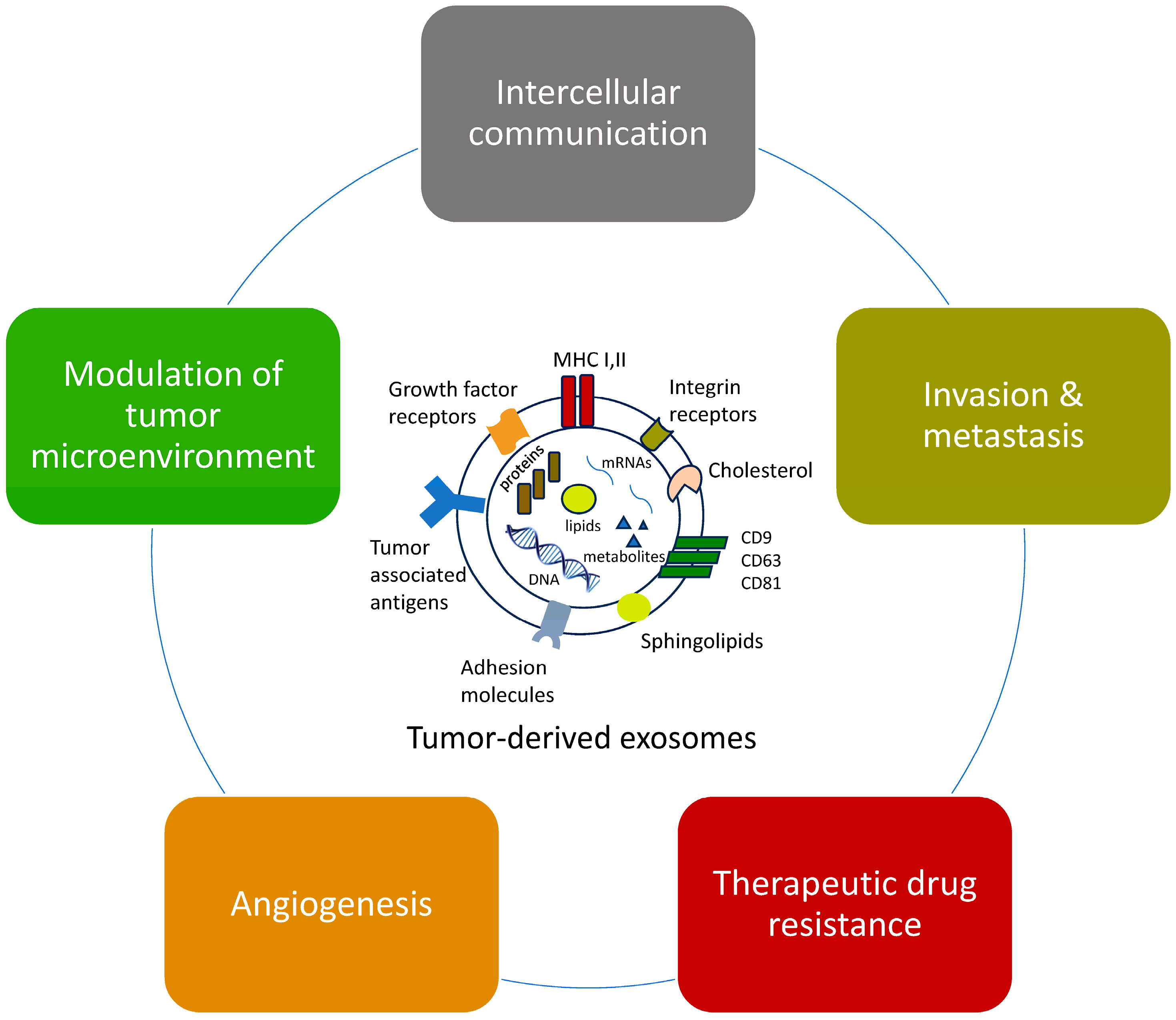
2. Functions of Tumor-Derived Exosomes (TDEs)

During the last few years, there has been an increasing effort to elucidate the impact of exosomes derived from cancer cells on the growth and metastasis of mesenchymal or epithelial tumors. TDEs are extracellular vesicles, larger than typical exosomes, that are formed and secreted into the extracellular space, and which contain a complex cargo of bioactive molecules, including oncoproteins, lipids, RNA, and DNA. However, it is important to highlight that the molecular cargoes of cancer-derived EVs comprise the oncogenic potential of cancer cells and the term “oncosomes” does not simply refer to their cellular origin [15]. In addition, the cargoes and functions of large and nanosized EVs with the same origin may also differ [16]. Nevertheless, even though they differentiate from typical exosomes, there is no consensus on the precise terminology of these vesicles [17].

2.1. Effects on Intercellular Communication

TDEs facilitate tumor cell communication with the microenvironment by transferring oncogenic molecules, such as nucleic acids and proteins, to adjacent cells, altering their behavior and promoting tumor progression (Figure 1). Rhabdomyosarcoma exosomes, for example, enhance the differentiation and proliferation of both neighboring fibroblasts and cells of the sarcoma itself, promoting angiogenesis, fibroblast migration, and infiltration, ultimately resulting in tumor growth [18]. In addition, the characteristic PAX3-FOXO1 gene fusion of alveolar rhabdomyosarcoma (aRMS) appears to be associated with miR-486-5p, which induces changes in the exosomal contents originating from myoblasts and acts as a pro-oncogenic signal to promote migration, infiltration, and colony formation [19].
Liposarcoma exosomes (LPS) have been shown to stimulate tumor-associated macrophages (TAMs) to release the pro-inflammatory cytokine IL-6, inducing tumor proliferation and metastasis with miR-92a-3p and miR-25-3p [20].
In myxofibrosarcoma (MFS), a malignant soft tissue tumor, EVs with miR-1260b affect normal surrounding fibroblasts and enhance tumor growth through downregulation of the tumor suppressor gene protocadherin 9 (PCDH9). In addition, overexpression of miR-1260b is associated with a more aggressive radiographic image, characteristic of invasive MFS [21].

2.2. Promotion of Metastasis

Exosomes can also modulate the metastatic spread of cancer by preparing the pre-metastatic niche since they can travel to distant organs, modifying the local environment to create a favorable niche for incoming tumor cells. They can also enhance tumor cell motility by transferring molecules that increase the invasive properties of cancer cells. This migratory potential is orchestrated by mutations on the TGF-β pathway that promote epithelial-to-mesenchymal transition (EMT) and metastasis through the suppression of E-cadherins and the induction of metalloproteinases (MMPs). This will eventually distort the extracellular matrix (ECM) to create a congenial environment for the tumor [22].
Tumors such as osteosarcoma (OS) affect mesenchymal cells in various ways, by increasing oxidative stress and lactate levels due to exposure to the tumor microenvironment, which can enhance the metastatic potential of osteosarcoma [23]. EVs from OS cell lines were shown to affect normal cells in the TME by enhancing their proliferation and migration capacity as well as by increasing their survivability and adhesion. These EVs exhibit an oncogenic potential, regardless of the presence of cancer cells and associated factors, including IL-6, TNF-α, and TGF-β with de novo MMP-9 and TNF-α expression [24]. Another study with similar results demonstrated that miR-21-5p and miR-148a, found in OS exosomes, could induce immortalization of the umbilical vein endothelial cells (HUVECs) [25]. As anticipated, these tumor exosomes consistently promoted tumor growth and metastasis through immunosuppression and interaction with the TME. The specific effects of exosomes on OS were consistent among many different studies and were in favor of tumor growth [26].
Cancer cell exosomes may influence the activity of mesenchymal cells through reprogramming and the acquisition of new properties. Adipose-derived stem cells (ADSCs) are pluripotent cells with innate angiogenic and proliferating abilities that stem from their exosomes. These cells are able to alter their functions depending on the molecular signals they are exposed to [27]. ADSCs from patients with prostate cancer can undergo mesenchymal–epithelial transition (MET), express specific markers, and mimic the primary tumor when exposed to tumor cell exosomes in vivo that contain oncomiRs such as miR-130b, miR-125b, and miR-155 [28]. Other cancer cell types (e.g., breast and ovarian cancer cells) secrete exosomes that can make ADSCs to resemble myofibroblasts (tumor-associated myofibroblast), possibly through a paracrine mechanism on the neighboring stromal cells of their microenvironment, thus influencing local tumor progression. The use of tumor exosomes was further shown to be associated with the increased expression of growth factors including TGF-β [29,30].

2.3. Effects on Axonogenesis

A particular function of TEDs in head and neck tumors is their involvement in tumor innervation. Exosomes were demonstrated to induce axonogenesis in a rat pheochromocytoma (PC12) cell line, a process of neo-innervation that was characterized by the formation of disordered nerve fibers, described as nerve twigs that were positive for β-III tubulin. This process was shown to be mediated by exosomes of cancer cells and not by exosomes of normal cells, differing from the perineural invasion seen during tumor extension [31].

2.4. Effects on Angiogenesis

Tumors have the potential to serve as “angiogenic switches”, altering their anti-angiogenic environment to a pro-angiogenic one. This function is facilitated by exosomes that promote new blood vessels’ formation by delivering pro-angiogenic factors such as VEGF and miRNAs to endothelial cells [32].
Tumor cells may also release proteins and TDEs containing tetraspanin 8, which can trigger transcriptional changes in endothelial cells, leading to the upregulation of pro-angiogenic genes and enhanced endothelial cell proliferation [33]. Other exosomal cargoes such as the oncogenic EGFR may be secreted from neoplastic cells to affect normal endothelial cells [34].
A study of A431 carcinoma cells subjected to hypoxia or reoxygenation showed that they displayed increased angiogenic and metastatic capabilities and stronger intercellular and cell-to-extracellular matrix adhesion, as well as enhanced invasiveness. Although the majority of these secreted proteins were of cytoplasmic or membrane origin [35], the hypoxic environment acts as the trigger for malignant cells to produce hypoxia-induced exosomes with miR-210, a process possibly mediated by HIF-1a [36].

2.5. Effects on Therapeutic Drug Resistance

Exosomes can contribute to therapeutic resistance by carrying as well as removing chemotherapeutic drugs from cancer cells, thus reducing drug efficacy. They can also transfer drug resistance-related proteins and miRNAs to sensitive cells, conferring resistance to treatment. In addition, some tumors are composed of cancer stem cells (CSCs) that can also produce exosomes and other EVs [37], contributing to tumor heterogeneity [38].
CSCs expressing CD133 in sarcomas or other cancer types also exhibit high levels of tumor-related genes and may be directly related to chemotherapy resistance and increased metastatic potential through various pathways [39]. For example, the chemotherapeutic resistance of CSCs has been studied in MCF-7 breast cancer cells. These CSCs had a noticeable high CD44 and low CD24 expression, along with Oct4 expression. Additionally, they could achieve anchorage-free growth in the form of tumor spheres that the chemotherapeutic drug paclitaxel was unable to inhibit [40].
Furthermore, exosomes can be armed with specific molecules bearing anti-tumor properties and can target cancer cells and CSCs. The use of miR-503-3p in ADSC-exos was shown to potentially suppress the initiation and progression of CSC growth, inhibit the formation of the characteristic spheroid formations by up to 69%, and block tumor growth in vivo. These molecules seem to exert some degree of negative regulatory action against characteristic genes of the CSCs such as Nanog, but without consistently suppressing them [41]. Of particular interest, CSCs can be reprogrammed into non-tumorigenic cells by exosomes released from osteogenically differentiated ADSCs (OD-EXOs) containing specific cargoes capable of differentiating CSCs to be less resistant to systemic therapies [42].

2.6. Modulation of the Tumor Microenvironment

The tumor microenvironment (TME) consists of a variety of different cell types, including stromal, vascular, and immune cells, as well as signaling molecules within the extracellular matrix, serving similar biological roles among cancer types ranging from angiogenesis, metastasis, and chemoresistance to immune regulation.
Low pH conditions in the TME characterize malignant tumors and may affect the release as well as the uptake of exosomes. In a study of melanoma cancer cells, the uptake of exosomes by cells of metastatic origin was higher compared to those released from primary tumors or normal cells. Moreover, their uptake by melanoma cells was inhibited after exposure to proton pump inhibitors and an increase in pH [43].
Exosomes influence various cells of the tumor microenvironment, including endothelial cells, fibroblasts, and immune cells, promoting a pro-tumorigenic environment. They can induce fibroblast activation, extracellular matrix remodeling, and suppression of anti-tumor immune responses.
Exosomal integrins participate in the organotropism of metastasis and may be used to detect organ-specific metastasis. Proteomic analysis of TDEs revealed distinct patterns of integrin expression, with α6β1 and α6β4 shown to be involved in lung metastasis, while exosomal integrin αvβ5 was correlated with liver metastasis. Exosomes derived from liver, lung, and brain tumor cells were studied in mice as well as humans and detected to fuse with normal cells at their intended destination. Their uptake was shown to activate Src phosphorylation and the expression of pro-inflammatory gene S100, thus playing an important role in the formation of the pre-metastatic niche [44].
Tumor-associated macrophages are mainly found within the tumor mass and interact with the TME by promoting or inhibiting tumor growth. The balance between the pro-tumorigenic and anti-tumorigenic functions is complex and relies on the cell phenotype at any given time. TAMs, like normal macrophages, can present with an M1 or M2 phenotype, affecting the tumor and its environment in various ways through exosomes. In the TME, M1-polarized TAMs may exhibit immunoinductive and tumor-suppressive behavior via specific EVs. These vesicles increased the pro-inflammatory cytokines such as IL-1β, TNF-α, and IL-6, and stimulated the apoptosis of osteosarcoma (OS) and melanoma cancer cells by increasing the activation of caspase-3 and caspase-7 [45]. On the contrary, immunosuppressive M2 polarization occurs at later stages and induces tumor growth. M2-TAM exosomes, rich in miR-221-3p, downregulate the SOCS3/JAK2/STAT3 axis, which promotes the malignant behavior of OS cells [46]. In another study, TAM-exosomes caused an increase in lncRNA LIFR-AS1, which suppresses miR-29a, a contributor to apoptosis and, thus, affects the growth and metastatic ability of OS through the miR-29a/NFIA pathway [47].
Exosomes can also drive the interaction of fibroblasts and cancer cells to advance sarcoma growth. The so-called CAFs (cancer-associated fibroblasts) are involved in the remodeling of the ECM and the generation of the pre-metastatic niche (Table 2). The exosomes of CAFs seem to depend on the type of TME of each tumor. For example, in the osteosarcoma TME, exosomal SNHG17 is involved in intercellular communication since it affects miR-2861, regulates the expression of MMP-2, and provides suitable conditions for the expansion and metastasis of neoplastic cells [48]. Additionally, EVs from OS cells, rich in TGF-β1, mediate fibroblast differentiation in the area of lung metastasis and prepare the ground for the metastatic niche [49].

3. TDEs Interact with Mesenchymal Cell Exosomes (MSC-exos) Within the Tumor Microenvironment

Research data suggest that exosomes from mesenchymal cells (MSC-exos) affect tumor growth through the induction of different complex signaling pathways and may promote tumorigenesis or act as tumor suppressors. Tumor growth, which relies on growth factors such as PDGFR, EGFR, VEGF, etc., is promoted by the activation of the respective receptors through various pathways such as Akt, PKC/PKB, and MAP kinases [50]. Although the number of studies with ADSCs in relation to malignant tumors is quite limited, it has been shown that the addition of ADSC-exos in breast cancer cells promotes tumor growth and migration through the Wnt/b-catenin signaling pathway along with Dkk1 and Axin2 target genes [51].
Another study about human ADSCs and dermatofibrosarcoma cells detected a significant tumor progression through the induction of growth factors and angiogenesis that was suggested to be mediated by exosomes [52]. Similar results were obtained upon investigation of ADSC-exos impact in osteosarcoma both in vitro and in vivo. ADSC-exos were shown to promote metastasis through the amplification of the COLGALT2 gene, which is also overexpressed in the osteosarcoma cells themselves. In addition, there was an overexpression of vimentin and MMP2/9 metalloproteinases and induction of the EMT, inducing more aggressive tumor behavior [53].
Accordingly, in ovarian tumors, ADSC-exos derived from omental adipocytes promoted progression and peritoneal metastasis via the MAPK, cyclin F, and other signaling pathways [54].
On the contrary, treatment with ADSC-exos in hepatocellular carcinomas was found to increase the tumor suppressor effect of NK T-cells, reduce the bulk of the tumor, and induce changes in the histopathology that correlated with lower tumor grade [55].
Studies focused on BMSC-exos in various cancer types demonstrated variable results, for example, overexpression of the exosomal protein lymphocyte cytosolic protein 1 (LCP1) from BMSCs promoted osteosarcoma growth via the JAK2/STAT3 pathway, while simultaneously suppressing the tumor suppressor miR-135a-5p [56]. The combination of ΒMSC-exos and SGC-7901 cells (subcutaneous tumor model of gastric cancer cell line) increased the ability of cancer cells to be transplanted, along with promoting a stronger proliferation potential through ERK 1/2 pathway activation, whereas the combination with cells of multiple myeloma led to enhanced survival via the p38, the c-Jun N-terminal kinase p53, and Akt pathway [57,58]. On the contrary, when BMSC-exos were injected into hepatocellular tumors, Kaposi sarcomas, and ovarian tumors in vitro and in vivo, resulted in increased cell death and reduction in the tumor, respectively [59]. A similar tumor suppressive effect of MSC-exos was detected in breast cancer cells, where the miR-16 cargoes mediated inhibition of VEGF and elicited an anti-angiogenic effect [60].
Altogether, research data indicate that exosomes and overall EVs produced by mesenchymal cells may have controversial to unpredictable effects that are associated with the origin and identity of their cargo when interacting with a tumor and its respective microenvironment. MSCs have the ability to absorb TDEs in a similar manner to the one in which they absorb other exosomes and can be altered to stimulate tumor growth via increased macrophage infiltration [61]. This process dynamically changes the chemokine profile of normal MSCs and turns them to tumorigenic cells that produce large amounts of CCL2 and CCL7 that act as ligands for macrophage recruitment.
As stated before, CAFs also play a vital role in tumor progression but their regulation and origin are still uncertain. Recent data suggest that TDEs may engage in CAF formulation and differentiation through the mechanism of endothelial-to-mesenchymal transition. Cancer cells that are depleted of exosomes fail to exploit their effects on fibroblasts and are incapable of promoting growth of the tumor-associated stroma [62]. In another study, exosomal TGF-β and other cargoes from TDEs were shown to promote the formation of CAFs from endothelial cells with normal MSCs, exerting negative feedback on the trans-differentiation of CAFs and, subsequently, alleviating tumor growth and metastasis [63].

4. Clinical Applications of Tumor-Derived Exosomes

With biotechnology currently thriving, researchers are trying to use new molecules as diagnostic markers as well as carriers for specific biomolecules such as micro-RNAs that will, ideally, have predictable and beneficial anti-tumor effects and enable the therapeutic use of exosomes. TDEs, being essential for communication between cells, are implicated in cell proliferation, signaling and differentiation, regulating oncogenesis, migration, and metastasis.
A variety of methods have been used for the isolation and characterization of exosomes, with the gold standard technique involving sequential centrifugation and ultracentrifugation to pellet exosomes. In addition, size-exclusion chromatography (SEC) can be applied to separate exosomes based on their size without, however, affecting their integrity, while precipitation-based kits using polymers can be used to precipitate exosomes from biofluids. Several microfluidic devices also employ lab-on-a-chip platforms with immunoaffinity or size-based separation for high-throughput capture of exosomes. Moreover, antibodies targeting exosome-specific markers (e.g., EpCAM, CD9, CD63, and CD81) to selectively bind TDEs can be used, as well as synthetic oligonucleotides (aptamers) that specifically recognize tumor exosomal markers [18,20,21,22,24,25]. Upon isolation, several techniques can be employed to analyze exosomes and detect cancer-specific molecular signatures. Nanoparticle tracking analysis can measure the size and concentration of exosomes while their structural details can be elucidated by scanning and transmission electron microscopy (SEM/TEM). Exosomal protein markers specific to tumors can be detected by Western blotting, mass spectrometry, and ELISA, while exosomal RNA can be detected by qPCR and RNA sequencing [64].
Importantly, TDEs can be detected in liquid biopsies, providing significant clinical insights for the early diagnosis of tumors and minimizing the invasive routine tissue biopsies along with their complications. The concentration of TDEs in biological fluids varies significantly depending on the type and stage of tumor as well as on the patient’s condition. It has been estimated that the total exosome concentration in human plasma or serum ranges from 108 to 1012 particles per ml while TDEs may constitute 10–50% of this total exosome amount, being 10–100 times higher in aggressive cancers. Increased TDE levels have been detected in lung, breast, and pancreatic cancers, as well as in glioblastoma. Their abundance is further increased in the advanced tumor stage, hypoxia, immune stress, and therapeutic schemes [64].
Exosomal and EV miRNAs that were directly associated with specific tumors have been further assigned to their molecular signature with diagnostic significance. Another study investigated similar vesicles identified by flow cytometry in the circulation and described them as large oncosomes (1–10 μm) that originate from the bulk of prostate tumor protrusions that can interact with endothelial cells, fibroblasts, and the other tumor cells. MMP-9, MMP-2, and other molecules such as caveolin-1 and ADP-ribosylation factor 6 were identified as cargoes of those large oncosomes [65]. Minciacchi et al. reported that there was a 25% difference in the cargoes of large and nano-sized oncosomes after quantitative proteomics, with molecules involved in invasion and tumor progression, like cathepsin proteases, tetraspanins, and growth factors, being abundant in nano-sized EVs. Further studies are needed to elucidate the specificity of these cathepsin-containing vesicles and to ascertain whether they could be considered as potential diagnostic markers [16].
Recent data have shown that not only miRNAs but many different molecules such as mRNA and lncRNA can be detected by next generation sequencing techniques (NGS) through analysis of the entire transcriptome (whole transcriptome RNA-sequencing) as well as by using novel techniques such as employing 3D-printed integrated microfluidic chips [66]. Finally, exosomes themselves can be enriched through surface markers or nano-flow cytometry for more reliable results and enhance the sensitivity of liquid biopsies in finding specific mutations and serve as circulating RNA biomarkers [67].
In an extensive analysis of mesothelioma cell exosomes, several tumor-specific protein cargoes were found that could serve as diagnostic targets. Chondrosarcoma exosomes carrying specific lncRNAs modulate the RAMP2-AS1/miR-2355-5p/VEGFR2 axis promoting angiogenesis, with RAMP2-AS1 being a potential biomarker of interest [68,69].
High expression of extracellular miR-675 has been detected in metastatic osteosarcoma cells but not in their non-metastatic counterparts [70]. In addition, extracellular miR-143, mainly found within exosomes, is involved in the biology of osteosarcoma metastasis through the regulation of the metalloproteinase MMP-13, indicating a potential prognostic marker [71].
Another interesting application of exosomes is their use as transporters of therapeutic molecules for targeted treatment in oncology. Investigation of the molecular composition of various exosomes revealed similar rapid clearance and distribution after intravenous administration, regardless of their unique protein or lipid cargo, potentially limiting their use as systemic anticancer therapies. On the contrary, when delivered intralesionally, the exosomes remained bound to the tumor tissue to a significantly greater extent. Furthermore, experiments in immunocompromised mice revealed the importance of non-specific immunity together with the complement protein C5 in the exosome clearance rate [72].
Exosomes that have been used as carriers of functional miRNAs, such as synthetic miR-143, can block the invasive and metastatic properties of osteosarcoma cells. Of interest, exosomal miR-143 increased after the administration of the synthetic analog into cells, while exosomes transported miR-143 to the respective subcellular location to reach its mRNA target in recipient cells more efficiently than other carriers such as liposomes. This evidence indicates that exosomes transported between cells change the expression of genes and may affect specific functions of osteosarcoma cells through the delivery of miRNAs [73].
Exosomal miR-15a directly targets GATA2 and the GATA2/MDM2 axis, which is overexpressed in OS, and drives continuous cell proliferation. Inhibition of this overexpression interrupts the dysregulation of the cell cycle and slows the onset of OS [74]. The selectivity of miR-15a for this pathway may have potential clinical applications since this overexpression is often an indicator of resistance to adjuvant therapies. Biotechnologically engineered exosomes rich in miR-15a can be potentially used in sarcomas that characteristically overexpress MDM2, such as well-differentiated and dedifferentiated liposarcomas [75,76].
Additional data showed that ADSC-derived exosomes are also an efficient in vivo delivery vehicle and exosomal miR-138-5p is a potential therapeutic molecule for the treatment of bladder cancer. ADSC-exos could infiltrate tumor tissues and release miR-138-5p to inhibit tumor growth [77]. Furthermore, engineered ADSC-EVs can secrete and transport miR-101, a microRNA present at very low levels in metastatic OS cells, limiting lung metastases [78]. Exosomal miR-1913 was shown to restrict OS growth through downregulation of NRSN2 [79], while miR-150-loaded exosomes target IGF2BP1 with similar effects in OS cells [80].
In a study of 11 cancer cell lines, TRAIL (TNF-related apoptosis-inducing ligand) was used in MSC-EVs to induce apoptosis in a dose-dependent manner without significant cytotoxicity [81]. Similar results were observed in the study by Lou et al. where ADSC-exos with miR-122 were used to increase the chemosensitivity of hepatocellular tumors to sorafenib in vivo [82]. Another investigation showed that the tumor suppressor miR-145 in both ADSCs and exosomes exerted a suppressive effect on prostate cancer cells by promoting apoptosis through inhibition of the anti-apoptotic protein Bcl-xL [83].
Tumor-derived exosomes can, therefore, enable personalized medicine and customized treatment (Table 3). The molecular content of TDEs can give important information on the genetic and molecular profile of a patient’s tumor, enabling personalized treatment strategies, customized to the specific characteristics of the cancer. Moreover, they can be used in exosome-based vaccines since they contain tumor antigens for stimulation of the immune response to cancer cells. This approach aims to train the immune system to recognize and attack cancer cells more effectively.
Unraveling the content of exosomes can also help identify mechanisms of resistance to therapy and enable the design of strategies to prevent or overcome resistance. Targeting exosome-mediated resistance pathways in combination with conventional therapies may improve treatment efficacy. The use of exosomes in liquid biopsies further allows for continuous monitoring of cancer patients with minimal invasiveness. This approach can detect changes in tumor dynamics and genetic mutations, providing insights into the evolving nature of the disease.

5. Conclusions

Extracellular vesicles are undoubtedly one of the main points of interest in current studies focused on elucidation of the molecular nature of cancer. Their applications range from non-invasive diagnostics and real-time monitoring of treatment responses to targeted drug delivery and personalized medicine. The interactions between malignant and mesenchymal stem cells seem to play a pivotal role in tumor biology and progression, and need thorough investigation. Nevertheless, this relationship may be the key to unlocking the unique properties of exosomes that will help clinicians to improve the accuracy of cancer diagnosis, provide tailored treatments to individual patients, and develop innovative therapeutic strategies. Tumor-derived exosomes and studies on oncosomes hold promise in revolutionizing the understanding of cancer evolution and clinical management. It is crucial, however, to properly evaluate the plethora of information collected by numerous studies in order to specifically determine their clinical significance in the future.

Author Contributions

Conceptualization: C.P.; data curation: K.S.P. and P.K.; formal analysis: K.S.P.; funding acquisition: C.P.; investigation: K.S.P. and P.K.; methodology: K.S.P. and P.K.; project administration: C.P.; resources: C.P.; software: K.S.P.; supervision: C.P.; validation: K.S.P. and P.K.; visualization: C.P.; writing—original draft: K.S.P. and P.K.; and writing—review and editing: K.S.P., P.K. and C.P. All authors have read and agreed to the published version of the manuscript.

Funding

This research received no external funding.

Conflicts of Interest

The authors declare no conflicts of interest.

References

  1. Johnstone, R.M.; Adam, M.; Hammond, J.R.; Orr, L.; Turbide, C. Vesicle Formation during Reticulocyte Maturation. Association of Plasma Membrane Activities with Released Vesicles (Exosomes). J. Biol. Chem. 1987, 262, 9412–9420. [Google Scholar] [CrossRef] [PubMed]
  2. Cocucci, E.; Meldolesi, J. Ectosomes and exosomes: Shedding the confusion between extracellular vesicles. Trends Cell Biol. 2015, 25, 364–372. [Google Scholar] [CrossRef] [PubMed]
  3. Ostrowski, M.; Carmo, N.B.; Krumeich, S.; Fanget, I.; Raposo, G.; Savina, A.; Moita, C.F.; Schauer, K.; Hume, A.N.; Freitas, R.P.; et al. Rab27a and Rab27b Control Different Steps of the Exosome Secretion Pathway. Nat. Cell Biol. 2010, 12, 19–30. [Google Scholar] [CrossRef] [PubMed]
  4. Hsu, C.; Morohashi, Y.; Yoshimura, S.; Manrique-Hoyos, N.; Jung, S.; Lauterbach, M.A.; Bakhti, M.; Grønborg, M.; Möbius, W.; Rhee, J.; et al. Regulation of Exosome Secretion by Rab35 and Its GTPase-Activating Proteins TBC1D10A–C. J. Cell Biol. 2010, 189, 223–232. [Google Scholar] [CrossRef]
  5. Lösche, W.; Scholz, T.; Temmler, U.; Oberle, V.; Claus, R.A. Platelet-Derived Microvesicles Transfer Tissue Factor to Monocytes but Not to Neutrophils. Platelets 2004, 15, 109–115. [Google Scholar] [CrossRef]
  6. Qu, Y.; Franchi, L.; Nunez, G.; Dubyak, G.R. Nonclassical IL-1β Secretion Stimulated by P2X7 Receptors Is Dependent on Inflammasome Activation and Correlated with Exosome Release in Murine Macrophages1. J. Immunol. 2007, 179, 1913–1925. [Google Scholar] [CrossRef]
  7. Yoon, Y.J.; Kim, O.Y.; Gho, Y.S. Extracellular Vesicles as Emerging Intercellular Communicasomes. BMB Rep. 2014, 47, 531–539. [Google Scholar] [CrossRef]
  8. Yamashita, T.; Takahashi, Y.; Nishikawa, M.; Takakura, Y. Effect of Exosome Isolation Methods on Physicochemical Properties of Exosomes and Clearance of Exosomes from the Blood Circulation. Eur. J. Pharm. Biopharm. 2016, 98, 1–8. [Google Scholar] [CrossRef]
  9. Mathivanan, S.; Fahner, C.J.; Reid, G.E.; Simpson, R.J. ExoCarta 2012: Database of Exosomal Proteins, RNA and Lipids. Nucleic Acids Res. 2012, 40, D1241–D1244. [Google Scholar] [CrossRef]
  10. Haqqani, A.S.; Delaney, C.E.; Tremblay, T.-L.; Sodja, C.; Sandhu, J.K.; Stanimirovic, D.B. Method for Isolation and Molecular Characterization of Extracellular Microvesicles Released from Brain Endothelial Cells. Fluids Barriers CNS 2013, 10, 4. [Google Scholar] [CrossRef]
  11. Welsh, J.A.; Goberdhan, D.C.I.; O’Driscoll, L.; Buzas, E.I.; Blenkiron, C.; Bussolati, B.; Cai, H.; Di Vizio, D.; Driedonks, T.A.P.; Erdbrügger, U.; et al. Minimal Information for Studies of Extracellular Vesicles (MISEV2023): From Basic to Advanced Approaches. J. Extracell. Vesicles 2024, 13, e12404. [Google Scholar] [CrossRef] [PubMed]
  12. Jaiswal, R.; Sedger, L.M. Intercellular Vesicular Transfer by Exosomes, Microparticles and Oncosomes—Implications for Cancer Biology and Treatments. Front. Oncol. 2019, 9, 125. [Google Scholar] [CrossRef]
  13. Di Vizio, D.; Kim, J.; Hager, M.H.; Morello, M.; Yang, W.; Lafargue, C.J.; True, L.D.; Rubin, M.A.; Adam, R.M.; Beroukhim, R.; et al. Oncosome Formation in Prostate Cancer: Association with a Region of Frequent Chromosomal Deletion in Metastatic Disease. Cancer Res. 2009, 69, 5601–5609. [Google Scholar] [CrossRef] [PubMed]
  14. Lee, T.H.; D’Asti, E.; Magnus, N.; Al-Nedawi, K.; Meehan, B.; Rak, J. Microvesicles as Mediators of Intercellular Communication in Cancer—The Emerging Science of Cellular ‘Debris’. Semin. Immunopathol. 2011, 33, 455–467. [Google Scholar] [CrossRef]
  15. Al-Nedawi, K.; Meehan, B.; Kerbel, R.S.; Allison, A.C.; Rak, J. Endothelial Expression of Autocrine VEGF upon the Uptake of Tumor-Derived Microvesicles Containing Oncogenic EGFR. Proc. Natl. Acad. Sci. USA 2009, 106, 3794–3799. [Google Scholar] [CrossRef]
  16. Minciacchi, V.R.; You, S.; Spinelli, C.; Morley, S.; Zandian, M.; Aspuria, P.-J.; Cavallini, L.; Ciardiello, C.; Sobreiro, M.R.; Morello, M.; et al. Large Oncosomes Contain Distinct Protein Cargo and Represent a Separate Functional Class of Tumor-Derived Extracellular Vesicles. Oncotarget 2015, 6, 11327–11341. [Google Scholar] [CrossRef]
  17. Meehan, B.; Rak, J.; Di Vizio, D. Oncosomes—Large and Small: What Are They, Where They Came From? J. Extracell. Vesicles 2016, 5, 33109. [Google Scholar] [CrossRef]
  18. Ghayad, S.E.; Rammal, G.; Ghamloush, F.; Basma, H.; Nasr, R.; Diab-Assaf, M.; Chelala, C.; Saab, R. Exosomes Derived from Embryonal and Alveolar Rhabdomyosarcoma Carry Differential miRNA Cargo and Promote Invasion of Recipient Fibroblasts. Sci. Rep. 2016, 6, 37088. [Google Scholar] [CrossRef]
  19. Ghamloush, F.; Ghayad, S.E.; Rammal, G.; Fahs, A.; Ayoub, A.J.; Merabi, Z.; Harajly, M.; Zalzali, H.; Saab, R. The PAX3-FOXO1 Oncogene Alters Exosome miRNA Content and Leads to Paracrine Effects Mediated by Exosomal miR-486. Sci. Rep. 2019, 9, 14242. [Google Scholar] [CrossRef]
  20. Casadei, L.; Calore, F.; Creighton, C.J.; Guescini, M.; Batte, K.; Iwenofu, O.H.; Zewdu, A.; Braggio, D.A.; Bill, K.L.; Fadda, P.; et al. Exosome-Derived miR-25-3p and miR-92a-3p Stimulate Liposarcoma Progression. Cancer Res. 2017, 77, 3846–3856. [Google Scholar] [CrossRef]
  21. Morita, T.; Fujiwara, T.; Yoshida, A.; Uotani, K.; Kiyono, M.; Yokoo, S.; Hasei, J.; Kunisada, T.; Ozaki, T. Clinical Relevance and Functional Significance of Cell-Free microRNA-1260b Expression Profiles in Infiltrative Myxofibrosarcoma. Sci. Rep. 2020, 10, 9414. [Google Scholar] [CrossRef] [PubMed]
  22. Coulouarn, C.; Factor, V.M.; Thorgeirsson, S.S. Transforming Growth Factor-β Gene Expression Signature in Mouse Hepatocytes Predicts Clinical Outcome in Human Cancer. Hepatology 2008, 47, 2059–2067. [Google Scholar] [CrossRef] [PubMed]
  23. Bonuccelli, G.; Avnet, S.; Grisendi, G.; Salerno, M.; Granchi, D.; Dominici, M.; Kusuzaki, K.; Baldini, N. Role of Mesenchymal Stem Cells in Osteosarcoma and Metabolic Reprogramming of Tumor Cells. Oncotarget 2014, 5, 7575–7588. [Google Scholar] [CrossRef] [PubMed]
  24. Urciuoli, E.; Giorda, E.; Scarsella, M.; Petrini, S.; Peruzzi, B. Osteosarcoma-derived Extracellular Vesicles Induce a Tumor-like Phenotype in Normal Recipient Cells. J. Cell. Physiol. 2018, 233, 6158–6172. [Google Scholar] [CrossRef]
  25. Raimondi, L.; De Luca, A.; Gallo, A.; Costa, V.; Russelli, G.; Cuscino, N.; Manno, M.; Raccosta, S.; Carina, V.; Bellavia, D.; et al. Osteosarcoma Cell-Derived Exosomes Affect Tumor Microenvironment by Specific Packaging of microRNAs. Carcinogenesis 2020, 41, 666–677. [Google Scholar] [CrossRef]
  26. Ye, H.; Hu, X.; Wen, Y.; Tu, C.; Hornicek, F.; Duan, Z.; Min, L. Exosomes in the Tumor Microenvironment of Sarcoma: From Biological Functions to Clinical Applications. J. Nanobiotechnol. 2022, 20, 403. [Google Scholar] [CrossRef]
  27. Papadopoulos, K.S.; Piperi, C.; Korkolopoulou, P. Clinical Applications of Adipose-Derived Stem Cell (ADSC) Exosomes in Tissue Regeneration. Int. J. Mol. Sci. 2024, 25, 5916. [Google Scholar] [CrossRef]
  28. Abd Elmageed, Z.Y.; Yang, Y.; Thomas, R.; Ranjan, M.; Mondal, D.; Moroz, K.; Fang, Z.; Rezk, B.M.; Moparty, K.; Sikka, S.C.; et al. Neoplastic Reprogramming of Patient-Derived Adipose Stem Cells by Prostate Cancer Cell-Associated Exosomes. Stem Cells 2014, 32, 983–997. [Google Scholar] [CrossRef]
  29. Cho, J.A.; Park, H.; Lim, E.H.; Lee, K.W. Exosomes from Breast Cancer Cells Can Convert Adipose Tissue-Derived Mesenchymal Stem Cells into Myofibroblast-like Cells. Int. J. Oncol. 2012, 40, 130–138. [Google Scholar] [CrossRef]
  30. Cho, J.A.; Park, H.; Lim, E.H.; Kim, K.H.; Choi, J.S.; Lee, J.H.; Shin, J.W.; Lee, K.W. Exosomes from Ovarian Cancer Cells Induce Adipose Tissue-Derived Mesenchymal Stem Cells to Acquire the Physical and Functional Characteristics of Tumor-Supporting Myofibroblasts. Gynecol. Oncol. 2011, 123, 379–386. [Google Scholar] [CrossRef]
  31. Madeo, M.; Colbert, P.L.; Vermeer, D.W.; Lucido, C.T.; Cain, J.T.; Vichaya, E.G.; Grossberg, A.J.; Muirhead, D.; Rickel, A.P.; Hong, Z.; et al. Cancer Exosomes Induce Tumor Innervation. Nat. Commun. 2018, 9, 4284. [Google Scholar] [CrossRef] [PubMed]
  32. Visconti, L.; Nelissen, K.; Deckx, L.; Akker, M.V.D.; Adriaensen, W.; Daniels, L.; Matheï, C.; Linsen, L.; Hellings, N.; Stinissen, P.; et al. Prognostic Value of Circulating Cytokines on Overall Survival and Disease-Free Survival in Cancer Patients. Biomark. Med. 2014, 8, 297–306. [Google Scholar] [CrossRef] [PubMed]
  33. Nazarenko, I.; Rana, S.; Baumann, A.; McAlear, J.; Hellwig, A.; Trendelenburg, M.; Lochnit, G.; Preissner, K.T.; Zöller, M. Cell Surface Tetraspanin Tspan8 Contributes to Molecular Pathways of Exosome-Induced Endothelial Cell Activation. Cancer Res. 2010, 70, 1668–1678. [Google Scholar] [CrossRef]
  34. Al-Nedawi, K.; Meehan, B.; Micallef, J.; Lhotak, V.; May, L.; Guha, A.; Rak, J. Intercellular Transfer of the Oncogenic Receptor EGFRvIII by Microvesicles Derived from Tumour Cells. Nat. Cell Biol. 2008, 10, 619–624. [Google Scholar] [CrossRef]
  35. Park, J.E.; Tan, H.S.; Datta, A.; Lai, R.C.; Zhang, H.; Meng, W.; Lim, S.K.; Sze, S.K. Hypoxic Tumor Cell Modulates Its Microenvironment to Enhance Angiogenic and Metastatic Potential by Secretion of Proteins and Exosomes. Mol. Cell Proteom. 2010, 9, 1085–1099. [Google Scholar] [CrossRef]
  36. King, H.W.; Michael, M.Z.; Gleadle, J.M. Hypoxic Enhancement of Exosome Release by Breast Cancer Cells. BMC Cancer 2012, 12, 421. [Google Scholar] [CrossRef]
  37. Tang, D.G. Understanding Cancer Stem Cell Heterogeneity and Plasticity. Cell Res. 2012, 22, 457–472. [Google Scholar] [CrossRef]
  38. Shackleton, M.; Quintana, E.; Fearon, E.R.; Morrison, S.J. Heterogeneity in Cancer: Cancer Stem Cells versus Clonal Evolution. Cell 2009, 138, 822–829. [Google Scholar] [CrossRef]
  39. Feng, B.-H.; Liu, A.-G.; Gu, W.-G.; Deng, L.; Cheng, X.-G.; Tong, T.-J.; Zhang, H.-Z. CD133+ Subpopulation of the HT1080 Human Fibrosarcoma Cell Line Exhibits Cancer Stem-like Characteristics. Oncol. Rep. 2013, 30, 815–823. [Google Scholar] [CrossRef]
  40. de la Mare, J.-A.; Sterrenberg, J.N.; Sukhthankar, M.G.; Chiwakata, M.T.; Beukes, D.R.; Blatch, G.L.; Edkins, A.L. Assessment of Potential Anti-Cancer Stem Cell Activity of Marine Algal Compounds Using an in Vitro Mammosphere Assay. Cancer Cell Int. 2013, 13, 39. [Google Scholar] [CrossRef]
  41. Seo, M.; Kim, S.M.; Woo, E.Y.; Han, K.-C.; Park, E.J.; Ko, S.; Choi, E.W.; Jang, M. Stemness-Attenuating miR-503-3p as a Paracrine Factor to Regulate Growth of Cancer Stem Cells. Stem Cells Int. 2018, 2018, 1–10. [Google Scholar] [CrossRef] [PubMed]
  42. Lee, K.S.; Choi, J.S.; Cho, Y.W. Reprogramming of Cancer Stem Cells into Non-Tumorigenic Cells Using Stem Cell Exosomes for Cancer Therapy. Biochem. Biophys. Res. Commun. 2019, 512, 511–516. [Google Scholar] [CrossRef] [PubMed]
  43. Parolini, I.; Federici, C.; Raggi, C.; Lugini, L.; Palleschi, S.; De Milito, A.; Coscia, C.; Iessi, E.; Logozzi, M.; Molinari, A.; et al. Microenvironmental pH Is a Key Factor for Exosome Traffic in Tumor Cells. J. Biol. Chem. 2009, 284, 34211–34222. [Google Scholar] [CrossRef] [PubMed]
  44. Hoshino, A.; Costa-Silva, B.; Shen, T.-L.; Rodrigues, G.; Hashimoto, A.; Tesic Mark, M.; Molina, H.; Kohsaka, S.; Di Giannatale, A.; Ceder, S.; et al. Tumour Exosome Integrins Determine Organotropic Metastasis. Nature 2015, 527, 329–335. [Google Scholar] [CrossRef]
  45. Lee, K.-M.; An, J.-H.; Yang, S.-J.; Park, S.-M.; Lee, J.-H.; Chae, H.-K.; Song, W.-J.; Youn, H.-Y. Influence of Canine Macrophage-Derived Extracellular Vesicles on Apoptosis in Canine Melanoma and Osteosarcoma Cell Lines. Anticancer Res. 2021, 41, 719–730. [Google Scholar] [CrossRef]
  46. Liu, W.; Long, Q.; Zhang, W.; Zeng, D.; Hu, B.; Liu, S.; Chen, L. miRNA-221-3p Derived from M2-Polarized Tumor-Associated Macrophage Exosomes Aggravates the Growth and Metastasis of Osteosarcoma through SOCS3/JAK2/STAT3 Axis. Aging 2021, 13, 19760–19775. [Google Scholar] [CrossRef]
  47. Zhang, H.; Yu, Y.; Wang, J.; Han, Y.; Ren, T.; Huang, Y.; Chen, C.; Huang, Q.; Wang, W.; Niu, J.; et al. Macrophages-Derived Exosomal lncRNA LIFR-AS1 Promotes Osteosarcoma Cell Progression via miR-29a/NFIA Axis. Cancer Cell Int. 2021, 21, 192. [Google Scholar] [CrossRef]
  48. Zhao, A.; Zhao, Z.; Liu, W.; Cui, X.; Wang, N.; Wang, Y.; Wang, Y.; Sun, L.; Xue, H.; Wu, L.; et al. Carcinoma-Associated Fibroblasts Promote the Proliferation and Metastasis of Osteosarcoma by Transferring Exosomal LncRNA SNHG17. Am. J. Transl. Res. 2021, 13, 10094–10111. [Google Scholar]
  49. Mazumdar, A.; Urdinez, J.; Boro, A.; Migliavacca, J.; Arlt, M.J.E.; Muff, R.; Fuchs, B.; Snedeker, J.G.; Gvozdenovic, A. Osteosarcoma-Derived Extracellular Vesicles Induce Lung Fibroblast Reprogramming. Int. J. Mol. Sci. 2020, 21, 5451. [Google Scholar] [CrossRef]
  50. Gu, H.; Ji, R.; Zhang, X.; Wang, M.; Zhu, W.; Qian, H.; Chen, Y.; Jiang, P.; Xu, W. Exosomes Derived from Human Mesenchymal Stem Cells Promote Gastric Cancer Cell Growth and Migration via the Activation of the Akt Pathway. Mol. Med. Rep. 2016, 14, 3452–3458. [Google Scholar] [CrossRef]
  51. Lin, R.; Wang, S.; Zhao, R.C. Exosomes from Human Adipose-Derived Mesenchymal Stem Cells Promote Migration through Wnt Signaling Pathway in a Breast Cancer Cell Model. Mol. Cell. Biochem. 2013, 383, 13–20. [Google Scholar] [CrossRef] [PubMed]
  52. Yuan, Z.; Zhu, Z.; Zhu, F.; Ding, F.; Wang, Y.; Wang, X.; Luo, X.; Yang, J.; Liu, F.; Sun, D. Impact of Human Adipose Tissue-Derived Stem Cells on Dermatofibrosarcoma Protuberans Cells in an Indirect Co-Culture: An in Vitro Study. Stem Cell Res. Ther. 2021, 12, 440. [Google Scholar] [CrossRef] [PubMed]
  53. Wang, Y.; Chu, Y.; Li, K.; Zhang, G.; Guo, Z.; Wu, X.; Qiu, C.; Li, Y.; Wan, X.; Sui, J.; et al. Exosomes Secreted by Adipose-Derived Mesenchymal Stem Cells Foster Metastasis and Osteosarcoma Proliferation by Increasing COLGALT2 Expression. Front. Cell Dev. Biol. 2020, 8, 353. [Google Scholar] [CrossRef]
  54. Qu, Q.; Liu, L.; Cui, Y.; Chen, Y.; Wang, Y.; Wang, Y. Exosomes from Human Omental Adipose-Derived Mesenchymal Stem Cells Secreted into Ascites Promote Peritoneal Metastasis of Epithelial Ovarian Cancer. Cells 2022, 11, 3392. [Google Scholar] [CrossRef]
  55. Ko, S.-F.; Yip, H.-K.; Zhen, Y.-Y.; Lee, C.-C.; Lee, C.-C.; Huang, C.-C.; Ng, S.-H.; Lin, J.-W. Adipose-Derived Mesenchymal Stem Cell Exosomes Suppress Hepatocellular Carcinoma Growth in a Rat Model: Apparent Diffusion Coefficient, Natural Killer T-Cell Responses, and Histopathological Features. Stem Cells Int. 2015, 2015, 853506. [Google Scholar] [CrossRef]
  56. Ge, X.; Liu, W.; Zhao, W.; Feng, S.; Duan, A.; Ji, C.; Shen, K.; Liu, W.; Zhou, J.; Jiang, D.; et al. Exosomal Transfer of LCP1 Promotes Osteosarcoma Cell Tumorigenesis and Metastasis by Activating the JAK2/STAT3 Signaling Pathway. Mol. Ther. Nucleic Acids 2020, 21, 900–915. [Google Scholar] [CrossRef]
  57. Zhu, W.; Huang, L.; Li, Y.; Zhang, X.; Gu, J.; Yan, Y.; Xu, X.; Wang, M.; Qian, H.; Xu, W. Exosomes Derived from Human Bone Marrow Mesenchymal Stem Cells Promote Tumor Growth in Vivo. Cancer Lett. 2012, 315, 28–37. [Google Scholar] [CrossRef]
  58. Wang, J.; Hendrix, A.; Hernot, S.; Lemaire, M.; De Bruyne, E.; Van Valckenborgh, E.; Lahoutte, T.; De Wever, O.; Vanderkerken, K.; Menu, E. Bone Marrow Stromal Cell–Derived Exosomes as Communicators in Drug Resistance in Multiple Myeloma Cells. Blood 2014, 124, 555–566. [Google Scholar] [CrossRef]
  59. Bruno, S.; Collino, F.; Deregibus, M.C.; Grange, C.; Tetta, C.; Camussi, G. Microvesicles Derived from Human Bone Marrow Mesenchymal Stem Cells Inhibit Tumor Growth. Stem Cells Dev. 2013, 22, 758–771. [Google Scholar] [CrossRef]
  60. Lee, J.-K.; Park, S.-R.; Jung, B.-K.; Jeon, Y.-K.; Lee, Y.-S.; Kim, M.-K.; Kim, Y.-G.; Jang, J.-Y.; Kim, C.-W. Exosomes Derived from Mesenchymal Stem Cells Suppress Angiogenesis by Down-Regulating VEGF Expression in Breast Cancer Cells. PLoS ONE 2013, 8, e84256. [Google Scholar] [CrossRef]
  61. Lin, L.Y.; Du, L.M.; Cao, K.; Huang, Y.; Yu, P.F.; Zhang, L.Y.; Li, F.Y.; Wang, Y.; Shi, Y.F. Tumour Cell-Derived Exosomes Endow Mesenchymal Stromal Cells with Tumour-Promotion Capabilities. Oncogene 2016, 35, 6038–6042. [Google Scholar] [CrossRef] [PubMed]
  62. Greening, D.W.; Gopal, S.K.; Mathias, R.A.; Liu, L.; Sheng, J.; Zhu, H.-J.; Simpson, R.J. Emerging Roles of Exosomes during Epithelial–Mesenchymal Transition and Cancer Progression. Semin. Cell Dev. Biol. 2015, 40, 60–71. [Google Scholar] [CrossRef] [PubMed]
  63. Yeon, J.H.; Jeong, H.E.; Seo, H.; Cho, S.; Kim, K.; Na, D.; Chung, S.; Park, J.; Choi, N.; Kang, J.Y. Cancer-Derived Exosomes Trigger Endothelial to Mesenchymal Transition Followed by the Induction of Cancer-Associated Fibroblasts. Acta Biomater. 2018, 76, 146–153. [Google Scholar] [CrossRef] [PubMed]
  64. Dilsiz, N. A comprehensive review on recent advances in exosome isolation and characterization: Toward clinical applications. Transl. Oncol. 2024, 50, 102121. [Google Scholar] [CrossRef]
  65. Di Vizio, D.; Morello, M.; Dudley, A.C.; Schow, P.W.; Adam, R.M.; Morley, S.; Mulholland, D.; Rotinen, M.; Hager, M.H.; Insabato, L.; et al. Large Oncosomes in Human Prostate Cancer Tissues and in the Circulation of Mice with Metastatic Disease. Am. J. Pathol. 2012, 181, 1573–1584. [Google Scholar] [CrossRef]
  66. Zhang, P.; Zhou, X.; He, M.; Shang, Y.; Tetlow, A.L.; Godwin, A.K.; Zeng, Y. Ultrasensitive Detection of Circulating Exosomes with a 3D-Nanopatterned Microfluidic Chip. Nat. Biomed. Eng. 2019, 3, 438–451. [Google Scholar] [CrossRef]
  67. Yu, W.; Hurley, J.; Roberts, D.; Chakrabortty, S.K.; Enderle, D.; Noerholm, M.; Breakefield, X.O.; Skog, J.K. Exosome-Based Liquid Biopsies in Cancer: Opportunities and Challenges. Ann. Oncol. 2021, 32, 466–477. [Google Scholar] [CrossRef]
  68. Greening, D.W.; Ji, H.; Chen, M.; Robinson, B.W.S.; Dick, I.M.; Creaney, J.; Simpson, R.J. Secreted Primary Human Malignant Mesothelioma Exosome Signature Reflects Oncogenic Cargo. Sci. Rep. 2016, 6, 32643. [Google Scholar] [CrossRef]
  69. Cheng, C.; Zhang, Z.; Cheng, F.; Shao, Z. Exosomal lncRNA RAMP2-AS1 Derived from Chondrosarcoma Cells Promotes Angiogenesis Through miR-2355-5p/VEGFR2 Axis. OncoTargets Ther. 2020, 13, 3291–3301. [Google Scholar] [CrossRef]
  70. Gong, L.; Bao, Q.; Hu, C.; Wang, J.; Zhou, Q.; Wei, L.; Tong, L.; Zhang, W.; Shen, Y. Exosomal miR-675 from Metastatic Osteosarcoma Promotes Cell Migration and Invasion by Targeting CALN1. Biochem. Biophys. Res. Commun. 2018, 500, 170–176. [Google Scholar] [CrossRef]
  71. Osaki, M.; Takeshita, F.; Sugimoto, Y.; Kosaka, N.; Yamamoto, Y.; Yoshioka, Y.; Kobayashi, E.; Yamada, T.; Kawai, A.; Inoue, T.; et al. MicroRNA-143 Regulates Human Osteosarcoma Metastasis by Regulating Matrix Metalloprotease-13 Expression. Mol. Ther. 2011, 19, 1123–1130. [Google Scholar] [CrossRef] [PubMed]
  72. Smyth, T.; Kullberg, M.; Malik, N.; Smith-Jones, P.; Graner, M.W.; Anchordoquy, T.J. Biodistribution and Delivery Efficiency of Unmodified Tumor-Derived Exosomes. J. Control. Release 2015, 199, 145–155. [Google Scholar] [CrossRef] [PubMed]
  73. Shimbo, K.; Miyaki, S.; Ishitobi, H.; Kato, Y.; Kubo, T.; Shimose, S.; Ochi, M. Exosome-Formed Synthetic microRNA-143 Is Transferred to Osteosarcoma Cells and Inhibits Their Migration. Biochem. Biophys. Res. Commun. 2014, 445, 381–387. [Google Scholar] [CrossRef] [PubMed]
  74. Wu, C.; Li, Z.; Feng, G.; Wang, L.; Xie, J.; Jin, Y.; Wang, L.; Liu, S. Tumor Suppressing Role of Serum-Derived Exosomal microRNA-15a in Osteosarcoma Cells through the GATA Binding Protein 2/Murine Double Minute 2 Axis and the P53 Signaling Pathway. Bioengineered 2021, 12, 8378–8395. [Google Scholar] [CrossRef]
  75. Thway, K. Well-Differentiated Liposarcoma and Dedifferentiated Liposarcoma: An Updated Review. Semin. Diagn. Pathol. 2019, 36, 112–121. [Google Scholar] [CrossRef]
  76. Hou, H.; Sun, D.; Zhang, X. The Role of MDM2 Amplification and Overexpression in Therapeutic Resistance of Malignant Tumors. Cancer Cell Int. 2019, 19, 216. [Google Scholar] [CrossRef]
  77. Liu, T.; Li, T.; Zheng, Y.; Xu, X.; Sun, R.; Zhan, S.; Guo, X.; Zhao, Z.; Zhu, W.; Feng, B.; et al. Evaluating Adipose-derived Stem Cell Exosomes as miRNA Drug Delivery Systems for the Treatment of Bladder Cancer. Cancer Med. 2022, 11, 3687–3699. [Google Scholar] [CrossRef]
  78. Zhang, K.; Dong, C.; Chen, M.; Yang, T.; Wang, X.; Gao, Y.; Wang, L.; Wen, Y.; Chen, G.; Wang, X.; et al. Extracellular Vesicle-Mediated Delivery of miR-101 Inhibits Lung Metastasis in Osteosarcoma. Theranostics 2020, 10, 411–425. [Google Scholar] [CrossRef]
  79. Zhou, J.; Xu, L.; Yang, P.; Lu, Y.; Lin, S.; Yuan, G. The Exosomal Transfer of Human Bone Marrow Mesenchymal Stem Cell-Derived miR-1913 Inhibits Osteosarcoma Progression by Targeting NRSN2. Am. J. Transl. Res. 2021, 13, 10178–10192. [Google Scholar]
  80. Xu, Z.; Zhou, X.; Wu, J.; Cui, X.; Wang, M.; Wang, X.; Gao, Z. Mesenchymal Stem Cell-Derived Exosomes Carrying microRNA-150 Suppresses the Proliferation and Migration of Osteosarcoma Cells via Targeting IGF2BP1. Transl. Cancer Res. TCR 2020, 9, 5323–5335. [Google Scholar] [CrossRef]
  81. Yuan, Z.; Kolluri, K.K.; Gowers, K.H.C.; Janes, S.M. TRAIL Delivery by MSC-derived Extracellular Vesicles Is an Effective Anticancer Therapy. J. Extracell. Vesicles 2017, 6, 1265291. [Google Scholar] [CrossRef] [PubMed]
  82. Lou, G.; Song, X.; Yang, F.; Wu, S.; Wang, J.; Chen, Z.; Liu, Y. Exosomes Derived from miR-122-Modified Adipose Tissue-Derived MSCs Increase Chemosensitivity of Hepatocellular Carcinoma. J. Hematol. Oncol. 2015, 8, 122. [Google Scholar] [CrossRef] [PubMed]
  83. Takahara, K.; Ii, M.; Inamoto, T.; Nakagawa, T.; Ibuki, N.; Yoshikawa, Y.; Tsujino, T.; Uchimoto, T.; Saito, K.; Takai, T.; et al. microRNA-145 Mediates the Inhibitory Effect of Adipose Tissue-Derived Stromal Cells on Prostate Cancer. Stem Cells Dev. 2016, 25, 1290–1298. [Google Scholar] [CrossRef] [PubMed]
Figure 1. Tumor-derived exosomes are involved in the modulation of tumor microenvironment, intercellular communication, angiogenesis, invasion, and metastasis, as well as drug resistance.
Ijms 26 03095 g001
Table 1. Similarities and differences between different types of EVs.
CharacteristicsEctosomesExosomesLarge Oncosomes
Diameter range100–350 nm50–100 nm1–10 μm
Accumulation sitePlasma membraneIntracellular MVBs Plasma membrane
ESCRT complex utilizationYes (Partially)YesYes (Partially)
Release procedurePinching offExocytosisPinching off
Release timing and amountEarly—high amountLate—controlledEarly—high mount
Possible markersTyA, C1qCD63, CD61, CD81, CD9 CK18, GAPDH, HSPA5
Table 2. Functions of tumor-derived exosomes (TDEs).
Type of Cancer CellsExosomal miRNATarget CellsMechanism of ActionEffectsReferences
RMSmiR-486-5pCAFs, fibroblasts, myoblastsAngiogenesis, migration, differentiation and proliferation of fibroblasts and cancer cellsTumor invasion and increased metastatic capacity[18,19]
LPSmiR-486-5p
miR-92a-3p
TAMsIncreased IL-6Tumor proliferation and increased metastatic capacity[20]
MFSmiR-1260bFibroblastsDown-regulation of PCDH9Tumor proliferation[21]
OSEVsMSCsEffect on the tumor microenvironment
TNF-α, IL-6, TGF-β, MMP-9
Oncogenic potential in the absence of cancer cells[23,24]
OSmiR-148a
miR-21-5p
HUVECsEffect on the tumor microenvironmentInduction of immortality in target cells[25]
ProstatemiR-125b, miR-130b miR-155ADSCsExpression of epithelial, neoplastic and angiogenic tumor markersMesenchymal-epithelial transition[28]
Breast-ADSCsOverexpression SDF-1, VEGF, CCL5, TGF-β, SMAD2 Convert ADSCs to myofibroblasts[29]
Ovaries-ADSCsOverexpression SDF-1, TGF-β, SMAD2Convert ADSCs to myofibroblasts[30]
RMS: rhabdomyosarcoma, LPS: liposarcoma, MFS: myxofibrosarcoma, OS: osteosarcoma, CAFs: cancer-associated fibroblasts, HUVECs: human umbilical vein endothelial cells, ADSCs: Adipose-derived stem cells and TAMs: tumor-associated macrophages.
Table 3. Mesenchymal cell exosomes affect the TME and tumor progression.
Origin of ExosomesTarget Cells Mechanism of ActionEffectsReference
ADSC-exosCSCsSuppression of spheroid formation of CSCs by miR-503-3pTumor suppression[40]
ADSC-exosCSCsIncreased expression of APLP, RUNX2, and BGLAPReprogramming of CSCs into non-tumorigenic cells[41]
ADSC-exosBreast cancerInduction of the Wnt/b-catenin signaling pathwayTumor progression[51]
ADSC-exosDFSPOverexpression of VEGF, HGF, and bFGF, PDGFRB, COL1A1Tumor progression[52]
ADSC-exosOSVimentin and MMP 2/9 overexpression, EMT inductionTumor progression[53]
ADSC-exosOvarian cancerFOXM1, Cyclin F, KIF20A, and MAPKTumor progression[54]
ADSC-exosHepatocellular carcinomaEnhancement of NK T-cell activityTumor suppression[55]
BMSC-exosOSLCP1, JAK2/STAT3 pathway, miR-135a-5p suppressionTumor progression[56]
BMSC-exosStomach cancerActivation of the ERK 1/2 pathwayTumor progression[57]
BMSC-exosMultiple myelomap53, p38 and Akt pathwaysTumor progression[58]
BMSC-exosHepatocellular carcinoma, ovarian cancer, Kaposi’s sarcomaDIRAS3, RBL2, RBL1, CDKN2B, CDKN1A, CCNE1, SKP2, CCND2, CUL3, GAPDHTumor suppression[59]
MSC-exosBreast cancermiR-16 cargoes of exosomes inhibit VEGFTumor suppression[60]
ADSC-exosBladder cancerTissue invasion and tumor growth suppression via miR-138-5pTumor suppression[77]
ADSC-EVsOSThey limit the development of pulmonary metastases through miR-101Tumor suppression[78]
ADSC-exosOSNegative regulation in NRSN2 by miR-1913Tumor suppression[79]
ADSC-exosOSNegative regulation of IGF2BP1 by miR-150Tumor suppression[80]
ADSC-exosHepatocellular cancerChemosensitivity of hepatocellular tumors to sorafenib via miR-122Tumor suppression[81]
ADSC-exosProstate cancerEnhancement of apoptosis through Bcl-xL inhibition via miR-145Tumor suppression[83]
CSCs: cancer stem cells, DFSP: dermatofibrosarcoma, ADSC: adipose tissue stem cells, OS: osteosarcoma, and BMSC: bone marrow stem cells.
Disclaimer/Publisher’s Note: The statements, opinions and data contained in all publications are solely those of the individual author(s) and contributor(s) and not of MDPI and/or the editor(s). MDPI and/or the editor(s) disclaim responsibility for any injury to people or property resulting from any ideas, methods, instructions or products referred to in the content.

