

Optimizing Chitin Extraction from Acheta domesticus: A Sustainable Approach Using Two Ultrafine Grinding Techniques

Abstract
1. Introduction
2. Results and Discussion
2.1. Index of Chitin Content in Crickets
2.2. Characterization of House Cricket Chitin
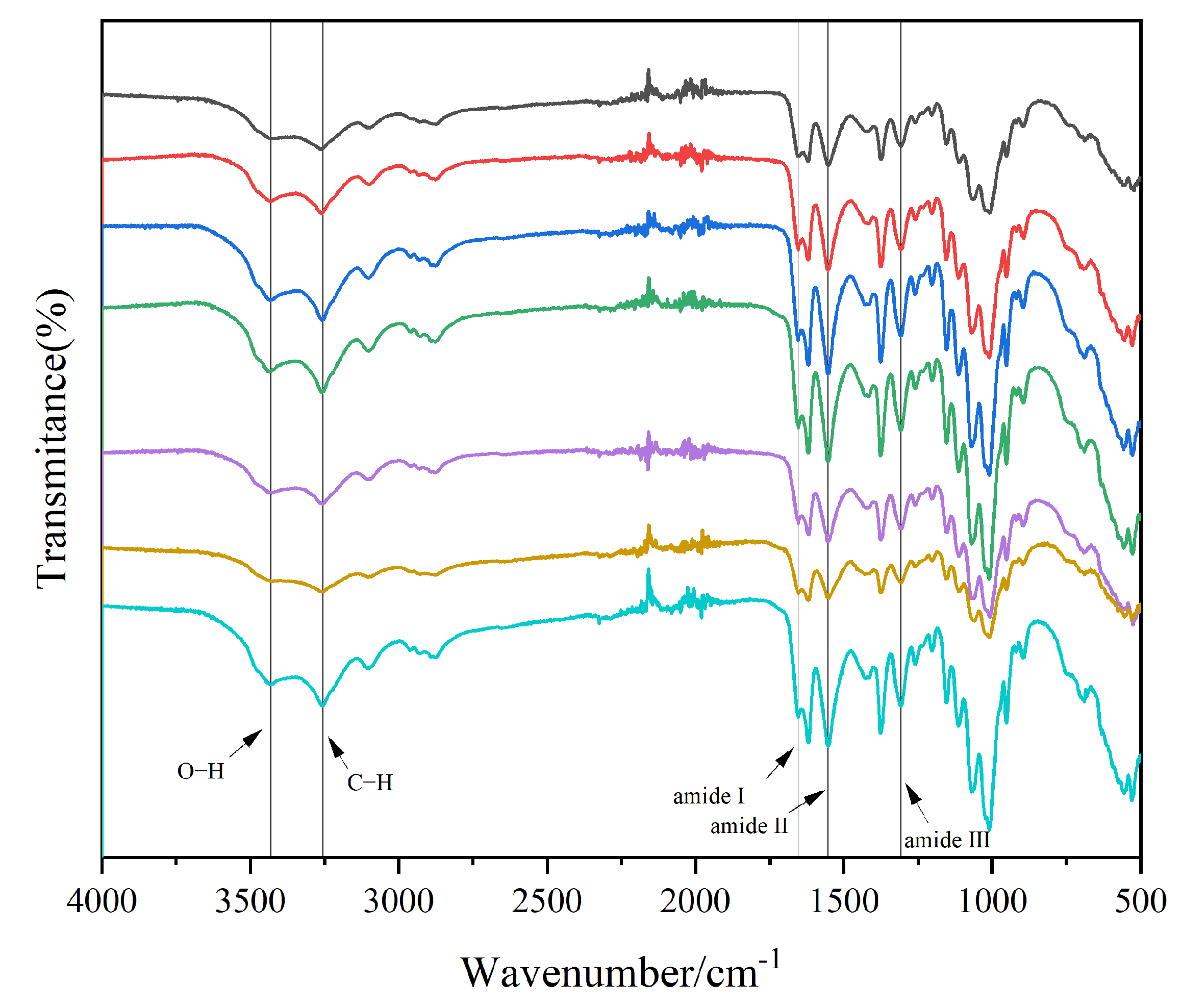
2.2.1. FTIR
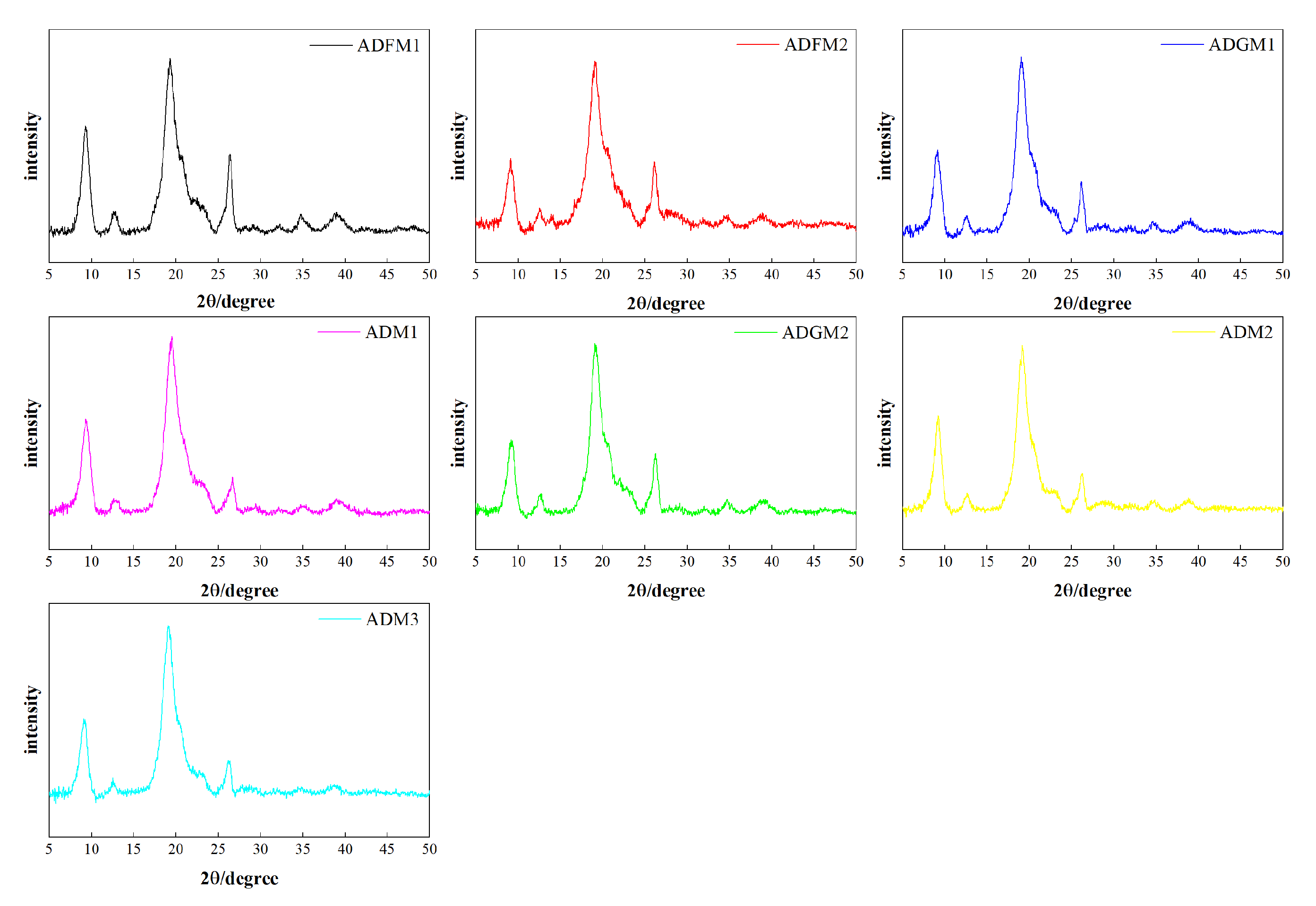
2.2.2. X-Ray Diffraction
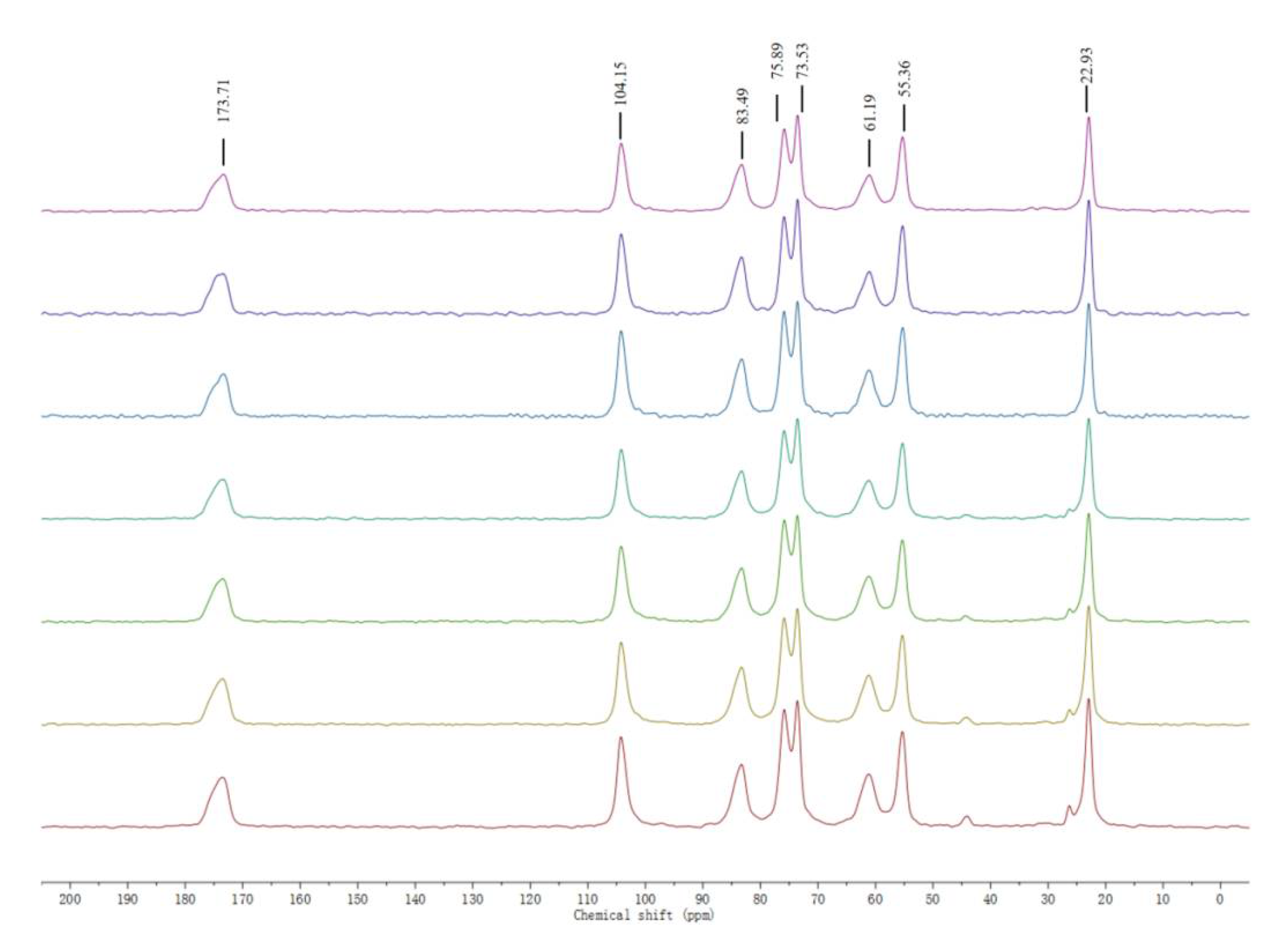
2.2.3. 13C NMR
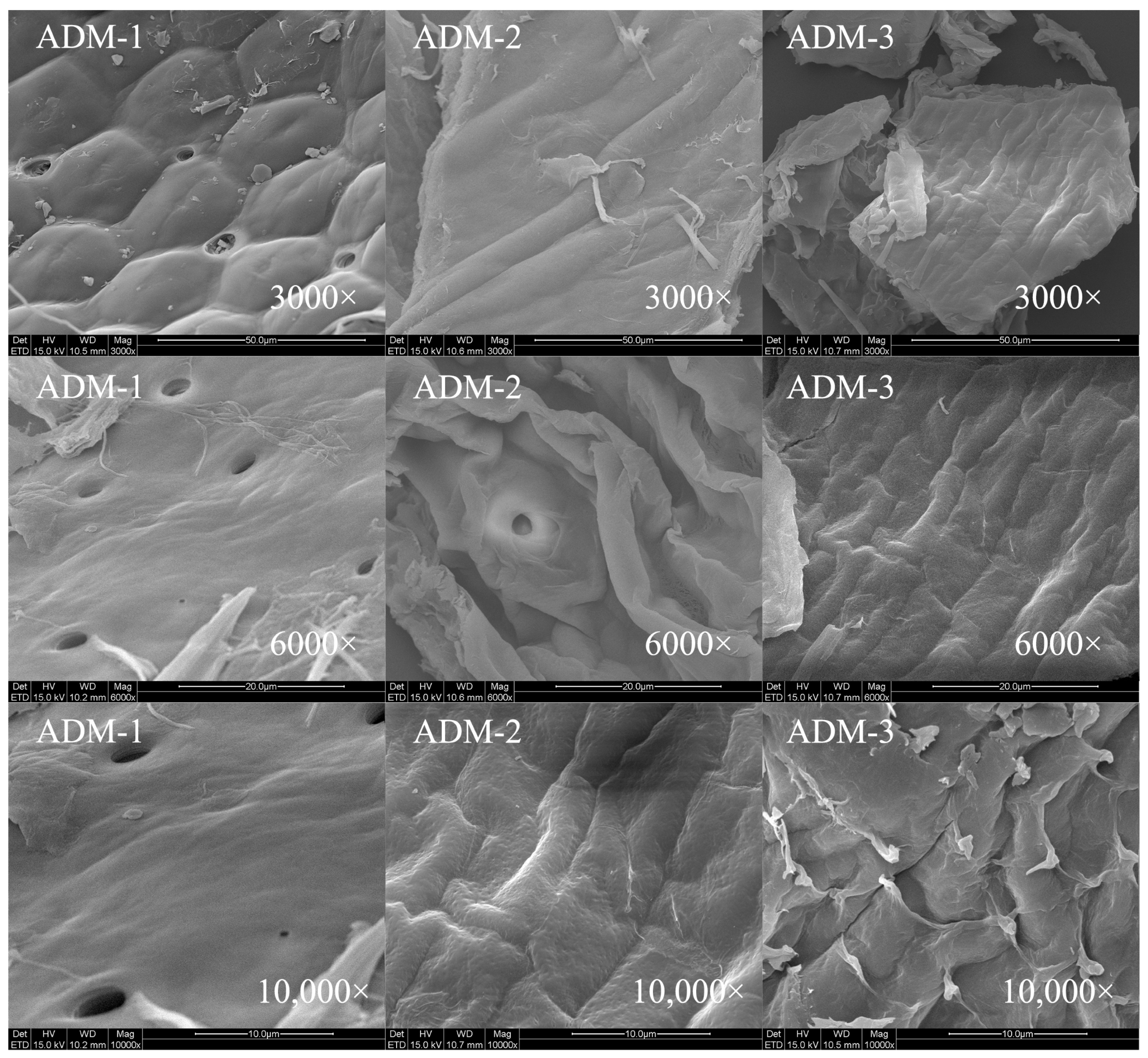
2.2.4. SEM
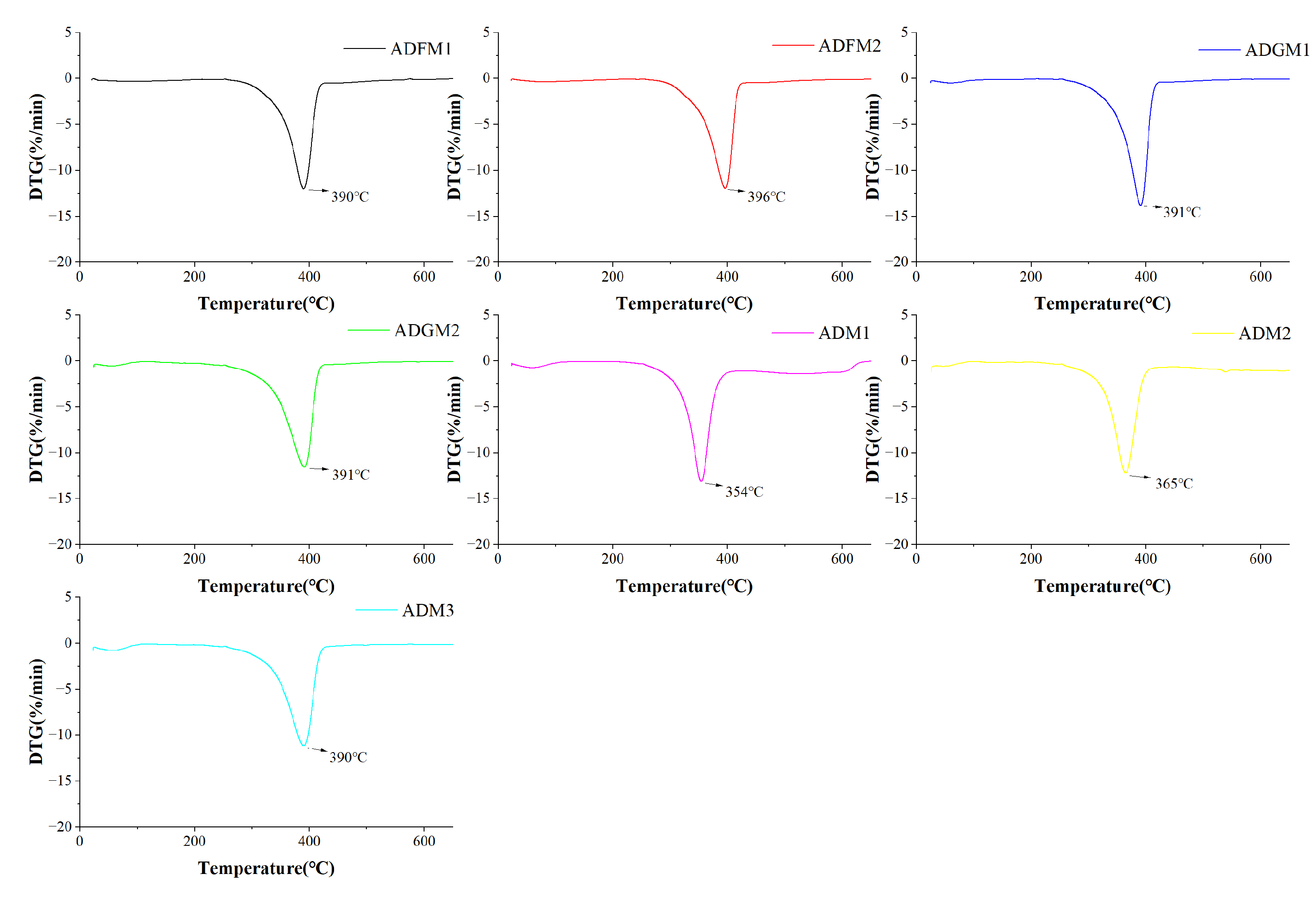
2.2.5. TGA
3. Materials and Methods
3.1. Biological Materials
3.2. Ultrafine Grinding
3.3. Chitin Extraction
3.4. Chemical Analysis
3.5. Viscosity Measurement
3.6. Water Oil Binding Ability
3.7. Fourier-Transform Infrared Spectroscopy (FTIR)
3.8. Nuclear Magnetic Resonance Spectroscopy(13C)
3.9. X-Ray Diffraction (XRD)
3.10. Scanning Electron Microscopy (SEM)
3.11. Thermogravimetric Analysis (TGA)
3.12. Statistical Analysis
4. Conclusions
Author Contributions
Funding
Institutional Review Board Statement
Informed Consent Statement
Data Availability Statement
Conflicts of Interest
References
- Pilco-Romero, G.; Chisaguano-Tonato, A.M.; Herrera-Fontana, M.E.; Chimbo-Gándara, L.F.; Sharifi-Rad, M.; Giampieri, F.; Battino, M.; Vernaza, M.G.; Álvarez-Suárez, J.M. House cricket (Acheta domesticus): A review based on its nutritional composition, quality, and potential uses in the food industry. Trends Food Sci. Technol. 2023, 142, 104226. [Google Scholar] [CrossRef]
- Rinaudo, M. Chitin and chitosan: Properties and applications. Prog. Polym. Sci. 2006, 31, 603–632. [Google Scholar] [CrossRef]
- Psarianos, M.; Dimopoulos, G.; Ojha, S.; Moreno Cavini, A.C.; Bußler, S.; Taoukis, P.; Schlüter, O.K. Effect of pulsed electric fields on cricket (Acheta domesticus) flour: Extraction yield (protein, fat and chitin) and techno-functional properties. Innov. Food Sci. Emerg. Technol. 2022, 76, 102908. [Google Scholar] [CrossRef]
- Ndiritu, A.; Kinyuru, J.; Onyango, A.; Kipkoech, C. Functional and microstructural characteristics of chitin extracted from field cricket, house cricket, and black soldier fly cocoons. Food Meas. 2023, 17, 5903–5912. [Google Scholar] [CrossRef]
- Kim, M.-W.; Song, Y.-S.; Han, Y.S.; Jo, Y.H.; Choi, M.H.; Park, Y.-K.; Kang, S.H.; Kim, S.-A.; Choi, C.; Jung, W.-J. Production of chitin and chitosan from the exoskeleton of adult two-spotted field crickets (Gryllus bimaculatus). Entomol. Res. 2017, 47, 279–285. [Google Scholar] [CrossRef]
- Gao, W.; Chen, F.; Wang, X.; Meng, Q. Recent advances in processing food powders by using superfine grinding techniques: A review. Compr. Rev. Food Sci. Food Saf. 2020, 19, 2222–2255. [Google Scholar] [CrossRef]
- Lokman, I.H.; Ibitoye, E.B.; Hezmee, M.N.M.; Goh, Y.M.; Zuki, A.B.Z.; Jimoh, A.A. Effects of chitin and chitosan from cricket and shrimp on growth and carcass performance of broiler chickens. Trop. Anim. Health Prod. 2019, 51, 2219–2225. [Google Scholar] [CrossRef]
- Kishida, K.; Mizuta, T.; Izawa, H.; Ifuku, S. Preparation of Nanochitin from Crickets and Comparison with That from Crab Shells. J. Compos. Sci. 2022, 6, 280. [Google Scholar] [CrossRef]
- Espinosa-Solís, A.; Velázquez-Segura, A.; Lara-Rodríguez, C.; Martínez, L.M.; Chuck-Hernández, C.; Rodríguez-Sifuentes, L. Optimizing Chitin Extraction and Chitosan Production from House Cricket Flour. Processes 2024, 12, 464. [Google Scholar] [CrossRef]
- Zhao, X.-Y.; Ao, Q.; Yang, L.-W.; Yang, Y.-F.; Sun, J.-C.; Gai, G.-S. Application of superfine pulverization technology in Biomaterial Industry. J. Taiwan Inst. Chem. Eng. 2009, 40, 337–343. [Google Scholar] [CrossRef]
- Ho, T.M.; Truong, T.; Bhandari, B. Methods to characterize the structure of food powders—A review. Biosci. Biotechnol. Biochem. 2017, 81, 651–671. [Google Scholar] [CrossRef]
- Ibitoye, E.B.; Lokman, I.H.; Hezmee, M.N.M.; Goh, Y.M.; Zuki, A.B.Z.; Jimoh, A.A. Extraction and physicochemical characterization of chitin and chitosan isolated from house cricket. Biomed. Mater. 2018, 13, 025009. [Google Scholar] [CrossRef]
- Rumpold, B.A.; Schlüter, O.K. Nutritional composition and safety aspects of edible insects. Mol. Nutr. Food Res. 2023, 57, 802–823. [Google Scholar] [CrossRef]
- Manel, C.; Khaled, C.; Riadh, K.; Qana, A.A.; Atef, J.; Sherif, M.A.S.K.; El, A.H. Physicochemical characterization of chitin extracted by different treatment sequences from an edible insect. Int. J. Biol. Macromol. 2023, 253, 127156. [Google Scholar] [CrossRef]
- Wasko, A.; Bulak, P.; Polak-Berecka, M.; Nowak, K.; Polakowski, C.; Bieganowski, A. The first report of the physicochemical structure of chitin isolated from. Int. J. Biol. Macromol. 2016, 92, 316–320. [Google Scholar] [CrossRef]
- Chantarasataporn, P.; Yoksan, R.; Visessanguan, W.; Chirachanchai, S. Water-based nano-sized chitin and chitosan as seafood additive through a case study of Pacific white shrimp (Litopenaeus vannamei). Food Hydrocoll. 2013, 32, 341–348. [Google Scholar] [CrossRef]
- Lokesh, S.; Soibam, N.; Porayil, L.; Amjad, K.B.; Binaya, B.N.; Xavier, K.A.M. Effect of chemical treatment duration on physicochemical, rheological, and functional properties of colloidal chitin. Food Hydrocoll. Health 2022, 2, 100091. [Google Scholar] [CrossRef]
- Younes, I.; Hajji, S.; Rinaudo, M.; Chaabouni, M.; Jellouli, K.; Nasri, M. Optimization of proteins and minerals removal from shrimp shells to produce highly acetylated chitin. Int. J. Biol. Macromol. 2016, 84, 246–253. [Google Scholar] [CrossRef]
- Kaya, M.; Seyyar, O.; Baran, T.; Turkes, T. Bat guano as new and attractive chitin and chitosan source. Front. Zool. 2014, 11, 59. [Google Scholar] [CrossRef]
- Erdogan, S.; Kaya, M. High similarity in physicochemical properties of chitin and chitosan from nymphs and adults of a grasshopper. Int. J. Biol. Macromol. 2016, 89, 118–126. [Google Scholar] [CrossRef]
- Kaya, M.; Sofi, K.; Sargin, I.; Mujtaba, M. Changes in physicochemical properties of chitin at developmental stages (larvae, pupa and adult) of Vespa crabro (wasp). Carbohydr. Polym. 2016, 145, 64–70. [Google Scholar] [CrossRef] [PubMed]
- Kandíc, L.J.; Mitríc, M.; Ignjatovíc, N. XRD analysis of calcium phosphate and biocomposite calcium phosphate/bioresorbable polymer. Mater. Sci. Forum 2006, 518, 507–512. [Google Scholar] [CrossRef]
- Silva, D.; Zuluaga, F.; Carlos, H. Evaluation of biocompatibility of chitosan films from the mycelium of Aspergillus niger in connective tissue of Rattus norvegicus. J. Mol. Genet. Med. 2015, 9, 1–8. [Google Scholar] [CrossRef]
- Hajji, S.; Younes, I.; Ghorbel-Bellaaj, O.; Hajji, R.; Rinaudo, M.; Nasri, M.; Jellouli, K. Structural differences between chitin and chitosan extracted from three different marine sources. Int. J. Biol. Macromol. 2014, 65, 298–306. [Google Scholar] [CrossRef]
- Ilyas, H.N.; Zia, K.M.; Rehman, S.; Ilyas, R.; Sultana, S. Utilization of shellfish industrial waste for isolation, purification, and characterizations of chitin from crustacean’s sources in Pakistan. J. Polym. Environ. 2021, 29, 2337–2348. [Google Scholar] [CrossRef]
- Kaya, M.; Baran, T. Description of a new surface morphology for chitin extracted from wings of cockroach (Periplaneta americana). Int. J. Biol. Macromol. 2015, 75, 7–12. [Google Scholar] [CrossRef]
- Ingole, P.G.; Ingole, N.P. Methods for separation of organic and pharmaceutical compounds by different polymer materials. Korean J. Chem. Eng. 2014, 31, 2109–2123. [Google Scholar] [CrossRef]
- Alexandre, T.P.; Julliana, I.S.; Juliana, C.G.; Jorge, N. Characterization of chitosan and chitin produced from silkworm crysalides. Carbohydr. Polym. 2006, 64, 98–103. [Google Scholar] [CrossRef]
- Yuan, B.-Q.; Yu, T.-H.; Chen, S.-C.; Zhang, Z.-Q.; Guo, Z.-K.; Huang, G.-X.; Xiao, J.-h.; Huang, D.-W. Physical and chemical characterization of chitin and chitosan extracted under different treatments from black soldier fly. Int. J. Biol. Macromol. 2024, 279, 135228. [Google Scholar] [CrossRef]
- AOAC. Official Methods of Analysis, 12th ed.; Association of Official Analytical Chemists: Washington, DC, USA, 1975. [Google Scholar]
- Chen, S.; Wei, X.; Sui, Z.; Guo, M.; Geng, J.; Xiao, J.; Huang, D. Preparation of antioxidant and antibacterial chitosan film from Periplaneta americana. Insects 2021, 12, 53. [Google Scholar] [CrossRef]
- Hamdi, M.; Hajji, S.; Affes, S.; Taktak, W.; Maalej, H.; Nasri, M.; Nasri, R. Development of a controlled bioconversion process for the recovery of chitosan from blue crab (Portunus segnis) exoskeleton. Food Hydrocoll. 2018, 77, 534–548. [Google Scholar] [CrossRef]
- Kasaai, M.R. A review of several reported procedures to determine the degree of N-acetylation for chitin and chitosan using infrared spectroscopy. Carbohydr. Polym. 2008, 71, 497–508. [Google Scholar] [CrossRef]
- Marei, N.H.; El-Samie, E.A.; Salah, T.; Saad, G.R.; Elwahy, A.H.M. Isolation and characterization of chitosan from different local insects in Egypt. Int. J. Biol. Macromol. 2016, 82, 871–877. [Google Scholar] [CrossRef]
- Moussout, H.; Ahlafi, H.; Aazza, M.; Bourakhouadar, M. Kinetics and mechanism of the thermal degradation of biopolymers chitin and chitosan using thermogravimetric analysis. Polym. Degrad. Stab. 2016, 130, 1–9. [Google Scholar] [CrossRef]


| Chitin Yield (%) | Moisture (%) | Protein (%) | Ash (%) | |
|---|---|---|---|---|
| ADFM-1 | 4.62 ± 0.02 e | 5.90 ± 0.39 bc | 1.90 ± 0.82 ab | 1.17 ± 0.16 c |
| ADFM-2 | 10.17 ± 0.09 a | 4.92 ± 0.28 a | 1.07 ± 0.46 a | 0.17 ± 0.09 ab |
| ADGM-1 | 6.46 ± 0.06 c | 4.87 ± 0.28 a | 1.33 ± 0.46 ab | 0.55 ± 0.15 b |
| ADGM-2 | 4.46 ± 0.10 e | 6.10 ± 0.39 c | 2.14 ± 0.46 bc | 1.39 ± 0.17 cd |
| ADM-1 | 9.61 ± 0.12 b | 5.35 ± 0.29 b | 2.31 ± 0.50 bc | 0.14 ± 0.04 a |
| ADM-2 | 9.34 ± 0.17 b | 8.30 ± 0.34 d | 2.97 ± 0.47 c | 0.37 ± 0.11 ab |
| ADM-3 | 5.46 ± 0.63 d | 5.87 ± 0.16 bc | 3.08 ± 0.53 c | 1.57 ± 0.47 d |
| DA (%) | Viscosity (kDa) | Crystallinity (%) | |
|---|---|---|---|
| ADFM-1 | 115 | 417 ± 5.69 a | 23 |
| ADFM-2 | 121 | 277 ± 4.04 b | 16 |
| ADGM-1 | 124 | 262 ± 4.73 c | 10 |
| ADGM-2 | 122 | 245 ± 4.93 d | 24 |
| ADM-1 | 119 | 218 ± 3.06 e | 24 |
| ADM-2 | 109 | 220 ± 7.10 e | 20 |
| ADM-3 | 118 | 238 ± 2.52 d | 20 |
| WBC (%) | FBC (%) | |
|---|---|---|
| ADFM-1 | 304.91 ± 1.67 g | 294.03 ± 2.11 e |
| ADFM-2 | 557.68 ± 5.99 e | 649.16 ± 10.00 b |
| ADGM-1 | 494.40 ± 7.56 f | 475.82 ± 17.39 d |
| ADGM-2 | 603.10 ± 11.51 d | 534.57 ± 26.58 c |
| ADM-1 | 1040.88 ± 31.10 a | 715.92 ± 3.30 a |
| ADM-2 | 915.34 ± 12.41 b | 709.30 ± 12.57 a |
| ADM-3 | 814.85 ± 14.32 c | 715.45 ± 7.00 a |
Disclaimer/Publisher’s Note: The statements, opinions and data contained in all publications are solely those of the individual author(s) and contributor(s) and not of MDPI and/or the editor(s). MDPI and/or the editor(s) disclaim responsibility for any injury to people or property resulting from any ideas, methods, instructions or products referred to in the content. |
© 2025 by the authors. Licensee MDPI, Basel, Switzerland. This article is an open access article distributed under the terms and conditions of the Creative Commons Attribution (CC BY) license (https://creativecommons.org/licenses/by/4.0/).
Share and Cite
Yuan, B.; Yu, T.; Huang, J.; Ren, X.; Huang, D.; Xiao, J. Optimizing Chitin Extraction from Acheta domesticus: A Sustainable Approach Using Two Ultrafine Grinding Techniques. Int. J. Mol. Sci. 2025, 26, 2938. https://doi.org/10.3390/ijms26072938
Yuan B, Yu T, Huang J, Ren X, Huang D, Xiao J. Optimizing Chitin Extraction from Acheta domesticus: A Sustainable Approach Using Two Ultrafine Grinding Techniques. International Journal of Molecular Sciences. 2025; 26(7):2938. https://doi.org/10.3390/ijms26072938
Chicago/Turabian StyleYuan, Binqiao, Tinghao Yu, Junkui Huang, Xinrui Ren, Dawei Huang, and Jinhua Xiao. 2025. "Optimizing Chitin Extraction from Acheta domesticus: A Sustainable Approach Using Two Ultrafine Grinding Techniques" International Journal of Molecular Sciences 26, no. 7: 2938. https://doi.org/10.3390/ijms26072938
APA StyleYuan, B., Yu, T., Huang, J., Ren, X., Huang, D., & Xiao, J. (2025). Optimizing Chitin Extraction from Acheta domesticus: A Sustainable Approach Using Two Ultrafine Grinding Techniques. International Journal of Molecular Sciences, 26(7), 2938. https://doi.org/10.3390/ijms26072938