Article

Determining the Role of OsAGP6P in Anther Development Within the Arabinogalactan Peptide Family of Rice (Oryza sativa)

1
State Key Laboratory of Conservation and Utilization of Subtropical Agricultural Biological Resources, Guangxi University, Nanning 530004, China
2
Guangxi Key Laboratory of Sugarcane Biology, College of Agriculture, Guangxi University, Nanning 530004, China
3
College of Life Science and Technology, Huazhong University of Science and Technology, Wuhan 430074, China
4
College of Informatics, Huazhong Agricultural University, Wuhan 430070, China
5
Centre for Crop Science, Queensland Alliance for Agriculture and Food Innovation, The University of Queensland, St Lucia 4072, Australia
*
Authors to whom correspondence should be addressed.
These authors contributed equally to this work.
Int. J. Mol. Sci. 2025, 26(6), 2616; https://doi.org/10.3390/ijms26062616
Submission received: 12 February 2025 / Revised: 11 March 2025 / Accepted: 11 March 2025 / Published: 14 March 2025
(This article belongs to the Special Issue Advances in Plant Genomics and Genetics: 2nd Edition)

Abstract

Arabinogalactan proteins (AGPs) are complex proteoglycans present in plant cell walls across the kingdom. They play crucial roles in biological functions throughout the plant life cycle. In this study, we identified 43 gene members of the AG peptide (an AGP subfamily) within the rice genome, detailing their structure, protein-conserved domains, and motif compositions for the first time. We also examined the expression patterns of these genes across 18 tissues and organs, especially the different parts of the flower (anthers, pollen, pistil, sperm cells, and egg cells). Interestingly, the expression of some AG peptides is mainly present in the pollen grain. Transcription data and GUS staining confirmed that OsAGP6P—a member of the AG peptide gene family—is expressed in the stamen during pollen development stages 11–14, which are critical for maturation as microspores form after meiosis of pollen mother cells. It became noticeable from stage 11, when exine formation occurred—specifically at stage 12, when the intine began to develop. The overexpression of this gene in rice decreased the seed-setting rate (from 91.5% to 30.5%) and plant height (by 21.9%) but increased the tillering number (by 34.1%). These results indicate that AGP6P contributes to the development and fertility of pollen, making it a valuable gene target for future genetic manipulation of plant sterility through gene overexpression or editing.

1. Introduction

Arabinogalactan proteins (AGPs) belong to the cell wall glycoprotein family rich in hydroxyproline [1]. AGPs have common features, with glycan chains rich in arabinose, galactose, and other carbohydrates making up 90–98% (w/w) of the molecular structure, while a protein backbone high in hydroxyproline comprises 2–10% (w/w) [2,3,4]. AGPs are found in various tissues of many higher plants—including seeds, roots, stems, leaves, and inflorescences—where they play a crucial role in several physiological processes [5,6].
AGPs are classified into classical and non-classical types based on protein backbones [2]. Classical AGPs typically have three parts: an N-terminal signal peptide; a central domain rich in proline, alanine, serine, and threonine residues (PAST); and a C-terminal glycosylphosphatidylinositol (GPI) anchor [7]. However, some studies also categorize AGPs as classical AGPs with only the first two parts and lacking GPI [8]. Classical AGPs consist of two types: Lys-rich classical AGPs are separated by Lys-rich regions [9], and arabinogalactan (AG) peptides—with a length ranging from 50 to 90 amino acids—have an amino acid composition in which at least 35% of the residues are PAST [8,10]. There are also many chimeric AGPs featuring various conserved domains that can be classified into three main subfamilies: fasciclin-like AGPs (FLAs), phytocyanin-like AGPs (PAGs), and xylogen-like AGPs (XYLPs) [11,12,13,14,15]. Classical AGPs [16] and AG peptides [17,18] can be highly glycosylated (~90% w/w) [19], whereas chimeric AGPs are moderately glycosylated [20]. Furthermore, other AGPs cannot be classified into any of these three families [16,21].
AGPs feature extensive glycosylation, typically found on the Ser and Thr residues of the protein core through O-glycosylation. AGPs are attached to the cell membrane via the GPI anchor [16]. The signal peptide is essential for ensuring that AGPs are synthesized, processed, and delivered to the correct location (cell membrane or extracellular space) [2,22]. These distinct domains and the classification help us understand the diverse roles AGPs play in plant biology, including their involvement in cell signaling, development, and responses to environmental stresses (salt stress, drought, and pathogens) [21,23,24,25,26,27]. AGPs involve many cytological processes, including cell expansion, differentiation, and embryogenesis [28,29]. AGPs have gained considerable interest due to their intricate structure and function. However, their activity is also influenced by distribution, the timing of expression, and environmental conditions within cells or tissues [30,31]. Understanding their precise localization and expression timing could reveal how AGPs contribute to the structural integrity and developmental processes.
Several studies in Arabidopsis have revealed that AGPs are essential for pollen development and PT germination and growth [7,32,33,34]. Also, AGPs have been reported during pollen development in plants other than Arabidopsis, such as those in the Brassica genus or rice [7,34]. For example, the OsFLA1 gene—from the FLA group—is found in spikelets, roots, stems, leaves, and leaf sheaths. It encodes a protein in the cytoplasm and nucleus that helps control pollen development [33]. In OsAGP13-RNAi plants, the cell walls in the flower spike axis are significantly thinner [35]. However, the information on the function of AG peptides in the rice pollen formation process is still limited. In addition, the mechanisms by which AGP influences pollen development are not fully understood. AGPs are part of the hormone response and signal transduction in plants. The expression of some AGP genes is affected by hormones like abscisic acid (ABA) and gibberellins (GAs) [36]. AtAGP30 regulates ABA signaling pathways, and the atagp30 mutant reduces the ABA-induced germination delay [37,38]. Calcium ion (Ca2+) signaling is crucial for cellular functions. Recent studies on AGP-Ca2+ oscillators have revealed the important role of AGP proteins in cells and their interaction with the auxin signaling network [39]. Despite these significant advances in understanding AGP functions in the dicot model plant Arabidopsis thaliana, their precise roles and their molecular mechanisms during pollen development in other plants remain elusive.
In a previous study, genomic studies in rice identified 69 AGPs, including 13 traditional classical AGPs, 15 AG peptides, 3 non-traditional AGPs, 3 early nodulin-like AGPs, 8 non-specific lipid transfer-like AGPs, and 27 fasciclin-like AGPs [10]. Some functions of these genes have been studied, while others remain unexplored. Given the advancements in genomic technologies (IIRGSP-1.0, MSU7, and RAP-DB) [40,41], particularly the recent release of the telomere-to-telomere (T2T) resolution of the Nipponbare rice genome [42], it is crucial to reassess and update our understanding of the AGP gene family in rice.
This study identified 43 classical AG peptide genes (designated OsAGP1P to OsAGP43P) from the Oryza sativa japonica genome. We systematically examined their gene structure, cis-acting elements, physicochemical properties, and conserved domains. The expression patterns of AG peptide genes across different growth stages and tissues were investigated. Specifically, OsAGP6P, one of the protein members, is found in the plasma membrane and is detectable during stages 11 to 14 of pollen development. Further transgenic expression confirmed its role in pollen development and various plant phenotypes. The findings will enhance the understanding of the structure and function of the AG peptides family in plants, which are vital for improving agricultural sustainability.

2. Results

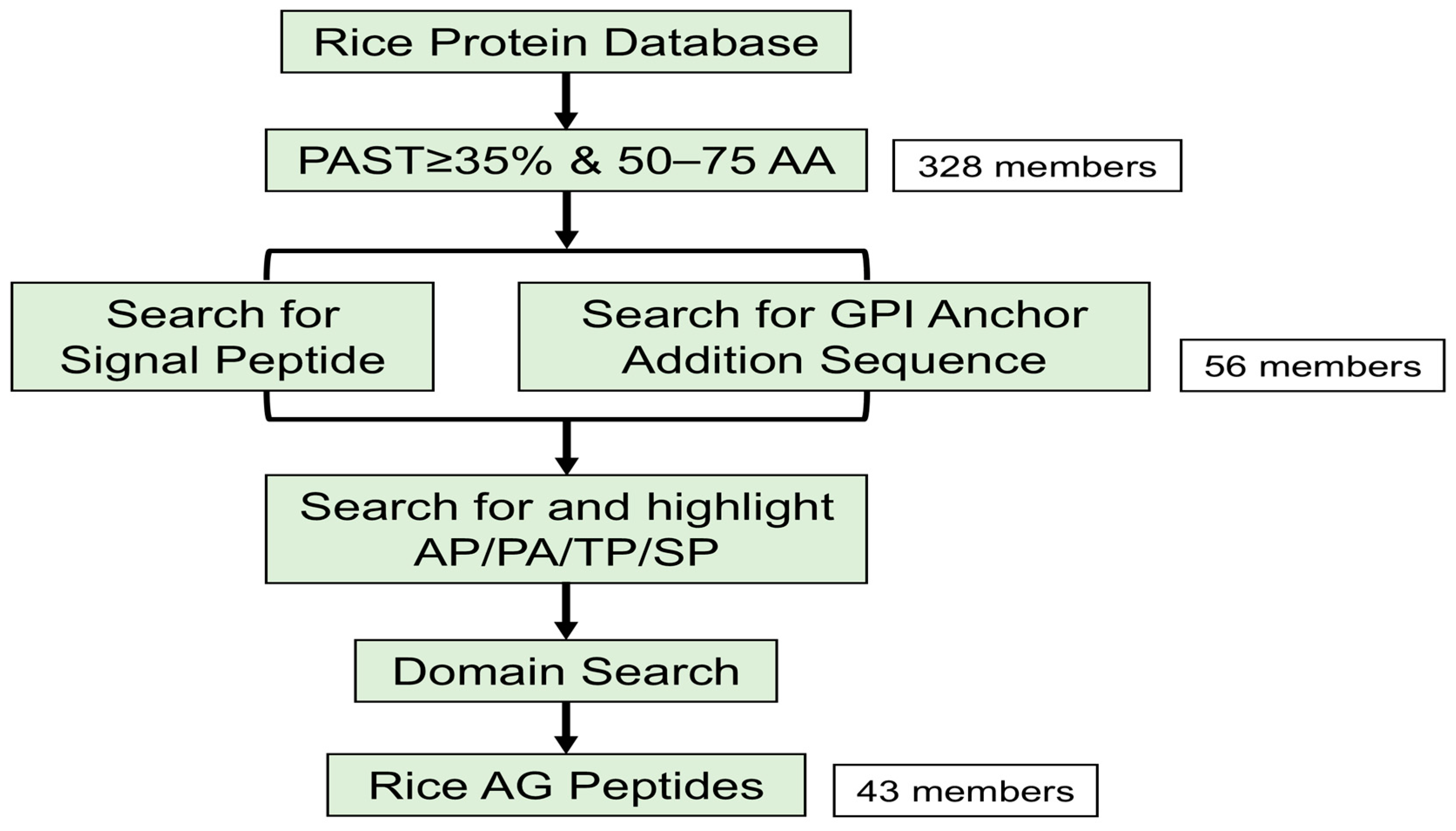
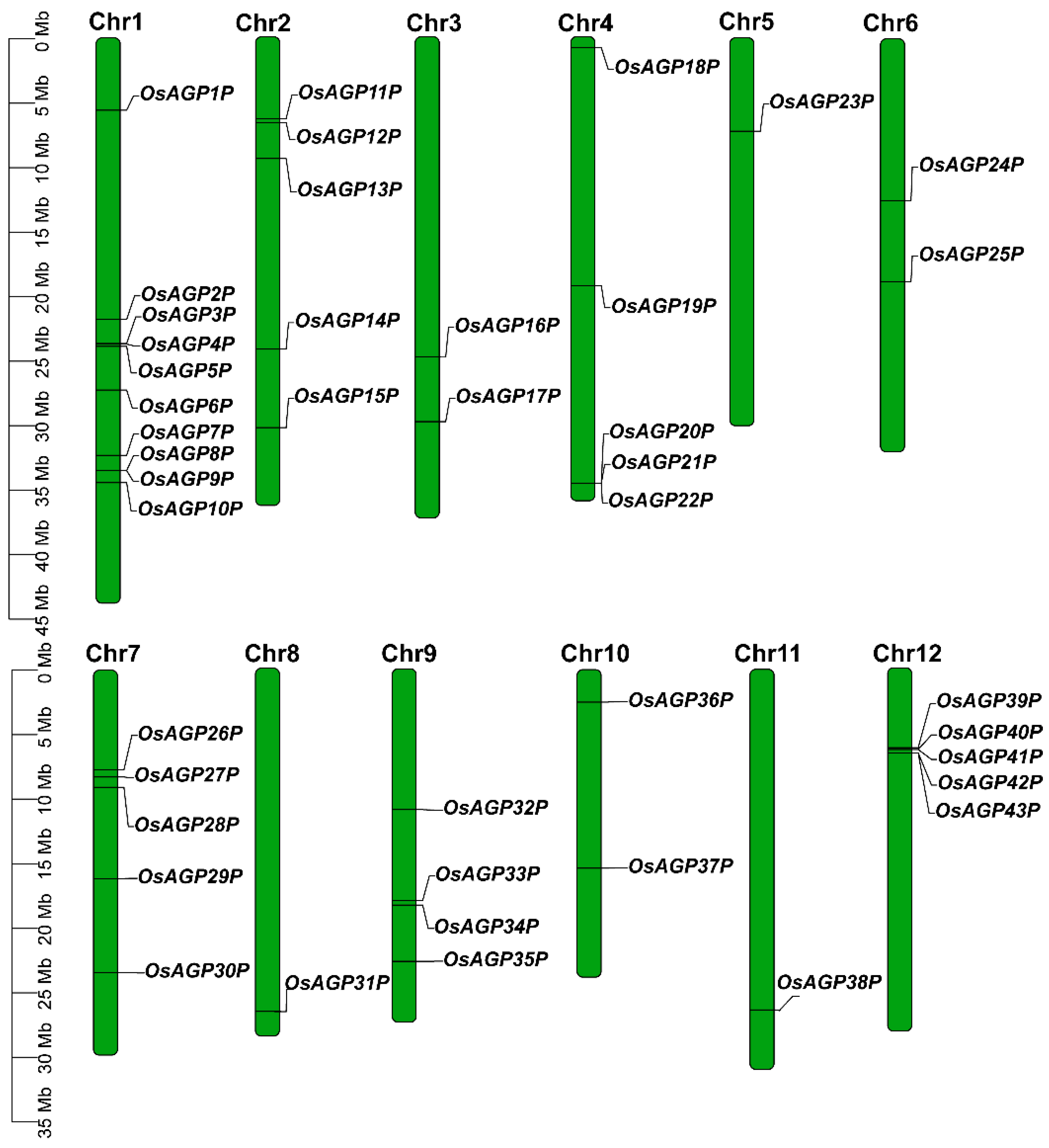
2.1. Identification of AG Peptide Genes in Rice

Considering the characteristics of AG peptides, we developed a detailed identification process (Figure 1). Initially, 328 potential AG peptides were identified in rice by searching for proteins between 50 and 75 amino acids long, with a biased amino acid composition of at least 35% PAST (Figure 1; Supplementary Materials, Table S2). Signal peptides are crucial for facilitating the glycosylation of AG peptides in the endoplasmic reticulum. Meanwhile, a glycosylphosphatidylinositol (GPI) anchor is required to anchor AG peptides to the plasma membrane. An additional screening process was performed on these 328 proteins to identify those that possess both a signal peptide and a GPI anchor (Supplementary Materials, Table S3). We also examined whether these proteins had dipeptide repeats (Ala–Hyp, Ser–Hyp, and Thr–Hyp), excluding proteins containing other domains (Supplementary Materials, Table S3). In total, 43 AG peptides were identified (Figure 1; Supplementary Materials, Table S2).
The 43 AG peptide genes identified were named OsAGP1P through OsAGP43P according to their chromosomal positions (Figure 2). These genes encode proteins with a length of 53–75 amino acids, with predicted molecular weights and isoelectric points from 5.09 kDa/4.00 to 7.79 kDa/12.10 (Table 1; Supplementary Materials, Table S4). The AG peptides were primarily predicted to localize in the cell membrane and nucleus. Some were also predicted to exist in cellular compartments like the Golgi apparatus, cytoplasm, and chloroplast (Supplementary Materials, Table S3). This multi-localization pattern suggests that these proteins may serve diverse roles in various physiological functions in plants.

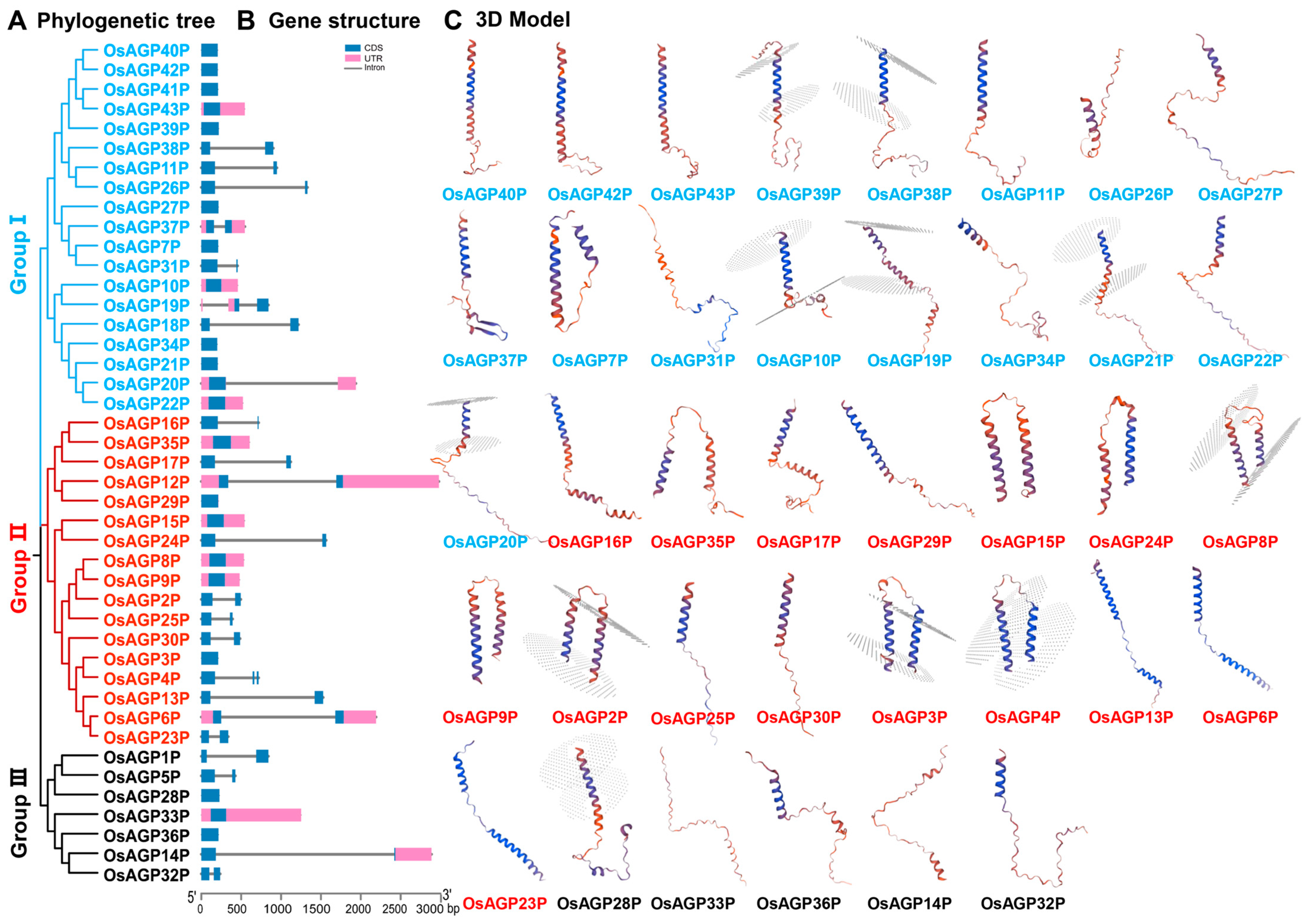
2.2. Phylogenetic and Protein Structure Analyses of AG Peptide Proteins in Rice

An unrooted phylogenetic tree was generated by aligning AG peptide protein sequences to illustrate their phylogenetic relationships (Figure 3A). The AG peptide proteins were classified into three groups containing 19, 17, and 7 members, respectively (Figure 3A). The number of exons among the AG peptide genes ranges from one to three, with most having only one exon (Figure 3B). Our study presented a detailed three-dimensional (3D) structure of the AG peptide protein (Figure 3C). Group I proteins typically contain single α-helices connected to coils, transition through a β-turn, and subsequently link to additional coils. Unlike Group I proteins, Group II proteins have a more compact structure, typically consisting of two α-helices connected by a β-turn and linked to coils. Group III proteins contain two to three short α-helices, with β-turn junctions between them, and coils at the C-terminus or N-terminus. These findings clarify the evolutionary relationships and structural diversity of AG peptide proteins, enhancing our understanding of their classification and functions.

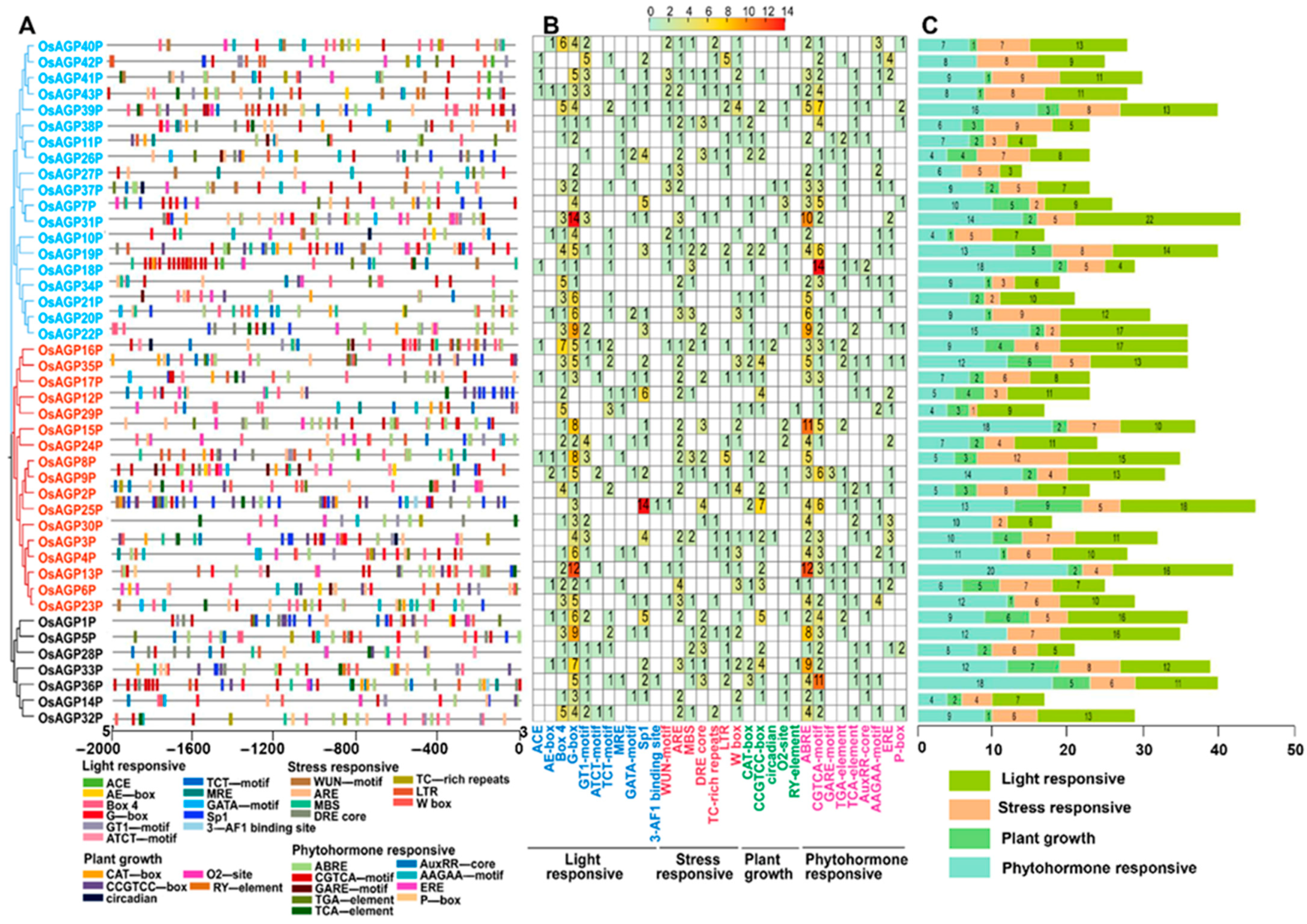
2.3. Identification of Cis-Regulatory Elements Within the Promoters of AG Peptide Genes

Cis-regulatory elements can significantly influence transcription regulation and the expression pattern of the associated genes. This study analyzed cis-regulatory elements based on the 2000-base-pair nucleotide sequences upstream of the start codon (ATG) for each AG peptide gene (Figure 4). In total, 32 types of elements were identified in the promoters of 43 AG peptide genes and categorized into four groups: light response (11), stress response (8), plant growth regulation (5), and phytohormone response (9) (Figure 4A,B). It was revealed that many identified elements were associated with light responsiveness, with the G-box element and Box 4 being the most frequently recognized (Figure 4B,C). The proportions indicate that the expression of the AG peptides gene was substantially affected by light.
Multiple plant hormone response elements, stress response elements, and plant growth regulation elements were also detected (Figure 4A). Elements associated with phytohormone responsiveness ranked second in frequency, while those concerning plant growth regulation were the least abundant (Figure 4C). These elements are essential for regulating gene expression during plant growth and development and responding to various stresses. The identified hormone response elements include the following: ABRE, TCA element, and AAGAA motif (abscisic acid); TGA element, AuxRP core, and p-box (auxin); CGTCA motif (MeJA); and ERE (ethylene). The stress response elements include the following: ARE elements (anaerobic induction); WUN motif (mechanical injury response); MBS and DRE core (drought stress response elements); and LTR (low-temperature response). Furthermore, various cis-elements related to plant growth—such as a CAT-box, CCGTCC-box, circadian, O2 site, and RY element—were also abundant. These findings suggest that AG peptide genes are regulated by hormones and environmental stresses, which govern plant development.

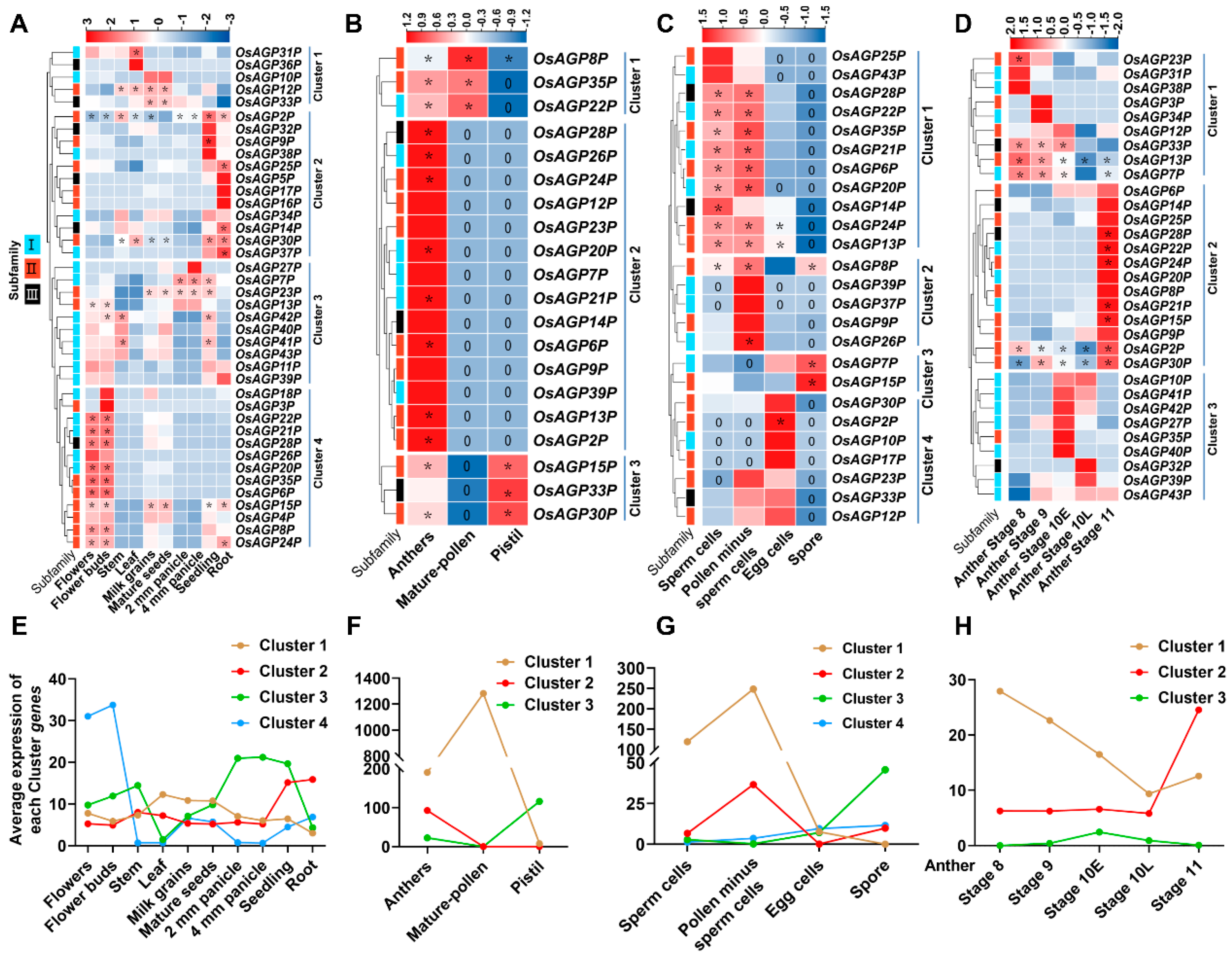
2.4. Expression Patterns of AG Peptide Genes in Multiple Tissues

The expression profiles of 43 AG peptide genes in various tissues exhibited divergent patterns and were classified into some clusters accordingly (Figure 5A,E). Most genes showed tissue-specific expression functions. Genes in Clusters 1, 2, and 3 were constitutively expressed. The genes in Cluster 1 (OsAGP12P/OsAGP31P/OsAGP33P) displayed higher expression levels in the leaves, milk grains, and mature seeds. The Cluster 2 genes (OsAGP2P/OsAGP9P/OsAGP14P/OsAGP25P/OsAGP30P/OsAGP37P) exhibited higher expression levels in the seedlings and roots. Cluster 3 genes (OsAGP7P/OsAGP13P/OsAGP23P/OsAGP41P/OsAG42P) showed higher expression in the stems, panicles, and seedlings. By contrast, Cluster 4 genes (OsAGP6P/OsAGP8P/OsAGP15P/OsAGP20P/OsAGP21P/OsAGP22P/OsAGP24P/OsAGP28P/OsAGP35P) presented preferential expression in the flowers and flower buds, with expression levels significantly higher than other tissues.
We further analyzed the expression of these AG peptide genes in different parts of a flower, including the anther, mature pollen, and pistil (Figure 5B,F). Interestingly, we found that all genes could be detected in the anther. Cluster 2 genes (OsAGP2P/OsAGP6P/OsAGP13P/OsAGP20P/OsAGP21P/OsAGP24P/OsAGP26P/OsAGP28P) were specifically expressed in the anther. In addition to the anther, the Cluster 3 genes (OsAG15P/OsAGP30P/OsAGP33P) and Cluster 1 genes (OsAG8P/OsAGP22P/OsAGP35P) were expressed in the pistil and mature pollen, respectively. Notably, the expression levels of these genes were high overall.
Furthermore, we investigated the expression of these AG peptide genes in sperm cells, pollen minus sperm cells, egg cells, and spores (Figure 5C,G). Cluster 1 genes (OsAGP6P/OsAGP13P/OsAGP14P/OsAGP20P/OsAGP21P/OsAGP22P/OsAGP24P/OsAGP28P/OsAGP35P) exhibited higher expression levels in sperm cells and pollen minus sperm cells. Cluster 2 genes (OsAGP8P/OsAGP26P) demonstrated preferential expression in pollen minus sperm cells. Cluster 3 genes (OsAGP7P/OsAGP115P) showed preferential expression in spores. In contrast, Cluster 4 genes showed preferential expression in egg cells.
Finally, we investigated the expression of these genes in anthers from stage 8 to stage 11 (Figure 5D,H). Cluster 1 displays a relatively high expression in one or two points between stage 8 and the early phase of stage 10, followed by a decrease at the later phase of stage 10 until stage 11. In contrast, Cluster 2 demonstrated lower expression during stage 8 to the later phase of stage 10 but was significantly higher at stage 11. Cluster 3 was expressed much higher in the early or later phases of stage 10. Obviously, the three clusters showed a distinctive complementary expression in anthers.

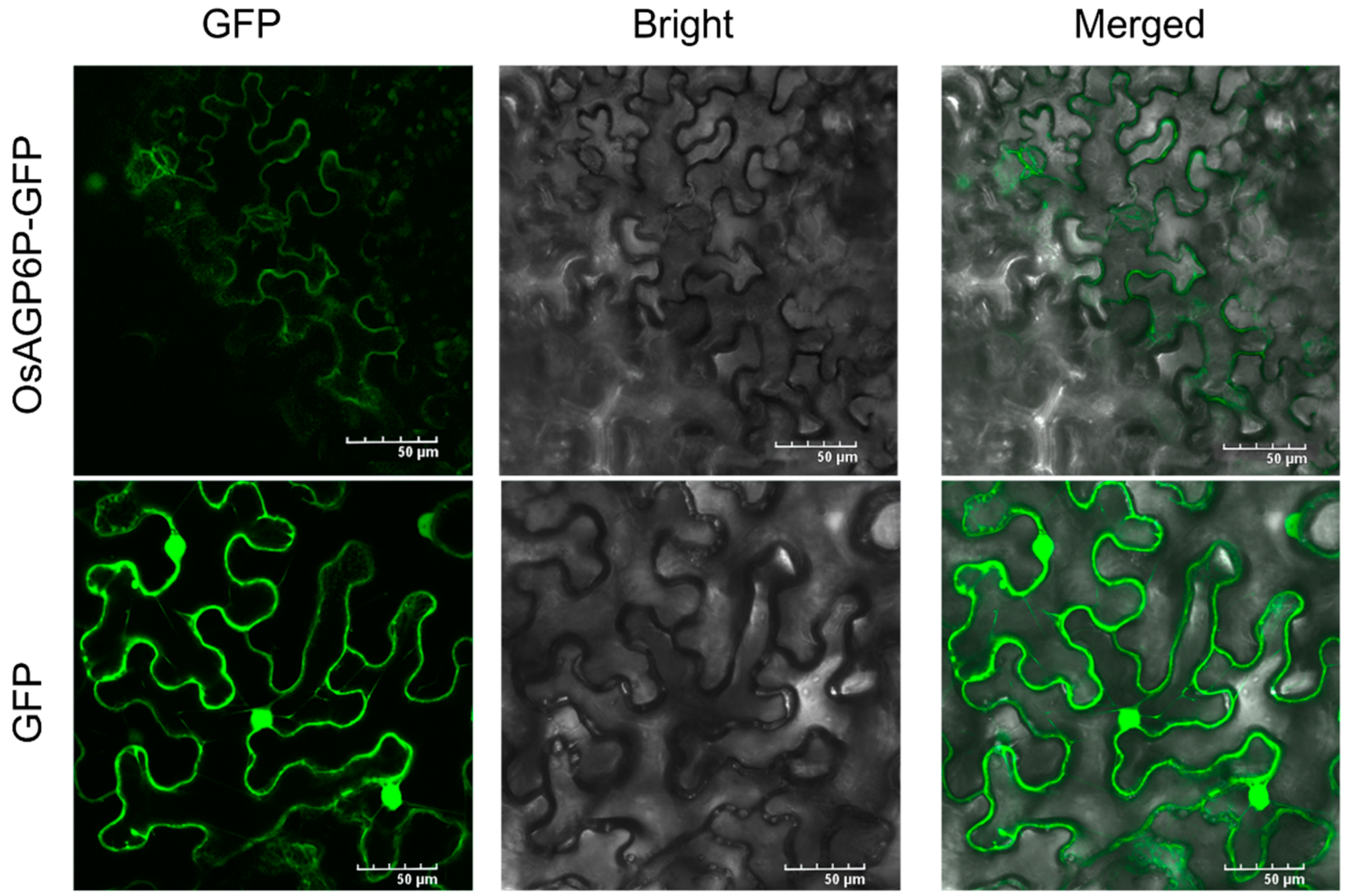
2.5. Subcellular Localization of the OsAGP6P Protein and the GUS Staining of OsAGP6P in Tissues and Organs

In this study, we found that one of the gene members, OsAGP6P, is preferentially higher in flowers, specifically in sperm cells and pollen (Figure 5). This result indicates that OsAGP6P may play a role in anther development and pollen formation in rice. Therefore, the OsAGP6P gene was chosen for further investigation.
The localization of the OsAGP6P protein was predicted in the plasma membrane using WoLF PSORT and Cell-Ploc 2.0. This result was verified as we found that GFP fluorescence was observed at the plasma membrane of tobacco leaf epidermal cells after the OsAGP6P-GFP fusion protein was transiently expressed in the leaves of four-week-old tobacco (Nicotiana benthamiana) (Figure 6). The plasma membrane localization of the OsAGP6P protein indicates its potential role as a small secreted protein.
Additionally, to investigate the expression of OsAGP6P at the tissue level further, GUS staining was performed on the tissues and organs at different stages of the ProOsAGP6P::GUS transgenic plants (Figure 7). Except for minimal staining in the young leaves of 14-day-old seedlings, no staining was observed in the roots or leaves at other vegetative stages (Figure 7A,B). Staining was not observed in the roots and leaves during the reproductive stage; however, it was evident in the basal internodes and the third internode of the rice stem. Transverse sections of the third internode revealed stronger staining in the vascular bundles of the stem (Figure 7C). Though insignificant, GUS staining in rice flowers began at stage 11 of anther development. The staining reached its darkest color at stage 12 (when microspores are formed after the meiosis of pollen mother cells) and continued until stage 14, when the rice flower fully bloomed (Figure 7D). These results indicate that OsAGP6P is expressed later during anther development. In detail, GUS staining can be observed in the stamens, specifically in the tapetum layer but not in the pollen at stage 11. However, at stage 13, slight GUS staining can be observed in pollen (Figure 7F,G). In contrast, the pistil showed no staining during anther development (Figure 7E). The findings indicate that the OsAGP6P protein primarily functions in the anther cell wall layer, although its effects on pollen tube growth cannot be excluded.

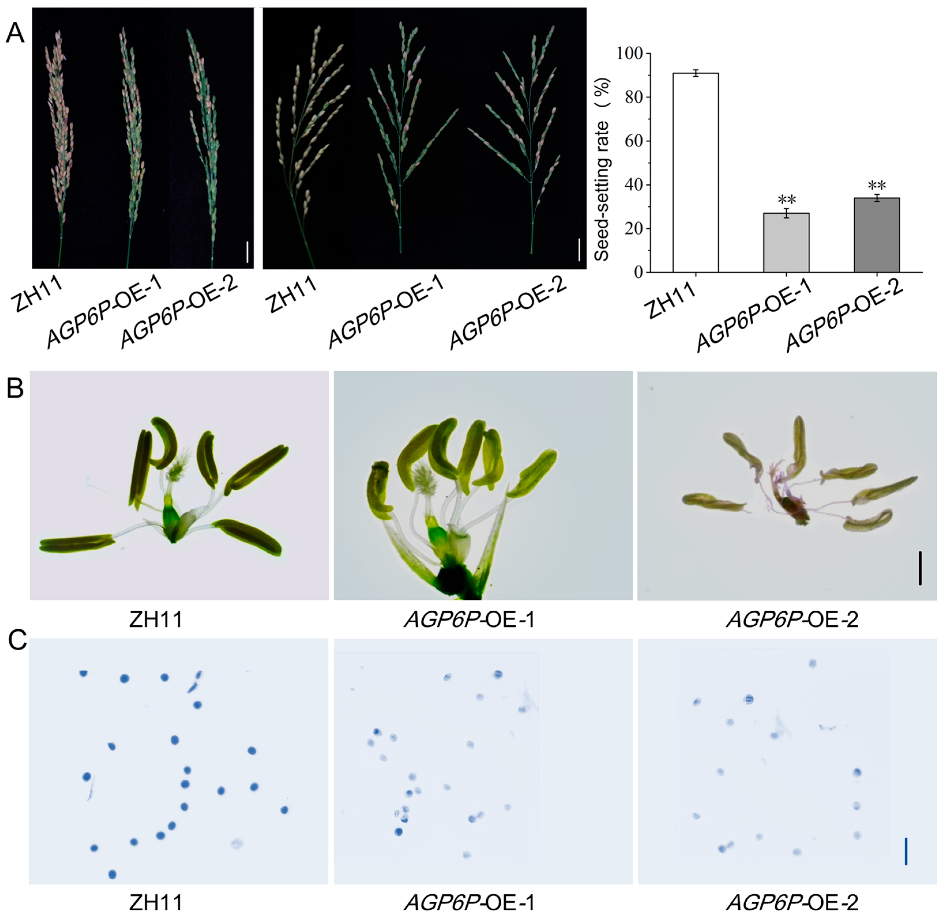
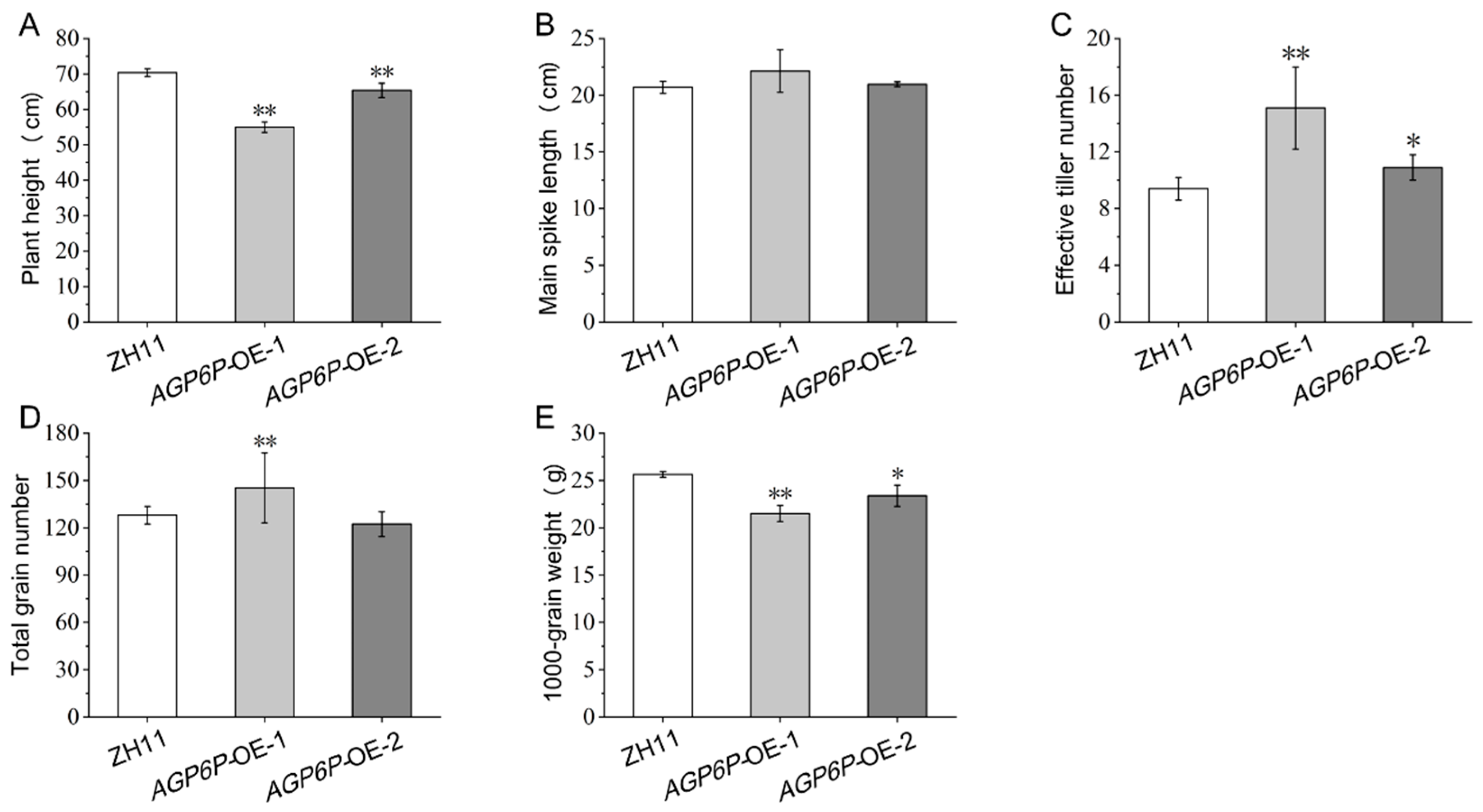
2.6. Phenotypic Observation of OsAGP6P Overexpression Plants

Plants from two positive independent overexpression lines (AGP6P-OE-1 and AGP6P-OE-2) and wild-type (WT) ZH11 were planted and grown in the field to examine the panicles at the mature stage. All positive plants exhibited a reduced seed-setting rate, while the negative plants exhibited no significant difference compared to the wild type (Figure 8A). These results indicated that overexpression of the OsAGP6P impaired the seed-setting rate and caused plant sterility in rice.
To further investigate the impact of OsAGP6P overexpression on the fertility of the plants, seed-setting rates were calculated, and the flowers of the two OE lines were examined. It was found that the overexpression of OsAGP6P hampered reproductive development in transgenic plants as they exhibited many green and empty grains, while the WT plants had golden-yellow seeds. The seed-setting rates were significantly lower in the two OE lines compared to the WT (from 91.5% to 27.2% and 34.1%) (Figure 8A). The anthers of the two OE lines seemed paler in color, had fewer filled pollen sacs, and were slightly curled (Figure 8B). After I2-KI staining, nearly all the pollen from the OE lines did not appear blue, indicating that they were inactive (Figure 8C).
To explore the effect of OsAGP6P on rice growth and development, several agronomic traits—including plant height, main spike length, effective tiller number, total grain number, and 1000-grain weight—were analyzed. No significant difference in main spike length was found between the OE lines and the WT ZH11 (Figure 9B). However, the OE lines showed a noticeable reduction in plant height and a significant increase in the effective tiller number compared to the WT ZH11 (Figure 9A,C). Compared with the wild type, the plant height of AGP6P-OE-1 decreased by 21.9%, while the effective tiller number increased by 60.6%. The AGP6P-OE-1 line had the shortest plant height (55.0 cm) and the highest effective tiller number (15.1), while the WT ZH11 had only 9.4 effective tillers on average (Figure 9C). In addition, the 1000-grain weights were significantly lower in the two OE lines, with the values 21.5g and 23.4g, respectively (Figure 9E). The results indicate that many yield components were affected by the overexpression of the gene OsAGP6P in rice.

3. Discussion

AGPs are subdivided according to the structure of their protein backbone into classical, lysine-rich, AG peptide, fasciclin-like (FLAs), non-classical, and chimeric AGPs [14,43,44,45]. The AG peptides are classical and possess complete structural features yet they are relatively under-researched. A genome-wide analysis of the AGP gene family should be performed as a first step in researching AGP gene families. Previous studies identified AGPs in rice but the information was fragmented and the number of members in each subfamily remained to be corrected.
In this study, with the recent availability of the latest genomic data from rice and the use of the “Python” programming language, AP peptides were identified. We tested various parameters to achieve the desired results. As expected, the number of AG peptides in this study was greatly increased compared to previous studies [10]. This computational methodology developed in this study can effectively differentiate PAST-rich proteins from others by applying specific thresholds (e.g., >35%) and can also be adapted for use with other species.
Based on these precise protein sequences, the diversity of AG peptides in rice can be analyzed in depth, and the phylogenetic analysis categorized them into three distinct groups. Additionally, we systematically elucidated the compositional characteristics and 3D structures of the AG peptides. AG peptides primarily consist of 1–2 α-helices, essential for protein recognition and binding, and all have a signal peptide that targets protein precursors to the endoplasmic reticulum (ER). In addition, we found that some AG peptides contain a GPI lipid anchor that was reported to tether the AGP molecules to the outer leaflet of the plasma membrane [4]. Many transmembrane proteins at the cell membrane feature α-helices that embed in the lipid bilayer, forming transmembrane channels or transporters. These structures are actively involved in the transmembrane transport of substances and the transmission of signals [46]. Some previous studies reported AGP proteins localized to multiple cellular structures, including the cell wall and the protoplast, or attached to the cell membrane via a GPI anchor at their C-terminus [47,48]. Lamport and Várnai [49] suggested that AGPs act as Ca2+ capacitors, indicating their role in Ca2+-dependent signaling pathways at the plasma membrane level. Another signaling pathway where AGPs may be significant is associated with the cleavage of their GPI anchor by phospholipase C [6,50]. Therefore, emphasizing structure and classification in this study was crucial for understanding AG peptide function in rice. It is important to note that the molecular size of AG peptides identified in this study ranges from approximately 5.09 kDa to 7.79 kDa. Thus, the AG peptides identified in our study can also be classified as small-molecule-secreted proteins (SSPs). SSPs are less than 250 amino acids (aa) in length and can be actively transported out of plant cells [51,52]. They regulate plant development and stress responses; studying them boosts plant resilience and productivity, making them a key research focus [53]. Therefore, our study serves as a valuable resource for investigating AG peptides in rice, revealing their role in plant development and male sterility while highlighting their potential targets for genetic engineering as a type of SSP.
Furthermore, the expression pattern of AG peptide genes was dynamically revealed during flowering, especially in the floral tissues as AGPs were frequently associated with pollen development [54,55]. Our study examined AG peptides in rice, particularly their roles in male reproductive development, establishing a crucial foundation for future research on the functions of AG peptides in rice development and stress response. We specifically demonstrated that OsAGP6P, a member of the rice AG peptide gene family, is associated with pollen development. Transcription data and GUS staining confirmed that the OsAGP6P protein is present in the stamen during stages 11–14 of pollen development. This generally represents critical transitions in pollen maturation, when microspores are formed after the meiosis of pollen mother cells. It was detectable from stage 11 (the late microspore stage when the exine formation occurs)—specifically at stage 12 (when the inner pollen wall, or intine, begins to develop beneath the exine). The results were consistent with the functions of AGPs, which have been reported to play a crucial role in anchoring primexine, followed by the formation of nexine and intine, as well as cellulose deposition, thereby providing essential structural support to the pollen grain [34]. It was shown that a change in the AGP expression pattern becomes evident from the early stages of the male gametophyte development [34]. In Arabidopsis, AGPs first appear in the cell walls of PMCs and endothelial cells. After meiosis, AGPs are found in the cytoplasm and primary cell wall of tetrads, forming a reticulated layer above the protectum of tetrads as well as in the locule-facing cell wall of the tapetum [56]. Although our study indicates that OsAGP6P is expressed at stages 11 and 14, we cannot dismiss the possibility that OsAGP6P is also expressed earlier (in PMCs) and contributes to pollen tube growth. Some studies have shown that variations in GPI biosynthesis affect pollen germination and pollen tube extension. For instance, the MAC207 antibody detects AGP proteins on the surface of mature pollen grains near the inner side of the plasma membrane [57]. AGP proteins were also observed in the cell walls of pollen tubes in Arabidopsis cultured in vitro [58]. AGP proteins profoundly impact the development of floral organs and ensure the proper timing of pollen grain germination and correct directional growth of pollen tubes [58,59]. Further work requires sensitive detection techniques like RNA in situ hybridization or immunolocalization using monoclonal antibodies specific to AGP sugar epitopes.
Our study has limitations that need future addressing, for example: (I) It is necessary to determine whether the OsAGP6P protein impacts pollen recognition at the stigma and the germination of pollen tubes, and whether it has functions in vegetative tissues. GUS staining also revealed that OsAGP6P expression occurs in leaves during the seedling stage and at the stem base during heading, potentially influencing agricultural traits in transgenic plants. (II) The precise mechanism underlying its mode of action still needs to be clarified. It has been reported that some transcription factors, such as novel microgametophyte defective mutant 1 (NMDM1; AT5G09250), can control the expression of AGPs for proper intine formation [60]. Are there any interacting partners that could explain their role in ensuring the proper timing of pollen grain development? Continued research in these areas is crucial for a deeper understanding of AGPs’ roles and could have implications for plant reproductive biology and crop improvement strategies.

4. Materials and Methods

4.1. The Identification of AG Peptide Proteins in Rice

The search criteria for AGPs referred to the guidelines [8,10,43,45,61]. A Python script was written to screen proteins with amino acid lengths between 50 and 75 and to calculate the PAST (Pro, Ala, Ser, Thr) percentage (greater than or equal to 35%, amino acid bias) for the candidate proteins (Supplementary Materials S1). The sequences of all annotated proteins in Nipponbare (Oryza sativa japonica) were downloaded from the Rice Super Pan-genome Information Resource Database (RiceSuperPIRdb, http://www.ricesuperpir.com/ (accessed on 7 November 2024)) [42]. Python 3.13.1 for Windows, macOS, UNIX, and Linux operating systems (https://www.python.org/ (accessed on 7 November 2024)) was used in this study.
The presence of N-terminal signal peptides of AG peptide proteins was predicted by the SignalP 5.0 Server (https://services.healthtech.dtu.dk/services/SignalP-5.0/ (accessed on 10 November 2024)) [62]. The presence of a C-terminal GPI anchor of AG peptide proteins was predicted using the GPI-SOM (http://gpi.unibe.ch, accessed on (accessed on 8 November 2024)) and the BIG-PI Plant Predictor (http://mendel.imp.ac.at/gpi/plant_server.html (accessed on 12 November 2024)).
After removing incomplete domains, the remaining proteins were chosen as AG peptide proteins for further analysis. The biochemical characteristics of AG peptide proteins, including molecular weight, instability index, isoelectric point (pI), and amino acid counts, were analyzed using TBtools [63]. The protein sequences for each gene were submitted to the website (http://www.csbio.sjtu.edu.cn/bioinf/plant-multi (accessed on 10 November 2024)) for predicting subcellular locations, following a previous study [64].

4.2. Phylogenetic Analysis, the Motifs Prediction, and the Three-Dimensional (3D) Structure Models of AG Peptide Proteins

Multiple alignments of AG peptide protein sequences in Oryza sativa were generated using MEGA 11 software [65]. Phylogenetic trees were constructed using a neighbor-joining algorithm and drawn using MEGA 11 software. Bootstrapping was implemented with 1000 replicates. Based on their sequence homology, all AG peptide proteins were clustered into distinct clades according to different subfamilies. The motifs within the AG peptide proteins were investigated using the Multiple Em for Motif Elicitation (MEME) (https://meme-suite.org/meme/ (accessed on 10 November 2024)). Three-dimensional (3D) protein models were constructed using the SWISS-MODEL platform (https://swissmodel.expasy.org (accessed on 10 November 2024)) [66,67].

4.3. Chromosomal Mapping, Gene Structure Analysis, and Promoter’s Cis-Element Analysis of AG Peptide Genes

TBtools software (version 2.019) was used to obtain and visualize the chromosomal location information of rice AG peptide genes. The intron–exon structure of the AG peptide genes was determined using TBtools, facilitating the integrated visualization of the motifs and the gene structure map [63]. The rice genome annotation file was downloaded from the Rice Super Pan-genome Information Resource Database (RiceSuperPIRdb, http://www.ricesuperpir.com/ (accessed on 7 November 2024)) [42]. The program Gtf/Gff3 Sequences Extract in TBtools software was used to extract upstream 2000-base-pair promoter sequences of the 43 AG peptides genes. AG peptides utilize “ep” or a similar variant as the suffix for the term “AGP”. This study employs the suffix “P” to differentiate the AG peptides from the traditional AGP members. Consequently, the genes of the AG peptide subfamily are referred to as AGP1P through AGP43P in this study. The upstream 2000-base-pair promoter sequences of each member were submitted to PlantCARE (http://bioinformatics.psb.ugent.be/webtools/plantcare/html/ (accessed on 7 November 2024)) [68] to forecast cis-elements and their potential function. The TBtools software was used to visualize the cis-element composition [63].

4.4. Expression Pattern Analysis of AG Peptide Genes

The transcriptomic data of 43 AG peptide genes were available on the Plant Public RNA-seq Database (PPRD) (https://plantrnadb.com/ (accessed on 10 November 2024)) [69]. Co-expression analysis was performed utilizing Euclidean distances in conjunction with the hierarchical clustering approach known as complete linkage clustering, with the analyses executed using TBtools [63]. To depict the overall expression profiles of each group, the average expression values of all co-expressed members within a cluster were calculated and represented in a line chart format.

4.5. Subcellular Localization Analysis of OsAGP6P Protein

The coding sequence (CDS) of the OsAGP6P gene (excluding the stop codon) was amplified from the rice variety ZH11. Under the control of the CaMV35S promoter, it was fused with the enhanced green fluorescent protein (eGFP) in the vector pD1301S to construct the plasmid OsAGP6P-eGFP. The plasmid was injected into Agrobacterium tumefaciens (EHA105) and transformed into the epidermal cells of 4–6-week-old tobacco (Nicotiana benthamiana L.) leaves according to the method of Kokkirala et al. [70]. After 36–48 h of injection, the subcellular localization of protein OsAGP6P-eGFP was observed using a FluoView FV3000 confocal microscope (Olympus, Tokyo, Japan).

4.6. The Construction and the Transformation of the Recombinant Vector pCAMBIA1381Z-OsAGP6P and GUS Staining of the Tissues from Transgenic Plants

The promoter region of the OsAGP6P gene (2000 bp upstream of the ATG) was obtained from the Rice Genome Annotation Project (RGAP) database. It was amplified from the DNA of ZH11 using the OsAGP6P-GUS primers (Supplementary Materials, Table S1). BamH I and EcoR I restriction sites were selected for cloning, and the amplified product was fused with the GUS-containing vector pCAMBIA1381Z to construct the recombinant expression vector pCAMBIA1381Z-OsAGP6P. The recombinant vector was inserted into DH5α competent cells for plating, and the colony was singled out for sequencing. After sequencing, the vector was transformed into the ZH11 callus via Agrobacterium-mediated transformation. Positive transgenic lines were identified by PCR using the hygromycin resistance gene detection primers (HygF and HygR) and further confirmed by β-glucuronidase (GUS) reporter gene-specific primers (OsAGP6P-GUS-IdentifyF and OsAGP6P-GUS-IdentifyR) (Supplementary Materials, Table S1). The primers were designed with Primer 5.0 and synthesized by Beijing Tsingke Biotechnology Co., Ltd.
The qualitative detection of the GUS reporter gene for OsAGP6P was conducted following the method of Lee and Schöffl [71]. The expression of OsAGP6P in different tissues was determined in the ProOsAGP6P::GUS (the β-glucuronidase (GUS) reporter gene driven by the promoter of OsAGP6P gene) transgenic lines. The expression of the β-glucuronidase (GUS) reporter gene was observed by histochemical staining [72]. For GUS staining, tissue samples were immersed in 90% acetone for 30 min, then stained in 5-bromo-4-chloro-3-indolyl-glucronide solution at 37 °C overnight. Then, the samples were transferred to 75% ethanol to remove chlorophyll and were observed under a stereomicroscope [73].

4.7. The Construction and the Investigation of Pollen Sterility of OsAGP6P Overexpression Transgenic Plants

For generating OsAGP6P overexpression lines, the coding sequence (CDS) of the OsAGP6P gene was amplified using the cDNA of ZH11 anther as the template, leaving out the stop codon and 20 base pairs from both the upstream and downstream parts of the cloning site. Primers were designed using the CEII tool, and BamHI and SmaI restriction enzyme sites were used (Supplementary Materials, Table S1). Gel electrophoresis checked the fragment size. Then, the fragment was cloned into the intermediate vector using a pEASY-Blunt Cloning Kit. Finally, the fragment was linked into the linearized binary expression vector pRHVcGFP, which has the enhanced green fluorescent protein (eGFP) sequence. The recombinant plasmid pRHVcGFP-OsAGP6P was introduced into the Zhonghua 11 (ZH11) by Agrobacterium-mediated transformation [74]. The positive transgenic lines were identified by PCR using the gene-specific primer combination OsAGP6P-pRYJ (Supplementary Materials, Table S1).

4.8. The Agronomic Traits and Statistical Analysis of OsAGP6P Overexpression Transgenic Plants

All overexpression plants and WT were grown at the experimental farm of Guangxi University (108° E, 22° N) in the 2023 rice growing season in Nanning, China. Seedlings approximately 35 days old were transplanted to a single-row plot, with a distance of 17 cm between plants within a row, and the rows were 27 cm apart. The field management followed essentially the normal agricultural practice. Pollen collected from OsAGP6P-overexpressing plants was stained with an I2-KI solution and examined using a stereomicroscope (Leica S8 APO, Wetzlar, Germany) to observe pollen viability. If the color of pollen grains appears blue–black, it indicates a high starch content and strong pollen vitality. The proportion of pollen in a microscopic field that can be stained can be calculated (%) using at least three microscopic fields from each observed sample. Only the 10 plants in the middle of each row were selected to measure agronomic traits, including plant height, main spike length, effective tiller number, total grain number, seed-setting rates, and 1000-grain weight. The method for determining the 1000-grain weight involved counting 100 fully developed and well-colored rice seeds, weighing them, and then converting the weight to the 1000-grain weight. Photos of the mature spikes and florets were taken during the measurements.

4.9. Statistical Analysis

Each experiment was conducted thrice, with data presented as means ± standard deviation (SD) from independent assays. Student’s t-test in GraphPad Prism version 8.0 was used to assess significance at the p < 0.01 level. Bar charts were generated using Origin 2018.

5. Conclusions

Arabinogalactan proteins (AGPs) are complex proteoglycans in nearly all plant organs, crucial for various biological functions in the plant life cycle. Here, we provide a computational methodology for screening AG peptides in rice and describe the classification, characteristics, and expression patterns of 43 AG peptides in detail. A focus is mainly placed on OsAGP6P, preferentially expressed in pollen during the later stages of anther development, encoding a small protein at the plasma membrane. Overexpressing OsAGP6P in rice reduced the seed-setting rate, pollen vitality, plant height, and 1000-grain weight while increasing the number of effective tillers, indicating its significant role in pollen ontogenesis and fertility. This understanding is essential for utilizing AG peptides in developing male-sterile lines, which are crucial for producing hybrid seeds.

Supplementary Materials

The following supporting information can be downloaded at https://www.mdpi.com/article/10.3390/ijms26062616/s1.

Author Contributions

S.S.: Data curation, Visualization, Writing—original draft, review and editing. Y.W.: Software, Data curation, Visualization. L.Z.: Methodology, Investigation. Z.Z.: Software, Data curation. X.L., M.Y. and H.Z.: Formal analysis. S.W.: Writing—original draft, Writing—review and editing, Conceptualization, Methodology, Software, Investigation, Visualization, Funding acquisition. L.W.: Writing—review and editing, Resources, Supervision, Project administration, Funding acquisition. All authors have read and agreed to the published version of the manuscript.

Funding

This research was funded by the Guangxi Science and Technology Support Initiative (Guike AD25069107), Guangxi Natural Science Foundation Project (grant numbers 2022GXNSFAA035479, 2023GXNSFBA026297, and 2024GXNSFGA010003), the Innovation Project of Guangxi Graduate Education (grant number YCBZ2023023), and the Department of Education of Guangxi Zhuang Autonomous Region.

Institutional Review Board Statement

Not applicable.

Informed Consent Statement

Not applicable.

Data Availability Statement

Data is contained within the article and Supplementary Materials.

Conflicts of Interest

The authors declare no conflicts of interest.

References

  1. Henry, J.S.; Ligrone, R.; Vaughn, K.C.; Lopez, R.A.; Renzaglia, K.S. Cell wall polymers in the Phaeoceros placenta reflect developmental and functional differences across generations. Bryophyt. Divers. Evol. 2021, 43, 265. [Google Scholar] [CrossRef] [PubMed]
  2. Showalter, A.M. Arabinogalactan-proteins: Structure, expression and function. Cell. Mol. Life Sci. CMLS 2001, 58, 1399–1417. [Google Scholar] [CrossRef]
  3. Hijazi, M.; Roujol, D.; Nguyen-Kim, H.; del Rocio Cisneros Castillo, L.; Saland, E.; Jamet, E.; Albenne, C. Arabinogalactan protein 31 (AGP31), a putative network-forming protein in Arabidopsis thaliana cell walls? Ann. Bot. 2014, 114, 1087–1097. [Google Scholar] [CrossRef]
  4. Silva, J.; Ferraz, R.; Dupree, P.; Showalter, A.M.; Coimbra, S. Three decades of advances in arabinogalactan-protein biosynthesis. Front. Plant Sci. 2020, 11, 610377. [Google Scholar] [CrossRef] [PubMed]
  5. Nguema-Ona, E.; Coimbra, S.; Vicré-Gibouin, M.; Mollet, J.C.; Driouich, A. Arabinogalactan proteins in root and pollen-tube cells: Distribution and functional aspects. Ann. Bot. 2012, 110, 383–404. [Google Scholar] [CrossRef] [PubMed]
  6. Hromadová, D.; Soukup, A.; Tylová, E. Arabinogalactan proteins in plant roots–an update on possible functions. Front. Plant Sci. 2021, 12, 674010. [Google Scholar] [CrossRef]
  7. Schultz, C.J.; Johnson, K.L.; Currie, G.; Bacic, A. The classical arabinogalactan protein gene family of Arabidopsis. Plant Cell 2000, 12, 1751–1767. [Google Scholar] [CrossRef]
  8. Han, T.; Dong, H.; Cui, J.; Li, M.; Lin, S.; Cao, J.; Huang, L. Genomic, molecular evolution, and expression analysis of genes encoding putative classical AGPs, lysine-rich AGPs, and AG peptides in Brassica rapa. Front. Plant Sci. 2017, 8, 397. [Google Scholar] [CrossRef]
  9. Kaur, D.; Moreira, D.; Coimbra, S.; Showalter, A.M. Hydroxyproline-O-galactosyltransferases synthesizing type ii arabinogalactans are essential for male gametophytic development in Arabidopsis. Front. Plant Sci. 2022, 13, 935413. [Google Scholar] [CrossRef]
  10. Ma, H.; Zhao, J. Genome-wide identification, classification, and expression analysis of the arabinogalactan protein gene family in rice (Oryza sativa L.). J. Exp. Bot. 2010, 61, 2647–2668. [Google Scholar] [CrossRef]
  11. MacMillan, C.P.; Taylor, L.; Bi, Y.; Southerton, S.G.; Evans, R.; Spokevicius, A. The fasciclin-like arabinogalactan protein family of Eucalyptus grandis contains members that impact wood biology and biomechanics. New Phytol. 2015, 206, 1314–1327. [Google Scholar] [CrossRef] [PubMed]
  12. Ma, H.; Zhao, H.; Liu, Z.; Zhao, J. The phytocyanin gene family in rice (Oryza sativa L.): Genome-wide identification, classification and transcriptional analysis. PLoS ONE 2011, 6, e25184. [Google Scholar] [CrossRef] [PubMed]
  13. Kobayashi, Y.; Motose, H.; Iwamoto, K.; Fukuda, H. Expression and genome-wide analysis of the xylogen-type gene family. Plant Cell Physiol. 2011, 52, 1095–1106. [Google Scholar] [CrossRef]
  14. Zhang, Y.; Zhou, F.; Wang, H.; Chen, Y.; Yin, T.; Wu, H. Genome-Wide Comparative Analysis of the Fasciclin-like Arabinogalactan Proteins (FLAs) in Salicacea and Identification of Secondary Tissue Development-Related Genes. Int. J. Mol. Sci. 2023, 24, 1481. [Google Scholar] [CrossRef] [PubMed]
  15. Ma, Y.; Johnson, K. Arabinogalactan proteins–multifunctional glycoproteins of the plant cell wall. Cell Surf. 2023, 9, 100102. [Google Scholar] [CrossRef]
  16. Showalter, A.M.; Keppler, B.D.; Lichtenberg, J.; Gu, D.; Welch, L.R. A bioinformatics approach to the identification, classification, and analysis of hydroxyproline-rich glycoproteins. Plant Physiol. 2010, 153, 485–513. [Google Scholar] [CrossRef]
  17. Johnson, K.L.; Cassin, A.M.; Lonsdale, A.; Wong, G.K.S.; Soltis, D.E.; Miles, N.W.; Melkonian, M.; Melkonian, B.; Deyholos, M.K.; Leebens-Mack, J. Insights into the evolution of hydroxyproline-rich glycoproteins from 1000 plant transcriptomes. Plant Physiol. 2017, 174, 904–921. [Google Scholar] [CrossRef]
  18. Johnson, K.L.; Cassin, A.M.; Lonsdale, A.; Bacic, A.; Doblin, M.S.; Schultz, C.J. Pipeline to identify hydroxyproline-rich glycoproteins. Plant Physiol. 2017, 174, 886–903. [Google Scholar] [CrossRef]
  19. Ma, Y.; Zeng, W.; Bacic, A.; Johnson, K. AGPs through time and space. Annu. Plant Rev. Online 2018, 1, 767–804. [Google Scholar] [CrossRef]
  20. Ma, Y.; Shafee, T.; Mudiyanselage, A.M.; Ratcliffe, J.; MacMillan, C.P.; Mansfield, S.D.; Bacic, A.; Johnson, K.L. Distinct functions of FASCICLIN-LIKE ARABINOGALACTAN PROTEINS relate to domain structure. Plant Physiol. 2023, 192, 119–132. [Google Scholar] [CrossRef]
  21. Leszczuk, A.; Kalaitzis, P.; Kulik, J.; Zdunek, A. Review: Structure and modifications of arabinogalactan proteins (AGPs). BMC Plant Biol. 2023, 23, 45. [Google Scholar] [CrossRef]
  22. Huang, G.Q.; Gong, S.Y.; Xu, W.L.; Li, W.; Li, P.; Zhang, C.J.; Li, D.D.; Zheng, Y.; Li, F.G.; Li, X.B. A fasciclin-like arabinogalactan protein, GhFLA1, is involved in fiber initiation and elongation of cotton. Plant Physiol. 2013, 161, 1278–1290. [Google Scholar] [CrossRef] [PubMed]
  23. Přerovská, T.; Jindřichová, B.; Henke, S.; Yvin, J.C.; Ferrieres, V.; Burketová, L.; Lipovová, P.; Nguema-Ona, E. Arabinogalactan protein-like proteins from Ulva lactuca activate immune responses and plant resistance in an oilseed crop. Front. Plant Sci. 2022, 13, 893858. [Google Scholar] [CrossRef] [PubMed]
  24. Pereira, A.M.; Pereira, L.G.; Coimbra, S. Arabinogalactan proteins: Rising attention from plant biologists. Plant Reprod. 2015, 28, 1–15. [Google Scholar] [CrossRef]
  25. Su, S.; Higashiyama, T. Arabinogalactan proteins and their sugar chains: Functions in plant reproduction, research methods, and biosynthesis. Plant Reprod. 2018, 31, 67–75. [Google Scholar] [CrossRef]
  26. Leszczuk, A.; Cybulska, J.; Skrzypek, T.; Zdunek, A. Properties of arabinogalactan proteins (AGPs) in apple (Malus × Domestica) fruit at different stages of ripening. Biology 2020, 9, 225. [Google Scholar] [CrossRef]
  27. Mareri, L.; Romi, M.; Cai, G. Arabinogalactan proteins: Actors or spectators during abiotic and biotic stress in plants? Plant Biosyst.-Int. J. Deal. All Asp. Plant Biol. 2019, 153, 173–185. [Google Scholar] [CrossRef]
  28. Suárez, C.; Zienkiewicz, A.; Castro, A.J.; Zienkiewicz, K.; Majewska-Sawka, A.; Rodríguez-García, M.I. Cellular localization and levels of pectins and arabinogalactan proteins in olive (Olea europaea L.) pistil tissues during development: Implications for pollen–pistil interaction. Planta 2013, 237, 305–319. [Google Scholar] [CrossRef]
  29. Rafińska, K.; Niedojadło, K.; Świdziński, M.; Niedojadło, J.; Bednarska-Kozakiewicz, E. Spatial and temporal distribution of arabinogalactan proteins during Larix decidua Mill. male gametophyte and ovule interaction. Int. J. Mol. Sci. 2021, 22, 4298. [Google Scholar] [CrossRef]
  30. Basu, D.; Wang, W.; Ma, S.; DeBrosse, T.; Poirier, E.; Emch, K.; Soukup, E.; Tian, L.; Showalter, A.M. Two hydroxyproline galactosyltransferases, GALT5 and GALT2, function in arabinogalactan-protein glycosylation, growth and development in Arabidopsis. PLoS ONE 2015, 10, e0125624. [Google Scholar] [CrossRef]
  31. Moreira, D.; Lopes, A.L.; Silva, J.; Ferreira, M.J.; Pinto, S.C.; Mendes, S.; Pereira, L.G.; Coimbra, S.; Pereira, A.M. New insights on the expression patterns of specific arabinogalactan proteins in reproductive tissues of Arabidopsis thaliana. Front. Plant Sci. 2022, 13, 1083098. [Google Scholar] [CrossRef] [PubMed]
  32. Kaur, D.; Held, M.A.; Smith, M.R.; Showalter, A.M. Functional characterization of hydroxyproline-O-galactosyltransferases for Arabidopsis arabinogalactan-protein synthesis. BMC Plant Biol. 2021, 21, 590. [Google Scholar] [CrossRef]
  33. Deng, Y.; Wan, Y.; Liu, W.; Zhang, L.; Zhou, K.; Feng, P.; Wang, N. OsFLA1 encodes a fasciclin-like arabinogalactan protein and affects pollen exine development in rice. Theor. Appl. Genet. 2022, 135, 1247–1262. [Google Scholar] [CrossRef] [PubMed]
  34. Foubert-Mendes, S.; Silva, J.; Ferreira, M.J.; Pereira, L.G.; Coimbra, S. A review on the function of arabinogalactan-proteins during pollen grain development. Plant Reprod. 2025, 38, 8. [Google Scholar] [CrossRef]
  35. Ma, T. Genome-Wide Analysis of Xylogen-Like Arabinogalactan Protein Gene Family and Biological Function Studies of OsAGP13 in Rice. Ph.D. Dissertation, Wuhan University, Wuhan, China, 2014. [Google Scholar]
  36. Li, Q.; Xu, F.; Chen, Z.; Teng, Z.; Sun, K.; Li, X.; Yu, J.; Zhang, G.; Liang, Y.; Huang, X.; et al. Synergistic interplay of ABA and BR signal in regulating plant growth and adaptation. Nat. Plants 2021, 7, 1108–1118. [Google Scholar] [CrossRef]
  37. Van Hengel, A.J.; Roberts, K. AtAGP30, an arabinogalactan-protein in the cell walls of the primary root, plays a role in root regeneration and seed germination. Plant J. 2003, 36, 256–270. [Google Scholar] [CrossRef] [PubMed]
  38. Van Hengel, A.J.; Barber, C.; Roberts, K. The expression patterns of arabinogalactan-protein AtAGP30 and GLABRA2 reveal a role for abscisic acid in the early stages of root epidermal patterning. Plant J. 2004, 39, 70–83. [Google Scholar] [CrossRef]
  39. Lopez-Hernandez, F.; Tryfona, T.; Rizza, A.; Yu, X.L.; Harris, M.O.; Webb, A.A.R.; Kotake, T.; Dupree, P. Calcium binding by arabinogalactan polysaccharides is important for normal plant development. Plant Cell 2020, 32, 3346–3369. [Google Scholar] [CrossRef]
  40. Sakai, H.; Lee, S.; Tanaka, T.; Numa, H.; Kim, J.; Kawahara, Y.; Wakimoto, H.; Yang, C.-C.; Iwamoto, M.; Abe, T.; et al. Rice Annotation Project Database (RAP-DB): An integrative and interactive database for rice genomics. Plant Cell Physiol. 2013, 54, e6. [Google Scholar] [CrossRef]
  41. Kawahara, Y.; Bastide, M.D.L.; Hamilton, J.P.; Kanamori, H.; McCombie, W.; Shu, O.; Schwartz, D.; Tanaka, T.; Wu, J.; Zhou, S.; et al. Improvement of the Oryza sativa Nipponbare reference genome using next generation sequence and optical map data. Rice 2013, 6, 4. [Google Scholar] [CrossRef]
  42. Shang, L.; He, W.; Wang, T.; Yang, Y.; Xu, Q.; Zhao, X.; Yang, L.; Zhang, H.; Li, X.; Lv, Y.; et al. A complete assembly of the rice Nipponbare reference genome. Mol. Plant 2023, 16, 1232–1236. [Google Scholar] [CrossRef] [PubMed]
  43. Schultz, C.J.; Rumsewicz, M.P.; Johnson, K.L.; Jones, B.J.; Gaspar, Y.M.; Bacic, A. Using genomic resources to guide research directions. The arabinogalactan protein gene family as a test case. Plant Physiol. 2002, 129, 1448–1463. [Google Scholar] [CrossRef]
  44. Tan, L.; Leykam, J.F.; Kieliszewski, M.J. Glycosylation motifs that direct arabinogalactan addition to arabinogalactan-proteins. Plant Physiol. 2003, 132, 1362–1369. [Google Scholar] [CrossRef] [PubMed]
  45. Gaspar, Y.; Johnson, K.L.; Mckenna, J.A.; Bacic, A.; Schultz, C.J. The complex structures of arabinogalactan-proteins and the journey towards understanding function. Plant Mol. Biol. 2001, 47, 161–176. [Google Scholar] [CrossRef]
  46. Woolfson, D.N. Understanding a protein fold: The physics, chemistry, and biology of α-helical coiled coils. J. Biol. Chem. 2023, 299, 104579. [Google Scholar] [CrossRef] [PubMed]
  47. Sardar, H.S.; Yang, J.; Showalter, A.M. Molecular interactions of arabinogalactan proteins with cortical microtubules and F-actin in Bright Yellow-2 tobacco cultured cells. Plant Physiol. 2006, 142, 1469–1479. [Google Scholar] [CrossRef]
  48. Bao, H.; Zheng, G. Distribution and Function of Arabinogalactan-proteins in Higher Plant. North. Hortic. 2016, 1, 185–191. [Google Scholar] [CrossRef]
  49. Lamport, D.T.; Várnai, P. Periplasmic arabinogalactan glycoproteins act as a calcium capacitor that regulates plant growth and development. New Phytol. 2013, 197, 58–64. [Google Scholar] [CrossRef]
  50. Olmos, E.; García De La Garma, J.; Gomez-Jimenez, M.C.; Fernandez-Garcia, N. Arabinogalactan proteins are involved in salt-adaptation and vesicle trafficking in tobacco by-2 cell cultures. Front. Plant Sci. 2017, 8, 1092. [Google Scholar] [CrossRef]
  51. Lease, K.A.; Walker, J.C. The Arabidopsis unannotated secreted peptide database, a resource for plant peptidomics. Plant Physiol. 2006, 142, 831–838. [Google Scholar] [CrossRef]
  52. Plett, J.M.; Yin, H.; Mewalal, R.; Hu, R.; Li, T.; Ranjan, P.; Jawdy, S.; Paoli, H.C.D.; Butler, G.; Burch-Smith, T.M.; et al. Populus trichocarpa encodes small, effector-like secreted proteins that are highly induced during mutualistic symbiosis. Sci. Rep. 2017, 7, 382. [Google Scholar] [CrossRef]
  53. Hu, X.L.; Lu, H.; Hassan, M.M.; Zhang, J.; Yuan, G.; Abraham, P.E.; Shrestha, H.K.; Solis, M.I.V.; Chen, J.G.; Tschaplinski, T.J. Advances and perspectives in discovery and functional analysis of small secreted proteins in plants. Hortic. Res. 2021, 8, 130. [Google Scholar] [CrossRef] [PubMed]
  54. Costa, M.; Nobre, M.S.; Becker, J.D.; Masiero, S.; Amorim, M.I.; Pereira, L.G.; Coimbra, S. Expression-based and co-localization detection of arabinogalactan protein 6 and arabinogalactan protein 11 interactors in Arabidopsis pollen and pollen tubes. BMC Plant Biol. 2013, 13, 7. [Google Scholar] [CrossRef] [PubMed]
  55. Lin, S.; Huang, L.; Miao, Y.; Yu, Y.; Peng, R.; Cao, J. Constitutive overexpression of the classical arabinogalactan protein gene BcMF18 in Arabidopsis causes defects in pollen intine morphogenesis. Plant Growth Regul. 2019, 88, 159–171. [Google Scholar] [CrossRef]
  56. Suzuki, T.; Narciso, J.O.; Zeng, W.; van de Meene, A.; Yasutomi, M.; Takemura, S.; Lampugnani, E.R.; Doblin, M.S.; Bacic, A.; Ishiguro, S. KNS4/UPEX1: A type II arabinogalactan β-(1, 3)-galactosyltransferase required for pollen exine development. Plant Physiol. 2017, 173, 183–205. [Google Scholar] [CrossRef] [PubMed]
  57. Samaj, J.; Ensikat, H.J.; Baluska, F.; Knox, J.P.; Volkmann, D. Immunogold localization of plant surface arabinogalactan-proteins using glycerol liquid substitution and scanning electron microscopy. J. Microsc. 1999, 193, 150–157. [Google Scholar] [CrossRef]
  58. Pereira, L.G.; Coimbra, S.; Oliveira, H.; Monteiro, L.; Sottomayor, M. Expression of arabinogalactan protein genes in pollen tubes of Arabidopsis thaliana. Planta 2006, 223, 374–380. [Google Scholar] [CrossRef]
  59. Coimbra, S.; Costa, M.; Mendes, M.A.; Pereira, A.M.; Pinto, J.; Pereira, L.G. Early germination of Arabidopsis pollen in a double null mutant for the arabinogalactan protein genes AGP6 and AGP11. Sex. Plant Reprod. 2010, 23, 199–205. [Google Scholar] [CrossRef]
  60. Mi, L.; Mo, A.; Yang, J.; Liu, H.; Ren, D.; Chen, W.; Long, H.; Jiang, N.; Zhang, T.; Lu, P. Arabidopsis novel microgametophyte defective mutant 1 is required for pollen viability via influencing intine development in Arabidopsis. Front. Plant Sci. 2022, 13, 814870. [Google Scholar] [CrossRef]
  61. Schultz, C.J.; Ferguson, K.L.; Lahnstein, J.; Bacic, A. Post-translational modifications of arabinogalactan-peptides of Arabidopsis thaliana: Endoplasmic reticulum and glycosylphosphatidylinositol-anchor signal cleavage sites and hydroxylation of proline. J. Biol. Chem. 2004, 279, 45503–45511. [Google Scholar] [CrossRef]
  62. Nielsen, H.; Tsirigos, K.D.; Brunak, S.; von Heijne, G. A brief history of protein sorting prediction. Protein J. 2019, 38, 200–216. [Google Scholar] [CrossRef]
  63. Chen, C.; Chen, H.; Zhang, Y.; Thomas, H.R.; Frank, M.H.; He, Y.; Xia, R. TBtools: An integrative toolkit developed for interactive analyses of big biological data. Mol. Plant 2020, 13, 1194–1202. [Google Scholar] [CrossRef]
  64. Chou, K.C.; Shen, H.B. Cell-ploc 2.0: An improved package of web-servers for predicting subcellular localization of proteins in various organisms. Nat. Sci. 2010, 2, 1090–1103. [Google Scholar] [CrossRef]
  65. Tamura, K.; Stecher, G.; Kumar, S. MEGA11: Molecular evolutionary genetics analysis version 11. Mol. Biol. Evol. 2021, 38, 3022–3027. [Google Scholar] [CrossRef]
  66. Bienert, S.; Waterhouse, A.; De Beer, T.A.; Tauriello, G.; Studer, G.; Bordoli, L.; Schwede, T. The SWISS-MODEL Repository—New features and functionality. Nucleic Acids Res. 2017, 45, D313–D319. [Google Scholar] [CrossRef]
  67. Waterhouse, A.; Bertoni, M.; Bienert, S.; Studer, G.; Tauriello, G.; Gumienny, R.; Heer, F.T.; de Beer, T.A.P.; Rempfer, C.; Bordoli, L.; et al. SWISS-MODEL: Homology modelling of protein structures and complexes. Nucleic Acids Res. 2018, 46, W296–W303. [Google Scholar] [CrossRef]
  68. Lescot, M.; Déhais, P.; Thijs, G.; Marchal, K.; Moreau, Y.; Van de Peer, Y.; Rouzé, P.; Rombauts, S. PlantCARE, a database of plant cis-acting regulatory elements and a portal to tools for in silico analysis of promoter sequences. Nucleic Acids Res. 2002, 30, 325–327. [Google Scholar] [CrossRef]
  69. Yu, Y.; Zhang, H.; Long, Y.; Shu, Y.; Zhai, J. PPRD: A comprehensive online database for expression analysis of ~45,000 plant public RNA-Seq libraries. Plant Biotechnol. J. 2022, 20, 806–808. [Google Scholar] [CrossRef]
  70. Kokkirala, V.R.; Peng, Y.; Abbagani, S.; Zhu, Z.; Umate, P. Subcellular localization of proteins of Oryza sativa L. in the model tobacco and tomato plants. Plant Signal. Behav. 2010, 5, 1336–1341. [Google Scholar] [CrossRef]
  71. Lee, J.H.; Schöffl, F. GUS activity staining in gels: A powerful tool for studying protein interactions in plants. Plant Mol. Biol. Report. 1995, 13, 346–354. [Google Scholar] [CrossRef]
  72. Jefferson, R.A.; Kavanagh, T.A.; Bevan, M.W. GUS fusions: Beta-glucuronidase as a sensitive and versatile gene fusion marker in higher plants. EMBO J. 1987, 6, 3901–3907. [Google Scholar] [CrossRef]
  73. Béziat, C.; Kleine-Vehn, J.; Feraru, E. Histochemical staining of β-glucuronidase and its spatial quantification. In Plant Hormones; Methods in Molecular Biology; Springer: Berlin/Heidelberg, Germany, 2017; pp. 73–80. [Google Scholar] [CrossRef]
  74. Yang, J.; Ji, L.; Liu, S.; Jing, P.; Hu, J.; Jin, D.; Wang, L.; Xie, G. The CaM1-associated CCaMK–MKK1/6 cascade positively affects lateral root growth via auxin signaling under salt stress in rice. J. Exp. Bot. 2021, 72, 6611–6627. [Google Scholar] [CrossRef]
Figure 1. Workflow diagram for the identification of AG peptides in rice.
Ijms 26 02616 g001
Figure 2. Chromosomal distribution of 43 AG peptide genes in rice. Chromosomes are represented by vertical bold bars with numerical orders at the top. The chromosome length scale (Mb) is indicated on the left side.
Ijms 26 02616 g002
Figure 3. Phylogenetic relationship and 3D models of AG peptide proteins in rice: (A) phylogenetic tree; (B) gene structure of 43 AG peptide genes with a scale bar indicating 500 bp; (C) 3D structural models. Blue boxes indicate the coding regions (CDS), pink boxes indicate 5′ and 3′ and untranslated regions (UTR), black lines indicate introns.
Ijms 26 02616 g003
Figure 4. Cis-regulatory element analysis of the AG peptide genes: (A) analysis of cis-elements in the promoter region of AG peptide genes; (B) heatmap of the number of cis-elements, the different colors represent the number of cis-elements; (C) the sum of cis-elements for each category is shown as a histogram.
Ijms 26 02616 g004
Figure 5. Hierarchical clustering of the expression profiles of the AP peptide genes in diverse tissues at different stages. (A,E) Expression of 43 AG peptide genes in various tissues exhibited divergent patterns. (B,F) Expression of 43 AG peptide genes in flower parts: anther, mature pollen, and pistil. (C,G) Expression of 43 AG peptide genes in sperm cells, pollen minus sperm cells, egg cells, and spores. (D,H) Expression of AG peptide genes at stages 8 to 11 of the anther development. The expression values of each point in (EH) were calculated from the mean values of the expression of all members in each cluster. The heatmap was created based on the log2 (TPM) values of AP peptide genes and normalized by row. A TPM value above 20 displays abundant genes and is marked with “*”. Differences in gene expression are colored, with red for high expression and blue for low expression.
Ijms 26 02616 g005
Figure 6. Subcellular location of OsAGP6P protein in Nicotona benthamiana. GFP is the empty vector pD1301S; OsAGP6P-GFP is the recombinant vector OsAGP6P-pD1301S. Scale bars, 50 μm.
Ijms 26 02616 g006
Figure 7. Histochemical GUS staining of ProOsAGP6P-GUS plants: (A) Stained tissues of roots and leaves at the seedling stage. Scale bar, 5 mm. (B) Stained tissues of roots and leaves at the vegetative stage. Scale bar, 5 mm. (C) Stained tissues of roots, basal internodes, third internodes, third internode slices, and leaves at heading stage. Scale bar, 5 mm. (D) Stained floral tissues from stages 8 to 14 of anther development. Scale bar, 2 mm. (E) The stained tissues of pistils, with stamens removed, show the 9th, 11th, and 13th stages of anther development from left to right. Scale bar, 500 μm. (F) Stained tissues of the anther, from left to right, represent the 11th and 13th stages of anther development. Scale bar, 200 μm. (G) Stained pollen tissues, shown from left to right, represent the 11th and 13th stages of anther development. Scale bar, 100 μm.
Ijms 26 02616 g007
Figure 8. Spikelets, seed-setting rate, florets, and pollen viability of rice with overexpression of OsAGP6P: (A) From left to right, spikelets of T1 generation rice overexpressing OsAGP6P, spikelets of T2 generation rice overexpressing OsAGP6P, seed-setting rate of T2 generation rice overexpressing OsAGP6P. Values represent the means, error bars depict SE (Scale bar, 10 cm. Student’s t-test, n = 10, ** p < 0.01). (B) Flowers of T2 generation rice overexpressing OsAGP6P. Scale bar, 1 mm. (C) I2-KI staining of pollen from T2 generation rice overexpressing OsAGP6P. Scale bar, 100 μm.
Ijms 26 02616 g008
Figure 9. The agronomic traits of ZH11 and OE lines (AGP6P-OE-1, AGP6P-OE-2) plants. (A) Plant height of ZH11 and OE lines. (B) Main spike length of ZH11 and OE lines. (C) Effective tiller number of ZH11 and OE lines. (D) Total grain number of ZH11 and OE lines. (E) 1000-grain weight of ZH11 and OE lines. Three repetitions for 10 plants each (3 replicates × 10 individuals). Values represent the means, error bars depict SE (Student’s t-test, n = 10, * p < 0.05, ** p < 0.01).
Ijms 26 02616 g009
Table 1. Identification and characterization of the AG peptides genes in rice.
AGIS IDLocus IDGene NamePAST
(%)
SPGPIAP/PA/SP/TP RepeatsAA
Size
AGIS_Os01g009100LOC_Os01g10550OsAGP1P35.62YesNo0/2/0/073
AGIS_Os01g032060LOC_Os01g37950OsAGP2P (OsAGP14)51.43YesNo4/2/0/070
AGIS_Os01g034750LOC_Os01g40880OsAGP3P40.00YesYes0/1/1/070
AGIS_Os01g034820LOC_Os01g40950OsAGP4P (OsAGP15)40.28YesNo3/3/0/072
AGIS_Os01g035100LOC_Os01g41230OsAGP5P44.93YesNo2/1/1/069
AGIS_Os01g039860LOC_Os01g46850OsAGP6P (OsAGP6P)40.58YesYes3/2/0/069
AGIS_Os01g047750LOC_Os01g55220OsAGP7P (OsAGP17)47.14YesYes4/3/0/070
AGIS_Os01g049500LOC_Os01g57030OsAGP8P (OsAGP18)58.82YesYes3/2/0/068
AGIS_Os01g049510LOC_Os01g57040OsAGP9P (OsAGP19)57.35YesYes4/3/0/068
AGIS_Os01g051100LOC_Os01g58590OsAGP10P37.50YesNo0/2/0/064
AGIS_Os02g010250LOC_Os02g12150OsAGP11P42.25YesNo0/2/3/071
AGIS_Os02g010720LOC_Os02g12654OsAGP12P40.91YesNo2/0/1/066
AGIS_Os02g014470LOC_Os02g16500OsAGP13P (OsAGP20)38.36YesYes4/0/1/073
AGIS_Os02g035140LOC_Os02g39290OsAGP14P43.75YesNo1/1/2/064
AGIS_Os02g044090LOC_Os02g48710OsAGP15P (OsAGP21)49.28YesYes2/2/0/069
AGIS_Os03g036830LOC_Os03g42990OsAGP6P43.84YesYes0/1/1/073
AGIS_Os03g043930LOC_Os03g50720OsAGP17P38.67YesNo1/0/0/175
AGIS_Os04g001290LOC_Os04g02380OsAGP18P39.13YesNo2/1/0/169
AGIS_Os04g027720NoneOsAGP19P38.81YesNo1/1/1/067
AGIS_Os04g050800LOC_Os04g57260OsAGP20P62.32YesNo4/4/2/069
AGIS_Os04g050820LOC_Os04g57270OsAGPp21P60.29YesNo4/4/2/068
AGIS_Os04g050830LOC_Os04g57280OsAGP22P57.97YesNo4/4/2/069
AGIS_Os05g010800LOC_Os05g12580OsAGP23P43.28YesYes2/1/1/067
AGIS_Os06g019180LOC_Os06g21410OsAGP24P (OsAGP25)45.95YesNo3/3/0/074
AGIS_Os06g027160LOC_Os06g30920OsAGP25P50.94YesNo3/4/0/053
AGIS_Os07g011720LOC_Os07g13319OsAGP26P41.79YesNo0/2/0/067
AGIS_Os07g012700LOC_Os07g14360OsAGP27P43.66YesNo1/2/2/071
AGIS_Os07g013810LOC_Os07g15530OsAGP28P36.00YesNo0/1/1/075
AGIS_Os07g023740LOC_Os07g27410OsAGP29P45.71YesNo3/0/3/070
AGIS_Os07g034440LOC_Os07g38630OsAGP30P (OsAGP27)40.63YesNo4/3/0/064
AGIS_Os08g038920LOC_Os08g41860OsAGP31P41.67YesNo0/1/0/172
AGIS_Os09g019700LOC_Os09g11780OsAGP32P41.67YesNo1/1/2/160
AGIS_Os09g029540LOC_Os09g22510OsAGP33P48.44YesNo1/2/0/064
AGIS_Os09g029970LOC_Os09g23430OsAGP34P37.88YesNo2/0/0/166
AGIS_Os09g036360LOC_Os09g30040OsAGP35P43.24YesNo1/0/1/074
AGIS_Os10g003720LOC_Os10g05150OsAGP36P35.21YesNo0/1/1/171
AGIS_Os10g022660LOC_Os10g28299OsAGP37P37.70YesNo2/0/2/061
AGIS_Os11g035190LOC_Os11g40850OsAGP38P36.62YesNo1/1/1/171
AGIS_Os12g010080LOC_Os12g11420OsAGP39P38.89YesNo1/0/2/072
AGIS_Os12g010260LOC_Os12g11610OsAGP40P42.65YesNo1/1/0/168
AGIS_Os12g010270LOC_Os12g11620OsAGP41P43.48YesNo1/0/0/169
AGIS_Os12g010590LOC_Os12g11980OsAGP42P42.65YesNo1/1/0/168
AGIS_Os12g010600LOC_Os12g11990OsAGP43P44.12YesNo1/0/0/168
SP: signal peptide. GPI: GPI anchor addition sequence. AP/PA/SP/TP repeats: Ala–Pro, Pro–Ala, Ser–Pro, or Thr–Pro repeats. Yes indicates the existence of this structure, No indicates the absence of this structure.
Disclaimer/Publisher’s Note: The statements, opinions and data contained in all publications are solely those of the individual author(s) and contributor(s) and not of MDPI and/or the editor(s). MDPI and/or the editor(s) disclaim responsibility for any injury to people or property resulting from any ideas, methods, instructions or products referred to in the content.

