Article

Mechanisms of mepA Overexpression and Membrane Potential Reduction Leading to Ciprofloxacin Heteroresistance in a Staphylococcus aureus Isolate

1
State Key Laboratory for Animal Disease Control and Prevention, South China Agricultural University, Guangzhou 510642, China
2
National Risk Assessment Laboratory for Antimicrobial Resistance of Animal Original Bacteria, South China Agricultural University, Guangzhou 510642, China
3
Guangdong Provincial Key Laboratory of Veterinary Pharmaceutics Development and Safety Evaluation, South China Agricultural University, Guangzhou 510642, China
*
Authors to whom correspondence should be addressed.
These authors contributed equally to this work.
Int. J. Mol. Sci. 2025, 26(5), 2372; https://doi.org/10.3390/ijms26052372
Submission received: 11 February 2025 / Revised: 2 March 2025 / Accepted: 4 March 2025 / Published: 6 March 2025
(This article belongs to the Section Molecular Microbiology)

Abstract

Heteroresistance has seriously affected the evaluation of antibiotic efficacy against pathogenic bacteria, causing misjudgment of antibiotics’ sensitivity in clinical therapy, leading to treatment failure, and posing a serious threat to current medical health. However, the mechanism of Staphylococcus aureus heteroresistance to ciprofloxacin remains unclear. In this study, heteroresistance to ciprofloxacin in S. aureus strain 529 was confirmed by antimicrobial susceptibility testing and population analysis profiling (PAP), with the resistance of subclonal 529_HR based on MIC being 8-fold that of the original bacteria. A 7-day serial MIC evaluation and growth curves demonstrate that their phenotype was stable, with 529_HR growing more slowly than 529, but reaching a plateau in a similar proportion. WGS analysis showed that there were 11 nonsynonymous mutations and one deletion gene between the two bacteria, but none of these SNPs were directly associated with ciprofloxacin resistance. Transcriptome data analysis showed that the expression of membrane potential related genes (qoxA, qoxB, qoxC, qoxD, mprF) was downregulated, and the expression of multidrug resistance efflux pump gene mepA was upregulated. The combination of ciprofloxacin and limonene restored the 529_HR MIC from 1 mg/L to 0.125 mg/L. Measurement of the membrane potential found that 529_HR had a lower potential, which may enable it to withstand the ciprofloxacin-induced decrease in membrane potential. In summary, we demonstrated that upregulation of mepA gene expression and a reduction in membrane potential are the main heteroresistance mechanisms of S. aureus to ciprofloxacin. Additionally, limonene may be a potentially effective agent to inhibit ciprofloxacin heteroresistance phenotypes.

1. Introduction

Staphylococcus aureus is one of the most well-known opportunistic pathogens, causing infections in humans and animals ranging from relatively mild skin and soft tissue infections to serious life-threatening diseases such as bacteremia, infective endocarditis, osteomyelitis, and pneumonia [1,2]. Of the primary pathogens linked to medication resistance in 2019, S. aureus, a member of the ESKAPE pathogen group, came in second place. Methicillin-resistant S. aureus (MRSA) infections produce higher morbidity and mortality than methicillin-sensitive S. aureus (MSSA) infections. MRSA caused over 100,000 fatalities due to antimicrobial resistance in 2019 [3]. Currently, vancomycin, daptomycin, ceflorin, and linezolid are the most commonly used antibiotics for treating MRSA infections [4]. However, the abuse of antibiotics and the evolution of bacteria have led to a progressive rise in S. aureus resistance in recent decades. With the increasing resistance of these bacteria to the antibiotics above, the investigation of alternative antimicrobial treatments is necessary. Fluoroquinolones have been mentioned as potentially effective medications for the treatment of MRSA [5], among which ciprofloxacin is a substitute for vancomycin for MRSA [6].
Ciprofloxacin exhibits broad antibacterial activity against both Gram-positive and Gram-negative bacteria [7]. However, the continual use of fluoroquinolone can result in therapeutic failure against S. aureus. The primary mechanism of fluoroquinolone resistance in S. aureus involves mutations in target genes for DNA helicase and topoisomerase IV, as well as overexpression of efflux pumps [8]. Mutations in genes such as gyrA and parC can lead to ciprofloxacin resistance in S. aureus [9]. Additionally, increased expression of multidrug-resistant efflux pump genes including norA and sdrM in the MFS family and mepA in the MATE family reduces the sensitivity of strains to ciprofloxacin [10,11]. It has been discovered that limonene functions as an efflux pump inhibitor to prevent the expression of the mepA efflux pump [12].
Heteroresistance refers to the phenomenon that there is a small part of a resistant subpopulation in a single isolated sensitive strain, with MIC >8-fold higher than the main population [13]. This phenomenon impedes antibiotic therapeutic evaluation of pathogenic bacteria, causing misjudgment of antibiotic sensitivity in clinical therapy and leading to treatment failure, posing a serious threat to current medical health. The prevalence of heteroresistant vancomycin-intermediate S. aureus increased 10-fold in 10 years, indicating serious and unavoidable clinical challenges for infection control and treatment [14,15]. Heteroresistance of S. aureus to antibiotics like daptomycin [16], omacycline [17], and illavacycline [18] has also been reported. However, heteroresistance of S. aureus to ciprofloxacin has not been reported. Heteroresistance poses challenges in detection and study due to phenotypic and genetic instability [13,19]. There are relatively few reports of heteroresistance, and the mechanism is not fully elucidated, requiring further exploration.
Gene expression heterogeneity is common in bacterial populations, with distinct bacteria exhibiting distinct transcriptional and metabolic patterns [20,21]. Membrane potential represents a free energy source regulating wide-ranging bacterial physiology and behavior, including transmembrane transport, flagellar movement, and antibiotic resistance [22,23]. Bacteria can respond to stress by regulating the flux of ions across the cell membrane, resulting in changes in the cell membrane potential. Additionally, measuring membrane potential can help determine whether a bacterial subpopulation is highly or poorly susceptible to antibiotics targeting ribosomes [24]. Some studies suggest that reduced metabolic activity stemming from decreased membrane potential may lead to persistence [25]. A decrease in membrane potential may reduce antibiotic sensitivity [26]; the electric potential (ΔΨ) component can be considered a potential antibacterial target.
In this study, we discovered that a clinical isolate of S. aureus GD18_SA_529 from the Guangdong Provincial Center for Disease Control and Prevention was heteroresistant to ciprofloxacin. Furthermore, we identified a subpopulation of the ciprofloxacin heteroresistant isolate and investigated the underlying mechanism of the heteroresistant phenotype.

2. Results

2.1. The Ciprofloxacin Heteroresistance of Staphylococcus aureus 529

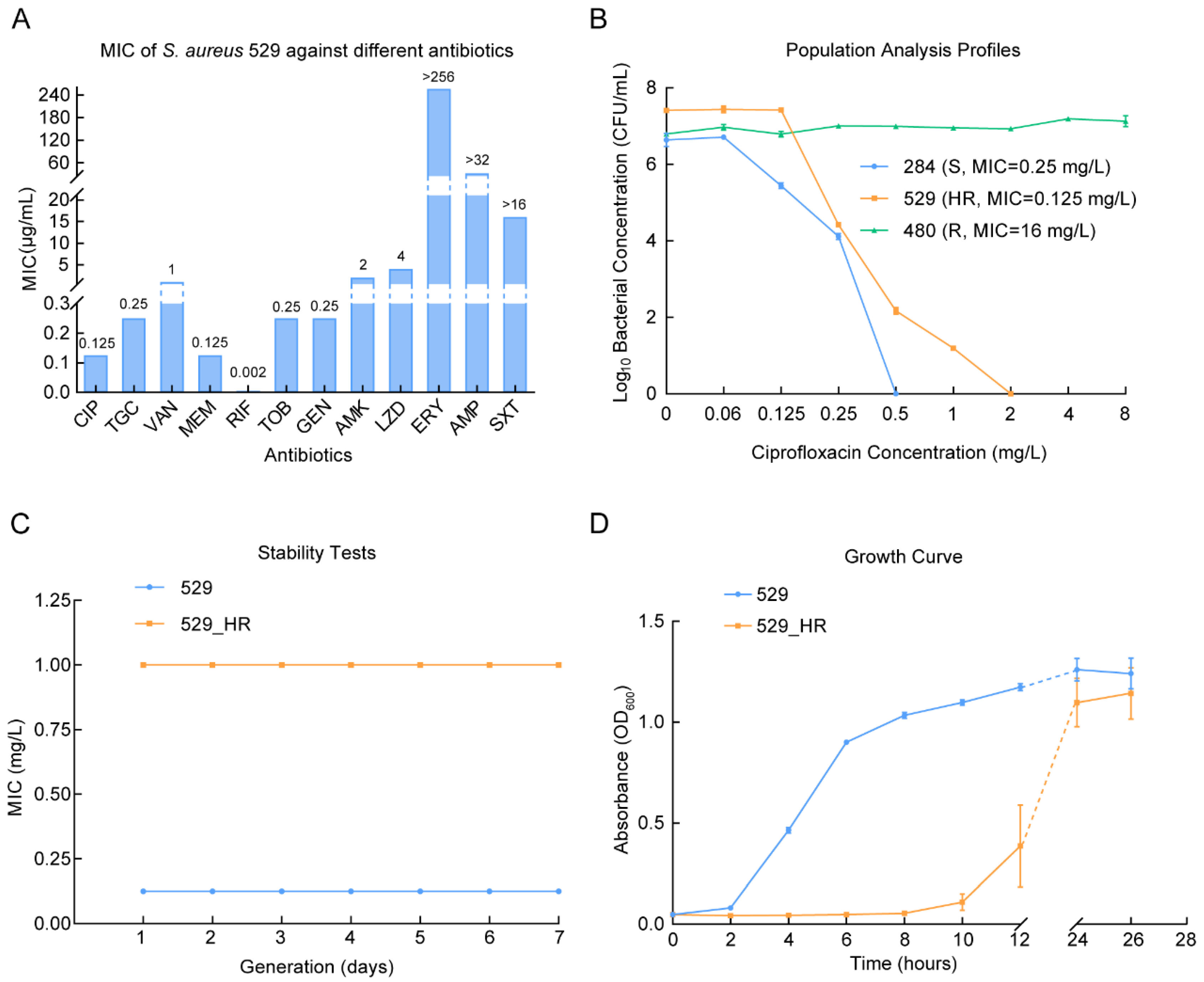
Antimicrobial susceptibility testing identified that S. aureus 529 was resistant to penicillin, macrolides, and sulfonamides. Conversely, it was not resistant to fluoroquinolones, aminoglycosides, beta-lactam, glycopeptides, glycylcyclines, oxazolidinones, and rifamycin antibiotics (Figure 1A). Interestingly, this strain was below the breakpoint of resistance to ciprofloxacin (MIC = 0.125 mg/L). However, PAP confirmed that this strain had a heteroresistant phenotype to ciprofloxacin, which could grow at 8-fold of the MIC ciprofloxacin concentration (1 mg/L) (Figure 1B). The 8-fold MIC ciprofloxacin heteroresistant subclone was isolated and named 529_HR. Furthermore, it was determined that the MBCs of 529 and 529_HR were 0.25 mg/L and 2 mg/L, respectively (Table S2). Both 529 and 529_HR established a stable MIC of ciprofloxacin after passaging on ciprofloxacin-free culture medium for 7 days (Figure 1C). By comparing the growth curves of 529 and 529_HR, it was discovered that the growth of 529_HR was slower than that of 529, but the proportion of bacteria that reached the plateau stage was comparable (Figure 1D). By calculating the specific growth rates of 529 and 529_HR during their respective logarithmic growth phases, it was determined that the specific growth rate of 529 was 0.205 and that of 529_HR was 0.139. This suggests the metabolic activities of these two strains derived from a single ancestor are different.

2.2. Genomic Characteristics Between 529 and 529_HR

To investigate the mechanism by which S. aureus strain 529 produces a heteroresistant phenotype, we conducted whole-genome sequencing of 529 and 529_HR. We obtained assembled genomes of 529 and 529_HR (N50529 = 174,991 and N50529_HR = 174,974), which annotated 2648 and 2647 genes, respectively. The total obtained number of annotated genes was comparable to the normal strain of S. aureus ATCC29213, which has 2586 genes. Each has a ~2.8 Mb genome containing two plasmids, pSJH101 and pMW2. The β-lactam resistance gene blaZ, macrolide resistance gene erm(A), aminoglycoside resistance gene spc, and rifamycin resistance gene rpoB exist in the genome. In addition, the genome also contains sdrM of the multi-resistance efflux pump MFS family and mepA of the MATE family (Table 1). Only a few variations were observed across the strains, including 11 nonsynonymous SNPs and a deletion (Table 2). Surprisingly, neither the SNPs nor the deletion genes were directly associated with resistance to ciprofloxacin. Of these nonsynonymous mutation sites, clfB is a member of the MSCRAMM (Microbial Surface Component Recognition Adhesive Matrix molecules) family of surface proteins, described as fibrinogen binding agglutination factors, asd is aspartate-semialdehyde dehydrogenase, which participates in the synthesis of cell wall components such as diaminopimelate, ebh is a hyperosmolarity resistance protein, which is an adhesion protein associated with cell walls, ssl3 is staphylococcal superantigen-like protein 3, which participates in immune regulation, and farB encodes the fatty acid resistant protein FarB. None of these mutations were associated with ciprofloxacin resistance, so genetic mutations do not contribute to understanding the mechanism of heteroresistance.

2.3. Transcriptional Regulation of the Heteroresistant Subclone 529_HR

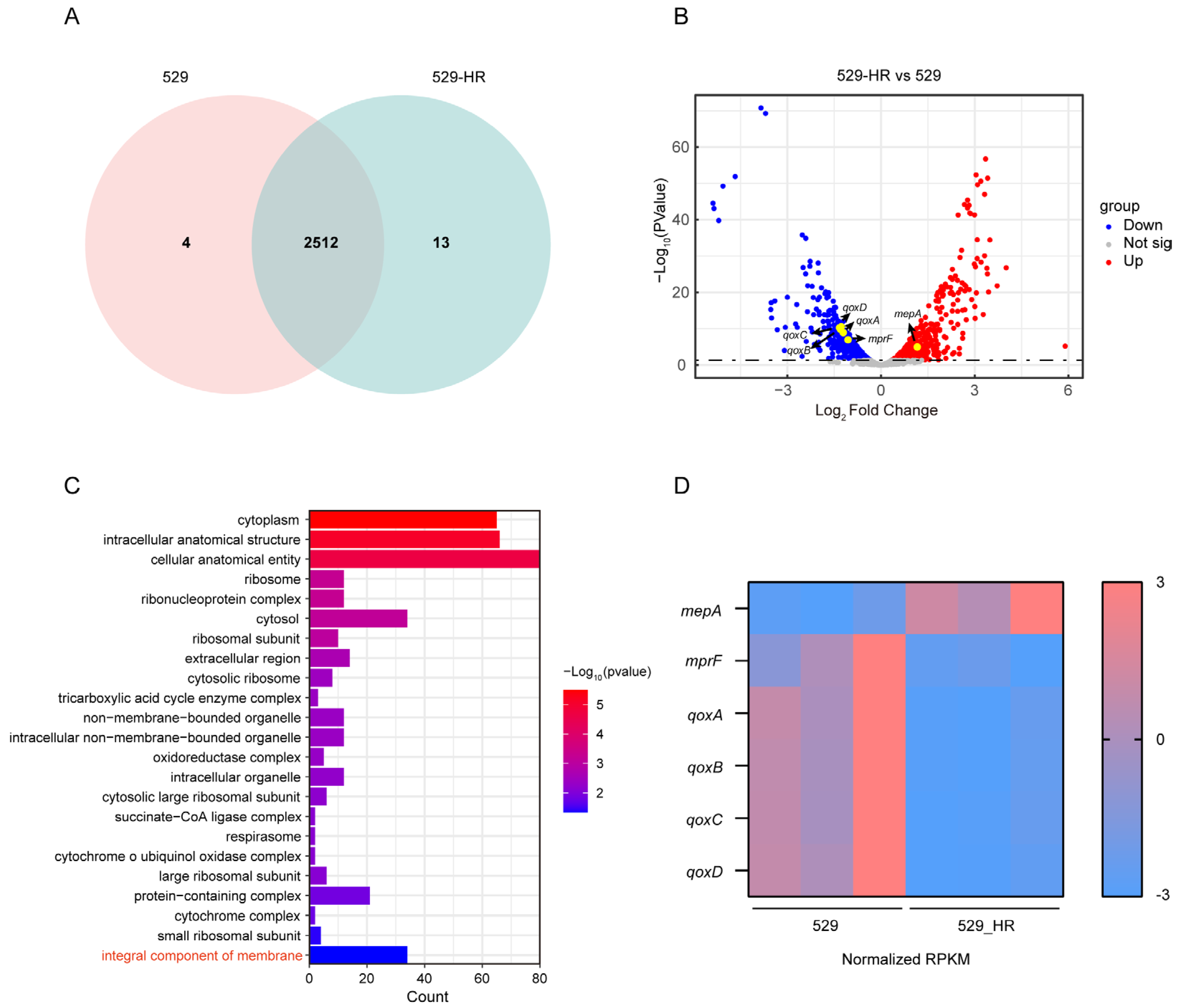
The transcriptional profiles of 529 and 529_HR were obtained via transcriptome sequencing. In total, 2516 genes were expressed in strain 529, while 2525 genes were expressed in strain 529_HR (Figure 2A). A total of 497 significant DEGs (differentially expressed genes) were identified between 529_HR and 529, including 301 up-regulated genes and 196 down-regulated genes (Figure 2B). GO cellular component analysis revealed that integral components of the membrane, intracellular anatomical structure, and cytochrome o ubiquinol oxidase complex were altered significantly (Figure 2C). Enriched biological process terms included metabolic processes such as organic acid catabolic process and amino acid catabolic process. Additionally, DEGs were enriched in molecular function terms including oxidoreduction-driven active transmembrane transporter activity, oxidoreductase activity, structural constituents of ribosomes, and so on (Figure S1). Notably, we observed down-regulation of genes associated with membrane potential (qoxA, qoxB, qoxC, qoxD, mprF) and up-regulation of the multidrug resistance efflux pump mepA among genes implicated in the integral component of the membrane term (Figure 2D and Figure S2). This suggests that the ciprofloxacin resistance of 529_HR may be associated with the up-regulated expression of mepA and altered membrane potential. Furthermore, the expression of mepA, qoxA, qoxB, and qoxC were validated by RT-qPCR (Figure S3). Moreover, the transcriptional levels of gyrA, gyrB, parC, and parE in strain 529_HR vs. strain 529 showed no statistically significant changes (log2 fold-change values: −0.47, −0.35, −0.70, −0.77, respectively; all magnitudes < 1). These results suggest that the expression of topoisomerase/gyrase-related genes is not substantially altered in strain 529-HR.

2.4. Limonene Changes the Heteroresistance Phenotype of 529

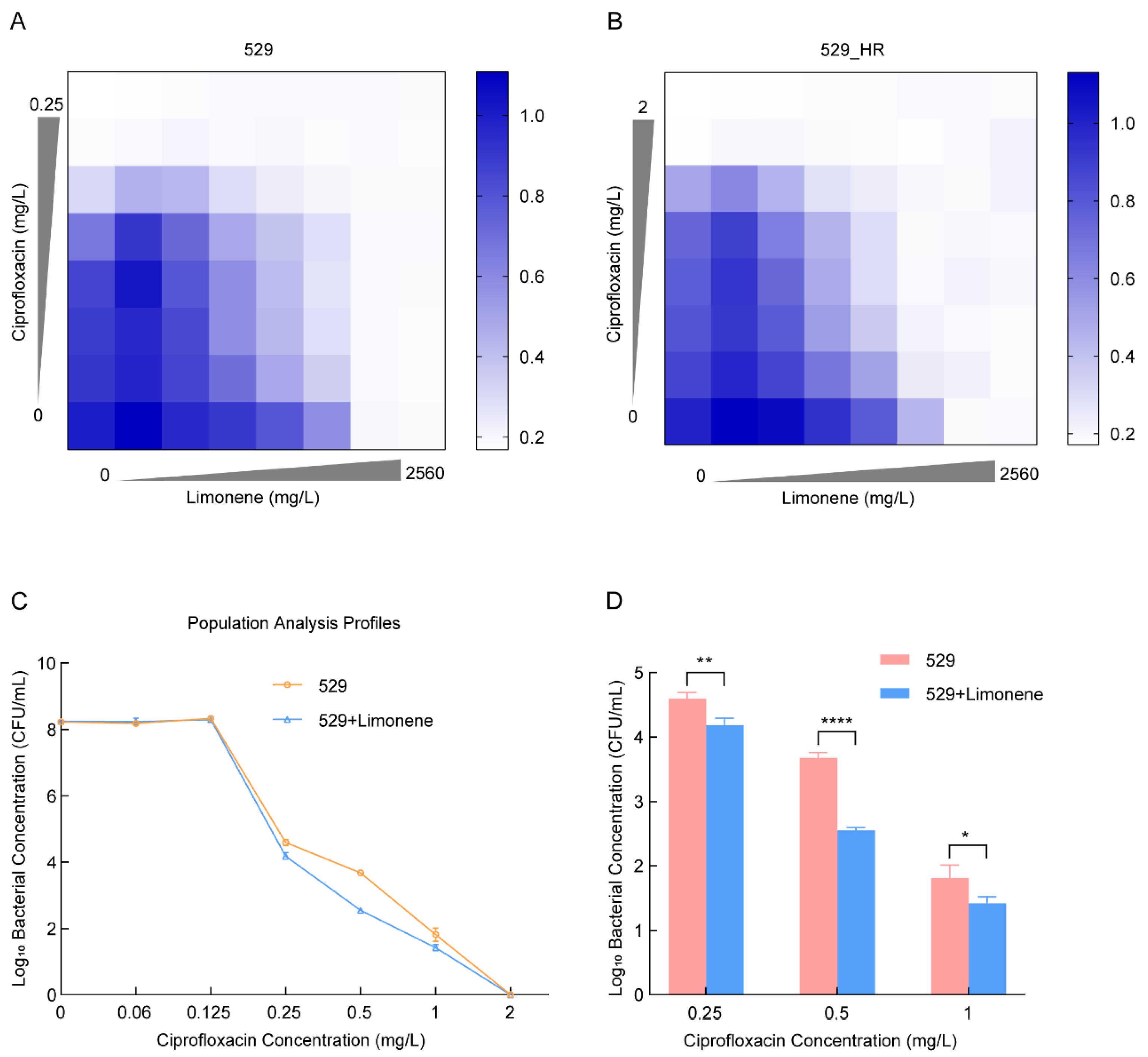
We utilized limonene to inhibit MepA efflux pump expression to confirm that mepA overexpression is a cause of S. aureus’s heteroresistance to ciprofloxacin. Combining ciprofloxacin and the efflux pump inhibitor limonene revealed 640 mg/L as the optimal concentration for inhibiting 529_HR (Figure 3A,B). At this concentration, 529_HR’s MIC to ciprofloxacin decreased from the original 1 mg/L to 0.06 mg/L (Figure 4D), restoring sensitivity to ciprofloxacin without affecting 529’s MIC. It was established that the up-regulation of mepA expression was one of the reasons for the increase in MIC in 529_HR. Thus, limonene may be a potential inhibitor of 529’s heteroresistance phenotype. PAP experiments showed limonene could indeed inhibit 529_HR’s resistant phenotype to some extent (Figure 3C). Treated with limonene and ciprofloxacin, 529’s surviving subpopulations significantly decreased, dropping 2.6, 13.5, and 2.6-fold under 0.25, 0.5, and 1 mg/L ciprofloxacin, respectively (Figure 3D). However, heteroresistance could not be completely suppressed (Figure 4D). These results indicate that up-regulated mepA is not the sole mechanism of heteroresistance.

2.5. Reduced Membrane Potential Led to Heteroresistance

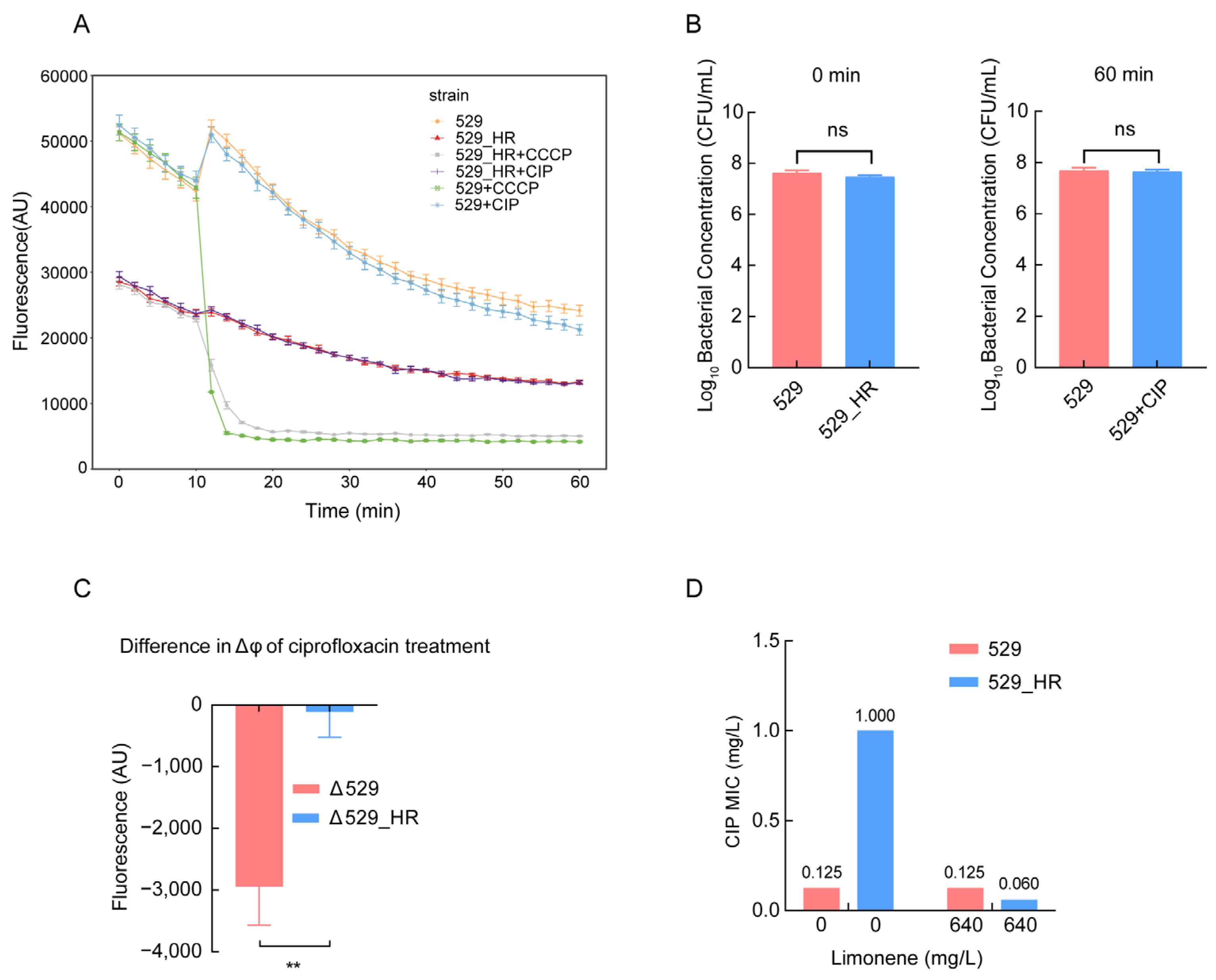
Analysis of the transcriptome results showed downregulation of membrane potential-related genes (qoxA, qoxB, qoxC, qoxD, mprF) in 529_HR (Figure 2D). These genes’ transcriptional regulation may reduce their membrane potential. Using a DiOC2(3) membrane potential fluorescence probe, the membrane potentials of 529 and 529_HR were measured. The latter was significantly lower, with a nine-fold reduction compared to the former (Figure 4C), consistent with transcriptome analysis findings. Moreover, 529’s membrane potential dropped dramatically with four-fold or higher MIC ciprofloxacin concentrations, but 529_HR’s remained relatively unchanged (Figure 4A and Figure S4). Plate counting showed no significant difference in viable bacteria between 529 and 529_HR at 0 min and no significant difference in 529 after 60 min ciprofloxacin treatment (Figure 4B). This suggests neither inconsistent bacterial quantity nor death caused the membrane potential change in 529 and 529_HR. The findings show 529_HR has a low potential, enabling it to withstand the ciprofloxacin-induced membrane potential decrease.

3. Discussion

Heteroresistance poses great challenges for evaluating antibiotic therapeutic effects on pathogenic bacteria, with clinically detected minimum inhibitory concentrations sometimes failing to inhibit pathogen growth, resulting in treatment failure and threatening modern medicine safety. Heteroresistance may also be an intermediate step towards developing resistance [27]. Fluoroquinolones like ciprofloxacin, an alternative to vancomycin, can treat MRSA infections. However, at present, there are few reports on the heteroresistance of S. aureus to fluoroquinolones. In this study, the mechanism of heteroresistance of S. aureus, belonging to the ESKAPE group, to ciprofloxacin was mainly explored.
Firstly, in this study, we identified a ciprofloxacin heteroresistant S. aureus strain 529, with both the parent strains and resistant subclone (529_HR) exhibiting stable inheritance, consistent with prior findings by Chuanzhen Zhang et al. [28]. Additionally, the growth rate of the resistant subclone was slower compared to the parent strain. Previous studies have linked low growth in Acinetobacter baumannii to colistin heteroresistance [29]. Similarly, vancomycin-heteroresistant S. aureus strains, including heteroresistant vancomycin-intermediate S. aureus (hVISA) and vancomycin-intermediate S. aureus (VISA) exhibited slower growth rates relative to fully sensitive strains [15]. Consequently, heteroresistance-related recurrent and persistent infections may stem from this phenomenon.
Many studies have shown that mutations, insertions, or deletions of genes associated with antibiotic resistance can lead to ciprofloxacin resistance, such as gyrA, parC, and rsxC mutations causing heteroresistance of Salmonella to ciprofloxacin [28]. Some reports have indicated that the high prevalence of heteroresistance in S. aureus is attributed to a large number of mutations occurring in core genes [30]. However, SNP analysis of the 529 and 529_HR genomes found none of their SNPs were associated with these genes. Our data exclude transcriptional dysregulation of DNA topoisomerases/gyrase as a determinant of ciprofloxacin heteroresistance in S. aureus 529_HR. Instead, mutations were found in genes such as fibrinogen binding agglutination factor, extracellular matrix binding protein, aspartate hemialdehyde dehydrogenase, and staphylococcal superantigen 3. These genes play different roles in different physiological activities of bacteria, such as encoding fatty acid resistance proteins and participating in immune regulation. It is worth noting that most of these mutated genes are related to bacterial cell wall formation, such as asd participating in the synthesis of cell wall components such as diaminopimelate, and ebh being an adhesion protein associated with cell walls. It has been reported that cell wall thickness in bacteria can lead to heteroresistance [31]. However, none of these genes were associated with cell wall thickening. Therefore, as none of these changes were linked to ciprofloxacin resistance nor cell wall thickening, DNA mutations could not explain the mechanism underlying S. aureus’s heteroresistance to ciprofloxacin.
Many studies have suggested that MATE family efflux pumps, including mepA, are associated with ciprofloxacin resistance [32,33]. We investigated the potential role of mepA in the mechanism of ciprofloxacin heteroresistance in S. aureus and found that mepA expression level in the ciprofloxacin heteroresistant subgroup was significantly higher than that in the parental strains. These results suggested that increased mepA expression might be a cause of heteroresistance.
Limonene is a secondary metabolite in phytochemistry, found in essential oil compositions of aromatic plants like Micrantha and tarragon [12]. It has been reported to inhibit the activity of S. aureus efflux pumps [34,35]. Freitas’ studies have shown that limonene can efficiently fit into the MepA structure and suppress MepA efflux systems, inhibiting bacterial resistance [12]. In this study, limonene decreased the MIC of heteroresistant subclones and increased sensitivity, while parent bacteria MIC remained unchanged. Indirectly, mepA overexpression may be the main cause of heteroresistance, suggesting its role in the potential drug-inhibiting ciprofloxacin heteroresistance phenotype. Additionally, many reports link changes in membrane potential to antibiotic resistance [22,26,36]. This study found that the membrane potential in 529_HR was lower than in 529, which was possibly caused by the downregulation of 529_HR membrane potential related genes (qoxA, qoxB, qoxC, qoxD, mprF). In S. aureus, mutations in these genes reduce membrane potential and gentamicin lethal activity [37]. Heteroresistant subclones may produce heteroresistance phenotypes by decreasing membrane potential and metabolic activity, suggesting that decreased membrane potential also contributes to heteroresistance.

4. Materials and Methods

4.1. Bacterial Strains

S. aureus GD18_SA_529, GD18_SA_284, and GD18_SA_480 (hereafter referred to as 529, 284, and 480, respectively) methicillin-resistant S. aureus isolates from the Guangdong Provincial Center for Disease Control and Prevention were used as test strains in this study. S. aureus ATCC29213 and E. coli ATCC25922 served as quality control strains and are listed in Table S1. Institutional Review Board or Institutional Animal Care and Use Committee approval was not required because our research did not involve human subjects or animal experimentation.

4.2. Antimicrobial Susceptibility Testing

The minimum inhibitory concentrations (MIC) were determined using the broth microdilution method per the European Committee on Antimicrobial Susceptibility Testing (EUCAST) guidelines for the following antibiotics: ciprofloxacin, tigecycline, vancomycin, meropenem, rifampicin, tobramycin, gentamycin, amikacin, linezolid, erythromycin, ampicillin, and trimethoprim/sulfamethoxazole [38,39,40]. The testing was performed in triplicate. S. aureus ATCC29213 and E. coli ATCC25922 were employed as quality control strains. The minimum bactericidal concentration (MBC) was determined using a modified broth microdilution method. Briefly, after obtaining the MIC, aliquots (100 μL) from wells showing no visible bacterial growth were subcultured onto fresh agar plates. After 24 h of incubation, MBC was recorded as the lowest concentration with ≤10 colony-forming units (CFUs).

4.3. Population Analysis Profiling

Population analysis profiling (PAP) was used to detect ciprofloxacin heteroresistance using standard published techniques [41]. Briefly, aliquots of 100 μL of 10-fold serially diluted overnight cultures of isolate were spread evenly on Mueller–Hinton (MH) agar plates containing ciprofloxacin at 0×, 1/2×, 1×, 2×, 4×, 8×, and 16× MIC. After overnight incubation at 37 °C, colonies were counted to calculate CFU/mL. The experiments were repeated three times.

4.4. Stability Evaluations and Growth Curve

The parental strain and the heteroresistant subclone (S. aureus GD18_SA_529_HR, referred to as 529_HR) were passaged on antibiotic-free MH agar plates every 24 h for 7 days, and the MICs were determined daily to monitor the stability of the phenotype [42]. Briefly, aliquots of bacterial suspension prepared from an overnight Luria–Bertani (LB) broth culture were 1000-fold diluted in 50 mL LB broth. Cultures were shaken at 180 rpm at 37 °C for 24 h, and the absorbance at 600 nm (OD600) was measured at 0, 2, 4, 6, 8, 10, 12, and 24 h. Each isolate was tested in triplicate [43].

4.5. Whole-Genome Sequence and Sequence Analysis

Whole-genome DNA from the 529 parental strain and heteroresistant subclone was extracted using a TIANamp Bacterial DNA Kit (TIANGEN, Beijing, China). WGS was performed on the Illumina NovaSeq 6000 platform (Illumina, San Diego, CA, USA) with 2 × 150 bp paired-end reads. SPAdes (v. 3.15.5) was used for genome assembly, and QUAST (v. 5.2) was used to assess the assemblies for quality metrics [44,45]. All assembled genomes were annotated using Prokka (v. 1.14.6) [46]. Resistance genes and plasmid replicon types were identified based on a comparison with the ResFinder and PlasmidFinder database [46,47]. Snippy (v. 4.6.0) (https://github.com/tseemann/snippy, accessed on 29 October 2023) was used to compute the genome alignment of strains, using S. aureus NCTC8325 as the reference sequence. The functions of differential mutant gene products were described by Gene Ontology (GO analysis; http://geneontology.org, accessed on 20 March 2023) [48].

4.6. RNA-seq and Transcriptomic Analysis

RNA from the 529 parental strain and heteroresistant subclone was extracted using OMEGA Bacteria RNA Kit (Omega Bio-tek, Norcross, GA, USA). RNA integrity was determined, and the mRNA library was constructed and sequenced on Illumina NovaSeq 6000 with 2 × 150 bp paired-end reads. Quality control and data filtering was performed using Fastp (v. 0.23.2) [49]. Reads were mapped to a S. aureus NCTC8325 mRNA reference sequence using FANSe2, and the parameters were L-160 E-11 I-1 S-14; genes with <10 mapped reads were considered unreliable and removed [50]. The mRNAs were normalized using reads per kilobase per million mapped reads (RPKM) [51]. Differential expression analysis used R (v. 4.3.1) with the edgeR (v. 3.42.4) package; cutoffs were Log2 fold change ≥ 1 or ≤−1 and p-value corrected for multiple guessing ≤ 0.05 [52]. Gene Ontology Enrichment Analysis of differentially expressed genes was conducted via Gene Ontology (GO analysis; http://geneontology.org, accessed on 20 March 2023) [48].

4.7. Determination of Limonene Inhibitory Effectiveness

The MICs of ciprofloxacin at different limonene concentrations were determined by checkerboard assay. In brief, each microplate well received 1 μL of working inoculum and 99 μL of MH broth medium. The wells were filled with 50 μL of ciprofloxacin 2-fold dilutions ranging in concentration from 1/32× MIC to 2× MIC and 50 μL of limonene 2-fold dilutions ranging in concentration from 0 mg/L to 2560 mg/L. After 12 h of incubation at 37 °C, OD600 was measured on the cultures [53]. An optimal concentration of limonene and varying amounts of diluted ciprofloxacin were added into MH agar plates. Aliquots of 100 μL of 10-fold serially diluted overnight cultures of isolates shaken at 180 rpm at 37 °C were equally disseminated on appropriate agar plates, and CFUs were calculated as previously described.

4.8. DiOC2(3) Membrane Potential Measurement

The exponential phase (OD600 of 0.4) of the parental strain and heteroresistant subclone were pelleted by centrifugation at 5000× g for 5 min to remove the growth medium. The cells were washed with 1× phosphate-buffered saline (PBS), then centrifuged, and resuspended. Then, 1 mM 3,3′-Diethyloxacarbocyannine iodide [DiOC2(3)] was added to the cells for a final concentration of 10 μM. A 96-well opaque microplate was filled with aliquots of 180 μL DiOC2(3)-loaded cells, then incubated for 20 min without light exposure. Cultures were quantified by measuring fluorescence (emission at 670 nm and excitation at 450 nm) every 2 min for 1 h. Ciprofloxacin was diluted and added to wells for 10 min at varying concentrations: 0.06 mg/L, 0.125 mg/L, 0.25 mg/L, 0.5 mg/L, 1 mg/L, and 2 mg/L. For disruption of ∆φ, carbonyl cyanide m-chlorophenyl hydrazine (CCCP) was used. Each isolate was tested three times, and the propensity of membrane potential to drop was validated by detecting the fluorescence of cells treated with 0.5 mg/L ciprofloxacin [54]. Aliquots of 100 μL of 10-fold serially diluted cultures of 0 h and 1 h DiOC2(3)-loaded cells were evenly spread on MH agar plates, and the CFU/mL was calculated as previously described.

4.9. RT-qPCR

The Vazyme HiScript III All-in-one RT SuperMix (Vazyme, Nanjing, China) was used to reverse transcribe the RNA sample, and quantitative reverse transcription-PCR (qRT-PCR) was performed in triplicate using gene-specific primers (Table S3), the Vazyme Taq Pro Universal SYBR qPCR Master Mix (Vazyme, Nanjing, China), and the Roche LightCycler 96 system (Roche, Basel, Switzerland). The initial denaturation for 30 s at 95 °C was followed by 40 cycles of denaturation for 10 s at 95 °C, annealing for 30 s at 60 °C, and extension for 30 s at 72 °C during PCR amplification [55].

5. Conclusions

To our knowledge, this is the first report of ciprofloxacin heteroresistance in S. aureus strains in China. Upregulation of mepA efflux pumps and a reduction in membrane potential may be the main heteroresistance mechanisms. Additionally, limonene might be a potentially effective agent to inhibit ciprofloxacin heteroresistance phenotypes.

Supplementary Materials

The following supporting information can be downloaded at: https://www.mdpi.com/article/10.3390/ijms26052372/s1.

Author Contributions

Conceptualization, X.L. (Xiaoping Liao) and X.L. (Xinlei Lian); Data curation, M.L. and Q.J.; Formal analysis, M.L., Q.J., X.L. (Xiaoling Long), M.J., J.W., Z.W., Y.R. and X.Y.; Investigation, M.L., Q.J. and X.Y.; Funding acquisition, J.S., Y.L. and X.L. (Xinlei Lian); Methodology, M.L., Q.J. and X.L. (Xinlei Lian); Software, M.L. and Q.J.; Visualization, M.L. and Q.J.; Writing—original draft, M.L. and Q.J.; Writing—review & editing, M.L., Q.J., X.L. (Xiaoling Long), M.J., J.W., Z.W., Y.R., X.Y., R.Z., H.R., J.S., Y.L., X.L. (Xiaoping Liao) and X.L. (Xinlei Lian). All authors have read and agreed to the published version of the manuscript.

Funding

This work was supported by the National Natural Science Foundation of China (Grant No. 32002336), Guangzhou Basic and Applied Basic Research Foundation (2023A04J0753), the Foundation for Innovative Research Groups of the National Natural Science Foundation of China (Grant No. 32121004), Local Innovative and Research Teams Project of Guangdong Pearl River Talents Program (2019BT02N054), National Key Research and Development Program of China (2022YFD1800400), Guangdong Provincial Key Research and Development Program (2023B0202150001), Laboratory of Lingnan Modern Agriculture Project (NT2021006), Double First-class Discipline Promotion Project (2023B10564003), Guangdong Major Project of Basic and Applied Basic Research (2020B0301030007), and the 111 Project (D20008).

Institutional Review Board Statement

Not applicable.

Informed Consent Statement

Not applicable.

Data Availability Statement

The datasets presented in this study can be found in online repositories. The raw data of 529 and 529_HR from WGS and RNA-seq are available in the NCBI Sequence Read Archive repository under BioProject ID PRJNA1051287 (accession numbers: SRR27185055 and SRR27185054) and PRJNA1051527 (accession numbers: SRR27185756 to SRR27185761).

Acknowledgments

We thank the Guangdong Provincial Center for Disease Control and Prevention for providing the Staphylococcus aureus used in this study.

Conflicts of Interest

The authors declare no conflicts of interest.

References

  1. Wang, M.; Buist, G.; van Dijl, J.M. Staphylococcus aureus cell wall maintenance—The multifaceted roles of peptidoglycan hydrolases in bacterial growth, fitness, and virulence. FEMS Microbiol. Rev. 2022, 46, fuac025. [Google Scholar] [CrossRef] [PubMed]
  2. Tong, S.Y.C.; Davis, J.S.; Eichenberger, E.; Holland, T.L.; Fowler, V.G. Staphylococcus aureus infections: Epidemiology, pathophysiology, clinical manifestations, and management. Clin. Microbiol. Rev. 2015, 28, 603–661. [Google Scholar] [CrossRef] [PubMed]
  3. Miller, W.R.; Arias, C.A. ESKAPE pathogens: Antimicrobial resistance, epidemiology, clinical impact and therapeutics. Nat. Rev. Microbiol. 2024, 22, 598–616. [Google Scholar] [CrossRef] [PubMed]
  4. Liu, W.-T.; Chen, E.-Z.; Yang, L.; Peng, C.; Wang, Q.; Xu, Z.; Chen, D.-Q. Emerging resistance mechanisms for 4 types of common anti-MRSA antibiotics in Staphylococcus aureus: A comprehensive review. Microb. Pathog. 2021, 156, 104915. [Google Scholar] [CrossRef]
  5. Ferreira, M.; Bessa, L.J.; Sousa, C.F.; Eaton, P.; Bongiorno, D.; Stefani, S.; Campanile, F.; Gameiro, P. Fluoroquinolone metalloantibiotics: A promising approach against methicillin-resistant Staphylococcus aureus. Int. J. Environ. Res. Public Health 2020, 17, 3127. [Google Scholar] [CrossRef]
  6. Mirzaie, A.; Peirovi, N.; Akbarzadeh, I.; Moghtaderi, M.; Heidari, F.; Yeganeh, F.E.; Noorbazargan, H.; Mirzazadeh, S.; Bakhtiari, R. Preparation and optimization of ciprofloxacin encapsulated niosomes: A new approach for enhanced antibacterial activity, biofilm inhibition and reduced antibiotic resistance in ciprofloxacin-resistant methicillin-resistance Staphylococcus aureus. Bioorg. Chem. 2020, 103, 104231. [Google Scholar] [CrossRef]
  7. Zhang, G.-F.; Liu, X.; Zhang, S.; Pan, B.; Liu, M.-L. Ciprofloxacin derivatives and their antibacterial activities. Eur. J. Med. Chem. 2018, 146, 599–612. [Google Scholar] [CrossRef]
  8. Mayumi, T.; Tong, W.; Yoshikuni, O.; Yoko, U.; Kenichi, S. Mechanism of quinolone resistance in Staphylococcus aureus. J. Infect. Chemother. 2000, 6, 131–139. [Google Scholar]
  9. Kwak, Y.G.; Truong-Bolduc, Q.C.; Bin Kim, H.; Song, K.H.; Kim, E.S.; Hooper, D.C. Association of norB overexpression and fluoroquinolone resistance in clinical isolates of Staphylococcus aureus from Korea. J. Antimicrob. Chemother. 2013, 68, 2766–2772. [Google Scholar] [CrossRef]
  10. McAleese, F.; Petersen, P.; Ruzin, A.; Dunman, P.M.; Murphy, E.; Projan, S.J.; Bradford, P.A. A novel MATE family efflux pump contributes to the reduced susceptibility of laboratory-derived Staphylococcus aureus mutants to tigecycline. Antimicrob. Agents Chemother. 2005, 49, 1865–1871. [Google Scholar] [CrossRef]
  11. Phillips-Jones, M.K.; Harding, S.E. Antimicrobial resistance (AMR) nanomachines-mechanisms for fluoroquinolone and glycopeptide recognition, efflux and/or deactivation. Biophys. Rev. 2018, 10, 347–362. [Google Scholar] [CrossRef] [PubMed]
  12. Freitas, P.R.; de Araújo, A.C.J.; dos Santos Barbosa, C.R.; Muniz, D.F.; de Almeida, R.S.; de Menezes, I.R.A.; da Costa, J.G.M.; Rodrigues, F.F.G.; Rocha, J.E.; Pereira-Junior, F.N.; et al. Inhibition of the MepA efflux pump by limonene demonstrated by in vitro and in silico methods. Folia Microbiol. 2021, 67, 15–20. [Google Scholar] [CrossRef] [PubMed]
  13. Andersson, D.I.; Nicoloff, H.; Hjort, K. Mechanisms and clinical relevance of bacterial heteroresistance. Nat. Rev. Microbiol. 2019, 17, 479–496. [Google Scholar] [CrossRef] [PubMed]
  14. Ho, C.M.; Hsueh, P.R.; Liu, C.Y.; Lee, S.Y.; Chiueh, T.S.; Shyr, J.M.; Tsao, S.M.; Chuang, Y.C.; Yan, J.J.; Wang, L.S.; et al. Prevalence and accessory gene regulator (agr) analysis of vancomycin-intermediate Staphylococcus aureus among methicillin-resistant isolates in Taiwan—SMART program, 2003. Eur. J. Clin. Microbiol. Infect. Dis. 2010, 29, 383–389. [Google Scholar] [CrossRef]
  15. Huang, S.-H.; Chen, Y.-C.; Chuang, Y.-C.; Chiu, S.-K.; Fung, C.-P.; Lu, P.-L.; Wang, L.-S.; Wu, T.-L.; Wang, J.-T. Prevalence of vancomycin-intermediate Staphylococcus aureus (VISA) and heterogeneous VISA among methicillin-resistant S. aureus with high vancomycin minimal inhibitory concentrations in Taiwan: A multicenter surveillance study, 2012–2013. J. Microbiol. Immunol. Infect. 2016, 49, 701–707. [Google Scholar] [CrossRef]
  16. Yu, Y.; Chen, Y.; Zhao, F.; Wang, H.; Sun, L.; Wei, X.; Jiang, S.; Ji, S. In-host evolution of daptomycin resistance and heteroresistance in methicillin-resistant Staphylococcus aureus strains from three endocarditis patients. J. Infect. Dis. 2020, 221 (Suppl. 2), S243–S252. [Google Scholar] [CrossRef]
  17. Bai, B.; Lin, Z.; Pu, Z.; Xu, G.; Zhang, F.; Chen, Z.; Sun, X.; Zheng, J.; Li, P.; Deng, Q.; et al. In vitro activity and heteroresistance of omadacycline against clinical Staphylococcus aureus isolates from China reveal the impact of omadacycline susceptibility by branched-chain amino acid transport system II carrier protein, Na/Pi cotransporter family protein, and fibronectin-binding protein. Front. Microbiol. 2019, 10, 2546. [Google Scholar] [CrossRef]
  18. Wang, Z.; Lin, Z.; Bai, B.; Xu, G.; Li, P.; Yu, Z.; Deng, Q.; Shang, Y.; Zheng, J. Eravacycline susceptibility was impacted by genetic mutation of 30S ribosome subunits, and branched-chain amino acid transport system II carrier protein, Na/Pi cotransporter family protein in Staphylococcus aureus. BMC Microbiol. 2020, 20, 189. [Google Scholar] [CrossRef]
  19. Roch, M.; Sierra, R.; Andrey, D.O. Antibiotic heteroresistance in ESKAPE pathogens, from bench to bedside. Clin. Microbiol. Infect. 2023, 29, 320–325. [Google Scholar] [CrossRef]
  20. Wang, T.; Shen, P.; He, Y.; Zhang, Y.; Liu, J. Spatial transcriptome uncovers rich coordination of metabolism in E. coli K12 biofilm. Nat. Chem. Biol. 2023, 19, 940–950. [Google Scholar] [CrossRef]
  21. Stewart, P.S.; Franklin, M.J. Physiological heterogeneity in biofilms. Nat. Rev. Microbiol. 2008, 6, 199–210. [Google Scholar] [CrossRef] [PubMed]
  22. Benarroch, J.M.; Asally, M. The microbiologist’s guide to membrane potential dynamics. Trends Microbiol. 2020, 28, 304–314. [Google Scholar] [CrossRef] [PubMed]
  23. Jin, X.; Zhang, X.; Ding, X.; Tian, T.; Tseng, C.K.; Luo, X.; Chen, X.; Lo, C.J.; Leake, M.C.; Bai, F. Sensitive bacterial V(m) sensors revealed the excitability of bacterial V(m) and its role in antibiotic tolerance. Proc. Natl. Acad. Sci. USA 2023, 120, e2208348120. [Google Scholar] [CrossRef] [PubMed]
  24. Lee, D.-y.D.; Galera-Laporta, L.; Bialecka-Fornal, M.; Moon, E.C.; Shen, Z.; Briggs, S.P.; Garcia-Ojalvo, J.; Süel, G.M. Magnesium flux modulates ribosomes to increase bacterial survival. Cell 2019, 177, 352–360.e13. [Google Scholar] [CrossRef]
  25. Verstraeten, N.; Knapen, W.J.; Kint, C.I.; Liebens, V.; Van den Bergh, B.; Dewachter, L.; Michiels, J.E.; Fu, Q.; David, C.C.; Fierro, A.C.; et al. Obg and membrane depolarization are part of a microbial bet-hedging strategy that leads to antibiotic tolerance. Mol. Cell 2015, 59, 9–21. [Google Scholar] [CrossRef]
  26. Damper, P.D.; Epstein, W. Role of the membrane potential in bacterial resistance to aminoglycoside antibiotics. Antimicrob. Agents Chemother. 1981, 20, 803–808. [Google Scholar] [CrossRef]
  27. Band, V.I.; Weiss, D.S. Heteroresistance to beta-lactam antibiotics may often be a stage in the progression to antibiotic resistance. PLoS Biol. 2021, 19, e3001346. [Google Scholar] [CrossRef]
  28. Zhang, C.-Z.; Zhang, Y.; Ding, X.-M.; Lin, X.-L.; Lian, X.-L.; Trampari, E.; Thomson, N.M.; Ding, H.-Z.; Webber, M.A.; Jiang, H.-X. Emergence of ciprofloxacin heteroresistance in foodborne Salmonella enterica serovar Agona. J. Antimicrob. Chemother. 2020, 75, 2773–2779. [Google Scholar] [CrossRef]
  29. Rodriguez, C.H.; Traglia, G.; Bastias, N.; Pandolfo, C.; Bruni, G.; Nastro, M.; Barrios, R.; Bavastro, E.M.; Rey, M.C.; Marques, I.A.; et al. Discrepancies in susceptibility testing to colistin in Acinetobacter baumannii: The influence of slow growth and heteroresistance. Int. J. Antimicrob. Agents 2019, 54, 587–591. [Google Scholar] [CrossRef]
  30. Heidarian, S.; Guliaev, A.; Nicoloff, H.; Hjort, K.; Andersson, D.I. High prevalence of heteroresistance in Staphylococcus aureus is caused by a multitude of mutations in core genes. PLoS Biol. 2024, 22, e3002457. [Google Scholar] [CrossRef]
  31. Okado, J.B.; Avaca-Crusca, J.S.; Oliveira, A.L.; Dabul, A.N.G.; Camargo, I.L.B.d.C. Daptomycin and vancomycin heteroresistance revealed among CC5-SCCmecII MRSA clone and in vitro evaluation of treatment alternatives. J. Glob. Antimicrob. Resist. 2018, 14, 209–216. [Google Scholar] [CrossRef] [PubMed]
  32. Truong-Bolduc, Q.C.; Wang, Y.; Reedy, J.L.; Vyas, J.M.; Hooper, D.C. Staphylococcus aureus efflux pumps and tolerance to ciprofloxacin and chlorhexidine following induction by mupirocin. Antimicrob. Agents Chemother. 2022, 66, e0184521. [Google Scholar] [CrossRef] [PubMed]
  33. Hassanzadeh, S.; Mashhadi, R.; Yousefi, M.; Askari, E.; Saniei, M.; Pourmand, M.R. Frequency of efflux pump genes mediating ciprofloxacin and antiseptic resistance in methicillin-resistant Staphylococcus aureus isolates. Microb. Pathog. 2017, 111, 71–74. [Google Scholar] [CrossRef] [PubMed]
  34. de Araújo, A.C.J.; Freitas, P.R.; Dos Santos Barbosa, C.R.; Muniz, D.F.; Ribeiro-Filho, J.; Tintino, S.R.; Júnior, J.P.S.; Filho, J.M.B.; de Sousa, G.R.; Coutinho, H.D.M. Modulation of drug resistance by limonene: Inhibition of efflux pumps in Staphylococcus aureus strains RN-4220 and IS-58. Curr. Drug Metab. 2021, 22, 110–113. [Google Scholar] [CrossRef]
  35. Justino de Araújo, A.C.; Freitas, P.R.; Rodrigues Dos Santos Barbosa, C.; Muniz, D.F.; Rocha, J.E.; Albuquerque da Silva, A.C.; Datiane de Morais Oliveira-Tintino, C.; Ribeiro-Filho, J.; Everson da Silva, L.; Confortin, C.; et al. GC-MS-FID characterization and antibacterial activity of the Mikania cordifolia essential oil and limonene against MDR strains. Food Chem. Toxicol. 2020, 136, 111023. [Google Scholar] [CrossRef]
  36. Yang, B.; Tong, Z.; Shi, J.; Wang, Z.; Liu, Y. Bacterial proton motive force as an unprecedented target to control antimicrobial resistance. Med. Res. Rev. 2023, 43, 1068–1090. [Google Scholar] [CrossRef]
  37. Vestergaard, M.; Nøhr-Meldgaard, K.; Ingmer, H. Multiple pathways towards reduced membrane potential and concomitant reduction in aminoglycoside susceptibility in Staphylococcus aureus. Int. J. Antimicrob. Agents 2018, 51, 132–135. [Google Scholar] [CrossRef]
  38. The European Committee on Antimicrobial Susceptibility Testing (EUCAST). Breakpoint Tables for Interpretation of MICs and Zone Diameters, Version 9.0; 2019. EUCAST. Available online: https://www.eucast.org/fileadmin/src/media/PDFs/EUCAST_files/Breakpoint_tables/v_9.0_Breakpoint_Tables.pdf (accessed on 19 December 2023).
  39. The European Committee on Antimicrobial Susceptibility Testing (EUCAST). EUCAST Reading Guide for Broth Microdilution; 2021. EUCAST. Available online: https://www.eucast.org/fileadmin/src/media/PDFs/EUCAST_files/MIC_testing/Reading_guide_BMD_v_3.0_2021.pdf (accessed on 19 December 2023).
  40. The European Committee on Antimicrobial Susceptibility Testing (EUCAST). Routine and Extended Internal Quality Control for MIC Determination and Disk Diffusion as Recommended by EUCAST, Version 13.2; 2023. EUCAST. Available online: https://www.eucast.org/fileadmin/src/media/PDFs/EUCAST_files/QC/v_13.2_EUCAST_QC_tables_routine_and_extended_QC.pdf (accessed on 19 December 2023).
  41. Dao, T.H.; Alsallaq, R.; Parsons, J.B.; Ferrolino, J.; Hayden, R.T.; Rubnitz, J.E.; Rafiqullah, I.M.; Robinson, D.A.; Margolis, E.B.; Rosch, J.W.; et al. Vancomycin heteroresistance and clinical outcomes in bloodstream infections caused by coagulase-negative Staphylococci. Antimicrob. Agents Chemother. 2020, 64, e00944-20. [Google Scholar] [CrossRef]
  42. Tian, Y.; Zhang, Q.; Wen, L.; Chen, J.; Van Tyne, D. Combined effect of polymyxin B and tigecycline to overcome heteroresistance in carbapenem-resistant Klebsiella pneumoniae. Microbiol. Spectr. 2021, 9, e00152-21. [Google Scholar] [CrossRef]
  43. Fan, Q.; Yan, C.; Shi, C.; Xu, Y.; Ma, Y.; Zhang, C.; Peng, X.; Xia, X. Inhibitory Effect of Coenzyme Q0 on the Growth of Staphylococcus aureus. Foodborne Pathog. Dis. 2019, 16, 317–324. [Google Scholar] [CrossRef]
  44. Bankevich, A.; Nurk, S.; Antipov, D.; Gurevich, A.A.; Dvorkin, M.; Kulikov, A.S.; Lesin, V.M.; Nikolenko, S.I.; Pham, S.; Prjibelski, A.D.; et al. SPAdes: A new genome assembly algorithm and its applications to single-cell sequencing. J. Comput. Biol. 2012, 19, 455–477. [Google Scholar] [CrossRef] [PubMed]
  45. Gurevich, A.; Saveliev, V.; Vyahhi, N.; Tesler, G. QUAST: Quality assessment tool for genome assemblies. Bioinformatics 2013, 29, 1072–1075. [Google Scholar] [CrossRef] [PubMed]
  46. Seemann, T. Prokka: Rapid prokaryotic genome annotation. Bioinformatics 2014, 30, 2068–2069. [Google Scholar] [CrossRef] [PubMed]
  47. Founou, L.L.; Founou, R.C.; Allam, M.; Ismail, A.; Essack, S.Y. Genome analysis of ESBL-producing Escherichia coli isolated from pigs. Pathogens 2022, 11, 776. [Google Scholar] [CrossRef]
  48. Ashburner, M.; Ball, C.A.; Blake, J.A.; Botstein, D.; Butler, H.; Cherry, J.M.; Davis, A.P.; Dolinski, K.; Dwight, S.S.; Eppig, J.T.; et al. Gene Ontology: Tool for the unification of biology. Nat. Genet. 2000, 25, 25–29. [Google Scholar] [CrossRef]
  49. Chen, S.; Zhou, Y.; Chen, Y.; Gu, J. fastp: An ultra-fast all-in-one FASTQ preprocessor. Bioinformatics 2018, 34, i884–i890. [Google Scholar] [CrossRef]
  50. Zhang, Z.; Xiao, C.-L.; Mai, Z.-B.; Lian, X.-L.; Zhong, J.-Y.; Jin, J.-j.; He, Q.-Y.; Zhang, G. FANSe2: A robust and cost-efficient alignment tool for quantitative next-generation sequencing applications. PLoS ONE 2014, 9, e94250. [Google Scholar] [CrossRef]
  51. Mortazavi, A.; Williams, B.A.; McCue, K.; Schaeffer, L.; Wold, B. Mapping and quantifying mammalian transcriptomes by RNA-Seq. Nat. Methods 2008, 5, 621–628. [Google Scholar] [CrossRef]
  52. Robinson, M.D.; McCarthy, D.J.; Smyth, G.K. edgeR: A Bioconductor package for differential expression analysis of digital gene expression data. Bioinformatics 2010, 26, 139–140. [Google Scholar] [CrossRef]
  53. Bellio, P.; Fagnani, L.; Nazzicone, L.; Celenza, G. New and simplified method for drug combination studies by checkerboard assay. MethodsX 2021, 8, 101543. [Google Scholar] [CrossRef]
  54. Kirchhoff, C.; Cypionka, H. Boosted membrane potential as bioenergetic response to anoxia in Dinoroseobacter shibae. Front. Microbiol. 2017, 8, 695. [Google Scholar] [CrossRef]
  55. Gazzola, S.; Cocconcelli, P.S. Vancomycin heteroresistance and biofilm formation in Staphylococcus epidermidis from food. Microbiology 2008, 154, 3224–3231. [Google Scholar] [CrossRef]
Figure 1. Verification of ciprofloxacin heteroresistance in S. aureus 529. (A) MIC of S. aureus 529 against different antibiotics. Abbreviations: CIP, ciprofloxacin; TGC, tigecycline; VAN, vancomycin; MEM, meropenem; RIF, rifampicin; TOB, tobramycin; GEN, gentamycin; AMK, amikacin; LZD, linezolid; ERY, erythromycin; AMP, ampicillin; SXT, trimethoprim/sulfamethoxazole. (B) Population analysis assays of the S. aureus 529, 284, 480 strains using ciprofloxacin. (C) Stability tests of the parental strain S. aureus 529 and heteroresistant subclone S. aureus 529_HR. (D) Growth curves of S. aureus 529 and 529_HR.
Ijms 26 02372 g001
Figure 2. Transcriptional regulation of S. aureus 529 and 529_HR. (A) Venn diagram of expressed genes in 529 and 529_HR. (B) Volcano plot of 529_HR vs. 529. The blue dot on the volcano plot indicates down-regulated genes (Log2 Fold change ≤ 0 and FDR < 0.05). The red dot on the volcano plot indicates up-regulated genes (Log2 Fold change ≥ 0 and FDR < 0.05). The genes labeled by the yellow plot are genes linked to membrane potential. (C) The chromosomal DEGs GO enrichment analysis in cellular components of 529_HR vs. 529. (D) Regulation of genes linked to membrane potential (mepA, mprF, qoxA, qoxB, qoxC, qoxD).
Ijms 26 02372 g002
Figure 3. Limonene combined with ciprofloxacin inhibited the heteroresistance phenotype of 529_HR. (A,B) Limonene combined with ciprofloxacin inhibited 529 and 529_HR at different concentrations. (C) Population analysis assays of S. aureus 529 with and without limonene using ciprofloxacin. (D) The amount of surviving 529 bacteria with and without limonene at 0.25, 0.5, and 1 mg/L ciprofloxacin. Note: * p < 0.05, ** p < 0.01, **** p < 0.0001.
Ijms 26 02372 g003
Figure 4. Changes in membrane potential lead to changes in the sensitivity of 529_HR to ciprofloxacin. (A) Strains 529 and 529_HR were continuously monitored for fluorescence for 1 h with and without ciprofloxacin and CCCP. Abbreviation: CCCP, carbonyl cyanide m-chlorophenyl hydrazine. (B) The amount of bacteria in 529 and 529_HR at 0 min and after 60 min of ciprofloxacin treatment. (C) ΔΨ values of 529 and 529_HR after ciprofloxacin treatment. (D) MIC values of 529 and 529_HR for ciprofloxacin at 0 mg/L and 640 mg/L limonene concentrations. Note: “ns” p ≥ 0.05, ** p < 0.01.
Ijms 26 02372 g004
Table 1. Resistance genes and efflux pumps of S. aureus GD18_SA_529.
TypeGeneSubstrate
Resistance genesBeta-lactamsblaZMethicillin
Macrolideserm(A)Erythromycin
AminoglycosidesspcSpectinomycin
RifamycinsrpoBRifampicin
Multidrug resistant efflux pumpsMFS familynorA, norB, norGCiprofloxacin
sdrMNorfloxacin
Acriflavine
Ethidium bromide
MATE familymepACiprofloxacin
Norfloxacin
Tigecycline
Table 2. SNPs between S. aureus GD18_SA_529 and GD18_SA_529_HR.
SNP PositionGeneSNPs
188,068ggtp.Val549Ser
294,112essG_6p.Leu7Gln
390,715ssl3p.Ile157Thr
390,730p.ThrAspMetThr162LysGluIleAsn
798,468sspp.Gln200_Ser201dup
1,043,951isdBp.Glu71_Thr72delinsValAlaLysProValAla
1,338,822asdp.Asp242Glu
1,378,171ebhp.Asp8843Gly
1,684,208lysP_2p.Glu375Gln
1,684,220p.Lys371Glu
2,246,711farB_1p.Thr253Ser
2,723,613clfBp.Asp663Glu
Disclaimer/Publisher’s Note: The statements, opinions and data contained in all publications are solely those of the individual author(s) and contributor(s) and not of MDPI and/or the editor(s). MDPI and/or the editor(s) disclaim responsibility for any injury to people or property resulting from any ideas, methods, instructions or products referred to in the content.