Article

Comparison of Staphylococcus pettenkoferi Isolated from Human Clinical Cases and Cat Carriers Regarding Antibiotic Susceptibility and Biofilm Production

by
Karolina Bierowiec
1,*,
Ashley Delmar
2,*,
Magdalena Karwańska
1,
Magdalena Siedlecka
1,
Aleksandra Kumala-Ćwikła
1,
Marta Książczyk
3 and
Katarzyna Kapczyńska
4
1
Department of Epizootiology and Clinic of Birds and Exotic Animals, Division of Infectious Diseases and Veterinary Administration, The Faculty of Veterinary Medicine in Wrocław, Wrocław University of Environmental and Life Sciences, 50-366 Wrocław, Poland
2
EZA Student Science Club, Department of Epizootiology and Clinic of Birds and Exotic Animals, Division of Infectious Diseases and Veterinary Administration, The Faculty of Veterinary Medicine in Wrocław, Wrocław University of Environmental and Life Sciences, 50-366 Wrocław, Poland
3
Department of Microbiology, Faculty of Biological Sciences, University of Wrocław, 51-148 Wrocław, Poland
4
Laboratory of Medical Microbiology, Hirszfeld Institute of Immunology and Experimental Therapy, Polish Academy of Sciences, 53-114 Wrocław, Poland
*
Authors to whom correspondence should be addressed.
Int. J. Mol. Sci. 2025, 26(5), 1948; https://doi.org/10.3390/ijms26051948
Submission received: 23 December 2024 / Revised: 14 February 2025 / Accepted: 18 February 2025 / Published: 24 February 2025
(This article belongs to the Special Issue Molecular Aspects of Bacterial Infection)

Abstract

:
Staphylococcus pettenkoferi (S. pettenkoferi) is a rare opportunistic bacterium not commonly found in healthy individuals or animals. S. pettenkoferi has increasing clinical significance in both veterinary and human medicine due to its multidrug resistance and biofilm-forming ability. This study analyzed 12 isolates of S. pettenkoferi collected from humans and cats and identified them using matrix-assisted laser desorption/ionization time-of-flight mass spectrometry (MALDI-TOF MS) and 16S rRNA and partial rpoB gene sequencing. All of the S. pettenkoferi were phenotypically resistant to penicillin, and almost all (except one human strain) were resistant to methicillin. Antibiotic susceptibility testing revealed a high prevalence of multidrug resistance in all human strains with frequent resistance to β-lactams, macrolides, and tetracyclines. A comparative analysis of human and feline isolates indicated the presence of shared resistance genes such as blaZ, mecA, and ermA. Biofilm production varied across isolates, with more potent biofilm formation abilities observed at elevated temperatures (39 °C) and time (48 h). These findings underscore the potential zoonotic risks of S. pettenkoferi and its role in managing multidrug-resistant infections.

1. Introduction

Staphylococcus pettenkoferi (S. pettenkoferi) is a rare staphylococcal species that has traditionally been described as a cause of bloodstream infections in humans [1,2,3,4]. Similar to other coagulase-negative staphylococci (CNS), S. pettenkoferi is a ubiquitous colonizer of skin and mucosal surfaces and plays a role as an opportunistic pathogen in immunocompromised patients [1,4,5]. S. pettenkoferi has also been isolated from wounds, ulcers, vaginal abscesses, and the frontal sinuses of humans [5,6,7]. Historically, most S. pettenkoferi isolates have been found among skin commensals and were considered clinically insignificant [6]. Although S. pettenkoferi appears to demonstrate similar pathogenicity and opportunistic potential as most other CNS, its role in human infections is still undetermined [6].
The first report of the presence of S. pettenkoferi in veterinary medicine was in 2013, when strains of the bacteria were isolated from a feeding dish and blanket found in a cat cage at a veterinary hospital [8]. While the bacteria are rarely isolated in animals, isolations of S. pettenkoferi from buccal samples of two rabbits, a cat, and a dog were reported [9]. In Poland, the prevalence of S. pettenkoferi in 520 healthy cats investigated as part of this study was 0.5% and bacterial isolates were found only in the conjunctival sacs [10]. According to the currently available literature, S. pettenkoferi was found to be present in the peritoneal fluid of a cat in India that was infected with peritonitis [11]. The strains isolated from the cat’s peritoneal fluid were multidrug-resistant, making them potentially dangerous to humans [8,9,11]. Previous reports concerning the antibiotic sensitivity of S. pettenkoferi strains pointed to the bacteria’s capability for resistance to multiple drug classes [8,11,12,13].
The increasing emergence of multidrug-resistant staphylococci poses a growing threat to both public health and veterinary medicine worldwide [6,8,11,13]. While much attention has been given to well-known pathogens such as Staphylococcus aureus in humans [14] and Staphylococcus pseudintermedius in cats and dogs (companion animals) [15], less common species such as S. pettenkoferi are gaining significance due to their multidrug resistance profiles and potential similarity to other CNS in terms of their ability to cause opportunistic infections [6,11,13]. These bacteria have evolved mechanisms to resist a broad spectrum of antibiotics, complicating treatment and increasing the burden of infections in both human and veterinary medicine [11,13]. Horizontal gene transfer (HGT) and recombination are recognized as genetic drivers in CNS. They function as gene flow regulators and significant reservoirs of genetic diversity for the entire genus [16]. Similar to other CNS, S. pettenkoferi may be a source of antibiotic resistance and virulent genes [13]. Despite these growing concerns, the pathogenic mechanisms, antimicrobial resistance patterns, and biofilm-forming capabilities of S. pettenkoferi remain poorly understood, limiting the ability to address its clinical and public health implications more effectively. In this context, our study aimed to bridge gaps in S. pettenkoferi knowledge by evaluating S. pettenkoferi isolates from both human and feline hosts in Wrocław, Poland. Our investigation combined phenotypic and genotypic analyses to assess the antimicrobial resistance, biofilm-forming ability, and pathogenicity of S. pettenkoferi isolates. By evaluating these characteristics, our study sought to provide critical insights into the epidemiology, clinical significance, and potential zoonotic risk of this underexplored species. Our study also sought to contribute to the broader understanding of multidrug-resistant staphylococci from a One Health perspective.

2. Results

All bacterial strains investigated as part of the study were identified as S. pettenkoferi using matrix-assisted laser desorption/ionization time-of-flight mass spectrometry (MALDI-TOF MS) with scores ≥ 2.0, the accepted species level identification score for S. pettenkoferi. A sequence analysis of the 16S rRNA gene and partial rpoB gene sequencing were also used to identify the strains. Using the collected spectra and bacterial identification, a principle component analysis (PCA) dendrogram and cluster were obtained, allowing for a visual depiction of the closeness of individual spectra (listed on the x-axis) to one another (Figure 1). The resulting dendrogram showed that spectra obtained from the investigated S. pettenkoferi allowed the strains to be distinguished from other staphylococcal species for proper species identification. Figure S1 in the Supplementary Materials section depicts a comparison of the MALDI-TOF spectra for all investigated S. pettenkoferi strains. Identification of the strains was further supported by 16s rRNA gene sequence analysis and by partial rpoB gene sequencing.
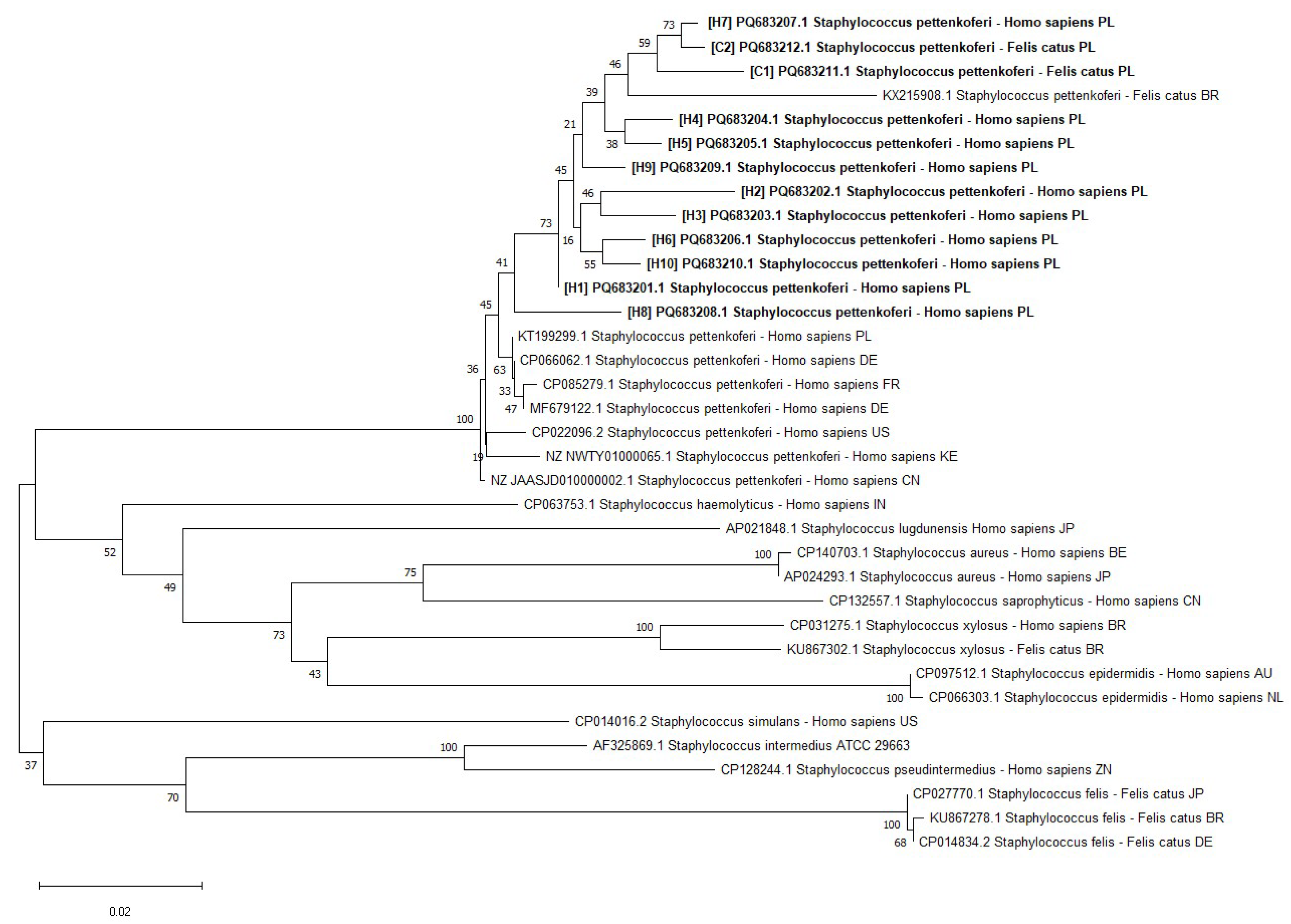
The investigated S. pettenkoferi strains were also examined with partial rpoB gene sequencing. The strains were found to be highly similar to one another, with the investigated animal strains being genetically similar to the investigated human strains. The resulting dendrogram is presented in Figure 2.
All investigated S. pettenkoferi strains were shown to be phenotypically resistant to penicillin, and all except one human strain were found to be resistant to methicillin. PCR genetic resistance testing supported these results. All of the investigated strains harbored the ermA gene, while seven also showed resistance to erythromycin on the phenotypic level. Only the S. pettenkoferi strains isolated from cats were not found to be multidrug resistant on the phenotypic level, while all S. pettenkoferi strains under investigation were MDR (multidrug resistant) according to the presence of these genetic determinants of resistance. Detailed profiles of S. pettenkoferi phenotypic and genotypic resistance are presented in Table 1.
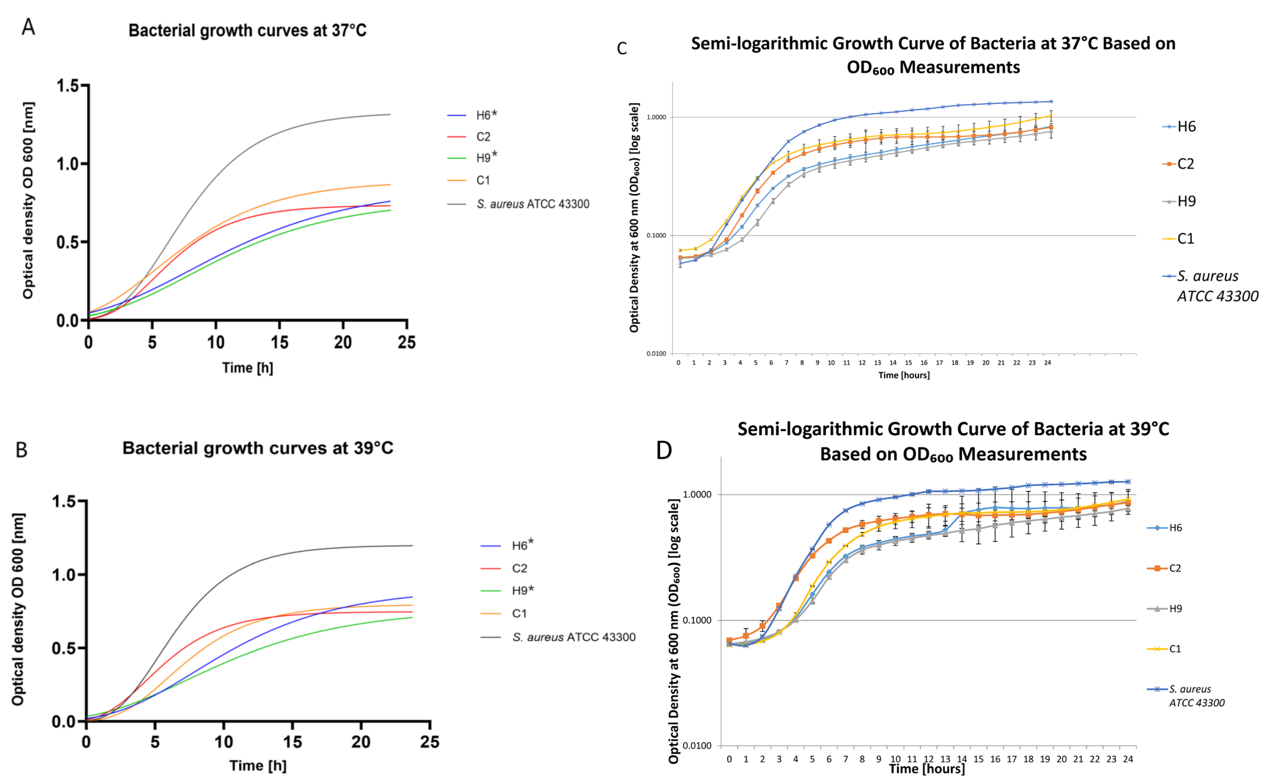
Physiological feline body temperature oscillates around 39 °C; thus, our study investigated the growth of human and feline S. pettenkoferi strains at the standard 37 °C and at 39 °C for 48 h. The growth curves for selected S. pettenkoferi strains are presented in Figure 3 as well as by all strains in Figures S2–S5 in the Supplementary Materials. The growth of all S. pettenkoferi of human origin was significantly slower than that of S. aureus ATCC 43300 at 39 °C (p < 0.05), while there was no significant difference in the growth of feline strains either at 37 °C or 39 °C. The detailed data connected with the growth of strains under investigation are presented in the Supplementary Materials in Tables S4 and S5. The biofilm production properties in vitro were tested using a microtiter plate test (MPT) with crystal violet staining. Additionally, considering the slower growth of S. pettenkoferi in media, biofilm production was measured after 24 and 48 h. The S. pettenkoferi typically showed weak or medium biofilm production after 24 h of incubation, regardless of the temperature. However, biofilm production was shown to increase at higher temperatures and after prolonged incubation. Detailed data can be found in Table 2. The presence of the two biofilm-related genes, icaA and bap, was also investigated. None of the investigated S. pettenkoferi strains tested positive for these genes.
The minimum inhibitory concentration (MIC) and minimum bactericidal concentration (MBEC) of the chosen antimicrobials were also compared. Detailed data are shown in Table S2 in the Supplementary Materials. The general tendency shown was that much higher concentrations of antimicrobials were needed to demonstrate an inhibitory or bactericidal effect on biofilms than on the bacteria’s planktonic forms. Only in the case of clindamycin activity in five strains (H1, H3, H5, H6, H9) did we observe an unusual situation in which the results of the MIC analysis were higher or comparable to MBEC.
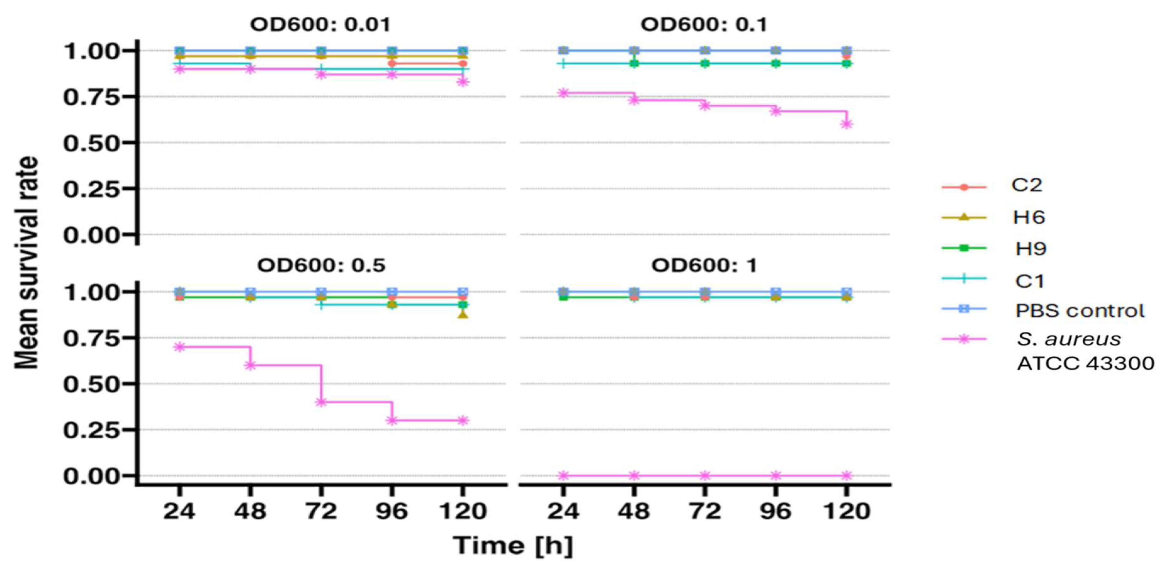
None of the tested S. pettenkoferi strains exhibited pathogenicity towards the G. mellonella larva model, as shown in the larval survival curves in Figure 4 below. Each strain was administered at four different optical densities: OD1600 = 1, OD2600 = 0.5, OD3600 = 0.1, and OD4600 = 0.01. Regardless of the size of the inoculum, the larval mortality rate was negligible, ranging from 0% to a maximum of 13%, as detailed in Table S3A in the Supplementary Materials. All tests were performed with at least three independent replicates. As a positive control, S. aureus ATCC 43300 was tested, which showed significant pathogenicity in the larva model, resulting in 100% larval mortality following injection with the highest infectious dose of S. aureus. The obtained results were consistent with the calculated 50% lethal dose (LD50) for the tested S. pettenkoferi strains and the control strain S. aureus ATCC 43300 (Table S3B in the Supplementary Materials). The S. pettenkoferi strains exhibited a high LD50 value in G. mellonella larvae, ranging from 1.52 × 10⁸ to 2.42 × 10¹⁴, whereas the LD50 for the S. aureus ATCC 43300 strain was significantly lower, at 1.76 × 10⁵.

3. Discussion

S. pettenkoferi is a rare bacterium that is not commonly found in healthy individuals or companion animals [10]. CNS are often identified at the genus level only, as definitive species identification can be complex and costly [17]. This practice contributes to the difficulty in determining the true prevalence of specific CNS species, including S. pettenkoferi. The implementation of MALDI-TOF MS in routine bacteriological protocols can significantly improve the identification of CNS at the species level. Several studies demonstrated the reliability and accuracy of MALDI-TOF MS in identifying S. pettenkoferi, even in cases where traditional identification methods failed [18,19,20,21]. The results obtained in the current study support these observations.
Presently, the ability of humans to act as carriers of S. pettenkoferi has not been investigated. In the previous literature, S. pettenkoferi was described as the causative agent of bloodstream infections [2,3,7,22]; however, the source of the infections was not further investigated. The presence of S. pettenkoferi was traditionally categorized as a contaminant or as a colonizer of indwelling catheters and was regarded as clinically irrelevant [12,19]. It is unclear whether companion animals are the source of bacteria in their owners or if companion animals simply act as accidental hosts of S. pettenkoferi. Our previous studies showed that S. pettenkoferi is rarely isolated from companion animals [10,23]. Despite this, our comparison of rpoB sequencing showed high similarity between regional S. pettenkoferi strains of human and feline origin. Based on these results, the possibility of host affinity and interspecies transmission should be further investigated.
The pathogenicity potential of S. pettenkoferi is also unclear. In the current study, the presence of biofilm-encoding genes such as bap and icaA was not detected; however, biofilm-forming properties were proven using MPT. Gen bap (biofilm-associated protein) promotes both the initial adhesion of strains to abiotic surfaces and intercellular adhesion. While gen icaA is a part of icaABCD, the operon encodes polysaccharide intercellular adhesin (PIA), the main component of biofilms [24]. Previous studies described icaA and bap as among the most frequently detected biofilm-encoding genes in CNS strains [24]. The icaABCD operon is frequently found in invasive strains of CNS, making it a potential marker to distinguish CNS from less harmful strains [25]. Other reports, however, did not find a correlation between the operon icaABCD and bap genes and biofilm-forming ability in phenotypic tests [24]. These biofilm-related genes were described previously in S. pettenkoferi strains [11,13,26]. Interestingly, previous investigations on clinical, animal, and environmental isolates reported no biofilm production [11,18]. Our study proves the biofilm-forming properties of all the S. pettenkoferi strains under investigation. The biofilm-forming ability was usually weak; nevertheless, the experiment showed that elongating the time of incubation and increasing the temperature improved biofilm production. In a study by Magnan et al. [13], 89.7% of isolates were able to produce biofilms and harbored a high content of biofilm-encoding genes. While S. pettenkoferi was associated with various infections, particularly in immunocompromised individuals, its specific features may predispose it to wound- and implant-related infections [5,13,26,27].
Due to their unique properties, such as the fact that their innate immune system displays many structural and functional similarities to the innate immune response of mammals, G. mellonella larvae serve as an ideal model for preliminary in vivo studies [28]. The use of this organism complements commonly applied in vitro methods (e.g., cell lines or organoids) [28]. G. mellonella larvae were repeatedly and successfully used as models to study specific mechanisms of pathogenicity and virulence factors of Staphylococcus species [29,30,31]. This insect model provides a rapid and accessible tool for monitoring the involvement of sRNA and mRNA in S. aureus pathogenesis and can also be applied to other human bacterial pathogens [32]. G. mellonella can also be used for other purposes, including studying the interactions between S. aureus and larval immune response as well as how bacteria induce the expression of immune-related peptides [32]. In contrast to other alternative insect models such as Caenorhabditis elegans or Drosophila melanogaster, G. mellonella larvae can be infected at 37 °C, which is a significant advantage in studies of Staphylococcus virulence and other human and animal pathogens [33]. To date, the G. mellonella larvae model has not been used to assess the pathogenicity of S. pettenkoferi strains. In our study, we demonstrated the low pathogenicity of this bacterial species in G. mellonella larvae. We tested a broad inoculum ranging from 106 to 109 CFU/mL, obtaining the highest larval mortality (around 13%) after injecting a single strain of S. pettenkoferi. As a positive control, we used the S. aureus ATCC strain, which exhibited significant pathogenicity towards the larvae and caused 100% mortality. This proved the low pathogenicity of the S. pettenkoferi strains under investigation. Ahmad-Mansour et al. [26] assessed the pathogenicity of S. pettenkoferi strains in human serum, where the bacteria were able to survive. The team also conducted tests on human keratinocytes, mouse macrophages, and human macrophages and demonstrated the virulence of S. pettenkoferi in a zebrafish model, causing significant embryonic mortality in this species [26].
Research on the antibiotic resistance patterns of S. pettenkoferi is still relatively limited compared to more common staphylococcal bacteria such as S. aureus. As with other CNS, S. pettenkoferi has the potential to develop resistance to various classes of antibiotics. The current study also confirmed that almost all the investigated strains (11/12) showed phenotypic resistance to beta-lactams and harbored the mecA gene. The ermA gene was detected in all the strains of S. pettenkoferi, which was connected to phenotypic resistance to macrolides in more than half of the strains. Our team also observed S. pettenkoferi’s multidrug-resistant properties. Similar results were described in work by Park et al. [12], where almost all S. pettenkoferi were resistant to penicillin, oxacillin, ciprofloxacin, and erythromycin (5/6). In another study, by Magnan et al. [13], 41.4% of tested S. pettenkoferi were shown to be resistant to penicillin G, while only one strain carried the mecA gene and was methicillin-resistant. All isolates were susceptible to gentamicin. In a report on a clinical infection caused by S. pettenkoferi in a cat, the tested strain was resistant to 36 antimicrobial drugs. The strain was found to be sensitive only to gentamicin, fluoroquinolones (enrofloxacin, norfloxacin, and levofloxacin), carbapenems (imipenem and meropenem), and tetracycline [11].
This study highlights the importance of the accurate species-level identification of CNS, particularly S. pettenkoferi, using advanced techniques like MALDI-TOF MS. While previously overlooked or misidentified, S. pettenkoferi demonstrates potential pathogenicity, including biofilm-forming abilities and multidrug resistance. Our findings suggest a possible zoonotic transmission and emphasize the need for further investigation into this emerging pathogen’s epidemiology, virulence factors, and clinical significance to both humans and companion animals. The increasing prevalence of antibiotic resistance in S. pettenkoferi underscores the importance of appropriate antibiotic use and the development of new therapeutic strategies to effectively manage infections caused by this bacterium.

4. Materials and Methods

4.1. S. pettenkoferi Origin and Identification

In this study, S. pettenkoferi isolates from human and animal origin were investigated. Materials from human participants (n = 10; H1–10) obtained from the Microbiology Laboratory of the Wrocław Medical University Foundation in Wrocław, Poland, were investigated. All bacterial isolates were obtained from the blood of clinical patients between the years 2021 and 2022. The animal isolates (n = 2, C1 and C2) were obtained from the conjunctiva of clinically healthy feline patients at the Department of Epizootiology and Clinic of Birds and Exotic Animals, Faculty of Veterinary Medicine at Wrocław University of Environmental and Life Science in Wrocław, Poland, between the years 2013 and 2019. The sampling procedure and initial identification were performed as previously described in the literature [10].
The investigated bacterial strains were identified as S. pettenkoferi via the Biotyper platform using a Bruker matrix-assisted laser desorption/ionization time-of-flight (MALDI-TOF) ultrafleXtreme mass spectrometer. All bacterial strains were subjected to ethanol/formic acid extraction sample preparation procedures performed for MALDI Biotyper analysis. The MALDI Biotyper analysis was performed at least twice for all investigated strains and two technical spots were prepared for each extract. Overnight growing colonies were suspended in water and were adjusted with ethanol, thoroughly suspended, and centrifuged. After removing the supernatant, the pellet was mixed with an appropriate amount of 70% formic acid followed by the addition of acetonitrile and was then re-mixed. The sample was then recentrifuged and 1 µL of supernatant was pipetted onto a polished steel MALDI plate, dried, and covered with 1 µL of 10 mg/mL HCCA (alpha-cyano-4-hydroxycinnamic acid) matrix solution in acetonitrile:water:TFA (trifluoroacetic acid) solution (50:47.5:2.5). The cut-off for species identification was set at a score value of ≥2.000. The Biotyper platform allows for comparison of the protein profiles of unknown microbial isolates with reference to mass spectra in a database used to identify microorganisms. The result of identification with the highest score was used for further analysis. Principal component analysis (PCA) was applied to reduce the dataset’s dimensionality. The PCA analysis was based on the peak profiles obtained from MALDI-TOF mass spectrometry [34].
In addition to Biotyper identification, the isolates were also identified as S. pettenkoferi using BLAST analysis of the 16S rRNA PCR product [35]. The obtained sequences were identified by comparison with sequences available in the GenBank database using the BLAST search algorithm (http://blast.ncbi.nlm.nih.gov/Blast.cgi; accessed on 5 December 2024). The accession numbers of the isolated strains were PQ636928.1–PQ636939.1. Additionally, an analysis of the partial fragment of the rpoB gene was performed [36]. The rpoB sequences can be found in GenBank as PQ683201–PQ683212. A phylogenetic tree based on partial rpoB gene sequences was constructed using MEGA 11 software [37]. Distances were calculated according to the Kimura two-parameter model and clustering was based on the neighbor-joining method, along with bootstrapping for 1000 replicates.

4.2. Antibiotic Resistance and Virulence Factors of S. pettenkoferi

All S. pettenkoferi isolates were screened for antibiotic susceptibility using both phenotypic and genotypic methods. The phenotypic resistance was performed using the disk diffusion method [amoxicillin with clavulanic acid (30 μg), ampicillin (10 μg), cefoxitin (30 μg), chloramphenicol (30 μg), ciprofloxacin (5 μg), erythromycin (15 μg), gentamicin (10 μg), clindamycin (2 μg), fusidic acid (10 μg), linezolid (30 μg), mupirocin (200 μg), oxacillin (1 μg), penicillin G (10 IU), rifampicin (5 μg), sulfamethoxazole/trimethoprim (25 μg), tetracycline (30 μg), tigecycline (15 μg), tobramycin (10 μg) (Oxoid Ltd., Basingstoke, UK) and marbofloxacin (5 μg) (MASTDISCS® AST (Mast Group Ltd., Liverpool, UK)] and MIC with the Sensititre™ EU Surveillance Staphylococcus EUST2 AST Plates (Thermo Fisher Scientific, Waltham, MA, USA). In the case of the MIC method, all solutions were allowed to reach room temperature prior to use. Three to five colonies were then selected from a fresh primary agar plate and suspended in sterile 0.9% NaCl, adjusted to 0.5 McFarland. Next, 100 µL of the suspension was transferred to a tube with 11 mL of Mueller–Hinton broth and was vortexed. Finally, 50 µL of the broth suspension was transferred to each well of the plate. Additionally, methicillin and teicoplanin resistances were detected using the broth microdilution method. The examined strains were inoculated in Mueller–Hinton broth (Oxoid, Basingstoke, UK) in a dilution series of oxacillin (16, 8, 4, 2, 1, 0.5, 0.25, 0.125, 0.0625, and 0.03125 µg/mL) and teicoplanin (128, 64, 32, 16, 8, 4, 2, 1, 0.5 µg/mL) (TOKU-E, Gent, Belgium) in water [38]. The inoculum was prepared according to the standard broth microdilution procedure described in the Clinical and Laboratory Standards Institute (CLSI) Performance Standards for Antimicrobial Disk and Dilution Susceptibility Tests for Bacteria Isolated From Animals, supplement VET01S [35]. Methicillin-resistant strains were detected using the CLSI criteria for CNS species for human and feline isolates, respectively [39,40]. S. aureus ATCC 29213 was used as a negative control, and S. aureus ATCC 43300 (mecA-positive) was used as a positive control. The double disk diffusion test (D-test) was performed on all isolates to detect inducible clindamycin resistance [41].
Antibiotic-resistant genotypes were identified using PCR analysis. The presence of genes involved in resistance to penicillin, aminoglycosides, β-lactamase, glycopeptides, macrolide-lincosamide-streptogramin, tetracyclines, fusidic acid, chloramphenicols, and mupirocin was determined using PCR. The detailed methods involved with the genetic determination of resistance and virulence factors are described in the Supplementary Materials (Table S1). S. pettenkoferi isolates that were found to be resistant to three or more classes of antimicrobial agents were interpreted as being MDR.

4.3. Growth Curves’ Evaluation and Biofilm Formation

The growth profiles of all S. pettenkoferi strains and reference strains were investigated using brain infusion bullion (BHI) (Oxoid, Basingstoke, UK). The overnight suspension of each strain in 5 mL of BHI was diluted to an optical density of around 0.1 at 600 nm (Eppendorf BioPhotometer 6131, Hamburg, Germany). Then, 500 µL of each bacterial suspension was added to three wells of a 48-well plate, incubated, and automatically shaken for 48 h at 37 °C and 39 °C. Each well’s absorbance at 600 nm was determined using an automatic absorbance reader (Spark Tecan, Männedorf, Switzerland). Growth curves were determined using GraphPad Prism 10.4.1 software (San Diego, CA, USA), as it was previously described by Magnan et al. (2022) [13]. To estimate growth kinetics, the per-capita growth rate was computed from OD readings using the calc_deriv function. The doubling time was then calculated using the doubling_time function. Data were analyzed in R 4.4.1 using the gcplyr 1.11.0 package [42]. The differences in the growth of S. pettenkoferi strains under investigation at 37 °C and 39 °C and comparison to S. aureus ATCC 43300 were investigated using the Mann–Whitney test with statistical significance at p < 0.5.
The presence of the two biofilm-related genes, icaA and bap, was determined by duplex polymerase chain reaction (PCR), as it was previously described in the literature [43], with the positive controls S. epidermidis PCM 2532 and S. epidermidis AIR08630, respectively.
All strains were tested for slime production using the static MTP test with crystal violet staining in a 96-well plate. After an overnight culture of all strains under investigation in 5 mL of BHI (Oxoid, Basingstoke, UK), the culture was diluted 1:100 in BHI and 200 μL of the suspensions was added into three wells in four plates each and incubated for 24 and 48 h at 37 °C and 39 °C. After incubation, the plates were washed twice with 200 μL of sterile phosphate-buffered saline. Next, the plate was allowed to air-dry for 2 h before being stained with 200 μL of 0.1% crystal violet for 10 min. After the subsequent washing (as described above), the staining bound to the bacteria was dissolved by adding 250 μL of 95% ethanol. The absorbance values were determined at 570 nm on a plate reader (Infinite F50, Tecan, Männedorf, Switzerland) after 24 and 48 h of incubation at 37 °C and 39 °C. Interpretation was performed as previously described by Płoneczka et al. [43]. The MTP procedure was carried out three times, in triplicate for each strain under investigation. The Minimal Biofilm Eradication Concentration was also calculated for selected antimicrobials. Serial dilutions of the antimicrobials were used in the following concentrations: clindamycin (TOKU-E, Gent, Belgium) at 16, 8, 4, 2, 1, 0.5, 0.25, 0.125, and 0.0625 µg/mL; vancomycin (TOKU-E, Gent, Belgium) and teicoplanin TOKU-E, Gent, Belgium) at 128, 64, 32, 16, 8, 4, 2, 1, and 0.5 µg/mL; and erythromycin and tetracycline (TOKU-E, Gent, Belgium) at 32, 16, 8, 4, 2, 1, 0.5, and 0.25 µg/mL. The tests were conducted and interpreted as previously described by Dudek et al. [44].

4.4. Pathogenicity Tests on Larvae Model

4.4.1. Preparation of Bacterial Suspension

Overnight cultures of the tested S. pettenkoferi strains [n = 4] were centrifuged at 3500 RPM. The resulting bacterial pellets were washed three times with sterile PBS buffer and then resuspended in PBS buffer to achieve an optical density of (1) OD600 = 1, (2) OD600 = 0.5, (3) OD600 = 0.1, and (4) OD600 = 0.01. The CFU/mL value for the prepared bacterial suspensions was determined using the culture method.

4.4.2. Larval Selection and Injection

The experiment used larvae from a self-maintained culture at the Department of Epizootiology and Clinic of Birds and Exotic Animals at Wrocław University of Environmental and Life Sciences. Larvae weighing 300 ± 30 mg were selected and placed in groups of n = 10 on sterile Petri dishes. Each larva was injected with 10 μL of bacterial suspension directly into the hemocoel using a Hamilton Bonaduz 100 μL microliter syringe. Negative control: larvae [n = 10] injected with 10 μL sterile PBS buffer. Positive control: larvae [n = 10] injected with 10 μL of suspension of Staphylococcus aureus MRSA strain ATCC 43300. The suspension of the reference strain was prepared as described above for S. pettenkoferi. The experiment was repeated at least three times. Larval survival was monitored over 120 h at 37 °C after inoculation, counting the numbers of live and dead larvae.

4.4.3. Analysis of the Results

The obtained data were analyzed statistically using GraphPad Prism software with a Kaplan–Meier algorithm. Survival curves were plotted based on the results. From the survival curves, the 50% lethal dose (LD50) for each bacterial strain was calculated. The LD50 represents the CFU/mL value of a given bacterial strain at which 50% of the larval population died. Calculations were performed using the method modified by Reed and Muench [45].
The percentage of larval mortality was calculated by determining the ratio of the number of dead larvae after 120 h of observation to the total number of larvae [n = 10] used for injection in a single experiment. The calculations were based on the average values for each bacterial strain and a specific CFU/mL concentration.

Supplementary Materials

The supporting information can be downloaded at https://www.mdpi.com/article/10.3390/ijms26051948/s1. References [46,47,48,49,50,51,52,53,54,55,56,57,58] are cited in the Supplementary Materials.

Author Contributions

Conceptualization, K.B., M.K. (Magdalena Karwańska) and M.S.; methodology, K.B., M.K. (Magdalena Karwańska), M.S., M.K. (Marta Książczyk) and K.K.; materials collection: K.B. and A.K.-Ć.; laboratory investigation, K.B., A.D., M.K. (Magdalena Karwańska), M.S., M.K. (Marta Książczyk) and K.K.; formal analysis, K.B., M.K. (Magdalena Karwańska), M.S. and M.K. (Marta Książczyk); writing—original draft preparation, K.B., A.D., M.K. (Magdalena Karwańska), M.S. and M.K. (Marta Książczyk); writing—review and editing, K.B. and A.D.; visualization, K.B., M.S., M.K. (Magdalena Karwańska), M.K. (Marta Książczyk) and K.K.; supervision, K.B.; project administration, K.B.; funding acquisition, K.B. All authors have read and agreed to the published version of the manuscript.

Funding

The APC/BPC is financed/co-financed by Wrocław University of Environmental and Life Sciences.

Institutional Review Board Statement

Not applicable.

Informed Consent Statement

Not applicable.

Data Availability Statement

All data are available in the article and Supplementary Materials.

Acknowledgments

The authors would like to thank Patrycja Trzeciak from the Microbiology Laboratory of the Wrocław Medical University Foundation in Wrocław, Poland, for sharing the human S. pettenkoferi strains used in the study.

Conflicts of Interest

The authors declare no conflicts of interest.

References

  1. Trülzsch, K.; Rinder, H.; Trček, J.; Bader, L.; Wilhelm, U.; Heesemann, J. Staphylococcus pettenkoferi, a novel staphylococcal species isolated from clinical specimens. Diagn. Microbiol. Infect. Dis. 2002, 43, 175–182. [Google Scholar] [CrossRef]
  2. Mammina, C.; Bonura, C.; Verde, M.S.; Fasciana, T.; Palma, D.M. A Fatal Bloodstream Infection by Staphylococcus pettenkoferi in an Intensive Care Unit Patient. Case Rep. Crit. Care. 2011, 2011, 612732. [Google Scholar] [CrossRef] [PubMed]
  3. Hashi, A.A.; Delport, J.A.; Elsayed, S.; Silverman, M.S. Staphylococcus pettenkoferi bacteremia: A case report and review of the literature. Can. J. Infect. Dis. Med. Microbiol. 2015, 26, 319–322. [Google Scholar] [CrossRef] [PubMed]
  4. Strong, C.; Cosiano, M.; Cabezas, M.; Barwatt, J.W.; Tillekeratne, L.G. Staphylococcus pettenkoferi Bacteremia in an American Intensive Care Unit. Case Rep. Infect. Dis. 2021, 29, 5235691. [Google Scholar] [CrossRef] [PubMed]
  5. Gisriel, S.D.; Jacobs, J.W. The first reported case of Staphylococcus pettenkoferi prosthetic joint infection. Microbes Infect. 2022, 24, 104978. [Google Scholar] [CrossRef]
  6. Ugaban, K.B.; She, R.C. The clinical significance of staphylococcus pettenkoferi: A retrospective review at a tertiary care medical center. Diagn. Microbiol. Infect. Dis. 2022, 102, 115592. [Google Scholar] [CrossRef]
  7. Kierzkowska, M.; Markowska, K.; Majewska, A. Knowledge, Attitude and Practice Regarding Staphylococcus pettenkoferi. Infect. Dis. Rep. 2022, 14, 112–120. [Google Scholar] [CrossRef]
  8. Weiss, S.; Kadlec, K.; Fessler, A.T.; Schwarz, S. Identification and characterization of methicillin-resistant Staphylococcus aureus, Staphylococcus epidermidis, Staphylococcus haemolyticus and Staphylococcus pettenkoferi from a small animal clinic. Vet. Microbiol. 2013, 167, 680–685. [Google Scholar] [CrossRef]
  9. Kaspar, U.; von Lützau, A.; Schlattmann, A.; Roesler, U.; Köck, R.; Becker, K. Zoonotic multidrug-resistant microorganisms among small companion animals in Germany. PLoS ONE 2018, 13, e0208364. [Google Scholar] [CrossRef]
  10. Bierowiec, K.; Korzeniowska-Kowal, A.; Wzorek, A.; Rypuła, K.; Gamian, A. Prevalence of Staphylococcus Species Colonization in Healthy and Sick Cats. Biomed. Res. Int. 2019, 20, 4360525. [Google Scholar] [CrossRef]
  11. Dutta, T.K.; Chakraborty, S.; Das, M.; Mandakini, R.; Vanrahmlimphuii Roychoudhury, P.; Ghorai, S.; Behera, S.K. Multidrug-resistant Staphylococcus pettenkoferi isolated from cat in India. Vet. World. 2018, 11, 1380–1384. [Google Scholar] [CrossRef] [PubMed]
  12. Park, S.; Chung, H.S.; Lee, M. Clinical and microbiological characteristics of six Staphylococcus pettenkoferi isolates from blood samples. Ann. Lab. Med. 2015, 35, 250–253. [Google Scholar] [CrossRef] [PubMed]
  13. Magnan, C.; Ahmad-Mansour, N.; Pouget, C.; Morsli, M.; Huc-Brandt, S.; Pantel, A.; Dunyach-Remy, C.; Sotto, A.; Molle, V.; Lavigne, J.P. Phenotypic and Genotypic Virulence Characterisation of Staphylococcus pettenkoferi Strains Isolated from Human Bloodstream and Diabetic Foot Infections. Int. J. Mol. Sci. 2022, 23, 15476. [Google Scholar] [CrossRef] [PubMed]
  14. Rai, A.; Khairnar, K. Overview of the risks of Staphylococcus aureus infections and their control by bacteriophages and bacteriophage-encoded products. Braz. J. Microbiol. 2021, 52, 2031–2042. [Google Scholar] [CrossRef]
  15. Lynch, S.A.; Helbig, K.J. The Complex Diseases of Staphylococcus pseudintermedius in Canines: Where to Next? Vet. Sci. 2021, 8, 11. [Google Scholar] [CrossRef]
  16. Smith, J.T.; Andam, C.P. Extensive Horizontal Gene Transfer within and between Species of Coagulase-Negative Staphylococcus. Genome Biol. Evol. 2021, 13, evab206. [Google Scholar] [CrossRef] [PubMed] [PubMed Central]
  17. Carretto, E.; Barbarini, D.; Couto, I.; De Vitis, D.; Marone, P.; Verhoef, J.; De Lencastre, H.; Brisse, S. Identification of coagulase-negative staphylococci other than Staphylococcus epidermidis by automated ribotyping. Clin. Microbiol. Infect. 2005, 11, 177–184. [Google Scholar] [CrossRef]
  18. Månsson, E.; Hellmark, B.; Stegger, M.; Skytt Andersen, P.; Sundqvist, M.; Söderquist, B. Genomic relatedness of Staphylococcus pettenkoferi isolates of different origins. J. Med. Microbiol. 2017, 66, 601–608. [Google Scholar] [CrossRef]
  19. Argemi, X.; Riegel, P.; Lavigne, T.; Lefebvre, N.; Grandpré, N.; Hansmann, Y.; Jaulhac, B.; Prévost, G.; Schramm, F. Implementation of Matrix-Assisted Laser Desorption Ionization-Time of Flight Mass Spectrometry in Routine Clinical Laboratories Improves Identification of Coagulase-Negative Staphylococci and Reveals the Pathogenic Role of Staphylococcus lugdunensis. J. Clin. Microbiol. 2015, 53, 2030–2036. [Google Scholar] [CrossRef]
  20. Becker, K.; Heilmann, C.; Peters, G. Coagulase-negative Staphylococci. Clin. Microbiol. Rev. 2014, 27, 870–926. [Google Scholar] [CrossRef]
  21. Kang, H.; Ryoo, N. Identification of Staphylococcus pettenkoferi Isolated from Blood Culture. Ann. Clin. Microbiol. 2019, 22, 77–79. [Google Scholar] [CrossRef]
  22. Tamura, D.; Kurane, K.; Koshu, K.; Shinjoh, M.; Tanaka, D.; Yamagishi1, H.; Kimura, Y.; Nakazato, E.; Yamagata, T. Clinical picture of bacteremia with Staphylococcus pettenkoferi in children. J. Clin. Images Med. Case Rep. 2023, 4, 2237. [Google Scholar] [CrossRef]
  23. Miszczak, M.; Korzeniowska-Kowal, A.; Wzorek, A.; Gamian, A.; Rypuła, K.; Bierowiec, K. Colonization of methicillin-resistant Staphylococcus species in healthy and sick pets: Prevalence and risk factors. BMC Vet. Res. 2023, 19, 85. [Google Scholar] [CrossRef] [PubMed]
  24. Gajewska, J.; Chajęcka-Wierzchowska, W. Biofilm Formation Ability and Presence of Adhesion Genes among Coagulase-Negative and Coagulase-Positive Staphylococci Isolates from Raw Cow’s Milk. Pathogens 2020, 9, 654. [Google Scholar] [CrossRef] [PubMed] [PubMed Central]
  25. Seng, R.; Kitti, T.; Thummeepak, R.; Kongthai, P.; Leungtongkam, U.; Wannalerdsakun, S.; Sitthisak, S. Biofilm formation of methicillin-resistant coagulase negative staphylococci (MR-CoNS) isolated from community and hospital environments. PLoS ONE 2017, 12, e0184172. [Google Scholar] [CrossRef] [PubMed] [PubMed Central]
  26. Ahmad-Mansour, N.; Plumet, L.; Huc-Brandt, S.; Magnan, C.; Yahiaoui-Martinez, A.; Kissa, K.; Pantel, A.; Lavigne, J.-P.; Molle, V. Investigating Pathogenicity and Virulence of Staphylococcus pettenkoferi: An Emerging Pathogen. Int. J. Mol. Sci. 2021, 22, 13614. [Google Scholar] [CrossRef]
  27. Loïez, C.; Wallet, F.; Pischedda, P.; Renaux, E.; Senneville, E.; Mehdi, N.; Courcol, R.J. Case of Osteomyelitis Caused by “Staphylococcus pettenkoferi”. J. Clin. Microbiol. 2007, 45, 1069–1071. [Google Scholar] [CrossRef]
  28. Kavanagh, K.; Sheehan, G. The Use of Galleria Mellonella Larvae to Identify Novel Antimicrobial Agents against Fungal Species of Medical Interest. J. Fungi 2018, 4, 113. [Google Scholar] [CrossRef]
  29. Ménard, G.; Rouillon, A.; Ghukasyan, G.; Emily, M.; Felden, B.; Donnio, P.Y. Galleria mellonella Larvae as an Infection Model to Investigate sRNA-Mediated Pathogenesis in Staphylococcus aureus. Front. Cell. Infect. Microbiol. 2021, 11, 631710. [Google Scholar] [CrossRef]
  30. Kaskatepe, B.; Ozturk, S. Assessment of synergistic activity of rhamnolipid and linezolid against methicillin-resistant Staphylococcus aureus in-vitro and in-vivo with Galleria mellonella larvae model. Microb. Pathog. 2023, 174, 105945. [Google Scholar] [CrossRef]
  31. Berryhill, B.A.; Gil-Gil, T.; Witzany, C.; Goldberg, D.A.; Vega, N.M.; Regoes, R.R.; Levin, B.R. The dynamics of Staphylococcal infection and their treatment with antibiotics and bacteriophage in the Galleria mellonella model system. bioRxiv 2024. [Google Scholar] [CrossRef]
  32. Sheehan, G.; Dixon, A.; Kavanagh, K. Utilization of Galleria mellonella larvae to characterize the development of Staphylococcus aureus infection. Microbiology. 2019, 165, 863–875. [Google Scholar] [CrossRef] [PubMed]
  33. Ramarao, N.; Nielsen-Leroux, C.; Lereclus, D. The Insect Galleria mellonella as a Powerful Infection Model to Investigate Bacterial Pathogenesis. J. Vis. Exp. 2012, 70, e4392. [Google Scholar] [CrossRef]
  34. Abdel Samad, R.; Al Disi, Z.; Mohammad Ashfaq, M.Y.; Wahib, S.M.; Zouari, N. The use of principle component analysis and MALDI-TOF MS for the differentiation of mineral forming Virgibacillus and Bacillus species isolated from sabkhas. RSC Adv. 2020, 10, 14606–14616. [Google Scholar] [CrossRef] [PubMed] [PubMed Central]
  35. Lane, D.J. 16S/23S rRNA Sequencing. In Nucleic Acid Techniques in Bacterial Systematics; Stackebrandt, E., Goodfellow, M., Eds.; John Wiley and Sons: New York, NY, USA, 1991; pp. 115–175. [Google Scholar]
  36. Drancourt, M.; Raoult, D. RpoB gene sequence-based identification of Staphylococcus species. J. Clin. Microbiol. 2002, 40, 1333–1338. [Google Scholar] [CrossRef]
  37. Tamura, K.; Stecher, G.; Kumar, S. MEGA11: Molecular Evolutionary Genetics Analysis version 11. Mol. Biol. Evol. 2021, 38, 3022–3027. [Google Scholar] [CrossRef]
  38. CLSI Document M07-A9; Methods for Dilution Antimicrobial Susceptibility Tests for Bacteria That Grow Aerobically. CLSI—Clinical and Laboratory Standards Institute: Wayne, PA, USA, 2012.
  39. CLSI Supplement VET01S; Performance Standards for Antimicrobial Disk and Dilution Susceptibility Tests for Bacteria Isolated From Animals. CLSI—Clinical and Laboratory Standards Institute: Wayne, PA, USA, 2020.
  40. CLSI Supplement M100; Performance Standards for Antimicrobial Susceptibility Testing. CLSI—Clinical and Laboratory Standards Institute: Wayne, PA, USA, 2023.
  41. Coutinho, V.d.L.S.; Paiva, R.M.; Reiter, K.C.; de-Paris, F.; Barth, A.L.; Machado, A.B. Distribution of erm genes and low prevalence of inducible resistance to clindamycin among staphylococci isolates. Braz. J. Infect. Dis. 2010, 14, 564–568. [Google Scholar] [CrossRef]
  42. Wright, R.C.T.; Wood, A.J.; Bottery, M.J.; Muddiman, K.J.; Paterson, S.; Harrison, E.; Brockhurst, M.A.; Hall, J.P.J. A chromosomal mutation is superior to a plasmid-encoded mutation for plasmid fitness cost compensation. PLoS Biol. 2024, 22, e3002926. [Google Scholar] [CrossRef] [PubMed] [PubMed Central]
  43. Płoneczka-Janeczko, K.; Lis, P.; Bierowiec, K.; Rypuła, K.; Chorbiński, P. Identification of bap and icaA genes involved in biofilm formation in coagulase negative staphylococci isolated from feline conjunctiva. Vet. Res. Commun. 2014, 38, 337–346. [Google Scholar] [CrossRef]
  44. Dudek, B.; Tymińska, J.; Szymczyk-Ziółkowska, P.; Chodaczek, G.; Migdał, P.; Czajkowska, J.; Junka, A. In Vitro Activity of Octenidine Dihydrochloride-Containing Lozenges against Biofilm-Forming Pathogens of Oral Cavity and Throat. Appl. Sci. 2023, 13, 2974. [Google Scholar] [CrossRef]
  45. Saganuwan, A.S. Application of median lethal concentration (LC50) of pathogenic microorganisms and their antigens in vaccine development. BMC Res. Notes 2020, 13, 289. [Google Scholar] [CrossRef] [PubMed]
  46. Mahdiyoun, S.M.; Kazemian, H.; Ahanjan, M.; Houri, H.; Goudarzi, M. Frequency of Aminoglycoside-Resistance Genes in Methicillin-Resistant Staphylococcus aureus (MRSA) Isolates from Hospitalized Patients. Jundishapur J. Microbiol. 2016, 9, e35052. [Google Scholar] [CrossRef] [PubMed]
  47. Bierowiec, K.; Płoneczka-Janeczko, K.; Rypuła, K. Diversity of antimicrobial-resistant pheno- and genotypes of Staphylococcus aureus from clinically healthy cats kept in city households. Berl. Und Munch. Tierarztl. Wochenschr. 2017, 130, 50–57. [Google Scholar] [CrossRef]
  48. Udo, E.E.; Boswihi, S.S.; Mathew, B.; Noronha, B.; Verghese, T. Resurgence of Chloramphenicol Resistance in Methicillin-Resistant Staphylococcus aureus Due to the Acquisition of a Variant Florfenicol Exporter (fexAv)-Mediated Chloramphenicol Resistance in Kuwait Hospitals. Antibiotics 2021, 10, 1250. [Google Scholar] [CrossRef]
  49. Strommenger, B.; Kettlitz, C.; Werner, G.; Witte, W. Multiplex PCR assay for simultaneous detection of nine clinically relevant antibiotic resistance genes in Staphylococcus aureus. J. Clin. Microbiol. 2003, 41, 4089–4094. [Google Scholar] [CrossRef]
  50. Martineau, F.; Picard, F.J.; Ke, D.; Paradis, S.; Roy, P.H.; Ouellette, M.; Bergeron, M.G. Development of a PCR assay for identification of staphylococci at genus and species levels. J. Clin. Microbiol. 2001, 39, 2541–2547. [Google Scholar] [CrossRef]
  51. Mason, W.J.; Blevins, J.S.; Beenken, K.; Wibowo, N.; Ojha, N.; Smeltzer, M.S. Multiplex PCR protocol for the diagnosis of staphylococcal infection. J. Clin. Microbiol. 2001, 39, 3332–3338. [Google Scholar] [CrossRef]
  52. Park, J.Y.; Fox, L.K.; Seo, K.S.; McGuire, M.A.; Park, Y.H.; Rurangirwa, F.R.; Sischo, W.M.; Bohach, G.A. Detection of classical and newly described staphylococcal superantigen genes in coagulase-negative staphylococci isolated from bovine intramammary infections. Vet. Microbiol. 2011, 147, 149–154. [Google Scholar] [CrossRef]
  53. Becker, K.; Roth, R.; Peters, G. Rapid and specific detection of toxigenic Staphylococcus aureus: Use of two multiplex PCR enzyme immunoassays for amplification and hybridization of staphylococcal enterotoxin genes, exfoliative toxin genes, and toxic shock syndrome toxin 1 gene. J. Clin. Microbiol. 1998, 36, 2548–2553. [Google Scholar] [CrossRef]
  54. Monday, S.R.; Bohach, G.A. Use of multiplex PCR to detect classical and newly described pyrogenic toxin genes in staphylococcal isolates. J. Clin. Microbiol. 1999, 37, 3411–3414. [Google Scholar] [CrossRef]
  55. Pereira, L.A.; Harnett, G.B.; Hodge, M.M.; Cattell, J.A.; Speers, D.J. Real-time PCR assay for detection of blaZ genes in Staphylococcus aureus clinical isolates. J. Clin. Microbiol. 2014, 52, 1259–1261. [Google Scholar] [CrossRef] [PubMed]
  56. Jung, J.H.; Yoon, E.J.; Choi, E.C.; Choi, S.S. Development of TaqMan probe-based real-time PCR method for erm(A),erm(B), and erm(C), rapid detection of macrolide-lincosamide-streptogramin B resistance genes, from clinical isolates. J. Microbiol. Biotechnol. 2009, 19, 1464–1469. [Google Scholar] [CrossRef] [PubMed]
  57. Kelley, K.; Cosman, A.; Belgrader, P.; Chapman, B.; Sullivan, D.C. Detection of methicillin-resistant Staphylococcus aureus by a duplex droplet digital PCR assay. J. Clin. Microbiol. 2013, 51, 2033–2039. [Google Scholar] [CrossRef] [PubMed]
  58. Peak, N.; Knapp, C.W.; Yang, R.K.; Hanfelt, M.M.; Smith, M.S.; Aga, D.S.; Graham, D.W. Abundance of six tetracycline resistance genes in wastewater lagoons at cattle feedlots with different antibiotic use strategies. Environ. Microbiol. 2007, 9, 143–151. [Google Scholar] [CrossRef] [PubMed]
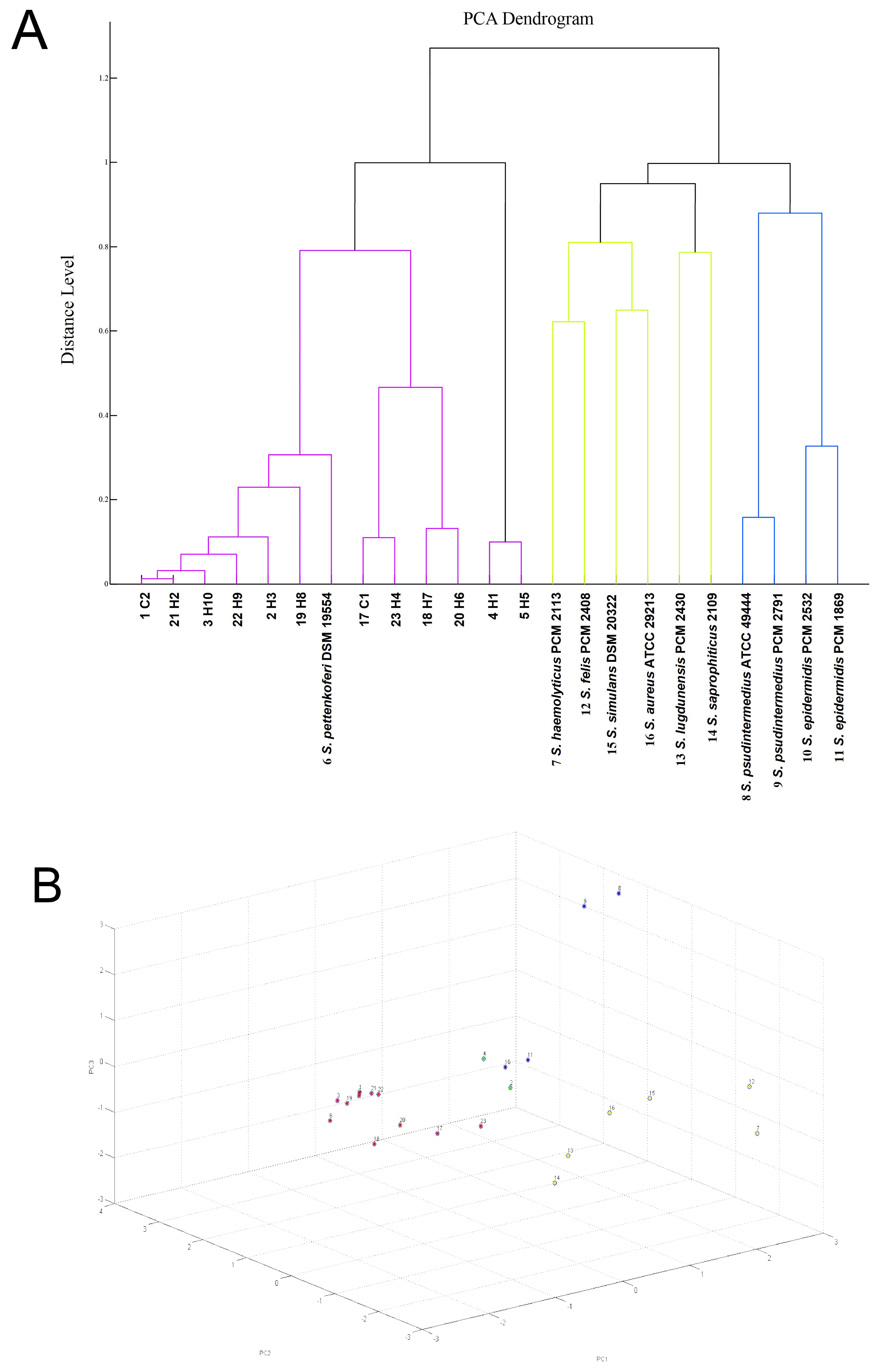
Figure 1. (A) Visual depiction of the closeness of individual spectra of S. pettenkoferi and chosen referential strains obtained with MALDI-TOF MS analysis. (B) PCA clustering of MALDI Biotyper spectra of S. pettenkoferi isolates and chosen referential strains: 1—C2, 2—H3, 3—H10, 4—H1, 5—H5, 6—S. pettenkoferi DSM 19554, 7—S. haemolyticus PCM 2113, 8—S. pseudintermedius ATCC 49444, 9—S. pseudintermedius PCM 2791, 10—S. epidermidis PCM 2532, 11—S. epidermidis PCM 1869, 12—S. felis PCM 2408, 13—S. lugdunensis PCM 2430, 14—S. saprophyticus 2109, 15—S. simulans DSM 20322, 16—S. aureus ATCC 29213, 17—C1, 18—H7, 19—H8, 20—H6, 21—H2, 22—H9, 23—H4.
Ijms 26 01948 g001
Figure 2. Phylogenetic tree based on partial rpoB gene sequencing of the genus Staphylococcus, including the sequences under investigation (C1, C2, H1–H10, indicated with bold font) and reference sequences from GenBank database. All sequences were obtained with MEGA 11. Genetic distance is indicated on the scale (the scale bar represents 2 nucleotide substitutions per 100 nucleotides).
Ijms 26 01948 g002
Figure 3. Growth curves of S. pettenkoferi of feline origin (C1 and C2) and human origin (H6 and H9) compared to S. aureus ATCC 43300 strain in (A) 37 °C and (B) in 39 °C; growth curves of bacterial strains (H6, C2, H9, C1, and S. aureus ATCC 43300) over a 24-h period in (C) 37 °C and (D) in 39 °C. Optical density (OD) at 600 nm was measured at hourly intervals. The data represent the mean OD values, with error bars indicating the standard deviation (SD), and the corresponding generation times (GT) ± SD. * a significant difference in growth compared to S. aureus ATCC 43300; p < 0.05.
Ijms 26 01948 g003
Figure 4. Survival curve of larvae [n = 10] following injection with the tested strains of S. pettenkoferi. The negative control consisted of larvae injected with sterile PBS buffer [PBS Control], while the positive control consisted of larvae injected with the S. aureus ATCC 43300 strain. Larval survival was monitored for 120 h.
Ijms 26 01948 g004
Table 1. The phenotypic and genotypic antimicrobial profiles of all S. pettenkoferi strains under investigation were determined using the MIC, disk diffusion method, and PCR reactions.
IDPhenotypic Antimicrobial ProfileGenotypic Antimicrobial Profile
C1P, OX, CblaZ, mecA, tet(L), ermA, vanA, aph3-IIIa
C2P, OXblaZ, mecA, ermA, vanA
H1P, OX, AMP, AMC, CIP, MAR, E, CL, SMXmecA, tet(L), ermA
H2P, OXblaZ, mecA, ermA, aph3-IIIa
H3P, OX, AMP, AMC, CIP, MAR, E, CL, SMXblaZ, mecA, tet(L), ter(M), ermA, aph3-IIIa
H4P, OX, AMC, TETblaZ, mecA, tet(M), ermA, fusB, aph3-IIIa
H5P, OX, AMP, AMC, CIP, MAR, E, CL, SMX, TMP, FDblaZ, mecA, tet(L), tet(M), ermA, mupA, fusB, aph3-IIIa
H6P, OX, AMP, AMC, CIP, MAR, E, CL, SMX, TMP, FD, MUPblaZ, mecA, tet(L), ermA, mupA, fusB, aph3-IIIa
H7P, CIP, E, CL, SMXblaZ, mecA, tet(L), ermA, vanA
H8P, OX, TET, SMXmecA, tet(L), ermA
H9P, OX, AMP, AMC, CIP, MAR, E, CL, SMX, TMP, FD, MUPblaZ, mecA, tet(L), ermA
H10P, OX, AMC, E, CL, SMXblaZ, mecA, tet(L), ermA
C1–C2: cats’ S. pettenkoferi strains; H1–H10: humans’ S. pettenkoferi strains; P = penicillin; OX = oxacillin, AMP—ampicillin; AMC = amoxicillin-clavulanate; CIP = ciprofloxacin; MAR = marbofloxacin; E = erythromycin; CL = clindamycin; TET = tetracycline; SMX = sulfamethoxazole; TMP = trimethoprim; FD = fusidic acid; MUP = mupirocin; C = chloramphenicol.
Table 2. The results of slime production by microtiter plate (MTP) test with crystal violet staining of S. pettenkoferi strains of human and feline origin after 24 and 48 h of incubation at 37 °C and 39 °C.
IDBiofilm Production at 37 °CBiofilm Production at 39 °C
After 24 hAfter 48 hAfter 24 hAfter 48 h
C1weakmediummediumstrong
C2weakstrongmediumstrong
H1weakmediumweakstrong
H2weakweakweakmedium
H3weakweakweakmedium
H4weakweakweakstrong
H5weakweakweakmedium
H6weakmediumweakstrong
H7weakweakweakmedium
H8weakmediumweakmedium
H9not presentmediumweakstrong
H10weakmediumweakstrong
S. pettenkoferi DSM 19554weakmediumweakmedium
S. aureus ATCC 43300mediumstrongmediumstrong
S. aureus ATCC 6532strongstrongstrongstrong
S. aureus ATCC 11632weakmediummediumstrong
S. pseudintermedius PCM 2791strongstrongstrongstrong
S. pseudintermedius ATCC 49444strongstrongstrongstrong
According to microtiter plate assay results; weak biofilm formers, 0.252 ≤ A570 < 0.504; medium-positive biofilm formers, 0.504  ≤  A570  <  1.008; strong biofilm formers, A570  ≥  1.008.
Disclaimer/Publisher’s Note: The statements, opinions and data contained in all publications are solely those of the individual author(s) and contributor(s) and not of MDPI and/or the editor(s). MDPI and/or the editor(s) disclaim responsibility for any injury to people or property resulting from any ideas, methods, instructions or products referred to in the content.