Article

Application of mRNA-Seq and Metagenomic Sequencing to Study Salmonella pullorum Infections in Chickens

1
State Key Laboratory of Livestock and Poultry Breeding, South China Agricultural University, Guangzhou 510642, China
2
College of Animal Science, South China Agricultural University, Guangzhou 510642, China
*
Authors to whom correspondence should be addressed.
Int. J. Mol. Sci. 2025, 26(4), 1448; https://doi.org/10.3390/ijms26041448
Submission received: 15 January 2025 / Revised: 3 February 2025 / Accepted: 5 February 2025 / Published: 9 February 2025
(This article belongs to the Section Molecular Microbiology)

Abstract

The disease caused by Salmonella pullorum has been demonstrated to exert a deleterious effect on the performance of poultry, giving rise to elevated mortality and considerable economic losses within the breeding industry. However, there is a paucity of research investigating the relationship between cecal gene expression and different isomer and Salmonella pullorum infection, and research on the relationship between intestinal microbiota and Salmonella pullorum infection is also limited. In this study, mRNA-Seq and metagenomic sequencing were performed on the cecal tissues and fresh feces of individuals who tested positive (n = 4) and negative (n = 4) for Salmonella pullorum, with the aim of exploring the chickens infected with Salmonella pullorum from two perspectives: the gene transcription level and the microbial level. The mRNA sequencing results revealed 1560 differentially expressed genes (DEGs), of which 380 genes were found to be up-regulated and 1180 genes were down-regulated. A number of genes were reported to be associated with immunity, including AQP8, SLC26A3, CBS, IFI6, DDX60, IL8L1 and IL8L2. Furthermore, a total of 1047 differentially expressed alternative splicings (DEASs) were identified through alternative splicing analysis, including CBS, SLC6A9, ILDR2, OCRL, etc. The joint analysis of DEGs and DEASs revealed 70 genes that exhibited both differentially expressed alternative splicings and differential expression, including CTNND1, TPM1, SPPL2A, etc. The results of metagenomic sequencing demonstrated that the abundances of Bacteroides, Firmicutes, and Verrucobacteria underwent a significant alteration subsequent to the infection of Salmonella pullorum. In summary, the present study conducted a preliminary exploration of the genetic basis of chickens infected with Salmonella pullorum. TPM1 and SPPL2A were found to be differentially expressed by mRNA-Seq, and differences in alternative splicing events. Furthermore, metagenomic sequencing revealed significant changes in the microbial communities of Bacteroidetes, Firmicutes, and Verrucobacteria during infection with Salmonella pullorum.

1. Introduction

The recessive white feather broiler demonstrates excellent production performance and hybridization advantages, which have played a significant role in the development of the high-quality chicken industry in China [1]. The serotypes of Salmonella enteritidis include Salmonella typhi and Salmonella pullorum [2]. While there are clear similarities between the two, the key difference is in the manner in which symptoms manifest and spread. Specifically, Salmonella typhi is primarily transmitted horizontally, whereas Salmonella pullorum is predominantly transmitted vertically [3,4]. Salmonella pullorum infection leads to a decrease in the laying rate and an increase in poultry mortality, resulting in significant economic losses for the breeding industry [5,6,7,8]. As demonstrated by Geng et al., the clinical signs exhibited by infected chicks within 21 days of age include white diarrhea, lethargy, dehydration, anorexia, and other symptoms, often accompanied by 100% mortality [9]. In adult chickens, the infection can result in a decline in egg production, hatching ability, and fertility. Furthermore, Salmonella pullorum has been observed to colonize the liver, spleen, and intestine for an extended duration, leading to adult chickens becoming recessive carriers [10,11].
It has been demonstrated that there are differences in the susceptibility and resistance of poultry to Salmonella pullorum in different genetic backgrounds and feeding environments [12]. Liu et al. demonstrated an association between polymorphisms in MyD88 and susceptibility to Salmonella pullorum [13]. Furthermore, Setta et al. demonstrated that Salmonella infection with HD11 resulted in increased levels of iNOS, the pro-inflammatory cytokine IL-6, and the chemokines CXCLi1 and CXCLi2 [14]. Alternative splicing (AS) is a process that involves the production of different mRNA splicing isomers through various splicing methods, with the purpose of regulating gene expression [15,16]. Research has demonstrated that variable splicing plays a significant role in the development of colitis. Eden et al. have demonstrated that gene expression and splicing events are altered in long-term ulcerative colitis [17]. Sun et al. demonstrated that SLC7A6-RI isomeric intron knockdown promotes colon cancer cell proliferation [18].
Salmonella pullorum is a bacterium that typically infects poultry via the fecal–oral route. Following infection, the bacterium colonizes the digestive tract of the host, primarily the ileum and cecum, and causes systemic infection. The ileum is widely regarded as the primary site of immune activation, but Salmonella enterica serotype enteritidis has been found to colonize the cecum for a more protracted duration [19]. The cecum has been shown to be a more conducive environment for long-term colonization than the ileum [20]. It has been demonstrated that intestinal microbiota have the capacity to maintain the normal function of intestinal villi, to regulate the immune response, and to protect the host from pathogens and symbiotic bacteria [21,22,23,24]. The gastrointestinal tract of poultry is a vital immune organ of the host, comprising numerous innate immune cells [25]. The impact of environment, feed, and gastrointestinal microbiota on chicken intestinal health is well documented [26]. Wang et al. demonstrated that the regulation of intestinal microbiota can enhance poultry growth performance, reduce intestinal inflammation, and restore intestinal mucosal damage [27]. Alterations or instability of the microbiome and changes in its biodiversity are characteristic of many gastrointestinal and metabolic diseases [28].
The multifactorial nature of Pullorum disease underscores the necessity for a comprehensive understanding of its regulatory mechanisms and therapeutic interventions. A systematic investigation into the alterations in intestinal gene expression, diverse splicing isomers, and microbial communities may hold significant promise in elucidating its pathophysiology and potential therapeutic targets. This study utilized mRNA-seq and metagenomic sequencing to identify numerous DEGs, DEASs, and microbial communities that exhibited significant alterations. These findings provide a theoretical foundation for further investigation into the genetic regulatory mechanisms and therapeutic interventions for Pullorum disease.

2. Results

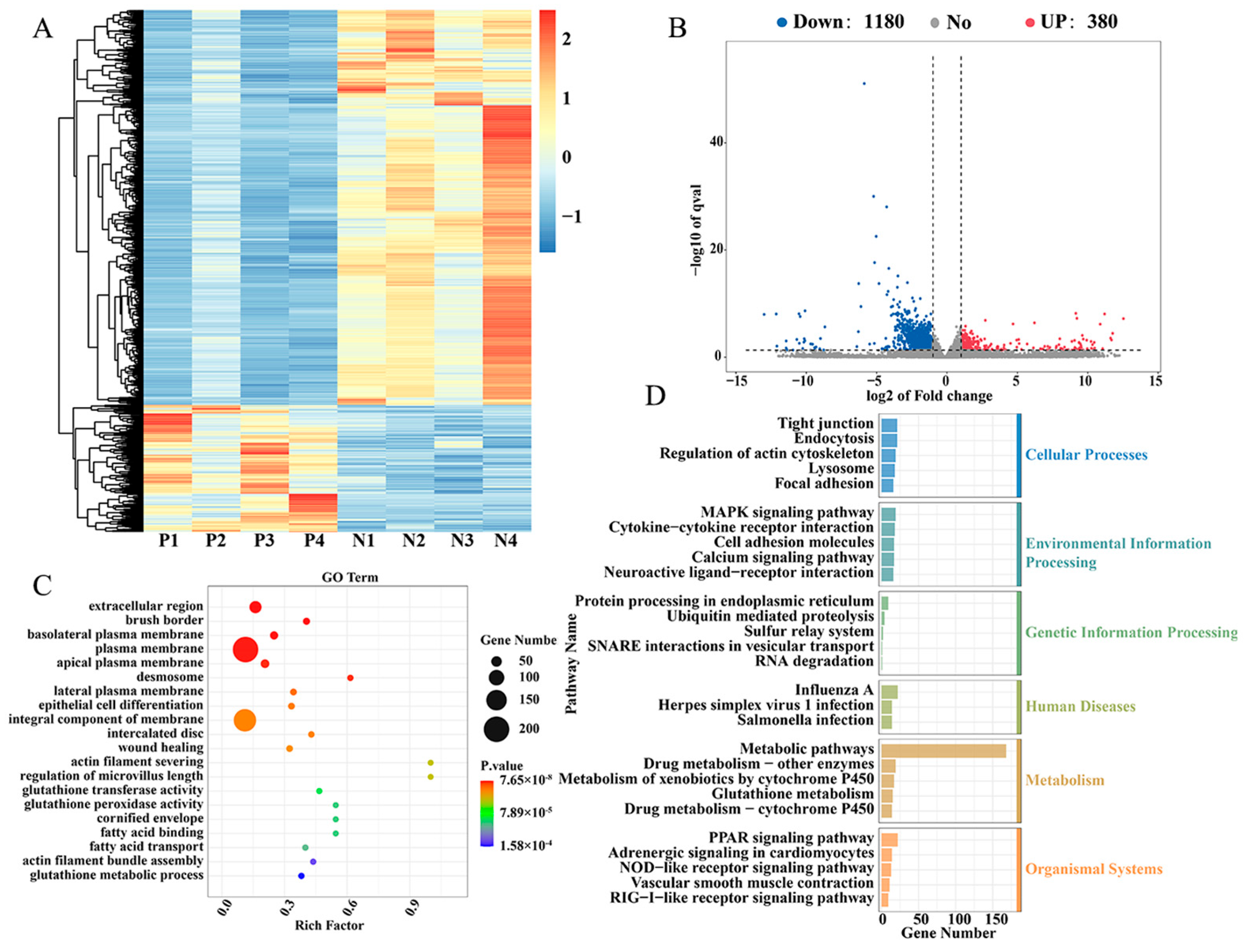
2.1. Different Expression of Genes in Cecal Tissues of Positive Group (P) and Negative Group (N) of Salmonella pullorum Infection

In order to investigate the genetic and molecular regulatory mechanisms potentially associated with Salmonella pullorum infection in recessive white feather broilers, a study was conducted in which the cecal tissues of the P with Salmonella pullorum infection (n = 4) and the N without Salmonella pullorum infection (n = 4) were subjected to transcriptome sequencing, and the results of the comparison between the sequencing data and the reference genomes are shown in Table S1. Utilizing the screening criteria of |log2FC| ≥ 1 and q < 0.05, 1560 DEGs were identified between the P and N groups, of which 380 genes were up-regulated and 1180 genes were down-regulated (Figure 1A,B). In order to explore the biological function of these DEGs, KEGG and GO enrichment pathway analysis was performed. GO enrichment analysis revealed that these DEGs were predominantly associated with protein binding, RNA transcription regulation, calcium ion binding, and gene expression regulation (Figure 1C). KEGG pathway enrichment analysis revealed that these DEGs were predominantly involved in metabolic signal transduction pathways, peroxisome proliferators-activated receptors (PPARs) signaling pathways, cell adhesion factors, and Salmonella infection (Figure 1D). These included AQP8, SLC26A3, CBS, IFI6, DDX60, IL8L1, IL8L2, etc. The top 20 differential genes are listed in Table 1.

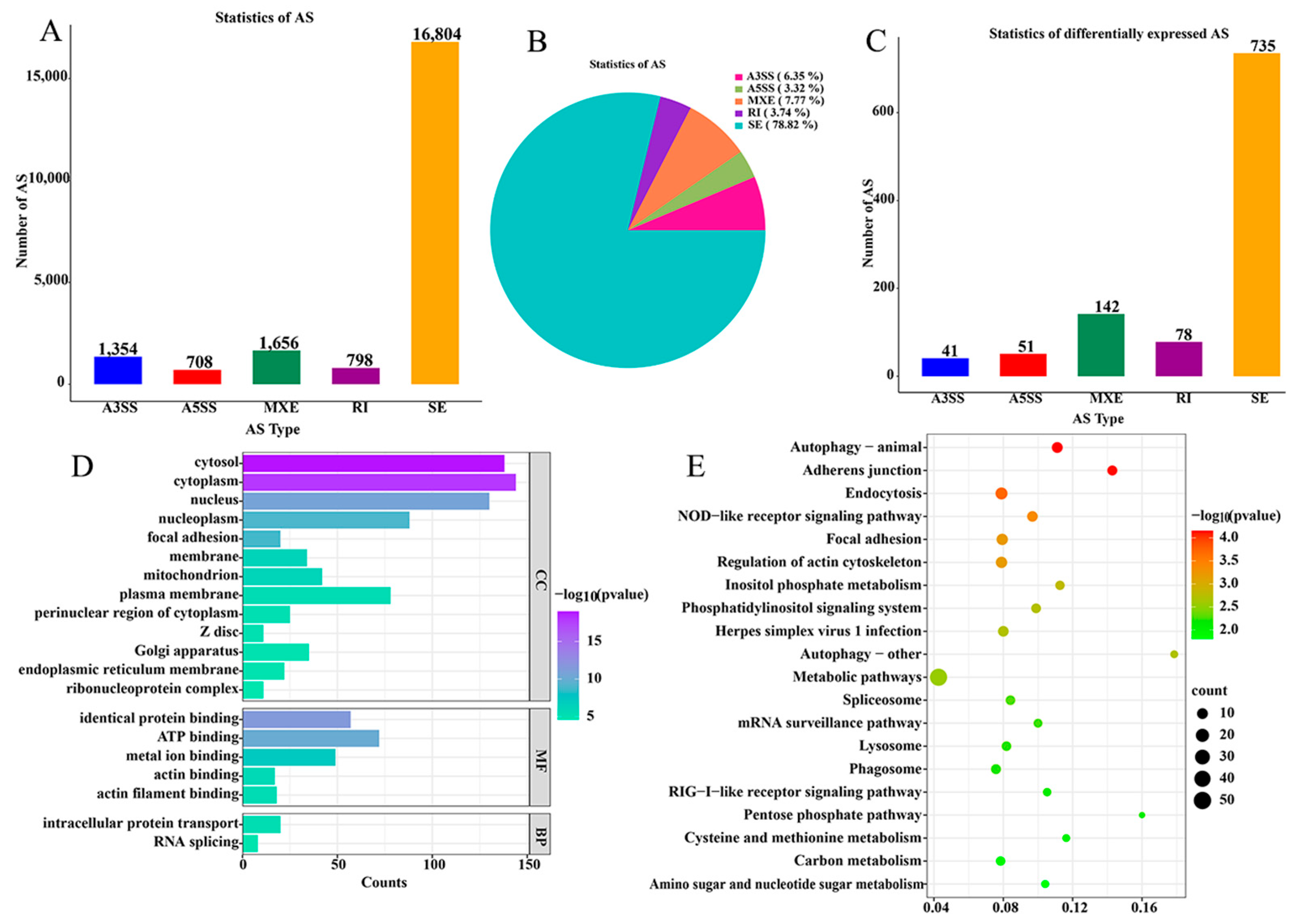
2.2. Differentially Expressed Alternative Splicings of Cecal Tissue in Positive (P) and Negative (N) Groups of Salmonella pullorum Infection

To investigate whether different transcripts are involved in the infection of Salmonella pullorum, we performed a variable splicing analysis of the mRNA data and found 21,320 ASs, of which 16,804 were SE, accounting for 78.82% of the total ASs (Figure 2A,B). All AS events were subjected to analysis using FDR < 0.05 and |IncLevel Difference| > 0.1 AS criteria for difference analysis. A total of 1047 ASs were found to be different, including 41 A3SS, 51 A5SS, 142 MXE, 78 RI, and 735 SE. (Figure 2C). The 1047 AS event was observed in 821 genes, which were subsequently analyzed using gene GO and KEGG. The results of GO and KEGG analyses showed that they were mainly enriched in cytoplasmic, cytosolic, and metabolic pathways (Figure 2D,E). Among them, genes related to immunity and inflammation are CBS, SLC6A9, ILDR2, OCRL, etc.

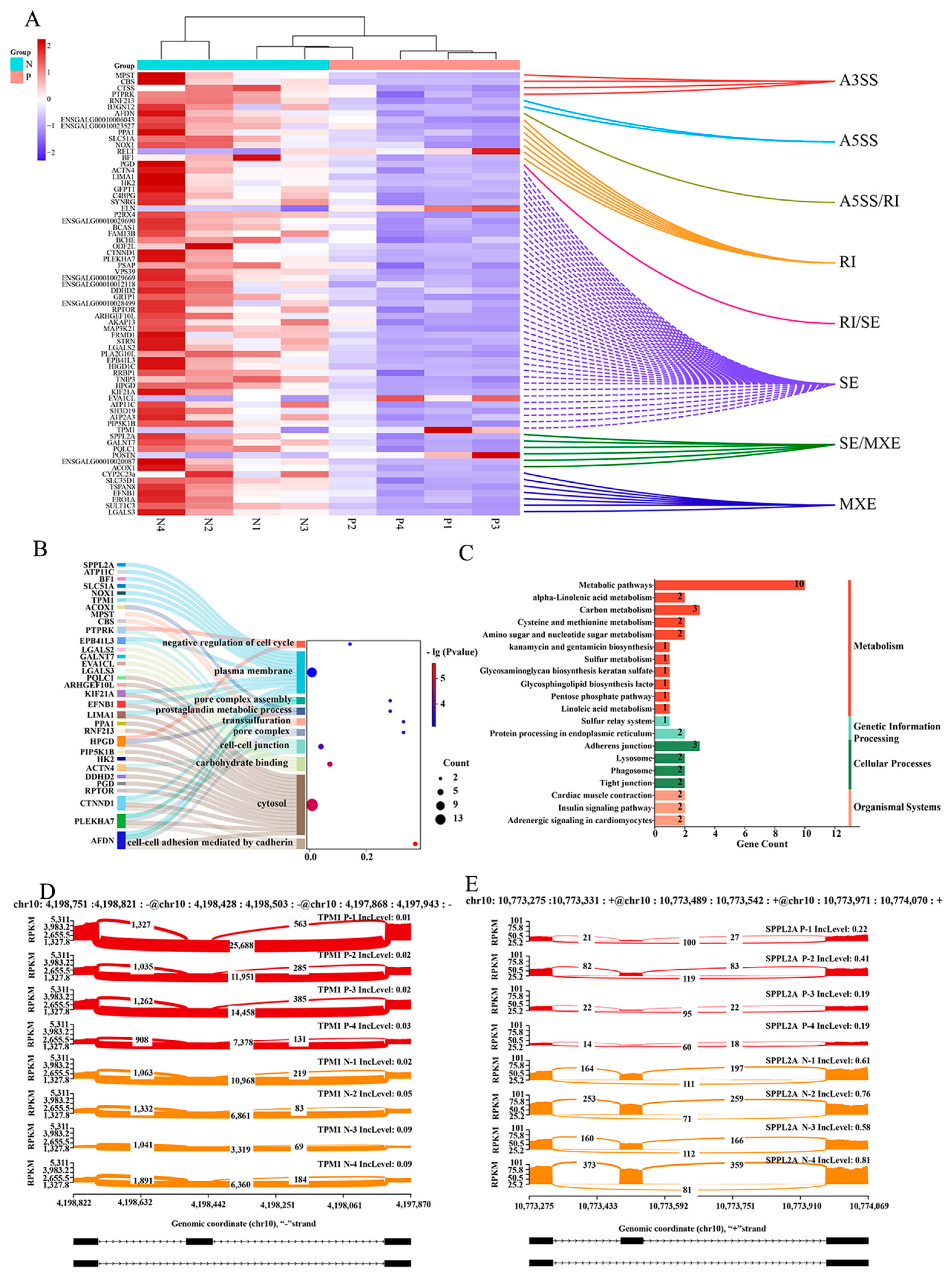
2.3. Combined DEGs and DEASs Analysis of Cecal Tissue from Positive (P) and Negative (N) Groups Infected with Salmonella pullorum

In order to further understand the changes of DEGs and DEASs in P and N groups after Salmonella infection, a joint analysis of DEGs and DEASs was performed. The analysis yielded results that indicated that 80 alternative splicing (AS) events were identified in 70 genes, and these ASs and gene expressions were simultaneously different. (Figure 3A). Subsequent GO and KEGG analyses of these 70 genes revealed that they were predominantly enriched in pathways related to the plasma membrane, cytoplasm, metabolism, and other processes (Figure 3B,C). These included CTNND1, TPM1, SPPL2A, etc. In this way, we visualized AS events with a high abundance of TPM1 and SPPL2A (Figure 3D,E).

2.4. The Metagenomic Sequencing of Cecal Tissue Was Conducted in the Positive (P) and Negative (N) Groups Infected with Salmonella pullorum

In order to further explore the changes in the intestinal microbiota in infected chickens after Salmonella pullorum infection, the metagenomic sequencing of fresh feces was performed on individuals in the P (n = 4) and N groups (n = 4). A total of 50.15 G of original data was obtained, and 40.36 G of effective data was obtained after pretreatment (Table 2). A composition analysis of the metagenomic data found that, at the gate level, the relative abundance of Actinobacteria (0.47% vs. 0.36%), Spirochaetes (0.45% vs. 0.30%), Synergistetes (0.27% vs. 0.23%), Euryarchaeota (0.26% vs. 0.14%), and Lentisphaerae (0.12% vs. 0.02%) was higher in the P group. In contrast, the N group exhibited a higher relative abundance of Bacteroidetes (31.27% vs. 27.92%), Firmicutes (16.05% vs. 13.03%), Proteobacteria (1.34% vs. 1.14%), and Deferribacteres (0.11% vs. 0.04%) (Figure 4A). At the genus level, g_Bacteroides and g_Akkermansia were more abundant in the P group. Conversely, the abundance of g_lactobacillus and g_pseudoflavonifractor was higher in the N group (Figure 4B,C). In order to distinguish biomarkers with significant abundance differences between positive and negative groups, LEfSe analysis was performed for the two groups. The LEfSe analysis revealed that Verrucomicrobia, Bacteroidaceae, and Akkermansia were more abundant in the P group. Conversely, Lactobacillus and Pseudoflavonifractor were abundant in the N group (Figure 4C).

3. Discussion

The highly infectious nature of the disease caused by Salmonella pullorum has a significant impact on performance and survival rates in the chicken production industry [6,8,29]. The current approach to purifying pullorum disease from the actual production involves the elimination of infected individuals. In this study, we collected cecal tissues from positive individuals (n = 4) and negative individuals (n = 4) to explore candidate genes and pathways associated with white dysentery infection in chickens at the transcriptome level. The KEGG enrichment analysis of DEGs revealed that these DEGs were predominantly enriched in metabolic signal transduction pathways, PPAR signaling pathways, cell adhesion factors, and Salmonella infection. Among them were AQP8, SLC26A3, CBS, IFI6, DDX60, IL8L1, IL8L2, and other candidate genes. The SLC26A3 protein plays an important role in maintaining the intestinal mucosal barrier, controlling intestinal inflammation, maintaining a stable intestinal pH value, and regulating intestinal flora [30,31,32]. SLC26A3 expression is decreased in inflammatory bowel disease. Kini et al. demonstrated that in SLC26A3 knockout mice, the cecum, colon, and mucosal adhesion flora were abnormal, with a loss of diversity, increased short-chain fatty acid production, and increased levels of pathogenic bacteria [33]. This result is consistent with the present study, in which SLC26A3 expression was reduced after infection with Salmonella pullorum infection, and beneficial bacteria were significantly reduced in the Salmonella pullorum infection group. IL8, a chemokine, has been demonstrated to play a pivotal role in the processes of inflammation and immune response within poultry species [34,35]. This assertion is further substantiated by the findings of Vu et al., which revealed that the expression level of IL8 in H5N1-resistant chickens exceeded that observed in H5N1-sensitive chickens [36]. Studies have demonstrated the pivotal role of cytokines as signaling molecules in the host’s defense against enteric pathogens, such as Salmonella. Following infection with Salmonella pullorum, there is an increase in IL8 expression within the host [37]. Furthermore, Swaggerty et al. demonstrated increased mRNA expression levels of proinflammatory cytokines IL6, IL8, and IL18 in individuals infected with Salmonella pullorum, despite the presence of only limited inflammatory infiltration [10,38]. This finding is consistent with the results of our study, which demonstrated a reduction in IL8 mRNA levels in chickens infected with Salmonella pullorum.
The application of alternative splicing analysis of mRNA data yielded the identification of 1047 distinct AS events in the cecum tissue of the P group and the N group. Furthermore, through the joint analysis with DEGs, it was found that 70 genes were not only differentially expressed but were also differentially spliced. These included CTNND1, TPM1, and SPPL2A, amongst others. Research has demonstrated that TPM1 generates multiple isomers through variable splicing and executes distinct functions under varying physiological and pathological conditions [39,40,41]. Gardina et al. demonstrated that there is a discrepancy between the AS of TPM1 colon cancer tumor tissue and normal colon tissue and that the isomer produced by the variable splicing is more highly expressed in colon cancer tumor tissue [42]. In this study, TPM1 IncLevel was found to be diminished in the P group, suggesting that exon jumps and the production of isomers are increased. This finding is in accordance with the results reported by Gardina et al.
Metagenomic sequencing analysis revealed significant changes in microflora abundance. Research has demonstrated that Salmonella has the capacity to destroy the dominant beneficial bacteria in the gut and modify the colonization of the host intestinal flora. This, in turn, results in the ability to resist the host’s normal intestinal flora and immune response, thereby creating a disadvantage for the normal intestinal flora [43,44,45]. Huang et al. demonstrated that Salmonella infection significantly reduced the relative abundance of Lachnoclostridium and Blautia, while phage CKT1 treatment significantly increased the proliferation of beneficial microflora in intestinal Firmicutes [46]. These included Lachnoclostridium, Ruminococcus, Lactobacillus, and pseudoflavonyifrtor. This finding is further corroborated by the study conducted by Ding et al., which demonstrated that the abundance of lactobacillus in the N group of Salmonella pullorum infected is higher than that in the P group [47]. This finding is corroborated by the present study, which demonstrates that the abundance of Firmicutes in the N group exceeds that of the P group, and that the abundance of Lactobacillus and Pseudoflavonifractor is higher in the N group.

4. Materials and Methods

4.1. Experimental Animal Management

In this study, subwing vein blood was collected from 100-day-old fourth-generation recessive white feather broilers. These chickens were bred by Guangzhou Jiangfeng Industrial Co., Ltd. (Guangzhou, Guangdong, China). A plate agglutination test was then performed to identify individuals infected with Salmonella pullorum. Subsequent to this, fresh feces were subjected to PCR, and the results of this analysis are presented in Figure S1. Individuals with plate agglutination results that were completely consistent with the PCR results were designated as positive or negative, and a total of eight individuals were euthanized. The cecum and its contents were then collected and placed in liquid ammonia, after which they were transported via dry ice to LC-Bio Technologies Co., Ltd. (Hangzhou, Zhejiang, China) for mRNA-seq and metagenomic sequencing, respectively. The animal experiment was approved by the Animal Health Professional Committee of South China Agricultural University (SCAU#2017015; 13 September 2017).

4.2. Serum Plate Agglutination Test

Subwing blood sampling was performed on 100-day-old recessive white feather broilers hens. Following the collection of blood, it was left to precipitate serum. Thereafter, serum and antigenic reagent (YDLH042, Shanghai, China) were added in a ratio of 1:1. The mixture was then shaken at room temperature for a period of 2–5 min. The presence of more than 50% agglutination was taken to indicate a positive result. In the absence of agglutination, a negative result was recorded. When the agglutination was observed to be between the two extremes, the result was deemed to be suspicious (Figure S2).

4.3. PCR Experiment

The extraction of deoxyribonucleic acid (DNA) was conducted utilizing a fecal DNA extraction kit (CW2091S, Shanghai, China). The subsequent PCR reaction was performed in strict accordance with the product instructions of the 2×Taq PCR MasterMix (B639295, Shanghai, China). The primer information is in Table S2.

4.4. mRNA-Seq of Cecal Tissues and Alternative Splicing Analysis

The total RNA was extracted from cecal tissues using TRIzol (Thermofisher,, CA, USA) according to the manufacturer’s instructions. The quantity and purity of the total RNA were then measured using a NanoDrop ND-1000 (NanoDrop, Wilmington, DE, USA) at Unikawa Bio, after which the RNA was analyzed using a Bioanalyzer 2100 (Agilent, CA, USA). The integrity of the RNA was detected, and mRNA sequencing was completed. Alternative splicing was detected using rMAST (rmats4.1.2), and the KOBAS software (http://bioinfo.org/kobas, accessed on 1 February 2025) was utilized to detect the enrichment of the Gene Ontology (GO) and Kyoto Encyclopedia of Genes and Genomes (KEGG) pathways in the differential genes [48]. This process was completed by LC-Bio Technologies Co., Ltd. (Hangzhou, Zhejiang, China).

4.5. DNA Extraction and Metagenomic Sequencing

The otal DNA was extracted from microbiome samples using the CTAB method. NovaSeq6000 was used for high-throughput sequencing, and the original sequencing data were assembled. MetaGeneMark (v3.26) was then used for CDS prediction, after which the predicted results were clustered. The reads of each sample were then compared to the CDS sequence library for TPM abundance calculation and species annotation information. Function annotation was achieved through comparison with various databases, including eggNOG, CAZy, CARD, PHI, MGEs, and VFDB. A statistical analysis of abundance and a comparative analysis of differences were performed at the species, function, and gene levels. This process was completed by LC-Bio Technologies Co., Ltd. (Hangzhou, Zhejiang, China).

5. Conclusions

In summary, in our study mRNA sequencing was utilized for the screening of candidate genes associated with Salmonella pullorum infection, including AQP8, SLC26A3, CBS, IFI6, DDX60, IL8L1, IL8L2, and others. Alternative splicing analysis identified the splicings associated with Salmonella pullorum infection, including CBS, SLC6A9, ILDR2, OCRL, CTNND1, TPM1, and SPPL2A. Metagenomic sequencing revealed that the relative abundance of p-verrucomicrobia increased and the relative abundance of p-Firmicutes decreased when chickens were infected Salmonella pullorum. The present study screened out candidate genes, AS isomers, and microbiota associated with Salmonella pullorum infection, thus providing a theoretical basis for the study of the regulatory mechanism and treatment of Salmonella pullorum infection. In subsequent studies, efforts should be made to restore the balance of beneficial microbes through targeted interventions, or by applying specific RNA editing techniques. This approach has the potential to offer a novel direction for the treatment and prevention of Salmonella pullorum infection.

Supplementary Materials

The following supporting information can be downloaded at: https://www.mdpi.com/article/10.3390/ijms26041448/s1.

Author Contributions

Conceptualization, X.C. and Z.F.; methodology, X.C. and Z.F.; software, X.C., Z.F., and J.W.; validation, C.Y. and J.W.; data curation, R.L., S.C., and X.W.; writing—original draft preparation, X.C.; writing—review and editing, X.C. and C.F.; visualization, X.C.; supervision, Q.L. and X.Z.; project administration, Q.L. and C.F.; funding acquisition, Q.L. and C.F. All authors have read and agreed to the published version of the manuscript.

Funding

This work was supported by STI2030—Major Projects, No.2023ZD04064.

Institutional Review Board Statement

The animal experiment was approved by the Animal Health Professional Committee of South China Agricultural University.

Informed Consent Statement

Not applicable.

Data Availability Statement

The data sets that have been either generated or analyzed during the course of this study are available for acquisition from the corresponding authors.

Conflicts of Interest

The authors declare that they have no conflicts of interest in the study.

Abbreviations

The following abbreviations are used in this manuscript:
DEGsDifferentially expressed genes
DEASsDifferentially expressed alternative splicings
P group positive group
N group negative group
ASAlternative splicing

References

  1. Liu, J.; Zhou, J.; Li, J.; Bao, H. Identification of candidate genes associated with slaughter traits in F2 chicken population using genome-wide association study. Anim. Genet. 2021, 52, 532–535. [Google Scholar] [CrossRef]
  2. Foley, S.L.; Johnson, T.J.; Ricke, S.C.; Nayak, R.; Danzeisen, J. Salmonella pathogenicity and host adaptation in chicken-associated serovars. Microbiol. Mol. Biol. Rev. 2013, 77, 582–607. [Google Scholar] [CrossRef] [PubMed]
  3. Li, X.; Nie, C.; Liu, Y.; Chen, Y.; Lv, X.; Wang, L.; Zhang, J.; Li, K.; Jia, Y.; Ban, L.; et al. A genome-wide association study explores the genetic determinism of host resistance to Salmonella pullorum infection in chickens. Genet. Sel. Evol. 2019, 51, 51. [Google Scholar] [CrossRef] [PubMed]
  4. He, T.; Hu, X.; Mi, J.; Hu, H.; Wang, H.; Qi, X.; Gao, L.; Zhang, Y.; Liu, C.; Wang, S.; et al. Ligilactobacillus salivarius XP132 with antibacterial and immunomodulatory activities inhibits horizontal and vertical transmission of Salmonella pullorum in chickens. Poult. Sci. 2024, 103, 104086. [Google Scholar] [CrossRef]
  5. Hu, Y.; Wang, Z.; Qiang, B.; Xu, Y.; Chen, X.; Li, Q.; Jiao, X. Loss and Gain in the Evolution of the Salmonella enterica Serovar Gallinarum Biovar Pullorum Genome. mSphere 2019, 4, e00627-18. [Google Scholar] [CrossRef]
  6. Kabeta, T.; Tolosa, T.; Duchateau, L.; Van Immerseel, F.; Antonissen, G. Prevalence and serotype of poultry salmonellosis in Africa: A systematic review and meta-analysis. Avian Pathol. 2024, 53, 325–349. [Google Scholar] [CrossRef] [PubMed]
  7. Shivaprasad, H.L.; Timoney, J.F.; Morales, S.; Lucio, B.; Baker, R.C. Pathogenesis of Salmonella enteritidis infection in laying chickens. I. Studies on egg transmission, clinical signs, fecal shedding, and serologic responses. Avian Dis. 1990, 34, 548–557. [Google Scholar] [CrossRef]
  8. Niu, Q.; Wang, X.; Qi, X.; Cao, C.; Yang, K.; Gu, C.; Zhou, Z.; Huang, Q. Identification of the gut microbiota affecting Salmonella pullorum and their relationship with reproductive performance in hens. Front. Microbiol. 2023, 14, 1216542. [Google Scholar] [CrossRef] [PubMed]
  9. Geng, S.; Jiao, X.; Barrow, P.; Pan, Z.; Chen, X. Virulence determinants of Salmonella Gallinarum biovar Pullorum identified by PCR signature-tagged mutagenesis and the spiC mutant as a candidate live attenuated vaccine. Vet. Microbiol. 2014, 168, 388–394. [Google Scholar] [CrossRef] [PubMed]
  10. Henderson, S.C.; Bounous, D.I.; Lee, M.D. Early events in the pathogenesis of avian salmonellosis. Infect. Immun. 1999, 67, 3580–3586. [Google Scholar] [CrossRef]
  11. Chappell, L.; Kaiser, P.; Barrow, P.; Jones, M.A.; Johnston, C.; Wigley, P. The immunobiology of avian systemic salmonellosis. Vet. Immunol. Immunopathol. 2009, 128, 53–59. [Google Scholar] [CrossRef] [PubMed]
  12. Li, X.; Nie, C.; Zhang, Z.; Wang, Q.; Shao, P.; Zhao, Q.; Chen, Y.; Wang, D.; Li, Y.; Jiao, W.; et al. Evaluation of genetic resistance to Salmonella pullorum in three chicken lines. Poult. Sci. 2018, 97, 764–769. [Google Scholar] [CrossRef] [PubMed]
  13. Liu, X.Q.; Wang, F.; Jin, J.; Zhou, Y.G.; Ran, J.S.; Feng, Z.Q.; Wang, Y.; Liu, Y.P. MyD88 Polymorphisms and Association with Susceptibility to Salmonella pullorum. BioMed Res. Int. 2015, 2015, 692973. [Google Scholar] [CrossRef] [PubMed]
  14. Setta, A.; Barrow, P.A.; Kaiser, P.; Jones, M.A. Immune dynamics following infection of avian macrophages and epithelial cells with typhoidal and non-typhoidal Salmonella enterica serovars; bacterial invasion and persistence, nitric oxide and oxygen production, differential host gene expression, NF-kappaB signalling and cell cytotoxicity. Vet. Immunol. Immunopathol. 2012, 146, 212–224. [Google Scholar] [CrossRef]
  15. Nikom, D.; Zheng, S. Alternative splicing in neurodegenerative disease and the promise of RNA therapies. Nat. Rev. Neurosci. 2023, 24, 457–473. [Google Scholar] [CrossRef]
  16. Zhao, Z.; Cai, Q.; Zhang, P.; He, B.; Peng, X.; Tu, G.; Peng, W.; Wang, L.; Yu, F.; Wang, X. N6-Methyladenosine RNA Methylation Regulator-Related Alternative Splicing (AS) Gene Signature Predicts Non-Small Cell Lung Cancer Prognosis. Front. Mol. Biosci. 2021, 8, 657087. [Google Scholar] [CrossRef]
  17. Low, E.N.D.; Mokhtar, N.M.; Wong, Z.; Raja Ali, R.A. Colonic Mucosal Transcriptomic Changes in Patients with Long-Duration Ulcerative Colitis Revealed Colitis-Associated Cancer Pathways. J. Crohns Colitis 2019, 13, 755–763. [Google Scholar] [CrossRef] [PubMed]
  18. Sun, C.; Zeng, B.; Zhou, J.; Li, N.; Li, M.; Zhu, C.; Xie, S.; Wang, Y.; Wang, S.; Wang, X. Analysis of SLC genes alternative splicing identifies the SLC7A6 RI isoform as a therapeutic target for colorectal cancer. Cancer Sci. 2024, 116, 233–247. [Google Scholar] [CrossRef]
  19. Simon, K.; de Vries Reilingh, G.; Kemp, B.; Lammers, A. Development of ileal cytokine and immunoglobulin expression levels in response to early feeding in broilers and layers. Poult. Sci. 2014, 93, 3017–3027. [Google Scholar] [CrossRef] [PubMed]
  20. Wigley, P. Salmonella enterica in the Chicken: How it has Helped Our Understanding of Immunology in a Non-Biomedical Model Species. Front. Immunol. 2014, 5, 482. [Google Scholar] [CrossRef]
  21. Liu, S.; Zhao, L.; Zhai, Z.; Zhao, W.; Ding, J.; Dai, R.; Sun, T.; Meng, H. Porcine Epidemic Diarrhea Virus Infection Induced the Unbalance of Gut Microbiota in Piglets. Curr. Microbiol. 2015, 71, 643–649. [Google Scholar] [CrossRef] [PubMed]
  22. Nicholson, J.K.; Holmes, E.; Kinross, J.; Burcelin, R.; Gibson, G.; Jia, W.; Pettersson, S. Host-gut microbiota metabolic interactions. Science 2012, 336, 1262–1267. [Google Scholar] [CrossRef]
  23. Dheer, R.; Davies, J.M.; Quintero, M.A.; Damas, O.M.; Deshpande, A.R.; Kerman, D.H.; Sawyer, W.P.; Pignac-Kobinger, J.; Ban, Y.; Fernandez, I.; et al. Microbial Signatures and Innate Immune Gene Expression in Lamina Propria Phagocytes of Inflammatory Bowel Disease Patients. Cell. Mol. Gastroenterol. Hepatol. 2020, 9, 387–402. [Google Scholar] [CrossRef] [PubMed]
  24. Ducatelle, R.; Goossens, E.; De Meyer, F.; Eeckhaut, V.; Antonissen, G.; Haesebrouck, F.; Van Immerseel, F. Biomarkers for monitoring intestinal health in poultry: Present status and future perspectives. Vet. Res. 2018, 49, 43. [Google Scholar] [CrossRef]
  25. Kogut, M.H.; Arsenault, R.J. Editorial: Gut Health: The New Paradigm in Food Animal Production. Front. Vet. Sci. 2016, 3, 71. [Google Scholar] [CrossRef] [PubMed]
  26. Shang, Y.; Kumar, S.; Oakley, B.; Kim, W.K. Chicken Gut Microbiota: Importance and Detection Technology. Front. Vet. Sci. 2018, 5, 254. [Google Scholar] [CrossRef]
  27. Wang, W.W.; Jia, H.J.; Zhang, H.J.; Wang, J.; Lv, H.Y.; Wu, S.G.; Qi, G.H. Supplemental Plant Extracts From Flos lonicerae in Combination With Baikal skullcap Attenuate Intestinal Disruption and Modulate Gut Microbiota in Laying Hens Challenged by Salmonella pullorum. Front. Microbiol. 2019, 10, 1681. [Google Scholar] [CrossRef]
  28. Aziz, Q.; Dore, J.; Emmanuel, A.; Guarner, F.; Quigley, E.M. Gut microbiota and gastrointestinal health: Current concepts and future directions. Neurogastroenterol. Motil. 2013, 25, 4–15. [Google Scholar] [CrossRef]
  29. Kang, X.; Yang, Y.; Meng, C.; Wang, X.; Liu, B.; Geng, S.; Jiao, X.; Pan, Z. Safety and protective efficacy of Salmonella pullorum spiC and rfaH deletion rough mutant as a live attenuated DIVA vaccine candidate. Poult. Sci. 2022, 101, 101655. [Google Scholar] [CrossRef]
  30. Kumar, A.; Priyamvada, S.; Ge, Y.; Jayawardena, D.; Singhal, M.; Anbazhagan, A.N.; Chatterjee, I.; Dayal, A.; Patel, M.; Zadeh, K.; et al. A Novel Role of SLC26A3 in the Maintenance of Intestinal Epithelial Barrier Integrity. Gastroenterology 2021, 160, 1240–1255.e1243. [Google Scholar] [CrossRef]
  31. Amiri, M.; Jiang, M.; Salari, A.; Xiu, R.; Alper, S.L.; Seidler, U.E. Reduced surface pH and upregulated AE2 anion exchange in SLC26A3-deleted polarized intestinal epithelial cells. Am. J. Physiol. Cell Physiol. 2024, 326, C829–C842. [Google Scholar] [CrossRef] [PubMed]
  32. Jayawardena, D.; Priyamvada, S.; Kageyama, T.; White, Z.; Kumar, A.; Griggs, T.F.; Majumder, A.; Akram, R.; Anbazhagan, A.N.; Sano, T.; et al. Loss of SLC26A3 Results in Colonic Mucosal Immune Dysregulation via Epithelial-Immune Cell Crosstalk. Cell. Mol. Gastroenterol. Hepatol. 2023, 15, 903–919. [Google Scholar] [CrossRef] [PubMed]
  33. Kini, A.; Singh, A.K.; Riederer, B.; Yang, I.; Tan, X.; di Stefano, G.; Tan, Q.; Xiao, F.; Xia, W.; Suerbaum, S.; et al. Slc26a3 deletion alters pH-microclimate, mucin biosynthesis, microbiome composition and increases the TNFalpha expression in murine colon. Acta Physiol. 2020, 230, e13498. [Google Scholar] [CrossRef] [PubMed]
  34. Dai, M.; Zhu, S.; An, Z.; You, B.; Li, Z.; Yao, Y.; Nair, V.; Liao, M. Dissection of key factors correlating with H5N1 avian influenza virus driven inflammatory lung injury of chicken identified by single-cell analysis. PLoS Pathog. 2023, 19, e1011685. [Google Scholar] [CrossRef] [PubMed]
  35. Song, X.; Hou, M.; Jiang, H.; Shen, X.; Xue, M.; Shao, Y.; Wang, L.; He, Q.; Zheng, L.; Tu, J.; et al. Hcp2a of type VI secretion system contributes to IL8 and IL1beta expression of chicken tracheal epithelium by affecting APEC colonization. Res. Vet. Sci. 2020, 132, 279–284. [Google Scholar] [CrossRef] [PubMed]
  36. Vu, T.H.; Hong, Y.; Truong, A.D.; Lee, J.; Lee, S.; Song, K.D.; Cha, J.; Dang, H.V.; Tran, H.T.T.; Lillehoj, H.S.; et al. Cytokine-cytokine receptor interactions in the highly pathogenic avian influenza H5N1 virus-infected lungs of genetically disparate Ri chicken lines. Anim. Biosci. 2022, 35, 367–376. [Google Scholar] [CrossRef] [PubMed]
  37. Eckmann, L.; Kagnoff, M.F. Cytokines in host defense against Salmonella. Microbes Infect. 2001, 3, 1191–1200. [Google Scholar] [CrossRef]
  38. Swaggerty, C.L.; Kogut, M.H.; Ferro, P.J.; Rothwell, L.; Pevzner, I.Y.; Kaiser, P. Differential cytokine mRNA expression in heterophils isolated from Salmonella-resistant and -susceptible chickens. Immunology 2004, 113, 139–148. [Google Scholar] [CrossRef]
  39. Vad, O.B.; Angeli, E.; Liss, M.; Ahlberg, G.; Andreasen, L.; Christophersen, I.E.; Hansen, C.C.; Moller, S.; Hellsten, Y.; Haunsoe, S.; et al. Loss of Cardiac Splicing Regulator RBM20 Is Associated With Early-Onset Atrial Fibrillation. JACC Basic. Transl. Sci. 2024, 9, 163–180. [Google Scholar] [CrossRef]
  40. Denkena, J.; Zaisser, A.; Merz, B.; Klinger, B.; Kuhl, D.; Bluthgen, N.; Hermey, G. Neuronal activity regulates alternative exon usage. Mol. Brain 2020, 13, 148. [Google Scholar] [CrossRef]
  41. Yang, Y.; Lee, G.C.; Nakagaki-Silva, E.; Huang, Y.; Peacey, M.; Partridge, R.; Gooding, C.; Smith, C.W.J. Cell-type specific regulator RBPMS switches alternative splicing via higher-order oligomerization and heterotypic interactions with other splicing regulators. Nucleic Acids Res. 2023, 51, 9961–9982. [Google Scholar] [CrossRef] [PubMed]
  42. Gardina, P.J.; Clark, T.A.; Shimada, B.; Staples, M.K.; Yang, Q.; Veitch, J.; Schweitzer, A.; Awad, T.; Sugnet, C.; Dee, S.; et al. Alternative splicing and differential gene expression in colon cancer detected by a whole genome exon array. BMC Genom. 2006, 7, 325. [Google Scholar] [CrossRef] [PubMed]
  43. Lawley, T.D.; Bouley, D.M.; Hoy, Y.E.; Gerke, C.; Relman, D.A.; Monack, D.M. Host transmission of Salmonella enterica serovar Typhimurium is controlled by virulence factors and indigenous intestinal microbiota. Infect. Immun. 2008, 76, 403–416. [Google Scholar] [CrossRef]
  44. Stecher, B.; Robbiani, R.; Walker, A.W.; Westendorf, A.M.; Barthel, M.; Kremer, M.; Chaffron, S.; Macpherson, A.J.; Buer, J.; Parkhill, J.; et al. Salmonella enterica serovar typhimurium exploits inflammation to compete with the intestinal microbiota. PLoS Biol. 2007, 5, 2177–2189. [Google Scholar] [CrossRef] [PubMed]
  45. Thiennimitr, P.; Winter, S.E.; Baumler, A.J. Salmonella, the host and its microbiota. Curr. Opin. Microbiol. 2012, 15, 108–114. [Google Scholar] [CrossRef] [PubMed]
  46. Huang, J.; Liang, L.; Cui, K.; Li, P.; Hao, G.; Sun, S. Salmonella phage CKT1 significantly relieves the body weight loss of chicks by normalizing the abnormal intestinal microbiome caused by hypervirulent Salmonella pullorum. Poult. Sci. 2022, 101, 101668. [Google Scholar] [CrossRef] [PubMed]
  47. Ding, J.; Zhou, H.; Luo, L.; Xiao, L.; Yang, K.; Yang, L.; Zheng, Y.; Xu, K.; He, C.; Han, C.; et al. Heritable Gut Microbiome Associated with Salmonella enterica Serovar Pullorum Infection in Chickens. mSystems 2021, 6, e01192-20. [Google Scholar] [CrossRef] [PubMed]
  48. Wu, J.; Mao, X.; Cai, T.; Luo, J.; Wei, L. KOBAS server: A web-based platform for automated annotation and pathway identification. Nucleic Acids Res. 2006, 34, W720–W724. [Google Scholar] [CrossRef]
Figure 1. mRNA-seq analysis of cecal tissue in positive group (P) and negative group (N) of Salmonella pullorum infection. (A,B) Heat map and volcano plot for N vs. P DEGs. (C) GO function enrichment analysis for DEGs. X axis: gene number; Y axis: GO terms. (D) KEGG pathways classification analysis for DEGs. X axis: gene number; Y axis: pathway.
Ijms 26 01448 g001
Figure 2. AS analysis of cecal tissue in the positive group (P) and the negative group (N) of Salmonella pullorum infection. (A) The following statistics were collated on the AS type, including A3SS, A5SS, MXE, RI, and SE. (B) The proportion of A3SS, A5SS, MXE, RI, and SE was counted. (C) DEASs in A3SS, A5SS, MXE, RI, and SE. (D) The GO function enrichment analysis for DEASs. (E) The KEGG pathways classification analysis for DEASs.
Ijms 26 01448 g002
Figure 3. A combined analysis of DEGs and DEASs. (A) A heat map of differentially expressed and AS genes and their AS types. (B,C) The GO and KEGG pathways enrichment analyses for differentially expressed and AS genes. (D,E) The visual presentation is intended to demonstrate the phenomenon of differential AS on TPM1 and SPPL2A.
Ijms 26 01448 g003
Figure 4. Microbiological analysis of Salmonella pullorum infection in the P group and the N group. (A) The top 20 species in terms of phylum-level abundance. (B) The top 20 species in terms of genus-level abundance. (C) LEfSe analyzed biomarkers that were significantly different in the Salmonella pullorum infection N and P groups.
Ijms 26 01448 g004
Table 1. The top 20 genes were differentially expressed in the negative group and positive group of Salmonella pullorum infection.
Gene NameN ExpressionP Expressionlog2(FC) (N/P)Up Down
AQP88631146.75−5.875936928down
ENSGALG0001001781210,723.5298−5.212645245down
RSAD22291.75118−4.288236383down
ENSGALG0001000170745,105.251414.75−5.028950395down
CBS94,320.752736.25−5.147365626down
ENSGALG000100217634732.75269−4.133430962down
ENSGALG000100050122873.25257.5−3.485079358down
SCNN1A4646.25674.25−2.809438512down
DDX605710206−4.830147732down
ENSGALG000100177195177.5−6.278950066down
ANPEP20,453.251817.75−3.531739751down
SLC26A31882104.75−4.179327946down
IFI618,342986.5−4.269812813down
EDN2532.7598−2.453229646down
COBL1703.5463.5−1.897709004down
SLC30A101501.25111.75−3.790121129down
EPSTI12068406.25−2.365929024down
SERPINB13981.75401.5−3.35058651down
LAMC21558.75138−3.512285305down
KRT4022,899.51603−3.858303875down
Table 2. The metagenomic sequencing data of Salmonella pullorum infection positive group and negative group.
SampleOriginal Data (G)Valid Data (G)Valid Data Ratio (%)Q20%Q30%GC%
P16.535.4589.5298.5695.5252.21
P26.355.3290.0498.5095.4351.18
P36.485.2988.3398.4995.4350.28
P46.345.1688.0298.5195.4952.07
N15.314.2687.1498.6495.8152.06
N26.214.7283.5098.5995.7051.47
N36.445.0585.8698.5895.7151.62
N46.495.1185.7298.6995.8851.20
Disclaimer/Publisher’s Note: The statements, opinions and data contained in all publications are solely those of the individual author(s) and contributor(s) and not of MDPI and/or the editor(s). MDPI and/or the editor(s) disclaim responsibility for any injury to people or property resulting from any ideas, methods, instructions or products referred to in the content.

