Article

Multi-Target Botanical Complex Attenuates Cellular Senescence via Bidirectional P21/P53/SIRT1 Regulation: Dual Model Validation

1
College of Pharmacy, Qinghai University, Xining 810016, China
2
College of Tibetan Medicine, Qinghai University, Xining 810016, China
*
Authors to whom correspondence should be addressed.
These authors contributed equally to this work.
Int. J. Mol. Sci. 2025, 26(23), 11394; https://doi.org/10.3390/ijms262311394
Submission received: 19 October 2025 / Revised: 20 November 2025 / Accepted: 23 November 2025 / Published: 25 November 2025
(This article belongs to the Section Biochemistry)

Abstract

Cellular senescence is a pivotal driver of aging and age-related diseases. This study aims to systematically investigate the anti-senescence effects and molecular mechanisms of a multi-component botanical complex (SBT) using both a D-galactose-induced senescence model in H9c2 cardiomyocytes and an H2O2-induced accelerated aging model in zebrafish. The SBT complex comprises Solms-laubachia eurycarpa, Bergenia purpurascens, Laccifer lacca, and Glycyrrhiza uralensis. Results demonstrated that SBT treatment significantly enhanced cell viability (increased from 52% to 85%) and reduced senescence-associated β-galactosidase (SA-β-gal) activity (from 41.2% to 20%). At the molecular level, SBT exerted bidirectional regulation of the P21/P53/SIRT1 axis, coordinately downregulating the pro-senescence proteins P53 and P21 while upregulating the longevity-associated deacetylase SIRT1. It also modulated the balance of apoptosis-related genes by suppressing Bax and enhancing Bcl-2 expression. In the zebrafish model, SBT significantly strengthened the antioxidant defense system, as indicated by increased activities of superoxide dismutase (SOD) and catalase (CAT), elevated glutathione (GSH) levels, and reduced malondialdehyde (MDA) content. These findings confirm that SBT exerts potent anti-senescence effects through bidirectional regulation of the P21/P53/SIRT1 signaling axis, enhanced antioxidant capacity, and inhibition of apoptosis, thereby providing a mechanistic foundation for the development of natural product-based interventions against aging and related diseases.

1. Introduction

Cellular senescence, characterized by the irreversible arrest of the cell cycle, is a fundamental hallmark of aging and a key driver of age-related functional decline and pathologies [1,2]. The molecular underpinnings of aging involve a complex network of processes, including genomic instability [3], epigenetic alterations [4], loss of proteostasis [5], and deregulated nutrient sensing [6]. Central among these are the p53 (Tumor Protein P53) and p21 (Cyclin-Dependent Kinase Inhibitor 1A) axis, and the sirtuin family of proteins, particularly SIRT1 (Sirtuin 1), which serve as pivotal regulators of the senescence program [7]. The p53 tumor suppressor protein is activated in response to diverse cellular stresses, such as DNA damage [8], and transcriptionally upregulates p21 [9]. p21, a cyclin-dependent kinase inhibitor, subsequently induces G1 phase cell cycle arrest, thereby promoting the senescent phenotype [10]. Conversely, SIRT1, an NAD+-dependent deacetylase [11], exerts anti-aging effects by deacetylating key substrates including p53, FOXOs (Forkhead box O transcription factors), and PGC-1α (Peroxisome proliferator-activated receptor gamma coactivator 1-alpha) [12]. This deacetylation modulates cellular stress resistance [13], metabolic homeostasis, and inflammatory responses [14]. Notably, SIRT1-mediated deacetylation directly inhibits p53’s transcriptional activity [15], forming a delicate bidirectional regulatory circuit that finely balances cellular senescence and survival [16].
Despite significant advances in understanding aging biology, the current therapeutic arsenal for intervening in the aging process remains limited [17,18]. Pharmacological agents like rapamycin, an mTOR inhibitor, have demonstrated lifespan extension in model organisms but are often hampered by side effects and challenges in clinical translation [19]. In this context, natural products present promising alternatives due to their inherent multi-target activities and favorable safety profiles [20]. Botanical preparations from unique ecosystems, such as high-altitude regions, have attracted particular interest for their distinctive phytochemical compositions and traditional ethnopharmacological uses [21]. A notable example is Srolo Bzhtang (SBT), a defined multi-herbal Tibetan formulation composed of Solms-laubachia eurycarpa, Bergenia purpurascens, Laccifer lacca, and Glycyrrhiza uralensis. Previous phytochemical studies have identified a complex mixture of bioactive constituents in SBT, including flavonoids (e.g., liquiritin and isoliquiritin from Glycyrrhiza uralensis), phenolic acids (e.g., bergenin from Bergenia purpurascens), tannins, and unique polysaccharides, which collectively contribute to its observed pharmacological effects [22]. Prior pharmacological investigations have confirmed SBT’s anti-inflammatory and antioxidant properties, primarily attributed to its ability to scavenge reactive oxygen species (ROS) and inhibit inflammatory cytokine signaling [23,24,25]. Building on this foundation, recent studies have further elucidated its efficacy in ameliorating pulmonary hypertension and attenuating airway inflammation [26,27,28]. However, its potential to directly counteract cellular aging, particularly within cardiac contexts and through modulation of the core P21/P53/SIRT1 senescence axis, remains largely unexplored and represents a significant gap in knowledge.
This study was therefore designed to systematically investigate the anti-senescence capacity and underlying molecular mechanisms of SBT. Utilizing a dual-model validation approach—comprising a D-galactose-induced senescence model in H9c2 cardiomyocytes and an H2O2-induced accelerated aging model in zebrafish [29,30]—we comprehensively evaluated the extract’s efficacy in mitigating senescence-associated phenotypes, oxidative stress parameters, and key molecular alterations within the P21/P53/SIRT1 signaling axis. Our integrated findings not only illuminate the novel anti-aging mechanisms of SBT but also position it as a compelling multi-target botanical candidate for development as a serotherapeutic intervention against age-related degenerative diseases.

2. Results

2.1. SBT Treatment Mitigates D-Galactose-Induced Oxidative Stress in H9c2 Cardiomyocytes

The protective effects of SBT against D-galactose (GAL)-induced oxidative damage in H9c2 cardiomyocytes were evaluated by measuring cell viability using the CCK-8 assay and intracellular ROS levels via fluorescence staining. Exposure to GAL significantly reduced cell viability to 52% of the control level (set as 100%). In contrast, SBT intervention markedly restored the survival rate to 85 ± 5.2% (p < 0.0001; Figure 1A). Fluorescence imaging revealed substantial ROS accumulation in the GAL-treated group, which was significantly attenuated by SBT treatment, indicating its potent antioxidant capacity (Figure 1C). These results demonstrate that SBT effectively alleviates GAL-induced cardiomyocyte injury through its antioxidant activity.

2.2. SBT Treatment Attenuates D-Galactose-Induced Cellular Senescence in H9c2 Cardiomyocytes

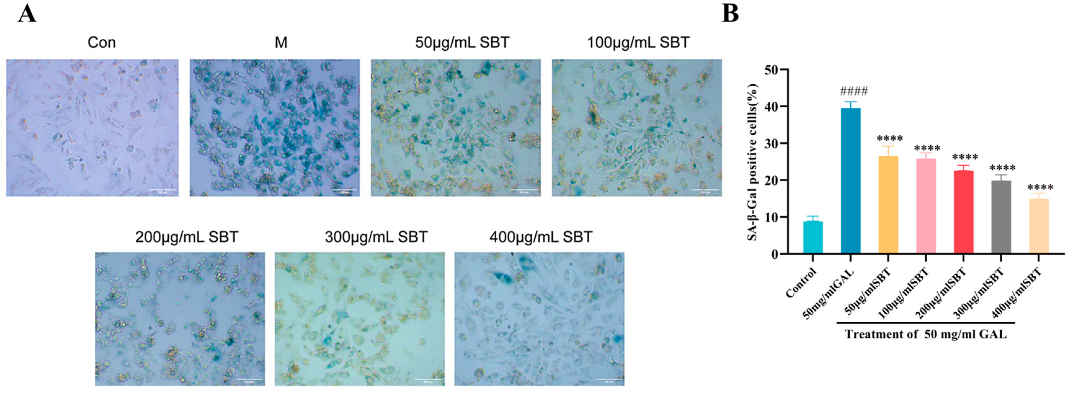
To investigate the anti-senescence effect of SBT, H9c2 cells were treated with GAL to establish a senescence model, which was assessed by SA-β-gal staining. The model group exhibited a significant increase in senescence burden (41.2% vs. control, p < 0.0001). In contrast, SBT treatment substantially reversed this effect, reducing the proportion of SA-β-gal-positive cells to 20 ± 5.0% (p < 0.0001; Figure 2). These results indicate that SBT effectively eliminates GAL-induced senescent cells.

2.3. SBT Alleviates D-Galactose-Induced Apoptosis in H9c2 Cardiomyocytes

To determine if SBT could inhibit programmed cell death in senescent H9c2 cells, we employed TUNEL staining. The results revealed that SBT administration markedly decreased the apoptotic rate compared to the GAL group (Figure 3A), underscoring its anti-apoptotic efficacy. In summary, these results indicate that SBT reduces apoptosis in H9c2 cardiomyocytes triggered by D-galactose.

2.4. SBT Modulates the P53/P21/SIRT1 Pathway in D-Galactose-Induced H9c2 Senescent Cells

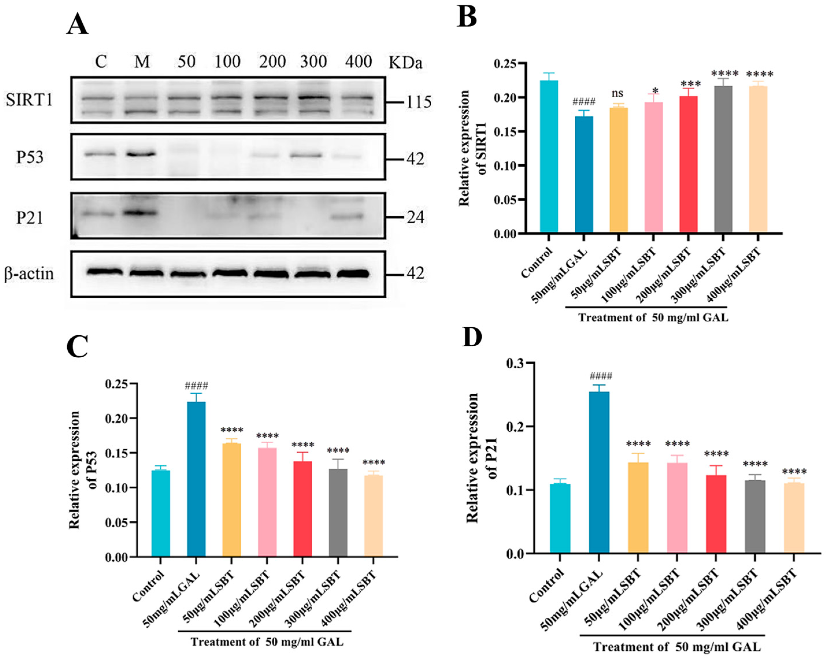
The P21/P53/SIRT1 axis serves as a core regulatory network governing senescence, integrating signals that modulate cell cycle progression, DNA damage response, and oxidative stress homeostasis. To explore SBT’s impact on this pathway, Western blotting quantified P21, P53, and SIRT1 protein levels. Our data showed that SBT treatment led to the downregulation of the pro-senescence proteins P21 and P53 (Figure 4A). Conversely, there was a marked increase in SIRT1 expression (Figure 4A), implicating the activation of this deacetylase in the cardioprotective mechanism of SBT. In summary, these results indicate that SBT modulates the P53/P21/SIRT1 pathway in H9c2 cardiomyocytes, suggesting a mechanism by which it may alleviate cellular senescence.

2.5. SBT Improves the Survival and Cardiac Function of H2O2-Treated Zebrafish

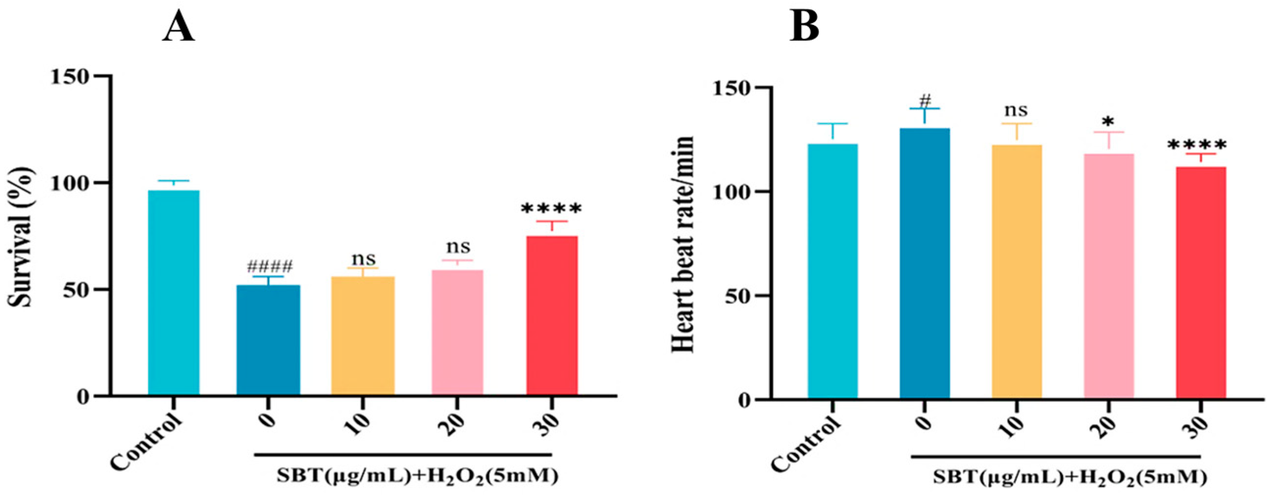
We further investigated the protective effects of SBT in an H2O2-induced zebrafish aging model. As illustrated in Figure 5A,B, SBT treatment significantly improved the survival rate and restored the heart rate of zebrafish larvae impaired by H2O2 exposure. Together, these results demonstrate that SBT effectively improves overall viability and cardiac function in a zebrafish model of accelerated aging.

2.6. SBT Enhances the Antioxidant Defense in H2O2-Treated Zebrafish

We further investigated the protective effects of SBT in an H2O2-induced zebrafish aging model by assessing its antioxidant capacity. Fluorescence-based ROS detection showed that the pronounced oxidative stress induced by H2O2 was markedly mitigated by SBT intervention (Figure 6A,B). Antioxidant enzyme activity and metabolite levels were additionally quantified. The administration of SBT improved the redox status, as evidenced by increased activities of SOD (Figure 6C) and CAT (Figure 6D), elevated GSH levels (Figure 6E), and reduced MDA content (Figure 6F). Together, these results demonstrate that SBT enhances the antioxidant defense system in a zebrafish model of accelerated aging.

2.7. SBT Inhibits Aging and Cell Death in Hydrogen Peroxide-Exposed Zebrafish Embryos

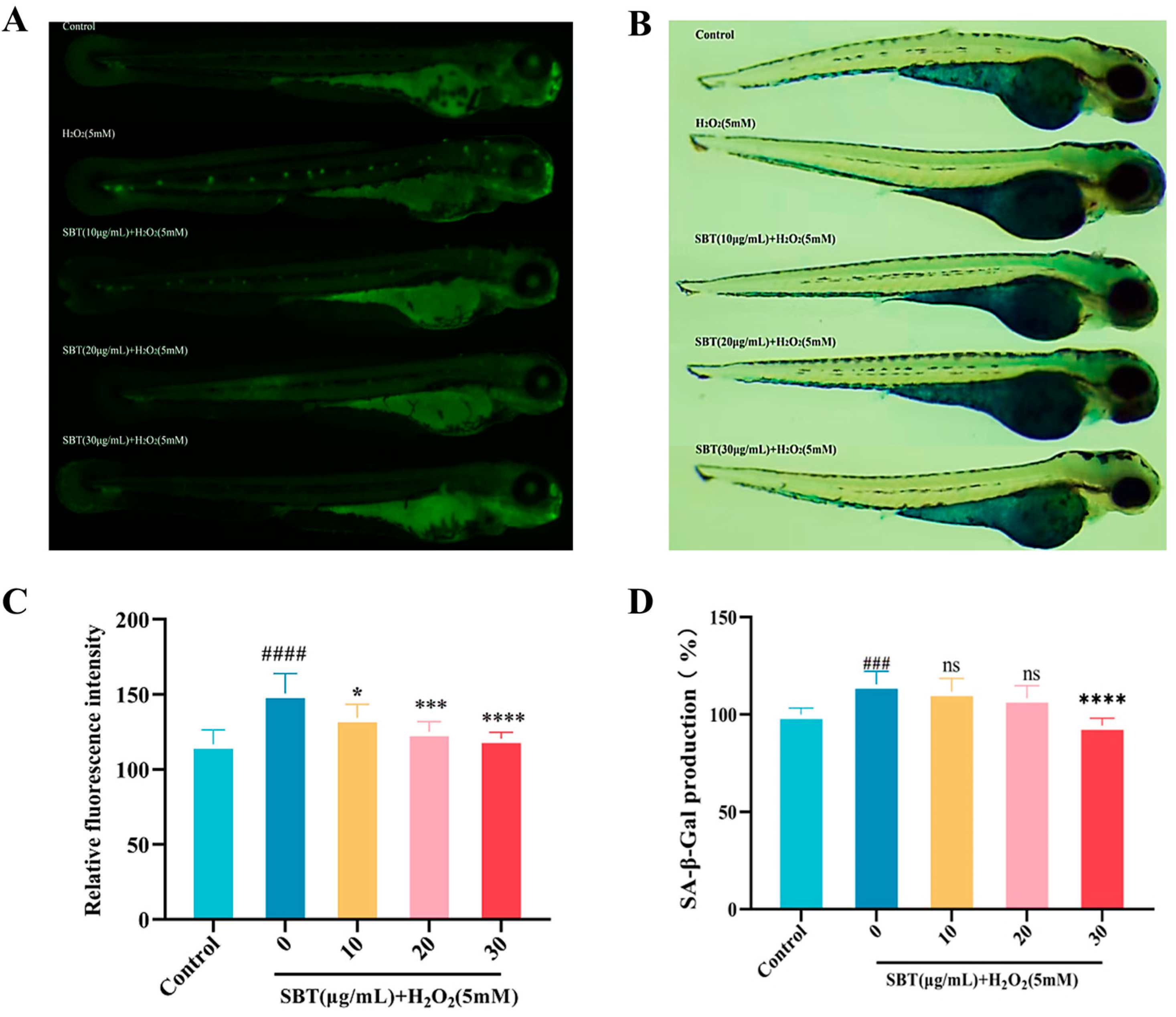
To investigate the potential of SBT in mitigating programmed cell death and delaying age-related decline in senescent zebrafish, acridine orange (AO) staining was performed. A significant increase in apoptotic signals (AO-positive) appeared in the heart tissue of zebrafish in the model cohort. In contrast, SBT intervention markedly reduced AO-positive signals (Figure 7A). Furthermore, the SA-β-gal assay demonstrated a marked escalation of senescence-linked enzymatic responses in the model cohort, an effect that was effectively reversed by SBT treatment (Figure 7B).

2.8. SBT Effects on P53/P21/SIRT1 Signaling Pathway

Western blot analysis assessed key protein expression in the SBT-modified P21/P53/SIRT1 signaling pathway. Figure 8A illustrates that SBT therapy markedly decreased P53 and P21 protein levels while concurrently increasing SIRT1 expression.

2.9. SBT-Mediated Modulation of Senescence-Associated Gene Transcription

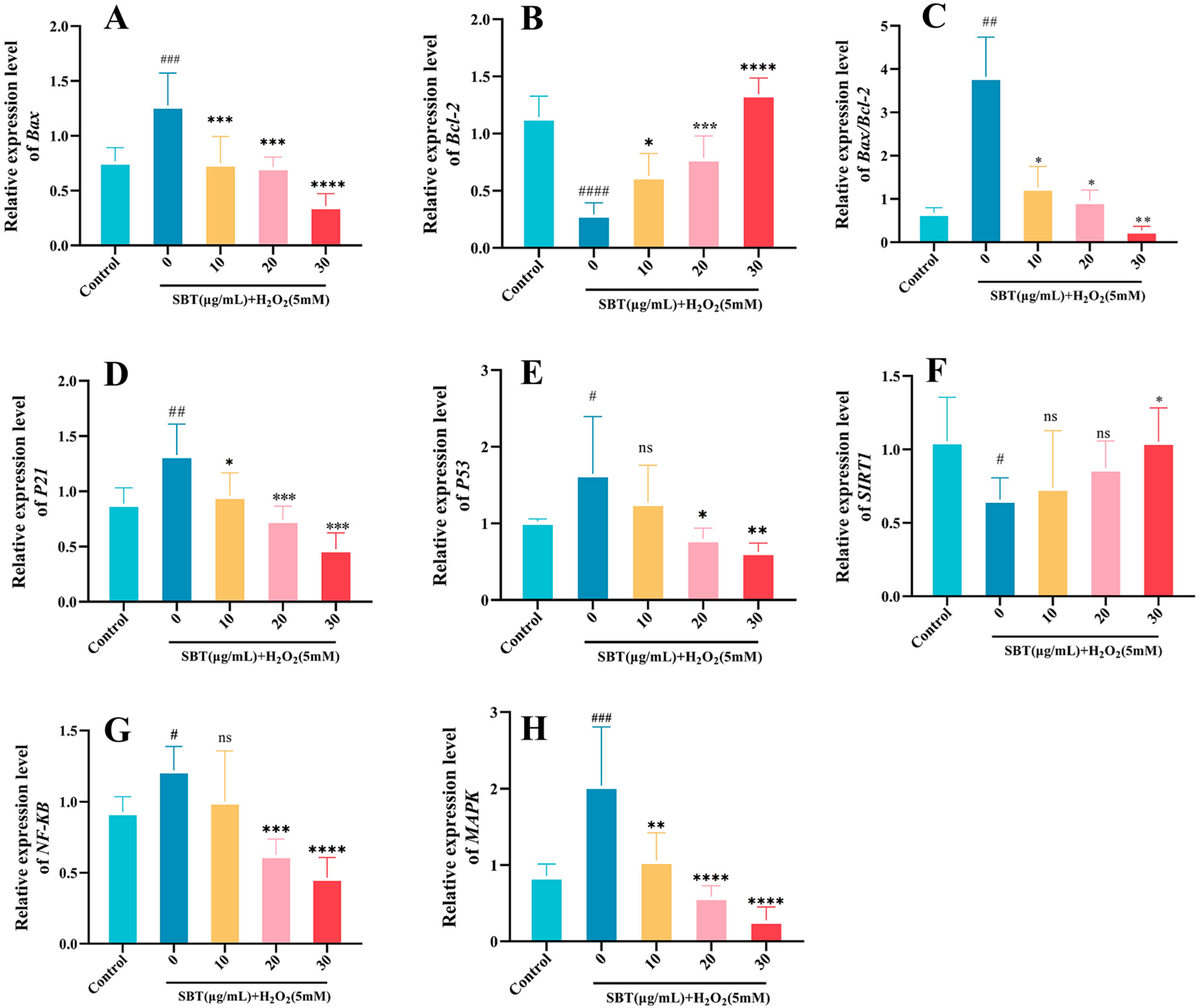
Quantitative PCR (q-PCR) assessed SBT’s transcriptional impacts through mRNA level measurements of key genes regulating apoptosis (Bax, Bcl-2), inflammation (NF-κB, MAPK), and senescence (P21, P53, SIRT1). The results demonstrated that SBT administration significantly suppressed Bax transcript levels while enhancing Bcl-2 expression (Figure 9A,B), resulting in a reduced Bax/Bcl-2 ratio (Figure 9C). A concurrent decrease was observed in P21 and P53 mRNA levels (Figure 9D,E), whereas SIRT1 expression was notably elevated (Figure 9F). Furthermore, NF-κB and MAPK signaling components showed marked transcriptional downregulation relative to the H2O2 group (Figure 9G,H). The collective data indicate that coordinated modulation of gene expression is the mechanism through which SBT attenuates cellular senescence.

3. Discussion

Our study provides compelling evidence that the botanical formulation Srolo Bzhtang (SBT) confers robust anti-senescence effects by orchestrating a multi-targeted response centered on the P21/P53/SIRT1 signaling axis [31]. Consistent findings in D-galactose-induced senescent H9c2 cardiomyocytes and H2O2-accelerated zebrafish aging models underscore the therapeutic potential of SBT in mitigating cellular aging and its associated functional decline [32].
A pivotal finding of this work is the elucidation of SBT’s ability to coordinately modulate the P53/P21/SIRT1 network, a crucial hub integrating stress responses with cellular fate decisions [33]. In senescent H9c2 cells, SBT not only rescued cell viability but also fundamentally altered the expression landscape of key senescence regulators, suppressing the pro-senescence proteins P53 and P21 while promoting the expression of the longevity-associated deacetylase SIRT1 [34]. The recapitulation of these molecular changes-downregulation of P53/P21 and upregulation of SIRT1-in the H2O2-induced zebrafish model strongly implies a conserved mechanism of action across species [35]. This coordinated regulation suggests a plausible model wherein SBT-mediated activation of SIRT1 leads to the deacetylation and functional inhibition of P53 [36], thereby attenuating the transcription of its downstream target P21 and subsequently alleviating cell cycle arrest driven by P21 and the establishment of the senescent phenotype [37,38].
First, our results clearly demonstrate that SBT exerts its anti-senescence effects through potent antioxidant activity. Beyond its core action on the senescence axis, SBT’s efficacy is deeply rooted in its potent capacity to reinforce the cellular defense system against oxidative stress [39]. In zebrafish, SBT treatment significantly bolstered the endogenous antioxidant arsenal, enhancing the activities of SOD and CAT and elevating GSH levels [40]. The concomitant reduction in MDA content indicates successful attenuation of lipid peroxidation [41]. This antioxidant capacity was corroborated in the H9c2 cell model, where SBT significantly reversed D-galactose-induced intracellular ROS accumulation (Figure 1), mirroring its in vivo effects in zebrafish. This comprehensive enhancement of the cellular antioxidant defenses effectively neutralizes excessive ROS accumulation, a key driver of aging, thereby preserving cellular integrity from oxidative insult [42,43].
Second, the anti-senescence profile of SBT is further augmented by its anti-apoptotic properties [44]. Our data demonstrate that SBT shifts the apoptotic balance towards cell survival, as evidenced by the downregulation of the pro-apoptotic gene Bax and the upregulation of the anti-apoptotic gene Bcl-2 [45]. This conclusion was directly supported by TUNEL staining in H9c2 cells (Figure 3) and AO staining in zebrafish (Figure 7A,C), showing that SBT markedly reduced apoptosis in the senescent milieu. qPCR results further confirmed the regulatory effect of SBT on the Bax/Bcl-2 ratio at the transcriptional level (Figure 9A–C), clarifying the molecular basis for its anti-apoptotic action.
Furthermore, our study revealed that SBT modulates senescence-associated inflammatory signaling, as evidenced by the downregulation of critical elements within the MAPK/NF-κB pathways, indicating a concomitant dampening of inflammatory and stress responses [46]. qPCR data showed that SBT treatment significantly reduced the mRNA levels of NF-κB and MAPK (Figure 9G,H), suggesting that SBT may further ameliorate the senescent microenvironment by suppressing SASP-associated inflammatory signaling. This multi-faceted intervention, targeting oxidative damage, programmed cell death, and associated inflammatory signaling, establishes an integrated defensive network against the drivers of cellular aging [47].
The experimental paradigm employed, leveraging the complementary strengths of the H9c2 cell line and the zebrafish model, provided a powerful platform for validation [48]. This dual-model system enabled a multi-tiered and comprehensive validation, spanning from cellular viability, oxidative stress, and senescence markers (SA-β-gal) to apoptosis, key signaling pathways (P21/P53/SIRT1, MAPK/NF-κB), and overall physiological functions (zebrafish survival and heart rate), thereby greatly enhancing the reliability of our conclusions [49,50]. Based on these findings, future investigations should prioritize several promising directions. First, employing genetic (e.g., siRNA) or pharmacological inhibition of SIRT1 is crucial to definitively establish a causal relationship between SIRT1 activation and the observed P53 deacetylation and subsequent anti-senescence effects. Second, a detailed pharmacokinetic profiling of SBT’s bioactive constituents is essential to understand their in vivo bioavailability and metabolism. Furthermore, deconvoluting the synergistic interactions among the multiple active components within the SBT formulation will be key to appreciating the holistic nature of its polypharmacological action.
In conclusion, this study delineates a novel mechanism by which SBT alleviates cellular senescence primarily through the coordinated targeting of the P21/P53/SIRT1 signaling axis. By reinforcing antioxidant defenses, inhibiting apoptosis, and modulating associated inflammatory signals, SBT effectively clears senescent cells and ameliorates aging-associated phenotypes. By enhancing the clearance of senescent cells and ameliorating aging-associated phenotypes through reinforced antioxidant defenses and suppressed apoptosis, SBT emerges as a promising, multi-targeted candidate for further development as a therapeutic or preventive agent for age-related degenerative conditions.

4. Materials and Methods

Srolo Bzhtang (abbreviated as SBT) was provided by the Pharmaceutical Preparation Department of Qinghai Tibetan Medical Hospital (Xining, China) (Batch No. 20130607). D-galactose was purchased from Sangon Biotech (Shanghai, China) Co., Ltd. (Shanghai, China) (Catalog Number A600215-0500).

4.1. Cell Culture and Drug Treatment

H9c2 rat cardiomyocytes obtained from the Cellular Repository of Peking Union Medical College (Beijing, China). Cells were maintained in high-glucose DMEM (BBI, Shanghai, China; Cat# E600003-0500) containing 10% fetal bovine serum (FBI, Catalog Number E600001-0500) and 1% penicillin/streptomycin (BBI, Catalog Number E607011-0100), Cultivated at 37 °C with 5% CO2 and humid conditions. Subculturing was performed with 0.25% trypsin-EDTA (BBI, Catalog Number E607002-0100) upon reaching 80–90% confluency. All cultures routinely undergo mycoplasma testing to ensure the absence of contamination. To induce cellular senescence, cells were subjected to 50 mg/mL D-galactose treatment for 24 h; control cells received an equal volume of complete medium. Following induction, the samples were categorized into the subsequent experimental subsets: normal control (untreated), model (GAL group; treated with 50 mg/mL D-galactose for 24 h), SBT-treated (exposed to D-galactose followed by 24 h incubation with SBT at 50, 100, 200, 300, or 400 μg/mL).

4.2. Cell Viability Assay H9c2

H9c2 cells seeded at 5 × 10³ cells/well in 96-well plates underwent 24-h incubation to facilitate cell attachment. After medium depletion, the cells were allocated into groups: the model and treatment groups were both exposed to 50 mg/mL D-galactose (GAL), in contrast to the control group, which was replenished with fresh complete medium. At the 24-h mark, the supernatant was aspirated. The treatment groups were then administered varying concentrations of SBT (50, 100, 200, 300, or 400 μg/mL), with the control and model groups receiving an equivalent volume of complete medium. Incubation proceeded for a further 24 h, at which point CCK-8 reagent (BBI, Catalog Number E606335-2000) was added to each well. The plate was incubated at 37 °C with 5% CO2 for 30 min. The absorption was measured at 450 nm via a microplate reader.

4.3. ROS Staining

ROS levels were assessed with the Beyotime DCFH-DA fluorescent dye (Shanghai, China; Catalog Number S0033S), following the supplier’s instructions. Briefly, H9c2 cells grown in 24-well plates were washed and incubated in a 10 μM DCFH-DA solution in serum-free conditions at 37 °C for a duration of 30 min. To eliminate residual dye, three washes with serum-free medium were performed post-staining. For the zebrafish larvae, the procedure was analogous: they were placed in a 10 μM DCFH-DA solution prepared in embryo medium and incubated at 37 °C for 30 min. Thereafter, the larvae underwent three 5-min washes in embryo medium to thoroughly clear any unincorporated probe. A confocal laser scanning microscope (ZEISS LSM 900, Oberkochen, Germany) was employed to capture fluorescence images. Finally, the relative ROS levels were quantified by analyzing the fluorescence intensity with ImageJ software (v1.48v, National Institutes of Health, Bethesda, MD, USA).

4.4. SA-β-Gal Staining

According to the manufacturer’s protocol, cellular senescence was evaluated by detecting SA-β-gal enzymatic function via a commercial assay (Beyotime, Catalog Number C0602). In brief, cells in 6-well plates were fixed for 10 min at room temperature, subjected to two PBS washes, and then subjected to an incubation period of 12 h in a stain solution at 37 °C. The percentage of cells positive for SA-β-gal was quantified by examination under a light microscope. For the zebrafish larvae, the procedure involved three rinses with embryo medium prior to fixation at 4 °C overnight. Following fixation, after triple washing with embryo medium, the larvae were incubated overnight at 37 °C in staining solution. Zebrafish SA-β-gal activity was captured via MZX100 stereomicroscope (Mshot, Guangzhou, China).

4.5. TUNEL Staining

Assessment of cell apoptosis was conducted using a TUNEL kit (Beyotime, Catalog Number C1086) from a commercial supplier as per the supplied instructions. The procedure commenced with the fixation of cells cultured in 6-well plates, using 4% paraformaldehyde treatment for 30 min at ambient temperature. Subsequently, a permeabilization step was performed to allow the assay reagents to penetrate the cells. Following this, the specimens underwent 60 min of dark incubation at 37 °C using the TUNEL reagent. Imaging was conducted on a ZEISS LSM 900 confocal microscope, and apoptotic cells were identified by the presence of red fluorescent nuclei in the acquired micrographs.

4.6. Western Blotting

RIPA buffer (Beyotime, Catalog Number P0013B) isolated proteins from collected cell cultures and zebrafish tissues. Following quantification of protein concentration with a BCA assay kit (Thermo Scientific, Waltham, MA, USA; Catalog Number 23225), Samples were thermally denatured in loading buffer at 100 °C for 10 min. Subsequently, the proteins underwent electrophoretic separation on 10% SDS-PAGE gels and were electrophoretically transferred to PVDF membranes using a wet transfer system. Membranes were incubated in 5% skim milk, followed by overnight primary antibody application at 4 °C. The primary antibodies used and their dilutions were: P21 (Proteintech, Chicago, IL, USA; Catalog Number 10355-1-AP; 1:1000), P53 (Proteintech, Catalog Number 100442-1-AP; 1:1000), and SIRT1 (Proteintech, Catalog Number 13161-1-AP; 1:1000). Following three TBST washes, the membranes underwent a 2-h incubation period at ambient temperature with HRP-conjugated secondary antibodies, specifically goat anti-rabbit IgG (BBI, Catalog Number D110058; dilution 1:5000) and goat anti-mouse IgG (BBI, Catalog Number D110087; dilution 1:5000). Signal development was achieved with an ECL chemiluminescence substrate (Millipore, Burlington, MA, USA; Product CodeWBKLS050), The band intensities were measured through grayscale analysis with ImageJ software (v1.48v, National Institutes of Health, Bethesda, MD, USA), utilizing β-actin (Proteintech, Catalog Number 8111S-1-RR; 1:5000) as a loading control.

4.7. Maintenance and Experimental Manipulation of Zebrafish

Zebrafish (AB wild-type strain) were obtained from the China Zebrafish Resource Center (Wuhan, China). Embryos were gathered through natural spawning, with a ratio of females to males of 1:2. For further experiments, normally developed embryos at 8 h post-fertilization (hpf) were chosen. Roughly 25 zebrafish embryos at 8 hpf were placed in 12-well plates filled with embryo medium and co-incubated in embryo water enriched with 5 mM H2O2 until they reached 24 hpf. After substituting with fresh embryo water, the embryos were maintained for an additional 2 days to create the H2O2-induced senescence model. The zebrafish embryos were subsequently divided into several groups: the control group (untreated); the hydrogen peroxide (H2O2) group (exposed to embryo water containing 5 mM H2O2 until 24 hpf); the SBT treatment group (embryos first received embryo water with 10, 20, or 30 μg/mL SBT for 1 h before adding embryo water with 5 mM H2O2, continuing co-incubation until 24 hpf).

4.8. Survival Rate and Heartbeat Rate/Time Analysis

At 8 h post-fertilization (hpf), zebrafish embryos were individually transferred into 12-well plates, with approximately 25 embryos per group. They were then exposed to embryo medium containing SBT at concentrations of 10, 20, or 30 μg/mL for 1 h. Following this pretreatment, the embryos were challenged with 5 mM H2O2 in embryo medium until 24 hpf. After the medium was exchanged for fresh embryo medium, the embryos were maintained for a further 48 h. Survival rates were determined by counting viable larvae, while heart rates were quantified for the surviving individuals under a stereomicroscope. Only viable larvae were subsequently used for further experiments.

4.9. Measurement of Antioxidant Capacity

The antioxidant capacity of zebrafish larvae was evaluated using commercial assay kits according to the manufacturers’ instructions. Specifically, superoxide dismutase (SOD) activity and malondialdehyde (MDA) concentration (an indicator of lipid peroxidation) were measured using the SOD activity assay kit (Beyotime, Catalog Number S0101) and MDA quantitative kit (Beyotime, Catalog Number S0131), respectively. Glutathione assay kits measured decreased GSH concentrations (Beyotime, Catalog Number S0052), while CAT activity was examined using a catalase-specific detection system (Beyotime, Catalog Number S0051). All absorbance measurements were conducted by following the standardized protocols provided in the respective kit manuals.

4.10. Acridine Orange (AO) Staining

Zebrafish larvae from each experimental group were incubated in embryo medium containing acridine orange (AO) staining solution (Sigma-Aldrich, St. Louis, MO, USA; Product Code A6014) at 37 °C for 20 min. Following incubation, the larvae underwent three washes in fresh embryo medium. Imaging by fluorescence was conducted with a stereo microscope fitted with the appropriate filters, and the acquired images were quantitatively analyzed for fluorescence intensity using ImageJ software (v1.48v, National Institutes of Health, Bethesda, MD, USA).

4.11. qRT-PCR

Following isolation from zebrafish larvae using TRIzol reagent (Thermo Fisher Scientific, Waltham, MA, USA; Catalog Number 15596026), RNA was converted to cDNA using PrimeScript RT reagent (Takara Bio, Kusatsu, Japan; Catalog Number RR047A). qPCR analysis was subsequently conducted with the aid of TB Green Premix Ex Taq II (Tli RNaseH Plus; Takara Bio, Catalog Number RR820A). All experimental steps were rigorously performed according to the manufacturers’ protocols to ensure reproducibility. Data normalization was achieved using β-actin as an endogenous control. Table 1 lists all primer sequences employed in the present study.

4.12. Statistical Analysis

Statistical processing was utilized with GraphPad Prism 9 software. Quantitative datasets were initially assessed for normality. Data that followed a normal distribution are presented as the mean ± standard deviation. Statistical comparisons between groups were performed using Student’s t-test or one-way ANOVA, with a p-value of less than 0.05 considered statistically significant.

Author Contributions

Conceptualization, C.Z. and S.S.; methodology, J.W. and N.D.; software, N.D.; validation, J.W., N.D. and N.W.; formal analysis, J.W. and D.S.; investigation, J.W. and J.L.; resources, C.Z. and S.S.; data curation, J.W. and N.W.; writing—original draft preparation, J.W. and N.D.; writing—review and editing, C.Z. and S.S.; visualization, J.L. and D.S.; supervision, C.Z. and S.S.; project administration, C.Z.; funding acquisition, C.Z. and S.S. All authors have read and agreed to the published version of the manuscript.

Funding

This study was funded by multiple sources, including the Applied Basic Research Project of Qinghai Province (2024-ZJ-737), the National Natural Science Foundation of China (32160233), the Qinghai University Experiment (Practice) Course Construction Project (SYKC-2024-13), the 2023 Scientific Research Project on Chinese and Tibetan Medicine of Qinghai Province (J20230016), the Tibetan Medicine Zuotai Inheritance and Innovation Fund Project (2024-ZT-Z-1), and the 2023 Bureau-level Scientific Research Project of the Tibetan Medicine Administration of the Xizang Autonomous Region (JJKT2023014).

Institutional Review Board Statement

The animal experiment was reviewed and approved by the Medical Science Research Ethics Committee of Qinghai University (Approval No. PJ202501-79).

Informed Consent Statement

Not applicable.

Data Availability Statement

The original contributions presented in the study are included in the article. Further inquiries can be directed to the corresponding author.

Conflicts of Interest

The authors declare that they have no known competing financial interests or personal relationships that could have appeared to influence the work reported in this paper.

Abbreviations

SBTSrolo Bzhtang
GALD-galactose
ROSReactive Oxygen Species
SA-β-galSenescence-Associated β-galactosidase
SODSuperoxide Dismutase
CATCatalase
GSHGlutathione
MDAMalondialdehyde
AOAcridine Orange
MAPKMitogen-Activated Protein Kinase
NF-κBNuclear Factor kappa-light-chain-enhancer of activated B cells
BaxBcl-2-Associated X protein
Bcl-2B-cell lymphoma 2
SIRT1Silent Information Regulator 1
DMEMDulbecco’s Modified Eagle Medium
FBSFetal Bovine Serum
CCK-8Cell Counting Kit-8
DCFH-DA2′,7′-Dichlorodihydrofluorescein diacetate
RIPARadioimmunoprecipitation Assay
SDS-PAGESodium Dodecyl Sulfate-Polyacrylamide Gel Electrophoresis
PVDFPolyvinylidene Fluoride
HRPHorseradish Peroxidase
ECLEnhanced Chemiluminescence
hpfHours Post-Fertilization
TBSTTris-Buffered Saline with Tween-20
ANOVAAnalysis of Variance

References

  1. Ramos, E.L.P.; Sousa Neto, I.V.; Pinto, A.P.; Cintra, D.E.; Ropelle, E.R.; Pauli, J.R.; de Freitas, E.C.; Mineo, T.W.P.; da Silva, A.S.R. Mechanisms underlying the interplay between autophagy and the inflammasome in age-related diseases: Implications for exercise immunology. Ageing Res. Rev. 2025, 110, 102821. [Google Scholar] [CrossRef]
  2. Sanada, F.; Hayashi, S.; Morishita, R. Targeting the hallmarks of aging: Mechanisms and therapeutic opportunities. Front. Cardiovasc. Med. 2025, 12, 1631578. [Google Scholar] [CrossRef] [PubMed]
  3. Qin, X.; Strand, S.H.; Lee, M.R.; Saraswathibhatla, A.; van IJzendoorn, D.G.P.; Zhu, C.; Vennam, S.; Varma, S.; Hall, A.; Factor, R.E.; et al. Single-Cell Expression Analysis of Ductal Carcinoma In Situ Identifies Complex Genotypic-Phenotypic Relationships Altering Epithelial Composition. Cancer Res. 2025, 85, 2302–2319. [Google Scholar] [CrossRef]
  4. Yang, J.; Xu, J.; Wang, W.; Zhang, B.; Yu, X.; Shi, S. Epigenetic regulation in the tumor microenvironment: Molecular mechanisms and therapeutic targets. Signal Transduct. Target. Ther. 2023, 8, 210. [Google Scholar] [CrossRef]
  5. Liu, B.; Qu, J.; Zhang, W.; Izpisua Belmonte, J.C.; Liu, G.H. A stem cell aging framework, from mechanisms to interventions. Cell Rep. 2022, 41, 111451. [Google Scholar] [CrossRef]
  6. Baechle, J.J.; Chen, N.; Makhijani, P.; Winer, S.; Furman, D.; Winer, D.A. Chronic inflammation and the hallmarks of aging. Mol. Metab. 2023, 74, 101755. [Google Scholar] [CrossRef] [PubMed]
  7. Duan, J.L.; Ruan, B.; Song, P.; Fang, Z.Q.; Yue, Z.S.; Liu, J.J.; Dou, G.R.; Han, H.; Wang, L. Shear stress-induced cellular senescence blunts liver regeneration through Notch-sirtuin 1-P21/P16 axis. Hepatology 2022, 75, 584–599. [Google Scholar] [CrossRef] [PubMed]
  8. Azzahrani, K.; Alqahtani, F. The Tumor Suppressor p53 Downregulates p107 (RBL1) Through p21–RB/E2F Signaling and Tandem E2F Sites. Int. J. Mol. Sci. 2025, 26, 9903. [Google Scholar] [CrossRef]
  9. Chen, Y.; Peng, C.; Chen, J.; Chen, D.; Yang, B.; He, B.; Hu, W.; Zhang, Y.; Liu, H.; Dai, L.; et al. WTAP facilitates progression of hepatocellular carcinoma via m6A-HuR-dependent epigenetic silencing of ETS1. Mol. Cancer 2019, 18, 127. [Google Scholar] [CrossRef]
  10. Ashraf, S.; Biglow, L.; Dotson, J.; Tirona, M.T. Pneumonitis and cellular immunodeficiency triggered by the CDK 4/6 inhibitor Abemaciclib. Transl. Breast Cancer Res. A J. Focus. Transl. Res. Breast Cancer 2022, 3, 9. [Google Scholar] [CrossRef]
  11. Chen, C.; Zhou, M.; Ge, Y.; Wang, X. SIRT1 and aging related signaling pathways. Mech. Ageing Dev. 2020, 187, 111215. [Google Scholar] [CrossRef]
  12. Abu Shelbayeh, O.; Arroum, T.; Morris, S.; Busch, K.B. PGC-1α Is a Master Regulator of Mitochondrial Lifecycle and ROS Stress Response. Antioxidants 2023, 12, 1075. [Google Scholar] [CrossRef]
  13. Li, Z.; Zhou, H.; Zhai, X.; Gao, L.; Yang, M.; An, B.; Xia, T.; Du, G.; Li, X.; Wang, W.; et al. MELK promotes HCC carcinogenesis through modulating cuproptosis-related gene DLAT-mediated mitochondrial function. Cell Death Dis. 2023, 14, 733. [Google Scholar] [CrossRef] [PubMed]
  14. Wang, H.; Kim, S.J.; Lei, Y.; Wang, S.; Wang, H.; Huang, H.; Zhang, H.; Tsung, A. Neutrophil extracellular traps in homeostasis and disease. Signal Transduct. Target. Ther. 2024, 9, 235. [Google Scholar] [CrossRef]
  15. Wei, X.; Xiong, X.; Wang, P.; Zhang, S.; Peng, D. SIRT1-mediated deacetylation of FOXO3 enhances mitophagy and drives hormone resistance in endometrial cancer. Mol. Med. 2024, 30, 147. [Google Scholar] [CrossRef]
  16. Lee, J.E.; Lee, H.; Baek, E.; Choi, B.; Yun, H.S.; Yoo, Y.K.; Lee, Y.S.; Song, G.J.; Cho, K.S. The role of glial and neuronal Eph/ephrin signaling in Drosophila mushroom body development and sleep and circadian behavior. Biochem. Biophys. Res. Commun. 2024, 720, 150072. [Google Scholar] [CrossRef]
  17. Tang, X.; Xu, Y.; Ou, N.; Tang, Y.; Chen, H. Update on late-onset hypogonadism: Current concepts, controversies, clinical diagnosis, pathogenesis, and treatment approaches. Sex. Med. Rev. 2025, 13, 652–662. [Google Scholar] [CrossRef]
  18. Tomasini, S.; Monteleone, E.; Altieri, A.; Margiotta, F.; Dardmeh, F.; Alipour, H.; Holm, A.; Kauppinen, S.; Panella, R. microRNA-22 Inhibition Stimulates Mitochondrial Homeostasis and Intracellular Degradation Pathways to Prevent Muscle Wasting. Int. J. Mol. Sci. 2025, 26, 9900. [Google Scholar] [CrossRef]
  19. Kaeberlein, T.L.; Green, A.S.; Haddad, G.; Hudson, J.; Isman, A.; Nyquist, A.; Rosen, B.S.; Suh, Y.; Zalzala, S.; Zhang, X.; et al. Evaluation of off-label rapamycin use to promote healthspan in 333 adults. GeroScience 2023, 45, 2757–2768. [Google Scholar] [CrossRef] [PubMed]
  20. Yang, Y.; Jiang, B.; Shi, L.; Wang, L.; Yang, Y.; Li, Y.; Zhang, Y.; Zhu, Z.; Zhang, X.; Liu, X. The potential of natural herbal plants in the treatment and prevention of non-small cell lung cancer: An encounter between ferroptosis and mitophagy. J. Ethnopharmacol. 2025, 346, 119555. [Google Scholar] [CrossRef] [PubMed]
  21. Qi, S.; Liang, X.; Wang, Z.; Jin, H.; Zou, L.; Yang, J. Potential Mechanism of Tibetan Medicine Liuwei Muxiang Pills against Colorectal Cancer: Network Pharmacology and Bioinformatics Analyses. Pharmaceuticals 2024, 17, 429. [Google Scholar] [CrossRef]
  22. Chen, T.; Su, S.; Yang, Z.; Zhang, D.; Li, Z.; Lu, D. Srolo Bzhtang reduces inflammation and vascular remodeling via suppression of the MAPK/NF-κB signaling pathway in rats with pulmonary arterial hypertension. J. Ethnopharmacol. 2022, 297, 115572. [Google Scholar] [CrossRef]
  23. Bian, X.; Chen, L.; Bian, X.; Li, L.; Liu, D.; Liu, S.; Xu, L.; Huo, X.; Yang, X. Protective effect of Tibetan medicine Qiwei Tiexie pills on liver injury induced by acetaminophen overdose: An integrated strategy of network pharmacology, metabolomics and transcriptomics. Phytomedicine Int. J. Phytother. Phytopharm. 2024, 123, 155221. [Google Scholar] [CrossRef]
  24. Wang, N.; Zhu, H. Geo-authentic Tibetan medicine: A traditional pharmacological resource for promoting human health and wellness. Front. Pharmacol. 2024, 15, 1432221. [Google Scholar] [CrossRef]
  25. Yang, J.; Karunarathna, S.C.; Patabendige, N.; Tarafder, E.; Lou, D.; Zhou, Y.; Hapuarachchi, K. Unveiling the Bioactive Compounds and Therapeutic Potential of Russula: A Comprehensive Review. J. Fungi 2025, 11, 341. [Google Scholar] [CrossRef]
  26. McMahon, S.; Spector, T.; Ramana, K.V. Significance of Macrophage-Mediated Inflammatory Response in Ocular Inflammatory Complications. Front. Biosci. (Landmark Ed.) 2025, 30, 26698. [Google Scholar] [CrossRef] [PubMed]
  27. Wang, J.; Gan, L.; Li, F.; Li, Q.; Wu, T.; Wu, Z.; Chen, P.; Scicluna, B.P.; Feng, X.; Gu, J.; et al. Tracheal epithelial cell-exosome-derived MiR-21-5p inhibits alveolar macrophage pyroptosis to resist pulmonary bacterial infection through PIK3CD-autophagy pathway. Life Sci. 2024, 336, 122340. [Google Scholar] [CrossRef] [PubMed]
  28. Chen, C.H.; Chen, Y.C.; Chang, Y.C.; Hung, C.H.; Huang, C.Y.; Tsai, C.L.; Sun, C.K.; Lin, H.Y. Taurine Rescues Cancer-induced Atrophy in Human Skeletal Muscle Cells via Ameliorating the Inflammatory Tumor Microenvironment. Anticancer Res. 2024, 44, 1963–1971. [Google Scholar] [CrossRef]
  29. Peng, Z.; Zhao, C.; Yang, Z.; Gong, S.; Du, Z. D-galactose-induced mitochondrial oxidative damage and apoptosis in the cochlear stria vascularis of mice. BMC Mol. Cell Biol. 2023, 24, 27. [Google Scholar] [CrossRef] [PubMed]
  30. Shen, E.; Wu, Y.; Ye, W.; Li, S.; Zhu, J.; Jiang, M.; Hu, Z.; Cao, G.; Yi, X.; Li, F.; et al. The FGF13-Caveolin-1 Axis: A Key Player in the Pathogenesis of Doxorubicin- and D-Galactose-Induced Premature Cardiac Aging. Adv. Sci. 2025, 12, e2501055. [Google Scholar] [CrossRef]
  31. Jing, L.; Su, S.; Zhang, D.; Li, Z.; Lu, D.; Ge, R. Srolo Bzhtang, a traditional Tibetan medicine formula, inhibits cigarette smoke induced airway inflammation and muc5ac hypersecretion via suppressing IL-13/STAT6 signaling pathway in rats. J. Ethnopharmacol. 2019, 235, 424–434. [Google Scholar] [CrossRef] [PubMed]
  32. Pu, L.; Yang, C.; Yu, L.; Li, S.; Liu, Y.; Liu, X.; Lai, X. Tibetan Medicines for the Treatment of Diabetic Nephropathy. Evid.-Based Complement. Altern. Med. ECAM 2021, 2021, 7845848. [Google Scholar] [CrossRef]
  33. Pan, L.; Gao, J.; Han, Y.; Shi, Y.; Tang, X.; Pu, L.; Lai, X.; Dongzhu, R.; Zhang, J.; Xiangmao, Q.; et al. The Treatment of Cholecystitis and Cholelithiasis by Tibetan Medicine. Evid.-Based Complement. Altern. Med. ECAM 2021, 2021, 9502609. [Google Scholar] [CrossRef]
  34. Fu, K.; Xu, M.; Zhou, Y.; Li, X.; Wang, Z.; Liu, X.; Meng, X.; Zeng, Y.; Zhang, H. The Status quo and way forwards on the development of Tibetan medicine and the pharmacological research of tibetan materia Medica. Pharmacol. Res. 2020, 155, 104688. [Google Scholar] [CrossRef]
  35. Yu, L.; Li, S.; Pu, L.; Yang, C.; Shi, Q.; Zhao, Q.; Meniga, S.; Liu, Y.; Zhang, Y.; Lai, X. Traditional Tibetan medicine: Therapeutic potential in rheumatoid arthritis. Front. Pharmacol. 2022, 13, 938915. [Google Scholar] [CrossRef]
  36. Liu, Z.; Wang, K.; Jiang, C.; Chen, Y.; Liu, F.; Xie, M.; Yim, W.Y.; Yao, D.; Qian, X.; Chen, S.; et al. Morusin Alleviates Aortic Valve Calcification by Inhibiting Valve Interstitial Cell Senescence Through Ccnd1/Trim25/Nrf2 Axis. Adv. Sci. 2024, 11, e2307319. [Google Scholar] [CrossRef]
  37. Wen, Y.; Zhang, X.; Liu, H.; Ye, H.; Wang, R.; Ma, C.; Duo, T.; Wang, J.; Yang, X.; Yu, M.; et al. SGLT2 inhibitor downregulates ANGPTL4 to mitigate pathological aging of cardiomyocytes induced by type 2 diabetes. Cardiovasc. Diabetol. 2024, 23, 430. [Google Scholar] [CrossRef]
  38. Ke, H.; Zhang, X.; Liang, S.; Zhou, C.; Hu, Y.; Huang, Q.; Wu, J. Study on the anti-skin aging effect and mechanism of Sijunzi Tang based on network pharmacology and experimental validation. J. Ethnopharmacol. 2024, 333, 118421. [Google Scholar] [CrossRef] [PubMed]
  39. Yin, Z.; Tian, L.; Kou, W.; Cao, G.; Wang, L.; Xia, Y.; Lin, Y.; Tang, S.; Zhang, J.; Yang, H. Xiyangshen Sanqi Danshen granules attenuated D-gal-induced C57BL/6J mouse aging through the AMPK/SIRT1 signaling pathway. Phytomed. Int. J. Phytother. Phytopharm. 2025, 136, 156213. [Google Scholar] [CrossRef] [PubMed]
  40. Liu, J.; Yang, Y.; He, Y.; Feng, C.; Ou, H.; Yang, J.; Chen, Y.; You, F.; Shao, B.; Bao, J.; et al. Erxian decoction alleviates cisplatin-induced premature ovarian failure in rats by reducing oxidation levels in ovarian granulosa cells. J. Ethnopharmacol. 2023, 304, 116046. [Google Scholar] [CrossRef]
  41. Liang, Y.; Liu, H.; Zhu, J.; Song, N.; Lu, Z.; Fang, Y.; Teng, J.; Dai, Y.; Ding, X. Inhibition of p53/miR-34a/SIRT1 axis ameliorates podocyte injury in diabetic nephropathy. Biochem. Biophys. Res. Commun. 2021, 559, 48–55. [Google Scholar] [CrossRef]
  42. Lv, X.; Liu, C.; Liu, S.; Li, Y.; Wang, W.; Li, K.; Hua, F.; Cui, B.; Zhang, X.; Yu, J.; et al. The cell cycle inhibitor P21 promotes the development of pulmonary fibrosis by suppressing lung alveolar regeneration. Acta Pharm. Sin. B 2022, 12, 735–746. [Google Scholar] [CrossRef]
  43. Tubita, A.; Lombardi, Z.; Tusa, I.; Lazzeretti, A.; Sgrignani, G.; Papini, D.; Menconi, A.; Gagliardi, S.; Lulli, M.; Dello Sbarba, P.; et al. Inhibition of ERK5 Elicits Cellular Senescence in Melanoma via the Cyclin-Dependent Kinase Inhibitor p21. Cancer Res. 2022, 82, 447–457. [Google Scholar] [CrossRef]
  44. Tao, P.; Zhang, H.F.; Zhou, P.; Wang, Y.L.; Tan, Y.Z.; Wang, H.J. Growth differentiation factor 11 alleviates oxidative stress-induced senescence of endothelial progenitor cells via activating autophagy. Stem Cell Res. Ther. 2024, 15, 370. [Google Scholar] [CrossRef]
  45. Wang, X.X.; Li, M.; Xu, X.W.; Zhao, W.B.; Jin, Y.M.; Li, L.L.; Qin, Z.H.; Sheng, R.; Ni, H. BNIP3-mediated mitophagy attenuates hypoxic-ischemic brain damage in neonatal rats by inhibiting ferroptosis through P62-KEAP1-NRF2 pathway activation to maintain iron and redox homeostasis. Acta Pharmacol. Sin. 2025, 46, 33–51. [Google Scholar] [CrossRef]
  46. Cui, Z.; Zhao, X.; Amevor, F.K.; Du, X.; Wang, Y.; Li, D.; Shu, G.; Tian, Y.; Zhao, X. Therapeutic application of quercetin in aging-related diseases: SIRT1 as a potential mechanism. Front. Immunol. 2022, 13, 943321. [Google Scholar] [CrossRef]
  47. Liu, J.; Wang, Z.; Liang, W.; Zhang, Z.; Deng, Y.; Chen, X.; Hou, Z.; Xie, Y.; Wang, Q.; Li, Y.; et al. Microglial TMEM119 binds to amyloid-β to promote its clearance in an Aβ-depositing mouse model of Alzheimer’s disease. Immunity 2025, 58, 1830–1846.e7. [Google Scholar] [CrossRef] [PubMed]
  48. Khan, A.O.; Rodriguez-Romera, A.; Reyat, J.S.; Olijnik, A.A.; Colombo, M.; Wang, G.; Wen, W.X.; Sousos, N.; Murphy, L.C.; Grygielska, B.; et al. Human Bone Marrow Organoids for Disease Modeling, Discovery, and Validation of Therapeutic Targets in Hematologic Malignancies. Cancer Discov. 2023, 13, 364–385. [Google Scholar] [CrossRef] [PubMed]
  49. Lee, J.; Kim, H.J. Normal Aging Induces Changes in the Brain and Neurodegeneration Progress: Review of the Structural, Biochemical, Metabolic, Cellular, and Molecular Changes. Front. Aging Neurosci. 2022, 14, 931536. [Google Scholar] [CrossRef] [PubMed]
  50. Frei, M.; Wirawan, R.; Wein, T.; Bracher, F. Lead Structure-Based Hybridization Strategy Reveals Major Potency Enhancement of SirReal-Type Sirt2 Inhibitors. Int. J. Mol. Sci. 2025, 26, 9855. [Google Scholar] [CrossRef] [PubMed]
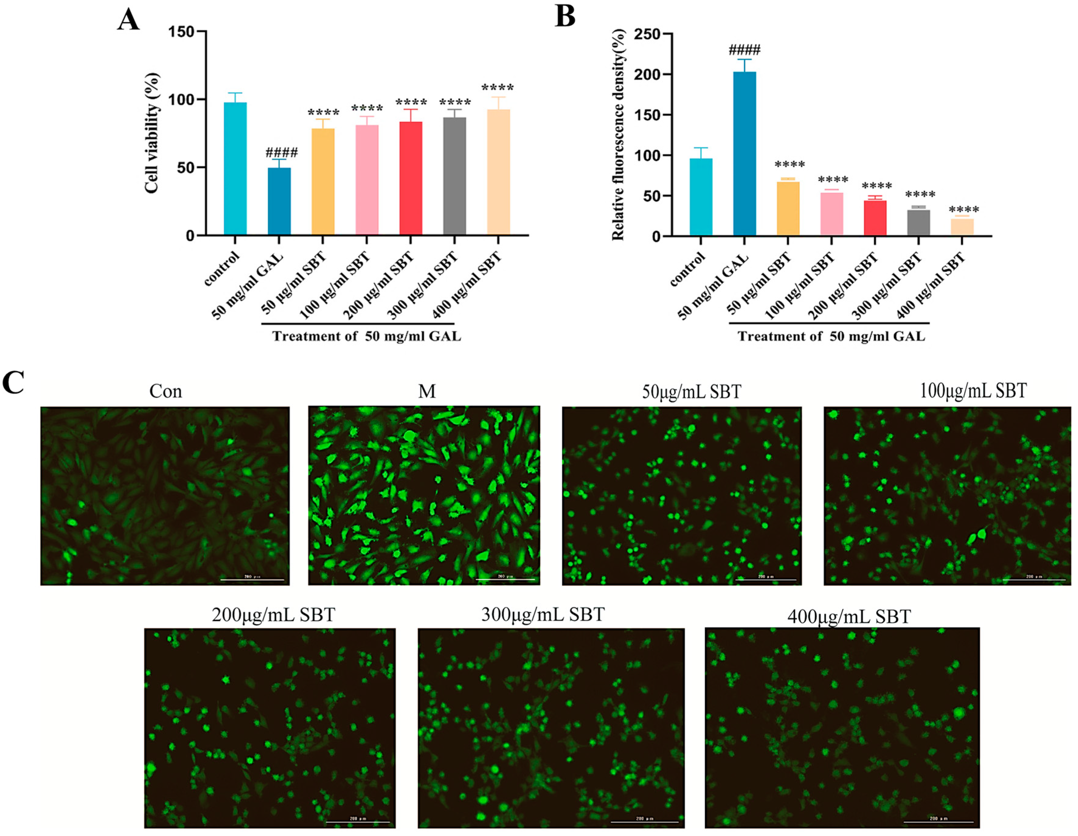
Figure 1. SBT alleviates D-galactose-induced oxidative damage in H9c2 cells. (A) CCK-8 assay assessment of cellular viability. (B) Quantitative analysis of intracellular ROS levels by fluorescence. (C) Representative micrographs of ROS fluorescence (magnification 20×, scale bar = 200 μm). #### p < 0.0001 versus the Control group; **** p < 0.0001 versus the GAL group.
Ijms 26 11394 g001
Figure 2. SBT attenuates D-galactose-induced cellular senescence in H9c2 cardiomyocytes. (A) Representative micrographs of SA-β-gal staining (magnification 20×, scale bar = 200 μm). (B) Quantitative analysis of the proportion of SA-β-gal-positive cells. #### p < 0.0001 versus the Control group; **** p < 0.0001 versus the GAL group.
Ijms 26 11394 g002
Figure 3. SBT inhibits apoptosis in H9c2 cells caused by D-galactose. (A) TUNEL staining (magnification 10×, scale bar = 100 μm). (B) Quantitative analysis of TUNEL-positive cells. #### p < 0.0001 versus the Control group; **** p < 0.0001 versus the GAL group.
Ijms 26 11394 g003
Figure 4. SBT regulates senescence-associated proteins in H9c2 cells. (A) Western blot analysis of P53, P21, and SIRT1 expression. (BD) Quantitative analysis of (B) P53, (C) P21, and (D) SIRT1. #### p < 0.0001 versus Control; **** p < 0.0001, *** p < 0.001, * p < 0.05 versus GAL group; ns, not significant.
Ijms 26 11394 g004
Figure 5. SBT exerts protective effects on survival and heart rate in H2O2-induced zebrafish model. (A) Survival rate analysis. (B) Quantitative assessment of heart rate. # p < 0.05, #### p < 0.0001 versus Control; **** p < 0.0001, * p < 0.05 versus H2O2 group; ns, not significant.
Ijms 26 11394 g005
Figure 6. SBT alleviates oxidative stress in H2O2-treated zebrafish larvae. (A) ROS staining. (B) Quantification of relative ROS fluorescence intensity. (C) SOD activity. (D) CAT activity. (E) GSH level. (F) MDA content. # p < 0.05, ### p < 0.001, #### p < 0.0001 versus Control; **** p < 0.0001, ** p < 0.01, * p < 0.05 versus H2O2 group; ns, not significant.
Ijms 26 11394 g006
Figure 7. SBT mitigates H2O2-induced apoptosis and cellular senescence in zebrafish larvae. (A) AO staining. (B) SA-β-gal staining. (C) AO fluorescence intensity. (D) Quantitative analysis of β-galactosidase staining. ### p < 0.001, #### p < 0.0001 versus Control group; **** p < 0.0001, *** p < 0.001, * p < 0.05 versus H2O2 group; ns, not significant.
Ijms 26 11394 g007
Figure 8. SBT modulates the P53/P21/SIRT1 pathway in zebrafish. (A) Representative Western blots of P21, P53, and SIRT1. (BD) Quantifying SIRT1, P53, and P21 protein levels. #### p < 0.0001 versus Control group; **** p < 0.0001, ** p < 0.01 versus H2O2 group.
Ijms 26 11394 g008
Figure 9. Effects of SBT on senescence-related gene expression in zebrafish. (AH) Quantification of mRNA for Bax, Bcl-2, P21, P53, SIRT1, NF-κB, and MAPK, and the Bax/Bcl-2 ratio. ### p < 0.001, ## p < 0.01, #### p < 0.0001, # p < 0.05 versus control group; **** p < 0.0001, *** p < 0.001, ** p < 0.01, * p < 0.05 versus H2O2 group; ns, not significant.
Ijms 26 11394 g009
Table 1. Primer sequences used in this study.
Gene NameForward Primer (5′-3′)Reverse Primer (5′-3′)
BaxCGGCATGGCGACAGGGATGCATAGCAGGAGACGGTGGTGATG
Bcl-2CTCCTTCTCATACTTCAGCCTCCACACCTTCAATGCCTCCTCCATCTTAC
MAPKGTCCTACAGCAGCACAACTTCTACTCAACCCACAACGAAACACTCAG
NF-κBCCTGTCTGTCTGTCTGTCTGTCTGTCGTGGTGTCGTTGCTCTTCTC
p21CCAGAGACGACACCGTTTATTGGAAGACTGAGGAATGGATCTTT
p53CGAGCCACTGCCATCTATAACTGATTGCCCTCCACTCTTATC
SIRT1CGCAAAGACATCAACACGTTAGCAGGAATCCCACAGGAAACA
β-actinTCGAGCAGGAGATGGGAACCCTCGTGGATACCGCAAGATTC
Disclaimer/Publisher’s Note: The statements, opinions and data contained in all publications are solely those of the individual author(s) and contributor(s) and not of MDPI and/or the editor(s). MDPI and/or the editor(s) disclaim responsibility for any injury to people or property resulting from any ideas, methods, instructions or products referred to in the content.