Review

A Scoping Review of Neurotoxic and Behavioral Outcomes Following Polychlorinated Biphenyl (PCB) Exposure in Post-Weaned Rodents

by
Nicole M. Breese
1,2,
Sophia G. Heim
1,
Riley J. Samuelson
3 and
Hans-Joachim Lehmler
1,2,*
1
Department of Occupational and Environmental Health, College of Public Health, The University of Iowa, Iowa City, IA 52242, USA
2
Interdisciplinary Graduate Program in Human Toxicology, The University of Iowa, Iowa City, IA 52242, USA
3
Hardin Library for the Health Sciences, University of Iowa Libraries, The University of Iowa, Iowa City, IA 52242, USA
*
Author to whom correspondence should be addressed.
Int. J. Mol. Sci. 2025, 26(22), 10829; https://doi.org/10.3390/ijms262210829
Submission received: 30 September 2025 / Revised: 3 November 2025 / Accepted: 4 November 2025 / Published: 7 November 2025
(This article belongs to the Collection New Advances in Molecular Toxicology)

Abstract

Polychlorinated biphenyls (PCBs) are persistent organic pollutants associated with neurodevelopmental toxicity, yet the effects of exposure during adolescence and adulthood remain underexplored. This scoping review evaluates the neurotoxic outcomes of post-weaning PCB exposure in rodent models. A comprehensive literature search was conducted across PubMed, Embase, and Scopus. Studies were screened according to PRISMA guidelines. Articles were included if they reported neurotoxic or behavioral outcomes in mice or rats exposed to PCBs during post-weaning stages. Thirty-five studies met the inclusion criteria, encompassing a variety of PCB congeners and mixtures administered via oral, inhalation, or intraperitoneal routes. Reported neurotoxic outcomes included histological and morphological brain changes, oxidative stress, disrupted calcium signaling, altered neurotransmitter systems, apoptosis, and gene expression alterations. These outcomes were assessed using diverse methodological approaches, including immunohistochemistry, biochemical assays, and gene expression profiling. Behavioral outcomes affected by PCB exposure included locomotion, anxiety-like behavior, learning and memory, motor coordination, and cognitive flexibility. Effects were often exposure-specific and sex-dependent, with limited female-focused studies and integrative molecular-behavioral assessments. These findings highlight the broad neurotoxic potential of PCBs following adolescent or adult exposure and underscore the need for further mechanistic, sex-specific research to inform health risk assessment and regulatory policy.

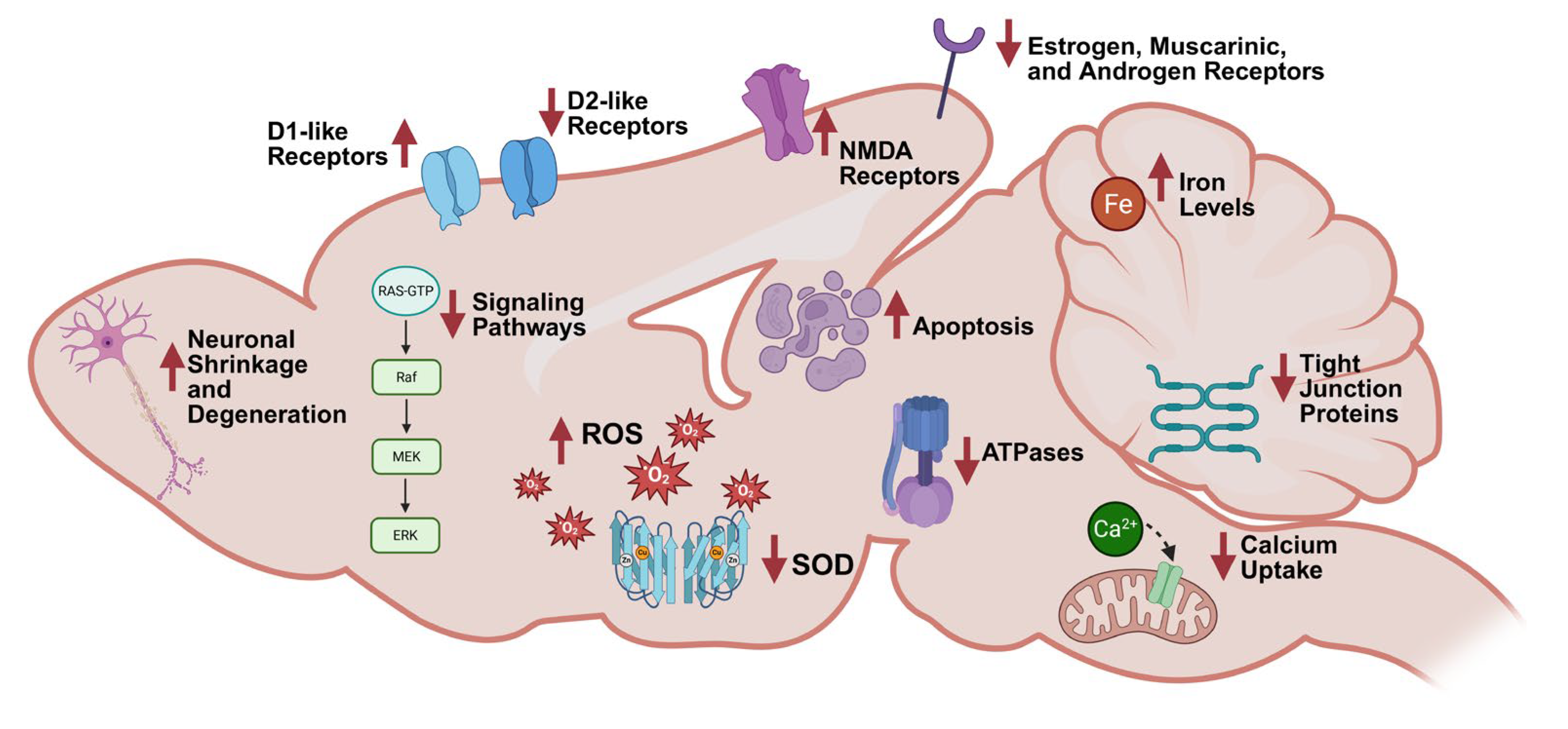
Graphical Abstract

1. Introduction

Polychlorinated biphenyls (PCBs) are a class of 209 synthetic chlorinated hydrocarbons that were manufactured for industrial and commercial products due to their electrical insulation properties and chemical stability [1]. Commercial PCB mixtures, sold in the United States (U.S.) under the trade name Aroclor, served as dielectric fluids in transformers and capacitors [1]. PCBs were also used as plasticizers and in lubricants, adhesives, and sealants in various building materials, such as caulking, floor finishes, and paints [1,2,3]. Although their manufacture was banned in the U.S. in 1979 under the Toxic Substances Control Act, PCBs continue to be pervasive in indoor environments [1]. PCB-containing caulks and sealants applied between the 1950s and 1970s continue to volatilize, emitting vapors that accumulate in indoor air and dust [4,5]. Studies in schools in the U.S. and Europe have documented the presence of PCBs in indoor air, raising significant concerns for public health [6,7,8,9,10,11]. Moreover, PCBs persist in the environment due to their resistance to degradation [12] and continue to be released into the environment from legacy sources [13,14]. Importantly, PCBs are inadvertently generated as byproducts of manufacturing processes [13,14,15].
Due to their lipophilicity and environmental persistence, PCBs bioaccumulate and biomagnify in the food chain, particularly in the fatty tissues of fish and other animals [16,17]. The bioaccumulation of PCBs is congener-dependent because physicochemical properties, three-dimensional structure, uptake, biotransformation, and excretion vary by congener [18,19,20]. Human exposure to PCBs occurs through ingestion of contaminated food and dermal contact [2,21]. Evidence from laboratory [22,23,24,25,26,27,28,29,30,31,32] and epidemiological studies [33,34,35,36,37] demonstrates that inhalation is an important route of current human PCB exposure. Human biomonitoring studies have detected PCBs in serum, adipose, and brain tissues across populations regardless of age, race, and socioeconomic status [38,39]. PCBs are a significant public health concern with well-documented toxic effects, including, but not limited to, carcinogenicity [40,41], immunotoxicity [42,43], reproductive toxicity [44,45], developmental toxicity [46,47], neurotoxicity [48,49], endocrine disruption [50,51], and cardiovascular impacts [52,53]. Epidemiological studies have established associations between PCB exposure and a range of adverse neurological outcomes, including decreased cognitive function and mood dysregulation, such as anxiety and depression [39,54].
Individual PCB congeners can be categorized into two functional classes based on structural and toxicological properties. Dioxin-like PCBs, which include congeners such as PCB 77 or PCB 126, can act like dioxins in the body [55]. Like dioxins, dioxin-like PCBs exert their toxicity through activation of the aryl hydrocarbon receptor (AhR) [56], with developmental effects as the most sensitive toxic endpoint [57]. In contrast, non-dioxin-like PCBs, such as PCB 180, act via AhR-independent pathways, including disruption of calcium homeostasis [58]. Non-dioxin-like PCBs have different toxicological activity, with more sensitivity in the liver and thyroid [59]. AhR-independent mechanisms involve activation of several nuclear receptors, such as the constitutive androstane receptor (CAR) and pregnane X receptor (PXR), both of which regulate the expression of genes involved in xenobiotic metabolism [60]. These mechanistic distinctions are essential for understanding the congener-specific risks associated with environmental PCB exposure. Other receptors also contribute to the diverse toxicological profiles of individual congeners. Ryanodine receptors (RyRs) are critical targets of non-dioxin-like PCBs, particularly ortho-substituted congeners, leading to dysregulated calcium signaling and altered neuronal excitability [61,62]. Estrogen receptors (ERs) and androgen receptors (ARs) may be modulated by specific hydroxylated PCB metabolites, acting as endocrine disruptors that interfere with hormone signaling [63,64]. Additionally, peroxisome proliferator-activated receptors (PPARs) are implicated in PCB-induced metabolic dysfunction, particularly through activation by PCBs [65,66].
PCB metabolism is a complex process involving multiple pathways [67,68,69]. PCBs undergo biotransformation through Phase I and Phase II metabolic pathways, primarily mediated by hepatic enzymes [70]. In Phase I metabolism, cytochrome P450 monooxygenases (CYPs), such as CYP1A and CYP2B isoforms, catalyze the oxidation of PCBs to form hydroxylated metabolites (OH-PCBs) [71,72,73,74]. PCB metabolism is congener-specific and strongly influenced by the number and position of chlorine atoms [75]. Non-ortho and mono-ortho-substituted PCBs are more readily metabolized than fully ortho-substituted or higher chlorinated congeners [76,77]. Some OH-PCBs can undergo further oxidation to form dihydroxylated PCBs [74], which may oxidize to form reactive PCB quinones, capable of redox cycling and inducing oxidative stress [78,79]. Phase II metabolism involves the conjugation of hydroxylated PCBs with polar functional groups to increase water solubility and promote excretion [80,81]. This includes glucuronidation by UDP-glucuronosyltransferases (UGTs) [82], sulfation by sulfotransferases (SULTs) [83], and glutathione conjugation via glutathione-S-transferases (GSTs) [84]. Additional processes include methoxylation of hydroxylated metabolites by O-methyltransferase (OMT) [85], and the formation of methyl sulfones via the mercapturic acid pathway [86]. Understanding these biotransformation pathways is crucial for interpreting differences in susceptibility and assessing the risk of neurotoxicity from PCB exposures.
While the neurodevelopmental toxicity of PCBs has been extensively studied in the context of prenatal and lactational exposure [48,87,88,89], much less is known about the consequences following post-weaning PCB exposure. Additionally, most preclinical models focus exclusively on either behavioral or molecular outcomes, limiting the ability to draw mechanistic connections between PCB-induced changes in neurochemistry and observed functional impairments. Furthermore, the sex-specific effects of PCB exposure during critical windows of exposure remain underexplored, despite increasing evidence that males and females may differ in their vulnerability and response to neurotoxicants [90]. To summarize the available evidence, a scoping review of the literature was conducted, with a specific focus on PCB exposure during the post-weaning period and its associated neurotoxic effects.

2. Methods

2.1. Selection of Articles

2.1.1. Search Strategy

The search strategy was adapted from the Preferred Reporting Items for Systematic Reviews and Meta-Analysis (PRISMA) best-practice guidelines and developed with a librarian trained in scoping reviews [91]. The scoping review, which complies with the PRISMA guidelines, was not preregistered. An outline of the search strategy, the inclusion and exclusion criteria, and the data extraction process is summarized in the flow chart shown in Figure 1. Briefly, a comprehensive search was conducted in PubMed, Scopus, and Embase using Boolean search terms that included combinations of “PCBs,” “neurotoxicity,” “mice,” “rats,” “adolescents,” and “adults” (see Supporting Information Table S1).
While the search strategy also included title/abstract search terms, a truncated form of the PubMed search with only controlled vocabulary (Medical Subject Headings) is as follows: (“Rats”[Mesh] OR “Murinae”[Mesh] OR “Mice”[Mesh]) AND (“Adolescent”[Mesh] OR “Child”[Mesh] OR “Minors”[Mesh] OR “Adult”[Mesh] OR “Middle Aged”[Mesh] OR “Aging”[Mesh] OR “Aged”[Mesh] OR “Aged, 80 and over”[Mesh] OR “Young Adult”[Mesh]) AND (“Polychlorinated Biphenyls”[Mesh] OR “Biphenyl Compounds”[Mesh] OR “Aroclors”[Mesh] OR “Dioxins and Dioxin-like Compounds”[Mesh] OR “Dioxins”[Mesh]) AND (“Attention Deficit Disorder with Hyperactivity”[Mesh] OR “Neurodevelopmental Disorders”[Mesh] OR “toxicity”[Subheading] OR “Neurocognitive Disorders”[Mesh] OR “Growth and Development”[Mesh] OR “Brain”[Mesh] OR “Central Nervous System”[Mesh] OR “Nervous System”[Mesh]). The searches were conducted in November 2023. These citations were imported into EndNote (Version 20.6, RRID:SCR_014001). The Bramer method [92] was employed to deduplicate references. Additional articles were excluded based on the predefined exclusion criteria, including non-English language, non-peer-reviewed work (e.g., editorials, commentaries), and irrelevant study types (e.g., reviews).

2.1.2. Title and Abstract Screening

The title and abstract screening were performed in parallel using both manual screening by researchers and automated screening with ChatGPT 4o (OpenAI, San Francisco, CA, USA) [93]. Two researchers independently screened the titles and abstracts to identify studies involving mouse, rat, human, or in vitro subjects exposed to PCBs or related chemicals with a reported neurotoxic outcome. Disagreements were resolved through discussion or by a third reviewer when consensus could not be reached.

2.1.3. Automated Screening Using ChatGPT

Given the complexity of this topic, which includes the large number of PCB congeners and mixtures, varying windows and routes of exposure, and diverse outcome measures, scoping reviews of this nature are inherently labor-intensive and time-consuming. To improve the rigor and scalability of this process, ChatGPT was utilized following a recently published method [93] to aid in literature screening and data extraction. Default temperature settings and standard conversation context were applied via the OpenAI web interface on a desktop computer (Intel Core i7 processor, 8 GB RAM, Windows 10, Google Chrome browser), with the “improve the model for everyone” feature disabled. No custom system prompts, external fine-tuning, or temperature adjustments were used. Each session was initialized with the same prompts to guide the model’s reasoning. ChatGPT was trained using a series of specific prompts (Table 1) to improve its ability to classify relevant studies [93]. First, ChatGPT was asked to define each category to ensure consistent classification. Subsequently, ChatGPT was asked to categorize each article into one or more of the following categories: (1) mouse, rat, human, or in vitro subjects, (2) PCB or related chemical exposure, (3) neurotoxic outcome, or (4) not related. ChatGPT was then prompted to classify each title and abstract, returning its classifications in a table format. ChatGPT was asked to use an “X” to indicate which categories applied to each article, with a brief explanation for each selection (Table 2). These results were transferred into Microsoft Excel for Microsoft 365 MSO (Version 2506, 64-bit, Windows) for further analysis.

2.1.4. Analysis of Screened Articles

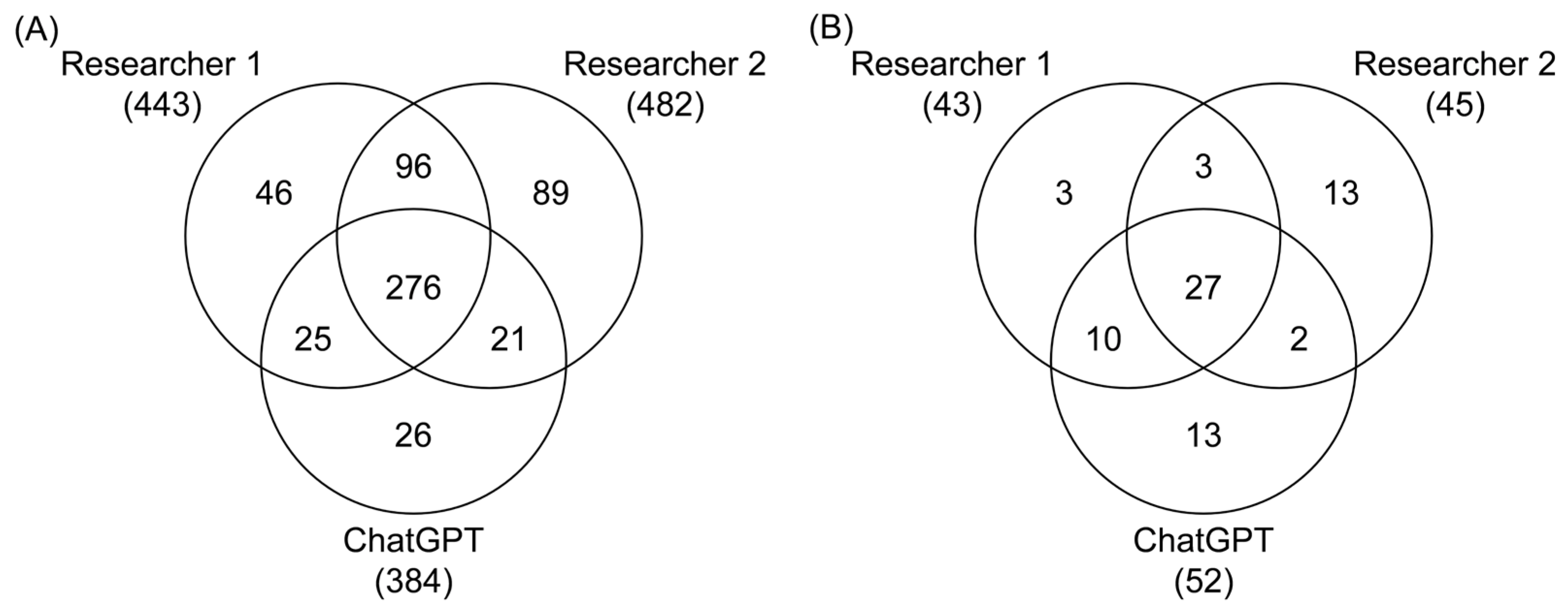
An article was selected for the next screening phase based on the classification of both researchers and ChatGPT. If only two of the three screeners (both researchers or one researcher and ChatGPT) selected an article, the article was re-evaluated by both researchers, with disagreements resolved by a third reviewer acting as a tiebreaker. Articles selected by only one screener were excluded from further screening (Figure 2).

2.2. Method Screening

2.2.1. Manual Screening of Methods Sections

Two independent researchers manually screened the methods sections of the selected articles. The aim was to identify studies involving mice or rats exposed to PCBs starting on postnatal day 21 or later, with reported neurotoxic or behavioral outcomes. Discrepancies between the two researchers were resolved through discussion or by a third reviewer, who served as a tiebreaker if necessary.

2.2.2. Automated Methods Screening Using ChatGPT

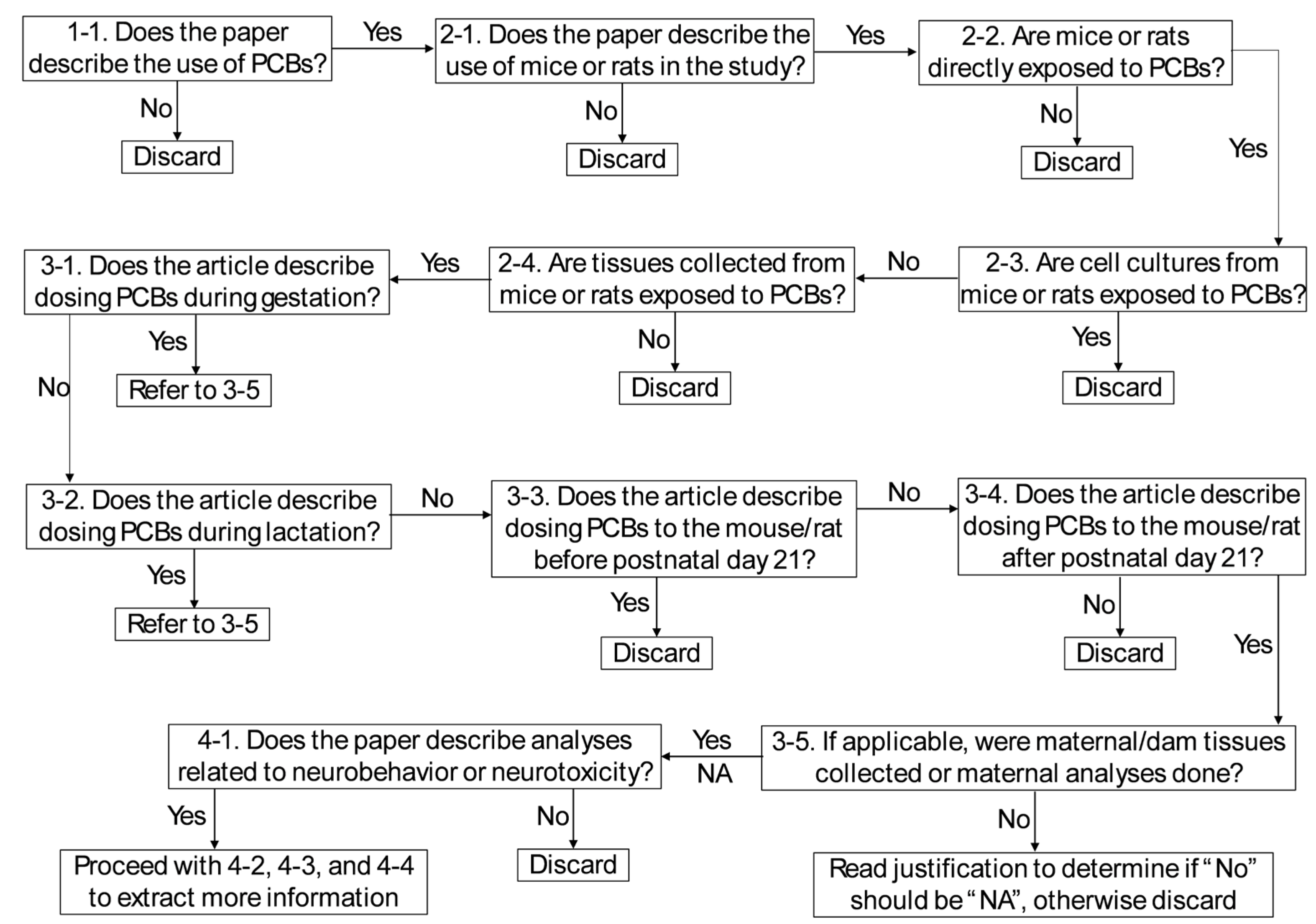
Subsequently, ChatGPT was utilized to screen the methods sections. Four key criteria were evaluated: (1) PCB exposure, (2) study subject (mice or rats), (3) dosing timeframe (postnatal day 21 or later), and (4) neurotoxic outcomes. Fourteen yes/no questions were created (Table 3) to guide ChatGPT’s evaluation of each article’s methods. ChatGPT was prompted to answer each question in a table format, with explanations for each “Yes” or “No” answer (Table 4). These results were copied into an Excel sheet and filtered to identify studies that met the predefined criteria. To ensure consistency and reproducibility, a decision tree (Figure 3) was developed to guide reviewers in interpreting and verifying ChatGPT’s responses. This decision tree outlined the logical sequence by which reviewers determined whether a study should be included, excluded, or flagged for further discussion.

2.2.3. Analysis of Methods Screening Results

An article was advanced to the full-text assessment if both researchers and ChatGPT identified the article as meeting the relevant criteria. If only two out of three screeners (both researchers or one researcher and ChatGPT) selected an article, the article was re-evaluated by both researchers. A third-party reviewer resolved any disagreements that arose as necessary. Articles selected by only one screener were excluded from further screening.

2.3. Full-Text Assessment

Prior to full-text assessment, a data extraction table was established to identify key information such as animal strain, sex, age, exposure compound(s), dose, route of administration, and behavioral or neurotoxic outcomes [94]. During full-text assessment, both researchers reviewed the entire article to extract key information. Five articles were identified as having indirect PCB exposure (e.g., through contaminated fish) and were excluded. Indirect exposure was defined as exposure resulting from naturally contaminated materials in which the PCB dose and congener profile could not be accurately determined or experimentally controlled. Such studies were excluded because the actual exposure conditions cannot be reproduced by other laboratories. The present review, therefore, focused on controlled experimental exposures in which PCB congeners or mixtures were deliberately administered via defined routes (e.g., oral gavage, inhalation, or intraperitoneal injection). From each included study, we extracted detailed information on study characteristics (author names, year of publication, country, and funding source), animal model features (species, strain, sex, age at exposure, and developmental stage), and exposure parameters (route, dose, duration, frequency, exposure window, and the specific PCB congeners or mixtures tested). Information on control conditions (e.g., vehicle-treated, untreated, or sham controls) was also recorded. Outcomes were classified as either neurotoxic (e.g., histological changes, alterations in neurotransmitter levels, or biomarkers of neurotoxicity) or behavioral (e.g., performance in learning and memory tasks, motor activity assessments, and anxiety-like behavior). Additional study design features were extracted, including sample size and methods of outcome assessment. Behavioral outcomes were grouped into broad domains, such as cognitive, motor, or anxiety-related, to enable synthesis across methodologies. All extracted information was organized into summary tables (Table A1, Table A2, Table A3, Table A4, Table A5, Table A6 and Table A7).

2.4. Limitations

Several limitations of the review process should be acknowledged. First, restricting the search to English-language publications may have led to the omission of relevant studies, particularly those published in non-indexed sources. The exclusion of gray literature and non-peer-reviewed studies may have further narrowed the scope. During title, abstract, and full-text screening, subjective decisions made by human reviewers and ChatGPT may have introduced bias, despite efforts to standardize the classification process. The application of strict exclusion criteria may have excluded studies with mixed exposures or partially relevant designs. Data extraction was complicated by inconsistent reporting of exposure doses, animal characteristics (e.g., strain, sex), and neurobehavioral outcomes, requiring simplification that may have obscured important nuances. As a scoping review, no formal assessment of study quality was conducted, and no quantitative synthesis was performed; thus, findings are descriptive and do not reflect effect sizes or dose–response relationships. Finally, focusing solely on post-weaning rodent models may limit generalizability to other developmental stages or species, and grouping diverse exposure paradigms may have masked important differences in congener-specific or temporal effects.

2.5. Statistical Analysis

The agreement of the human reviewers and ChatGPT was evaluated using Cohen’s and Fleiss’ kappa at the title/abstract and methods screening steps of the review process. Cohen’s kappa evaluates the agreement between two reviewers and was developed to consider the likelihood that raters may make guesses on some variables due to uncertainty [95]. Kappa values were calculated for each of the comparisons (Reviewer 1 & Reviewer 2, Reviewer 1 & ChatGPT, and Reviewer 2 & ChatGPT) to determine agreement between raters. Values > 0.21 are considered “Fair”, values > 0.41 are considered “Moderate”, values > 0.61 are considered “Substantial”, and values > 0.81 are considered “Almost Perfect”. Fleiss’ kappa is similar to Cohen’s and allows for more than two raters to be evaluated [96].

3. Results and Discussion

3.1. Article Selection

An outline of the search strategy, the inclusion and exclusion criteria established a priori, and the data extraction process are summarized in the flow chart shown in Figure 1. This search identified a total of 6166 citations. The Bramer method [92] was employed to remove 2425 duplicate articles. Subsequently, 477 additional articles were excluded based on the predefined exclusion criteria. This process resulted in 3264 articles being eligible for title and abstract screening, which was performed independently by two Reviewers and ChatGPT. During title and abstract screening, Researcher 1 identified 443 articles, Researcher 2 identified 482 articles, and ChatGPT identified 348 articles that met the inclusion criteria. This resulted in 276 articles selected by all three screeners, 142 articles selected by two reviewers or one reviewer and ChatGPT, and 161 selected by only one reviewer. These 161 articles were excluded from the review (Figure 2A). The 142 articles selected by two reviewers or ChatGPT were reevaluated by the two human reviewers, with a third reviewer serving as a tiebreaker. Of these articles, 84 were selected for inclusion, resulting in 360 articles proceeding to the methods screening phase. During method screening, Researcher 1 identified 43 relevant articles, Researcher 2 identified 45 articles, and ChatGPT identified 52 articles. This resulted in 27 articles being selected by all three screeners, and 44 articles underwent re-evaluation (Figure 2B), ultimately leading to 40 articles being chosen for full-text assessment. During full-text assessment, 5 articles were identified as having indirect PCB exposure (e.g., through PCB-contaminated fish [97,98,99,100] or food grown on PCB-contaminated sediment [101]) and were excluded, resulting in 35 articles selected for inclusion. Overall, the studies investigated outcomes primarily in male rats and mice with 24 studies examining only males, 5 investigating males and females, and 6 studies examining only female rodents. In addition to these 35 studies, other relevant literature is cited throughout the manuscript to provide background, methodological comparisons, or mechanistic insights.
Screening time was measured using a start–stop method, where each reviewer recorded the duration required to complete screening for each batch of abstracts or methods, and an average time was calculated for all articles. Researchers spent an average of 54.4 h screening titles and abstracts, while using ChatGPT reduced this time to about 12 h. During the methods screening stage, each researcher invested around 36 h, whereas ChatGPT required only about 9 h. These findings suggest that ChatGPT can significantly reduce time spent during the initial screening phases.

3.2. Inter-Reviewer Reliability Assessment

During the title and abstract screening stage, Reviewer 1 and Reviewer 2 demonstrated the highest percentage of agreement and Cohen’s kappa, while their agreement with ChatGPT was lower (Table 5). The fixed-marginal Fleiss’ kappa value for Reviewer 1, Reviewer 2, and ChatGPT was 0.73, with a 95% confidence interval of 0.68–0.77, indicating significant agreement and an overall agreement of 93.81%. During the methods screening stage, Reviewer 1 and ChatGPT showed the highest percent agreement and Cohen’s kappa, while agreement with Reviewer 2 was lower for both Reviewer 1 and ChatGPT (Table 6). Fleiss’ kappa for the method selection step was determined for Reviewer 1, Reviewer 2, and ChatGPT. There was an overall agreement of 91.85%, with a fixed-marginal kappa of 0.64 and a 95% confidence interval of 0.49–0.78, indicating moderate to significant agreement. Consistent with earlier research [93], our results support the integration of ChatGPT and similar large language models as effective tools for scoping review.

3.3. Neurotoxic Outcomes

This section provides a scoping review of the neurotoxic effects of PCBs observed in rodent models, focusing on histological, molecular, and biochemical endpoints, in the studies identified in the scoping review. Of the 27 studies reporting neurotoxic outcomes, 22 studies examined only males, 2 investigated males and females, and 3 studies examined only female rodents. Studies examined a range of exposure routes (oral, inhalation, intraperitoneal), as well as PCB mixtures (e.g., Aroclors 1254, 1221, and 1260) and individual PCB congeners (e.g., PCB 180 and 153). Outcomes reported in these studies include structural and morphological changes in various brain regions, alterations in calcium homeostasis, oxidative stress, neurotransmitter dynamics, apoptotic signaling, or changes in gene and protein expression. These findings highlight the broad neurotoxic potential of PCBs, with brain region and sex-specific differences that may underlie PCB-induced neurotoxic effects. Table A1, Table A2, Table A3 and Table A4 provide an overview of neurotoxic outcomes across exposure types and analyses.

3.3.1. Histological/Morphological Changes

Structural alterations in the brain, including neuronal degeneration, tissue atrophy, and cell loss, are indicators of neurotoxic damage and neurodegeneration [102]. Only a few studies have investigated the effects of PCBs on histological or morphological endpoints in female rats. Female Sprague-Dawley rats exposed to inhaled Aroclor 1254 alone, or in combination with Aroclor 1221, did not show histopathological changes in the brain [24,25].
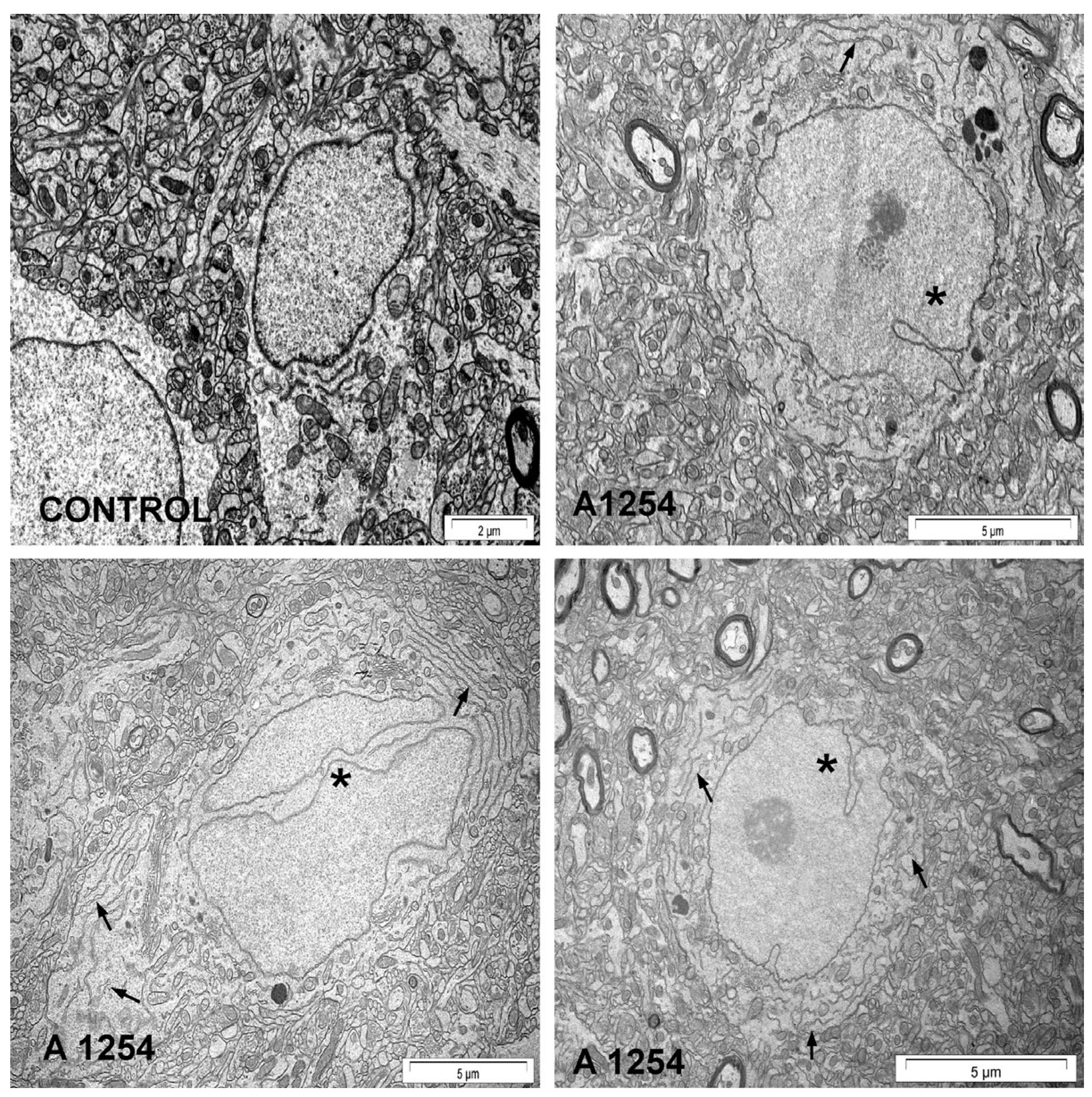
In contrast, male Wistar rats exposed intraperitoneally (i.p.) to Aroclor 1254 exhibited significant histological and morphological changes across various brain regions [103,104,105,106,107,108,109,110]. In the cerebral cortex, neuronal shrinkage [104], degeneration [104,106], and the presence of pyknotic nuclei, accompanied by increased peri-neuronal spaces [103,104], were reported following PCB exposure. There was also an increase in pyknotic cell bodies, nonhomologous cytoplasm, and intense basophilic appearance in the cerebral cortex [107]. In contrast, pyramidal, Betz, and stellate cells, and myelin density were notably decreased in this region [107]. In the cerebellum, Purkinje neurons showed shrinkage [103,105], degeneration [103,105,106], and moderate atrophy [103]. The hippocampus displayed degeneration of pyramidal cells [103,108,109], neuronal shrinkage [108,109], and disruption in the Cornus Ammonis 4 region [110]. Male Wistar rats exposed to oral Aroclor 1254 showed a decreased number of neurons, and transmission electron microscopy identified irregularly shaped and lobed neuronal nuclei in the forebrain cortex and hippocampus, and enlargement and local edema of the rough endoplasmic reticulum in the hippocampus [111] (Figure 4).

3.3.2. Calcium Modulation

Calcium homeostasis is essential for neuronal signaling, synaptic plasticity, and cell survival, with its disruption a hallmark of excitotoxicity and neurodegeneration [112]. Disruption of calcium signaling was observed across multiple brain regions in male rats exposed to Aroclor 1254, including alterations in calcium channel expression, kinase activity, and intracellular calcium handling [104,109,111,113,114]. Male Wistar rats exposed to i.p. Aroclor 1254 exhibited increased mRNA expression of Cacna1d in the cerebral cortex [104], but decreased mRNA expression of Cacna1d in the hippocampus [109]. PCB exposure decreased protein kinase A (PKA) alpha levels and increased protein expression of N-methyl-D-aspartate (NMDA) receptors, calpain, and N-type calcium channel protein alpha1B in the cerebral cortex [113]. In the hippocampus of male Wistar rats exposed to oral Aroclor 1254, PCBs decreased the maximum NMDA-induced calcium release [111]. Male Long-Evans rats exposed to Aroclor 1254 orally had reduced calcium uptake in the microsomes across the frontal cortex, cerebellum, and striatum [114]. Additionally, PCBs decreased calcium uptake in the mitochondria of the cerebellum and decreased total protein kinase C (PKC) activity while increasing membrane-bound PKC activity [114].

3.3.3. Oxidative Stress and DNA Modifications

Oxidative damage and impaired redox balance can compromise neuronal integrity, while alterations in DNA and antioxidant defenses can affect gene expression and neural resilience [115]. Female Sprague-Dawley rats exposed to inhaled Aroclor 1254 showed reduced reactive oxygen and nitrogen species (ROS/RNS) levels in the brain, whereas oxidized lipid markers, MDA and 4-HNE, were not altered following PCB inhalation (Figure 5) [24]. In a second study, female Sprague-Dawley rats exposed to an inhaled mixture of Aroclor 1254 and Aroclor 1221 did not have altered brain ROS/RNS levels [25].
Oxidative stress was observed in the brains of male rats following i.p. exposure to Aroclor 1254, characterized by elevated lipid and protein oxidation, disrupted expression of antioxidant enzymes, and increased DNA fragmentation across multiple brain regions [103,104,105,108,110,116,117]. Male Wistar rats exposed i.p. to Aroclor 1254 showed increased levels of lipid peroxidation [104,105,108,110,116,117,118], hydroxyl radicals [110,117,118], hydrogen peroxide [104,105,108,110,116,117], and protein carbonyls [104,105,108,116] in the cerebellum [105,117,118], cerebral cortex [104,117,118], and hippocampus [108,110,116,117,118]. PCB exposure decreased glutathione levels [117,118] and mRNA expression of copper/zinc superoxide dismutase (Cu/Zn SOD) [103] and GPx-4 [103] in the cerebellum [103,117,118], cerebral cortex [103,117,118], and the hippocampus of male rats [103,117,118]. Furthermore, PCB exposure resulted in elevated levels of thiobarbituric acid reactive substances (TBARS) in the hippocampus [110] and resulted in DNA fragmentation [108]. PCB exposure led to a significant decrease in the levels of key antioxidant enzymes in the brain [103,110]. PCB exposure decreased mRNA expression of Cu/Zn SOD and glutathione peroxidase (GPx)-4 in the cerebellum, cerebral cortex, and hippocampus [103]. Additionally, PCBs decreased the concentrations of SOD, catalase (CAT), GPx, glutathione-S-transferase (GST), and glutathione reductase (GR) in the hippocampus [110]. Furthermore, PCB exposure lowered the specific activities of total SOD, Cu/Zn SOD, and manganese SOD (Mn SOD) across the cerebellum, cerebral cortex, and hippocampus, along with a decrease in the specific activity of GPx in these same regions [103]. Male and female Sprague-Dawley rats exposed to oral PCB 180 exhibited decreased glutathione levels in the cerebral cortex of males [119].
Oral exposure of male C57BL/6 mice to Aroclor 1254 induced significant oxidative stress in both the striatum and cerebellum [120]. PCB exposure increased the oxidized lipid markers MDA and 4-HNE in both regions, with greater accumulation observed in the striatum than in the cerebellum [120]. Additionally, PCB exposure led to an increase in the number of oxidized proteins in both the striatum and cerebellum. In response to this oxidative stress, PCB exposure also upregulated the protein expression of heme oxygenase-1 (HO-1) in both brain regions [120]. Furthermore, PCB exposure led to increased Mn SOD protein levels in the striatum and cerebellum, as well as an increase in Cu/Zn SOD protein expression in both regions [120].

3.3.4. Neurotransmitters and Receptors

Disruptions in neurotransmitter systems and receptor signaling can impair synaptic transmission, cognitive function, mood regulation, and motor control [121]. The following sections examine how PCB exposure disrupts key neurotransmitter systems in rodent brains, including dopamine (DA) metabolism, and receptor/transporter expression; serotonin and norepinephrine dynamics; glutamatergic signaling and transporter regulation; and the biosynthesis machinery for neurotransmitters.
Biosynthesis of Neurotransmitters
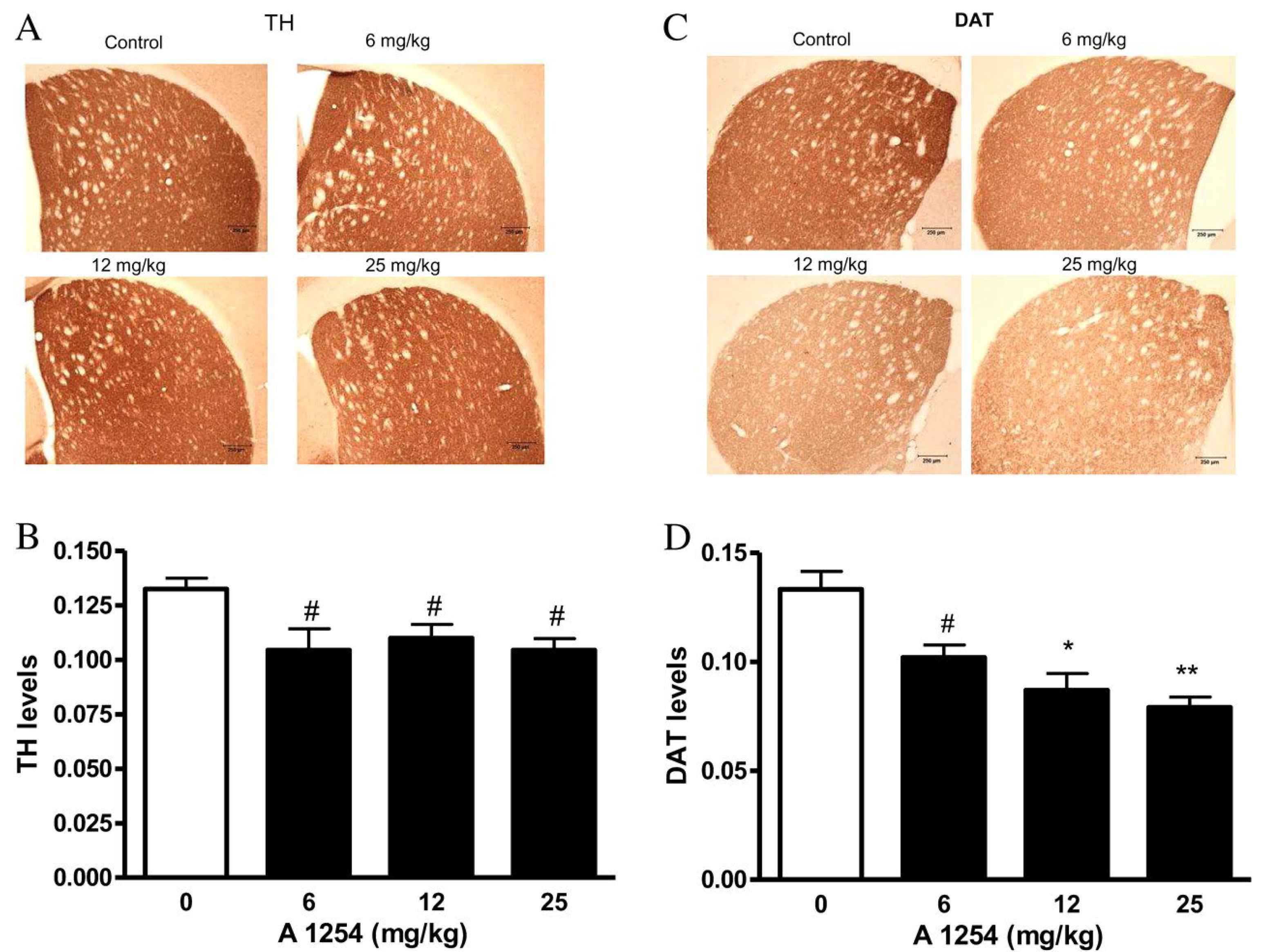
Alterations in the biosynthesis of neurotransmitters in the brain can reduce the efficiency of neurotransmission and impair the function of neural networks [122]. Most studies investigating the effects of PCBs on neurotransmitter synthesis to date have focused on the expression and activity of tyrosine hydroxylase (TH), the rate-limiting enzyme of catecholamine biosynthesis [123]. Male Wistar rats exposed i.p. to Aroclor 1254 did not exhibit any changes in the mRNA expression of TH in the cerebellum or hippocampus. However, PCB exposure resulted in a decrease in protein expression of tyrosine hydroxylase in both the cerebellum and hippocampus [105,109]. Male Long-Evans rats exposed orally to Aroclor 1254 showed no changes in TH immunoreactivity or specific activity in the striatum [114]. Male C57BL/6 mice exposed orally to Aroclor 1254 exhibited a decrease in TH concentration in the striatum across all exposure groups (Figure 6A,B) [120].
Dopamine and Metabolites
DA plays critical roles in reward, motivation, and motor control; dysregulation is linked to neurological diseases [124,125]. Several studies have indicated PCBs influence levels of DA and its metabolites [126,127,128,129,130,131]. Consistent with these results, male Sprague-Dawley rats exposed orally to Aroclor 1254 exhibited varying effects on DA concentrations in striatal dialysate, as determined by a three-way analysis of variance (ANOVA) [132]. PCB exposure did not show a main effect on DA levels in the dialysate, but the interaction between PCB exposure and exposure duration was significant [132]. Specifically, PCB exposure increased DA concentrations after 3 days of exposure, but decreased DA concentrations after 1, 2, and 8 weeks [132]. In contrast, PCB exposure did not alter DA levels in the striatum [132]. Analogous studies have not been reported in female Sprague-Dawley rats. However, female Sprague-Dawley rats exposed to inhaled Aroclor 1254 and 1221 did not exhibit any alterations in DA or 3,4-dihydroxyphenylacetic acid (DOPAC) levels in the striatum [25].
Male Wistar rats exposed orally to Aroclor 1254 exhibited significant alterations in DA and its metabolites in specific brain regions [133]. PCB exposure resulted in a decrease in DA levels in the striatum, accompanied by reduced levels of its metabolites DOPAC and homovanillic acid (HVA) [133]. This exposure also resulted in a decrease in the DOPAC/DA and HVA/DA ratios in the striatum [133]. Similarly, while PCB exposure did not affect DA levels in the lateral olfactory tract, PCBs significantly reduced the levels of DOPAC, HVA, DOPAC/DA, and HVA/DA ratios in this region [133]. In a separate animal study, male Wistar rats exposed orally to Aroclor 1254 and 1260 showed significant changes in DA metabolism in the brain [134]. PCB exposure decreased DA and DOPAC concentrations in the caudate; however, PCB exposure did not affect HVA concentrations in either the caudate or the lateral olfactory tract, and DA and DOPAC levels remained unchanged in the lateral olfactory tract [134]. The DOPAC/DA ratio was decreased only in the lateral olfactory tract. At the same time, PCB exposure did not affect the HVA/DA ratios in either the caudate or lateral olfactory tract [134].
There is also evidence that PCB exposure alters brain DA homeostasis in male Long-Evans rats and male C57BL/6 mice. Analogous studies in the corresponding female animals have not been reported to date. Briefly, male Long-Evans rats exposed orally to Aroclor 1254 did not exhibit any changes in the levels of DA or HVA in the frontal cortex or striatum [114]. However, PCB exposure did lead to an increase in DOPAC levels in the striatum of exposed rats compared to controls [114]. Male C57BL/6 mice exposed orally to Aroclor 1254 exhibited an increase in DA levels; however, PCB exposure did not alter the levels of DOPAC, HVA, or the HVA/DA ratio in the striatum [120].
Dopamine Receptors and Transporters
Changes in dopamine receptor expression alter synaptic responsiveness and are implicated in neurodevelopmental and neuropsychiatric disorders [135]. Male and female Sprague-Dawley rats exposed orally to PCB 180 observed no alterations in specific binding to D1/D5 dopamine receptors in the cerebellum [119].
Male Wistar rats exposed i.p. to Aroclor 1254 exhibited significant changes in the expression of dopamine receptors in various brain regions [104,105,109]. PCB exposure increased the mRNA and protein expression of excitatory D1-like receptors (DRD1 and DRD5) in the cerebellum while decreasing the mRNA and protein expression of inhibitory D2-like receptors (DRD2, DRD3, and DRD4) in this region [105]. Similarly, PCB exposure increased mRNA expression for D1-like receptors in the cerebral cortex, while reducing mRNA expression for D2-like receptors [104]. In the hippocampus, PCB exposure led to increased mRNA and protein expression of the excitatory D1-like receptors and decreased expression of the inhibitory D2-like receptors [109]. These studies in rats did not investigate changes in the expression of dopamine transporter (DAT). However, male C57BL/6 mice exposed orally to Aroclor 1254 showed a decrease in the concentration of DAT in the striatum [120] (Figure 6C,D).
Serotonin and Norepinephrine
Serotonin and norepinephrine (NE) are key modulators of mood, arousal, and stress response, where disruptions are associated with anxiety, depression, and cognitive deficits [136]. Several studies using synaptosomes suggest that PCB mixtures may affect serotonin homeostasis [128,131,137]. Moreover, the following evidence from rodent studies suggests alterations in serotonin metabolism in the brain following exposure to PCBs.
Male Wistar rats exposed orally to Aroclor 1254 and 1260 exhibited region-specific alterations in serotonin metabolism [138,139]. PCB exposure decreased serotonin levels in the frontal cortex and hippocampus, while increasing serotonin concentrations in the lateral olfactory tract; however, no changes were observed in serotonin concentrations in the brainstem or hypothalamus [139]. PCB exposure also increased 5-hydroxyindoleacetic acid (5-HIAA), the primary metabolite of serotonin, concentrations in the frontal cortex and lateral olfactory tract, but did not affect 5-HIAA concentrations in the hippocampus, brainstem, or hypothalamus [139]. The 5-HIAA/serotonin ratio was elevated in the frontal cortex, hippocampus, brainstem, and hypothalamus, though no changes were observed in the lateral olfactory tract [139]. Exposure to oral Aroclor 1254 and 1260 also showed region-specific changes in NE concentration. PCB exposure decreased NE concentrations in both the dorsal frontal cortex and hippocampus; however, PCBs did not affect NE concentrations in the medial-basal hypothalamus or brainstem [138].
Male Long-Evans rats exposed orally to Aroclor 1254 did not show any changes in the concentrations of NE or serotonin in the frontal cortex or striatum [114]. However, PCB exposure did increase the concentration of 5-HIAA in the striatum [114]. Male Wistar rats exposed orally to Aroclor 1254 did not show any changes in the concentration or activity of serotonin and NE in the striatum and lateral olfactory tract [133].
Glutamate
Glutamate is vital for synaptic plasticity as the primary excitatory neurotransmitter, where dysregulation contributes to excitotoxicity and neuronal injury [140]. Male Wistar rats exposed i.p. to Aroclor 1254 exhibited decreased mRNA expression of several key proteins relevant to glutamate synthesis and signaling in the cerebral cortex [113]. PCB exposure reduced the mRNA expression of the glial glutamate aspartate transporter (GLAST), brain-derived neurotrophic factor (BDNF), and neurotrophin tyrosine kinase receptor B (NTRKB) [113]. Additionally, PCB exposure decreased the mRNA expression of downstream signaling molecules, including microtubule-associated protein 1 (MAP1), extracellular signal-regulated kinase 1 and 2 (ERK1 and ERK2), and the transcription factor cyclic AMP-response element binding protein (CREB) [113].
Male Wistar rats exposed orally to Aroclor 1254 exhibited several changes in glutamate uptake and transporter expression in freshly prepared whole-brain homogenates, synaptosomal fractions, and glial plasmalemmal vesicle (GPV) fractions. The rate of glutamate uptake into synaptosomes was increased, yet reduced in GPV fractions prepared from PCB-exposed male rats compared to the respective controls [141]. Additionally, the rate of glutamate accumulation was diminished at the point of maximal uptake in the GPV fraction [141]. PCB exposure also increased KCl-stimulated glutamate release in the synaptosomal fraction but did not alter the rate of released [3H] glutamate in the GPV fractions [141]. Furthermore, PCB exposure decreased glutamate transporter 1 (GLT-1) mRNA and protein expression in the forebrain, while GLT-1 remained unchanged in the cerebellum [141]. Similarly, GLAST mRNA and protein expression were unchanged in both the forebrain and cerebellum [141]. PCB exposure increased the protein level of excitatory amino acid carrier 1 (EAAC1) in forebrain homogenates, although mRNA levels remained unchanged. In contrast, PCB exposure in cerebellar homogenates decreased EAAC1 mRNA levels and showed a trend toward reduced protein levels [141].
Male rats exposed to oral Aroclor 1254 also exhibited significant alterations in NMDA-evoked glutamate signaling [111]. PCB exposure abolished NMDA-evoked glutamate release and reduced basal microdialysate glutamate concentration in hippocampal microdialysate [111]. The concentration of basal cyclic guanosine monophosphate (cGMP), a second messenger in glutamate neurotransmission, was not affected; however, PCB exposure decreased NMDA-evoked cGMP accumulation [111]. No studies investigated glutamate in female rats.

3.3.5. Apoptosis

Apoptotic signaling in the brain, which plays a critical role in normal neuronal pruning during adolescent brain development [142], can also lead to programmed cell death in the adult brain, contributing to neurodegeneration and the loss of neural circuitry [143]. Male Wistar rats exposed i.p. to Aroclor 1254 exhibited significant changes in apoptosis-related signaling pathways in the cerebral cortex, cerebellum, and hippocampus [106,107,108]. PCB exposure increased nuclear factor kappa B (NF-kB) mRNA expression in both the cerebral cortex and cerebellum [106], as well as increased mRNA expression of pro-apoptotic proteins BH3 interacting-domain death agonist (Bid), BCL2-associated death promoter (Bad), BCL2-associated X protein (Bax), caspase 3 and 9 (CASP3 and CASP9) in the cerebral cortex [106,107]. Additionally, PCB exposure decreased the mRNA expression of the anti-apoptotic protein B-cell leukemia/lymphoma 2 (Bcl2) in the cerebral cortex [106,107]. In the hippocampus, PCB exposure increased both mRNA and protein expression of pro-apoptotic genes Bad, Bid, and Bax, while decreasing the mRNA and protein expression of the anti-apoptotic gene Bcl2 [108]. PCB exposure also led to increased mRNA and protein expression of pro-inflammatory markers, including tumor necrosis factor alpha (TNFα), NF-kB, tumor necrosis factor receptor superfamily member 6 (Fas), and Fas ligand (FasL) in the hippocampus [108], as well as increased FasL and caspase 8 (CASP8) mRNA expression in the cerebral cortex and cerebellum [106]. Furthermore, PCB exposure increased the mRNA and protein expression of CASP3, CAP8, and CASP9 in the hippocampus, along with elevated caspase 3 activity in this region [108].
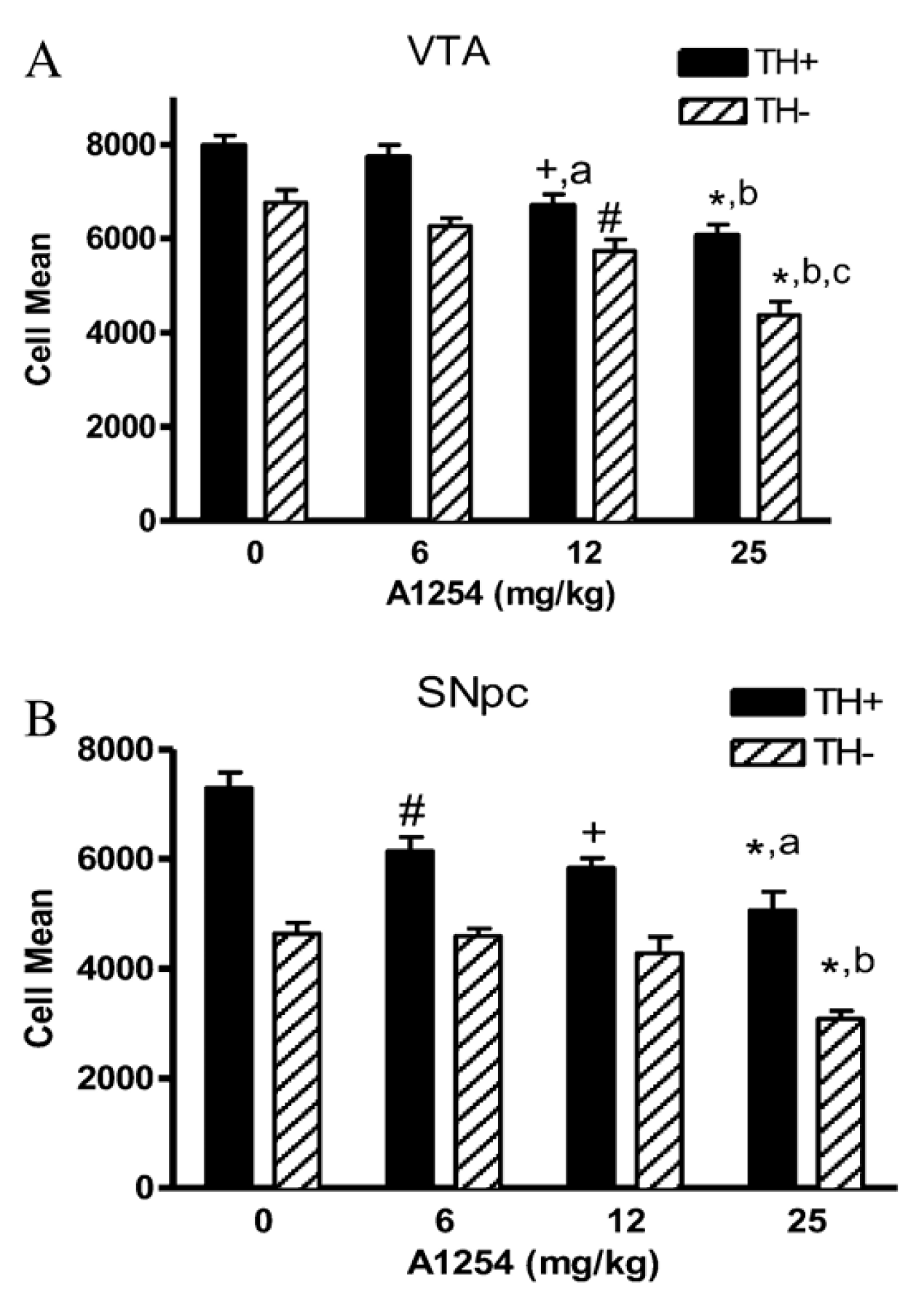
Male C57BL/6 mice exposed orally to Aroclor 1254 exhibited increased cell death in both TH-positive (TH+) and TH-negative (TH−) neurons in the substantia nigra pars compacta (SNpc) and ventral tegmental area (VTA) [120] (Figure 7).

3.3.6. Additional Drivers of PCB-Induced Neurotoxicity

Additional molecular and cellular processes, including enzyme activities, receptor expression [144], iron homeostasis [145], amino acid levels [146], and gene expression have been investigated for their potential role in PCB neurotoxicity.
Enzymes
Changes in enzymatic activity can impact neurotransmitter metabolism, energy production, and ionic gradients, all of which are essential for normal neuronal function [147,148,149,150]. Male Sprague-Dawley rats exposed orally to Aroclor 1254 were evaluated for changes in nitric oxide synthase (NOS) activity using NADPH-diaphorase (NADPH-d) following hyperosmotic challenge. Hyperosmotic challenge involves i.p. injection of hypertonic saline and temporary water deprivation to induce intracellular dehydration and stimulate NOS activity in the brain. NOS-containing neurons were visualized using NADPH-d histochemistry, an established staining method in which reaction product intensity reflects NOS enzymatic activity due to the shared use of NADPH as a cofactor. Quantification was based on staining density in the supraoptic nucleus (SON). No significant difference in NADPH-d staining density in the SON was observed between hyperosmotic PCB-treated rats and hyperosmotic controls [151].
Male Wistar rats that were exposed to Aroclor 1254 via i.p. injection showed a reduction in the activity of membrane-bound ATPases [104,105,109,117], which are crucial for maintaining the ionic environment and signaling processes of neurons across brain regions [152,153,154,155]. Briefly, PCB exposure led to a reduction in the mean activity of Na+/K+ [104,105,109,117], Ca2+ [104,105,109,117], and Mg2+ ATPases [104,105,109,117] in the cerebral cortex [104,117], cerebellum [105,117], and hippocampus [109,117].
Creatine kinase plays a role in brain energetics and neurotransmitter trafficking between glial cells and neurons across multiple brain regions [156,157]. Male Wistar rats exposed i.p. to Aroclor 1254 showed a reduction in the activity of creatine kinase [104,105,118]. PCB exposure lowered the mean creatine kinase activity in the cerebral cortex [104,118], cerebellum [105,118], and hippocampus [118].
Acetylcholinesterase (AChE) is not only responsible for terminating synaptic transmission at cholinergic synapses but also has multifunctional, non-enzymatic roles in the brain that play a potential role in neurodegeneration [158,159]. Male Wistar rats exposed i.p. to Aroclor 1254 also exhibited a decrease in the activity of AChE [104,105,117]. PCB exposure reduced the mean AChE activity in the cerebellum [105,117], cerebral cortex [104,117], and hippocampus [117].
Receptors
Receptor dysregulation alters neuronal responsiveness and signaling pathways that influence cognition, behavior, and neuroendocrine balance [160,161,162]. Male and female Sprague-Dawley rats exposed i.p. to Aroclor 1221 exhibited sex-specific changes in the expression of several receptors in specific brain regions. In male rats, PCB exposure decreased mRNA expression of the androgen receptor and mu opioid receptor (Oprm1) in the preoptic area [163]. Additionally, PCB exposure increased mRNA expression of the Oprm1 in the prefrontal cortex of male rats [163]. Notably, higher mRNA expression levels of the Oprm1 in the prefrontal cortex were positively associated with increased time spent near the no-hormone stimulus animal during the sociosexual preference test, suggesting a mechanistic link between Oprm1 expression and behavioral outcomes [163].
PCB exposure decreased the mRNA expression of vasopressin receptor 1a (Avpr1a) in the lateral septum of male rats exposed i.p. to Aroclor 1221 [163]. This study reported no changes in Avpr1a mRNA expression observed in the BNST, MeA, PVN, or VMH [163]. Male Wistar rats that were i.p. exposed to Aroclor 1254 exhibited reduced mRNA expression of estrogen receptors α and β in the hippocampus [116].
Female Sprague-Dawley rats exposed orally to PCB 153 exhibited changes in muscarinic receptor (MR) density across different brain regions. PCB exposure increased MR density in the cerebral cortex while decreasing MR density in the cerebellum; PCB exposure did not alter MR density in the hippocampus or striatum [164].
Male and female Sprague-Dawley rats exposed orally to PCB 180 did not show any alterations in high- or low-affinity sites on nicotinic receptors in the cerebellum [119].
Iron Metabolism
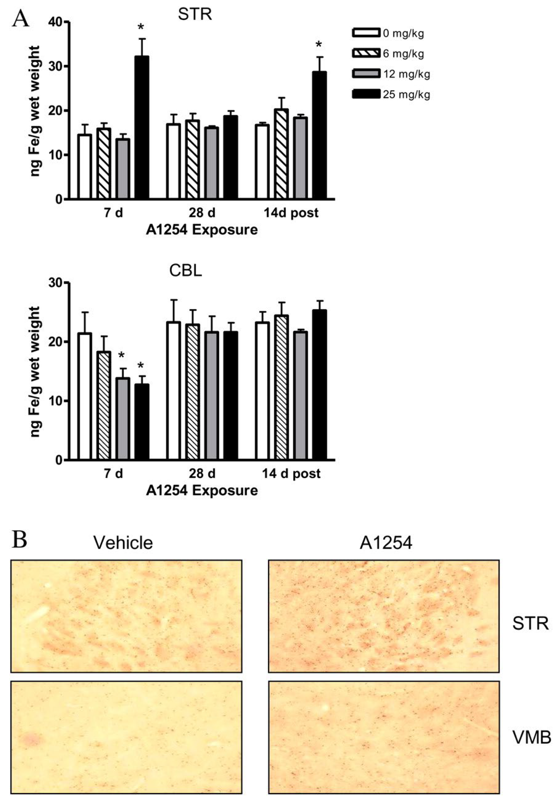
Iron accumulation in the brain can catalyze oxidative reactions and promote neurodegeneration [165,166]. Male C57BL/6 mice exposed orally to Aroclor 1254 exhibited significant changes in iron metabolism in the brain. PCB exposure elevated total iron (bound and unbound)in the striatum after 7 days of exposure and 2 weeks post-exposure, while exposure decreased total iron in the cerebellum at 7 days (Figure 8A) [120]. Additionally, PCB exposure increased the number of iron-positive cells in the striatum, but did not alter iron staining patterns in the cerebellum (Figure 8B) [120]. Further, PCB exposure increased ferritin levels in the striatum at 7 days of exposure, but ferritin levels decreased after 14 days of exposure, particularly in the 25 mg/kg group at 14 days post-exposure [120]. Finally, PCB exposure increased transferrin receptor 1 (TfR1) protein levels in the striatum [120].
Amino Acids
Amino acids serve as neurotransmitters and metabolic precursors; imbalances can impair neurotransmission and neuron-glia interactions [167,168,169]. Male and female Sprague-Dawley rats exposed to oral PCB 180 did not exhibit any changes in aspartate, glutamate, serine, glutamine, glycine, taurine, alanine, or γ-amino butyric acid in the cerebral cortex [119].
Male Wistar rats exposed orally to Aroclor 1254 exhibited reductions in basal levels of amino acids in hippocampal microdialysates. PCB exposure decreased the basal concentration of arginine [111]. However, PCB exposure did not alter the basal levels or NMDA-evoked release of taurine, nor did exposure alter aspartate accumulation, and no NMDA-evoked aspartate release observed [111].
Gene Expression
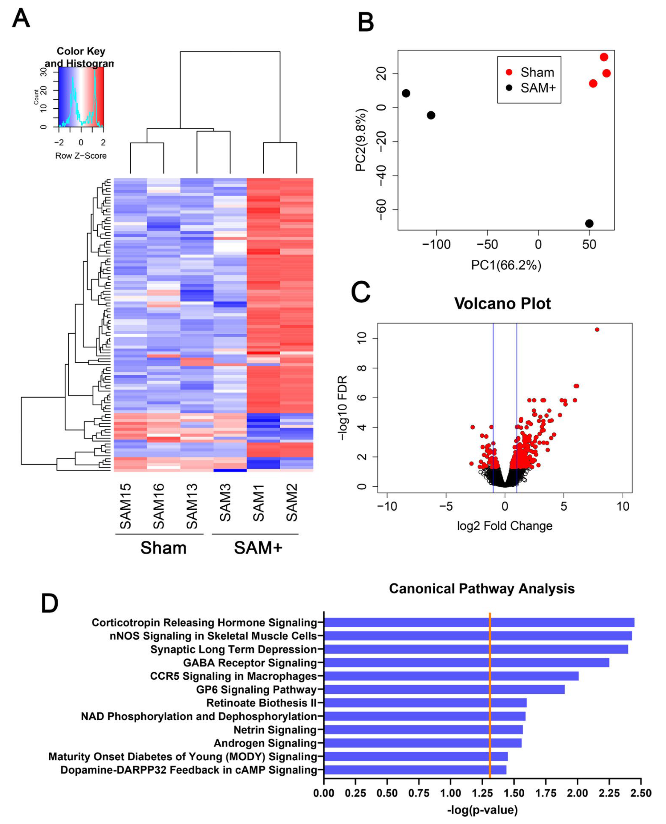
Gene expression changes reveal shifts in molecular signaling that may underlie structural or functional brain alterations [170,171,172]. Female Sprague-Dawley rats exposed to inhaled Aroclor 1254 and 1221 exhibited changes in differentially expressed genes (DEGs) and alterations in canonical pathways in the brain [25]. In the whole brain, PCB exposure resulted in 274 upregulated genes and 58 downregulated genes, as well as pathways related to neurotransmitter signaling, cognitive dysfunction, vascular function, and immune response (Figure 9) [25].
Male Wistar rats i.p. exposed to Aroclor 1254 exhibited significant changes in the expression of tight junctional proteins in the hippocampus. PCB exposure led to a decrease in mRNA expression of integral membrane proteins, including Occludin (Ocln) and Claudin-5 (Cldn5), as well as cytoplasmic accessory proteins Zona Occludens-1 and -2 (ZO-1 and ZO-2), Afadin (AF-6), and the Junction Adhesion Molecule-3 (Jam3) [116].
Male Wistar rats i.p. exposed to Aroclor 1254 exhibited decreased mRNA and protein expression of the cytoskeletal neurofilament light chain (Nefl) in neuronal cells of the cerebral cortex and cerebellum [106].
Signaling Pathways
Disruption of intracellular signaling cascades, such as MAPK/ERK and cGMP, affects synaptic plasticity, learning, and neuronal survival [173,174]. Male Wistar rats exposed orally to Aroclor 1254 exhibited a disruption in NMDA receptor–dependent intracellular signaling. While basal cGMP levels were unaffected, PCB exposure significantly reduced NMDA-evoked cGMP accumulation [111].
Male Wistar rats i.p. exposed to Aroclor 1254 exhibited significant changes in the expression of proteins involved in growth-stimulatory signaling pathways in the hippocampus. PCB exposure resulted in a significant reduction in expression of the neurotrophic ligand BDNF, and the BDNF receptor NTRKB [116]. Additionally, PCB exposure suppressed components of neurotrophin-associated signal transduction pathways, including the Ras, Raf, and MAPK/ERK signaling cascade [116].
Cytokine/Chemokine
Neuroinflammation driven by cytokines and chemokines contributes to blood–brain barrier disruption, glial activation, and neuronal injury [175,176]. Female Sprague-Dawley rats exposed to inhaled Aroclor 1254 and 1221 did not exhibit altered cytokine or chemokine concentrations in whole brain homogenate [25].
DNA Methylation
Epigenetic modifications such as DNA methylation regulate gene expression in the brain and may mediate long-term effects of toxic exposures on neural development and behavior [177,178,179]. Male and female Sprague-Dawley rats exposed i.p.to Aroclor 1221 did not show any alterations in DNA methylation in the bed nucleus of the stria terminalis (BNST), medial amygdala (MeA), paraventricular nucleus (PVN), or ventromedial hypothalamus (VMH) [163].

3.4. Behavioral Outcomes

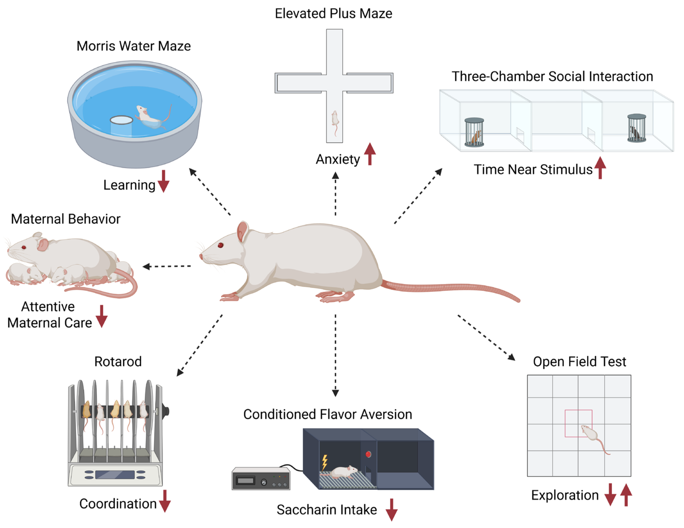
This section summarizes the effects of PCB exposure on behavioral outcomes in rodent models, based on data from studies using various PCB mixtures (e.g., Aroclors 1221, 1248, 1254) and congeners (e.g., PCB 77, PCB 180), exposure routes (oral, i.p., inhalation, subcutaneous), and timepoints (acute, subchronic, chronic). Of the 15 studies reporting neurotoxic outcomes, 6 studies examined only males, 4 investigated males and females, and 5 studies examined only female rodents. Behavioral domains assessed include locomotion, exploration, anxiety-like responses, learning and memory, motor coordination, social and sociosexual behavior, operant conditioning, maternal behavior, cognitive flexibility, and impulsivity. Both male and female rats and mice were studied, although several studies focused on males alone. The results reveal PCB-induced behavioral alterations that are exposure-dependent and often sex-specific, with some effects emerging only after repeated testing or in particular test phases. Table A5, Table A6 and Table A7 provide an overview of behavioral outcomes across exposure types and test paradigms.

3.4.1. Locomotor and Exploration

Locomotor and exploratory behaviors are commonly assessed to detect disruptions in central nervous system (CNS) function, including alterations in arousal, motivation, and sensorimotor processing [180]. These behaviors are typically evaluated using the Open Field Test (OFT), which measures spontaneous movement in a novel arena and quantifies metrics such as distance traveled, zone preference (inner vs. outer), and habituation over repeated trials [181]. Alterations in locomotor activity or exploration patterns may indicate neurotoxic effects on dopaminergic or serotonergic systems, as well as impairments in habituation or anxiety regulation [182]. Changes in horizontal and vertical activity levels can further reflect disruptions in motor coordination or motivation resulting from neurotoxic insult [183].
Male and female Sprague-Dawley rats orally exposed to PCB 180 showed changes in locomotion and exploration [119]. During the OFT, performed 24 days after exposure (first test day), females exhibited a significant dose-dependent increase in the percentage of time and distance traveled in the inner zone of the open field, while the percentage of time and distance in the outer zone of the open field decreased [119]. These behavioral differences diminished over five days of repeated testing, with significant interactions between exposure and test day for both time and distance in the inner zone. Habituation metrics, calculated as the ratio of behavior across days 2–5 to day 1, revealed dose-dependent impairments for time and distance [119]. The total distance moved across both zones exhibited a quadratic dose–response pattern, with increased activity at intermediate doses but no consistent changes over time. In contrast, no significant behavioral changes were detected in males exposed to PCB 180 at any exposure level [119].
Male Wistar rats i.p. exposed to Aroclor 1254 exhibited changes in locomotion and exploration [107,110]. One study reported decreased exploratory behavior, number of crossings in peripheral and center squares, and number of grooming in rats exposed to PCBs in the OFT [107]. Rats experienced an increased number of fecal boli, grooming, rearing, and freeze bouts compared to control animals [107]. However, a second study reported increased exploratory behavior, locomotion in all squares of the field, and time spent in outer squares in male rats after PCB exposure in the OFT [110]. They also reported no change in the number of fecal boli, grooming, and freeze bouts compared to control animals [110]. Although both studies assessed locomotor and exploratory behaviors in the OFT after 30 days of i.p. exposure to 2 mg/kg/day Aroclor 1254 in corn oil, methodological differences may explain the contrasting results. Specifically, variations in the testing apparatus (arena size, wall height, and floor color), lighting, and starting position may influence anxiety-like and exploratory behaviors, potentially contributing to the increased center avoidance and fecal boli in the first study versus the increased locomotion and exploration in the second.
Male Long-Evans rats exposed to oral Aroclor 1245 at varying doses exhibited changes in locomotion and exploration. Rats repeatedly exposed to Aroclor 1254 at 30 mg/kg demonstrated significantly reduced horizontal motor activity compared to both control and 10 mg/kg groups; however, vertical activity was unaffected by PCB exposure [114]. Rats acutely exposed to Aroclor 1254 exhibited dose-dependent effects on motor activity. Briefly, exposure to 1000 mg/kg resulted in significantly decreased horizontal motor activity at both 45 min and 7 days after exposure [184]. Vertical activity was significantly reduced at 300 mg/kg and above, 45 min post-dosing. No significant effects were observed at 100 mg/kg [184]. Repeated administration of Aroclor 1254 resulted in significant reductions in horizontal motor activity on days 7 and 14 at 100 mg/kg, days 14 to 42 at 30 mg/kg, and day 22 at 10 mg/kg, with full recovery observed by day 49 at 10 and 30 mg/kg [184]. Vertical activity was significantly reduced from day 22 to 42 at 30 mg/kg and on days 7 and 14 at 100 mg/kg, while no vertical activity changes were observed at 10 mg/kg. An additional follow-up study confirmed no changes in activity at doses ≤10 mg/kg given for six weeks [184].
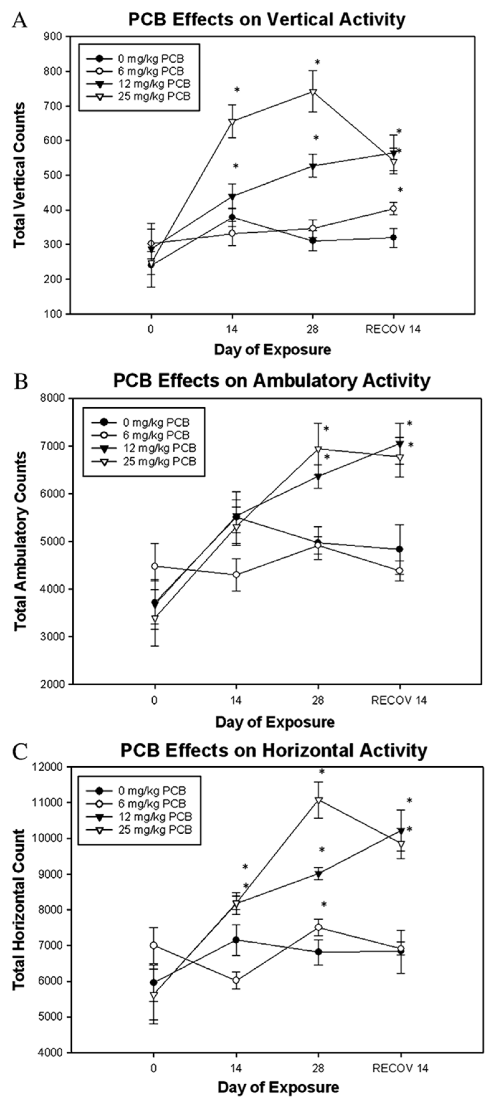
Male C57BL/6 mice orally exposed for 4 weeks to Aroclor 1245 doses also exhibited changes in locomotion and exploration; however, these effects differ from those observed in rats described above. Briefly, mice exposed to 12 and 25 mg/kg Aroclor 1254 showed elevated vertical activity after 2 weeks, which further increased by week 4 and remained elevated for at least 2 weeks post-exposure [120] (Figure 10). Similarly, these higher dose groups exhibited increases in ambulatory activity by the end of the 4-week exposure period, which continued after cessation of exposure [120]. While mice exposed to 6 mg/kg showed decreased ambulation at week 2, their activity did not differ significantly from that of controls over time. Horizontal activity was also significantly increased in mice exposed to 12 and 25 mg/kg Aroclor 1254 starting at week 2 and remained elevated through the post-exposure period [120].

3.4.2. Anxiety-like Behavior

Anxiety-like behavior is frequently assessed in rodents to detect changes in CNS function related to emotional regulation, risk assessment, and behavioral inhibition [185]. The Elevated Plus Maze (EPM) relies on rodents’ natural aversion to open, elevated spaces and preference for enclosed arms to infer anxiety levels [186,187]. In the EPM test, metrics such as the time spent in open versus closed arms, the number of open arm entries, and the total arm entries are quantified to evaluate anxiety-related behavior and general activity [188,189]. Alterations in EPM performance may reflect neurotoxic impacts on GABAergic, serotonergic, or glutamatergic signaling pathways, which are critical in the modulation of anxiety and stress responses [185,187]. Changes in open arm avoidance can indicate heightened anxiety or impaired behavioral inhibition due to neurotoxic exposures [186]. Similarly, the light-dark box test [190] utilizes rodents’ natural aversion to brightly lit environments, with increased time spent in the dark compartment and fewer transitions reflecting elevated anxiety [191]. This test is sensitive to pharmacological and toxicological manipulations of anxiety-regulating pathways and complements EPM findings in behavioral phenotyping [191].
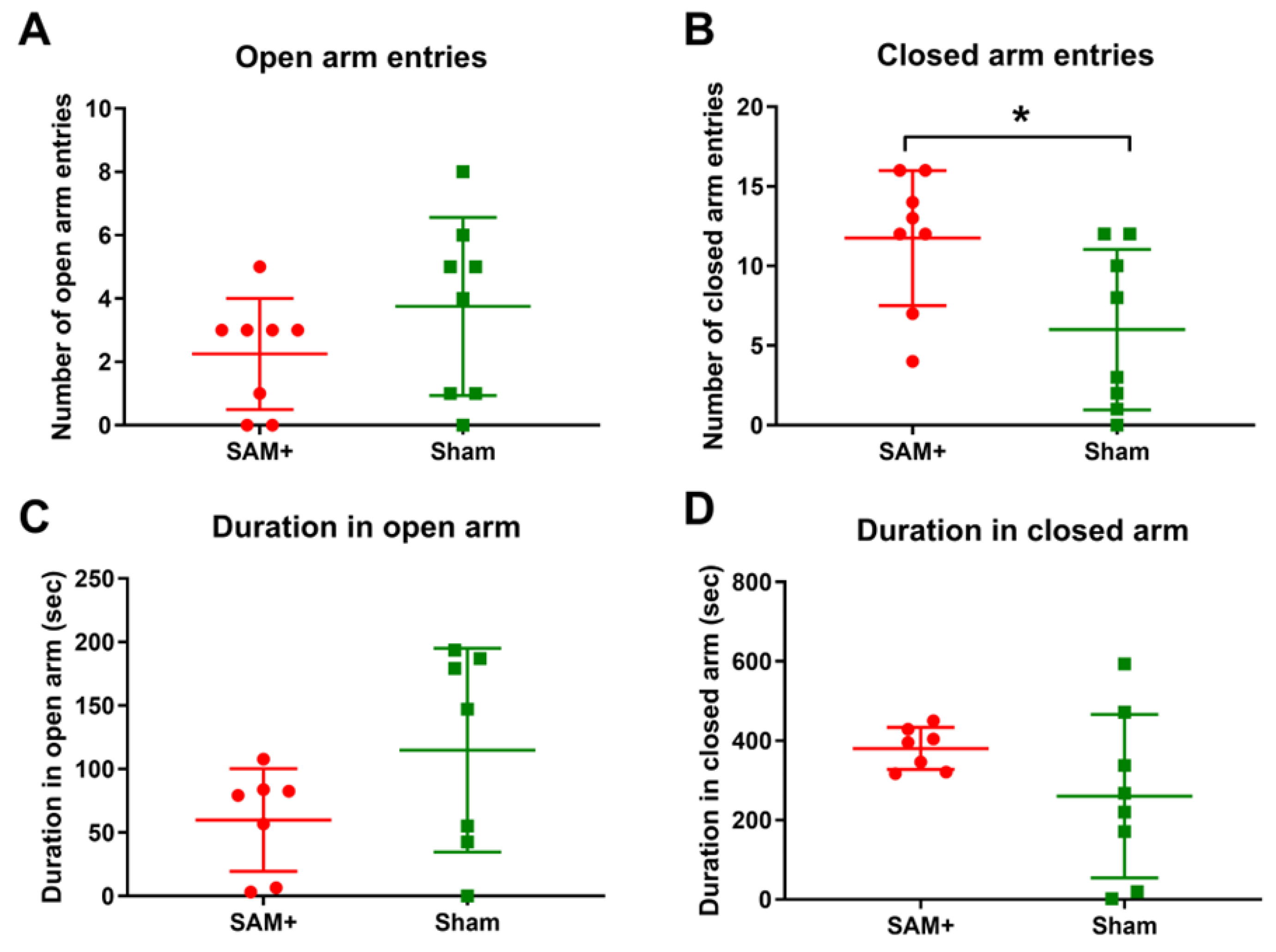
Male and female Sprague-Dawley rats exposed to i.p. Aroclor 1221 did not exhibit changes in anxiety-like behavior [192]. In the EPM, there was no change in time spent in the arms or the number of entries into open/closed arms during juvenile or adult periods [192]. During the light-dark box test, there was no change in time or number of entries in the light side of the box [192]. In contrast, female Sprague-Dawley rats exposed to a mixture of inhaled Aroclor 1221 and 1254 showed changes in anxiety-like behavior. In the EPM test, the female rats exhibited an increase in the duration and number of entries in the closed arm [25] (Figure 11). Male Wistar rats exposed to i.p. Aroclor 1254 also exhibited changes in the EPM. Similarly to the inhalation study, there was a significant increase in closed arm entries, while the time spent in the open and closed arms was significantly decreased [107].

3.4.3. Learning and Memory

Learning and memory behaviors are widely assessed to evaluate the integrity of cognitive function and identify disruptions in processes such as spatial navigation, working memory, and associative learning [193,194]. These are commonly examined using well-established behavioral tests, including the Morris water maze (MWM) [195], the eight-arm radial maze [196], and the conditioned flavor aversion (CFA) test [197]. The MWM assesses spatial learning and memory by requiring animals to locate a hidden platform in a pool of water using distal visual cues, providing quantitative measures such as escape latency, path length, and time spent in the target quadrant during probe trials [198,199]. The 8-arm radial maze evaluates working and reference memory by tracking arm entries and re-entries during baited trials, revealing deficits in short-term memory retention or spatial strategy use [200]. CFA tests associative learning by pairing a novel flavor with an aversive stimulus, measuring subsequent avoidance behavior to detect impairments in conditioned learning [201]. Alterations in performance across these tasks can reflect neurotoxic effects on hippocampal, cortical, or limbic system circuits critical for memory encoding, retrieval, and aversive learning [202,203,204].
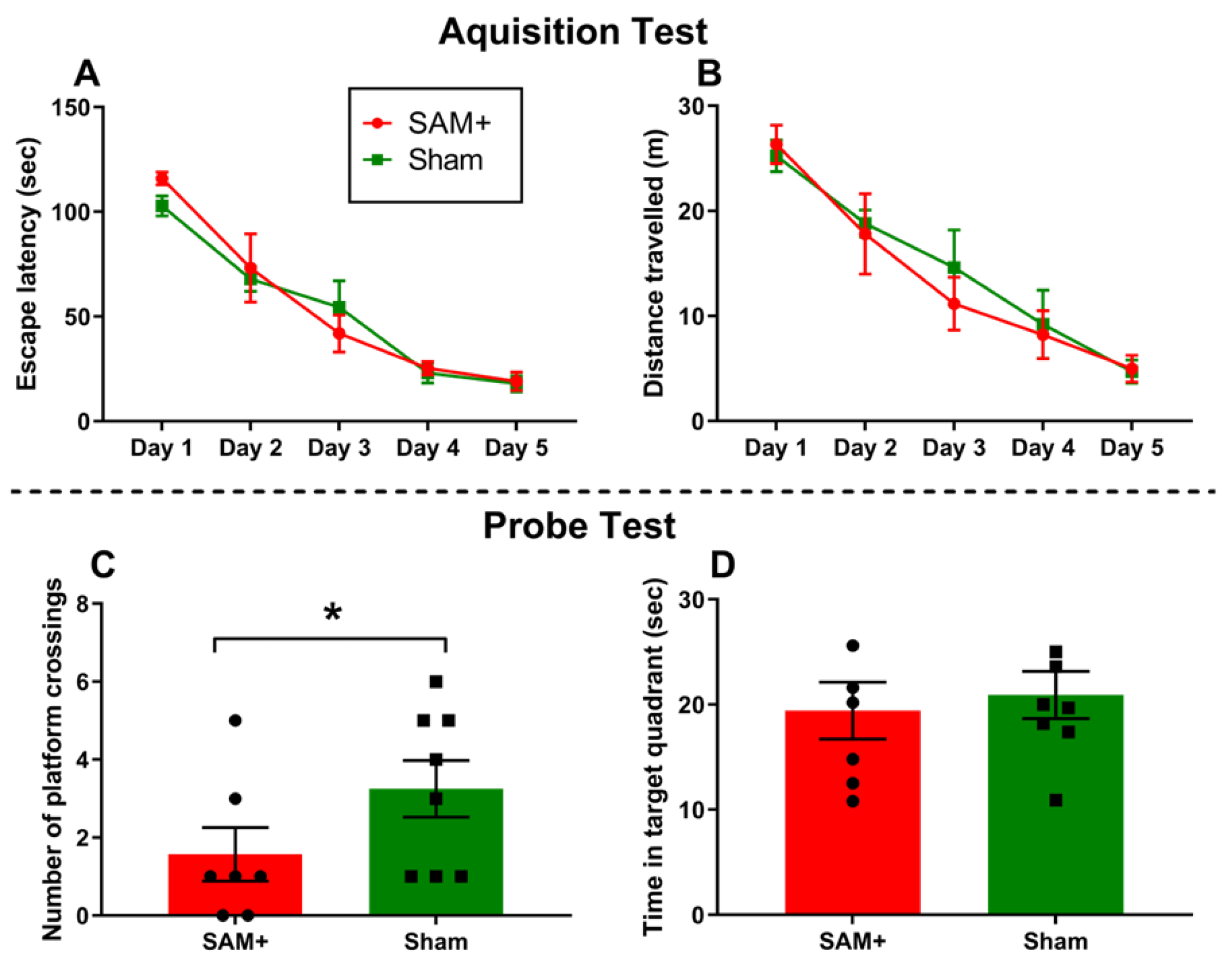
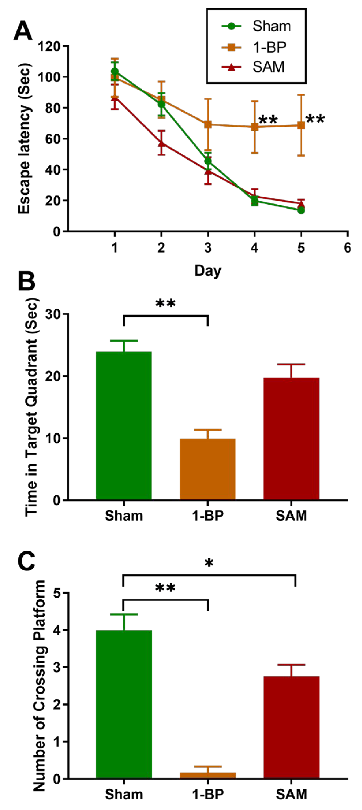
Female Sprague-Dawley rats exposed to inhaled Aroclor 1254 alone, or in combination with Aroclor 1221, showed changes in the MWM. There was no significant difference in escape latency, travel distance, or time spent in the target quadrant in PCB-exposed rats; however, PCB-exposed rats exhibited fewer platform crossings than controls [24,25] (Figure 12 and Figure 13).
Male Wistar rats i.p. exposed to Aroclor 1254 exhibited changes in learning and memory with the 8-arm radial maze [110]. In the total memory error (TME) analysis, PCB-exposed rats showed an initial decline in TME, followed by inconsistent performance with no sustained improvement in later trials [110]. In the long-term memory error (LTME) analysis, PCB-exposed rats showed an increase in LTME over time [110]. Rats exposed to PCBs exhibited erratic performance with no apparent reduction in short-term memory errors (STME) throughout the experiment [110].
Male Long-Evans rats exposed to various doses of oral Aroclor 1245 exhibited changes in the flavor aversion test. Following acute exposure, rats had reduced saccharin preference at doses ≥25 mg/kg compared to vehicle-treated controls [184]. The reduction in saccharin intake occurred independently of total fluid intake, which was significantly decreased only at the 15 mg/kg dose. Repeated exposure to Aroclor 1254 also showed aversive effects on flavor preference [184]. In the first conditioning experiment, repeated pairing of saccharin with 15 mg/kg Aroclor 1254 significantly decreased daily saccharin intake; however, the 7.5 mg/kg dose had no effect when saccharin was the sole fluid available. In the following choice test conducted after 14 pairings, rats treated with 15 mg/kg Aroclor 1254 demonstrated a reduction in saccharin preference. A follow-up experiment using repeated doses of 3.75 and 7.5 mg/kg Aroclor 1254 over 14 days revealed no significant effects on saccharin intake, either during conditioning or in subsequent choice testing [184]. Total fluid intake during the choice test was also reduced following repeated exposure to 15 mg/kg Aroclor 1254 [184].

3.4.4. Motor Coordination and Balance

Motor coordination and balance are key components of neuromotor function and are frequently evaluated to detect impairments in cerebellar or sensorimotor integrity following toxicant exposure or neurological insult [205,206]. These are commonly assessed using well-established behavioral assays, such as the accelerating rotarod test, which provides sensitive measures of motor performance and motor learning [207,208]. In this task, rodents are placed on a rotating rod that gradually accelerates, and latency to fall is recorded across repeated trials [209]. This test evaluates a combination of balance, coordination, grip strength, and motor learning, with decreased performance indicating deficits in neuromuscular control or cerebellar function [210]. Alterations in rotarod performance can reflect disruptions in central motor pathways, including cerebellar Purkinje cell function, basal ganglia signaling, or corticospinal tract integrity [211,212,213].
Male Wistar rats exposed to i.p. Aroclor 1254 exhibited differences in motor coordination in the rotarod test. PCB-exposed rats did not stay on the rotating rod as long as controls and had an increased number of falls compared with controls [107].

3.4.5. Social and Sociosexual Behavior

Social and sociosexual behaviors are commonly assessed to evaluate neural circuits responsible for social motivation, recognition, and affiliative communication [214]. These behaviors are typically examined using tests such as the three-chamber social interaction test, which measures preference for a chamber containing an animal of the same species versus an empty chamber [215], or the social novelty test, which distinguishes recognition and preference for familiar versus novel stimulus animals [216]. Outcomes such as latency to approach, time spent in proximity to the stimulus animal, and number of transitions between chambers provide insight into social engagement and discrimination [217]. Sociosexual behaviors may also be assessed through ultrasonic vocalizations (USVs), with specific call types reflecting affiliative or sexually motivated communication [218]. Alterations in social investigation, recognition, or sociosexual USVs can signal disruptions in neural systems involved in social reward processing, olfactory signaling, or anxiety regulation, potentially indicating neurodevelopmental or neurotoxic effects [218,219].
Male and female Sprague-Dawley rats exposed to Aroclor 1221 i.p. showed differences in social and sociosexual behavior. In the first stage of the social approach test, where juvenile mice were given the choice between a chamber containing a stimulus animal and an empty chamber, no significant effects of PCB exposure were observed on the total time spent in the area near the stimulus animal [192]. However, juvenile PCB-exposed females exhibited a significantly increased latency to approach the area near the stimulus animal. No effect was observed in males [192]. In the second stage of the test, assessing social recognition and preference by offering a choice between a familiar and a novel stimulus animal, PCB exposure did not influence the time spent near, or latency to reach, either the familiar or novel animal [192]. PCB exposure did not alter sociosexual ultrasonic vocalizations, but sociosexual choice was altered. In males, PCB exposure significantly increased the time spent near a no-hormone (non-estrous) female stimulus [192]. While the time spent near the hormone-treated female was not significantly altered, the combined increase in time spent near both stimulus animals resulted in an overall increase in total interaction time among juvenile-exposed males [192]. PCB exposure did not alter affiliate USVs, but affiliative behavior was changed in females with an increased latency to hop [192].

3.4.6. Operant Conditioning and Behavioral Flexibility

Operant conditioning paradigms are widely used to assess learning, motivation, and behavioral flexibility, providing insight into the reward-related and executive function pathways in the CNS [220,221]. These tasks, including fixed interval (FI), extinction (EXT), and transition schedule testing, allow for the quantification of response patterns such as lever pressing rates, response bursts, and extinction persistence [222,223]. Alterations in operant performance can reflect disruptions in dopaminergic and corticostriatal circuitry, which are critical for timing, reinforcement processing, and behavioral inhibition [224,225]. Changes in response trajectories across FIs or increased responding during EXT phases may indicate neurotoxic effects on impulse control or motivational drive [226,227]. Additionally, heightened response rates during transitions between reinforcement schedules may signal impaired adaptability and behavioral regulation, often linked to prefrontal or striatal dysfunction [228,229]. Therefore, these patterns are valuable in identifying neurobehavioral toxicity following exposure to environmental contaminants, such as PCBs.
Male Sprague-Dawley rats exposed orally to Aroclor 1248 exhibited alterations in operant training tests [230]. Rats exposed to PCB significantly increased lever pressing rate during FI sessions compared to controls. A mixed ANOVA revealed a main effect of segment duration, with lever presses increasing progressively across the 30 s segments [230]. Follow-up analysis revealed that the PCB and control groups exhibited distinct response trajectories over the 120 s interval. Specifically, the PCB group showed significantly greater lever pressing than controls by 90 s and 120 s, but not at earlier time points. During the last six sessions, there was no change in response bursts, and no significant differences were observed in the number of reinforcements delivered across groups during the final six sessions [230].
During EXT testing in male rats, the PCB-food group exhibited an increase in responding across the 5 min EXT period, in contrast to the relatively steady pattern observed in controls. No significant differences were detected between the PCB and control groups in extinction response patterns [230]. During transition training testing, male rats exposed to PCB exhibited altered behavioral responses; there was an increase in lever pressing during the first session of a new schedule compared to the second in PCB-exposed rats. Specifically, during continuous reinforcement (CRF) and FI schedules, the PCB group demonstrated a significant main effect of schedule session, with higher response counts in earlier sessions. PCB-exposed rats displayed more response bursts than control animals [230].
During EXT components associated with transition schedules, the PCB-exposed male rats pressed significantly more than the control rats. Across FI schedules, PCB-exposed rats exhibited the greatest number of extinction presses during FI 30 s and progressively fewer across the 06- and 120 s reinforcement conditions [230]. A significant three-way interaction (Groups × Associated Schedule × Schedule Session) confirmed that the pattern of extinction responding across sessions was uniquely altered in the PCB group compared to controls [230].
Male and female Sprague-Dawley rats exposed to inhaled Aroclor 1248 exhibited changes in operant training tests. PCB-exposed rats showed significantly higher response rates during the final stable-state sessions of the 120 s FI, 5 min EXT schedule [231]. A mixed ANOVA revealed a main effect of group and segment, as well as a significant interaction between group and segment. Follow-up comparisons showed that the PCB group exhibited elevated responding specifically at 90 and 120 s into the FI interval, with no significant differences from controls at the 30 or 60 s segments [231]. Male rats exposed to PCBs responded significantly more than male controls, while female rats in the PCB group did not differ from female controls. Additionally, males in the PCB group pressed significantly more than females. During the final two stable-state sessions, response bursts were more frequent in the PCB group compared to controls [231].

3.4.7. Reproductive and Maternal Behavior

Reproductive and maternal behaviors are frequently assessed in rodent models to identify alterations in neuroendocrine and motivational systems that regulate social, sexual, and caregiving behaviors [232]. Female sexual behavior is commonly evaluated using lordosis and pacing paradigms, which measure receptivity, sexual motivation, and control over mating interactions based on latency to approach, return after mounts or intromissions, and lordosis response [233]. Maternal behavior is a sensitive indicator of CNS integrity and hormonal regulation and is typically assessed through pup-directed activities such as licking/grooming, nursing posture, nest-building, and pup retrieval [234,235]. Changes in these behaviors may reflect alterations in hypothalamic-pituitary-gonadal or -adrenal axis signaling, dopaminergic systems, or disruptions in circuits responsible for social motivation [236,237].
Adult female Long-Evans rats exposed to i.p. Aroclor 1221 and 1254 during lactation did not show changes in reproductive behavior [238]. Briefly, female sexual behavior was assessed using traditional lordosis tests to measure receptivity and pacing tests that evaluated sexual motivation and control through latency and escape behaviors in a two-compartment chamber [238]. There were no changes observed in approach latency, mount return latency, intromission return latency, post-ejaculatory refractory period, or lordosis quotient for pacing and non-pacing [238].
In a separate study, female Long-Evans rats exposed to PCB 77 subcutaneously from gestational days 6-18 exhibited changes in maternal behavior. PCB exposure significantly increased the amount of time dams spent licking and grooming their pups, as well as the number of nursing bouts and the time spent on the nest [239]. High crouch nursing posture, associated with attentive maternal care, was significantly reduced in PCB-exposed rats [239]. However, PCB exposure did not change the time spent engaged in maternal autogrooming, or the time spent nursing pups [239].
Female Swiss Albino mice exposed orally to a mixture of six non-dioxin-like PCBs, including PCBs 28, 52, 101, 138, 153, and 180, did not show changes in maternal behavior. Briefly, there was no change in nest-building activity or the time to retrieve a pup during the nursing period after PCB exposure [240].

3.4.8. Cognitive Flexibility and Decision-Making

Cognitive flexibility and decision-making are commonly assessed using operant-based paradigms that test an animal’s ability to modify behavioral strategies in response to changing rules and reinforcement contingencies [222]. These tasks assess the functional integrity of prefrontal and striatal circuits involved in attentional control, behavioral inhibition, and adaptive learning [241]. One assessment is the set-shifting and reversal learning task, which consists of three sequential phases. In the visual cue discrimination phase, animals learn to associate a visual stimulus (an illuminated cue light) with a food reward, requiring attention to external cues [242]. This is followed by the set-shifting (position discrimination) phase, in which the rule shifts to a fixed spatial location, testing the animal’s ability to inhibit the prior rule and adopt a new response strategy [242]. The final reversal learning phase requires animals to reverse the spatial contingency, thereby assessing behavioral flexibility and the capacity for response inhibition [242]. Performance metrics such as total errors to criterion, trial omissions, response latency, and error subtypes (perseverative or regressive errors) provide a characterization of learning strategies and potential deficits in executive function [243]. Therefore, assessments of cognitive flexibility and decision-making provide a means of detecting disruptions in executive function associated with PCB exposure, shedding light on the potential neurobehavioral effects of environmental pollutants on the prefrontal-striatal circuitry.
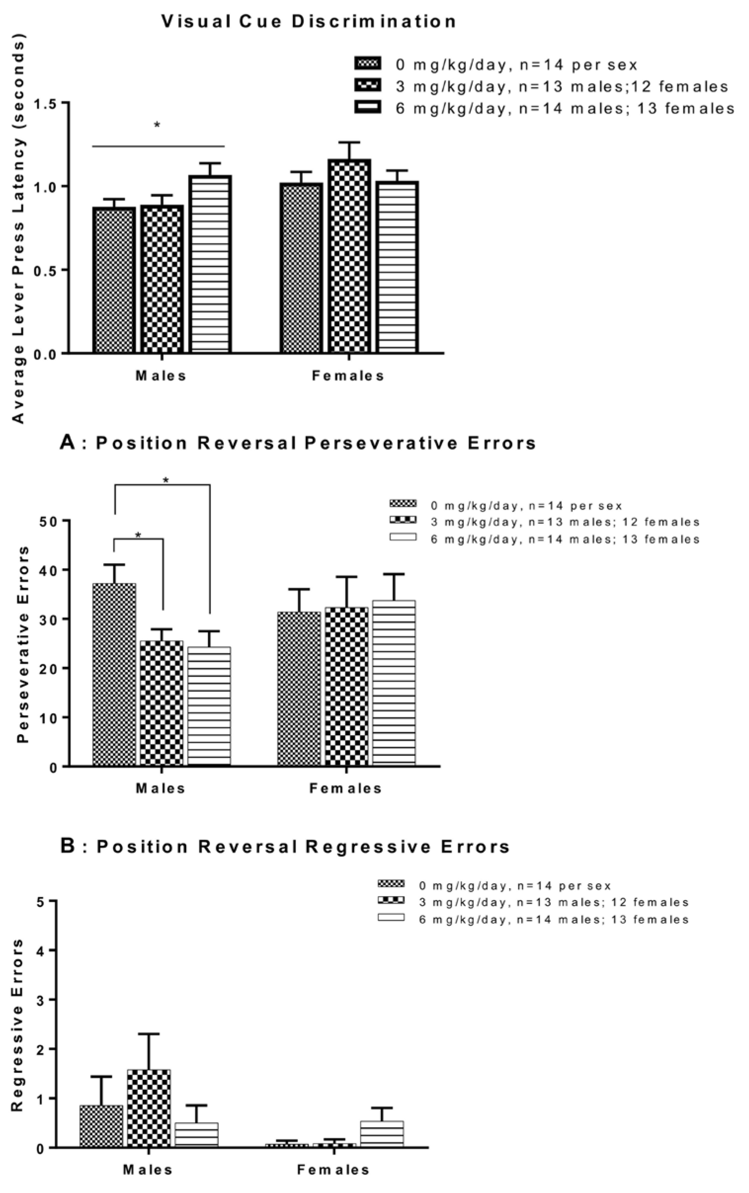
Male and female Long-Evans rats exposed to the Fox River Mixture [244,245,246,247] orally exhibited changes in cognitive flexibility and decision-making. In the set-shifting test during the visual cue discrimination phase, there were no significant effects of PCB exposure on total errors to criterion in either sex [248]. In contrast, a significant increase in trial omissions was observed among male rats in the 6 mg/kg group compared to both the 0 mg/kg and 3 mg/kg groups [248]. Female rats exhibited no exposure-related differences in omissions or errors. A significant main effect of exposure on lever press latency was also observed in male rats during this phase, with the 6 mg/kg group taking longer to respond (Figure 14, Top) [248].
In the position discrimination (set-shift) phase, there were no significant effects of exposure on total errors to criterion in Long-Evans rats exposed to the Fox River Mixture, irrespective of sex [248]. Similarly, no group differences were observed during the first 20 trials, during which reinforcement followed the previous visual cue rule. Error subtype analysis (perseverative, regressive, and never-reinforced errors) during the set-shift phase also revealed no significant differences across exposure groups for either sex [248].
In the position reversal phase, male rats exposed to the Fox River Mixture made significantly fewer total errors to reach criterion compared to controls [248]. No exposure-related effects were observed in female rats. Error subtype analysis during reversal learning showed that males in PCB exposure groups made significantly fewer perseverative errors than controls, with no differences in regressive errors [248] (Figure 14A,B). No differences in perseverative or regressive errors were observed in females [248].

3.4.9. Impulsivity and Timing

Impulse control and response inhibition are critical aspects of executive function, often evaluated in animal models using operant-based schedules such as differential reinforcement of low rates of responding (DRL) [249]. These tests require subjects to withhold responses for a specified interval to receive reinforcement, providing a measure of impulsivity and behavioral regulation [249]. Performance on DRL schedules can reflect functional integrity of prefrontal cortical and serotonergic systems [250]. Schedule-specific metrics such as total lever presses, efficiency ratios, and performance across increasing delay intervals (1 to 15 s) are used to detect subtle impairments in timing, motor control, and inhibitory capacity [250]. DRL tasks are valuable tools for assessing the long-term effects of exposures to environmental pollutants, such as PCBs, on cognitive and executive functions.
Male and female Long-Evans rats exposed to the Fox River Mixture orally were evaluated for DRL to assess impulse control. During initial DRL training at the 1 s interval (DRL1), there were no significant effects of PCB exposure on total lever presses or response efficiency in either sex [248]. In the 5 s interval phase (DRL5), both males and females improved performance across sessions; however, there were no exposure-related differences [248].
In the 10 s interval phase (DRL10), there was no main effect of session or exposure in male and female rats. A significant session × exposure interaction was observed for total lever presses in males, with a steeper decline in responding across sessions in the 3 mg/kg group compared to other groups; however, no differences were observed in the 0 or 6 mg/kg groups [248]. Females exhibited a significant session × exposure interaction, but there were no differences within groups across sessions. Efficiency ratios during DRL10 did not differ by exposure in either sex [248].
During the 30-session 15 s interval phase (DRL15), no significant effects of PCB exposure were detected in total presses or efficiency ratios across five-session blocks in either sex. In the DRL extinction phase, where reinforcement was withheld, no significant exposure effects were observed in either sex [248].

4. Conclusions

4.1. Neurotoxic Outcomes

Collectively, the studies reviewed provide robust laboratory evidence that polychlorinated biphenyl (PCB) exposure causes widespread neurotoxic effects in rodent models, affecting histological, molecular, and biochemical endpoints across multiple brain regions (Figure 15). Structural damage, including neuronal degeneration and regional atrophy, is consistently reported in male animals, while data in females remain limited. Disruption of calcium signaling, induction of oxidative stress, and activation of apoptotic pathways were common findings, alongside marked alterations in neurotransmitter levels, receptor expression, and mRNA expression. Notably, PCBs impair dopaminergic, serotonergic, and glutamatergic systems and interfere with intracellular signaling cascades essential for synaptic function and neuronal survival. Changes in iron homeostasis, amino acid balance, and neuroinflammatory mediators further support the complex nature of PCB-induced neurotoxicity. Differences in outcomes are shaped by several key experimental factors. The PCB congener or mixture studied influences the type and severity of effects. Dose also plays a critical role, as higher exposure levels tend to yield more drastic effects, while lower doses often yield subtle or no effects. The exposure route also contributes to variability, with inhalation, oral, and i.p. exposures resulting in sometimes contradictory effects. Finally, the timing of exposure can determine whether effects manifest acutely or persist chronically. Acute exposures can transiently increase molecular responses in the brain, while prolonged exposures lead to lasting effects, such as hyperactivity in the weeks following PCB exposure [251]. Importantly, many outcomes demonstrate brain region–specific and sex-dependent variability, underscoring the need to consider sex as a biological variable in PCB neurotoxicity studies. These findings highlight the complex mechanisms by which PCBs disrupt nervous system function, with implications for understanding the risk of neurodevelopmental and neurodegenerative disorders following PCB exposure.

4.2. Behavioral Outcomes

Additionally, PCBs can significantly disrupt behavioral functions in adolescent or adult rodents, with effects varying by congener or mixture, dose, exposure route, sex, and timing. PCB exposure often led to changes across various outcomes, including locomotion, anxiety-like behavior, learning and memory, motor coordination, social interaction, operant conditioning, maternal behavior, cognitive flexibility, and impulsivity, indicating dysfunction in the central nervous system following PCB exposure (Figure 16). However, the consistency of these outcomes across studies is mixed. For example, some investigations report increased locomotion following exposure, while others observe reduced or unchanged activity, even under comparable exposure conditions, suggesting that methodological variables (e.g., test apparatus, lighting, starting position) strongly influence outcomes. Motor coordination deficits have been observed in some rotarod studies, although these findings are not consistently replicated. Anxiety-related behaviors and patterns of social interaction also vary across studies, with some showing increased avoidance or changed preferences, while others indicate no impact of PCB exposure. Notably, behavioral effects were often sex-specific and exposure-dependent, with several studies reporting differing outcomes based on dose or the specific testing phase. Some effects, such as increased activity or changes in social preferences, indicate hyperactivity or disinhibition, whereas others demonstrate memory impairments following PCB exposure. Despite variability across studies, this review demonstrates that PCB exposure of adolescent or adult rodents adversely affects neurobehavioral health.

4.3. Limitations

Despite evidence of PCB-induced neurotoxicity in adolescent or adult rodents, several critical gaps remain that limit mechanistic understanding and the translation of findings to human health and risk assessment. A major limitation is the small number of studies using female animals, despite evidence of sex-specific variability in outcomes [252]. This imbalance of studies using male rodents compared to those including females likely reflects a historical bias toward the use of male animals in toxicology studies to avoid confounding by estrous cycle-related variability. Consequently, the scarcity of data on females limits our understanding of sex-specific susceptibility and may obscure distinct neurotoxic mechanisms. Furthermore, there is often a disconnect between biochemical endpoints and behavioral assessments, with few studies integrating these measures in the same study to directly link mechanistic alterations to functional deficits (Table 7). Moreover, behavioral assessments themselves are frequently limited to a narrow range of tests, often focusing on gross locomotor or anxiety-like measures without incorporating sensitive, domain-specific paradigms for learning, memory, sensory processing, or executive function [253]. The lack of standardized testing and laboratory variability further limits comparison across studies [253]. Application of advanced techniques, such as spatial transcriptomics, proteomics, and metabolomics, would enable high-resolution characterization of PCB-induced molecular and cellular changes across brain regions. Dose–response relationships also remain incompletely defined, with limited integration of congener or mixture composition, dose, exposure timing, and route of administration into study designs, particularly for environmentally relevant, chronic low-dose exposures. While oxidative stress, calcium signaling, neurotransmitter systems, and apoptotic pathways are frequently implicated, causal links between these mechanisms and behavioral outcomes remain unclear. This highlights the need for multi-omics integration to identify key drivers of toxicity [254,255]. In addition, alternatives to traditional animal models, such as human-derived brain organoids and microphysiological systems, offer promising paths to reduce animal use and generate human-specific mechanistic insights [256,257]. Finally, there is a lack of longitudinal studies tracking neurobiological and behavioral outcomes across the lifespan, which are essential to assess the persistence or progression of PCB-induced effects, including potential contributions to aging-related neurodegeneration.
Table 7. Summary of behavioral assays, associated domains, implicated brain regions, and proposed molecular mechanisms underlying PCB-induced neurobehavioral alterations in rodent models. This table integrates findings across major behavioral paradigms, including locomotor, anxiety-like, cognitive, motor, social, and maternal behaviors, and links observed outcomes to key neuroanatomical targets and molecular pathways. Proposed mechanistic interpretations provide cross-scale connections between cellular/molecular disruptions and system-level behavioral manifestations.
Behavioral
Test
Behavioral
Domain
Key Brain
Regions
Implicated
Principal Molecular
Pathways/
Mechanisms
Proposed Mechanistic
Interpretation (Cross-Scale Link)
References
OFTLocomotor and
Exploration; Anxiety-Like Behavior
Striatum,
Prefrontal
Cortex,
Amygdala
Dopaminergic
signaling disruption; serotonergic/GABAergic imbalance; Ca2+ dysregulation
Altered dopaminergic tone and excitatory–inhibitory
imbalance led to hyperactivity or increased thigmotaxis indicative of anxiety-like responses.
[258,259,260]
EPMAnxiety-Like BehaviorAmygdala, HippocampusSerotonergic and
GABAergic disruption; oxidative stress
Neurochemical imbalance
enhances amygdala excitability, increasing anxiety and reducing open-arm exploration.
[187,261,262]
Light–Dark BoxAnxiety-Like BehaviorAmygdala, Prefrontal
Cortex
Serotonergic and GABAergic imbalance; neuroinflammationHeightened inflammatory and neurotransmitter dysregulation promotes avoidance of the light zone and risk-averse exploration.[263,264,265]
MWMLearning and MemoryHippocampus, CortexOxidative stress; Ca2+ homeostasis disruption; BDNF–TrkB axis impairment; thyroid hormone signalingOxidative injury and reduced neurotrophic support impair hippocampal plasticity and
spatial learning.
[266,267,268]
8-Arm
Radial Maze
Learning and MemoryHippocampus,
Prefrontal
Cortex
Oxidative stress; mitochondrial dysfunctionEnergy deficits and oxidative damage reduce accuracy and
increase latency in memory
retrieval.
[198,269,270]
CFALearning and MemoryInsular Cortex, AmygdalaSynaptic plasticity gene dysregulation; oxidative stressImpaired synaptic encoding of aversive cues due to oxidative stress and reduced immediate-early gene activation.[271,272,273]
RotarodMotor Coordination and BalanceCerebellum, Motor Cortex, StriatumDopaminergic disruption; mitochondrial dysfunction; apoptosisMotor impairments arise from
cerebellar mitochondrial toxicity and dopaminergic neuron loss.
[274,275,276]
Three-Chamber Social
Interaction
Social and
Sociosexual Behavior
Prefrontal
Cortex,
Hippocampus, Amygdala
BDNF–TrkB axis
disruption;
neuroinflammation
Reduced neurotrophic signaling and cytokine-mediated synaptic pruning impair social approach behavior.[217,277,278]
Social
Novelty Test
Social and
Sociosexual Behavior
Hippocampus, Prefrontal
Cortex
BDNF–TrkB
impairment; oxidative stress
Diminished hippocampal
plasticity and oxidative injury
impair discrimination between familiar and novel animals.
[279,280,281]
USVSocial and
Sociosexual Behavior
Amygdala, Periaqueductal GraySynaptic plasticity gene dysregulation; neuroinflammationAltered synaptic signaling and neuroimmune activation
disrupt communication and
social bonding cues.
[218,282,283]
Operant ConditioningOperant Conditioning and Behavioral FlexibilityStriatum,
Prefrontal
Cortex
Dopaminergic
disruption; oxidative stress
Impaired reward processing and task acquisition due to
frontostriatal dysfunction.
[284,285,286]
Maternal CareReproductive and Maternal BehaviorHypothalamus,
Prefrontal
Cortex
Thyroid hormone disruption; dopaminergic signalingEndocrine disruption and
altered reward circuits reduce pup-directed behaviors.
[232,287,288]
Set-Shifting/Reversal LearningCognitive Flexibility and Decision-MakingPrefrontal
Cortex
Neuroinflammation; synaptic gene dysregulationPrefrontal cytokine activation and reduced synaptic
remodeling impair task-
switching and adaptability.
[289,290,291]
In addition to these research gaps, several methodological considerations related to the use of large language models in the review process should be acknowledged. ChatGPT was used to assist with text summarization and data organization. Because the “improve the model for everyone” feature was not enabled, study data or text was not shared for model training, safeguarding data privacy. We acknowledge that large language models may exhibit biases reflective of their training data and have limited context retention when processing complex or lengthy text. To mitigate these limitations, all AI-generated outputs were reviewed and verified by human researchers for accuracy and consistency with the source material. Moreover, as large language model outputs are not fully reproducible due to model updates and stochastic variation, all ChatGPT responses used in data extraction were documented to promote transparency and reproducibility.

4.4. Future Directions and Translational Outlook

Future research should leverage emerging technologies, including spatial transcriptomics, proteomics, and metabolomics, to achieve high-resolution mapping of PCB-induced molecular and cellular alterations across brain regions and developmental stages. Integrating congener composition, dose, exposure timing, and route of administration into study designs will enhance environmental relevance and mechanistic interpretation. Key translational questions regarding PCB neurotoxicity remain: What molecular pathways at the cellular level causally link PCB exposure to behavioral outcomes, and do alterations in oxidative stress, calcium homeostasis, and neurotransmission jointly drive these effects? How do the neurotoxic mechanisms and dose–response relationships differ between legacy PCBs and inadvertently produced PCB congeners? How does the route of exposure, such as inhalation, oral ingestion, or dermal absorption, influence the neurotoxic outcomes and behavioral phenotypes observed in experimental models? Do region- and cell type–specific transcriptional responses predict distinct neurobehavioral phenotypes? Can human-derived brain organoids and microphysiological systems replicate mixture- and dose–response effects observed in vivo, thereby improving human relevance while reducing animal use? Finally, do PCB-induced neurobiological alterations persist across the lifespan or accelerate aging-related neurodegeneration? Addressing these questions through multi-omics integration and longitudinal study designs will establish a translational framework connecting mechanistic discoveries in experimental models with human health risk assessment.

Supplementary Materials

The following supporting information can be downloaded at https://www.mdpi.com/article/10.3390/ijms262210829/s1.

Author Contributions

Conceptualization, N.M.B. and H.-J.L.; methodology, N.M.B.; validation, N.M.B. and H.-J.L.; formal analysis, N.M.B., S.G.H. and R.J.S.; investigation, N.M.B., S.G.H. and R.J.S.; data curation, N.M.B. and S.G.H.; writing—original draft preparation, N.M.B.; writing—review and editing, N.M.B., S.G.H., R.J.S. and H.-J.L.; visualization, N.M.B.; supervision, H.-J.L.; project administration, H.-J.L.; funding acquisition, H.-J.L. All authors have read and agreed to the published version of the manuscript.

Funding

This research was funded by the National Institute of Environmental Health Sciences, grant numbers P30ES005605, P42ES013661, R01ES014901, R01ES031098, and R01ES034691, where the authors’ own work is cited.

Institutional Review Board Statement

Not applicable.

Informed Consent Statement

Not applicable.

Data Availability Statement

The original data presented in the study are openly available in Iowa Research Online at 10.25820/data.008102.

Acknowledgments

During the preparation of this manuscript/study, the author(s) used ChatGPT 4o for the purposes of data analysis and extraction. The authors have reviewed and edited the output and take full responsibility for the content of this publication.

Conflicts of Interest

The authors declare no conflicts of interest. The funders had no role in the design of the study; in the collection, analyses, or interpretation of data; in the writing of the manuscript; or in the decision to publish the results.

Abbreviations

The following abbreviations are used in this manuscript:
5-HIAA5-hydroxyindoleacetic acid
AChEAcetylcholinesterase
AhRAryl hydrocarbon receptor
ANOVAAnalysis of variance
ArsAndrogen receptors
AF-6Afadin
Avpr1aVasopressin receptor 1a
BadBCL2-associated death promoter
BaxBCL2-associated X protein
Bcl2B-cell leukemia/lymphoma 2
BDNFBrain-derived neurotrophic factor
BidBH3 interacting-domain death agonist
BNSTBed nucleus of the stria terminalis
CARConstitutive androstane receptor
CATCatalase
CASPCaspase
CBLCerebellar
CFAConditioned flavor aversion
cGMPCyclic guanosine monophosphate
Cldn5Claudin-5
CNSCentral nervous system
COMTCatechol-O-methyltransferase
CREBCyclic AMP-response element binding protein
CRFContinuous reinforcement
Cu/Zn SODCu/Zn superoxide dismutase
CYPsCytochrome P450 monooxygenases
DADopamine
DATDopamine transporter
DOPAC3,4-dihydroxyphenylacetic acid
DRLDifferential reinforcement of low rates of responding
DRL1DRL training at the 1 s interval
DRL10DRL training at the 10 s interval
DRL15DRL training at the 15 s interval
DRL5DRL training at the 5 s interval
EAC1Excitatory amino acid carrier 1
EPMElevated Plus Maze
ERKExtracellular signal-regulated kinase
ERsEstrogen receptors
EXTExtinction
FasTumor necrosis factor receptor superfamily member 6
FasLFas ligand
FIFixed interval
GLASTGlutamate aspartate transporter
GLT-1Glutamate transporter 1
GPVGlial plasmalemmal vesicle
GPxGlutathione peroxidase
GRGlutathione reductase
GSTsGlutathione-S-transferases
HO-1Heme oxygenase-1
HVAHomovanillic acid
i.p.Intraperitoneal
Jam3Junction adhesion molecule-3
LTMELong-term memory error
MAPMicrotubule-associated protein
MeAMedial amygdala
Mn SODManganese SOD
MRMuscarinic receptor
MWMMorris water maze
NAPDH-dNADPH-diaphorase
NENorepinephrine
NeflNeurofilament light chain
NF-kBNuclear factor kappa B
NMDAN-methyl-D-aspartate
NOSNitric oxide synthase
NTKRBNeurotrophin tyrosine kinase receptor B
OclnOccludin
OFTOpen Field Test
OH-PCBsHydroxylated metabolites
Oprm1mu opioid receptor
PCBsPolychlorinated biphenyls
PKAProtein Kinase A
PKCProtein Kinase C
PPARsPeroxisome proliferator-activated receptors
PRISMAPreferred Reporting Items for Systematic Reviews and Meta-Analysis
PVNParaventricular nucleus
PXRPregnane X receptor
RNSReactive nitrogen species
ROSReactive oxygen species
RyRsRyanodine receptors
SAMSchool Air Mixture
SNpcSubstantia nigra pars compacta
SONSupraoptic nucleus
STMEShort-term memory errors
STRStriatal
SULTSSulfotransferases
TBARSThiobarbituric acid reactive substances
THTyrosine hydroxylase
TH−TH-negative
TH+Tyrosine hydroxylase-positive
TMETotal memory error
TNFaTumor necrosis factor alpha
UGTsUDP-glucuronosyltransferases
U.S.United States
USVsUltrasonic vocalizations
VMHVentromedial hypothalamus
VTAVentral tegmental area
ZOZona Occludens

Appendix A

Table A1. Summary of Studies Investigating Neurotoxic Outcomes in Sprague Dawley Rats Following PCB Exposure.
PCB
Source
Dose
(mg/kg/day) a
Exposure
Route
VehicleExposure
Period
AgeN bSexBrain RegionAnalysisResult cRef
A1254 d62.1 ± 17.7 μg/m3Inh eLA f28 daysAdult
(239 ± 8g)
8F gWhole
Brain
Histology H&E h- Gross histopathological changes in HC i
- Features of necrosis or apoptosis
[24]
A1254
A1221
45.5 ± 5.9 μg/m3InhLA6 d/w j for
13 weeks
Adult
(241 ± 9g)
S k: 8
E l: 12
FWhole
Brain
Histology H&E- Histopathological changes[25]
A125462.1 ± 17.7 μg/m3InhLA28 daysAdult
(239 ± 8g)
8FWhole
Brain
OS m Markers↓ ROS n/RNS o[24]
A1254
A1221
45.5 ± 5.9 μg/m3InhLA6 d/w for 13 weeksAdult
(241 ± 9g)
S: 8
E: 12
FWhole
Brain
OS Markers- ROS/RNS[25]
A12210 or 1IP p4% DMSO qPND24 r, 26, 28PND249–12M s/FBNST t
MeA u
PVN v
VMH w
DNA Methylation- DNA methylation[163]
A12540 or 25PO xCO y on VW z3 days, or 1, 2, or 8 weeks12 weeks3–4MStriatal
Dialysate
DA aa Concentrations↑ DA after 3 days
↓ DA after 1, 2, and 8 weeks
[132]
A12540 or 25POCO on VW3 days, or 1, 2, or 8 weeks12 weeks3–6MST bbDA Concentrations- DA levels[132]
A1254
A1221
45.5 ± 5.9 μg/m3InhLA6 d/w for 13 weeksAdult
(241 ± 9g)
S: 8
E: 12
FSTDA and DOPAC cc Concentrations- DA and DOPAC levels[25]
PCB 1800, 19.4, 64.3, 194, 643, 2000, 6430 or 13000POCO28 days6 weeks5M/FCE ddDA and Nicotinic Receptors- Specific binding to DRD1 ee/DRD5 receptors
- High or low affinity sites on nicotinic receptor
[119]
PCB 1800, 19.4, 64.3, 194, 643, 2000, 6430 or 13,000POCO28 days6 weeks5M/FCEBrain AA ff↓ GSH gg in males
- γ-Amino butyric acid, aspartate, glutamate, serine, glycine, taurine, glutamine, alanine,
[119]
A1254
A1221
45.5 ± 5.9 μg/m3InhLA6 d/w for 13 weeksAdult
(241 ± 9g)
S: 8
E: 12
FWhole Brain Cytokine and Chemokine- Cytokine or chemokine concentrations[25]
A12210 or 1IP4% DMSOPND24, 26, 28P249–12M/FPoA hhGene Expression↓ Expression of Ar ii and Oprm1 jj in males[163]
A12210 or 1IP4%
DMSO
PND24, 26, 28P249–12M/FPFC kkGene Expression↑ Expression of Oprm1 in males[163]
A12210 or 1IP4%
DMSO
PND24, 26, 28P249–12M/FLateral septumGene Expression↓ Expression of Avpr1a ll in males[163]
A12210 or 1IP4% DMSOP24, 26, 28P249–12M/FPFCGene × behavior interactions↑ Correlation of no-hormone stimulus time and Oprm1 [163]
A12210 or 1IP4% DMSOP24, 26, 28P249–12M/FBNST
MeA
PVN
VMH
Gene Expression- Gene expression[163]
A1254
A1221
45.5 ± 5.9 μg/m3InhLA6 d/w for 13 weeksAdult
(241 ± 9g)
S: 8
E: 12
FBrainRNAseq and Pathway Analysis↑ 274 genes
↓ 58 genes
Pathways altered NS, CD, VF, + IR
[25]
A12540 or 30POCO in Cheeto14 days3–5 months3–6MSON mmHyperosmotic Challenge and NOS Activity- NAPDH-d density[151]
PCB 1530 or 20POCOGD10-GD16 nn15 weeks5FCC
CE
HC
ST
Muscarinic Receptor↑ MR oo density in CC
↓ MR density in CE
- MR density in HC or ST
[164]
a Unless otherwise noted. b N, Number of animals per group and sex. c (-) no change; (↑) significant increase; (↓) significant decrease. d A, Aroclor. e Inh, Inhalation. f LA, Lab air. g F, Female. h H&E, Hematoxylin and eosin staining. i HC, Hippocampus. j d/w, Days per week. k S, Sham. l E, Exposed. m OS, Oxidative stress. n ROS, Reactive oxygen species. o RNS, Reactive nitrogen species. p IP, Intraperitoneal injection. q DMSO, Dimethyl sulfoxide. r PND, Postnatal day. s M, Male. t BNST, Bed nucleus of the stria terminalis. u MeA, Medial amygdala. v PVN, Paraventricular nucleus. w VMH, Ventromedial hypothalamus. x PO, Oral. y CO, Corn oil. z VW, Vanilla wafer. aa DA, Dopamine. bb ST, Striatum. cc DOPAC, 3,4-dihydroxyphenylacetic acid. dd CE, Cerebellum. ee DRD, Dopamine receptor D. ff AA, Amnio acid. gg GSH, Glutathione. hh PoA, Preoptic area. ii Ar, Androgen receptor. jj Oprm1, Mu opioid receptor. kk PFC, Prefrontal cortex. ll Avpr1a, Arginine vasopressin receptor 1A. mm SON, Supraoptic nucleus. nn GD, Gestational day. oo MR, Muscarinic receptor.
Table A2. Summary of Studies Investigating Neurotoxic Outcomes in Wistar Rats Following PCB Exposure.
PCB
Source
Dose
(mg/kg/day)
Exposure
Route
VehicleExposure
Period
AgeN aSexBrain RegionAnalysisResult bRef
A1254 c0 or 2IP dCO e30 daysAdult
(180–200 g)
6M fCC g
CE h
Histology Methylene Blue↑ Degenerated neurons[106]
A12540 or 2IPCO30 daysAdult
(180–200 g)
6MCEHistology H&E i↑ Shrinkage and degeneration of Purkinje neurons[105]
A12540 or 2IPCO30 daysAdult
(180–200 g)
4MCCHistology H&E/Luxol Fast Blue↑ Pyknotic CB j, NC k, IBA l
↓ Pyramidal, Betz, stellate cells, myelin density
[107]
A12540 or 2IPCO30 daysPND90 m6MCCHistology H&E↑ Neuronal shrinkage and degeneration
↑ Pyknotic nuclei with PPNS n
[104]
A12540 or 2IPCO30 daysPND906MHC oHistology Cresyl Fast Violet↑ Disruption in CA4 p[110]
A12540 or 2IPCO30 daysPND906MHCHistology H&E↑ Degeneration of pyramidal cells
↑ Neuronal shrinkage of HC layer
[109]
A12540 or 2IPCO30 daysPND906MHCHistology H&E↑ Degeneration of pyramidal cells
↑ Neuronal shrinkage of HC layer
[108]
A12540 or 2IPCO30 daysPND906MCE
CC
HC
Histology H&E↑ Pyknotic nuclei with PPNS in CC
↑ Degeneration of pyramidal cells in HC
↑ Purkinje cells in CE with shrinkage, degeneration, and moderate atrophy
[103]
A12540 or 10PO qRO r14 daysYoung Adult
(200–250 g)
DND sMHCHistology Toluidine Blue↓ Number of neurons[111]
A12540 or 10PORO14 daysYoung Adult
(200–250 g)
DNDMFBC t
HC
Transmission Electron Microscopy↑ Damaged neuron nuclei in FBC and HC
↑ RER u damage in HC
[111]
A12540 or 2IPCO30 daysPND906MCCCacna1d v
Expression
↑ mRNA expression of Cacna1d[104]
A12540 or 2IPCO30 daysPND906MHCCacna1d Expression↓ mRNA expression of Cacna1d[109]
A12540 or 2IPCO30 daysAdult
(180–200 g)
6MCCProtein Expression↑ NMDA w receptor expression
↓ PKAxα levels
↑ Calpain expression
↑ NcaCP y level
[113]
A12540 or 10PORO14 daysYoung Adult
(200–250 g)
DNDMHCCa2+ Efflux↓ NMDA Ca2+ release[111]
A12540 or 2IPCO30 daysAdult6MCEOS z Markers↑ LP aa, HP bb, and PC cc[105]
A12540 or 2IPCO30 daysPND906MCE
HC
CC
mRNA Expression↓ Expression of Cu/Zn SOD dd
↓ Expression of GPx-4 ee
[103]
A12540 or 2IPCO30 daysPND906MCCOS Markers↑ LP, HP, and PC[104]
A12540 or 2IPCO30 daysPND906MHCOS Markers↑ LP, HP, HR ff, and TBARS gg[110]
A12540 or 2IPCO30 daysPND906MHCOS Markers↑ LP, HP, and PC[116]
A12540 or 2IPCO30 daysPND906MHCOS Markers↑ LP, HP, and PC[108]
A12540 or 2IPCO30 daysPND906MCE
HC
CC
OS Markers↑ LP and HR
↓ Glutathione
[118]
A12540 or 2IPCO30 daysPND906MCE
HC
CC
OS Markers↑ LP, HP, and HR
↓ Glutathione
[117]
A12540 or 2IPCO30 daysPND906MHCAntioxidant Enzymes↓ SOD, CAT hh, GPx, GST ii, and GR jj[110]
A12540 or 2IPCO30 daysPND906MCE
HC
CC
Antioxidant Enzyme Activity↓ Total SOD, Cu/Zn and Mn SOD
↓ Activity of GPx
[103]
A12540 or 2IPCO30 daysPND906MHCDNA FragmentationStreak of fragmentation[108]
A12540 or 2IPCO30 daysPND906MCCATPase Activity↓ Na/K, Ca, and Mg ATPase activity[104]
A12540 or 2IPCO30 daysAdult
(180–200 g)
6MCEATPase Activity↓ Na/K, Ca, and Mg ATPase activity[105]
A12540 or 2IPCO30 daysPND906MHCATPase Activity↓ Na/K, Ca, and Mg ATPase activity[109]
A12540 or 2IPCO30 daysPND906MCE
CC
HC
ATPase Activity↓ Na/K, Ca, and Mg ATPase activity[117]
A1254
A1260
0, 500 or 1000POCO1 dayAdult
(250–300 g)
36MCaudate
LOT kk
Dopamine
Metabolites
↓ DA and DOPAC in caudate
↓ DOPAC/DA ratio in LOT
- DA, DOPAC in LOT
- HVA ll, HVA/DA ratio
[134]
A12540, 500 or 1000POAOC mm30 daysAdult
(350–375 g)
9MST nnDopamine
Metabolites
↓ DA, HVA, and DOPAC/DA, HVA/DA ratios[133]
A12540, 500 or 1000POAOC30 daysAdult
(350–375 g)
9MLOTDopamine
Metabolites
↓ DOPAC, HVA, and DOPAC/DA, HVA/DA ratios
- DA levels
[133]
A1254
A1260
0, 500 or 1000POCO1 dayAdult
(250–300 g)
36MFC ooNeurotransmitter Concentrations↓ 5-HT pp
↑ 5-HIAA qq, 5-HIAA/5-HT ratio
[139]
A1254
A1260
0, 500 or 1000POCO1 dayAdult
(250–300 g)
36MHCNeurotransmitter Concentrations↓ 5-HT
↑ 5-HIAA/5-HT ratio levels
- 5-HIAA levels
[139]
A1254
A1260
0, 500 or 1000POCO1 dayAdult
(250–300 g)
36MLOTNeurotransmitter Concentrations↑ 5-HT and 5-HIAA
- 5-HIAA/5HT ratio
[139]
A1254
A1260
0, 500 or 1000POCO1 dayAdult
(250–300 g)
36MBS rr
HT ss
Neurotransmitter Concentrations↑ 5-HIAA/5HT ratio
- 5-HT and 5-HIAA
[139]
A12540, 500 or 1000POAOC30 daysAdult
(350–375 g)
9MST
LOT
Neurotransmitter Concentration/Activity- NE tt or 5-HT[133]
A1254
A1260
0, 500 or 1000POCO1 dayAdult
(250–300 g)
36MDFC uu
HC
NE
Concentration
↓ NE[138]
A1254
A1260
0, 500 or 1000POCO1 dayAdult
(250–300 g)
36MMBH vv
BS
NE
Concentration
- NE[138]
A12540 or 2IPCO30 daysAdult
(180–200 g)
6MCEDR ww Expression↑ DRD1 xx and DRD5 yy mRNA and protein
↓ DRD2 zz, DRD3 aaa and DRD4 bbb mRNA and protein
[105]
A12540 or 2IPCO30 daysPND906MCCDR Expression↑ DRD1 and DRD5 mRNA
↓ DRD2, DRD3, and DRD4 mRNA
[104]
A12540 or 2IPCO30 daysPND906MHCDR Expression↑ DRD1 and DRD5 mRNA and protein
↓ DRD2, DRD3 and DRD4 mRNA and protein
[109]
A12540 or 2IPCO30 daysAdult
(180–200 g)
6MCETH ccc Expression↓ TH protein
- TH mRNA
[105]
A12540 or 2IPCO30 daysPND906MHCTH expression- TH mRNA[109]
A12540 or 2IPCO30 daysAdult
(180–200 g)
6MCC
CE
Apoptotic Gene Expression↑ NF-kB ddd expression[106]
A12540 or 2IPCO30 daysAdult
(180–200 g)
4MCCBcl2 eee Signaling Protein↑ Bid fff, Bad ggg, Bax hhh, CASP3 iii, CASP9 jjj
↓ Bcl2
[107]
A12540 or 2IP CO30 daysPND906MHCBcl2 Signaling mRNA↑ Bid, Bad, Bax, CASP3, CASP9
↓ Bcl2
[108]
A12540 or 2IPCO30 daysAdult
(180–200 g)
6MCC
CE
Bcl2 Signaling mRNA↑ Bad
↓ Bcl2
[106]
A12540 or 2IPCO30 daysPND906MHCApoptotic Signaling Expression↑ TNFa kkk, NF-kB, Fas lll, and FasL mmm mRNA and protein[108]
A12540 or 2IPCO30 daysAdult
(180–200 g)
6MCC
CE
Apoptotic Gene Expression↑ FasL and CASP8 nnn[106]
A12540 or 2IPCO30 daysPND906MHCCaspase Expression and Activity↑ CASP8, 9, and 3 mRNA and protein
↑ CASP3 activity
[108]
A12540 or 10PORO14 daysYoung Adult (200–250 g)DNDMHC micro-dialysatecGMP and Amino Acid Levels ↓ NDMA-evoked cGMP and Glu
↓ Basal Glu, Arg, and Gln
- Basal cGMP, Tau, Asp
[111]
A12540 or 2IPCO30 daysPND906MCCCreatine Kinase Activity↓ Creatine kinase activity[104]
A12540 or 2IPCO30 daysPND906MCE
CC
HC
Creatine Kinase Activity↓ Creatine kinase activity[118]
A12540 or 2IPCO30 daysAdult
(180–200 g)
6MCEFunctional Enzyme Activity↓ Creatine kinase A and AChE ooo[105]
A12540 or 2IPCO30 daysPND906MCCAChE Activity↓ Mean AChE activity[104]
A12540 or 2IPCO30 daysPND906MCE
CC
HC
AChE Activity↓ Specific activity of AChE[117]
A12540 or 2IPCO30 daysAdult
(180–200 g)
6MCCNeurotrophin Signaling Expression↓ Expression of GLAST ppp, BDNF qqq, NTRKB rrr, MAP1 sss, ERK1 ttt, ERK2 uuu, CREB vvv[113]
A12540 or 10PORO14 daysYoung Adult (200–250 g)DNDMFB
CE
Glutamate Transporter Expression↓ GLT-1 www mRNA and protein in FB
- GLT-1 in CE
- GLAST in CE and FB
[141]
A12540 or 10PORO14 daysYoung Adult (200–250 g)DNDMWhole Brain SS xxx and GS yyy[3H] Glutamate Transport↑ Glutamate uptake in SS
↓ Glutamate uptake in GS
↓ Accumulation of amino acids
↑ KCl-stimulated release of glutamate
- Rate of [3H] glutamate release
[141]
A12540 or 10PORO14 daysYoung Adult (200–250 g)DNDMFB
CE
Glutamate Transporter Expression↑ EAAC1 zzz protein in FB
↓ EAAC1 mRNA in FB
↓ EAAC1 mRNA and protein in CE
[141]
A12540 or 2IPCO30 daysPND906MHCmRNA Expression↓ Estrogen α and β
↓ Occludin, claudin, ZO1 aaaa and 2, AF6 bbbb and JAM cccc
[116]
A12540 or 2IPCO30 daysPND906MHCProtein Expression↓ Ras dddd + Raf eeee protein levels[116]
A12540 or 2IPCO30 daysAdult
(180–200 g)
6MCC
CE
mRNA and Protein Expression↓ Nefl ffff mRNA and protein [106]
a N, Number of animals per group and sex. b (-) no change; (↑) significant increase; (↓) significant decrease. c A, Aroclor. d IP, Intraperitoneal injection. e CO, Corn oil. f M, Male. g CC, Cerebral cortex. h CE, Cerebellum. i H&E, Hematoxylin and eosin staining. j CB, Cell body. k NC, Nonhomologous cytoplasm. l IBA, Intense basophilic appearance. m PND, Postnatal day. n PPNS, Predominant peri-neuronal spaces. o HC, Hippocampus. p CA4, Cornu Ammonis 4. q PO, Oral. r RO, Rape oil. s DND, Did not describe. t FBC, Forebrain cortex. u RER, Rough endoplasmic reticulum. v Cacna1d, Calcium channel, voltage dependent L-type, α 1 D subunit. w NMDA, N-methyl-D-aspartate. x PKA, Protein kinase A. y NCaCP, N-type Ca2+ channel protein α1B. z OS, Oxidative Stress. aa LP, Lipid peroxidation. bb HP, Hydrogen peroxide. cc PC, Protein carbonyls. dd SOD, Superoxide dismutase. ee GPx-4, Glutathione peroxidase 4. ff HR, Hydroxyl radicals. gg TBARS, Thiobarbituric acid reactive substances. hh CAT, Catalase. ii GST, Glutathione S-transferase. jj GR, Glutathione reductase. kk LOT, Lateral olfactory tract. ll HVA, Homovanillic acid. mm AOC, Acetone on chow. nn ST, Striatum. oo FC, Frontal cortex. pp 5-HT, Serotonin. qq 5-HIAA, 5-hydroxyindoleacetic acid. rr BS, Brainstem. ss HT, Hypothalamus. tt NE, Norepinephrine. uu DFC, Dorsal frontal cortex. vv MBH, Medial-basal hypothalamus. ww DR, Dopamine receptor. xx DRD1, Dopamine receptor D1. yy DRD5, Dopamine receptor D5. zz DRD2, Dopamine receptor D2. aaa DRD3, Dopamine receptor D3. bbb DRD4, Dopamine receptor D4. ccc TH, Tyrosine hydroxylase. ddd NF-kB, Nuclear factor kappa B. eee Bcl2, B-cell leukemia/lymphoma 2. fff Bid, BH3 interacting-domain death agonist. ggg Bad, BCL2-associated death promoter. hhh Bax, BCL2-associated X protein. iii CASP3, Caspase 3. jjj CASP9, Caspase 9. kkk TNFa, Tumor necrosis factor alpha. lll Fas, Tumor necrosis factor receptor superfamily member 6. mmm FasL, Fas ligand. nnn CASP8, Caspase 8. ooo AChE, Acetylcholinesterase. ppp GLAST, Glutamate-aspartate transporter. qqq BDNF, Brain-derived neurotrophic factor. rrr NTRKB, Neurotrophin tyrosine kinase receptor B. sss MAP1, Microtubule-associated protein. ttt ERK1, Extracellular signal-regulated kinase 1. uuu ERK2, Extracellular signal-regulated kinase 2. vvv CREB, Cyclic AMP response element binding protein. www GLT-1, Glutamate transporter 1. xxx SS, Synaptosomes. yyy GS, Gliosomes. zzz EAAC1, Excitatory amino acid carrier 1. aaaa ZO1, Zonula occludens-1. bbbb AF6, Afadin. cccc JAM, Junctional adhesion molecule. dddd Ras, Rat sarcoma. eeee Raf, Rapidly accelerated fibrosarcoma. ffff Nefl, Neurofilament light polypeptide.
Table A3. Summary of Studies Investigating Neurotoxic Outcomes in Long-Evans Rats Following PCB Exposure.
PCB
Source
Dose
(mg/kg/day) a
Exposure
Route
VehicleExposure
Period
AgeN bSexBrain RegionAnalysisResult cRef
A1254 d0, 10 or 30PO eCO f5 d/w g,
4 weeks
PND60 h26, 25, 35M iFC j, CE k, ST l, MS m, and MT nCa2+ Buffering and PKC o↓ Ca2+ uptake in microsomes
↓ Ca2+ uptake in mitochondria of CE
↓ Total PKC activity in CE
↑ Membrane-bound PKC activity in CE
[114]
A12540, 10 or 30POCO5 d/w,
4 weeks
PND6026, 25, 35MFC
ST
Neurotransmitter Concentrations- 5-HT p, NE q, DA r, HVA s
↑ DOPAC t and 5-HIAA u in ST
↑ NE, DA, DOPAC, HVA, and 5-HIAA in ST vs. FC
[114]
A12540, 10 or 30POCO5 d/w,
4 weeks
PND6026, 25, 35MSTTyrosine Hydroxylase- TH v immunoreactivity
- TH specific activity
[114]
a Unless otherwise noted. b N, Number of animals per group and sex. c (-) no change; (↑) significant increase; (↓) significant decrease. d A, Aroclor. e PO, Oral. f CO, Corn Oil. g d/w, days per week. h PND, Postnatal day. i M, Male. j FC, Frontal cortex. k CE, Cerebellum. l ST, Striatum. m MS, Microsomes. n MT, Mitochondria. o PKC, Protein kinase C. p 5-HT, Serotonin. q NE, Norepinephrine. r DA, Dopamine. s HVA, Homovanillic acid. t DOPAC, 3,4-Dihydroxyphenylacetic acid. u 5-HIAA, 5-Hydroxyindoleacetic acid. v TH, Tyrosine Hydroxylase.
Table A4. Summary of Studies Investigating Neurotoxic Outcomes in C57Bl/6 Mice Following PCB Exposure.
PCB
Source
Dose
(mg/kg/day)
Exposure
Route
VehicleExposure
Period
AgeN aSexBrain RegionAnalysisResult bRef
A1254 c0, 6, 12 or 25PO dSO e on VW f4 weeks9 weeks20M fST g, CE hOS g Markers↑ MDA h, 4-HNE i, PO j
↑ Accumulation in ST than CE
[120]
A12540, 6, 12 or 25POSO on VW4 weeks9 weeks20MST, CEOS Protein Expression↑ HO-1 k, MnSOD l, CuZnSOD protein expression[120]
A12540, 6, 12 or 25POSO on VW4 weeks9 weeks20MSTDA m and
Metabolites
↑ DA levels
- DOPAC n, HVA o, HVA/DA ratio
[120]
A12540, 6, 12 or 25POSO on VW4 weeks9 weeks20MSTTH p and DAT q Expression↓ TH and DAT [120]
A12540, 6, 12 or 25POSO on VW4 weeks9 weeks20MST
CE
Iron levels↑ Iron and Iron+ cells in ST
↓ Iron levels CE
- Iron staining CE
[120]
A12540, 6, 12 or 25POSO on VW4 weeks9 weeks20MSTIron Regulatory Protein Expression↑ Ferritin levels at 7 days
↓ Ferritin levels at 14 days
↑ TfR1 r after 28 days
[120]
A12540, 6, 12 or 25POSO on VW4 weeks9 weeks20MSNPC s
VTA t
Neuronal cell number↑ Cell death in TH+ and TH− cells [120]
a N, Number of animals per group and sex. b (-) no change; (↑) significant increase; (↓) significant decrease. c A, Aroclor. d PO, Oral. e SO, Soybean oil. f VW, Vanilla wafer. g OS, Oxidative stress. h MDA, Malondialdehyde. i 4-HNE, 4-Hydroxynonenal. j PO, Protein oxidation. k HO-1, Heme oxygenase. l SOD, Superoxide dismutase. m DA, Dopamine. n DOPAC, 3,4-Dihydroxyphenylacetic acid. o HVA, Homovanillic acid. p TH, Tyrosine hydroxylase. q DAT, Dopamine transporter. r TfR1, Transferrin receptor protein 1. s SNPC, Substantia nigra pars compacta. t VTA, Ventral tegmental area.
Table A5. Summary of Studies Investigating Neurobehavioral Outcomes in Wistar Rats Following PCB Exposure.
PCB
Source
Dose
(mg/kg/day) a
Exposure
Route
VehicleExposure
Period
AgeN bSexBehavioral CategoryAnalysisResult cRef
PCB 1800, 19.4, 64.3, 194, 643, 2000, 6430 or 13,000 μg/kg/dayPO dCO e28 days6 weeks5M f
F g
Locomotor and ExplorationOpen Field Test↑ Time and distance moved in inner zone in F
↓ Habituation across days 2–5 for time and distance moved in the inner zone
↑/↓ Quadratic relationship between dose and total distance moved
- Habituation of total activity
- Observed in M
[119]
A1221 h0 or 1IP i4% DMSO jPND24 k, 26, 28PND24VM l: 12
EM m: 10
VF n: 10
EF o: 12
M
F
AnxietyJuvenile
Elevated Plus Maze
- Time spent on arms and number of entries into open/closed arms[192]
A12210 or 1IP4% DMSOPND24, 26, 28PND24VM: 12
EM: 10
VF: 10
EF: 12
M
F
AnxietyAdult Elevated Plus Maze- Time spent on arms or number of entries into open/closed arms in adults exposed to juvenile PCB[192]
A1254
A1221
45.5 ± 5.9 μg/m3Inh pLA q6 d/w r for 13 weeksAdult
(241 ± 9 g)
S s: 8
E t: 12
FAnxietyElevated Plus Maze↑ Duration and number of entries in the closed arm[25]
A12210 or 1IP4% DMSOPND24, 26, 28PND24VM: 12
EM: 10
VF: 10
EF: 12
F
M
AnxietyJuvenile Light/Dark Box - Time spent on the light side[192]
A12210 or 1IP4% DMSOPND24, 26, 28PND24VM: 12
EM: 10
VF: 10
EF: 12
F
M
AnxietyAdult Light/Dark Box- Time spent on the light side[192]
A12210 or 1IP4% DMSOPND24, 26, 28PND24VM: 12
EM: 10
VF: 10
EF: 12
F
M
Social and Sociosexual BehaviorJuvenile Sociability- Time near stimulus
- Choice of familiar or novel animal
↑ Latency to stimulus in F
[192]
A12210 or 1IP4%
DMSO
PND24, 26, 28PND24VM: 12
EM: 10
VF: 10
EF: 12
F
M
Social and Sociosexual BehaviorAdult Socio-sexual USV u- Number of USVs produced [192]
A12210 or 1IP4% DMSOPND24, 26, 28PND24VM: 12
EM: 10
VF: 10
EF: 12
F
M
Social and Sociosexual BehaviorAdult Socio-sexual Choice↑ Time in hormone stimulus zone
↑ Time with stimuli
[192]
A12480 or 1 mL oil containing 0.5 μg/g PCBPOCOR v30 daysPND3714MOperant Conditioning and Behavioral FlexibilityOperant Training Fixed Interval Presses↑ Lever presses[230]
A12480 or 1 mL oil containing 0.5 μg/g PCBPOCOR30 daysPND3714MOperant Conditioning and Behavioral FlexibilityOperant Training Response Bursts- Response bursts[230]
A12480 or 1 mL oil containing 0.5 μg/g PCBPOCOR30 daysPND3714MOperant Conditioning and Behavioral FlexibilityOperant Training Reinforcements Delivered- Mean number of reinforcements delivered[230]
A12480 or 1 mL oil containing 0.5 μg/g PCBPOCOR30 daysPND3714MOperant Conditioning and Behavioral FlexibilityOperant Training
Extinction Presses
- Mean number of lever presses[230]
A12480 or 1 mL oil containing 0.5 μg/g PCBPOCOR30 daysPND3714MOperant Conditioning and Behavioral FlexibilityOperant Training Transition Presses↑ Lever presses in the first session than the second[230]
A12480 or 562 ng/m3InhLA30 daysPND37S: 23
E: 24
M
F
Operant Conditioning and Behavioral FlexibilityOperant Training↑ Bar pressing rates, differences over time, hyperactivity at 90 and 120 s, and number of response bursts
↑ Number of responses in males
[231]
A125462.1 ± 17.7 μg/m3InhLA28 daysAdult
(239 ±8 g)
8FLearning and MemoryMorris Water Maze- Escape latency
↓ Platform crossings
[24]
A1254
A1221
45.5 ± 5.9 μg/m3InhLA6 d/w for 13 weeksAdult
(241 ± 9 g)
S: 8
E: 12
FLearning and MemoryMorris Water Maze- Escape latency, travel distance, or time in target quadrant
↓ Platform crossings
[25]
A12210 or 1IP4% DMSOPND24, 26, 28PND24VM: 12
EM: 10
VF: 10
EF: 12
F
M
Social and Sociosexual BehaviorAffiliative USV- Number of USVs produced[192]
A12210 or 1IP4% DMSOPND24, 26, 28PND24VM: 12
EM: 10
VF: 10
EF: 12
F
M
Social and Sociosexual BehaviorAffiliative Behavior↑ Latency to hop in F
- Number of hops
- Nape contacts
[192]
a Unless otherwise noted. b N, Number of animals per group and sex. c (-) no change; (↑) significant increase; (↓) significant decrease. d PO, Oral. e CO, Corn oil. f M, Male. g F, Female. h A, Aroclor. i IP, Intraperitoneal injection. j DMSO, Dimethyl sulfoxide. k PND, Postnatal day. l VM, Vehicle male. m EM, Exposed male. n VF, Vehicle female. o EF, Exposed female. p Inh, Inhalation. q LA, Lab air. r d/w, Days per week. s S, Sham. t E, Exposed. u USV, Ultrasonic vocalization. v COR, Corn oil on chow.
Table A6. Summary of studies Investigating Neurobehavioral Outcomes in Sprague-Dawley Rats Following PCB Exposure.
PCB
Source
Dose
(mg/kg/day) a
Exposure
Route
VehicleExposure
Period
AgeN bSexBrain RegionAnalysisResult cRef
A1254 d62.1 ± 17.7 μg/m3Inh eLA f28 daysAdult
(239 ± 8g)
8 F gWhole
Brain
Histology H&E h- Gross histopathological changes in HC i
- Features of necrosis or apoptosis
[24]
A1254
A1221
45.5 ± 5.9 μg/m3InhLA6 d/w j for
13 weeks
Adult
(241 ± 9 g)
S k: 8
E l: 12
FWhole
Brain
Histology H&E- Histopathological changes[25]
A1254 m0, 10 or 30PO nCO o5 d/w p for 4 weeksPND60 q26, 25, 35M rLocomotor and ExplorationMotor Activity↓ Horizontal activity in 30 mg/kg/day
-Vertical activity
[114]
A12540, 100, 300 or 1000POCO1 dayPND679MLocomotor and ExplorationMotor Activity↓ Horizontal activity in 1000 mg/kg/day
↓ Vertical activity in 300 and 1000 mg/kg/day
[184]
A12540, 10, 30 or 100POCO5 d/w for 4 weeks sPND679MLocomotor and ExplorationMotor Activity↓ Horizontal activity
↓ Vertical activity
[184]
A12540, 1, 3 or 10POCO5 d/w for 6 weeksPND679MLocomotor and ExplorationMotor Activity- Horizontal and vertical activity[184]
A1221
A1254
0, 2.5 or 5 IP tSO u1 weekPND6012F vReproductive and Maternal BehaviorLordosis and Pacing Test- Approach latency, mount return latency, intromission return latency, postejaculatory refractory period, and lordosis quotient[238]
PCB 770 or 2SQ wCOGD6x-18Adult6FReproductive and Maternal BehaviorMaternal Auto-grooming- Time spent auto-grooming[239]
PCB 770 or 2SQCOGD6-18Adult6FReproductive and Maternal BehaviorLicking and Grooming Pups↑ Time spent licking and grooming pups[239]
PCB 770 or 2SQCOGD6-18Adult6FReproductive and Maternal BehaviorNursing Bouts↑ Number of nursing bouts[239]
PCB 770 or 2SQCOGD6-18Adult6FReproductive and Maternal BehaviorNursing- Time nursing[239]
PCB 770 or 2SQCOGD6-18Adult6FReproductive and Maternal BehaviorTime on Nest↑ Time spent on nest[239]
PCB 770 or 2SQCOGD6-18Adult6FReproductive and Maternal BehaviorHigh Crouch Nursing Posture↓ Time in high crouch nursing posture[239]
FRM y,z0, 3 or 6POCOPND27-50PND2714, 13, 14F
M
Cognitive Flexibility and Decision-MakingSet Shifting—Omissions and Response Latencies↑ Average trial omissions in M
- Position discrimination and position reversal omissions in M
- Set shifting omissions
[248]
FRM0, 3 or 6POCOPND27-50PND2714, 13, 14F
M
Cognitive Flexibility and Decision-MakingSet Shifting—Visual Cue Discrimination↑ Errors to criterion in M
- Errors to criterion in F
[248]
FRM0, 3 or 6POCOPND27-50PND2714, 13, 14F
M
Cognitive Flexibility and Decision-MakingSet Shifting—Shift to Position- Number of errors[248]
FRM0, 3 or 6POCOPND27-50PND2714, 13, 14F
M
Cognitive Flexibility and Decision-MakingSet Shifting—Response Reversal↓ Number of errors in M
- Number of errors in F
[248]
FRM0, 3 or 6POCOPND27-50PND2714, 13, 14F
M
Cognitive Flexibility and Decision-MakingSet Shifting—Error Analyss↓ Number of pervasive errors in M[248]
FRM0, 3 or 6POCOPND27-50PND2714, 13, 14F
M
Impulsivity and TimingDRL aa—Training- Total presses or efficiency ratio [248]
FRM0, 3 or 6POCOPND27-50PND2714, 13, 14F
M
Impulsivity and TimingDRL -
DRL15 training
- Total presses or efficiency ratio[248]
FRM0, 3 or 6POCOPND27-50PND2714, 13, 14F
M
Impulsivity and TimingDRL—Extinction- Main effect of exposure or exposure × session interaction[248]
A12540, 100 or 300POCO1 dayPND766MLearning and MemoryFlavor Aversion↓ Saccharin intake
- Fluid intake
[184]
A12540, 10 or 30POCO1 dayPND766MLearning and MemoryFlavor Aversion↓ Saccharin intake
- Fluid intake
[184]
A12540, 15 or 25POCO1 dayPND766MLearning and MemoryFlavor Aversion↓ Saccharin intake
↓ Fluid intake
[184]
A12540, 7.5 or 15POCO14 daysPND766MLearning and MemoryFlavor Aversion↓ Saccharin intake
↓ Fluid intake 15mg/kg
[184]
A12540, 3.5 or 7.5POCO14 daysPND766MLearning and MemoryFlavor Aversion- Saccharin intake[184]
a Unless otherwise noted. b N, Number of animals per group and sex. c (-) no change; (↑) significant increase; (↓) significant decrease. d A, Aroclor. e Inh, Inhalation. f LA, Lab air. g F, Female. h H&E, Hematoxylin and eosin staining. i HC, Hippocampus. j d/w, Days per week. k S, Sham. l E, Exposed. m A, Aroclor. n PO, Oral. o CO, Corn oil. p d/w, Days per week. q PND, Postnatal day. r M, Male. s 100 mg/kg group only dosed for 2 weeks. t IP, Intraperitoneal injection. u SO, Soybean oil. v F, Female. w SQ, Subcutaneous injection. x GD, Gestational day. y FRM, Fox river mixture. z FRM mimics the congener profile found in walleye taken from the Fox River in northeast Wisconsin. FRM composition by mass %: 35% Aroclor 1242, 35% Aroclor 1248, 15% Aroclor 1254, 15% Aroclor 1260. aa DRL, Differential reinforcement of low rates of responding.
Table A7. Summary of Studies Investigating Neurobehavioral Outcomes in C57Bl/6 Mice and Swiss Albino Rats Following PCB Exposure.
PCB
Source
Dose
(mg/kg/day) a
Exposure
Route
VehicleExposure
Period
AgeN bSexBrain RegionAnalysisResult cRef
C57Bl/6 Mice
A1254 d0, 6, 12 or 25PO eVWC f SO g4 weeks9 weeks20M hLocomotor and ExplorationLocomotor Activity↑ Vertical, horizontal, and ambulatory activity[120]
Swiss Albino Mice
Mixture i (PCB 28, 52, 101, 138, 153, 180)0, 1, 10, or 100 ng/kg/dayPORO jGD k 0-2111 weeks10F lReproductive and Maternal BehaviorNext Building Activity- Nest quality [240]
Mixture (PCB 28, 52, 101, 138, 153, 180)0, 1, 10, or 100 ng/kg/dayPOROGD 0-2111 weeks10FReproductive and Maternal BehaviorPup Retrieving Test- Latency period to retrieve pup and carry back to nest[240]
a Unless otherwise noted. b N, Number of animals per group and sex. c (-) no change; (↑) significant increase. d A, Aroclor. e PO, Oral. f VWC, Vanilla wafer cookie. g SO, Soybean oil. h M, Male. i Mixture composition by mass %: 2% PCB 28, 6% PCB 52, 12% PCB 101, 32% PCB 138, 37% PCB 153, 11% PCB 180. j RO, Rape oil. k GD, Gestational day. l F, Female.

References

  1. Learn About Polychlorinated Biphenyls. Available online: www.epa.gov/pcbs/learn-about-polychlorinated-biphenyls (accessed on 16 March 2025).
  2. IARC. Polychlorinated Biphenyls and Polybrominated Biphenyls; IARC Monographs on the Evaluation of Carcinogenic Risks to Humans; International Agency for Research on Cancer: Lyon, France, 2016. [Google Scholar]
  3. Erickson, M.D.; Kaley, R.G. Applications of polychlorinated biphenyls. Environ. Sci. Pollut. Res. Int. 2011, 18, 135–151. [Google Scholar] [CrossRef]
  4. MacIntosh, D.L.; Minegishi, T.; Fragala, M.A.; Allen, J.G.; Coghlan, K.M.; Stewart, J.H.; McCarthy, J.F. Mitigation of building-related polychlorinated biphenyls in indoor air of a school. Environ. Health 2012, 11, 24. [Google Scholar] [CrossRef]
  5. Thomas, K.; Xue, J.; Williams, R.; Jones, P.; Whitaker, D. Polychlorinated Biphenyls (PCBs) in School Buildings: Sources, Environmental Levels, and Exposures; United States Environmental Protection Agency (U.S. EPA): Washington, DC, USA, 2012. [Google Scholar]
  6. Herrick, R.F.; McClean, M.D.; Meeker, J.D.; Baxter, L.K.; Weymouth, G.A. An unrecognized source of PCB contamination in schools and other buildings. Environ. Health Perspect. 2004, 112, 1051–1053. [Google Scholar] [CrossRef]
  7. Herrick, R.F.; Meeker, J.D.; Altshul, L. Serum PCB levels and congener profiles among teachers in PCB-containing schools: A pilot study. Environ. Health 2011, 10, 56. [Google Scholar] [CrossRef] [PubMed]
  8. Herrick, R.F.; Stewart, J.H.; Allen, J.G. Review of PCBs in US schools: A brief history, an estimate of the number of impacted schools, and an approach for evaluating indoor air samples. Environ. Sci. Pollut. Res. Int. 2016, 23, 1975–1985. [Google Scholar] [CrossRef] [PubMed]
  9. Bannavti, M.K.; Jahnke, J.C.; Marek, R.F.; Just, C.L.; Hornbuckle, K.C. Room-to-room variability of airborne polychlorinated biphenyls in schools and the application of air sampling for targeted source evaluation. Environ. Sci. Technol. 2021, 55, 9460–9468, Correction in Environ. Sci. Technol. 2021, 55, 10892. [Google Scholar] [CrossRef] [PubMed]
  10. Hua, J.B.X.; Marek, R.F.; Hornbuckle, K.C. Polyurethane foam emission samplers to identify sources of airborne polychlorinated biphenyls from glass-block windows and other room surfaces in a Vermont school. Environ. Sci. Technol. 2023, 57, 14310–14318. [Google Scholar] [CrossRef]
  11. Bräuner, E.V.; Andersen, Z.J.; Frederiksen, M.; Specht, I.O.; Hougaard, K.S.; Ebbehøj, N.; Bailey, J.; Giwercman, A.; Steenland, K.; Longnecker, M.P.; et al. Health effects of PCBs in residences and schools (HESPERUS): PCB—Health cohort profile. Sci. Rep. 2016, 6, 24571. [Google Scholar] [CrossRef]
  12. Borja, J.; Taleon, D.M.; Auresenia, J.; Gallardo, S. Polychlorinated biphenyls and their biodegradation. Process Biochem. 2005, 40, 1999–2013. [Google Scholar] [CrossRef]
  13. Hu, D.; Hornbuckle, K.C. Inadvertent polychlorinated biphenyls in commercial paint pigments. Environ. Sci. Technol. 2010, 44, 2822–2827. [Google Scholar] [CrossRef]
  14. Liu, X.; Mullin, M.R.; Egeghy, P.; Woodward, K.A.; Compton, K.C.; Nickel, B.; Aguilar, M.; Folk Iv, E. Inadvertently generated PCBs in consumer products: Concentrations, fate and transport, and preliminary exposure assessment. Environ. Sci. Technol. 2022, 56, 12228–12236. [Google Scholar] [CrossRef]
  15. Mastin, J.; Harner, T.; Schuster, J.K.; South, L. A review of PCB-11 and other unintentionally produced PCB congeners in outdoor air. Atmos. Pollut. Res. 2022, 13, 101364. [Google Scholar] [CrossRef]
  16. Louis, C.; Covaci, A.; Crocker, D.E.; Debier, C. Lipophilicity of PCBs and fatty acids determines their mobilisation from blubber of weaned northern elephant seal pups. Sci. Total Environ. 2016, 541, 599–602. [Google Scholar] [CrossRef]
  17. Harrad, S. Beyond the Stockholm Convention: An Introduction to Current Issues and Future Challenges in POPs Research. In Persistent Organic Pollutants; John Wiley and Sons: Hoboken, NJ, USA, 2009; pp. 1–4. [Google Scholar]
  18. Hornbuckle, K.C.; Carlson, D.L.; Swackhamer, D.L.; Baker, J.E.; Eisenreich, S.J. Polychlorinated Biphenyls in the Great Lakes. In Persistent Organic Pollutants in the Great Lakes; Hites, R.A., Ed.; Springer: Berlin/Heidelberg, Germany, 2006; pp. 13–70. [Google Scholar]
  19. Boon, J.P.; Oostingh, I.; van der Meer, J.; Hillebrand, M.T.J. A model for the bioaccumulation of chlorobiphenyl congeners in marine mammals. Eur. J. Pharmacol. 1994, 270, 237–251. [Google Scholar] [CrossRef]
  20. Borlakoglu, J.T.; Haegele, K.D. Comparative aspects on the bioaccumulation, metabolism and toxicity with PCBs. Comp. Biochem. Physiol. C Toxicol. Pharmacol. 1991, 100, 327–338. [Google Scholar] [CrossRef]
  21. Weitekamp, C.A.; Phillips, L.J.; Carlson, L.M.; DeLuca, N.M.; Cohen Hubal, E.A.; Lehmann, G.M. A state-of-the-science review of polychlorinated biphenyl exposures at background levels: Relative contributions of exposure routes. Sci. Total Environ. 2021, 776, 145912. [Google Scholar] [CrossRef] [PubMed]
  22. Bullert, A.J.; Li, X.; Gautam, B.; Wang, H.; Adamcakova-Dodd, A.; Wang, K.; Thorne, P.S.; Lehmler, H.-J. Distribution of 2,2′,5,5′-tetrachlorobiphenyl (PCB52) metabolites in adolescent rats after acute nose-only inhalation exposure. Environ. Sci. Technol. 2024, 58, 6105–6116. [Google Scholar] [CrossRef]
  23. Helm-Kwasny, B.K.; Bullert, A.; Wang, H.; Chimenti, M.S.; Adamcakova-Dodd, A.; Jing, X.; Li, X.; Meyerholz, D.K.; Thorne, P.S.; Lehmler, H.-J.; et al. Upregulation of fatty acid synthesis genes in the livers of adolescent female rats caused by inhalation exposure to PCB52 (2,2′,5,5′-tetrachlorobiphenyl). Environ. Toxicol. Pharmacol. 2024, 110, 104520. [Google Scholar] [CrossRef]
  24. Wang, H.; Adamcakova-Dodd, A.; Flor, S.; Gosse, L.; Klenov, V.E.; Stolwijk, J.M.; Lehmler, H.J.; Hornbuckle, K.C.; Ludewig, G.; Robertson, L.W.; et al. Comprehensive subchronic inhalation toxicity assessment of an indoor school air mixture of PCBs. Environ. Sci. Technol. 2020, 54, 15976–15985. [Google Scholar] [CrossRef] [PubMed]
  25. Wang, H.; Adamcakova-Dodd, A.; Lehmler, H.J.; Hornbuckle, K.C.; Thorne, P.S. Toxicity assessment of 91-day repeated inhalation exposure to an indoor school air mixture of PCBs. Environ. Sci. Technol. 2022, 56, 1780–1790. [Google Scholar] [CrossRef] [PubMed]
  26. Dhakal, K.; Uwimana, E.; Adamcakova-Dodd, A.; Thorne, P.S.; Lehmler, H.-J.; Robertson, L.W. Disposition of phenolic and sulfated metabolites after inhalation exposure to 4-chlorobiphenyl (PCB3) in female rats. Chem. Res. Toxicol. 2014, 27, 1411–1420. [Google Scholar] [CrossRef]
  27. Hu, X.; Adamcakova-Dodd, A.; Lehmler, H.-J.; Gibson-Corley, K.; Thorne, P.S. Toxicity evaluation of exposure to an atmospheric mixture of polychlorinated biphenyls by nose-only and whole-body inhalation regimens. Environ. Sci. Technol. 2015, 49, 11875–11883. [Google Scholar] [CrossRef]
  28. Dhakal, K.; Adamcakova-Dodd, A.; Lehmler, H.-J.; Thorne, P.S.; Robertson, L.W. Sulfate conjugates are urinary markers of inhalation exposure to 4-chlorobiphenyl (PCB3). Chem. Res. Toxicol. 2013, 26, 853–855. [Google Scholar] [CrossRef] [PubMed]
  29. Hu, X.; Adamcakova-Dodd, A.; Lehmler, H.-J.; Hu, D.; Kania-Korwel, I.; Hornbuckle, K.C.; Thorne, P.S. Time course of congener uptake and elimination in rats after short-term inhalation exposure to an airborne polychlorinated biphenyl (PCB) mixture. Environ. Sci. Technol. 2010, 44, 6893–6900. [Google Scholar] [CrossRef]
  30. Hu, X.; Adamcakova-Dodd, A.; Lehmler, H.-J.; Hu, D.; Hornbuckle, K.; Thorne, P.S. Subchronic inhalation exposure study of an airborne polychlorinated biphenyl mixture resembling the Chicago ambient air congener profile. Environ. Sci. Technol. 2012, 46, 9653–9662. [Google Scholar] [CrossRef] [PubMed]
  31. Hu, X.; Lehmler, H.-J.; Adamcakova-Dodd, A.; Thorne, P.S. Elimination of inhaled 3,3′-dichlorobiphenyl and the formation of the 4-hydroxylated metabolite. Environ. Sci. Technol. 2013, 47, 4743–4751. [Google Scholar] [CrossRef]
  32. Hu, X.; Adamcakova-Dodd, A.; Thorne, P.S. The fate of inhaled 14C-labeled PCB11 and its metabolites in vivo. Environ. Int. 2014, 63, 92–100. [Google Scholar] [CrossRef]
  33. Ampleman, M.D.; Martinez, A.; DeWall, J.; Rawn, D.F.K.; Hornbuckle, K.C.; Thorne, P.S. Inhalation and dietary exposure to PCBs in urban and rural cohorts via congener-specific measurements. Environ. Sci. Technol. 2015, 49, 1156–1164. [Google Scholar] [CrossRef]
  34. Lehmann, G.M.; Christensen, K.; Maddaloni, M.; Phillips, L.J. Evaluating health risks from inhaled polychlorinated biphenyls: Research needs for addressing uncertainty. Environ. Health Perspect. 2015, 123, 109–113. [Google Scholar] [CrossRef] [PubMed]
  35. Hammel, S.C.; Andersen, H.V.; Knudsen, L.E.; Frederiksen, M. Inhalation and dermal absorption as dominant pathways of PCB exposure for residents of contaminated apartment buildings. Int. J. Hyg. Environ. Health 2023, 247, 114056. [Google Scholar] [CrossRef]
  36. Othman, N.; Ismail, Z.; Selamat, M.I.; Sheikh Abdul Kadir, S.H.; Shibraumalisi, N.A. A review of polychlorinated biphenyls (PCBs) pollution in the air: Where and how much are we exposed to? Int. J. Environ. Res. Public Health 2022, 19, 13923. [Google Scholar] [CrossRef]
  37. Heiger-Bernays, W.J.; Tomsho, K.S.; Basra, K.; Petropoulos, Z.E.; Crawford, K.; Martinez, A.; Hornbuckle, K.C.; Scammell, M.K. Human health risks due to airborne polychlorinated biphenyls are highest in New Bedford Harbor communities living closest to the harbor. Sci. Total Environ. 2020, 710, 135576. [Google Scholar] [CrossRef] [PubMed]
  38. Li, X.; Hefti, M.M.; Marek, R.F.; Hornbuckle, K.C.; Wang, K.; Lehmler, H.J. Assessment of polychlorinated biphenyls and their hydroxylated metabolites in postmortem human brain samples: Age and brain region differences. Environ. Sci. Technol. 2022, 56, 9515–9526. [Google Scholar] [CrossRef]
  39. Sasaki, N.; Jones, L.E.; Morse, G.S.; Carpenter, D.O. Mixture effects of polychlorinated biphenyls (PCBs) and three organochlorine pesticides on cognitive function in Mohawk Adults at Akwesasne. Int. J. Environ. Res. Public Health 2023, 20, 1148. [Google Scholar] [CrossRef]
  40. Robertson, L.W.; Ludewig, G. Polychlorinated biphenyl (PCB) carcinogenicity with special emphasis on airborne PCBs. Gefahrst Reinhalt Luft 2011, 71, 25–32. [Google Scholar]
  41. Silberhorn, E.M.; Glauert, H.P.; Robertson, L.W. Carcinogenicity of polyhalogenated biphenyls: PCBs and PBBs. Crit. Rev. Toxicol. 1990, 20, 440–496. [Google Scholar] [CrossRef]
  42. Barrett, J.R. Diminished protection? Early childhood PCB exposure and reduced immune response to vaccinations. Environ. Health Perspect. 2010, 118, A445. [Google Scholar] [CrossRef] [PubMed]
  43. Heilmann, C.; Grandjean, P.; Weihe, P.; Nielsen, F.; Budtz-Jørgensen, E. Reduced antibody responses to vaccinations in children exposed to polychlorinated biphenyls. PLoS Med. 2006, 3, e311. [Google Scholar] [CrossRef]
  44. Meeker, J.D.; Hauser, R. Exposure to polychlorinated biphenyls (PCBs) and male reproduction. Syst. Biol. Reprod. Med. 2010, 56, 122–131. [Google Scholar] [CrossRef]
  45. Wu, C.; Du, X.; Liu, H.; Chen, X.; Ge, K.; Meng, R.; Zhang, Z.; Zhang, H. Advances in polychlorinated biphenyls-induced female reproductive toxicity. Sci. Total Environ. 2024, 918, 170543. [Google Scholar] [CrossRef] [PubMed]
  46. Govarts, E.; Nieuwenhuijsen, M.; Schoeters, G.; Ballester, F.; Bloemen, K.; de Boer, M.; Chevrier, C.; Eggesbø, M.; Guxens, M.; Krämer, U.; et al. Birth weight and prenatal exposure to polychlorinated biphenyls (PCBs) and dichlorodiphenyldichloroethylene (DDE): A meta-analysis within 12 European birth cohorts. Environ. Health Perspect. 2012, 120, 162–170. [Google Scholar] [CrossRef] [PubMed]
  47. Ulbrich, B.; Stahlmann, R. Developmental toxicity of polychlorinated biphenyls (PCBs): A systematic review of experimental data. Arch. Toxicol. 2004, 78, 252–268, Correction in Arch. Toxicol. 2004, 78, 483–487. [Google Scholar] [CrossRef] [PubMed]
  48. Pessah, I.N.; Lein, P.J.; Seegal, R.F.; Sagiv, S.K. Neurotoxicity of polychlorinated biphenyls and related organohalogens. Acta Neuropathol. 2019, 138, 363–387. [Google Scholar] [CrossRef] [PubMed]
  49. Schantz, S.L. Developmental neurotoxicity of PCBs in humans: What do we know and where do we go from here? Neurotoxicol. Teratol. 1996, 18, 217–227. [Google Scholar] [CrossRef]
  50. Lee, D.H.; Lee, I.K.; Jin, S.H.; Steffes, M.; Jacobs, D.R., Jr. Association between serum concentrations of persistent organic pollutants and insulin resistance among nondiabetic adults: Results from the National Health and Nutrition Examination Survey 1999–2002. Diabetes Care 2007, 30, 622–628. [Google Scholar] [CrossRef]
  51. Buha Djordjevic, A.; Antonijevic, E.; Curcic, M.; Milovanovic, V.; Antonijevic, B. Endocrine-disrupting mechanisms of polychlorinated biphenyls. Curr. Opin. Toxicol. 2020, 19, 42–49, Correction in Curr. Opin. Toxicol. 2022, 32, 100375. [Google Scholar] [CrossRef]
  52. Everett, C.J.; Mainous, A.G., 3rd; Frithsen, I.L.; Player, M.S.; Matheson, E.M. Association of polychlorinated biphenyls with hypertension in the 1999–2002 National Health and Nutrition Examination Survey. Environ. Res. 2008, 108, 94–97. [Google Scholar] [CrossRef]
  53. Grimm, F.A.; Klaren, W.D.; Li, X.; Lehmler, H.J.; Karmakar, M.; Robertson, L.W.; Chiu, W.A.; Rusyn, I. Cardiovascular effects of polychlorinated biphenyls and their major metabolites. Environ. Health Perspect. 2020, 128, 77008. [Google Scholar] [CrossRef]
  54. Kilburn, K.H.; Warsaw, R.H.; Shields, M.G. Neurobehavioral dysfunction in firemen exposed to polycholorinated biphenyls (PCBs): Possible improvement after detoxification. Arch. Environ. Health 1989, 44, 345–350. [Google Scholar] [CrossRef]
  55. Learn About Dioxin. Available online: www.epa.gov/dioxin/learn-about-dioxin (accessed on 11 September 2025).
  56. Zhang, W.; Sargis, R.M.; Volden, P.A.; Carmean, C.M.; Sun, X.J.; Brady, M.J. PCB 126 and other dioxin-like PCBs specifically suppress hepatic PEPCK expression via the aryl hydrocarbon receptor. PLoS ONE 2012, 7, e37103. [Google Scholar] [CrossRef]
  57. Dioxins and Dioxin-Like Substances. Available online: www.who.int/teams/environment-climate-change-and-health/chemical-safety-and-health/health-impacts/chemicals/dioxins (accessed on 11 September 2025).
  58. Langeveld, W.T.; Meijer, M.; Westerink, R.H. Differential effects of 20 non-dioxin-like PCBs on basal and depolarization-evoked intracellular calcium levels in PC12 cells. Toxicol. Sci. 2012, 126, 487–496. [Google Scholar] [CrossRef]
  59. Bjermo, H.; Darnerud, P.O.; Lignell, S.; Pearson, M.; Rantakokko, P.; Nälsén, C.; Enghardt Barbieri, H.; Kiviranta, H.; Lindroos, A.K.; Glynn, A. Fish intake and breastfeeding time are associated with serum concentrations of organochlorines in a Swedish population. Environ. Int. 2013, 51, 88–96. [Google Scholar] [CrossRef]
  60. Gährs, M.; Roos, R.; Andersson, P.L.; Schrenk, D. Role of the nuclear xenobiotic receptors CAR and PXR in induction of cytochromes P450 by non-dioxinlike polychlorinated biphenyls in cultured rat hepatocytes. Toxicol. Appl. Pharmacol. 2013, 272, 77–85. [Google Scholar] [CrossRef]
  61. Pessah, I.N.; Cherednichenko, G.; Lein, P.J. Minding the calcium store: Ryanodine receptor activation as a convergent mechanism of PCB toxicity. Pharmacol. Ther. 2010, 125, 260–285. [Google Scholar] [CrossRef]
  62. Yaghoobi, B.; Miller, G.W.; Holland, E.B.; Li, X.; Harvey, D.; Li, S.; Lehmler, H.J.; Pessah, I.N.; Lein, P.J. Ryanodine receptor-active non-dioxin-like polychlorinated biphenyls cause neurobehavioral deficits in larval zebrafish. Front. Toxicol. 2022, 4, 947795. [Google Scholar] [CrossRef]
  63. Takeuchi, S.; Shiraishi, F.; Kitamura, S.; Kuroki, H.; Jin, K.; Kojima, H. Characterization of steroid hormone receptor activities in 100 hydroxylated polychlorinated biphenyls, including congeners identified in humans. Toxicology 2011, 289, 112–121. [Google Scholar] [CrossRef]
  64. Amir, S.; Shah, S.T.A.; Mamoulakis, C.; Docea, A.O.; Kalantzi, O.I.; Zachariou, A.; Calina, D.; Carvalho, F.; Sofikitis, N.; Makrigiannakis, A.; et al. Endocrine disruptors acting on estrogen and androgen pathways cause reproductive disorders through multiple mechanisms: A review. Int. J. Environ. Res. Public Health 2021, 18, 1464. [Google Scholar] [CrossRef]
  65. Shan, Q.; Li, H.; Chen, N.; Qu, F.; Guo, J. Understanding the multiple effects of PCBs on lipid metabolism. Diabetes Metab. Syndr. Obes. 2020, 13, 3691–3702. [Google Scholar] [CrossRef]
  66. Sheikh, I.A.; Khweek, A.A.; Beg, M.A. Peroxisome proliferator-activated receptors as potential targets for carcinogenic activity of polychlorinated biphenyls: A computational perspective. Anticancer Res. 2016, 36, 6117. [Google Scholar] [CrossRef]
  67. Grimm, F.A.; Hu, D.; Kania-Korwel, I.; Lehmler, H.J.; Ludewig, G.; Hornbuckle, K.C.; Duffel, M.W.; Bergman, Å.; Robertson, L.W. Metabolism and metabolites of polychlorinated biphenyls. Crit. Rev. Toxicol. 2015, 45, 245–272. [Google Scholar] [CrossRef]
  68. Zhang, C.-Y.; Li, X.; Flor, S.; Ruiz, P.; Kruve, A.; Ludewig, G.; Lehmler, H.-J. Metabolism of 3-chlorobiphenyl (PCB 2) in a human-relevant cell line: Evidence of dechlorinated metabolites. Environ. Sci. Technol. 2022, 56, 12460–12472. [Google Scholar] [CrossRef]
  69. Ernhart, C.B. A critical review of low-level prenatal lead exposure in the human: 1. Effects on the fetus and newborn. Reprod. Toxicol. 1992, 6, 9–19. [Google Scholar] [CrossRef]
  70. Matthews, H.B.; Kato, S. The metabolism and disposition of halogenated aromatics. Ann. N.Y. Acad. Sci. 1979, 320, 131–137. [Google Scholar] [CrossRef]
  71. Mise, S.; Haga, Y.; Itoh, T.; Kato, A.; Fukuda, I.; Goto, E.; Yamamoto, K.; Yabu, M.; Matsumura, C.; Nakano, T.; et al. From the cover: Structural determinants of the position of 2,3′,4,4′,5-pentachlorobiphenyl (CB118) hydroxylation by mammalian cytochrome P450 monooxygenases. Toxicol. Sci. 2016, 152, 340–348. [Google Scholar] [CrossRef]
  72. Yabu, M.; Haga, Y.; Itoh, T.; Goto, E.; Suzuki, M.; Yamazaki, K.; Mise, S.; Yamamoto, K.; Matsumura, C.; Nakano, T.; et al. Hydroxylation and dechlorination of 3,3′,4,4′-tetrachlorobiphenyl (CB77) by rat and human CYP1A1s and critical roles of amino acids composing their substrate-binding cavity. Sci. Total Environ. 2022, 837, 155848. [Google Scholar] [CrossRef]
  73. Warner, N.A.; Martin, J.W.; Wong, C.S. Chiral polychlorinated biphenyls are biotransformed enantioselectively by mammalian cytochrome P-450 isozymes to form hydroxylated metabolites. Environ. Sci. Technol. 2009, 43, 114–121. [Google Scholar] [CrossRef]
  74. Lu, Z.; Kania-Korwel, I.; Lehmler, H.-J.; Wong, C.S. Stereoselective formation of mono- and dihydroxylated polychlorinated biphenyls by rat cytochrome P450 2B1. Environ. Sci. Technol. 2013, 47, 12184–12192. [Google Scholar] [CrossRef]
  75. Kania-Korwel, I.; Lehmler, H.J. Chiral polychlorinated biphenyls: Absorption, metabolism and excretion--a review. Environ. Sci. Pollut. Res. Int. 2016, 23, 2042–2057. [Google Scholar] [CrossRef]
  76. Agency for Toxic Substances and Disease Registry. Interaction Profile for: Persistent Chemicals Found in Breast Milk (Chlorinated Dibenzo-p-Dioxins, Hexachlorobenzene, p,p′-DDE, Methylmercury, and Polychlorinated Biphenyls); Agency for Toxic Substances and Disease Registry: Atlanta, GA, USA, 2004. [Google Scholar]
  77. Safe, S.H. Polychlorinated biphenyls (PCBs): Environmental impact, biochemical and toxic responses, and implications for risk assessment. Crit. Rev. Toxicol. 1994, 24, 87–149. [Google Scholar] [CrossRef]
  78. Song, Y.; Wagner, B.A.; Lehmler, H.-J.; Buettner, G.R. Semiquinone radicals from oxygenated polychlorinated biphenyls: Electron paramagnetic resonance studies. Chem. Res. Toxicol. 2008, 21, 1359–1367. [Google Scholar] [CrossRef]
  79. Amaro, A.R.; Oakley, G.G.; Bauer, U.; Spielmann, H.P.; Robertson, L.W. Metabolic activation of PCBs to quinones:  Reactivity toward nitrogen and sulfur nucleophiles and influence of superoxide dismutase. Chem. Res. Toxicol. 1996, 9, 623–629. [Google Scholar] [CrossRef]
  80. Tehrani, R.; Van Aken, B. Hydroxylated polychlorinated biphenyls in the environment: Sources, fate, and toxicities. Environ. Sci. Pollut. Res. Int. 2014, 21, 6334–6345. [Google Scholar] [CrossRef]
  81. Liu, J.; Tan, Y.; Song, E.; Song, Y. A critical review of polychlorinated biphenyls metabolism, metabolites, and their correlation with oxidative stress. Chem. Res. Toxicol. 2020, 33, 2022–2042. [Google Scholar] [CrossRef]
  82. Tampal, N.; Lehmler, H.-J.; Espandiari, P.; Malmberg, T.; Robertson, L.W. Glucuronidation of hydroxylated polychlorinated biphenyls (PCBs). Chem. Res. Toxicol. 2002, 15, 1259–1266. [Google Scholar] [CrossRef]
  83. Ekuase, E.J.; Liu, Y.; Lehmler, H.-J.; Robertson, L.W.; Duffel, M.W. Structure–activity relationships for hydroxylated polychlorinated biphenyls as inhibitors of the sulfation of dehydroepiandrosterone catalyzed by human hydroxysteroid sulfotransferase SULT2A1. Chem. Res. Toxicol. 2011, 24, 1720–1728. [Google Scholar] [CrossRef]
  84. Song, Y.; Wagner, B.A.; Witmer, J.R.; Lehmler, H.-J.; Buettner, G.R. Nonenzymatic displacement of chlorine and formation of free radicals upon the reaction of glutathione with PCB quinones. Proc. Natl. Acad. Sci. USA 2009, 106, 9725–9730. [Google Scholar] [CrossRef]
  85. Rezek, J.; Macek, T.; Mackova, M.; Triska, J.; Ruzickova, K. Hydroxy-PCBs, methoxy-PCBs and hydroxy-methoxy-PCBs: Metabolites of polychlorinated biphenyls formed in vitro by tobacco cells. Environ. Sci. Technol. 2008, 42, 5746–5751. [Google Scholar] [CrossRef]
  86. Mio, T.; Sumino, K. Mechanism of biosynthesis of methylsulfones from PCBs and related compounds. Environ. Health Perspect. 1985, 59, 129–135. [Google Scholar] [CrossRef]
  87. Klocke, C.; Sethi, S.; Lein, P.J. The developmental neurotoxicity of legacy vs contemporary polychlorinated biphenyls (PCBs): Similarities and differences. Environ. Sci. Pollut. Res. Int. 2020, 27, 8885–8896. [Google Scholar] [CrossRef]
  88. Klocke, C.; Lein, P.J. Evidence implicating non-dioxin-like congeners as the key mediators of polychlorinated biphenyl (PCB) developmental neurotoxicity. Int. J. Mol. Sci. 2020, 21, 1013. [Google Scholar] [CrossRef]
  89. Bell, M.R. Endocrine-disrupting actions of PCBs on brain development and social and reproductive behaviors. Curr. Opin. Pharmacol. 2014, 19, 134–144. [Google Scholar] [CrossRef]
  90. Torres-Rojas, C.; Jones, B.C. Sex differences in neurotoxicogenetics. Front. Genet. 2018, 9, 196. [Google Scholar] [CrossRef]
  91. Moher, D.; Liberati, A.; Tetzlaff, J.; Altman, D.G.; Group, P. Preferred reporting items for systematic reviews and meta-analyses: The PRISMA statement. J. Clin. Epidemiol. 2009, 62, 1006–1012. [Google Scholar] [CrossRef]
  92. Bramer, W.M.; Giustini, D.; de Jonge, G.B.; Holland, L.; Bekhuis, T. De-duplication of database search results for systematic reviews in EndNote. J. Med. Libr. Assoc. 2016, 104, 240–243, Correction in J. Med. Libr. Assoc. 2017, 105, 111. [Google Scholar] [CrossRef]
  93. Alshami, A.; Elsayed, M.; Ali, E.; Eltoukhy, A.E.E.; Zayed, T. Harnessing the power of ChatGPT for automating systematic review process: Methodology, case study, limitations, and future directions. Systems 2023, 11, 351. [Google Scholar] [CrossRef]
  94. Pollock, D.; Peters, M.D.J.; Khalil, H.; McInerney, P.; Alexander, L.; Tricco, A.C.; Evans, C.; de Moraes, É.; Godfrey, C.M.; Pieper, D.; et al. Recommendations for the extraction, analysis, and presentation of results in scoping reviews. JBI Evid. Synth. 2023, 21, 520–532. [Google Scholar] [CrossRef]
  95. McHugh, M.L. Interrater reliability: The kappa statistic. Biochem. Med. 2012, 22, 276–282. [Google Scholar] [CrossRef]
  96. Fleiss, J.L.; Nee, J.C.; Landis, J.R. Large sample variance of kappa in the case of different sets of raters. Psychol. Bull. 1979, 86, 974–977. [Google Scholar] [CrossRef]
  97. Beattie, M.K.; Gerstenberger, S.; Hoffman, R.; Dellinger, J.A. Rodent neurotoxicity bioassays for screening contaminated Great Lakes fish. Environ. Toxicol. Chem. 1996, 15, 313–318. [Google Scholar] [CrossRef]
  98. Hertzler, D.R. Neurotoxic behavioral effects of Lake Ontario salmon diets in rats. Neurotoxicol. Teratol. 1990, 12, 139–143. [Google Scholar] [CrossRef]
  99. Stewart, P.; Pagano, J.; Sargent, D.; Darvill, T.; Lonky, E.; Reihman, J. Effects of Great Lakes fish consumption on brain PCB pattern, concentration, and progressive-ratio performance. Environ. Res. 2000, 82, 18–32. [Google Scholar] [CrossRef]
  100. Daly, H.B. Laboratory rat experiments show consumption of Lake Ontario salmon causes behavioral changes: Support for wildlife and human research results. J. Great Lakes Res. 1993, 19, 784–788. [Google Scholar] [CrossRef]
  101. Flanigan, K.A.S.; Czuba, M.I.; Riesgo, V.R.; Rúa, M.A.; Stevenson, L.M.; Willing, J. Developmental exposure to corn grown on Lake Erie dredged material: A preliminary analysis. Front. Behav. Neurosci. 2023, 17, 987239. [Google Scholar] [CrossRef] [PubMed]
  102. Wilson, D.M.; Cookson, M.R.; Van Den Bosch, L.; Zetterberg, H.; Holtzman, D.M.; Dewachter, I. Hallmarks of neurodegenerative diseases. Cell 2023, 186, 693–714. [Google Scholar] [CrossRef]
  103. Venkataraman, P.; Selvakumar, K.; Krishnamoorthy, G.; Muthusami, S.; Rameshkumar, R.; Prakash, S.; Arunakaran, J. Effect of melatonin on PCB (Aroclor 1254) induced neuronal damage and changes in Cu/Zn superoxide dismutase and glutathione peroxidase-4 mRNA expression in cerebral cortex, cerebellum and hippocampus of adult rats. Neurosci. Res. 2010, 66, 189–197. [Google Scholar] [CrossRef]
  104. Pratheepa Kumari, R.; Selvakumar, K.; Bavithra, S.; Zumaana, R.; Krishnamoorthy, G.; Arunakaran, J. Role of quercetin on PCBs (Aroclor-1254) induced impairment of dopaminergic receptor mRNA expression in cerebral cortex of adult male rats. Neurochem. Res. 2011, 36, 1344–1352. [Google Scholar] [CrossRef] [PubMed]
  105. Bavithra, S.; Selvakumar, K.; Pratheepa Kumari, R.; Krishnamoorthy, G.; Venkataraman, P.; Arunakaran, J. Polychlorinated biphenyl (PCBs)-induced oxidative stress plays a critical role on cerebellar dopaminergic receptor expression: Ameliorative role of quercetin. Neurotox. Res. 2012, 21, 149–159. [Google Scholar] [CrossRef]
  106. Bavithra, S.; Selvakumar, K.; Krishnamoorthy, G.; Venkataraman, P.; Arunakaran, J. Melatonin attenuates polychlorinated biphenyls induced apoptosis in the neuronal cells of cerebral cortex and cerebellum of adult male rats-In vivo. Environ. Toxicol. Pharmacol. 2013, 36, 152–163. [Google Scholar] [CrossRef]
  107. Bavithra, S.; Selvakumar, K.; Sundareswaran, L.; Arunakaran, J. Neuroprotective effect of melatonin against PCBs induced behavioural, molecular and histological changes in cerebral cortex of adult male Wistar rats. Neurochem. Res. 2017, 42, 428–438. [Google Scholar] [CrossRef]
  108. Selvakumar, K.; Bavithra, S.; Suganthi, M.; Benson, C.S.; Elumalai, P.; Arunkumar, R.; Krishnamoorthy, G.; Venkataraman, P.; Arunakaran, J. Protective role of quercetin on PCBs-induced oxidative stress and apoptosis in hippocampus of adult rats. Neurochem. Res. 2012, 37, 708–721. [Google Scholar] [CrossRef]
  109. Selvakumar, K.; Bavithra, S.; Krishnamoorthy, G.; Ganesh, A.; Venkataraman, P.; Arunakaran, J. Impact of quercetin on PCBs (Aroclor-1254)-induced impairment of dopaminergic receptors expression in hippocampus of adult male Wistar rats. Biomed. Prev. Nutr. 2013, 3, 42–52. [Google Scholar] [CrossRef]
  110. Selvakumar, K.; Bavithra, S.; Ganesh, L.; Krishnamoorthy, G.; Venkataraman, P.; Arunakaran, J. Polychlorinated biphenyls induced oxidative stress mediated neurodegeneration in hippocampus and behavioral changes of adult rats: Anxiolytic-like effects of quercetin. Toxicol. Lett. 2013, 222, 45–54. [Google Scholar] [CrossRef]
  111. Hilgier, W.; Łazarewicz, J.W.; Strużynska, L.; Frontczak-Baniewicz, M.; Albrecht, J. Repeated exposure of adult rats to Aroclor 1254 induces neuronal injury and impairs the neurochemical manifestations of the NMDA receptor-mediated intracellular signaling in the hippocampus. Neurotoxicology 2012, 33, 16–22. [Google Scholar] [CrossRef]
  112. Zündorf, G.; Reiser, G. Calcium dysregulation and homeostasis of neural calcium in the molecular mechanisms of neurodegenerative diseases provide multiple targets for neuroprotection. Antioxid. Redox Signal. 2011, 14, 1275–1288. [Google Scholar] [CrossRef] [PubMed]
  113. Bavithra, S.; Sugantha Priya, E.; Selvakumar, K.; Krishnamoorthy, G.; Arunakaran, J. Effect of melatonin on glutamate: BDNF signaling in the cerebral cortex of polychlorinated biphenyls (PCBs)—Exposed adult male rats. Neurochem. Res. 2015, 40, 1858–1869. [Google Scholar] [CrossRef] [PubMed]
  114. Kodavanti, P.R.; Derr-Yellin, E.C.; Mundy, W.R.; Shafer, T.J.; Herr, D.W.; Barone, S.; Choksi, N.Y.; MacPhail, R.C.; Tilson, H.A. Repeated exposure of adult rats to Aroclor 1254 causes brain region-specific changes in intracellular Ca2+ buffering and protein kinase C activity in the absence of changes in tyrosine hydroxylase. Toxicol. Appl. Pharmacol. 1998, 153, 186–198. [Google Scholar] [CrossRef] [PubMed]
  115. Salim, S. Oxidative stress and the central nervous system. J. Pharmacol. Exp. Ther. 2017, 360, 201–205. [Google Scholar] [CrossRef]
  116. Selvakumar, K.; Bavithra, S.; Krishnamoorthy, G.; Arunakaran, J. Impact of quercetin on tight junctional proteins and BDNF signaling molecules in hippocampus of PCBs-exposed rats. Interdiscip. Toxicol. 2019, 11, 294–305. [Google Scholar] [CrossRef]
  117. Venkataraman, P.; Krishnamoorthy, G.; Vengatesh, G.; Srinivasan, N.; Aruldhas, M.M.; Arunakaran, J. Protective role of melatonin on PCB (Aroclor 1254) induced oxidative stress and changes in acetylcholine esterase and membrane bound ATPases in cerebellum, cerebral cortex and hippocampus of adult rat brain. Int. J. Dev. Neurosci. 2008, 26, 585–591. [Google Scholar] [CrossRef]
  118. Venkataraman, P.; Krishnamoorthy, G.; Selvakumar, K.; Arunakaran, J. Oxidative stress alters creatine kinase system in serum and brain regions of polychlorinated biphenyl (Aroclor 1254)-exposed rats: Protective role of melatonin. Basic Clin. Pharmacol. Toxicol. 2009, 105, 92–97. [Google Scholar] [CrossRef]
  119. Viluksela, M.; Heikkinen, P.; Van Der Ven, L.T.M.; Rendel, F.; Roos, R.; Esteban, J.; Korkalainen, M.; Lensu, S.; Miettinen, H.M.; Savolainen, K.; et al. Toxicological profile of ultrapure 2,2′,3,4,4′,5,5′- heptachlorbiphenyl (PCB 180) in adult rats. PLoS ONE 2014, 9, e104639. [Google Scholar] [CrossRef]
  120. Lee, D.W.; Notter, S.A.; Thiruchelvam, M.; Dever, D.P.; Fitzpatrick, R.; Kostyniak, P.J.; Cory-Slechta, D.A.; Opanashuk, L.A. Subchronic polychlorinated biphenyl (Aroclor 1254) exposure produces oxidative damage and neuronal death of ventral midbrain dopaminergic systems. Toxicol. Sci. 2012, 125, 496–508. [Google Scholar] [CrossRef] [PubMed]
  121. Teleanu, R.I.; Niculescu, A.G.; Roza, E.; Vladâcenco, O.; Grumezescu, A.M.; Teleanu, D.M. Neurotransmitters-key factors in neurological and neurodegenerative disorders of the central nervous system. Int. J. Mol. Sci. 2022, 23, 5954. [Google Scholar] [CrossRef]
  122. Nimgampalle, M.; Chakravarthy, H.; Sharma, S.; Shree, S.; Bhat, A.R.; Pradeepkiran, J.A.; Devanathan, V. Neurotransmitter systems in the etiology of major neurological disorders: Emerging insights and therapeutic implications. Ageing Res. Rev. 2023, 89, 101994. [Google Scholar] [CrossRef]
  123. Daubner, S.C.; Le, T.; Wang, S. Tyrosine hydroxylase and regulation of dopamine synthesis. Arch. Biochem. Biophys. 2011, 508, 1–12. [Google Scholar] [CrossRef]
  124. Speranza, L.; Miniaci, M.C.; Volpicelli, F. The role of dopamine in neurological, psychiatric, and metabolic disorders and cancer: A complex web of interactions. Biomedicines 2025, 13, 492. [Google Scholar] [CrossRef] [PubMed]
  125. Money, K.M.; Stanwood, G.D. Developmental origins of brain disorders: Roles for dopamine. Front. Cell. Neurosci. 2013, 7, 260. [Google Scholar] [CrossRef] [PubMed]
  126. Enayah, S.H.; Vanle, B.C.; Fuortes, L.J.; Doorn, J.A.; Ludewig, G. PCB95 and PCB153 change dopamine levels and turn-over in PC12 cells. Toxicology 2018, 394, 93–101. [Google Scholar] [CrossRef]
  127. Dreiem, A.; Okoniewski, R.J.; Brosch, K.O.; Miller, V.M.; Seegal, R.F. Polychlorinated biphenyls and polybrominated diphenyl ethers alter striatal dopamine neurochemistry in synaptosomes from developing rats in an additive manner. Toxicol. Sci. 2010, 118, 150–159. [Google Scholar] [CrossRef]
  128. Mariussen, E.; Mørch Andersen, J.; Fonnum, F. The effect of polychlorinated biphenyls on the uptake of dopamine and other neurotransmitters into rat brain synaptic vesicles. Toxicol. Appl. Pharmacol. 1999, 161, 274–282. [Google Scholar] [CrossRef]
  129. Chishti, M.A.; Fisher, J.P.; Seegal, R.F. Aroclors 1254 and 1260 reduce dopamine concentrations in rat striatal slices. Neurotoxicology 1996, 17, 653–660. [Google Scholar]
  130. Seegal, R.F.; Bush, B.; Shain, W. Lightly chlorinated ortho-substituted PCB congeners decrease dopamine in nonhuman primate brain and in tissue culture. Toxicol. Appl. Pharmacol. 1990, 106, 136–144. [Google Scholar] [CrossRef]
  131. Mariussen, E.; Fonnum, F. The effect of polychlorinated biphenyls on the high affinity uptake of the neurotransmitters, dopamine, serotonin, glutamate and GABA, into rat brain synaptosomes. Toxicology 2001, 159, 11–21. [Google Scholar] [CrossRef] [PubMed]
  132. Seegal, R.F.; Okoniewski, R.J.; Brosch, K.O.; Bemis, J.C. Polychlorinated biphenyls alter extraneuronal but not tissue dopamine concentrations in adult rat striatum: An in vivo microdialysis study. Environ. Health Perspect. 2002, 110, 1113–1117. [Google Scholar] [CrossRef] [PubMed]
  133. Seegal, R.F.; Bush, B.; Brosch, K.O. Sub-chronic exposure of the adult rat to Aroclor 1254 yields regionally-specific changes in central dopaminergic function. Neurotoxicology 1991, 12, 55–65. [Google Scholar] [PubMed]
  134. Seegal, R.F.; Brosch, K.O.; Bush, B. Polychlorinated biphenyls produce regional alteration of dopamine metabolism in rat brain. Toxicol. Lett. 1986, 30, 197–202. [Google Scholar] [CrossRef]
  135. Zhang, Y.-Q.; Lin, W.-P.; Huang, L.-P.; Zhao, B.; Zhang, C.-C.; Yin, D.-M. Dopamine D2 receptor regulates cortical synaptic pruning in rodents. Nat. Commun. 2021, 12, 6444. [Google Scholar] [CrossRef]
  136. Jiang, Y.; Zou, D.; Li, Y.; Gu, S.; Dong, J.; Ma, X.; Xu, S.; Wang, F.; Huang, J.H. Monoamine neurotransmitters control basic emotions and affect major depressive disorders. Pharmaceuticals 2022, 15, 1203. [Google Scholar] [CrossRef]
  137. Rosin, D.L.; Martin, B.R. Neurochemical and behavioral effects of polychlorinated biphenyls in mice. Neurotoxicology 1981, 2, 749–764. [Google Scholar]
  138. Seegal, R.F.; Bush, B.; Brosch, K.O. Polychlorinated biphenyls induced regional changes in brain norepinephrine concentrations in adult rats. Neurotoxicology 1985, 6, 13–23. [Google Scholar]
  139. Seegal, R.F.; Brosch, K.O.; Bush, B. Regional alterations in serotonin metabolism induced by oral exposure of rats to polychlorinated biphenyls. Neurotoxicology 1986, 7, 155–165. [Google Scholar]
  140. Mattson, M.P. Glutamate and neurotrophic factors in neuronal plasticity and disease. Ann. N.Y. Acad. Sci. 2008, 1144, 97–112. [Google Scholar] [CrossRef]
  141. Strużyńska, L.; Sulkowski, G.; Dąbrowska-Bouta, B. Aroclor 1254 selectively inhibits expression of glial GLT-1 glutamate transporter in the forebrain of chronically exposed adult rat. Toxicology 2012, 300, 12–18. [Google Scholar] [CrossRef]
  142. Hollville, E.; Romero, S.E.; Deshmukh, M. Apoptotic cell death regulation in neurons. FEBS J. 2019, 286, 3276–3298. [Google Scholar] [CrossRef] [PubMed]
  143. Chi, H.; Chang, H.Y.; Sang, T.K. Neuronal cell death mechanisms in major neurodegenerative diseases. Int. J. Mol. Sci. 2018, 19, 3082. [Google Scholar] [CrossRef] [PubMed]
  144. Rehman, S.; Rahimi, N.; Dimri, M. G Protein Coupled Receptors; StatPearls: St. Petersburg, FL, USA, 2023; NBK518966. [Google Scholar]
  145. Salvador, G.A.; Uranga, R.M.; Giusto, N.M. Iron and mechanisms of neurotoxicity. Int. J. Alzheimers Dis. 2010, 2011, 720658. [Google Scholar] [CrossRef] [PubMed]
  146. Gillessen, T.; Budd, S.L.; Lipton, S.A. Excitatory amino acid neurotoxicity. Adv. Exp. Med. Biol. 2002, 513, 3–40. [Google Scholar] [CrossRef]
  147. Garthwaite, J. Concepts of neural nitric oxide-mediated transmission. Eur. J. Neurosci. 2008, 27, 2783–2802. [Google Scholar] [CrossRef]
  148. Benarroch, E.E. Na+, K+ -ATPase. Neurology 2011, 76, 287–293. [Google Scholar] [CrossRef]
  149. Andres, R.H.; Ducray, A.D.; Schlattner, U.; Wallimann, T.; Widmer, H.R. Functions and effects of creatine in the central nervous system. Brain Res. Bull. 2008, 76, 329–343. [Google Scholar] [CrossRef]
  150. Soreq, H.; Seidman, S. Acetylcholinesterase—New roles for an old actor. Nat. Rev. Neurosci. 2001, 2, 294–302. [Google Scholar] [CrossRef]
  151. Coburn, C.G.; Watson-Siriboe, A.; Hou, B.; Cheetham, C.; Gillard, E.R.; Lin, L.; León-Olea, M.; Sánchez-Islas, E.; Mucio-Ramírez, S.; Currás-Collazo, M.C. Permanently compromised NADPH-diaphorase activity within the osmotically activated supraoptic nucleus after in utero but not adult exposure to Aroclor 1254. Neurotoxicology 2015, 47, 37–46. [Google Scholar] [CrossRef]
  152. Zhang, X.; Lee, W.; Bian, J.S. Recent advances in the study of Na+/K+ -ATPase in neurodegenerative diseases. Cells 2022, 11, 4075. [Google Scholar] [CrossRef] [PubMed]
  153. Huang, S.; Dong, W.; Lin, X.; Bian, J. Na+/K+-ATPase: Ion pump, signal transducer, or cytoprotective protein, and novel biological functions. Neural Regen. Res. 2024, 19, 2684–2697. [Google Scholar] [CrossRef]
  154. Mata, A.M.; Sepulveda, M.R. Plasma membrane Ca-ATPases in the nervous system during development and ageing. World J. Biol. Chem. 2010, 1, 229–234. [Google Scholar] [CrossRef] [PubMed]
  155. Nozadze, E.; Arutinova, N.; Tsakadze, L.; Shioshvili, L.; Leladze, M.; Dzneladze, S.; Chkadua, G. Molecular mechanism of Mg-ATPase activity. J. Membr. Biol. 2015, 248, 295–300. [Google Scholar] [CrossRef] [PubMed]
  156. Hemmer, W.; Wallimann, T. Functional aspects of creatine kinase in brain. Dev. Neurosci. 1993, 15, 249–260. [Google Scholar] [CrossRef]
  157. Adhihetty, P.J.; Beal, M.F. Creatine and its potential therapeutic value for targeting cellular energy impairment in neurodegenerative diseases. Neuromolecular Med. 2008, 10, 275–290. [Google Scholar] [CrossRef]
  158. Zimmerman, G.; Soreq, H. Termination and beyond: Acetylcholinesterase as a modulator of synaptic transmission. Cell Tissue Res. 2006, 326, 655–669. [Google Scholar] [CrossRef]
  159. Knorr, D.Y.; Demirbas, D.; Heinrich, R. Multifaceted promotion of apoptosis by acetylcholinesterase. Front. Cell Death 2023, 2, 1169966. [Google Scholar] [CrossRef]
  160. Yohn, S.E.; Harvey, P.D.; Brannan, S.K.; Horan, W.P. The potential of muscarinic M1 and M4 receptor activators for the treatment of cognitive impairment associated with schizophrenia. Front. Psychiatry 2024, 15, 1421554. [Google Scholar] [CrossRef]
  161. Reeves, K.C.; Shah, N.; Muñoz, B.; Atwood, B.K. Opioid receptor-mediated regulation of neurotransmission in the brain. Front. Mol. Neurosci. 2022, 15, 919773. [Google Scholar] [CrossRef]
  162. Hwang, W.J.; Lee, T.Y.; Kim, N.S.; Kwon, J.S. The role of estrogen receptors and their signaling across psychiatric disorders. Int. J. Mol. Sci. 2021, 22, 373. [Google Scholar] [CrossRef] [PubMed]
  163. Bell, M.R.; Hart, B.G.; Gore, A.C. Two-hit exposure to polychlorinated biphenyls at gestational and juvenile life stages: 2. Sex-specific neuromolecular effects in the brain. Mol. Cell Endocrinol. 2016, 420, 125–137. [Google Scholar] [CrossRef] [PubMed]
  164. Coccini, T.; Randine, G.; Castoldi, A.F.; Grandjean, P.; Ostendorp, G.; Heinzow, B.; Manzo, L. Effects of developmental co-exposure to methylmercury and 2,2′,4,4′,5,5′-hexachlorobiphenyl (PCB153) on cholinergic muscarinic receptors in rat brain. Neurotoxicology 2006, 27, 468–477. [Google Scholar] [CrossRef]
  165. Gao, Q.; Yiyang, Z.; Yu, C.; Wei, H.; Wenwen, J.; Chunting, Z.; Hao, Y.; Jianshun, L.; Zhenlang, L.; Lin, W. Role of iron in brain development, aging, and neurodegenerative diseases. Ann. Med. 2025, 57, 2472871. [Google Scholar] [CrossRef]
  166. Levi, S.; Ripamonti, M.; Moro, A.S.; Cozzi, A. Iron imbalance in neurodegeneration. Mol. Psychiatry 2024, 29, 1139–1152. [Google Scholar] [CrossRef] [PubMed]
  167. Zwingmann, C.; Leibfritz, D. Glial–Neuronal Shuttle Systems. In Handbook of Neurochemistry and Molecular Neurobiology: Brain Energetics; Integration of Molecular and Cellular Processes; Lajtha, A., Gibson, G.E., Dienel, G.A., Eds.; Springer: Boston, MA, USA, 2007; pp. 197–238. [Google Scholar]
  168. Meriney, S.D.; Fanselow, E.E. Amino Acid Neurotransmitters. In Synaptic Transmission; Meriney, S.D., Fanselow, E.E., Eds.; Academic Press: Cambridge, MA, USA, 2019; pp. 399–419. [Google Scholar]
  169. Albrecht, J.; Sidoryk-Węgrzynowicz, M.; Zielińska, M.; Aschner, M. Roles of glutamine in neurotransmission. Neuron Glia Biol. 2010, 6, 263–276. [Google Scholar] [CrossRef]
  170. Cairns, N.J.; Lee, V.M.; Trojanowski, J.Q. The cytoskeleton in neurodegenerative diseases. J. Pathol. 2004, 204, 438–449. [Google Scholar] [CrossRef]
  171. Zhao, F.; Li, B.; Yang, W.; Ge, T.; Cui, R. Brain-immune interaction mechanisms: Implications for cognitive dysfunction in psychiatric disorders. Cell Prolif. 2022, 55, e13295. [Google Scholar] [CrossRef]
  172. Hudson, N.; Campbell, M. Tight Junctions of the Neurovascular Unit. Front. Mol. Neurosci. 2021, 14, 752781. [Google Scholar] [CrossRef] [PubMed]
  173. Ota, K.T.; Pierre, V.J.; Ploski, J.E.; Queen, K.; Schafe, G.E. The NO-cGMP-PKG signaling pathway regulates synaptic plasticity and fear memory consolidation in the lateral amygdala via activation of ERK/MAP kinase. Learn. Mem. 2008, 15, 792–805. [Google Scholar] [CrossRef]
  174. Albert-Gascó, H.; Ros-Bernal, F.; Castillo-Gómez, E.; Olucha-Bordonau, F.E. MAP/ERK signaling in developing cognitive and emotional function and Its effect on pathological and neurodegenerative processes. Int. J. Mol. Sci. 2020, 21, 4471. [Google Scholar] [CrossRef] [PubMed]
  175. Ziebell, J.M.; Morganti-Kossmann, M.C. Involvement of pro- and anti-inflammatory cytokines and chemokines in the pathophysiology of traumatic brain injury. Neurotherapeutics 2010, 7, 22–30. [Google Scholar] [CrossRef] [PubMed]
  176. Ramesh, G.; MacLean, A.G.; Philipp, M.T. Cytokines and chemokines at the crossroads of neuroinflammation, neurodegeneration, and neuropathic pain. Mediat. Inflamm. 2013, 2013, 480739. [Google Scholar] [CrossRef]
  177. Mallick, R.; Duttaroy, A.K. Epigenetic modification impacting brain functions: Effects of physical activity, micronutrients, caffeine, toxins, and addictive substances. Neurochem. Int. 2023, 171, 105627. [Google Scholar] [CrossRef]
  178. Rasmi, Y.; Shokati, A.; Hassan, A.; Aziz, S.G.; Bastani, S.; Jalali, L.; Moradi, F.; Alipour, S. The role of DNA methylation in progression of neurological disorders and neurodegenerative diseases as well as the prospect of using DNA methylation inhibitors as therapeutic agents for such disorders. IBRO Neurosci. Rep. 2023, 14, 28–37. [Google Scholar] [CrossRef]
  179. Lossi, L.; Castagna, C.; Merighi, A. An An overview of the epigenetic modifications in the brain under normal and pathological conditions. Int. J. Mol. Sci. 2024, 25, 3881. [Google Scholar] [CrossRef]
  180. Pisula, W. Exploratory Behavior. In Encyclopedia of Animal Cognition and Behavior; Vonk, J., Shackelford, T.K., Eds.; Springer International Publishing: Cham, Switzerland, 2022; pp. 2558–2566. [Google Scholar] [CrossRef]
  181. Võikar, V.; Stanford, S.C. The Open Field Test. In Psychiatric Vulnerability, Mood, and Anxiety Disorders: Tests and Models in Mice and Rats; Harro, J., Ed.; Springer: New York, NY, USA, 2023; pp. 9–29. [Google Scholar] [CrossRef]
  182. Leussis, M.P.; Bolivar, V.J. Habituation in rodents: A review of behavior, neurobiology, and genetics. Neurosci. Biobehav. Rev. 2006, 30, 1045–1064. [Google Scholar] [CrossRef]
  183. Graham, D.L.; Meyer, J.S.; Stanwood, G.D. Behavioral Phenotyping in Developmental Neurotoxicology—Simple Approaches Using Unconditioned Behaviors in Rodents. In Handbook of Developmental Neurotoxicology, 2nd ed.; Slikker, W., Paule, M.G., Wang, C., Eds.; Academic Press: Cambridge, MA, USA, 2018; pp. 287–308. [Google Scholar]
  184. Nishida, N.; Farmer, J.D.; Kodavanti, P.R.; Tilson, H.A.; MacPhail, R.C. Effects of acute and repeated exposures to Aroclor 1254 in adult rats: Motor activity and flavor aversion conditioning. Fundam. Appl. Toxicol. 1997, 40, 68–74. [Google Scholar] [CrossRef]
  185. Rodgers, R.J.; Dalvi, A. Anxiety, defence and the elevated plus-maze. Neurosci. Biobehav. Rev. 1997, 21, 801–810. [Google Scholar] [CrossRef] [PubMed]
  186. Himanshu; Dharmila; Sarkar, D.; Nutan. A review of behavioral tests to evaluate different types of anxiety and anti-anxiety effects. Clin. Psychopharmacol. Neurosci. 2020, 18, 341–351. [Google Scholar] [CrossRef]
  187. Walf, A.A.; Frye, C.A. The use of the elevated plus maze as an assay of anxiety-related behavior in rodents. Nat. Protoc. 2007, 2, 322–328. [Google Scholar] [CrossRef]
  188. Komada, M.; Takao, K.; Miyakawa, T. Elevated plus maze for mice. J. Vis. Exp. 2008, 22, 1088. [Google Scholar] [CrossRef]
  189. La-Vu, M.; Tobias, B.C.; Schuette, P.J.; Adhikari, A. To approach or avoid: An introductory overview of the study of anxiety using rodent assays. Front. Behav. Neurosci. 2020, 14, 145. [Google Scholar] [CrossRef] [PubMed]
  190. Crawley, J.; Goodwin, F.K. Preliminary report of a simple animal behavior model for the anxiolytic effects of benzodiazepines. Pharmacol. Biochem. Behav. 1980, 13, 167–170. [Google Scholar] [CrossRef] [PubMed]
  191. Campos-Cardoso, R.; Godoy, L.D.; Lazarini-Lopes, W.; Novaes, L.S.; dos Santos, N.B.; Perfetti, J.G.; Garcia-Cairasco, N.; Munhoz, C.D.; Padovan, C.M. Exploring the light/dark box test: Protocols and implications for neuroscience research. J. Neurosci. Methods 2023, 384, 109748. [Google Scholar] [CrossRef]
  192. Bell, M.R.; Thompson, L.M.; Rodriguez, K.; Gore, A.C. Two-hit exposure to polychlorinated biphenyls at gestational and juvenile life stages: 1. Sexually dimorphic effects on social and anxiety-like behaviors. Horm. Behav. 2016, 78, 168–177. [Google Scholar] [CrossRef]
  193. Lang, B.; Kahnau, P.; Hohlbaum, K.; Mieske, P.; Andresen, N.P.; Boon, M.N.; Thöne-Reineke, C.; Lewejohann, L.; Diederich, K. Challenges and advanced concepts for the assessment of learning and memory function in mice. Front. Behav. Neurosci. 2023, 17, 1230082. [Google Scholar] [CrossRef]
  194. Sridhar, S.; Khamaj, A.; Asthana, M.K. Cognitive neuroscience perspective on memory: Overview and summary. Front. Hum. Neurosci. 2023, 17, 1217093. [Google Scholar] [CrossRef]
  195. Morris, R.G.M. Spatial localization does not require the presence of local cues. Learn. Motiv. 1981, 12, 239–260. [Google Scholar] [CrossRef]
  196. Olton, D.S.; Samuelson, R.J. Remembrance of places passed: Spatial memory in rats. J. Exp. Psychol. Anim. Behav. Process. 1976, 2, 97–116. [Google Scholar] [CrossRef]
  197. Garcia, J.; Kimeldorf, D.J.; Koelling, R.A. Conditioned aversion to saccharin resulting from exposure to gamma radiation. Science 1955, 122, 157–158. [Google Scholar] [CrossRef]
  198. Vorhees, C.V.; Williams, M.T. Morris water maze: Procedures for assessing spatial and related forms of learning and memory. Nat. Protoc. 2006, 1, 848–858. [Google Scholar] [CrossRef]
  199. Vorhees, C.V.; Williams, M.T. Assessing spatial learning and memory in rodents. ILAR J. 2014, 55, 310–332. [Google Scholar] [CrossRef]
  200. Dudchenko, P.A. An overview of the tasks used to test working memory in rodents. Neurosci. Biobehav. Rev. 2004, 28, 699–709. [Google Scholar] [CrossRef]
  201. Bermúdez-Rattoni, F. Molecular mechanisms of taste-recognition memory. Nat. Rev. Neurosci. 2004, 5, 209–217. [Google Scholar] [CrossRef]
  202. Matsumoto, K.; Tanaka, K. The role of the medial prefrontal cortex in achieving goals. Curr. Opin. Neurobiol. 2004, 14, 178–185. [Google Scholar] [CrossRef]
  203. Squire, L.R. Memory systems of the brain: A brief history and current perspective. Neurobiol. Learn. Mem. 2004, 82, 171–177. [Google Scholar] [CrossRef]
  204. Olton, D.S.; Paras, B.C. Spatial memory and hippocampal function. Neuropsychologia 1979, 17, 669–682. [Google Scholar] [CrossRef] [PubMed]
  205. Manto, M.; Bower, J.M.; Conforto, A.B.; Delgado-García, J.M.; da Guarda, S.N.F.; Gerwig, M.; Habas, C.; Hagura, N.; Ivry, R.B.; Mariën, P.; et al. Consensus paper: Roles of the cerebellum in motor control—The diversity of ideas on cerebellar involvement in movement. Cerebellum 2012, 11, 457–487. [Google Scholar] [CrossRef]
  206. Tilson, H.A. Behavioral indices of neurotoxicity: What can be measured? Neurotoxicol. Teratol. 1987, 9, 427–443. [Google Scholar] [CrossRef]
  207. Dunham, N.W.; Miya, T.S. A note on a simple apparatus for detecting neurological deficit in rats and mice. J. Am. Pharm. Assoc. 1957, 46, 208–209. [Google Scholar] [CrossRef]
  208. Jones, B.J.; Roberts, D.J. The quantitative measurement of motor in coordination in naive mice using an accelerating rotarod. J. Pharm. Pharmacol. 1968, 20, 302–304. [Google Scholar] [CrossRef] [PubMed]
  209. Carter, R.J.; Morton, J.; Dunnett, S.B. Motor coordination and balance in rodents. Curr. Protoc. Neurosci. 2001, 15, 8.12.1–8.12.14. [Google Scholar] [CrossRef]
  210. Mann, A.; Chesselet, M.-F. Techniques for Motor Assessment in Rodents. In Movement Disorders, 2nd ed.; LeDoux, M.S., Ed.; Academic Press: Boston, MA, USA, 2015; pp. 139–157. [Google Scholar]
  211. Ölveczky, B.P. Motoring ahead with rodents. Curr. Opin. Neurobiol. 2011, 21, 571–578. [Google Scholar] [CrossRef] [PubMed]
  212. Meredith, G.E.; Kang, U.J. Behavioral models of Parkinson’s disease in rodents: A new look at an old problem. Mov. Disord. 2006, 21, 1595–1606. [Google Scholar] [CrossRef] [PubMed]
  213. Plowman, E.K.; Kleim, J.A. Behavioral and neurophysiological correlates of striatal dopamine depletion: A rodent model of Parkinson’s disease. J. Commun. Disord. 2011, 44, 549–556. [Google Scholar] [CrossRef]
  214. Willott, J.F. Handbook of Mouse Auditory Research: From Behavior to Molecular Biology; CRC Press: Boca Raton, FL, USA, 2001. [Google Scholar]
  215. Moy, S.S.; Nadler, J.J.; Perez, A.; Barbaro, R.P.; Johns, J.M.; Magnuson, T.R.; Piven, J.; Crawley, J.N. Sociability and preference for social novelty in five inbred strains: An approach to assess autistic-like behavior in mice. Genes Brain Behav. 2004, 3, 287–302. [Google Scholar] [CrossRef]
  216. Rein, B.; Ma, K.; Yan, Z. A standardized social preference protocol for measuring social deficits in mouse models of autism. Nat. Protoc. 2020, 15, 3464–3477. [Google Scholar] [CrossRef]
  217. Cum, M.; Santiago Pérez, J.A.; Wangia, E.; Lopez, N.; Wright, E.S.; Iwata, R.L.; Li, A.; Chambers, A.R.; Padilla-Coreano, N. A systematic review and meta-analysis of how social memory is studied. Sci. Rep. 2024, 14, 2221. [Google Scholar] [CrossRef]
  218. Premoli, M.; Memo, M.; Bonini, S.A. Ultrasonic vocalizations in mice: Relevance for ethologic and neurodevelopmental disorders studies. Neural Regen. Res. 2021, 16, 1158–1167. [Google Scholar] [CrossRef]
  219. Mercado, E.; Zhuo, J. Do rodents smell with sound? Neurosci. Biobehav. Rev. 2024, 167, 105908. [Google Scholar] [CrossRef]
  220. Carlsson, M.A.; Swedberg, M.D.B. A behavioural operant discrimination model for assessment and pharmacological manipulation of visual function in rats. Brain Res. 2010, 1321, 78–87. [Google Scholar] [CrossRef] [PubMed]
  221. Dayan, P.; Balleine, B.W. Reward, motivation, and reinforcement learning. Neuron 2002, 36, 285–298. [Google Scholar] [CrossRef] [PubMed]
  222. Staddon, J.E.; Cerutti, D.T. Operant conditioning. Annu. Rev. Psychol. 2003, 54, 115–144. [Google Scholar] [CrossRef]
  223. Rossi, M.A.; Yin, H.H. Methods for studying habitual behavior in mice. Curr. Protoc. Neurosci. 2012, 8, 8.29.1–8.29.9. [Google Scholar] [CrossRef]
  224. Schultz, W. Predictive reward signal of dopamine neurons. J. Neurophysiol. 1998, 80, 1–27. [Google Scholar] [CrossRef] [PubMed]
  225. Shiflett, M.W.; Balleine, B.W. Molecular substrates of action control in cortico-striatal circuits. Prog. Neurobiol. 2011, 95, 1–13. [Google Scholar] [CrossRef]
  226. Nist, A.N.; Shahan, T.A. The extinction burst: Impact of reinforcement time and level of analysis on measured prevalence. J. Exp. Anal. Behav. 2021, 116, 131–148. [Google Scholar] [CrossRef]
  227. Bari, A.; Robbins, T.W. Inhibition and impulsivity: Behavioral and neural basis of response control. Prog. Neurobiol. 2013, 108, 44–79. [Google Scholar] [CrossRef]
  228. Dalley, J.W.; Everitt, B.J.; Robbins, T.W. Impulsivity, compulsivity, and top-down cognitive control. Neuron 2011, 69, 680–694. [Google Scholar] [CrossRef]
  229. Burton, C.L.; Longaretti, A.; Zlatanovic, A.; Gomes, G.M.; Tonini, R. Striatal insights: A cellular and molecular perspective on repetitive behaviors in pathology. Front. Cell. Neurosci. 2024, 18, 1386715. [Google Scholar] [CrossRef]
  230. Berger, D.F.; Lombardo, J.P.; Jeffers, P.M.; Hunt, A.E.; Bush, B.; Casey, A.; Quimby, F. Hyperactivity and impulsiveness in rats fed diets supplemented with either Aroclor 1248 or PCB-contaminated St. Lawrence river fish. Behav. Brain Res. 2001, 126, 1–11. [Google Scholar] [CrossRef]
  231. Lombardo, J.P.; Berger, D.F.; Hunt, A.; Carpenter, D.O. Inhalation of polychlorinated biphenyls (PCB) produces hyperactivity in rats. J. Toxicol. Environ. Health A 2015, 78, 1142–1153. [Google Scholar] [CrossRef]
  232. Bridges, R.S. Neuroendocrine regulation of maternal behavior. Front. Neuroendocrinol. 2015, 36, 178–196. [Google Scholar] [CrossRef] [PubMed]
  233. Erskine, M.S. Solicitation behavior in the estrous female rat: A review. Horm. Behav. 1989, 23, 473–502. [Google Scholar] [CrossRef]
  234. Fleming, A.S.; Rosenblatt, J.S. Maternal behavior in the virgin and lactating rat. J. Comp. Physiol. Psychol. 1974, 86, 957–972. [Google Scholar] [CrossRef]
  235. Gammie, S.C. Current models and future directions for understanding the neural circuitries of maternal behaviors in rodents. Behav. Cogn. Neurosci. Rev. 2005, 4, 119–135. [Google Scholar] [CrossRef] [PubMed]
  236. Curley, J.P.; Champagne, F.A. Influence of maternal care on the developing brain: Mechanisms, temporal dynamics and sensitive periods. Front. Neuroendocrinol. 2016, 40, 52–66. [Google Scholar] [CrossRef] [PubMed]
  237. Xie, Y.; Huang, L.; Corona, A.; Pagliaro, A.H.; Shea, S.D. A dopaminergic reward prediction error signal shapes maternal behavior in mice. Neuron 2023, 111, 557–570. [Google Scholar] [CrossRef]
  238. Chung, Y.W.; Nunez, A.A.; Clemens, L.G. Effects of neonatal polychlorinated biphenyl exposure on female sexual behavior. Physiol. Behav. 2001, 74, 363–370. [Google Scholar] [CrossRef]
  239. Cummings, J.A.; Nunez, A.A.; Clemens, L.G. A cross-fostering analysis of the effects of PCB 77 on the maternal behavior of rats. Physiol. Behav. 2005, 85, 83–91. [Google Scholar] [CrossRef]
  240. Elnar, A.A.; Diesel, B.; Desor, F.; Feidt, C.; Bouayed, J.; Kiemer, A.K.; Soulimani, R. Neurodevelopmental and behavioral toxicity via lactational exposure to the sum of six indicator non-dioxin-like-polychlorinated biphenyls (∑6 NDL-PCBs) in mice. Toxicology 2012, 299, 44–54. [Google Scholar] [CrossRef] [PubMed]
  241. Dalley, J.W.; Cardinal, R.N.; Robbins, T.W. Prefrontal executive and cognitive functions in rodents: Neural and neurochemical substrates. Neurosci. Biobehav. Rev. 2004, 28, 771–784. [Google Scholar] [CrossRef]
  242. Floresco, S.B.; Block, A.E.; Tse, M.T.L. Inactivation of the medial prefrontal cortex of the rat impairs strategy set-shifting, but not reversal learning, using a novel, automated procedure. Behav. Brain Res. 2008, 190, 85–96. [Google Scholar] [CrossRef]
  243. Brady, A.M.; Floresco, S.B. Operant procedures for assessing behavioral flexibility in rats. J. Vis. Exp. 2015, 96, e52387. [Google Scholar] [CrossRef]
  244. Li, X.; Suh, Y.P.; Cui, J.Y.; Lehmler, H.-J. Dataset for Characterization of the Fox River Mixture (FRM); Westra, B., Ed.; University of Iowa: Iowa City, IA, USA, 2023. [Google Scholar] [CrossRef]
  245. Kostyniak, P.J.; Hansen, L.G.; Widholm, J.J.; Fitzpatrick, R.D.; Olson, J.R.; Helferich, J.L.; Kim, K.H.; Sable, H.J.K.; Seegal, R.F.; Pessah, I.N.; et al. Formulation and characterization of an experimental PCB mixture designed to mimic human exposure from contaminated fish. Toxicol. Sci. 2005, 88, 400–411. [Google Scholar] [CrossRef] [PubMed]
  246. Cheng, S.L.; Li, X.; Lehmler, H.J.; Phillips, B.; Shen, D.; Cui, J.Y. Gut microbiota modulates interactions between polychlorinated biphenyls and bile acid homeostasis. Toxicol. Sci. 2018, 166, 269–287. [Google Scholar] [CrossRef] [PubMed]
  247. Wilson, R.J.; Suh, Y.P.; Dursun, I.; Li, X.; da Costa Souza, F.; Grodzki, A.C.; Cui, J.Y.; Lehmler, H.-J.; Lein, P.J. Developmental exposure to the Fox River PCB mixture modulates behavior in juvenile mice. Neurotoxicology 2024, 103, 146–161. [Google Scholar] [CrossRef]
  248. Monaikul, S.; Eubig, P.; Floresco, S.; Schantz, S. Strategy set-shifting and response inhibition in adult rats exposed to an environmental polychlorinated biphenyl mixture during adolescence. Neurotoxicol. Teratol. 2017, 63, 14–23. [Google Scholar] [CrossRef]
  249. Ferster, C.B.; Skinner, B.F. Schedules of Reinforcement; Appleton-Century-Crofts: New York, NY, USA, 1957. [Google Scholar]
  250. Liao, R.-M.; Pattij, T. Neural basis of operant behaviors maintained on the differential-reinforcement-of-low-rate (DRL) schedule in rodents. Brain Res. Bull. 2022, 185, 1–17. [Google Scholar] [CrossRef] [PubMed]
  251. Tilson, H.A.; Davis, G.J.; McLachlan, J.A.; Lucier, G.W. The effects of polychlorinated biphenyls given prenatally on the neurobehavioral development of mice. Environ. Res. 1979, 18, 466–474. [Google Scholar] [CrossRef]
  252. Beery, A.K. Inclusion of females does not increase variability in rodent research studies. Curr. Opin. Behav. Sci. 2018, 23, 143–149. [Google Scholar] [CrossRef]
  253. Hånell, A.; Marklund, N. Structured evaluation of rodent behavioral tests used in drug discovery research. Front. Behav. Neurosci. 2014, 8, 252. [Google Scholar] [CrossRef] [PubMed]
  254. Liu, X.; Peng, T.; Xu, M.; Lin, S.; Hu, B.; Chu, T.; Liu, B.; Xu, Y.; Ding, W.; Li, L.; et al. Spatial multi-omics: Deciphering technological landscape of integration of multi-omics and its applications. J. Hematol. Oncol. 2024, 17, 72. [Google Scholar] [CrossRef]
  255. Shi, C.; Cheng, L.; Yu, Y.; Chen, S.; Dai, Y.; Yang, J.; Zhang, H.; Chen, J.; Geng, N. Multi-omics integration analysis: Tools and applications in environmental toxicology. Environ. Pollut. 2024, 360, 124675. [Google Scholar] [CrossRef] [PubMed]
  256. Alam El Din, D.-M.; Moenkemoeller, L.; Loeffler, A.; Habibollahi, F.; Schenkman, J.; Mitra, A.; van der Molen, T.; Ding, L.; Laird, J.; Schenke, M.; et al. Human neural organoid microphysiological systems show the building blocks necessary for basic learning and memory. Commun. Biol. 2025, 8, 1237. [Google Scholar] [CrossRef]
  257. Lee, C.-J.; Nam, Y.; Rim, Y.A.; Ju, J.H. Advanced animal replacement testing strategies using stem cell and organoids. Int. J. Stem Cells 2025, 18, 107–125. [Google Scholar] [CrossRef]
  258. Zhang, X.Y.; Diaz-delCastillo, M.; Kong, L.; Daniels, N.; MacIntosh-Smith, W.; Abdallah, A.; Domanski, D.; Sofrenovic, D.; Yeung, T.P.S.; Valiente, D.; et al. A systematic review and meta-analysis of thigmotactic behaviour in the open field test in rodent models associated with persistent pain. PLoS ONE 2023, 18, e0290382. [Google Scholar] [CrossRef]
  259. Spielewoy, C.; Roubert, C.; Hamon, M.; Nosten-Bertrand, M.; Betancur, C.; Giros, B. Behavioural disturbances associated with hyperdopaminergia in dopamine-transporter knockout mice. Behav. Pharmacol. 2000, 11, 279–290. [Google Scholar] [CrossRef]
  260. Bertan, F.; Wischhof, L.; Sosulina, L.; Mittag, M.; Dalügge, D.; Fornarelli, A.; Gardoni, F.; Marcello, E.; Di Luca, M.; Fuhrmann, M.; et al. Loss of ryanodine receptor 2 impairs neuronal activity-dependent remodeling of dendritic spines and triggers compensatory neuronal hyperexcitability. Cell Death Differ. 2020, 27, 3354–3373, Correction in Cell Death Differ. 2021, 28, 1134. [Google Scholar] [CrossRef] [PubMed]
  261. Hogg, S. A review of the validity and variability of the elevated plus-maze as an animal model of anxiety. Pharmacol. Biochem. Behav. 1996, 54, 21–30. [Google Scholar] [CrossRef]
  262. León-Rodríguez, A.; Fernández-Arjona, M.d.M.; Grondona, J.M.; Pedraza, C.; López-Ávalos, M.D. Anxiety-like behavior and microglial activation in the amygdala after acute neuroinflammation induced by microbial neuraminidase. Sci. Rep. 2022, 12, 11581. [Google Scholar] [CrossRef]
  263. Comai, S.; De Gregorio, D.; Posa, L.; Ochoa-Sanchez, R.; Bedini, A.; Gobbi, G. Dysfunction of serotonergic activity and emotional responses across the light-dark cycle in mice lacking melatonin MT2 receptors. J. Pineal. Res. 2020, 69, e12653. [Google Scholar] [CrossRef]
  264. Miller, S.M.; Piasecki, C.C.; Lonstein, J.S. Use of the light-dark box to compare the anxiety-related behavior of virgin and postpartum female rats. Pharmacol. Biochem. Behav. 2011, 100, 130–137. [Google Scholar] [CrossRef]
  265. Antunes, G.F.; Gouveia, F.V.; Kuroki, M.A.; Oliveira Martins, D.; Pagano, R.L.; Pinheiro Campos, A.C.; Martinez, R.C.R. Neuroinflammation in the prefrontal-amygdala-hippocampus network is associated with maladaptive avoidance behaviour. Heliyon 2024, 10, e30427. [Google Scholar] [CrossRef]
  266. Patki, G.; Solanki, N.; Atrooz, F.; Allam, F.; Salim, S. Depression, anxiety-like behavior and memory impairment are associated with increased oxidative stress and inflammation in a rat model of social stress. Brain Res. 2013, 1539, 73–86. [Google Scholar] [CrossRef]
  267. Xu, L.; Zhu, L.; Zhu, L.; Chen, D.; Cai, K.; Liu, Z.; Chen, A. Moderate exercise combined with enriched environment enhances learning and memory through BDNF/TrkB signaling pathway in rats. Int. J. Environ. Res. Public Health 2021, 18, 8283. [Google Scholar] [CrossRef] [PubMed]
  268. Othman, M.Z.; Hassan, Z.; Che Has, A.T. Morris water maze: A versatile and pertinent tool for assessing spatial learning and memory. Exp. Anim. 2022, 71, 264–280. [Google Scholar] [CrossRef] [PubMed]
  269. Olesen, M.A.; Torres, A.K.; Jara, C.; Murphy, M.P.; Tapia-Rojas, C. Premature synaptic mitochondrial dysfunction in the hippocampus during aging contributes to memory loss. Redox Biol. 2020, 34, 101558. [Google Scholar] [CrossRef] [PubMed]
  270. Comyn, T.; Preat, T.; Pavlowsky, A.; Plaçais, P.-Y. Mitochondrial plasticity: An emergent concept in neuronal plasticity and memory. Neurobiol. Dis. 2024, 203, 106740. [Google Scholar] [CrossRef]
  271. Yamamoto, T.; Ueji, K. Brain mechanisms of flavor learning. Front. Syst. Neurosci. 2011, 5, 76. [Google Scholar] [CrossRef]
  272. Yiannakas, A.; Rosenblum, K. The insula and taste learning. Front. Mol. Neurosci. 2017, 10, 335. [Google Scholar] [CrossRef]
  273. Kargl, D.; Kaczanowska, J.; Ulonska, S.; Groessl, F.; Piszczek, L.; Lazovic, J.; Buehler, K.; Haubensak, W. The amygdala instructs insular feedback for affective learning. eLife 2020, 9, e60336. [Google Scholar] [CrossRef] [PubMed]
  274. Surmeier, D.J. Determinants of dopaminergic neuron loss in Parkinson’s disease. FEBS J. 2018, 285, 3657–3668. [Google Scholar] [CrossRef]
  275. Colebrooke, R.E.; Humby, T.; Lynch, P.J.; McGowan, D.P.; Xia, J.; Emson, P.C. Age-related decline in striatal dopamine content and motor performance occurs in the absence of nigral cell loss in a genetic mouse model of Parkinson’s disease. Eur. J. Neurosci. 2006, 24, 2622–2630. [Google Scholar] [CrossRef]
  276. Shiotsuki, H.; Yoshimi, K.; Shimo, Y.; Funayama, M.; Takamatsu, Y.; Ikeda, K.; Takahashi, R.; Kitazawa, S.; Hattori, N. A rotarod test for evaluation of motor skill learning. J. Neurosci. Methods 2010, 189, 180–185. [Google Scholar] [CrossRef]
  277. Ni, R.J.; Yuan, W.J.; Wang, Y.Y.; Yang, X.; Wei, J.X.; Zhao, L.S.; Wang, Q.; Tang, X.D.; Ma, X.H. Microglia-mediated inflammation and synaptic pruning contribute to sleep deprivation-induced mania in a sex-specific manner. Transl. Psychiatry 2025, 15, 285. [Google Scholar] [CrossRef]
  278. Krasner, H.; Ong, C.V.; Hewitt, P.; Vida, T.A. From stress to synapse: The neuronal atrophy pathway to mood dysregulation. Int. J. Mol. Sci. 2025, 26, 3219. [Google Scholar] [CrossRef] [PubMed]
  279. Walton, J.C.; Selvakumar, B.; Weil, Z.M.; Snyder, S.H.; Nelson, R.J. Neuronal nitric oxide synthase and NADPH oxidase interact to affect cognitive, affective, and social behaviors in mice. Behav. Brain Res. 2013, 256, 320–327. [Google Scholar] [CrossRef] [PubMed]
  280. Sun, Y.-X.; Su, Y.-A.; Wang, Q.; Zheng, J.-Y.; Zhang, C.-C.; Wang, T.; Liu, X.; Ma, Y.-N.; Li, X.-X.; Zhang, X.-Q.; et al. The causal involvement of the BDNF-TrkB pathway in dentate gyrus in early-life stress-induced cognitive deficits in male mice. Transl. Psychiatry 2023, 13, 173. [Google Scholar] [CrossRef] [PubMed]
  281. Chen, S.; He, L.; Huang, A.J.Y.; Boehringer, R.; Robert, V.; Wintzer, M.E.; Polygalov, D.; Weitemier, A.Z.; Tao, Y.; Gu, M.; et al. A hypothalamic novelty signal modulates hippocampal memory. Nature 2020, 586, 270–274. [Google Scholar] [CrossRef]
  282. Sharif, A.; Matsumoto, J.; Choijiljav, C.; Badarch, A.; Setogawa, T.; Nishijo, H.; Nishimaru, H. Characterization of ultrasonic vocalization-modulated neurons in rat motor cortex based on their activity modulation and axonal projection to the periaqueductal gray. eNeuro 2024, 11, ENEURO.0452-23.2024. [Google Scholar] [CrossRef]
  283. Michael, V.; Goffinet, J.; Pearson, J.; Wang, F.; Tschida, K.; Mooney, R. Circuit and synaptic organization of forebrain-to-midbrain pathways that promote and suppress vocalization. eLife 2020, 9, e63493. [Google Scholar] [CrossRef]
  284. Arnsten, A.F. Stress signalling pathways that impair prefrontal cortex structure and function. Nat. Rev. Neurosci. 2009, 10, 410–422. [Google Scholar] [CrossRef]
  285. Johnson, A.W.; Jaaro-Peled, H.; Shahani, N.; Sedlak, T.W.; Zoubovsky, S.; Burruss, D.; Emiliani, F.; Sawa, A.; Gallagher, M. Cognitive and motivational deficits together with prefrontal oxidative stress in a mouse model for neuropsychiatric illness. Proc. Natl. Acad. Sci. USA 2013, 110, 12462–12467. [Google Scholar] [CrossRef]
  286. Ríos-Bonilla, K.M.; Aga, D.S.; Lee, J.; König, M.; Qin, W.; Cristobal, J.R.; Atilla-Gokcumen, G.E.; Escher, B.I. Neurotoxic effects of mixtures of perfluoroalkyl substances (PFAS) at environmental and human blood concentrations. Environ. Sci. Technol. 2024, 58, 16774–16784. [Google Scholar] [CrossRef]
  287. Meaney, M.J.; Diorio, J.; Francis, D.; Weaver, S.; Yau, J.; Chapman, K.; Seckl, J.R. Postnatal handling increases the expression of cAMP-inducible transcription factors in the rat hippocampus: The effects of thyroid hormones and serotonin. J. Neurosci. 2000, 20, 3926–3935. [Google Scholar] [CrossRef] [PubMed]
  288. Day, K.R.; Shea, S.D. Dopamine dynamics underlying maternal motivation and reward. Neurosci. Res. 2025, 218, 104928. [Google Scholar] [CrossRef]
  289. Bissonette, G.B.; Powell, E.M. Reversal learning and attentional set-shifting in mice. Neuropharmacology 2012, 62, 1168–1174. [Google Scholar] [CrossRef] [PubMed]
  290. Zipp, F.; Bittner, S.; Schafer, D.P. Cytokines as emerging regulators of central nervous system synapses. Immunity 2023, 56, 914–925. [Google Scholar] [CrossRef] [PubMed]
  291. Liss, A.; Siddiqi, M.T.; Marsland, P.; Varodayan, F.P. Neuroimmune regulation of the prefrontal cortex tetrapartite synapse. Neuropharmacology 2025, 269, 110335. [Google Scholar] [CrossRef] [PubMed]
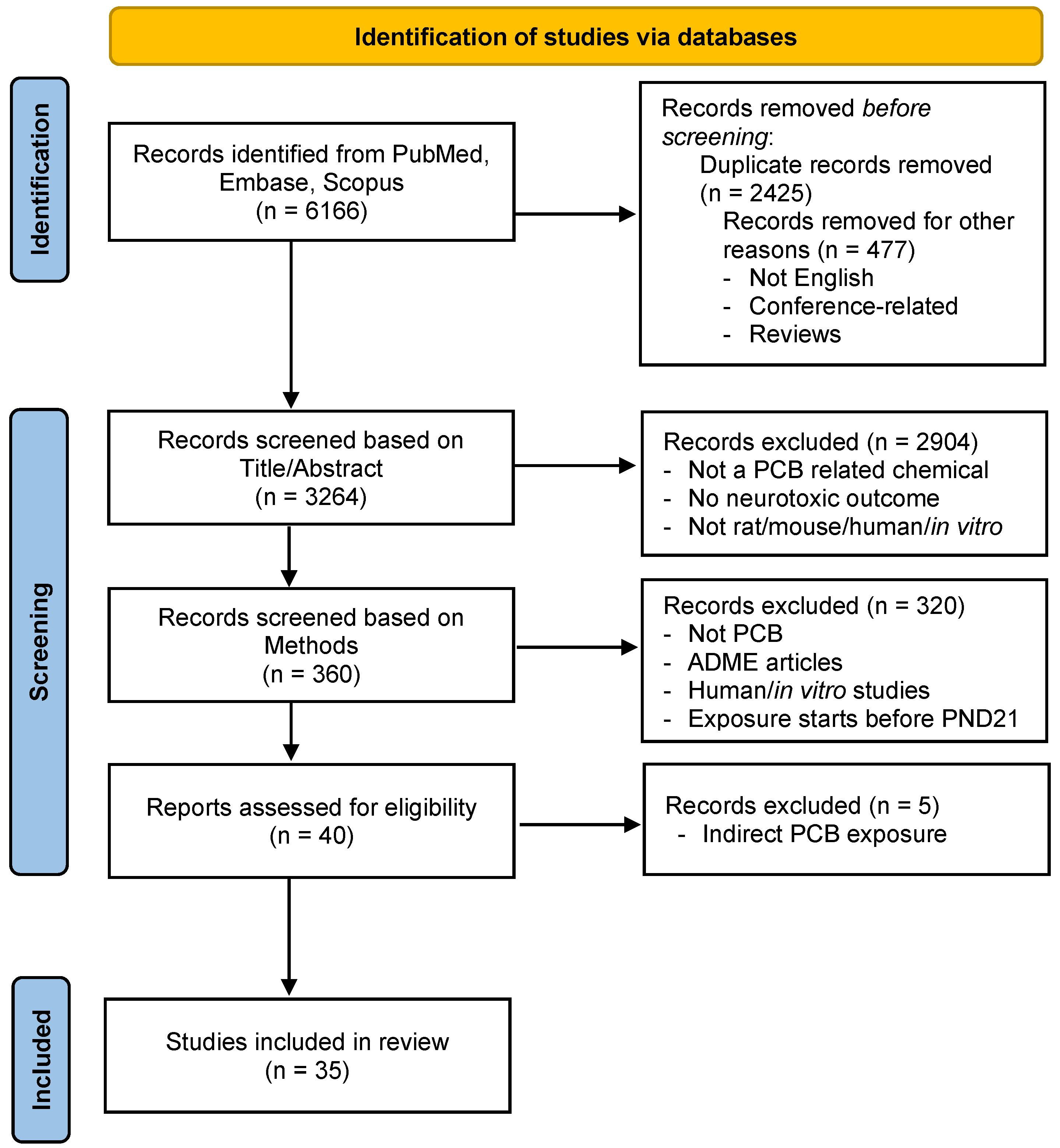
Figure 1. A Preferred Reporting Items for Systematic Reviews and Meta-Analysis (PRISMA) flow diagram for study identification, inclusion, and exclusion [88].
Ijms 26 10829 g001
Figure 2. Venn diagram comparing article selections based on (A) title/abstract between the two researchers and ChatGPT and (B) methods section between the two researchers and ChatGPT. The total number of articles selected by each reviewer is in parentheses.
Ijms 26 10829 g002
Figure 3. Decision tree guiding human reviewers in filtering ChatGPT-generated responses to the 14 yes/no questions used for the analysis of the methods sections.
Ijms 26 10829 g003
Figure 4. Electron micrograph showing the effect of PCBs on hippocampal sections in PCB-exposed adult male Wistar rats. (Upper Left) Control section. (Upper Right, Lower Left, Lower Right) PCB exposed sections. * Hypochromatic appearance of neurons and irregularly shaped and lobed cell nuclei. Arrows indicate enlargement and local edema of rough endoplasmic reticulum. Reprinted from [111], with permission from Elsevier.
Ijms 26 10829 g004
Figure 5. Biomarkers of oxidative stress (ROS/RNS) concentration in tissue homogenate after subchronic exposure to School Air Mixture (SAM, Aroclor 1254) vapor. Values are normalized by protein content and show mean ± standard error (n = 5 for the brain). ** p < 0.01, Student’s t-test, compared to sham exposure. Reprinted with permission from [24]. Copyright 2025 American Chemical Society.
Ijms 26 10829 g005
Figure 6. Tyrosine hydroxylase (TH) and dopamine transporter (DAT) and levels in the striatum of male C57BL/6 mice exposed orally to Aroclor 1254. (A) Thirty micrometer striatal sections were stained from mice 2 weeks after a 28-day exposure period for TH by immunohistochemistry. Scale bar represents 250 micrometers. (B) Staining levels were quantified by densitometry for TH. (C) Thirty micrometer striatal sections stained (immunohistochemistry) from mice 2 weeks after 28-day exposure. Scale bar represents 250 micrometers. (D) Staining levels quantified by densitometry for DA. # p < 0.05, * p < 0.01, and ** p < 0.001 from vehicle control [120] by permission of Oxford University Press.
Ijms 26 10829 g006
Figure 7. Effect of PCBs on neurons and tyrosine hydroxylase in the striatum of male C57BL/6 mice. TH-positive (TH+) and TH-negative (TH−) dopaminergic neurons counted from (A) VTA and (B) SNpc sections taken from mice 2 weeks following a 28-day exposure. Bars represent mean ± SEM, n = 7–8, # p < 0.01, + p < 0.001, and * p < 0.0001 from control; a p < 0.05 from 6 and 12 mg/kg A1254; b p < 0.005 from 6 and 12 mg/kg A1254; and c p < 0.001 from 12 mg/kg A1254 [120] by permission of Oxford University Press.
Ijms 26 10829 g007
Figure 8. Effect of PCB exposure on iron in the brain of male C57BL/6 mice. (A) Striatal (STR) and cerebellar (CBL) tissue analyzed for total iron levels by atomic absorption spectroscopy (mean ± SEM; n = 4). * p < 0.05 from vehicle-control by ANOVA. (B) 30 µm sections of the striatum and midbrain (VMB) were processed for Perl’s staining and visualized microscopically. Representative brain sections (n = 3 per exposure group) are shown. Densitometric values were acquired using Image J software (mean ± SEM; n = 3). * p < 0.05 from vehicle control [120] by permission of Oxford University Press.
Ijms 26 10829 g008
Figure 9. Effect of PCB exposure on gene expression in the brains of female Sprague-Dawley rats. RNA sequencing data visualization from brain tissues of School Air Mixture+ (SAM+, Aroclors 1245 and 1221) and sham (each n = 3). (A) Hierarchical clustering analysis and heatmap; each column represents an animal, and each row stands for a gene. (B) PCA plot displaying all six samples along PC1 and PC2. (C) Volcano plot visualizing the log2 fold change against the -log 10 false discovery rate (FDR). Dots in red indicate FDR < 0.05, dots in black indicate FDR ≥ 0.05, and blue vertical lines represent a fold change of 2. (D) Ingenuity pathway analysis (IPA)-identified statistically significant canonical pathways (p < 0.05) for the differentially expressed genes. The orange vertical line represents the threshold p-value of 0.05. Reprinted with permission from [25]. Copyright 2025 American Chemical Society.
Ijms 26 10829 g009
Figure 10. Effect of PCBs on activity in open field test in male C57BL/6 mice. (A) Total vertical, (B) ambulatory, and (C) horizontal locomotor activities were measured in mice exposed to vehicle or 6–25 mg/kg A1254. Data represents total activity counts ± SEM (n = 10) at week 0 (baseline) and at the end of exposure weeks 2, 4, and 2 weeks postexposure. * Denotes significant differences (p < 0.0001) from vehicle control at the same time point by repeated measures ANOVA [120] by permission of Oxford University Press.
Ijms 26 10829 g010
Figure 11. Effect of PCBs on elevated plus maze performance in female Sprague-Dawley rats exposed to School Air Mixture+ (SAM+, Aroclors 1245 and 1221). (A,B) show open- and closed-arm entries, respectively. (C,D) shows the time spent in open and closed arms. Values are mean ± SE (n = 8). * p < 0.05, Student’s t-test. Reprinted with permission from [25]. Copyright 2025 American Chemical Society.
Ijms 26 10829 g011
Figure 12. Effect of PCBs on Morris water maze performance in female Sprague-Dawley rats exposed to School Air Mixture+ (SAM+, Aroclors 1245 and 1221). The five-day acquisition test is illustrated in (A), showing escape latency, and (B) showing swimming distance traveled. Data from day 6 probe test shows (C), the number of platform crossings, and (D), the time spent in the target quadrant. Values are mean ± SE (n = 7 for SAM+, n = 8 for sham). * p < 0.05, Student’s t-test. Reprinted with permission from [25]. Copyright 2025 American Chemical Society.
Ijms 26 10829 g012
Figure 13. Effect of PCBs on Morris water maze performance in female Sprague-Dawley rats exposed to School Air Mixture (SAM, Aroclor 1245) or 1-bromopropane (1-BP, positive control). Five-day acquisition test (A) showing escape latency. Data from day 6 probe test showing (B), time spent in the target quadrant and (C), number of platform crossings. Data expressed as mean ± standard error (n = 8 for SAM and 1-BP, n = 6 for sham). * p < 0.05, ** p < 0.01, vs. sham group. (A) Two-way repeated-measures ANOVA. (B,C) One-way ANOVA with Bonferroni correction for multiple comparisons. Reprinted with permission from [24]. Copyright 2025 American Chemical Society.
Ijms 26 10829 g013
Figure 14. Effects of PCB exposure on cognitive flexibility and decision-making in male and female Long-Evans rats. Results are reported as mean ± SEM for all measures. “*” denotes a significant p value of p < 0.05. (Top) The average lever press latency of males and females during the visual cue discrimination phase of the set-shifting task. (A,B) The types of errors made during the reversal phase of the set-shifting task. Reprinted from [248], with permission from Elsevier.
Ijms 26 10829 g014
Figure 15. Summary of effects of PCB exposure on biochemical outcomes in the rodent brain. Red arrows indicate changes in direction. ROS: Reactive Oxygen Species. SOD: Superoxide Dismutase. NMDA: N-methyl-D-aspartate. Locations of images do not necessarily represent spatial location. Created with BioRender.com.
Ijms 26 10829 g015
Figure 16. Summary of effects of PCB exposure on behavioral outcomes in rodents. Red arrows indicate changes in direction. Created with BioRender.com. See Table 7 for a summary of behavioral assays, associated domains, implicated brain regions, and proposed molecular mechanisms underlying PCB-induced neurobehavioral alterations in rodent models.
Ijms 26 10829 g016
Table 1. Questions asked ChatGPT to feed the model with information about the topic.
IDQuestion
1What are polychlorinated biphenyls?
2How are people exposed to PCBs?
3How do PCBs impact health?
4How are PCBs neurotoxic?
5How do PCBs impact behavior?
6How do PCBs impact neurobehavior?
7How are PCBs studied in mice and rats?
Table 2. ChatGPT’s response to the request for classification of titles/abstracts.
Author/Title/YearMouse/Rat/Human/In Vitro
Related
PCB or
Related Chemicals
Neurotoxic Outcome ListedNot
Related
Explanation
Nilsson, C. B. et al. (2000). “2,3,7,8-tetrachlorodibenzo-p-dioxin increases serum and kidney retinoic acid levels and kidney retinol esterification in the rat.”XX Involves rats and TCDD, a related chemical to PCBs.
Nilsson, E. et al. (2012). “Environmentally induced epigenetic transgenerational inheritance of ovarian disease.”XX Involves rats and various environmental chemicals, including dioxins.
Ning, S. and X. Xiaobai (1997). “Reductive metabolism of 4-nitrobiphenyl by rat liver fraction.”X Involves rats but not related to PCBs or neurotoxic outcomes.
Ninou, I. et al. (2018). “Pharmacologic targeting of the ATX/LPA axis attenuates bleomycin-induced pulmonary fibrosis.” XNot related to PCBs or neurotoxic outcomes.
Nishida, N. et al. (1997). “Effects of acute and repeated exposures to Aroclor 1254 in adult rats: motor activity and flavor aversion conditioning.”XXX Involves rats, Aroclor 1254 (a type of PCB), and studies neurotoxic outcomes.
Table 3. Questions asked ChatGPT to extract information from each article.
IDQuestion
1. PCBs
1-1Does the paper describe the use of PCBs? Must only be PCB congeners, not related substances. If the answer is no, do not proceed any further with the assessment.
2. Mouse/Rat
2-1Does the paper describe the use of mice or rats in the study?
2-2Are mice or rats directly exposed to PCBs?
2-3Are cell cultures from mice or rats exposed to PCBs?
2-4Are tissues collected from mice or rats exposed to PCBs?
3. Exposure Timeframe
3-1Does the article describe dosing PCBs during gestation?
3-2Does the article describe dosing PCBs during lactation?
3-3Does the article describe dosing PCBs to the mouse/rat before postnatal day 21?
3-4Does the article mention dosing after weaning/post-natal day 21?
3-5If applicable, were maternal/dam tissues collected or maternal analyses done?
4. Neuro-Outcomes
4-1Does the paper describe analyses related to neurobehavior or neurotoxicity?
4-2Does the paper describe behavioral analysis?
4-3Does the paper describe gene analysis?
4-4Does the paper describe receptor changes?
Table 4. ChatGPT’s response to the request for information extraction from the methods section.
Author/Title/YearSub-PartYes/NoExplanation
Coburn, C. G. et al., 2015. Permanently compromised NADPH-diaphorase activity within the osmotically activated supraoptic nucleus after in utero but not adult exposure to Aroclor 1254. Neurotoxicology, 47, 37–46.1-1YesThe paper describes the use of Aroclor 1254, a commercial PCB mixture.
2-1YesThe study uses male Sprague-Dawley rats.
2-2YesRats were exposed to Aroclor 1254 through cheese puff snacks infused with the PCB mixture.
2-3NoThe study does not describe the use of cell cultures from mice or rats exposed to PCBs.
2-4YesBrain tissues were collected from rats exposed to Aroclor 1254 for histochemical analysis.
3-1YesIn utero exposure was accomplished by feeding pregnant dams cheese puff snacks infused with Aroclor 1254 for 10 days during gestation (GD 10–19).
3-2NoThe article does not describe dosing PCBs during lactation.
3-3YesPups were exposed to PCBs in utero and allowed to suckle until weaning at PND 22.
3-4YesAdult rats were exposed to PCBs at 3–5 months old, and some were allowed to age until 14–16 months before sacrifice.
3-5NoThe study does not mention collecting maternal/dam tissues or conducting maternal analyses.
4-1YesThe paper describes the assessment of NADPH-diaphorase activity, which is related to neurotoxicity.
4-2NoThe paper does not describe behavioral analysis.
4-3NoThe paper does not describe gene analysis.
4-4NoThe paper does not describe receptor changes.
Table 5. Cohen’s kappa values and interpretations for inter-rater reliability of title/abstract screening selections. A: The total number of instances that both raters said were correct. B: The total number of instances that Rater 2 said was incorrect, but Rater 1 said were correct. C: The total number of instances that Rater 1 said was incorrect, but Rater 2 said were correct. D: The total number of instances that both raters said were incorrect.
ComparisonABCDAgreementKappaSE95% CIInterpretation
Reviewer 1 & Reviewer 237271110271194.45%0.7720.020.740–0.804Substantial Agreement
Reviewer 1 & ChatGPT30114247277494.21%0.7290.020.692–0.765Substantial Agreement
Reviewer 2 & ChatGPT29751185273192.77%0.6750.020.637–0.714Substantial Agreement
Table 6. Cohen’s kappa values and interpretations for inter-rater reliability for method screening selections. A: The total number of instances that both raters said were correct. B: The total number of instances that Rater 2 said was incorrect, but Rater 1 said were correct. C: The total number of instances that Rater 1 said was incorrect, but Rater 2 said were correct. D: The total number of instances that both raters said were incorrect.
ComparisonABCDAgreementKappaSE95% CIInterpretation
Reviewer 1 & Reviewer 230131530292.22%0.6380.060.514–0.761Moderate-
Substantial Agreement
Reviewer 1 & ChatGPT3761530294.17%0.7460.050.642–0.849Moderate-
Almost Perfect Agreement
Reviewer 2 & ChatGPT29231629289.17%0.5360.070.407–0.664Fair-Moderate Agreement
Disclaimer/Publisher’s Note: The statements, opinions and data contained in all publications are solely those of the individual author(s) and contributor(s) and not of MDPI and/or the editor(s). MDPI and/or the editor(s) disclaim responsibility for any injury to people or property resulting from any ideas, methods, instructions or products referred to in the content.