Article

A Celsr3 Mutation Linked to Tourette Disorder Disrupts Cortical Dendritic Patterning and Striatal Cholinergic Interneuron Excitability

by
Cara Nasello
1,2,3,†,
G. Duygu Yilmaz
1,2,†,
Lauren A. Poppi
3,4,
Tess F. Kowalski
1,2,4,
K. T. Ho-Nguyen
1,4,
Junbing Wu
1,4,
Matthew Matrongolo
1,4,
Joshua K. Thackray
1,3,
Anna Shi
1,4,
Nicolas L. Carayannopoulos
1,
Nithisha Cheedalla
1,
Julianne McGinnis
1,4,
Jasmine Chen
1,2,
Adyan Khondker
1,2,
Fadel Tissir
5,6,
Gary A. Heiman
3,
Jay A. Tischfield
3 and
Max A. Tischfield
1,2,4,*
1
Department of Cell Biology and Neuroscience, Rutgers, The State University of New Jersey, Piscataway, NJ 08854, USA
2
Keck Center for Collaborative Neuroscience, Rutgers, The State University of New Jersey, 604 Allison Road, D251, Piscataway, NJ 08854, USA
3
Department of Genetics and the Human Genetics Institute of New Jersey, Rutgers, The State University of New Jersey, Piscataway, NJ 08854, USA
4
Child Health Institute of New Jersey, Robert Wood Johnson Medical School, New Brunswick, NJ 08901, USA
5
College of Health and Life Sciences, Hamad Bin Khalifa University, Doha 34110, Qatar
6
Laboratory of Developmental Neurobiology, Institute of Neuroscience, Université Catholique de Louvain, 1200 Brussels, Belgium
*
Author to whom correspondence should be addressed.
These authors contributed equally to this work.
Int. J. Mol. Sci. 2025, 26(21), 10307; https://doi.org/10.3390/ijms262110307
Submission received: 15 August 2025 / Revised: 14 October 2025 / Accepted: 16 October 2025 / Published: 23 October 2025

Abstract

Tourette Disorder (TD) is a prevalent neurodevelopmental condition characterized by chronic motor and vocal tics. A mechanistic understanding of both the genetic etiology and brain pathophysiology remains poor. To gain insight into the molecular underpinnings of TD, we have generated a novel mouse model expressing an orthologous human mutation in CELSR3, a high-confidence TD risk gene. This putative damaging de novo variant, R774H, causes an amino acid substitution within the fifth cadherin repeat. Unlike previous Celsr3 TD models and Celsr3 constitutive null mice, mice homozygous for the R774H amino acid substitution are viable. They have grossly normal forebrain development and no changes to the density of cortical and striatal interneuron subpopulations. However, 3D geometric analysis of cortical pyramidal neurons revealed changes to dendritic patterning and the types and distributions of spines. Furthermore, patch clamp recordings in cholinergic interneurons located within the sensorimotor striatum uncovered mild intrinsic hyperexcitability and changes to spine density. Despite these changes, Celsr3R774H homozygous mice do not show repetitive motor behaviors at baseline nor motor learning impairments. However, Celsr3R774H homozygous males have sensorimotor gating deficits, a behavioral phenotype observed in both humans with TD and previously reported mouse models. Our findings suggest human mutations in CELSR3 may affect dendritic patterning, spine formation and/or turnover, and the firing properties of neurons within cortico-striatal circuits.

1. Introduction

Tourette Disorder (TD) is a childhood-onset neurodevelopmental disorder characterized by chronic vocal and motor tics, which are often elicited in response to somatosensory phenomena known as premonitory sensations [1]. TD manifests with neuropsychiatric comorbidities including attention-deficit hyperactivity disorder, obsessive–compulsive disorder, autism spectrum disorder, as well as mood, anxiety, and sleep disorders, which altogether underscore its multifaceted pathophysiology [2,3,4,5]. TD is a heritable disorder, with a concordance rate approaching 80% in monozygotic twins [6]. Human imaging studies have revealed structural and functional changes spanning cortico-striatal-thalamo-cortical (CSTC) and basal ganglia networks that govern the selection, planning, and control of volitional behavioral output [7,8,9,10,11].
Structural and functional changes in sensorimotor control loops likely contribute to the pathology of TD [12,13,14,15]. Wide-spread alterations in cortical thickness (grey matter) and intercortical innervation patterns (white matter) have been reported in TD subjects, although findings vary. Both increased and decreased grey matter volumes have been observed in the somatosensory and motor cortices, the putamen, thalamus, hypothalamus, and midbrain nuclei [16,17,18,19,20]. Functional magnetic resonance imaging consistently shows increased activity in these areas and a stronger connectivity between the sensorimotor cortex and the putamen, suggesting a heightened descending drive from sensorimotor areas [10,21,22]. In contrast, studies of higher-order associative cortices show mostly decreased grey matter volume and diminished functional activity in orbitofrontal, prefrontal, parietal, occipital and premotor cortices [16,21,23,24]. Increased insular, hippocampal and amygdalar volumes in patients with strong premonitory sensations may be structural consequences of tic regulation [20,25,26]. Given TD’s developmental trajectory, determining whether these structural changes are causal or compensatory is challenging.
These structural differences can be due to microscopic changes to neural circuits. For example, a change in cortical thickness might reflect underlying changes to the neuropil (dendritic arborization, synaptogenesis, glial processes), to neuronal composition and distribution, or to myelination patterns within surrounding white matter. Immunohistochemical approaches on post-mortem brain tissue provide a more detailed picture, but these findings are limited to the striatum. Post-mortem brain tissue of subjects with severe, refractory TD has revealed signs of parvalbumin and cholinergic interneuron loss in striatal regions and may account for tics and related behaviors in at least some affected individuals [27,28,29,30]. Focal disinhibition of the dorsal sensorimotor striatum via application of GABAA receptor antagonists in rodents and non-human primates can trigger severe motor stereotypies [31,32,33], whereas disinhibition in the ventral striatum leads to vocalizations [34,35]. Furthermore, targeted ablation of cholinergic interneurons in the rodent dorsal striatum causes motor stereotypies as well as perseverative behaviors following acute stress or amphetamine challenge [36,37]. While these animal models have provided valuable insights for understanding the pathogenesis of TD, they have notable limitations as the experimental manipulations were performed in adult animals and likely fail to model the types of neurodevelopmental changes found in humans.
Historically, the limited availability of preclinical genetic models for TD has hindered the development of targeted treatment [14]. Despite the prevalence of TD (~0.5–1% of the population) [38], gene coding variants have been identified in relatively few families, and genome-wide association studies have yielded few clues, often failing to replicate in subsequent studies [39]. However, recurrent de novo coding variants in several genes, including CELSR3, NIPBL, and WWC1, have been identified using large scale trio based whole-exome sequencing [14,40]. Of the identified high-confidence candidates, multiple likely damaging or loss-of-function mutations in several domains of CELSR3 have been identified in simplex trios and multiplex families [41].
CELSR3 encodes a protocadherin cell adhesion G protein-coupled receptor that is critical for axon guidance, dendritic patterning, and the formation of synapses. CELSR3 is required for the development and guidance of major forebrain axon tracts, such as the anterior commissure and internal capsule, which contains corticostriatal, thalamocortical, and corticothalamic axons [42,43,44]. During development, CELSR3 is required for the establishment of basal ganglia pathways, including axonal projections from areas such as the striatum, subthalamic nucleus, and the substantia nigra pars compacta to the globus pallidus [45]. Mice homozygous for a GFP knock-in allele (ablating Celsr3 expression) suggest Celsr3 may be required for the tangential migration of interneurons, although these findings have not been replicated in other Celsr3 mutant models [46,47]. In adult animals, Celsr3 expression is maintained in subpopulations of cortical and striatal interneurons, as well as cerebellar Purkinje neurons [48]. Thus, Celsr3 expression patterns in the brain and its necessity for the development of CSTC and basal ganglia circuitry make it an attractive candidate to model TD in mice.
We previously reported that mice expressing orthologous human mutations within the second laminin G-like domains of Celsr3 show sensorimotor gating deficits, repetitive motor behaviors, and changes to reward learning and electrically evoked striatal dopamine release [49]. In the present study, we have developed a novel model for TD that expresses an orthologous human amino acid substitution, R774H (in humans, R783H), within the fifth protocadherin repeat of the extracellular domain of Celsr3. We investigated the impact of the Celsr3R774H amino acid substitution on mouse behavior and brain development. We hypothesized that Celsr3R774H mutant mice would show similar behavioral changes to those previously described in other Celsr3 models of TD [49] as well as changes to axon guidance and/or dendritic patterning. Unlike our previously reported models, mice heterozygous for the R774H amino acid substitution do not show behavioral changes in the paradigms tested. However, males homozygous for the R774H amino acid substitution have sensorimotor gating deficits that are absent in affected females, while homozygous females, but not males, have increased marble burying, demonstrating sex-specific phenotypes in this model. In agreement with our previous findings, we do not see evidence of either cortical or striatal interneuron loss in homozygous mutants, and the development of major white matter tracts in the forebrain also appears grossly normal. However, we find subtle changes to dendritic patterning and synapse formation in deep layer cortical pyramidal neurons, as well as changes to striatal cholinergic interneuron excitability. Our findings demonstrate that human mutations in CELSR3 are sufficient to cause discernible changes to neurite development and synapse formation, supporting a framework in which impairments in the ability of neurons to functionally integrate into CSTC loops might underlie TD.

2. Results

Celsr3R774H/+ and Celsr3R774H/R774H animals on a pure C57BL/6 background were born at normal Mendelian ratios, had normal weights, and were indistinguishable from littermate controls (no hair loss or skin lesions). By contrast, we previously reported that homozygous Celsr3C1906Y and Celsr3S1894Qfs*2 mice are perinatal lethal, like Celsr3 null mice, suggesting the Celsr3R774H mutant protein retains partial function (Figure 1a). Furthermore, protein levels in whole-brain lysates were normal in homozygous Celsr3R774H neonates, by contrast with previous models (Figure 1b). Thus, the R774H substitution in the 5th protocadherin repeat appears to exert milder effects versus those found in the second laminin G-like domain.

2.1. Celsr3R774H/R774H Mice Show Sex-Specific Sensorimotor Gating Deficits

TD subjects show deficits in sensorimotor gating as measured by prepulse inhibition (PPI) of the acoustic or tactile startle reflex [50,51]. Similarly, several genetic mouse models of TD have sensorimotor gating deficits [49,52,53]. These findings suggest that perturbations to sensorimotor gating may be useful as a behavioral screening test to validate animal models of TD. Therefore, we tested sensorimotor gating in Celsr3R774H mice using an acoustic PPI paradigm. All Celsr3R774H mice showed comparable levels of baseline acoustic startle reflex as wild-type control littermates (Figure 1c). Both male and female Celsr3R774H/+ mice showed no differences in PPI of acoustic startle (Figure 1d,e). However, PPI was mildly, yet significantly, reduced in male Celsr3R774H/R774H mice compared to wild-type littermate controls (Sidak’s multiple comparisons testing, pp71: p = 0.6, pp77: p = 0.03 and pp81, p = 0.06, Figure 1d). Female Celsr3R774H/R774H mice did not show significant PPI deficits at any of the dB prepulses tested (Figure 1e). Due to our findings showing changes only in homozygous mice, we restricted our focus to Celsr3R774H/R774H animals for subsequent experiments.

2.2. Celsr3R774H/R774H Mice Do Not Show Hyperactivity or Repetitive Motor Behaviors in the Open Field

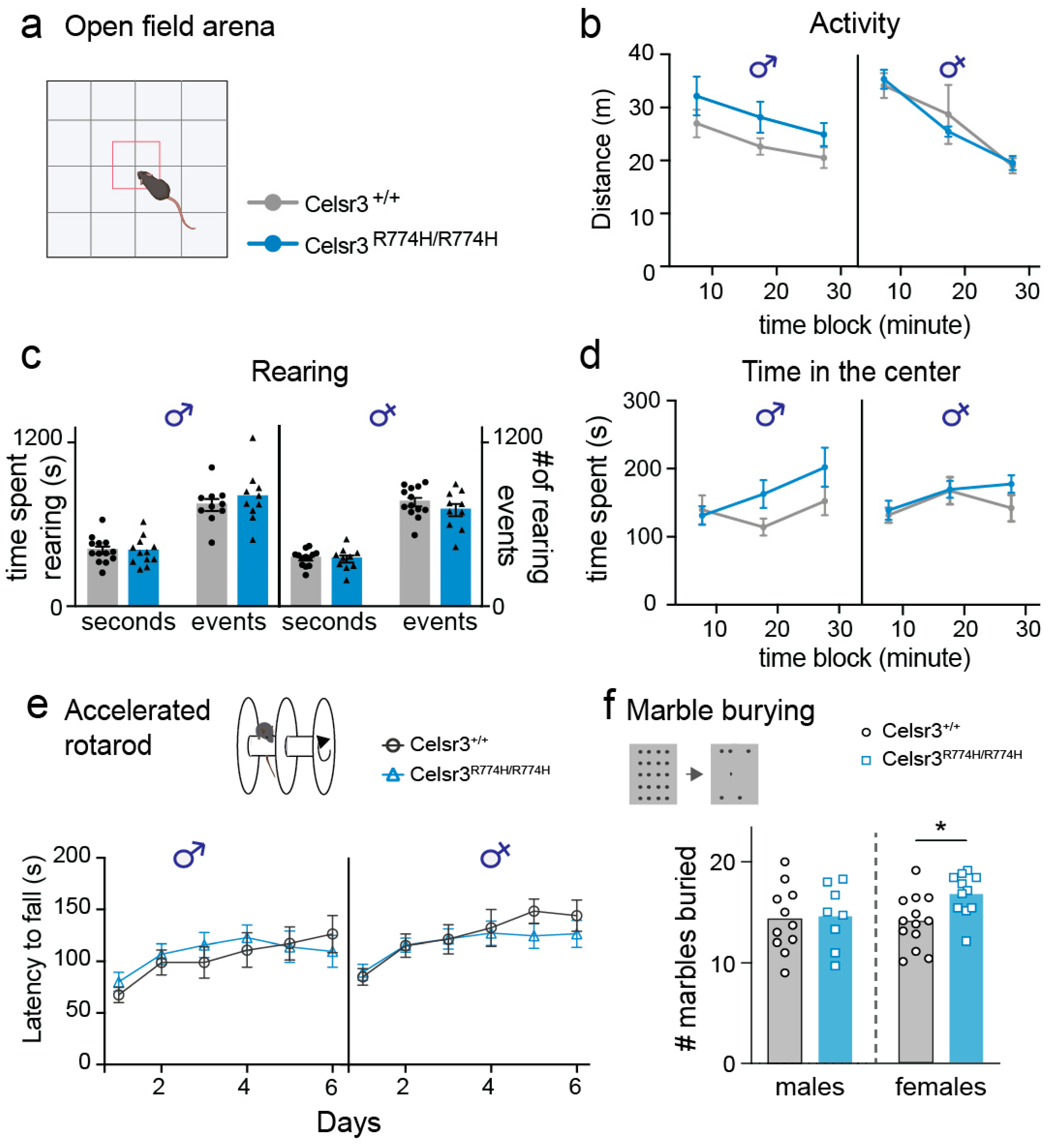
Celsr3C1906Y/+ and Celsr3S1894Rfs*/+ mice have increased locomotion and repetitive rearing in an open field arena; therefore, we tested Celsr3R774H/R774H mice and their wild-type littermates for similar behavioral changes. Celsr3R774H/R774H male and female mice did not show an increase in overall activity compared to wild-type littermate controls (Figure 2a,b). However, males trended upward suggesting a milder—but shared—increased activity phenotype with Celsr3C1906Y/+ and Celsr3S1894Rfs*/+ mutants (Figure 2b, p = 0.16, 2-way RM-ANOVA). Next, we looked at the number of rearing events and time spent rearing. The number of events and amount of time spent rearing were equivalent between Celsr3R774H/R774H male and female mice versus wild-type littermates (Figure 2c). We next looked at changes in the time spent in the center of the arena, a measure of exploratory behavior and anxiety (Figure 2d). Celsr3R774H/R774H male and female mice spent equivalent amounts of time in the center compared to wild-type littermate controls. However, both male and female Celsr3R774H/R774H mice spent more time in the center during the third 10 min testing period compared to the first 10 min period, suggesting changes to exploratory behavior and/or anxiety levels as time progressed. (Figure 2d Sidak’s multiple comparisons, male: p = 0.044 female, p = 0.049, 2-way RM-ANOVA).

2.3. Celsr3R774H/R774H Mice Exhibit Normal Motor Coordination as Measured by Accelerated Rotarod

Mutations in Celsr3 are reported to alter rotarod performance in mice. Mice with conditional knockout of Celsr3 within Purkinje cells in the cerebellum have worse performance in the rotarod, whereas our previously reported Celsr3 TD models perform better [48,49]. Therefore, we tested balance and motor coordination using an accelerated rotarod protocol. Celsr3R774H/R774H male and female mice showed comparable latency to fall as wild-type littermate controls (males: p = 0.76, females: p = 0.62, 2-way RM-ANOVA), suggesting motor coordination and learning is normal and intact in these animals (Figure 2e).

2.4. Female Celsr3R774H/R774H Mice Show Perseverative Digging Behavior

It has been previously reported that mutations in Celsr3 mice can cause sex-specific changes to perseverative digging behavior [49,54]. We examined perseverative behaviors using the marble burying assay, a test of repetitive digging behavior (Figure 2f). Male Celsr3R774H/R774H mice buried similar numbers of marbles compared to wild-type littermates; however, female Celsr3R774H/R774H mice buried a significantly higher number of marbles on average compared to littermate controls (p = 0.013; two-tailed t-test), suggesting sex-specific changes to object-oriented perseverative behavior.

2.5. Axon Tract Development Is Grossly Normal in Celsr3R774H/R774H Mice

The gross anatomy and overall size of Celsr3R774H/R774H mouse brains appeared normal. Given the central role Celsr3 has on axonal tract development in the forebrain, we evaluated the major CSTC pathways (Figure 3a). Antibody labelling against neuronal cell adhesion protein L1 in embryonic day (E) 18.5 brain sections showed that the development and trajectories of major forebrain axon tracts in the internal capsule, anterior commissure, and corpus callosum were grossly normal in Celsr3R774H/R774H mice (Figure 3b). By comparison, these tracts are absent in Celsr3 null mutants [47]. Striatonigral axons in the direct pathway terminating in the globus pallidus internus and substantia nigra were visualized by crossing Drd1a-Cre and R26:Ai14 reporter lines (Supplementary Table S1). Using widefield fluorescence microscopy, striatonigral fiber tracts showed normal development and terminated appropriately in the globus pallidus internus in adult Drd1a-Cre;Celsr3R774H/R774H;R26:Ai14 mice (Figure 3c). Next, we crossed A2a-Cre and R26:Ai14 reporter lines (Supplementary Table S1) to visualize striatopallidal axons that terminate in the globus pallidus externus. We also did not detect any major qualitative differences in the pattern of tdTomato-positive fibers terminating in the globus pallidus externus of A2A-Cre;Celsr3R774H/R774H;R26:Ai14 animals compared to littermate controls. By contrast, both striatopallidal and striatonigral axon tracts fail to form in mice with constitutive loss of Celsr3 [45]. Axon pathfinding appeared normal as we did not observe instances of wandering axons, bundles, or mis-innervation by direct and indirect pathway terminals (Figure 3c,d). Thus, the Celsr3R774H amino acid substitution within the fifth cadherin repeat does not affect the ability of the protein to regulate axon guidance in the forebrain in a manner that is detectable with the qualitative anatomical techniques used.

2.6. Celsr3R774H/R774H Mice Have Organized Cortical Layering and Do Not Show Interneuron Loss

Cortical layering, as assessed by TBR1, CTIP2, and SATB2 immunostaining, was normal in Celsr3R774H/R774H animals compared to littermate controls (Figure 4a). The relative radial thickness of each cortical layer was also normal in Celsr3R774H/R774H animals (Figure 4b), and nearest neighbor (NN) analysis showed normal distribution of labelled cortical neurons (Figure 4b, p = 0.2275, 2-way ANOVA). Celsr3 is expressed by E13.5 in the ganglionic eminences, which give rise to cortical and striatal interneurons, and regulates the tangential migration of cortical interneurons [46]. Immunolabeling against parvalbumin showed that the density of cortical parvalbumin interneurons was normal in Celsr3R774H/R774H mice, despite variable staining differences that occurred in thick sections from both wild-type and mutant mice (Figure 4d,e). Using Somatostatin-Cre and the R26:Ai14 reporter line to lineage label somatostatin interneurons, there were also no differences in density within the cortex of Celsr3R774H/R774H mice (Figure 4f,g). Thus, cell proliferation and the radial and tangential migration of cortical pyramidal neurons and interneurons, respectively, appeared normal in Celsr3R774H/R774H animals.

2.7. Cortical Pyramidal Neuron Dendritic Patterning Is Affected in Celsr3R774H/R774H Mice

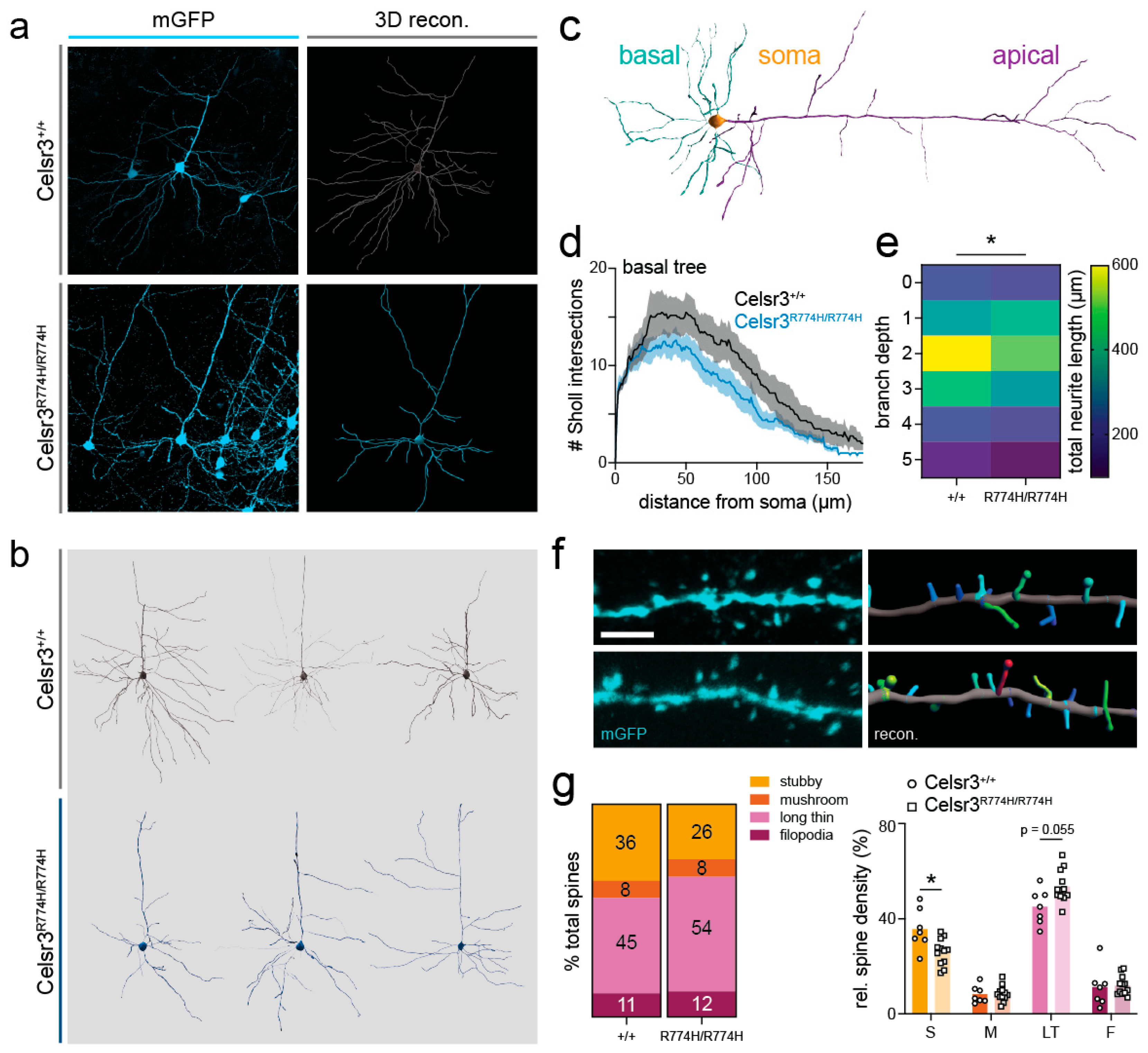
Celsr3 is required for neurite development and dendritic patterning in the cortex and hippocampus [55,56]. Given broad Celsr3 expression across cortical PV interneurons, we attempted to examine whether the dendritic arborizations of cortical parvalbumin interneurons were properly patterned in Celsr3R774H/R774H mice by using a Cre-dependent viral sparse cell labelling approach to mark parvalbumin (PV) interneurons with GFP (Figure 5a,b) [57]. We crossed a PV-2A-Cre allele onto the Celsr3R774H/R774H background and injected the virus into the somatosensory cortex. Most labelled neurons localized to deep layer 5 of the cortex but surprisingly, most were not positive for parvalbumin immunostaining. Instead, these neurons had typical cortical pyramidal neuron morphology with basal and long apical dendrites. Crossing these animals to the R26:Ai14 reporter line showed diffuse td-Tomato expression throughout the cortex, suggesting the PV-2A-Cre allele went germline, consistent with previous reports [57].
Nonetheless, 3D neuronal reconstructions revealed that the basal dendrites of Celsr3R774H/R774H deep layer 5 pyramidal neurons were less arborized than littermate controls (Figure 5b). Basal dendrites were analyzed separately by excluding apical branches from the dataset (Figure 5c). Sholl analysis revealed a genotype effect for the complexity of Celsr3R774H/R774H pyramidal neuron basal dendrites (Figure 5d Celsr3+/+ n = 6; Celsr3R774H/R774H n = 8; 2-way ANOVA genotype effect p < 0.001). The area under the Sholl curve was 1639 ± 40.05 and 1204 ± 27.62 for Celsr3+/+ and Celsr3R774H/R774H, respectively. There was also a significant genotype effect when comparing branch depth, which reflects the number of times a dendrite has branched since leaving the soma (Figure 5e; p = 0.0271, 2-way ANOVA). There was no significant difference in the number of branch points (Celsr3+/+ = 16.33 ± 2.81, Celsr3R774H/R774H = 15.22 ± 1.52, p = 0.7110, unpaired t-test) or dendritic straightness (Celsr3+/+ = 0.9411 ± 0.003, Celsr3R774H/R774H = 0.9298 ± 0.006, p = 0.1805, unpaired t-test). We also did not see evidence of increased number of self-crossings.
There was no difference in the density of spines along the secondary basal dendrites between Celsr3+/+ (8.60/10 µm) and Celsr3R774H/R774H (8.94/10 µm) mice (Figure 5f). However, when spines were classified according to morphology (e.g., stubby, mushroom, long-thin, filopodia, see Appendix A.2 Table A2 for criteria), and the relative densities were compared using the ClassifySpines IMARIS plug-in, the proportion of stubby and long-thin spines detected along a single length of dendrite appeared shifted in Celsr3R774H/R774H mice (Figure 5g). There was also a significant reduction in stubby spines in Celsr3R774H/R774H animals (p = 0.033), and a strong trend toward an increase in long thin spines (p = 0.055, multiple t-tests corrected for multiple comparisons). Thus, the Celsr3R774H amino acid substitution in the homozygous state is sufficient to alter dendritic patterning as well as the types and distributions of spines in deep layer cortical pyramidal neurons.

2.8. Celsr3R774H/R774H Cholinergic Interneurons Have Altered Membrane Properties and Spine Density

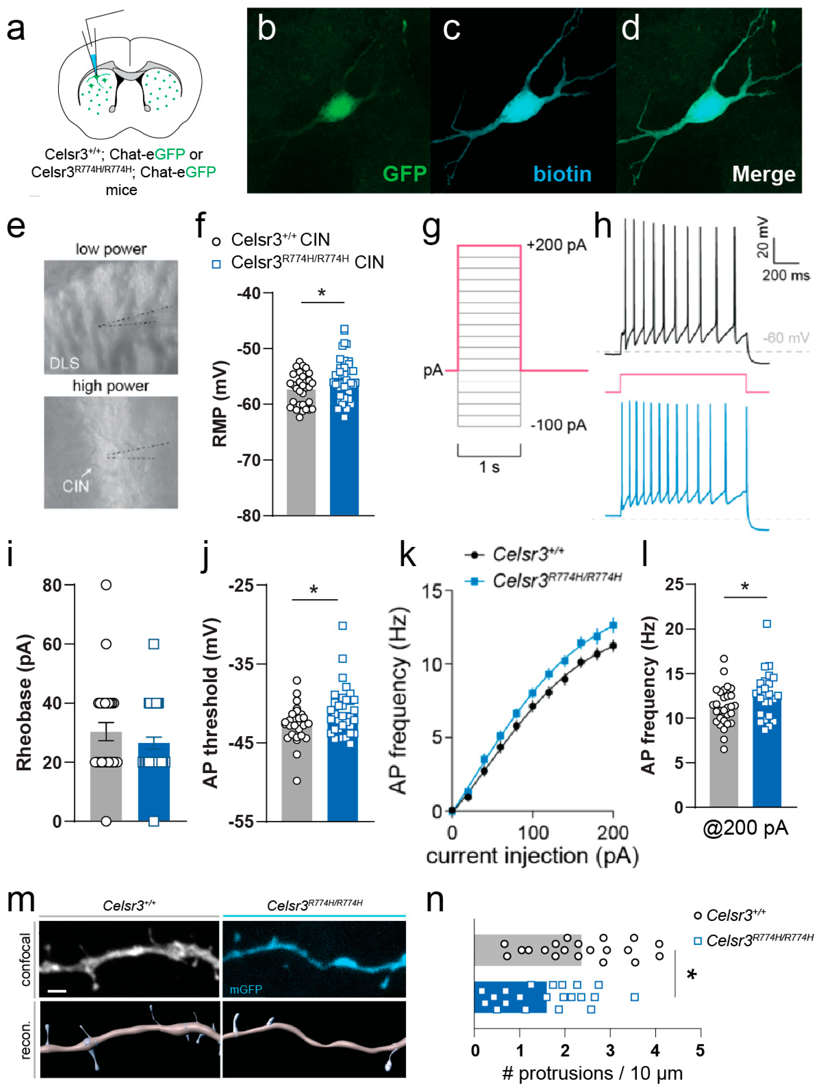
Loss of striatal interneurons, including cholinergic interneurons (CINs), have been reported in adults with severe, refractory TD [28,30,43]. Although we previously reported that the numbers and positioning of striatal parvalbumin interneurons and CINs were normal in Celsr3C1906Y/+ and Celsr3S1894Rfs*2/+ mice [49], we nonetheless asked if mutations in the cadherin domain affected the numbers or positioning of CINs. We crossed Celsr3R774H/R774H mutants with ChAT-eGFP reporter mice and compared the numbers and distributions of CINs from selected axial positions in the striatum (Figure 6a,b). Consistent with our previous models, we did not see any changes in the numbers (Figure 6c) or distributions (Figure 6d) of CINs.
Although we did not detect signs of striatal interneuron loss in Celsr3R774H/R774H mice, it is possible that the mutation may alter the active and/or passive membrane properties of these cells. Celsr3 is widely expressed throughout CINs (Supplementary Figure S1a,b). By comparison, in a Celsr3eGFP/+ reporter mouse, we observed less colocalization between GFP expressing cells and PV+ interneurons, although their difference failed to reach significance (Supplementary Figure S1c,d, Wilcoxon signed-rank test, one-tailed, median difference between ChAT-PV: 0.33, p = 0.125). Given the significance of CINs in TD literature, we next examined their electrophysiological properties in Celsr3R774H/R774H mice. CINs located within the dorsolateral striatum of both Celsr3+/+ and Celsr3R774H/R774H mice had characteristically large somata, a tonic firing profile, with varying levels frequency adaptation between cells, and upon membrane breakthrough, had a relatively depolarized resting membrane potential (RMP, Figure 7a,d). Passive membrane properties were not significantly different between Celsr3+/+ and Celsr3R774H/R774H mice (Figure 7e–h, see Appendix A.1 Table A1 for more information). Membrane impedance (Rm) was 184.8 ± 8.82 MΩ and 205.1 ± 11.89 MΩ for Celsr3+/+ (n = 31) and Celsr3R774H/R774H (n = 39) CINs, respectively (p = 0.4238, Mann–Whitney test). Membrane capacitance (Cm) was 33.52 ± 1.10 pF and 34.05 ± 0.97 pF for Celsr3+/+ and Celrs3R774H/R774H CINs, respectively (p = 0.7214, t-test). Membrane time constant (tau) was 2.88 ± 0.16 ms and 2.91 ± 0.16 ms for Celsr3+/+ and Celsr3R774H/R774H CINs, respectively (p = 0.8870, t-test). Resting membrane potential (RMP) was on average more depolarized in Celsr3R774H/R774H CINs (p = 0.037, t-test, Figure 7f). Rheobase (minimum current injection step required to elicit an action potential) was not significantly affected (p = 0.3505, Mann–Whitney test, Figure 7i). The action potential (AP) threshold was significantly more depolarized in Celsr3R774H/R774H CINs (p = 0.0456, t-test, Figure 7j). The f/I plots for Celsr3+/+ (n = 29) and Celsr3R774H/R774H (n = 25) required different nonlinear fits (p < 0.001, Figure 7k). This indicated a tendency for Celsr3R774H/R774H CINs to fire at a higher frequency in response to somatic current injection compared with Celsr3+/+ CINs. AP frequency was significantly higher in Celsr3R774H/R774H compared to Celsr3+/+ CINs with 200 pA current injection (p = 0.038, t-test, Figure 7l). Thus, Celsr3R774H is sufficient to alter the membrane properties of cholinergic interneurons.
In a subset of neurons, we assessed changes to dendrite morphology using biotin filling during recording and post hoc anatomical recovery. Celsr3R774H/R774H CINs showed changes to neurite complexity compared to Celsr3+/+ CINs (Sholl analysis, n = 6, p < 0.001, 2way ANOVA, (Supplemental Figure S2a–c). The number of branch points trended towards an increase in Celsr3R774H/R774H CINs (p = 0.06, t-test, Supplemental Figure S2d), whereas neurite straightness trended towards a decrease in Celsr3R774H/R774H CINs (p = 0.05, t-test, Supplemental Figure S2e). In addition, while fractal dimension (DB) was similar between Celsr3+/+ and Celsr3R774H/R774H CINs (p = 0.2636, Mann–Whitney test, Supplemental Figure S2f), lacunarity was significantly increased in Celsr3R774H/R774H CINs compared to controls (p = 0.0379, t-test, Supplemental Figure S2g), potentially indicating that dendritic branches are distributed unevenly in mutant cells. Finally, although the distribution of dendritic spines on CINs is much sparser compared to neighboring medium spiny neurons, the average spine density along second order dendrites was decreased in Celsr3R774H/R774H mice compared to controls (p = 0.0184, t-test, Figure 7m,n). Given the widespread GFP expression observed in Celsr3GFP/+ mice in adult CINs, these observations may reflect cell-autonomous changes resulting from mutant Celsr3, and could account, at least in part, to changes in membrane properties and firing activities.

3. Discussion

In this study, we sought to expand on the available animal models carrying mutations in Celsr3, a high-confidence TD gene, by presenting a phenotypic analysis of a novel mouse model engineered to express a putative damaging variant that causes an amino acid substitution within the fifth extracellular cadherin repeat (R774H). This mutation is orthologous to a human variant, R783H, that was identified previously in a whole exome sequencing study [5]. While heterozygous mutants showed no overt behavioral abnormalities, homozygous Celsr3R774H/R774H males exhibited deficits in sensorimotor gating as measured by prepulse inhibition, whereas homozygous females displayed increased perseverative digging behavior. At the gross anatomical level, we observed no obvious disruptions to major forebrain fiber tracts or changes to interneuron distribution in the cortex and striatum. However, we identified a reduction in the complexity of basal dendritic arbors in deep layer pyramidal neurons located within the primary somatosensory cortex, accompanied by altered dendritic spine distributions. We also observed changes to the membrane properties and firing activities of striatal CINs in ex vivo brain slices, with altered spine density along second order dendrites. Taken together, these results suggest mutations that reside within the extracellular cadherin repeats of Celsr3 have the potential to disrupt neurite patterning (without gross changes to white matter tracts), synapse formation/turnover, and the firing properties of neurons. These findings highlight the importance of characterizing distinct TD-associated mutations in Celsr3 found across different functional domains to uncover how specific molecular insults may disrupt brain development and contribute to the pathogenesis of TD either through shared or distinct ways. As such, these findings provide a roadmap for extended analyses in Celsr3C1906Y/+ and Celsr3S1894Rfs*2/+ models, both of which show more pronounced behavioral phenotypes, suggesting the anatomical phenotypes observed in the current study may be present, and potentially more severe, in those models as well.
Celsr3R774H/R774H males have PPI deficits, consistent with findings in Celsr3C1906Y/+ and Celsr3S1894Rfs*2/+ males. In those models, however, PPI deficits were also observed in females. Notably, PPI deficits were reported in males, but not females, in Celsr3GFP/+ mice, which are haploinsufficient and show some similar behavioral changes compared to Celsr3C1906Y/+ and Celsr3S1894Rfs*2/+ TD models [49,54]. PPI deficits in Celsr3R774H/R774H males, despite being present only in homozygous mutants, agree with the idea that PPI deficits and changes to sensorimotor gating may be a defining phenotype in TD mouse models [14]. By contrast to males, Celsr3R774H/R774H females bury more marbles, suggesting changes to perseverative or compulsive-like behaviors. This data may be reflective of human studies which suggest that complex tics can resemble compulsions often performed in a ritualistic manner and may be more common in females with TD versus males [3,58]. Interestingly, Celsr3 heterozygous KO mice show no changes to marble burying [54], and Celsr3C1906Y/+ females bury slightly fewer marbles [49]. Although extensive loss of CINs can cause repetitive digging [59], we observed no changes in the amount or distribution of CINs in Celsr3R774H/R774H mice, suggesting that this phenotype is not caused by changes to interneuron density/location. Although Celsr3R774H/R774H male and female mice may display some distinct behavioral phenotypes, anatomical changes noted above did not appear to skew according to sex, although larger datasets could show differences.
The milder behavioral changes that Celsr3R774H/R774H mice show contrast with more widespread and pronounced changes seen in our previously described models and may reflect more retainment of protein function. Unlike Celsr3C1906Y/+ and Celsr3S1894Rfs*2/+ TD models, Celsr3R774H/R774H homozygous mice are viable at birth and Celsr3 protein levels are unchanged [49]. Notably, both S1894Rfs*2 and C1906Y mutations occur in the second laminin G-like repeat while R774H is located within the fifth cadherin repeat. The fact that R774H affects a different functional domain, one of many extracellular cadherin repeats, and does not change overall protein levels suggest it causes partial loss-of-function effects on the protein, which may account for the milder behavioral phenotypes compared with Celsr3C1906Y/+ and Celsr3S1894Rfs*2/+ TD models. Furthermore, the plethora of neuropsychiatric comorbidities and tic severity along the TD spectrum would seem to support the notion of diverging phenotypes between animal models. Since homozygous Celsr3R774H/R774H mice were viable and heterozygous animals had no behavioral phenotypes, we focused our studies on homozygotes. It is important to note, however, that the human subjects are all heterozygous for their respective de novo mutations. Furthermore, it is possible that Celsr3R774H results in haploinsufficiency in humans--but not in mice--which would account for the lack of behavioral phenotypes in heterozygous mutants.
Anatomically, we observed no gross disruptions to major forebrain white matter tracts or changes to cortical layering. Constitutive loss of Celsr3 affects axon guidance and the development of white matter tracts within the internal capsule, which includes the corticostriatal, corticothalamic, and thalamocortical fibers that comprise CSTC pathways [44,47]. The effects on axon guidance are quite severe and easily observed using lipophillic dyes and widefield fluorescent microscopy. While Celsr3 is required cell autonomously for corticospinal and corticostriatal axon pathfinding, it guides thalamocortical and corticothalamic axons in a non-cell autonomous manner via its activity in guidepost neurons [44]. Additionally, Celsr3 is required for the formation of axon tracts within basal ganglia circuits [45]. Unlike Celsr3 constitutive null animals, the Celsr3R774H amino acid substitution does not cause appreciable misrouting of axons or loss of white matter tracts. However, the ability of axons to terminally branch and/or synapse appropriately onto neurons may be altered, which was not assessed in the present study. Further investigation across multiple Celsr3 TD models is necessary to determine if such disruptions occur and what their functional impacts might be.
Deep-layer cortical pyramidal neurons in homozygous Celsr3R774H mice have atrophic basal dendrites, suggesting human mutations in CELSR3 may affect the ability of neurons to pattern their dendritic arborizations and receptive fields. This unexpected finding hinged upon a leaky germline PV-2A-Cre, which has been previously reported [57]. Reduced complexity of basal dendrites in deep layer pyramidal neurons agrees with previous findings in conditional Celsr3:Dlx5/6-Cre:Celsr3FLX/FLX:Thy1-YFP KO mice, in which the number and length of basal dendrites on deep layer cortical pyramidal neurons is significantly reduced [56]. Reduced numbers of dendritic spines are also observed. Furthermore, hippocampal CA1 neurons also showed atrophic basal dendrites and loss of dendritic spines in Celsr3FLX/FLX:Foxg1-Cre mice [55]. Notably, loss of deep layer cortical neurons is also observed in these mutants, presumably due to defects in axon guidance and the failure of subcortical fiber projections to develop, resulting in neuronal cell death [56]. These phenotypes are not observed in Celsr3R774H/R774H mice, suggesting partial loss-of-function mutations that affect the extracellular cadherin repeats of Celsr3 are still sufficient to alter dendritic patterning. These changes to grey matter are likely functionally important. A more compact dendritic tree limits the temporal and spatial window for integrating coincident excitatory inputs, effectively sharpening the neuron’s tuning to afferent activity [60]. While this increases selectivity for specific sensorimotor inputs, it likely leads to reduced integrative capacity compromising the fidelity of thalamocortical transmission and subsequent cortical output [61]. Such a bias could alter context-dependent modulation of behavior and contribute to the rigid, stimulus-bound action pattern characteristic of TD. Notably, Celsr3 is widely expressed in cortical inhibitory interneurons, raising the possibility that altered pyramidal cell structure interacts with disrupted inhibitory tone to further perturb local circuit computations and downstream read-out [62,63,64].
We also found changes to the types and distributions of dendritic spines along the secondary basal dendrite of cortical pyramidal neurons, with a significant loss of stubby spines and a trend towards an increase in long-thin spines, suggesting the extracellular cadherin repeats of Celsr3 are important both for dendritic patterning and regulating the type/distribution of dendritic spines. We also saw changes to dendritic patterning and reduced spine density along the secondary dendrites of striatal CINs. These morphological changes are consistent with heightened spine turnover and increased synaptic plasticity [65]. Although the functional significance of these changes needs to be tested, this shift in spine distribution may indicate an imbalance between synaptic stabilization and remodeling, affecting how sensorimotor experiences refine cortical circuits over time [66,67]. Taken together, these dendritic and spine-level alterations may constrain the ability of cortical pyramidal neurons (or CINs) to adaptively encode sensorimotor contingencies, which could in turn impact the efficiency and flexibility of corticostriatal communication and control over motor responses. Future studies should investigate the functional consequences of these structural changes in both thalamocortical and corticostriatal pathways, and their behavioral correlates, to better elucidate the circuit-level underpinnings of impaired action control in TD.
Celsr3 is expressed by ~E13.5 in the mouse ganglionic eminences, which produce cortical and striatal interneurons [68]. While the role of Celsr3 in the tangential migration of interneurons from the preganglionic eminences has been debated [55], Celsr3GFP knock-in mice show disrupted tangential interneuron migration and cortical interneuron loss [46]. In these mice, cortical interneuron loss occurs when tangentially migrating interneurons become trapped at the boundary between the cortex and the striatum. This is accompanied by an increase in calretinin-expressing interneurons in the striatum, which are abnormally distributed compared to control animals [46]. Post-mortem studies of brains of adults with severe refractory TD report loss of striatal parvalbumin interneurons and CINs [27,28]. However, CINs and parvalbumin interneurons show normal distribution and density in all reported Celsr3 TD models to date [49]. Additionally, Hdc KO, Ash1l heterozygotes, and WWC1W88C/W88C TD mouse models do not show signs of CIN or parvalbumin interneuron loss in the cortex and/or striatum [52,69,70]. Consistent with these findings, we did not observe loss of cortical parvalbumin or somatostatin interneurons, and the numbers/distribution of CINs was normal in homozygous Celsr3R774H/R774H mice. Thus, our findings, along with those in previously described TD mouse models, suggest that striatal interneuron loss does not constitute a common biomarker of TD, and may be limited to a subset of subjects at the severe end of the TD spectrum.
Although we have not observed CIN loss in any of our Celsr3 TD models, we discovered that TD-associated mutations may alter the membrane properties and firing activities of neurons, and potentially spine formation and/or turnover. In Celsr3R774H/R774H mice, CINs fired APs at a modestly higher frequency than their control counterparts, indicating a subtle shift in intrinsic conductances that govern RMP, AP threshold, and discharge dynamics [71]. Elevated firing in Celsr3R774H/R774H CINs could reflect more nuanced changes in dopaminergic and/or muscarinic M2 acetylcholine receptor intracellular signaling [72]. Whether this phenotype arises directly from striatal circuit alterations or cell-autonomous effects of the mutant protein or, alternatively, indirectly as a homeostatic adaptation to reduced descending cortical input remains an open question. In the latter case, CIN “up-gain” could serve as a compensatory mechanism: CINs may amplify the gain of corticostriatal information flow due to diminished coincident excitatory drive from pruned deep-layer S1 pyramidal neurons. Through nicotinic receptor facilitation, the increased gain would enhance the salience of weak cortical inputs onto medium spiny neurons (MSNs) [73,74].
The downstream consequences of CIN hyperexcitability are broad. Increased tonic acetylcholine levels would enhance corticostriatal release probability and, at least temporarily, increase collateral inhibition between MSNs in a temporally diffuse manner [75]. Since collateral inhibition is asymmetric, this process would preferentially affect the indirect pathway MSNs [75]. Even small changes in intrinsic excitability can influence the coordinated activity of CIN populations, potentially altering their burst-pause modes which normally entrain with dopamine release [76,77]. Over time, the disruption of pause-rebound patterns in CINs could impair dopamine release dynamics and corticostriatal plasticity, ultimately weakening MSN-MSN inhibitory scaffolding, and compromising ensemble selectivity [78,79]. Therefore, changes in spontaneous CIN firing patterns and dopamine signaling in this model should also be investigated in vivo. Cortical afferents to striatal CINs preferentially originate from the associative cortices [80], suggesting that altered excitability in CINs could further bias the integration of higher-order cortical inputs. Such a mechanism is consistent with functional imaging studies in TD [19,81]. Of note, we did not expand our analyses to other cortical regions, which are an avenue for future studies.
In summary, our findings in Celsr3R774H/R774H mice point to subtle but detectable changes in behavioral signatures associated with TD; morphological differences suggesting alterations in how cortical neurons pattern their receptive fields within CSTC loops; the capabilities of these neurons to regulate the types and distributions of dendritic spines; and subtle changes in striatal CIN excitability and spine density. These results support the idea that human mutations in CELSR3 may contribute to TD not through gross structural abnormalities or interneuron loss, but by impairing the integration and signaling capacity of cortical neurons and/or interneurons within critical CSTC circuits. However, whether or not these morphological changes extend to genetic models expressing mutations in other functional domains of Celsr3 remains to be investigated.
There are some notable limitations to this study. Despite some mild phenotypic differences between sexes, sex was not used as a variable in the anatomical and ex vivo electrophysiological studies, and instead we used a mixed sample of mice from either sex. Additionally, while we did not observe gross anatomical changes to forebrain axon tracts, we cannot rule out finer changes to axon branching or termination patterns, which needs to be investigated at higher resolution using confocal microscopy. This is important considering that sparse cell labeling revealed changes to dendritic patterning in deep layer cortical neurons. Furthermore, some of our analyses, including assessment of dendritic patterning, could benefit from a larger number of samples. However, in many respects, the present findings, including loss of basal dendrite complexity on deep layer cortical neurons and changes to spine density, agree with previous findings in conditional Celsr3 mutant mice [56,57], adding confidence that the results will hold true with larger numbers of mice.

4. Materials and Methods

4.1. Mouse Lines

All experimental procedures were conducted in accordance with Rutgers Institutional Animal Care and Use Committee (IACUC) guidelines (PROTO201702623, 18 December 2023). Mice were group-housed in individually ventilated cages under a standard 12 h light/dark schedule, with controlled temperature and humidity, and ad libitum access to water and standard chow. Unless otherwise stated, all mice used in this study were young adults (P30–60). Mouse lines used in this study are shown in Supplementary Table S1.
CRISPR/Cas9 was used within the Rutgers Gene Editing Shared Resource to produce an R774H amino acid substitution, which maps onto the fifth cadherin repeat (Figure 1a) and corresponds to R783H in the human protein. The following single-stranded oligodeoxynucleotide template was used for targeted insertion via homology directed repair: [CAATCGGCCTGAGTTCACCATGAAAGAGTACCACCTTCGGCTCAATGAGGACGCAGCTGTAGGCACCAGTGTGGTCAGTGTGACTGCGGTAGATCACGATGCTAACAGCGCTATCAGCTACCAAATCACGGGTGGCAACACTCGGAACCGATTTGCCATC]. The following guide RNA was co-injected: [GGTAGTCGATGGTTTAGTGCCCA]. The targeted insertion added a restriction fragment length polymorphism that ablated a site recognized by Taq1 and 15 base pairs downstream of the targeted insertion. Chimeric mice were crossed with wild-type C57BL/6 animals and resulting heterozygous R774H mutant mice were backcrossed again with wild type C57BL/6 mice (Supplementary Table S1) for at least three generations. The following Cre recombinase (Drd1-Cre, A2a-Cre, Sst-Cre, Pvalb-Cre) and reporter lines (Celsr3-eGFP, Ai14, Chat-eGFP, and Pvalb-tdT) were crossed with the Celsr3R774H line to generate double and triple transgenic lines. The Celsr3-eGFP knock-in mouse line was generously provided by Prof. Mario Capecchi, University of Utah, and Prof. Qiang Wu, Shanghai Jiao Tong University [46].

4.2. Histology & Immunostaining

4.2.1. Tissue Collection

For the experiments involving neonates, P0 mouse pups (Celsr3+/+ and Celsr3R774H/R774H littermates, both sexes) were sacrificed by rapid decapitation and brains were quickly removed and processed further. For all other anatomy experiments, young adult (2–4 mo) mice were deeply anaesthetized via intraperitoneal injection of ketamine and xylazine prior to transcardial perfusion with 0.1 M PBS followed by 4% PFA in 0.1 M PBS. Extracted brains were post-fixed in 4% PFA overnight at 4 °C. Following post-fixing, brains were either embedded in 3% agarose and sectioned on a vibratome (Leica VT1200S, Wetzlar, Germany) or incubated overnight in 30% sucrose/0.1 M PBS solution at 4 °C for cryoprotection and sectioned on a cryostat (Leica CM1950, Leica Biosystems, Wetzlar, Germany) the following day.

4.2.2. Immunofluorescent Labeling

The typical protocol for immunofluorescent labelling consisted of 0.1 M PBS washes, followed by a 1–3-h of incubation at room temperature in either normal donkey serum or normal goat serum, depending on the host species of secondary antibodies used. This was followed by incubation in primary antibody solution, at 4 °C overnight, 3–5 washes in 0.1 M PBS, incubation in secondary antibody solution for 1–2 h at room temperature, 3–5 washes in 0.1 M PBS, and finally mounting onto glass microscope slides (VWR, Radnor, PA, USA) using Fluoromount-G mounting media (Southern Biotech, Birmingham, AL, USA). Primary antibodies and concentrations used for fluorescent imaging were as follows: rat anti-L1 (1:500, MAB5272, Millipore, Burlington, MA, USA), NeuroTrace 435/455 Nissl (1:500, N21479, Invitrogen), rabbit anti-µOR (1:1000, 24216, immunoStar, Hudson, WI, USA), mouse anti-Satb2 (1:50, AB51502, Abcam), rat anti-Ctip2 (1:1000, AB18465, Abcam), rabbit anti-Foxp2 (1:1000, AB16406, Abcam), goat anti-parvalbumin (PV) (1:1000, PVG-213, Swant, Burgdorf, Switzerland), rabbit anti-RFP (1:1000, 600-401-379, Rockland), chicken anti-GFP (1:500, GFP-1020, Aves Labs, Davis, CA, USA), goat anti-choline acetyltransferase (ChAT) (1:200, AB144P, Millipore-Sigma), and guinea pig anti-parvalbumin (PV) (1:2000, GP72, Swant). Secondary antibodies for fluorescent imaging: goat anti-rat Alexa Fluor 546 (A11010, Thermo Fisher, Waltham, MA, USA), donkey anti-rabbit Alexa Fluor 647 (A31571, Thermo Fisher), goat anti-mouse Alexa Fluor 546 (A11003, Thermo Fisher), goat anti-rat Alexa Fluor 488 (A11006, Thermo Fisher), goat anti-rabbit Alexa Fluor 647 (A21244, Thermo Fisher), donkey anti-goat Alexa Fluor 488 (A11055, Thermo Fisher), donkey anti-rabbit Alexa Fluor 546 (A11081, Thermo Fisher), donkey anti-chicken Alexa Fluor 488 (A78948, Thermo Fisher), donkey anti-goat Alexa Fluor 546 (A11056, Thermo Fisher), and donkey anti-guinea pig Alexa Fluor 647 (706-605-148, JAX, Bar Harbor, ME, USA).

4.3. Western Blot Analysis

Whole brain tissue was collected from neonates. The tissue was homogenized using a Dounce homogenizer and lysed in Synper (Thermo Fisher, 87793). Following lysing, the tissue was spun at 5000× g for 10 min. The supernatant was collected for Western blot. A 7% Tris Acetate gel was used for SDS-PAGE (Thermo Fisher, EA0358BOX). Samples were treated with reducing agent and 4X loading dye (Thermo Fisher, NP0009). Following incubation at 95 °C/5 min, 25 µg of protein was loaded into each well. Electrophoresis was run at 150 V for 1 h. Transfer was completed on a PVDF membrane (0.2 µm pore size) on ice for 70 V/1.5 h. The membrane was blocked in 5% non-fat milk for 1 h at room temperature. Primary antibody was incubated overnight at 4 °C. Celsr3 antibody (gift from Fadel Tissir [47]) was used at a ratio of 1:300 and Beta-Actin antibody (75377, Neuromab, Davis, CA, USA) was used at a ratio of 1:2500 in 5% non-fat milk. After incubation, the membrane was washed 6X/5 min in 0.1% PBS-Tween-20. Secondary antibodies were incubated for 30 min at room temperature (Rabbit-anti-Guinea Pig-HRP, Invitrogen PA1-28597, and mouse anti-Rabbit-HRP, SC2357, Waltham, MA, USA). ImageJ version 1.54p was used to calculate the area under the curve for each protein band. A ratio of Celsr3 to the housekeeper Beta-Actin was used to calculate relative protein concentrations. Statistics were created using Student’s t-test.

4.4. Viral Sparse Cell Labeling

Mice were anesthetized with 1–3% vaporized isoflurane in oxygen (1 L/min) and placed on a stereotaxic frame. Pvalb-Cre/+; Celsr3R774H/R774H animals and Pvalb-Cre/+; Celsr3+/+ littermate control animals were injected with a cocktail of 2 adenoviruses (AAV9-TRE-DIO-vCre and AAV9-TRE-vDIO-GFP-tTA) diluted in sterile saline (1:1:18 ratio of AAV9-TRE-DIO-vCre to AAV9-TRE-vDIO-GFP-tTA to 0.9% NaCl) bilaterally into S1 (+/− 1.80 ML, 0.00 AP, −1.75 DV; 500 nL each injection at a rate of 100 nL/min). This sparse labelling system, provided by Dr. Minmin Luo, Tsinghua University, consists of a controller vector that contains a Tetracycline Response Element promoter (TRE) and a Cre-dependent expressioncassette (double-floxed inverse open reading frame) encoding a mutated Cre-recombinase (vCre) that only recognizes vLoxP sites [57]. The amplifier vector contains a vCre-dependent expression cassette encoding membrane-anchored GFP (mGFP) and the tetracycline-controlled transactivator (tTA) downstream of an internal ribosome entry site. When these viruses are injected into mice that express Cre-recombinase, vCre is flipped into the correct reading frame. vCre can then flip the amplifier expression cassette into the correct orientation, resulting in GFP and tTA expression. Under basal conditions, the TRE promoter is “leaky” and provides very low levels of vCre expression, and only a few neurons will produce enough vCre to flip the amplifier expression cassette into the right orientation. In these sparsely populated neurons, tTA can bind to the TRE promotor on both the control and amplifier vectors, boosting mGFP expression in a positive feedback loop. Following surgery, buprenorphine SR (1.5 mg/kg), carprofen (5 mg/kg) and sterile saline were administered for 3 days post-surgery and the health and welfare of mice were closely monitored. 3 weeks post-surgery, mice were transcardially perfused as described above.

4.5. Microscopy & Image Analysis

All anatomy data was acquired using confocal microscopy (Zeiss LSM 700 or Zeiss LSM 800) except for direct and indirect pathway visualization studies where data were collected on a Leica M165FC stereomicroscope with CoolLED illumination. Side-by-side qualitative comparisons were made for—major axon tracts (neonates, 110 µm thick sections on a vibratome), direct and indirect pathways (Drd1-Cre/+; Celsr3+/+; Ai14/+, Drd1-Cre/+; Celsr3R774H/R774H; Ai14/+, A2a-Cre/+; Celsr3+/+; Ai14/+, and A2a-Cre/+; Celsr3R774H/R774H; Ai14/+ mice, 120 µm thick sections on a vibratome), and Nissl-B and mu-opioid receptors (Celsr3+/+ and Celsr3R774H/R774H mice, 40 µm, cryosectioning) using either 10× or 20× objectives and a z-stack tile approach. Quantitative image analysis was done with Imaris (RRID:SCR_007370), Fiji (ImageJ, U.S. National Institutes of Health, Bethesda, MD, US, version 1.54p), and GraphPad Prism 9 (as described below). All images were optimized for presentation using linear adjustments in Fiji (ImageJ).

4.5.1. Cortical Layer Labeling

Tissue was cryosectioned at 60 µm (Celsr3+/+ and Celsr3R774H/R774H, both sexes). Images were acquired with a 20× objective and z-stack tile approach. Images were analyzed offline in Imaris. Total cortical depth was measured in S1 cortex from the pial surface to the outer edge of the external capsule. Cortical layer thicknesses were measured along the same axis, guided by the fluorescent layer markers. Cortical layer thicknesses were calculated as a % of total cortical thickness. The Spots function was used within ROIs to determine the density and nearest neighbor distribution of labelled populations within each defined cortical layer. Spots data were exported into Excel (Microsoft) for further analysis.

4.5.2. Interneuron Counting

Fixed brain tissue (Celsr3+/+, Celsr3R774H/R774H, Sst-Cre/+:Celsr3+/+:Ai14/+, Sst-Cre/+:Celsr3R774H/R774H:Ai14/+, Celsr3+/+:Chat-eGFP and Celsr3R774H/R774H:Chat-eGFP) was sliced with a vibratome at 60 µm (for PVIN and SSTIN counts) or 120 µm (for CIN counts) Image data were acquired using a 20× objective and z-stack tile approach with a maximum step size of 2 µm. Images were analyzed offline and blinded to genotype in Fiji (Image J). Interneuron counts were quantitatively compared at 4 predefined anterio-posterior axis positions relative to bregma: position 1 (1.53 to 0.85 mm), position 2 (0.85 to 0.13 mm), position 3 (0.13 to −0.59 mm), and position 4 (−0.59 to −1.31 mm) [73].

4.5.3. Anatomical Recovery of Cortical Pyramidal Neurons

Fixed brain tissue was sectioned on a vibratome at 110 µm. mGFP expressing cells were imaged at 20× using a z-stack tile approach with maximum z-steps of 1 µm. For spine counts, secondary dendrites were imaged on a Leica LSM800 confocal microscope using a 63× oil immersion lens with minimum z-step distance (0.46 µm) and post hoc deconvolution. z-stack tile images were imported into Imaris and neurites were semi-automatically traced using the autodepth feature in Filaments. Tracing was performed independently by two different experimenters and blinded to mouse genotype. Somas were rendered using Surfaces for illustration purposes only. Spines were detected semiautomatically, and diameters were recomputed using the shortest distance from distance map algorithm. Spines were classified into 4 distinguished classes: stubby, mushroom, long thin, and filopodia using the ClassifySpines Xtension and specified criteria (Supplementary Table S2). All Filaments and ClassifySpines data were exported into Excel for further analysis.

4.5.4. Celsr3 and Interneuron Colocalization

Fixed brain tissue was cryosectioned at 50 µm. Matched striatal slices were mounted and imaged with a 20× objective and a z-stack tile approach with a maximum step size of 3.5 µm. Cell counts were performed using ImageJ ROI manager and manual cell counter.

4.6. Behavioral Assays

4.6.1. Prepulse Inhibition of the Acoustic Startle Reflex

Prepulse inhibition was run as previously described on 10–12 week old male and female mice [49,53]. Briefly, mice were placed into a startle chamber above an accelerometer (SR-Lab, San Diego Systems, CA, USA). After a 5 min habituation period mice were subjected to five types of trial: 120 dB startle pulse alone, no pulse, and three prepulse trial types (6, 12 and 16 dB above background) followed by a 120 dB startle stimulus. The intertrial interval averaged 15 s ranging from 8–23 s. The prepulse stimulus was 20ms in length and the startle pulse was 40ms in length. Background white noise was set to 65 dB.

4.6.2. Open Field Arena

Mice (Celsr3+/+ and Celsr3R774H/R774H, both sexes) were brought to the experiment room and allowed to habituate for 30 min. Individual mice were placed at the same edge of the open field arena (40 cm × 40 cm, Med Associates, legacy, Fairfax, VT, USA) and allowed to freely explore for 30 min. The center of the arena was defined as the middle 10 cm × 10 cm of the arena.

4.6.3. Accelerated Rotarod Test

The Rota-rod apparatus (LE8205, Panlab, Hollistin, MA, USA) was used to assess motor learning capabilities of mice. Mice were placed on the rod and the rod was started at 4 rpm and accelerated to 40 rpm in a timespan of 5 min. The time each mouse fell off the rotarod was recorded automatically (latency). Mice were given at least 1 min recovery time between trials. Mice were tested for 5 trials per day over 6 consecutive days. The apparatus was cleaned and dried between trials. The average latency to fall for each mouse per day was plotted.

4.6.4. Marble Burying Assay

Mice (Celsr3+/+ and Celsr3R774H/R774H, both sexes) were gently placed into a rectangular arena with a 5 cm base of Beta Chip bedding (Northeastern Products, Warrensburg, NY, USA), with 20 glass marbles placed on top of the bedding in a 4 × 5 matrix. After 30 min, each mouse was returned to its home cage and the number of marbles buried counted. Marbles were counted as ‘buried’ if 50% or more was underneath the bedding.

4.7. Ex Vivo Electrophysiology

Mice (Celsr3+/+; Chat-eGFP and Celsr3R774H/R774H; Chat-eGFP, both sexes) were anesthetized with an intraperitoneal injection of ketamine-xylazine solution prior to rapid decapitation and brain dissection [82]. Coronal 300 µm sections were taken on a vibratome (Leica VT1200S) in ice-cold sucrose substituted cerebrospinal fluid (aCSF) containing (in mM): 250 sucrose, 25 NaHCO3, 10 glucose, 2.5 KCl, 1 NaH2PO4, 1 MgCl and 2.5 CaCl2. Ringers’ solutions were continually bubbled with 95% O2/5% CO2 to maintain oxygenation and neutral pH. Sections were allowed to recover for 1 h at room temperature in normal aCSF (118 mM NaCl substituted for sucrose) prior to recording. aCSF was continually bubbled with 95% O2/5% CO2. Evoked action potential characterization was done using a potassium gluconate based internal solution containing (in mM): 135 K.gluconate, 8 NaCl, 10 HEPES, 0.1 EGTA, 0.3 Na3GTP, and 2 Mg2ATP. Biotin hydrobromide (0.2%, Biotium) was added to the internal solution. Data were amplified using a Multiclamp 200B amplifier, digitized using a Digidata 1550A, and acquired using pClamp11 (Molecular Devices, CA, USA, RRID:SCR_011323). Series resistance (Rs), membrane resistance (Rm), membrane capacitance (Cm), and resting membrane potential (RMP) were measured at the beginning of recording and monitored throughout. Evoked AP characteristics were recorded within 1 min of membrane breakthrough. Bridge balances were applied in current clamp mode. Voltages have not been corrected for liquid junction potential. Cholinergic interneurons in the dorsolateral striatum were fluorescence targeted via their expression of eGFP, and their identities were confirmed physiologically via relatively depolarized RMPs (~−55mV), prominent voltage sag, slow AHP currents, and relatively wide action potential waveforms. AP threshold was measured using the first derivative of the AP and was defined as the voltage at which dV/dt = 10 mV·s−1. Data were excluded from analysis if Rs > 30 MOhm or if ∆Rs > 20% over the course of the recording. Electrophysiology data were analyzed offline in Axograph X (Axograph, Sydney, Australia, RRID:SCR_014284). At the end of recording, slices were dropped into 4% PFA for post hoc anatomical recovery. Slices were kept in 4% PFA overnight at 4 °C, washed in 0.1 M PBS, then stored in 0.1 M PBS + 5 mM NaN3 at 4 °C until further processing.

4.8. Statistical Analyses

Parametric methods were used whenever possible to compare between two or more means. Normality was assessed using Shapiro–Wilk tests, and Levene’s test was used to assess homogeneity of variances. Main and interaction effects were evaluated for significance using an α level of 0.05. All comparisons were made with two-tailed tests, unless otherwise specified. Significant effects were followed by pre-planned multiple comparisons, with Bonferroni corrected α values. Unless otherwise specified, data was presented as mean ± standard error of the mean (SEM). For data that would not satisfy parametric test assumptions, non-parametric alternatives were used, and data is shown as median ± inter quartile range (IQR). All descriptive and inferential statistical analyses were done using GraphPad Prism 9 (RRID:SCR_002798; GraphPad Software, Boston, Massachusetts USA, http://www.graphpad.com (accessed on 1 June 2021)).

Supplementary Materials

The following supporting information can be downloaded at: https://www.mdpi.com/article/10.3390/ijms262110307/s1.

Author Contributions

Conceptualization and methodology, M.A.T., J.A.T., G.A.H., L.A.P., J.W. and C.N.; investigation and formal analyses, C.N., G.D.Y., L.A.P., K.T.H.-N., T.F.K., J.W., M.M., A.S., N.L.C., N.C., J.M., J.K.T., J.C., A.K.; writing—original draft preparation, M.A.T., L.A.P., G.D.Y., C.N.; writing—review and editing, M.A.T., C.N., G.D.Y., L.A.P., T.F.K.; data curation and visualization, L.A.P., G.D.Y., T.F.K., C.N.; supervision, M.A.T.; resources and funding acquisition, F.T., G.A.H., M.A.T., J.A.T. All authors have read and agreed to the published version of the manuscript.

Funding

This research was supported by grants from the National Institute of Mental Health (R01MH115958), the Tourette Association of America (grant held by M.A.T.), National Institute of Neurological Disorders and Stroke (F31 grant held by T.F.K.), the Robert Wood Johnson Foundation (#74260), and a grant from the New Jersey Center for Tourette Syndrome.

Institutional Review Board Statement

The animal study protocol was approved by the Institutional Animal Care and Use Committee (IACUC) of Rutgers University guidelines (PROTO201702623, 18 December 2023).

Informed Consent Statement

Not applicable.

Data Availability Statement

The data is available from the corresponding author upon request.

Acknowledgments

We would like to thank A.K. for their valuable contributions to the early stages of this study. We also thank Minmin Luo for providing the sparse cell labeling viral constructs. We would also like to thank the Yingling family for their generosity.

Conflicts of Interest

The authors declare no conflicts of interest.

Abbreviations

The following abbreviations are used in this manuscript:
TDTourette Disorder
GABAGamma-aminobutyric acid
CELSR3Cadherin EGF LAG seven-pass G-type receptor 3
NIPBLNipped-B-like
WWC1WW and C2 domain-containing protein 1
CSTCCortico-striato-thalamo-cortical (circuit)
PPIPrepulse inhibition
μ-ORμ-opioid receptor
NNNearest neighbor
PVParvalbumin
PVINParvalbumin-expressing interneuron
SSTINSomatostatin-expressing interneuron
APAnteroposterior
GFPGreen fluorescent protein
YFPYellow fluorescent protein
RFPRed fluorescent protein
APAction potential
RMPResting membrane potential
KOKnockout (genetically modified organism lacking a specific gene)
CINCholinergic interneuron
PBSPhosphate-buffered saline
PFAParaformaldehyde
TRETetracycline Response Element
CRISPRClustered Regularly Interspaced Short Palindromic Repeats (genome editing technology)
ROIRegion of interest

Appendix A

Appendix A.1

Table A1. Electrophysiological characterization of striatal cholinergic interneurons (CINs).
CharacteristicCelsr3+/+Celsr3R774H/R774Hp-Value
Input resistance (Rm), MΩ 0.3218 *
n31 cells, 8 mice38 cells, 7 mice
Min, max87.18, 282.8107.8, 463.3
Mean184.8207.6
SD49.0973.55
SEM8.81711.93
Lower 95% CI, upper 95% CI166.8, 202.8183.4, 231.8
Membrane capacitance (Cm), pF 0.7207 #
n31 cells, 8 mice38 cells, 7 mice
Min, max22.13, 46.3620.41, 47.34
Mean33.5234.05
SD6.1355.995
SEM1.1020.9725
Lower 95% CI, upper 95% CI31.27, 35.7732.08, 36.02
Membrane time constant (tau), ms 0.8948 #
n10 cells, 3 mice17 cells, 3 mice
Min, max2.114, 3.5351.192, 3.999
Mean2.8822.914
SD0.49470.6517
SEM0.15640.1581
Lower 95% CI, upper 95% CI2.528, 3.2362.579, 3.249
Resting membrane potential, mV 0.0229 #
n31 cells, 8 mice38 cells, 7 mice
Min, max−62.33, −52.36−62.35, −46.46
Mean−57.34−55.39
SD2.8633.857
SEM0.51420.6256
Lower 95% CI, upper 95% CI−58.39, −56.29−56.66, −54.13
Rheobase, pA 0.3561 *
n27 cells, 7 mice34 cells, 6 mice
Min, max0, 800, 60
Mean30.3726.47
SD16.0511.78
SEM3.0892.020
Lower 95% CI, upper 95% CI24.02, 36.7222.36, 30.58
AP latency, ms 0.6189 *
n27 cells, 7 mice34 cells, 6 mice
Min, max78.76, 783.530.96, 974.3
Mean294.8283.7
SD191.0220.2
SEM36.7537.77
Lower 95% CI, upper 95% CI219.2, 370.3206.8, 360.5
AP threshold, mV 0.0752 *
n27 cells, 7 mice34 cells, 6 mice
Min, max−49.82, −37.08−45.14, −30.14
Mean−42.65−41.08
SD2.4053.120
SEM0.46290.5350
Lower 95% CI, upper 95% CI−43.60, −41.70−42.17, −39.99
AP rise time, ms 0.1049 #
n27 cells, 7 mice34 cells, 6 mice
Min, max0.4664, 0.69150.4326, 0.6723
Mean0.56790.5428
SD0.061690.05702
SEM0.011870.009778
Lower 95% CI, upper 95% CI0.5435, 0.59230.5229, 0.5627
AP peak amplitude (from threshold), mV 0.3684 #
n27 cells, 7 mice34 cells, 6 mice
Min, max74.18, 92.2975.65, 92.39
Mean83.3484.37
SD4.5164.301
SEM0.86910.7376
Lower 95% CI, upper 95% CI81.56, 85.1382.87, 85.87
AP half width, ms 0.1739 #
n27 cells, 7 mice34 cells, 6 mice
Min, max1.693, 2.9601.462, 3.210
Mean2.2322.101
SD0.32090.4034
SEM0.061770.06919
Lower 95% CI, upper 95% CI2.105, 2.3591.960, 2.241
AP decay time, ms 0.5801 #
n27 cells, 7 mice34 cells, 6 mice
Min, max1.963, 3.4251.677, 2.057
Mean2.6142.545
SD0.39720.5428
SEM0.076440.09308
Lower 95% CI, upper 95% CI2.457, 2.7712.355, 2.734
AHP peak amplitude, mV 0.5227 #
n24 cells, 7 mice33 cells, 6 mice
Min, max−22.48, −9.902−24.66, −9.931
Mean−17.13−17.70
SD3.4993.223
SEM0.71420.5610
Lower 95% CI, upper 95% CI−18.60, −15.65−18.85, −16.56
AHP time to peak, ms 0.5649 #
n24 cells, 7 mice33 cells, 6 mice
Min, max8.848, 82.159.671, 79.57
Mean40.8943.14
SD15.7213.56
SEM3.2092.360
Lower 95% CI, upper 95% CI34.25, 47.5338.34, 47.95
* Mann–Whitney U test, two-tailed. # Unpaired t-test, two tailed. AHP, afterhyperpolarization; AP, action potential; CI, confidence interval; SD, standard deviation; SEM, standard error of the mean.

Appendix A.2

Table A2. Criteria for spine classification.
Spine ClassCriteria
Stubby Spine length < 1 μm
MushroomSpine length < 3 μm and spine head width > spine neck width × 2
Long Thin Spine head width ≥ spine neck width
Filopodia True

References

  1. Leckman, J.F.; Bloch, M.H.; Scahill, L.; King, R.A. Tourette Syndrome: The Self Under Siege. J. Child Neurol. 2006, 21, 642–649. [Google Scholar] [CrossRef]
  2. Hartmann, A.; Worbe, Y.; Black, K.J. Tourette syndrome research highlights from 2017. F1000Research 2018, 7, 1122. [Google Scholar] [CrossRef]
  3. Hirschtritt, M.E.; Lee, P.C.; Pauls, D.L.; Dion, Y.; Grados, M.A.; Illmann, C.; King, R.A.; Sandor, P.; McMahon, W.M.; Lyon, G.J.; et al. Lifetime Prevalence, Age of Risk, and Genetic Relationships of Comorbid Psychiatric Disorders in Tourette Syndrome. JAMA Psychiatry 2015, 72, 325. [Google Scholar] [CrossRef]
  4. Robertson, M.M.; Cavanna, A.E.; Eapen, V. Gilles de la Tourette Syndrome and Disruptive Behavior Disorders: Prevalence, Associations, and Explanation of the Relationships. J. Neuropsychiatry Clin. Neurosci. 2015, 27, 33–41. [Google Scholar] [CrossRef]
  5. Willsey, A.J.; Morris, M.T.; Wang, S.; Willsey, H.R.; Sun, N.; Teerikorpi, N.; Baum, T.B.; Cagney, G.; Bender, K.J.; Desai, T.A.; et al. The Psychiatric Cell Map Initiative: A Convergent Systems Biological Approach to Illuminating Key Molecular Pathways in Neuropsychiatric Disorders. Cell 2018, 174, 505–520. [Google Scholar] [CrossRef]
  6. Price, R.A. A Twin Study of Tourette Syndrome. Arch. Gen. Psychiatry 1985, 42, 815. [Google Scholar] [CrossRef] [PubMed]
  7. Draper, A.; Jackson, S.R. Alterations in structural connectivity may contribute both to the occurrence of tics in Gilles de la Tourette syndrome and to their subsequent control. Brain 2015, 138, 244–245. [Google Scholar] [CrossRef] [PubMed]
  8. Jackson, G.M.; Draper, A.; Dyke, K.; Pépés, S.E.; Jackson, S.R. Inhibition, Disinhibition, and the Control of Action in Tourette Syndrome. Trends Cogn. Sci. 2015, 19, 655–665. [Google Scholar] [CrossRef] [PubMed]
  9. Kuo, H.-Y.; Liu, F.-C. Synaptic Wiring of Corticostriatal Circuits in Basal Ganglia: Insights into the Pathogenesis of Neuropsychiatric Disorders. eNeuro 2019, 6, ENEURO.0076-19.2019. [Google Scholar] [CrossRef]
  10. Wang, Z.; Maia, T.V.; Marsh, R.; Colibazzi, T.; Gerber, A.; Peterson, B.S. The Neural Circuits That Generate Tics in Tourette’s Syndrome. Am. J. Psychiatry 2011, 168, 1326–1337. [Google Scholar] [CrossRef]
  11. Worbe, Y.; Malherbe, C.; Hartmann, A.; Pélégrini-Issac, M.; Messé, A.; Vidailhet, M.; Lehéricy, S.; Benali, H. Functional immaturity of cortico-basal ganglia networks in Gilles de la Tourette syndrome. Brain 2012, 135, 1937–1946. [Google Scholar] [CrossRef] [PubMed]
  12. Felling, R.J.; Singer, H.S. Neurobiology of Tourette Syndrome: Current Status and Need for Further Investigation. J. Neurosci. 2011, 31, 12387–12395. [Google Scholar] [CrossRef]
  13. Hashemiyoon, R.; Kuhn, J.; Visser-Vandewalle, V. Putting the Pieces Together in Gilles de la Tourette Syndrome: Exploring the Link Between Clinical Observations and the Biological Basis of Dysfunction. Brain Topogr. 2017, 30, 3–29. [Google Scholar] [CrossRef] [PubMed]
  14. Kowalski, T.F.; Wang, R.; Tischfield, M.A. Genetic advances and translational phenotypes in rodent models for Tourette disorder. Curr. Opin. Neurobiol. 2025, 90, 102967. [Google Scholar] [CrossRef]
  15. Worbe, Y.; Hartmann, A. Neuroimaging of Gilles de la Tourette syndrome. In Magnetic Resonance Imaging in Movement Disorders, 1st ed.; Tuite, P., Dagher, A., Eds.; Cambridge University Press: Cambridge, UK, 2013; pp. 121–133. [Google Scholar] [CrossRef]
  16. Draganski, B.; Martino, D.; Cavanna, A.E.; Hutton, C.; Orth, M.; Robertson, M.M.; Critchley, H.D.; Frackowiak, R.S. Multispectral brain morphometry in Tourette syndrome persisting into adulthood. Brain 2010, 133, 3661–3675. [Google Scholar] [CrossRef]
  17. Fahim, C.; Yoon, U.; Das, S.; Lyttelton, O.; Chen, J.; Arnaoutelis, R.; Rouleau, G.; Sandor, P.; Frey, K.; Brandner, C.; et al. Somatosensory–motor bodily representation cortical thinning in Tourette: Effects of tic severity, age and gender. Cortex 2010, 46, 750–760. [Google Scholar] [CrossRef]
  18. Tinaz, S.; Belluscio, B.A.; Malone, P.; Van Der Veen, J.W.; Hallett, M.; Horovitz, S.G. Role of the sensorimotor cortex in tourette syndrome using multimodal imaging: Multimodal Neuroimaging in Tourette Syndrome. Hum. Brain Mapp. 2014, 35, 5834–5846. [Google Scholar] [CrossRef]
  19. Worbe, Y.; Gerardin, E.; Hartmann, A.; Valabrégue, R.; Chupin, M.; Tremblay, L.; Vidailhet, M.; Colliot, O.; Lehéricy, S. Distinct structural changes underpin clinical phenotypes in patients with Gilles de la Tourette syndrome. Brain 2010, 133, 3649–3660. [Google Scholar] [CrossRef]
  20. Greene, D.J.; Williams, A.C., III; Koller, J.M.; Schlaggar, B.L.; Black, K.J.; The Tourette Association of America Neuroimaging Consortium. Brain structure in pediatric Tourette syndrome. Mol. Psychiatry 2017, 22, 972–980, Erratum in Mol. Psychiatry 2020, 25, 3112. https://doi.org/10.1038/mp.2016.194. [Google Scholar] [CrossRef]
  21. Neuner, I.; Werner, C.J.; Arrubla, J.; Stöcker, T.; Ehlen, C.; Wegener, H.P.; Schneider, F.; Shah, N.J. Imaging the where and when of tic generation and resting state networks in adult Tourette patients. Front. Hum. Neurosci. 2014, 8, 362. [Google Scholar] [CrossRef] [PubMed]
  22. Stern, E.; Silbersweig, D.A.; Chee, K.-Y.; Holmes, A.; Robertson, M.M.; Trimble, M.; Frith, C.D.; Frackowiak, R.S.J.; Dolan, R.J. A Functional Neuroanatomy of Tics in Tourette Syndrome. Arch. Gen. Psychiatry 2000, 57, 741. [Google Scholar] [CrossRef]
  23. Marsh, R.; Zhu, H.; Wang, Z.; Skudlarski, P.; Peterson, B.S. A Developmental fMRI Study of Self-Regulatory Control in Tourette’s Syndrome. Am. J. Psychiatry 2007, 164, 955–966. [Google Scholar] [CrossRef]
  24. Tobe, R.H.; Bansal, R.; Xu, D.; Hao, X.; Liu, J.; Sanchez, J.; Peterson, B.S. Cerebellar morphology in Tourette syndrome and obsessive-compulsive disorder. Ann. Neurol. 2010, 67, 479–487. [Google Scholar] [CrossRef] [PubMed]
  25. Jackson, S.R.; Loayza, J.; Crighton, M.; Sigurdsson, H.P.; Dyke, K.; Jackson, G.M. The role of the insula in the generation of motor tics and the experience of the premonitory urge-to-tic in Tourette syndrome. Cortex 2020, 126, 119–133. [Google Scholar] [CrossRef] [PubMed]
  26. Peterson, B.S.; Choi, H.A.; Hao, X.; Amat, J.A.; Zhu, H.; Whiteman, R.; Liu, J.; Xu, D.; Bansal, R. Morphologic Features of the Amygdala and Hippocampus in Children and Adults With Tourette Syndrome. Arch. Gen. Psychiatry 2007, 64, 1281. [Google Scholar] [CrossRef] [PubMed]
  27. Kalanithi, P.S.A.; Zheng, W.; Kataoka, Y.; DiFiglia, M.; Grantz, H.; Saper, C.B.; Schwartz, M.L.; Leckman, J.F.; Vaccarino, F.M. Altered parvalbumin-positive neuron distribution in basal ganglia of individuals with Tourette syndrome. Proc. Natl. Acad. Sci. USA 2005, 102, 13307–13312. [Google Scholar] [CrossRef]
  28. Kataoka, Y.; Kalanithi, P.S.A.; Grantz, H.; Schwartz, M.L.; Saper, C.; Leckman, J.F.; Vaccarino, F.M. Decreased number of parvalbumin and cholinergic interneurons in the striatum of individuals with Tourette syndrome. J. Comp. Neurol. 2010, 518, 277–291. [Google Scholar] [CrossRef]
  29. Rapanelli, M.; Frick, L.R.; Pittenger, C. The Role of Interneurons in Autism and Tourette Syndrome. Trends Neurosci. 2017, 40, 397–407. [Google Scholar] [CrossRef]
  30. Wang, Y.; Fasching, L.; Wu, F.; Suvakov, M.; Huttner, A.; Berretta, S.; Roberts, R.; Leckman, J.F.; Fernandez, T.V.; Abyzov, A.; et al. Interneuron Loss and Microglia Activation by Transcriptome Analyses in the Basal Ganglia of Tourette Disorder. Biol. Psychiatry 2025, 98, 260–270. [Google Scholar] [CrossRef]
  31. Bronfeld, M.; Yael, D.; Belelovsky, K.; Bar-Gad, I. Motor tics evoked by striatal disinhibition in the rat. Front. Syst. Neurosci. 2013, 7, 50. [Google Scholar] [CrossRef]
  32. McCairn, K.W.; Bronfeld, M.; Belelovsky, K.; Bar-Gad, I. The neurophysiological correlates of motor tics following focal striatal disinhibition. Brain 2009, 132, 2125–2138. [Google Scholar] [CrossRef]
  33. Worbe, Y.; Baup, N.; Grabli, D.; Chaigneau, M.; Mounayar, S.; McCairn, K.; Féger, J.; Tremblay, L. Behavioral and Movement Disorders Induced by Local Inhibitory Dysfunction in Primate Striatum. Cereb. Cortex 2009, 19, 1844–1856. [Google Scholar] [CrossRef]
  34. McCairn, K.W.; Nagai, Y.; Hori, Y.; Ninomiya, T.; Kikuchi, E.; Lee, J.-Y.; Suhara, T.; Iriki, A.; Minamimoto, T.; Takada, M.; et al. A Primary Role for Nucleus Accumbens and Related Limbic Network in Vocal Tics. Neuron 2016, 89, 300–307. [Google Scholar] [CrossRef]
  35. Sagalajev, B.; Lennartz, L.; Mokhtari, N.; Szpak, M.; Uyar, M.S.; Schüller, T.; Baldermann, J.C.; Andrade, P.; Visser-Vandewalle, V.; Sesia, T. Frequent vocalizations and deep brain stimulation-responsive hyperkinesia in a striatal disinhibition rat model for Tourette syndrome. Int. J. Neuropsychopharmacol. 2025, 28, pyaf039. [Google Scholar] [CrossRef] [PubMed]
  36. Cadeddu, R.; Van Zandt, M.; Santovito, L.S.; Odeh, K.; Anderson, C.J.; Flanagan, D.; Nordkild, P.; Pinna, G.; Pittenger, C.; Bortolato, M. Prefrontal allopregnanolone mediates the adverse effects of acute stress in a mouse model of tic pathophysiology. Neuropsychopharmacology 2023, 48, 1288–1299. [Google Scholar] [CrossRef] [PubMed]
  37. Xu, M.; Kobets, A.; Du, J.-C.; Lennington, J.; Li, L.; Banasr, M.; Duman, R.S.; Vaccarino, F.M.; DiLeone, R.J.; Pittenger, C. Targeted ablation of cholinergic interneurons in the dorsolateral striatum produces behavioral manifestations of Tourette syndrome. Proc. Natl. Acad. Sci. USA 2015, 112, 893–898. [Google Scholar] [CrossRef]
  38. Scharf, J.M.; Miller, L.L.; Gauvin, C.A.; Alabiso, J.; Mathews, C.A.; Ben-Shlomo, Y. Population prevalence of Tourette syndrome: A systematic review and meta-analysis. Mov. Disord. 2015, 30, 221–228. [Google Scholar] [CrossRef] [PubMed]
  39. Yu, D.; Sul, J.H.; Tsetsos, F.; Nawaz, M.S.; Huang, A.Y.; Zelaya, I.; Illmann, C.; Osiecki, L.; Darrow, S.M.; Hirschtritt, M.E.; et al. Interrogating the Genetic Determinants of Tourette’s Syndrome and Other Tic Disorders Through Genome-Wide Association Studies. Am. J. Psychiatry 2019, 176, 217–227. [Google Scholar] [CrossRef]
  40. Willsey, A.J.; Fernandez, T.V.; Yu, D.; King, R.A.; Dietrich, A.; Xing, J.; Sanders, S.J.; Mandell, J.D.; Huang, A.Y.; Richer, P.; et al. De Novo Coding Variants Are Strongly Associated with Tourette Disorder. Neuron 2017, 94, 486–499.e9. [Google Scholar] [CrossRef]
  41. Wang, S.; Mandell, J.D.; Kumar, Y.; Sun, N.; Morris, M.T.; Arbelaez, J.; Nasello, C.; Dong, S.; Duhn, C.; Zhao, X.; et al. De Novo Sequence and Copy Number Variants Are Strongly Associated with Tourette Disorder and Implicate Cell Polarity in Pathogenesis. Cell Rep. 2018, 24, 3441–3454, Erratum in Cell Rep. 2018, 25, 3544. https://doi.org/10.1016/j.celrep.2018.12.024. [Google Scholar] [CrossRef]
  42. Takeichi, M. The cadherin superfamily in neuronal connections and interactions. Nat. Rev. Neurosci. 2007, 8, 11–20. [Google Scholar] [CrossRef]
  43. Wu, J.; Poppi, L.A.; Tischfield, M.A. Planar cell polarity and the pathogenesis of Tourette Disorder: New hypotheses and perspectives. Dev. Biol. 2022, 489, 14–20. [Google Scholar] [CrossRef]
  44. Zhou, L.; Bar, I.; Achouri, Y.; Campbell, K.; De Backer, O.; Hebert, J.M.; Jones, K.; Kessaris, N.; De Rouvroit, C.L.; O’Leary, D.; et al. Early Forebrain Wiring: Genetic Dissection Using Conditional Celsr3 Mutant Mice. Science 2008, 320, 946–949. [Google Scholar] [CrossRef]
  45. Jia, Z.; Guo, Y.; Tang, Y.; Xu, Q.; Li, B.; Wu, Q. Regulation of the Protocadherin Celsr3 Gene and Its Role in Globus Pallidus Development and Connectivity. Mol. Cell. Biol. 2014, 34, 3895–3910. [Google Scholar] [CrossRef] [PubMed]
  46. Ying, G.; Wu, S.; Hou, R.; Huang, W.; Capecchi, M.R.; Wu, Q. The Protocadherin Gene Celsr3 Is Required for Interneuron Migration in the Mouse Forebrain. Mol. Cell. Biol. 2009, 29, 3045–3061. [Google Scholar] [CrossRef] [PubMed]
  47. Tissir, F.; Bar, I.; Jossin, Y.; Goffinet, A.M. Erratum: Corrigendum: Protocadherin Celsr3 is crucial in axonal tract development. Nat. Neurosci. 2005, 8, 451–457, Erratum in Nat. Neurosci. 2006, 9, 147. https://doi.org/10.1038/nn0106-147a. [Google Scholar] [CrossRef]
  48. Zhou, Q.; Qin, J.; Liang, Y.; Zhang, W.; He, S.; Tissir, F.; Qu, Y.; Zhou, L. Celsr3 is required for Purkinje cell maturation and regulates cerebellar postsynaptic plasticity. iScience 2021, 24, 102812. [Google Scholar] [CrossRef] [PubMed]
  49. Nasello, C.; Poppi, L.A.; Wu, J.; Kowalski, T.F.; Thackray, J.K.; Wang, R.; Persaud, A.; Mahboob, M.; Lin, S.; Spaseska, R.; et al. Human mutations in high-confidence Tourette disorder genes affect sensorimotor behavior, reward learning, and striatal dopamine in mice. Proc. Natl. Acad. Sci. USA 2024, 121, e2307156121. [Google Scholar] [CrossRef]
  50. Swerdlow, N.R.; Karban, B.; Ploum, Y.; Sharp, R.; Geyer, M.A.; Eastvold, A. Tactile prepuff inhibition of startle in children with Tourette’s syndrome: In search of an “fMRI-friendly” startle paradigm. Biol. Psychiatry 2001, 50, 578–585. [Google Scholar] [CrossRef]
  51. Castellanos, F.X.; Fine, E.J.; Kaysen, D.; Marsh, W.L.; Rapoport, J.L.; Hallett, M. Sensorimotor gating in boys with Tourette’s syndrome and ADHD: Preliminary results. Biol. Psychiatry 1996, 39, 33–41. [Google Scholar] [CrossRef]
  52. Lv, J.; Liang, S.; Qin, P.; Liu, X.; Ge, X.; Guo, Y.; Xia, S.; Jing, W.; Lu, Y.; Zhang, T.; et al. WWC1 mutation drives dopamine dysregulation and synaptic imbalance in Tourette’s syndrome. Sci. Adv. 2025, 11, eadr4588. [Google Scholar] [CrossRef]
  53. Baldan, L.C.; Williams, K.A.; Gallezot, J.-D.; Pogorelov, V.; Rapanelli, M.; Crowley, M.; Anderson, G.M.; Loring, E.; Gorczyca, R.; Billingslea, E.; et al. Histidine Decarboxylase Deficiency Causes Tourette Syndrome: Parallel Findings in Humans and Mice. Neuron 2014, 81, 77–90, Erratum in Neuron 2014, 82, 1186–1187. https://doi.org/10.1016/j.neuron.2014.05.023. [Google Scholar] [CrossRef]
  54. Cadeddu, R.; Branca, C.; Braccagni, G.; Musci, T.; Piras, I.S.; Anderson, C.J.; Capecchi, M.R.; Huentelman, M.J.; Moos, P.J.; Bortolato, M. Tic-related behaviors in Celsr3 mutant mice are contributed by alterations of striatal D3 dopamine receptors. Mol. Psychiatry 2025, 30, 3912–3924. [Google Scholar] [CrossRef] [PubMed]
  55. Feng, J.; Xu, Y.; Wang, M.; Ruan, Y.; So, K.-F.; Tissir, F.; Goffinet, A.; Zhou, L. A Role for Atypical Cadherin Celsr3 in Hippocampal Maturation and Connectivity. J. Neurosci. 2012, 32, 13729–13743. [Google Scholar] [CrossRef] [PubMed]
  56. Zhou, L.; Gall, D.; Qu, Y.; Prigogine, C.; Cheron, G.; Tissir, F.; Schiffmann, S.N.; Goffinet, A.M. Maturation of “Neocortex Isole” In Vivo in Mice. J. Neurosci. 2010, 30, 7928–7939. [Google Scholar] [CrossRef]
  57. Luo, L.; Ambrozkiewicz, M.C.; Benseler, F.; Chen, C.; Dumontier, E.; Falkner, S.; Furlanis, E.; Gomez, A.M.; Hoshina, N.; Huang, W.-H.; et al. Optimizing Nervous System-Specific Gene Targeting with Cre Driver Lines: Prevalence of Germline Recombination and Influencing Factors. Neuron 2020, 106, 37–65.e5. [Google Scholar] [CrossRef]
  58. Garris, J.; Quigg, M. The female Tourette patient: Sex differences in Tourette Disorder. Neurosci. Biobehav. Rev. 2021, 129, 261–268. [Google Scholar] [CrossRef] [PubMed]
  59. Martos, Y.V.; Braz, B.Y.; Beccaria, J.P.; Murer, M.G.; Belforte, J.E. Compulsive Social Behavior Emerges after Selective Ablation of Striatal Cholinergic Interneurons. J. Neurosci. 2017, 37, 2849–2858. [Google Scholar] [CrossRef]
  60. Schaefer, A.T.; Larkum, M.E.; Sakmann, B.; Roth, A. Coincidence Detection in Pyramidal Neurons is Tuned by Their Dendritic Branching Pattern. J. Neurophysiol. 2003, 89, 3143–3154. [Google Scholar] [CrossRef]
  61. Constantinople, C.M.; Bruno, R.M. Deep Cortical Layers Are Activated Directly by Thalamus. Science 2013, 340, 1591–1594. [Google Scholar] [CrossRef]
  62. Gentet, L.J.; Kremer, Y.; Taniguchi, H.; Huang, Z.J.; Staiger, J.F.; Petersen, C.C.H. Unique functional properties of somatostatin-expressing GABAergic neurons in mouse barrel cortex. Nat. Neurosci. 2012, 15, 607–612. [Google Scholar] [CrossRef]
  63. Paulsen, O.; Moser, E. A model of hippocampal memory encoding and retrieval: GABAergic control of synaptic plasticity. Trends Neurosci. 1998, 21, 273–278. [Google Scholar] [CrossRef]
  64. Wiecki, T.V.; Frank, M.J. A computational model of inhibitory control in frontal cortex and basal ganglia. Psychol. Rev. 2013, 120, 329–355. [Google Scholar] [CrossRef]
  65. Alvarez, V.A.; Sabatini, B.L. Anatomical and Physiological Plasticity of Dendritic Spines. Annu. Rev. Neurosci. 2007, 30, 79–97. [Google Scholar] [CrossRef]
  66. Fox, K.; Wong, R.O.L. A Comparison of Experience-Dependent Plasticity in the Visual and Somatosensory Systems. Neuron 2005, 48, 465–477. [Google Scholar] [CrossRef]
  67. Yuste, R.; Bonhoeffer, T. Genesis of dendritic spines: Insights from ultrastructural and imaging studies. Nat. Rev. Neurosci. 2004, 5, 24–34. [Google Scholar] [CrossRef] [PubMed]
  68. Tissir, F.; Goffinet, A.M. Expression of planar cell polarity genes during development of the mouse CNS. Eur. J. Neurosci. 2006, 23, 597–607. [Google Scholar] [CrossRef] [PubMed]
  69. Abdurakhmanova, S.; Chary, K.; Kettunen, M.; Sierra, A.; Panula, P. Behavioral and stereological characterization of Hdc KO mice: Relation to Tourette syndrome. J. Comp. Neurol. 2017, 525, 3476–3487. [Google Scholar] [CrossRef]
  70. Liu, S.; Tian, M.; He, F.; Li, J.; Xie, H.; Liu, W.; Zhang, Y.; Zhang, R.; Yi, M.; Che, F.; et al. Mutations in ASH1L confer susceptibility to Tourette syndrome. Mol. Psychiatry 2020, 25, 476–490. [Google Scholar] [CrossRef]
  71. Mainen, Z.F.; Sejnowski, T.J. Influence of dendritic structure on firing pattern in model neocortical neurons. Nature 1996, 382, 363–366. [Google Scholar] [CrossRef] [PubMed]
  72. Kosillo, P.; Zhang, Y.-F.; Threlfell, S.; Cragg, S.J. Cortical Control of Striatal Dopamine Transmission via Striatal Cholinergic Interneurons. Cereb. Cortex 2016, 26, 4160–4169. [Google Scholar] [CrossRef]
  73. Zhou, F.; Wilson, C.J.; Dani, J.A. Cholinergic interneuron characteristics and nicotinic properties in the striatum. J. Neurobiol. 2002, 53, 590–605. [Google Scholar] [CrossRef]
  74. Nelson, A.B.; Bussert, T.G.; Kreitzer, A.C.; Seal, R.P. Striatal Cholinergic Neurotransmission Requires VGLUT3. J. Neurosci. 2014, 34, 8772–8777. [Google Scholar] [CrossRef] [PubMed]
  75. Taverna, S.; Ilijic, E.; Surmeier, D.J. Recurrent Collateral Connections of Striatal Medium Spiny Neurons Are Disrupted in Models of Parkinson’s Disease. J. Neurosci. 2008, 28, 5504–5512. [Google Scholar] [CrossRef]
  76. Zhang, Y.-F.; Luan, P.; Qiao, Q.; He, Y.; Zatka-Haas, P.; Zhang, G.; Lin, M.Z.; Lak, A.; Jing, M.; Mann, E.O.; et al. An axonal brake on striatal dopamine output by cholinergic interneurons. Nat. Neurosci. 2025, 28, 783–794. [Google Scholar] [CrossRef]
  77. Chuhma, N.; Tanaka, K.F.; Hen, R.; Rayport, S. Functional Connectome of the Striatal Medium Spiny Neuron. J. Neurosci. 2011, 31, 1183–1192. [Google Scholar] [CrossRef] [PubMed]
  78. Burke, D.A.; Rotstein, H.G.; Alvarez, V.A. Striatal Local Circuitry: A New Framework for Lateral Inhibition. Neuron 2017, 96, 267–284. [Google Scholar] [CrossRef] [PubMed]
  79. Frank, M.J. Dynamic Dopamine Modulation in the Basal Ganglia: A Neurocomputational Account of Cognitive Deficits in Medicated and Nonmedicated Parkinsonism. J. Cogn. Neurosci. 2005, 17, 51–72. [Google Scholar] [CrossRef]
  80. Klug, J.R.; Engelhardt, M.D.; Cadman, C.N.; Li, H.; Smith, J.B.; Ayala, S.; Williams, E.W.; Hoffman, H.; Jin, X. Differential inputs to striatal cholinergic and parvalbumin interneurons imply functional distinctions. eLife 2018, 7, e35657. [Google Scholar] [CrossRef]
  81. Müller-Vahl, K.R.; Kaufmann, J.; Grosskreutz, J.; Dengler, R.; Emrich, H.M.; Peschel, T. Prefrontal and anterior cingulate cortex abnormalities in Tourette Syndrome: Evidence from voxel-based morphometry and magnetization transfer imaging. BMC Neurosci. 2009, 10, 47. [Google Scholar] [CrossRef]
  82. De Oliveira, R.B.; Graham, B.; Howlett, M.C.H.; Gravina, F.S.; Oliveira, M.W.S.; Imtiaz, M.S.; Callister, R.J.; Lim, R.; Brichta, A.M.; Van Helden, D.F. Ketamine anesthesia helps preserve neuronal viability. J. Neurosci. Methods 2010, 189, 230–232. [Google Scholar] [CrossRef] [PubMed]
Figure 1. R774H mutation causes no changes to relative Celsr3 protein levels, and Celsr3R774H-mutant mice show mild PPI deficits. (a) Known domains of CELSR3 protein and location of arginine to histidine substitution (R774H, red arrow) within the fifth cadherin repeat, as well as previous mutations that were tested in mice that are located in laminin G-like domain (C1906Y, and S1894Rfs*2, red arrow). (b) Western blot results for Celsr3+/+ and Celsr3R774H/R774H. Relative protein levels that were obtained from the gel (left), are plotted on the right. Protein levels are comparable between genotypes, indicating preserved Celsr3 amounts (t(4) = 0.54, p = 0.62, independent samples t-test). (c) Prepulse inhibition (PPI) paradigm, mice did not show any difference in baseline startle magnitudes (left: males (presented as median ± IQR), Krusakal Wallis, p = 0.072 (approximate); right: females, F(2, 32) = 0.2761, p = 0.76, ordinary 1-way ANOVA). (d) Male homozygous mice (Celsr3R774H/R774H, triangles) had significantly reduced PPI compared to heterozygous (Celsr3R774H/+, squares) and wild-type littermate controls (Celsr3+/+, circles; F(1, 21) = 4.675, p = 0.042, 2-way ANOVA, Sidak’s multiple comparisons testing, pp71: p = 0.6, pp77: p = 0.03 and pp81, p = 0.06). (e) Female mice showed no significant changes to their PPI phenotype (F(1, 21) = 0.7, p = 0.4; Celsr3+/+/Celsr3R774H/+/Celsr3R774H/R774H, males: n = 10/15/13, Females: n = 11/12/12). p < 0.05 (*).
Ijms 26 10307 g001
Figure 2. Celsr3R774H-mutant mice do not exhibit hyperactivity or motor learning deficits, but females show increased compulsive behavior. (a) Open field test was used to assess activity levels and exploratory behavior in Celsr3 mice (Celsr3+/+/Celsr3R774H/R774H, males: n = 10/12, females: n = 12/12) (b) Open field results show no changes to distance traveled in the open field, indicating no hyperactivity (2-way RM-ANOVA, males: F(1,20) = 2, p = 0.17, females: F(1,21) = 0.02, p = 0.88). (c) Total rearing time (Student’s t-test, males: p = 0.87, females: p = 0.87) and events (Student’s t-test, males: p = 0.45, females: p = 0.28) were similar between genotypes (Celsr3+/+: circles, Celsr3R774H/R774H: triangles). (d) Time spent in the center region of the open field arena was comparable between genotypes (2-way RM-ANOVA, males: F(1.20) = 1.6, p = 0.22, and females: F(1,20) = 0.5, p = 0.45); however, Celsr3R774H/R774H mice of both sexes spent significantly more time in the center in the last 10 min of testing compared to the first 10 min (Sidak’s multiple comparisons test, males: p = 0.04, females: p = 0.049) Celsr3+/+ behaved similarly in the first ten minutes and the last ten minutes of testing (Sidak’s multiple comparisons test, males: p = 0.94, females, 0.87). (e) Rotarod test in the accelerated condition revealed no significant changes to motor adaptation between genotypes (2-way RM-ANOVA, males: F(1,17) = 0.09, p = 0.76, females: F(1,17) = 0.25, p = 0.62, Celsr3+/+/Celsr3R774H/R774H, males: n = 9/10, females: n = 11/8). (f) Marble burying assay revealed increased perseverative-like behavior in Celsr3R774H/R774H female mice (Student’s t-test, males: p = 0.5, females: p = 0.013, Celsr3+/+/Celsr3R774H/R774H males: n = 12/8, females: n = 14/11). p < 0.05 (*).
Ijms 26 10307 g002
Figure 3. Homozygous point mutation in Celsr3 does not perturb gross organization of the mouse brain. (a) Depiction of P0 axonal projections—thalamocortical and corticothalamic projections (coronal view at axial position shown in panel b). Neonate (P0) brains were compared using different markers to reveal gross anatomical changes. (b) L1 antibody labelling in coronal sections of the P0 mouse brain shows fiber tracts in Celsr3+/+ (left) and Celsr3R774H/R774H (right) mice. Scale bar represents 500 µm. (c) Sagittal view of direct pathway axon tracts in adult Celsr3+/+ (top) and Celsr3R774H/R774H (bottom) based on Ai14 expression under control of Drd1-Cre. (yellow rectangle) zoom in of direct pathway axon tracts (d) Sagittal view of indirect pathway fiber tracts in Celsr3+/+ (top) and Celsr3R774H/R774H (bottom) mice based on Ai14 expression under control of A2a-Cre, (yellow rectangle) zoom in of indirect fiber tracts. Scale bar represents 1 mm. Thal—thalamic nuclei, GP—globus pallidus, Str—striatum, Hpc—hippocampus, LV—lateral ventricle, Ctx—cortex, ic—internal capsule.
Ijms 26 10307 g003
Figure 4. Cortical layering and inhibitory interneuron patterning is not significantly impacted by the R774H amino acid substitution in Celsr3. (a) Representative image of cortical layers in a Celsr3R774H/R774H mouse somatosensory (S1) cortex (large image). Scale bar represents 200 µm. Representative ROIs of Celsr3+/+ and Celsr3R774H/R774H (smaller images), with layer positions I-VI marked. Scale bar represents 100 µm. (b) Relative cortical layer thickness in Celsr3+/+ (left bar, n = 3) and Celsr3R774H/R774H (right bar, n = 3, p = 0.9742, Chi-square test). Nearest neighbor distances across labelled populations within defined layers (p = 0.2275, 2-way ANOVA). (white rectangle) zoom in of cortical layers (c) Celsr3-eGFP expression in mouse S1 cortex co-labelled for parvalbumin (PV). Scale bar represents 200 µm. (d) Representative images of cortical PVINs in Celsr3+/+ (left) and Celsr3R774H/R774H (right) mice. Scale bar represents 1 mm. (white rectangle) zoom in of cortical PVINs (e) Comparison of cortical PVIN density at four different AP positions (Celsr3+/+ n = 8, Celsr3R774H/R774H n = 7, p = 0.4159, 2-way ANOVA). (f) Representative images of cortical SSTINs in Sst-Cre/+:Celsr3+/+:Ai14/+ (left) and Sst-Cre/+:Celsr3R774H/R774H:Ai14/+ (right) mice. Scale bar represents 1 mm. (white rectangle) zoom in of cortical SSTINs (g) Comparison of cortical SSTIN density at four different AP positions (p = 0.8944, 2-way ANOVA).
Ijms 26 10307 g004
Figure 5. Basal dendrites of Celsr3-mutant cortical pyramidal neurons show reduced complexity. (a) Representative images of confocal images (left) and their 3D reconstructions (right). Scale bar represents 100 µm. (b) Representative reconstructions of cortical pyramidal neurons from Celsr3+/+ (top, grey) and Celsr3R774H/R774H (bottom, blue) mice (n = 3 mice for each genotype). Scale bar represents 50 µm. (c) Schematic showing denotation of basal dendrites (blue) versus apical dendrites (purple). (d) Sholl analysis of basal dendrites of Celsr3+/+ (n = 6, black) and Celsr3R774H/R774H (n = 8, blue) neurons (genotype effect p < 0.001, 2-way ANOVA). Shaded area represents SEM. (e) Heat map comparing total neurite length vs. branch depths (p = 0.0271, 2-way ANOVA). (f) Representative confocal images of secondary dendrites (left) and their 3D reconstruction and classification (right). Scale bar represents 2 µm. (g) Relative spine density by class: stubby (S), mushroom (M), long thin (LT) and filopodia (F) in Celsr3+/+ and Celsr3R774H/R774H mice (stubby spines p = 0.03, long thin spines p = 0.055, multiple Holm-Šídák t-test with multiple comparison correction). p < 0.05 (*).
Ijms 26 10307 g005
Figure 6. Celsr3R774H-mutant mice have no detectable loss of cholinergic striatal interneurons. (a) Representative images of Celsr3+/+; Chat-eGFP and Celsr3R774H/R774H; Chat-eGFP striatum. Scale bar represents 500 µm. (b) Four axial positions were chosen to quantify the density of interneurons. (c) Density of GFP+ Cholinergic interneurons in Celsr3+/+ (n = 7) and Celsr3R774H/R774H (n = 10) striatum at four axial positions (p = 0.6728, 2-way ANOVA, ns: not significant). (d) Distance to the nearest neighbor (NN) from counted cholinergic interneurons in Celsr3+/+ (n = 7) and Celsr3R774H/R774H (n = 10) striatum at four axial positions (p > 0.05, 2-way ANOVA). (e) Distribution of distances to cholinergic neurons’ nearest neighbors in one representative axial position (P3).
Ijms 26 10307 g006
Figure 7. Striatal cholinergic interneurons show mild intrinsic hyperexcitability. (a) Schematic of a coronal slice from mouse brain showing approximate AP levels, and locations of recordings. 300 um slices were made from double-transgenic mice (Celsr3+/+;ChAT-GFP n = 8, or Celsr3R774H/R774H; ChAT-GFP n = 7) to record (b) endogenously GFP expressing cholinergic interneurons (CINs). (c) Recorded cells were filled with biotin, (d) which were verified to coexpress GFP post hoc. (e) DIC images during recording at low power (top) showing placement of electrode in the dorsolateral striatum and high power (bottom) showing placement of electrode on an identified CIN. (f) Resting membrane potential (RMP) of recorded Celsr3+/+ (n = 31) and Celsr3R774H/R774H (n = 35) Cholinergic interneurons (p = 0.0386, two-tailed t-test). (g) Depolarizing current injection ladder used to characterize evoked action potentials in current clamp mode. (h) Representative traces of a Celsr3+/+ (top, black trace) and a Celsr3R774H/R774H (lower, blue trace) tonically firing CIN in response to 200 pA current injection (red step). (i) Rheobase of Celsr3+/+ and Celsr3R774H/R774H Cholinergic interneurons (p = 0.3505, Mann–Whitney test). (j) Action potential (AP) threshold of Celsr3+/+ and Celsr3R774H/R774H Cholinergic interneurons (p = 0.0456, unpaired t-test). (k) f/I plot of Celsr3+/+ (n = 26) and Celsr3R774H/R774H Cholinergic interneurons (n = 19, left plot, p < 0.0001, nonlinear fit—different curve for each dataset). (l) AP frequency @ 200 pA injection for Celsr3+/+ and Celsr3R774H/R774H Cholinergic interneurons (right graph, p = 0.0382, two-tailed t test). (m) Representative images of confocal maximum intensity projections of second order dendrite ROIs in Celsr3+/+ (left, top) and Celsr3R774H/R774H (right, top) mice and their 3D reconstructions and semiautomatic spine detection (lower panels). Scale bar represents 2 µm. (n) Average spine density on second order dendrites (p = 0.0184, t-test). p < 0.05 (*).
Ijms 26 10307 g007
Disclaimer/Publisher’s Note: The statements, opinions and data contained in all publications are solely those of the individual author(s) and contributor(s) and not of MDPI and/or the editor(s). MDPI and/or the editor(s) disclaim responsibility for any injury to people or property resulting from any ideas, methods, instructions or products referred to in the content.