Correction

Correction: An et al. Integrin β1-Mediated Cell−Cell Adhesion Augments Metformin-Induced Anoikis. Int. J. Mol. Sci. 2019, 20, 1161

Center of Growth, Metabolism and Aging, Key Laboratory of Bio-Resource and Eco-Environment, Ministry of Education, College of Life Sciences, Sichuan University, Chengdu 610064, China
*
Author to whom correspondence should be addressed.
Int. J. Mol. Sci. 2025, 26(20), 10014; https://doi.org/10.3390/ijms262010014
Submission received: 14 August 2025 / Accepted: 30 September 2025 / Published: 15 October 2025
(This article belongs to the Section Molecular Oncology)
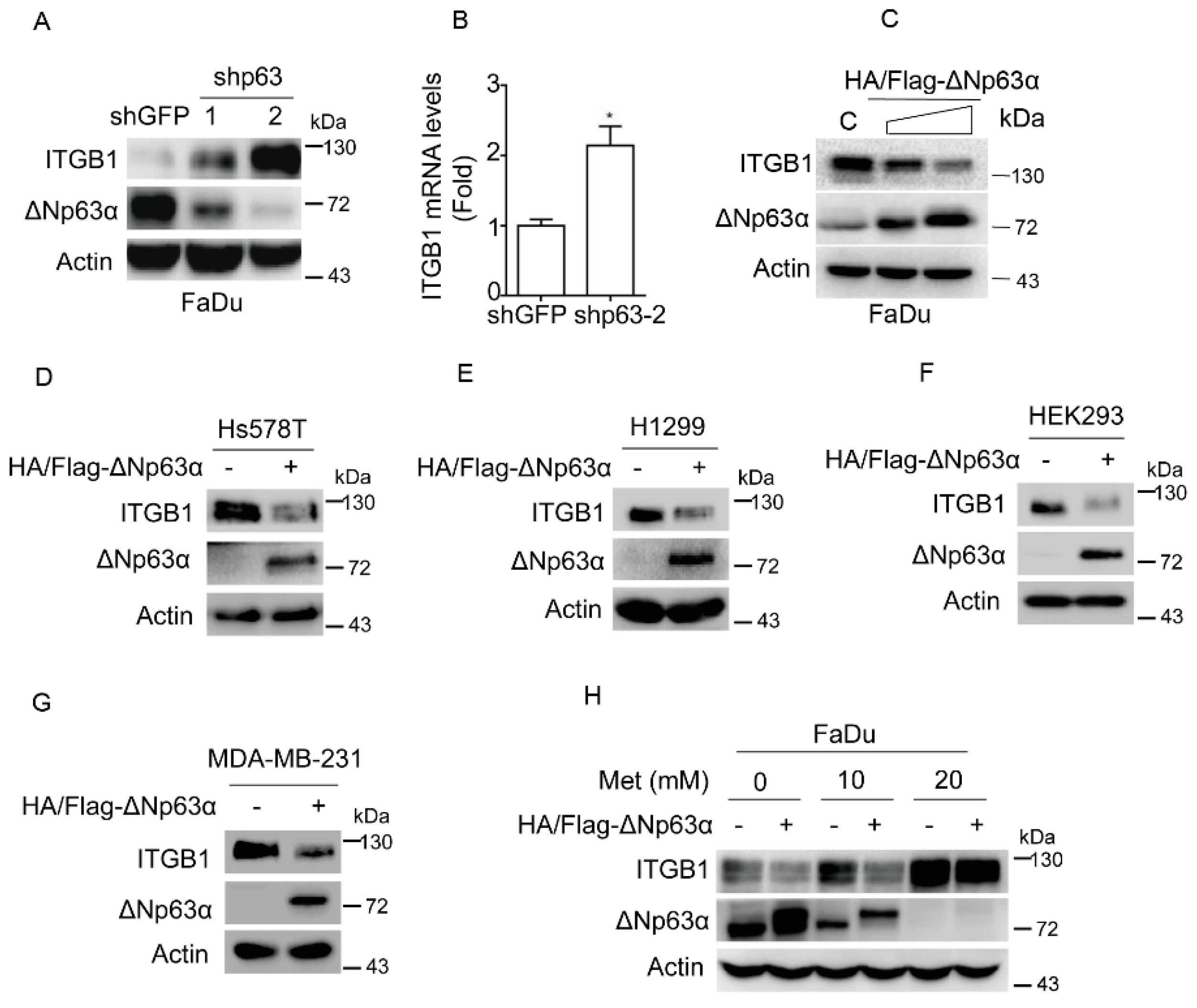
In the original publication [1], there were two mistakes in Figure 2 and Figure 3 as published. 1. In Figure 2D, the image shown in panel shGFP−24 h is incorrect; 2. In Figure 3C, the western blot images are incorrect. The corrected Figure 2 and Figure 3 appear below. The authors state that the scientific conclusions are unaffected. This correction was approved by the Academic Editor. The original publication has also been updated.

Reference

  1. An, T.; Zhang, Z.; Li, Y.; Yi, J.; Zhang, W.; Chen, D.; Ao, J.; Xiao, Z.-X.; Yi, Y. Integrin β1-Mediated Cell–Cell Adhesion Augments Metformin-Induced Anoikis. Int. J. Mol. Sci. 2019, 20, 1161. [Google Scholar] [CrossRef] [PubMed]
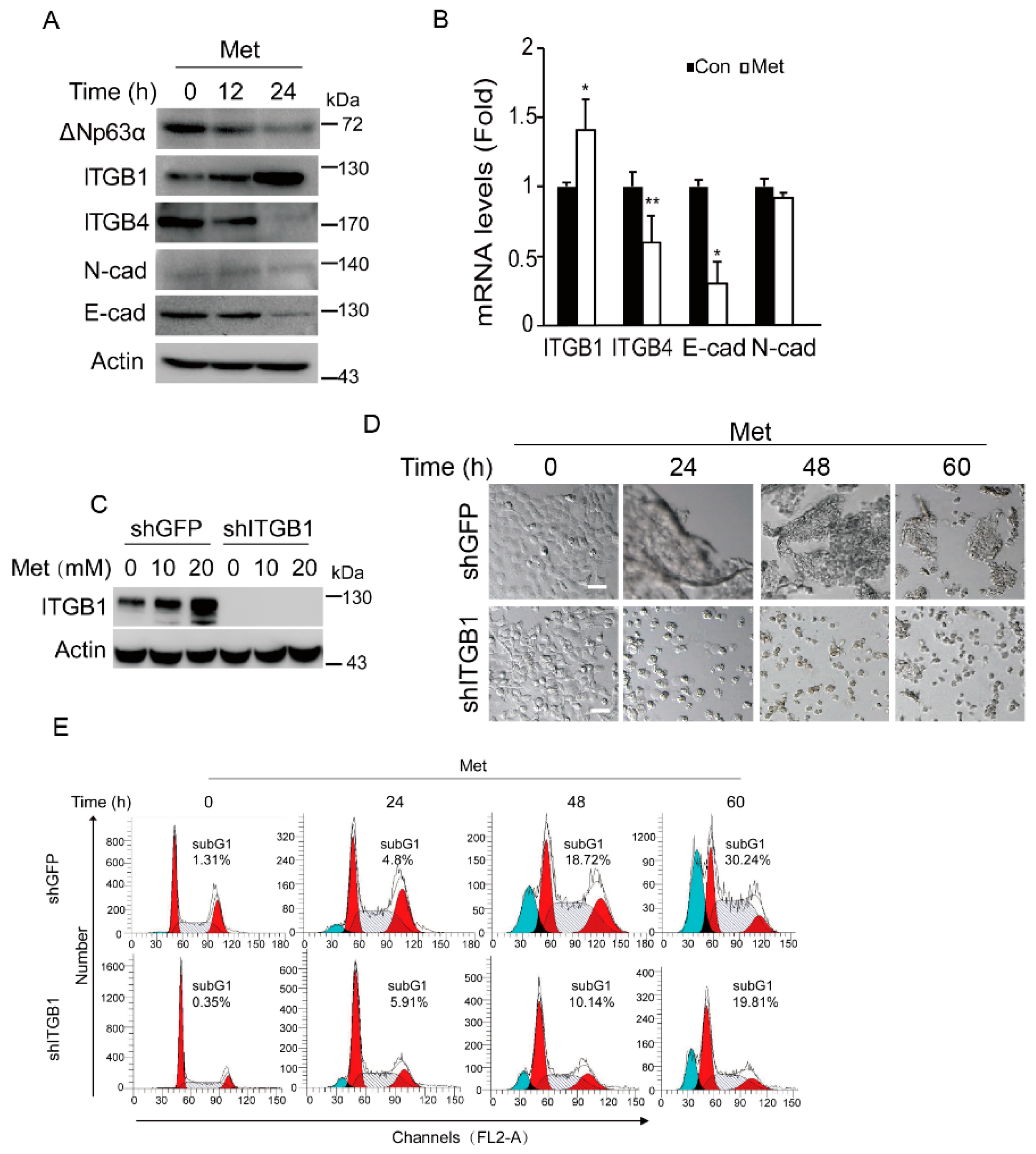
Figure 2. Metformin induces expression of integrin β1 resulting in cell aggregation and promoting anoikis. (A,B) FaDu cells were treated with metformin (20 mM, and thereafter) for an indicated time (0, 12, or 24 h) in DMEM-LG. Cells were subjected to western blot analyses (A) or subjected to Q-PCR analyses (cells were collected at 18 h) (B). Statistical analyses were based on Student’s t-test. Data were presented as means ± s.d. **. p < 0.05; *, p < 0.05. (C) FaDu cells stably expressing shITGB1 or shGFP were treated with metformin (0, 10, or 20 mM) in DMEM-LG for 24 h. Cells were subjected to western blot analyses. (D,E) FaDu cells stably expressing shITGB1 or shGFP were treated with metformin in DMEM-LG for an indicated time (0, 24, 48, or 60 h). Representative images were shown (D) and cells were subjected to FACS analyses (E, Red indicates G1 and G2 phases, blue marks the subG1 population (apoptotic cells), and the slash line denotes the S phase). Scale bars = 50 µm. For western blotting, all PVDF membranes derived from the transfers were cut at appropriate areas and reacted with an indicated primary antibody.
Ijms 26 10014 g002
Figure 3. Metformin suppresses expression of ΔNp63α, resulting in upregulation of integrin β1. (A,B) FaDu cells stably expressing shp63-1, shp63-2 or shGFP were subjected to western blot analyses (A) or subjected to Q-PCR analyses (B). Data are presented as means ± s.d. *, p < 0.05. (C) FaDu cells stably overexpressing HA/Flag-ΔNp63α or vector were subjected to western blot analyses, with sample processing controls run on separate gels. (DG) Ectopic expression of HA/Flag-ΔNp63α in Hs578T (D), H1299 (E), HEK293 (F), and MDA-MB-231 (G) cells were subjected to western blot analyses. (H) FaDu cells stably overexpressing HA/Flag-ΔNp63α or vector were treated with metformin (0, 10, or 20 mM) for 24 h in DMEM-LG. Cells were subjected to western blot analyses. All western blot membranes were cut at appropriate areas and reacted with an indicated primary antibody.
Ijms 26 10014 g003
Disclaimer/Publisher’s Note: The statements, opinions and data contained in all publications are solely those of the individual author(s) and contributor(s) and not of MDPI and/or the editor(s). MDPI and/or the editor(s) disclaim responsibility for any injury to people or property resulting from any ideas, methods, instructions or products referred to in the content.