Review

YKL-40 in Virus-Associated Liver Disease: A Translational Biomarker Linking Fibrosis, Hepatocarcinogenesis, and Liver Transplantation

1
Solid Organ Transplant Unit, Department of Anesthesiology, Intensive Care and Pain Medicine, Clinical Hospital Merkur, Zajceva 19, 10000 Zagreb, Croatia
2
Immediate Medical Care Unit, Saint James Hospital, SLM-1030 Sliema, Malta
3
Department of Medical Biochemistry and Hematology, Faculty of Pharmacy and Biochemistry, A. Kovacica 1, 10000 Zagreb, Croatia
4
Department of Laboratory Diagnostics, University Hospital Centre Zagreb, Kispaticeva 12, 10000 Zagreb, Croatia
5
Solid Organ Transplant Unit, Department of Surgery, Clinical Hospital Merkur, Zajceva 19, 10000 Zagreb, Croatia
6
Solid Organ Transplant Unit, Department of Gastroenterology, Clinical Hospital Merkur, Zajceva 19, 10000 Zagreb, Croatia
7
School of Medicine, University of Mostar, Zrinskog Frankopana 34, 88000 Mostar, Bosnia and Herzegovina
*
Author to whom correspondence should be addressed.
Int. J. Mol. Sci. 2025, 26(19), 9584; https://doi.org/10.3390/ijms26199584
Submission received: 27 August 2025 / Revised: 26 September 2025 / Accepted: 30 September 2025 / Published: 1 October 2025

Abstract

Virus-associated hepatocellular carcinoma (HCC) remains a major global health burden despite effective antiviral therapies. Chronic infection with hepatitis B (HBV), hepatitis C (HCV), and hepatitis D (HDV) promotes malignant transformation through overlapping pathways of fibrosis, immune dysregulation, and microenvironmental remodeling. YKL-40, a glycoprotein secreted by hepatic stellate cells, hepatocytes under stress, macrophages, and endothelial cells, has emerged as a marker that reflects stromal activation rather than direct hepatocyte injury. Its expression is reinforced by profibrotic and angiogenic circuits, and circulating concentrations correlate with advanced fibrosis, residual risk after viral suppression, and oncologic outcomes. This review synthesizes current evidence on YKL-40 across HBV, HCV, and HDV cohorts, with emphasis on its role in bridging molecular mechanisms to clinical applications. We examine its utility in non-invasive fibrosis assessment, longitudinal monitoring after antiviral therapy, and prognostic modeling in HCC. Particular attention is given to its potential in the liver transplant pathway, where YKL-40 may refine eligibility beyond morphology, inform bridging therapy response, and predict post-transplant recurrence or graft fibrosis. Remaining challenges include its lack of disease specificity, assay variability, and limited multicenter validation. Future integration of YKL-40 into multimarker, algorithm-based frameworks could enable risk-adaptive strategies that align surveillance and transplant decisions with the evolving biology of virus-associated liver disease.

Graphical Abstract

1. Introduction

Hepatocellular carcinoma (HCC) is the most common form of primary liver cancer and accounts for a substantial proportion of global cancer mortality [1]. Many cases develop in the context of chronic viral hepatitis, particularly from persistent infection with hepatitis B virus (HBV), hepatitis C virus (HCV), or hepatitis D virus (HDV) [2,3]. Antiviral strategies such as nucleos(t)ide analogs for HBV and direct-acting antivirals (DAAs) for HCV have significantly improved therapeutic options. However, important challenges remain, with persistent disparities in diagnosis and screening, limited access to antiviral treatment in many regions, and the continuing risk of virus-associated HCC even after viral suppression or clearance [4,5].
The progression from chronic viral hepatitis to HCC arises through cumulative hepatocellular injury, with activation of hepatic stellate cells (HSCs) driving extracellular matrix (ECM) deposition and fibrosis, while chronic inflammation and dysregulated regenerative signaling promote oncogenic transformation [6,7].
Current serum biomarkers for HCC surveillance, including alpha-fetoprotein (AFP) and des-gamma-carboxy prothrombin (DCP), have limited diagnostic performance, particularly in early-stage disease or in tumors lacking typical secretory phenotypes [8]. In virus-associated disease, where virologic control reduces inflammatory activity but does not abolish carcinogenic risk, biologically informative biomarkers are needed to refine risk-targeted surveillance, enable earlier detection and response assessment (including molecular residual disease), and complement morphologic criteria along the liver transplantation (LT) pathway (candidate selection, bridging/downstaging, post-LT surveillance). Consequently, current work spans complementary biomarker classes, such as tumor-derived proteins (e.g., glypican-3 and osteopontin), liquid biopsy analytes (circulating tumor DNA and microRNAs, including extracellular vesicle cargo), and extracellular vesicles themselves, which probe oncoprotein expression, genomic/epigenetic alterations, and vesicle-mediated stromal-immune crosstalk [9,10,11,12,13]. An overview of conventional and emerging biomarkers across the viral hepatitis-HCC-LT continuum is summarized in Table 1.
YKL-40 (chitinase-3-like protein 1, CHI3L1) is a glycoprotein produced by activated HSCs, macrophages, endothelial cells, and stressed hepatocytes. By engaging ECM turnover and inflammatory-angiogenic signaling, it serves as a readout of stromal activation central to virus-associated carcinogenesis [14]. Circulating concentrations correlate with fibrosis stage, aggressive tumor biology, and adverse outcomes in chronic liver disease and HCC, thereby complementing tumor-derived markers and imaging [15,16]. Accordingly, a structured evaluation of YKL-40 across viral etiologies and along the LT pathway is warranted.
Despite these associations, the virus-specific contributions of YKL-40 and its translational potential in guiding LT remain incompletely understood. In this narrative review, we critically synthesize current evidence on YKL-40 in HBV-, HCV-, and HDV-associated disease, emphasizing its mechanistic basis, clinical applications, and implications for biologically adaptive decision-making across the fibrosis-carcinogenesis-transplant continuum.

2. Mechanistic and Clinical Relevance of YKL-40 in Virus-Associated Fibrosis and Hepatocarcinogenesis

2.1. HBV: Viral Persistence, Fibrogenic Signaling, and Oncogenic Remodeling

The persistence of HBV is maintained through covalently closed circular DNA (cccDNA), a stable episomal template that remains transcriptionally active despite antiviral suppression [17]. Continuous viral gene expression creates a state of chronic immune disturbance, with exhausted T cells, increased secretion of interleukin-6 (IL-6) and tumor necrosis factor-alpha (TNF-α), and expansion of regulatory immune subsets that blunt antiviral cytotoxicity [18,19,20]. These signals converge on HSCs, where profibrogenic mediators such as transforming growth factor-beta (TGF-β), connective tissue growth factor (CTGF), and hepatocyte-derived exosomes initiate ECM deposition and architectural remodeling [21,22].
Sustained stimulation gradually reprograms stellate cells into a fibrogenic state characterized by enhanced collagen synthesis and reduced matrix degradation, supported by contractile and proliferative changes that consolidate their myofibroblastic phenotype [23,24,25,26,27]. Scar tissue becomes further stabilized through collagen cross linking by lysyl oxidase-like 2 (LOXL2), which limits the potential for reversal [28]. Insights from single-cell transcriptomic studies add nuance, showing that activation is not uniform across the liver but varies by zone, with periportal and pericentral stellate subsets contributing in distinct ways to the fibrotic landscape [29,30]. This heterogeneity has clinical significance because fibrosis may progress silently despite normal alanine aminotransferase (ALT) levels [31]. In this context, circulating YKL-40 has emerged as an informative biomarker, with elevated levels reported in HBV cohorts with advanced fibrosis and potential utility for staging and disease monitoring [32,33].
As fibrosis advances, the altered microenvironment facilitates oncogenic transformation. Epigenetic changes such as aberrant DNA methylation and dysregulated noncoding RNAs weaken tumor-suppressive programs, while HBV DNA integration activates oncogenes including telomerase reverse transcriptase (TERT) and cyclin E1 (CCNE1), promoting clonal expansion of genetically unstable hepatocytes [34,35,36,37]. These processes destabilize genomic integrity and create conditions permissive for malignant evolution [38].
HBV also promotes cancer through direct molecular drivers. The hepatitis B virus X protein (HBx) is central to this process. It impairs DNA repair, disrupts cell-cycle checkpoints, and alters chromosomal organization [39,40]. HBx can engage chromatin remodeling machinery such as SWI/SNF and displace Polycomb repressors, opening proto-oncogenic loci to transcriptional activation [41,42]. In parallel, interferon signaling and antigen presentation are dampened, and apoptotic thresholds are shifted via mitochondrial and death-receptor pathways, weakening immune surveillance [43,44]. Although no direct studies have linked HBx activity to YKL-40 expression, the fibrogenic and inflammatory environment fostered by HBV provides a biologically coherent basis for its up-regulation, emphasizing the need for studies that clarify its virus-specific contributions and translational potential. The key processes of viral persistence, fibrogenic signaling, and oncogenic remodeling in HBV are summarized in Figure 1.

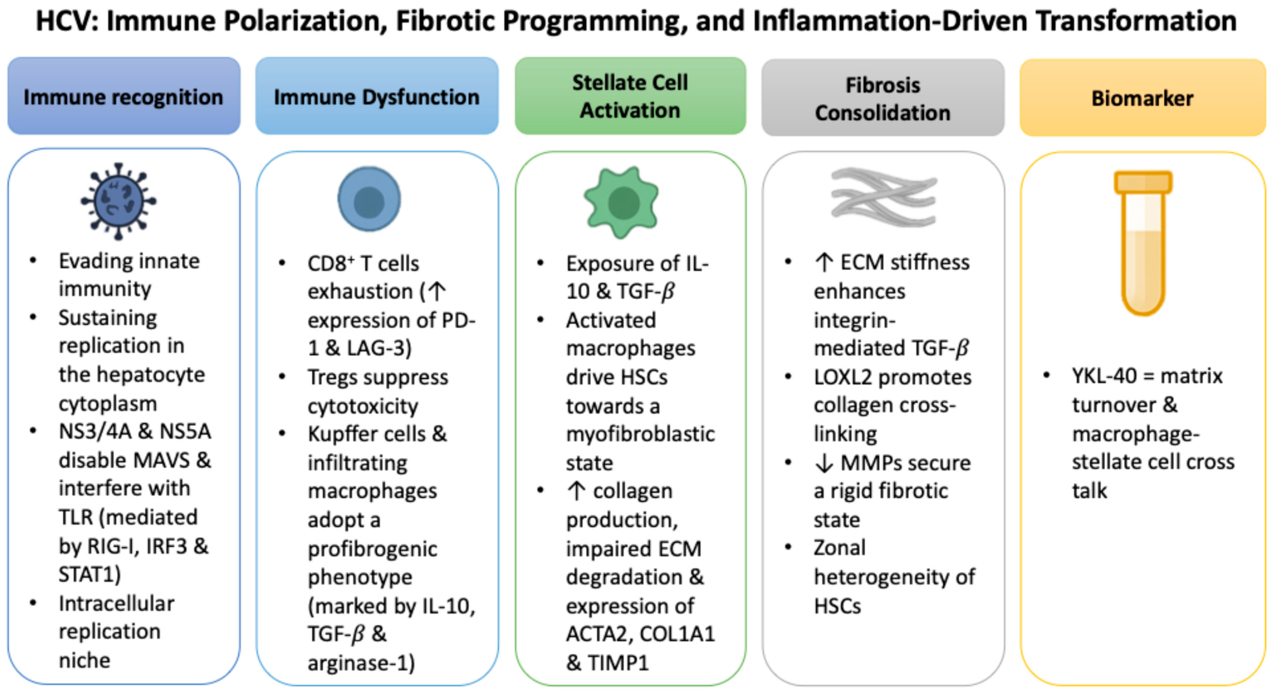
2.2. HCV: Immune Polarization, Fibrotic Programming, and Inflammation-Driven Transformation

Unlike HBV, which achieves persistence through cccDNA reservoirs and integration into the host genome, HCV maintains chronic infection by evading innate immune recognition while sustaining replication in the hepatocyte cytoplasm [45,46]. Viral proteins such as NS3/4A and NS5A disable mitochondrial antiviral signaling protein (MAVS) and interfere with toll-like receptor (TLR)-dependent interferon cascades, weakening antiviral defenses mediated by retinoic acid-inducible gene I (RIG-I), interferon regulatory factor 3 (IRF3), and signal transducer and activator of transcription 1 (STAT1) [47,48].
Beyond immune evasion, HCV viral proteins actively drive profibrogenic signaling. Experimental studies have shown that NS5A and core proteins induce oxidative stress through NADPH oxidases and mitochondrial pathways, while simultaneously activating NF-κB and STAT3 signaling [49]. These pathways serve as established transcriptional activators of YKL-40, providing a mechanistic link between HCV protein activity and YKL-40 upregulation [49].
The YKL-40 promoter contains functional response elements for both NF-κB and STAT3, rendering it highly responsive to these signaling cascades [50]. HCV NS3 and NS5A proteins sustain STAT3 activation through prolonged phosphorylation, directly enhancing YKL-40 transcription and linking viral replication to persistent stromal activation and fibrogenesis [50].
Persistent viral replication and protein activity create an environment of chronic, low-grade inflammation rather than direct cytopathic damage. Virus-specific CD8+ T cells become progressively exhausted, with elevated expression of inhibitory receptors such as programmed cell death protein 1 (PD-1) and lymphocyte activation gene 3 (LAG-3) [51]. Regulatory T cells (Tregs) expand and further suppress cytotoxic responses, while Kupffer cells and infiltrating macrophages polarize toward a profibrogenic phenotype marked by IL-10, TGF-β, and arginase-1 [52].
These immune changes converge on HSCs, initiating ECM deposition and stromal remodeling. Reactive oxygen species (ROS), pro-inflammatory cytokines such as IL-1β, and chemokines like CXCL10 reinforce this fibrogenic circuit, creating a self-perpetuating inflammatory loop [53,54]. In this milieu, serum YKL-40 levels are consistently higher in HCV patients with advanced fibrosis, reflecting both matrix turnover and the intensity of macrophage-stellate cell crosstalk [55].
Macrophage polarization further stabilizes this fibrogenic axis. Under sustained IL-10 and TGF-β exposure, alternatively activated macrophages drive HSCs toward a myofibroblastic state characterized by increased collagen production, impaired ECM degradation, and expression of markers such as ACTA2, COL1A1, and TIMP1 [56,57,58]. Kupffer cells and infiltrating macrophages amplify this circuit through secretion of CCL2 and osteopontin, which recruit additional inflammatory and profibrogenic cells into perisinusoidal regions [59,60].
As fibrosis matures, structural cues reinforce its persistence. Increasing ECM stiffness enhances integrin-mediated activation of latent TGF-β, locking HSCs into a self-sustaining fibrogenic program [61]. This process is amplified by lysyl oxidase-like 2 (LOXL2), which promotes collagen crosslinking [62], while downregulation of matrix metalloproteinases (MMPs) limits turnover and enforces rigidity [63]. Insights from single-cell transcriptomics reveal zone-specific heterogeneity of HSC activation, with central vein-associated subsets showing dominant collagen production and distinct expression of ACTA2, remodeling enzymes, and chemokine receptors [64]. Endothelial signaling and oxygen gradients further refine these spatial responses, integrating immune, vascular, and metabolic cues into the evolving fibrotic architecture [65]. Figure 2 summarizes the sequence of immune dysfunction, fibrotic remodeling, and YKL-40 induction in HCV-associated liver disease.

2.3. HDV: Intensified Immunopathology and Fibrotic Escalation

HDV depends on the hepatitis B surface antigen (HBsAg) for assembly and propagation, making HBV co- or superinfection a prerequisite for its persistence [66]. Clinically, HBV/HDV coinfection follows a more aggressive course than HBV alone, with accelerated progression to cirrhosis, earlier decompensation, and higher risk of HCC [67].
The large hepatitis D antigen (L-HDAg) is a central driver of this pathogenic phenotype [68]. Beyond its direct viral functions, L-HDAg promotes an inflammatory and profibrogenic milieu within the liver. Kupffer cells and infiltrating monocytes adopt activated states and release cytokines such as TNF-α and IL-1β, which sustain stellate-cell activity and drive collagen I accumulation [68]. Together these mechanisms provide a foundation for the rapid fibrotic trajectory observed in HBV/HDV coinfection.
Additional studies show that L-HDAg also promotes oxidative stress through nicotinamide adenine dinucleotide phosphate (NADPH) oxidase and activates STAT3 and nuclear factor kappa-light-chain-enhancer of activated B cells (NF-κB) signaling [69]. It further disrupts endoplasmic reticulum homeostasis, triggers unfolded-protein-response pathways, and perturbs proteostasis, thereby maintaining a chronically inflamed hepatic niche reinforced by type I interferon activity [69]. These signals accelerate stellate-cell activation and matrix deposition, deepening the fibrotic response.
Clinical evidence supports these mechanistic observations, demonstrating that fibrosis progresses more rapidly in HBV/HDV coinfection compared with HBV monoinfection, in line with the heightened inflammatory burden [70]. Adaptive immunity is also reshaped. Virus-specific CD8+ T cells gradually acquire features of exhaustion, regulatory pathways expand, and cytotoxic lymphocytes lose functional capacity. This imbalance limits viral control while amplifying hepatocellular injury, hastening cirrhosis and elevating oncogenic risk compared with HBV alone [71,72].
Direct evaluation of YKL-40 in HDV infection is not yet available. Even so, the profibrotic and inflammatory profile of HBV/HDV coinfection creates a biologically plausible setting for its up-regulation. Evidence from HBV and HCV cohorts, where higher YKL-40 levels correlate with advanced fibrosis and adverse outcomes, provides a strong rationale for testing whether similar associations are present in HDV. Addressing this gap will be important for determining whether YKL-40 can serve as a clinically useful biomarker in this uniquely aggressive viral context. Future research could employ multi-omics approaches, such as single-cell transcriptomics of HDV-infected hepatocytes or stellate cells, to identify cellular sources and regulatory networks driving YKL-40 expression. In parallel, clinical cohort studies with paired serum and liver biopsy data could define the diagnostic and prognostic performance of YKL-40 in HBV/HDV coinfection, establishing its role as a potential biomarker across disease stages. Figure 3 provides a visual summary of the intensified immunopathology and rapid fibrotic escalation characteristic of HBV/HDV coinfection, illustrating how L-HDAg-driven immune dysregulation, oxidative stress, and stellate-cell activation converge on YKL-40 induction and heightened oncogenic risk.

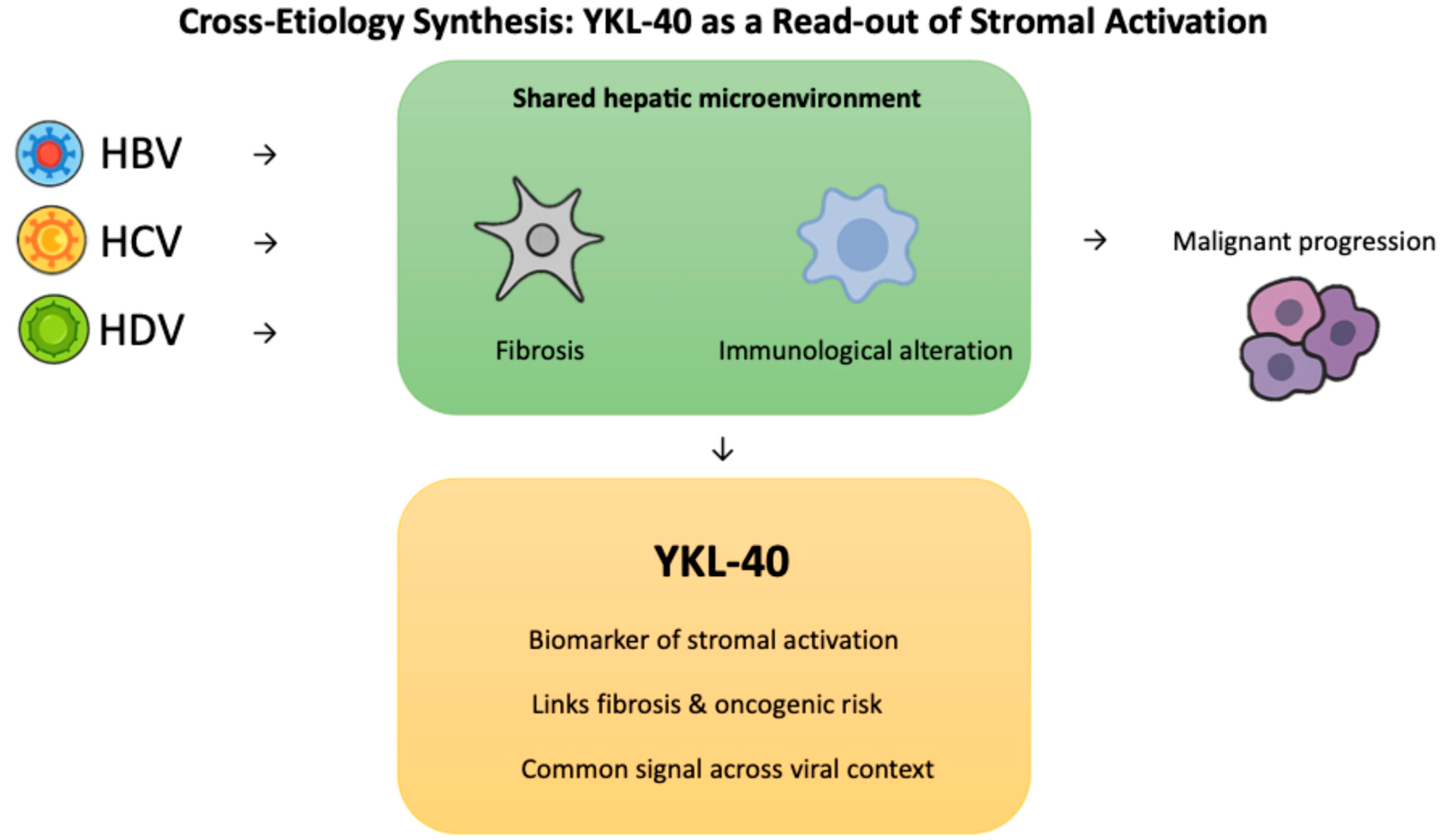
2.4. Cross-Etiology Synthesis: YKL-40 as a Read-Out of Stromal Activation

HBV, HCV, and HDV maintain persistence through different virological strategies, yet over time they drive the liver toward a similar endpoint in which the hepatic microenvironment becomes fibrotic and immunologically altered, creating conditions that favor malignant progression [73,74,75]. Within this shared landscape, YKL-40 reflects stromal activation that connects fibrosis with oncogenic risk, providing a common signal across viral contexts [76].
The convergent and virus-specific pathways underlying YKL-40 induction are depicted in Figure 4 and summarized in Table 2, highlighting how HBV, HCV, and HDV engage distinct viral drivers that ultimately activate overlapping profibrotic and inflammatory cascades.

3. YKL-40 as a Translational Biomarker in Virus-Associated Liver Disease

3.1. Molecular Basis of YKL-40 Expression

YKL-40 stands out among candidate biomarkers because it is produced by different cell populations that act together in the chronically injured liver. This explains why it is consistently measurable in both tissue and circulation and why its signal is not limited to hepatocyte injury alone [77,78].
The strongest evidence for YKL-40 production comes from HSCs, which secrete it as they adopt a matrix-producing phenotype during fibrogenesis [79,80]. Hepatocytes also contribute when exposed to stress, particularly at the edges of fibrous septa, where staining is most pronounced in HDV, followed by HCV and HBV [79]. Macrophages add further expression during inflammatory activation [77,78], while endothelial and epithelial cells increase secretion under hypoxia or pro-angiogenic conditions [77,80]. These diverse sources position YKL-40 as a multicellular stress response that reflects how parenchymal, stromal, and vascular compartments adapt under chronic injury.
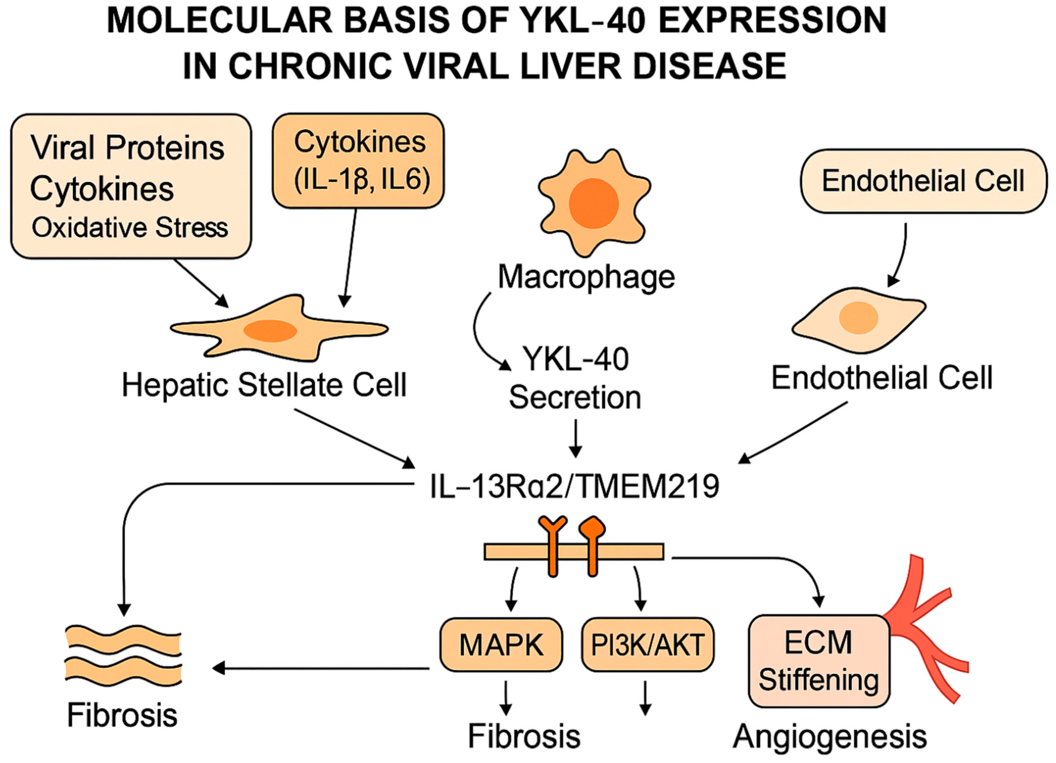
Its induction is reinforced by inflammatory and stress-responsive circuits. IL-1β and IL-6 activate STAT3 and NF-κB, embedding YKL-40 in transcriptional programs that remain active long after acute injury subsides [50]. Oxidative stress, matrix stiffening, and hypoxia further sustain expression, which explains why serum concentrations remain elevated in advanced fibrosis even when ALT values normalize [81].
Secreted YKL-40 then links these upstream signals to structural remodeling. Binding to interleukin-13 receptor subunit alpha-2 (IL-13Rα2) together with transmembrane protein 219 activates extracellular signal-regulated kinase (ERK), protein kinase B (AKT), and Wingless-related integration site/β-catenin (Wnt/β-catenin) signaling [82]. In addition to being a marker of fibrosis, YKL-40 actively participates in a positive feedback loop that amplifies fibrogenesis. Upon secretion, YKL-40 binds to IL-13Rα2 in complex with TMEM219, triggering downstream MAPK and PI3K/AKT signaling cascades. These pathways promote HSC proliferation and collagen synthesis, reinforcing ECM deposition and stiffening. Increasing matrix stiffness in turn sustains stellate cell activation and further upregulates YKL-40 expression, creating a self-perpetuating cycle of fibrosis progression [82,83]. Figure 5 illustrates these interconnected mechanisms, showing how viral proteins, cytokines, and oxidative stress activate multiple liver cell populations to secrete YKL-40, which in turn drives fibrosis, angiogenesis, and ECM stiffening through MAPK and PI3K/AKT signaling pathways.
This mechanistic loop explains why circulating YKL-40 levels closely track disease severity and remain elevated even when upstream injury markers, such as ALT, return to normal. Interactions with syndecan-1 and integrins αvβ3 and αvβ5 amplify motility and ECM turnover in endothelial and stromal cells [83]. These overlapping receptor pathways align with the consistent observation that YKL-40 is concentrated at fibrotic and angiogenic interfaces in diseased tissue [79,84].
Functional studies illustrate the consequences of this biology. In stellate cells, YKL-40 stimulates proliferation and collagen synthesis, accelerating matrix accumulation [78,79]. In endothelial models, it enhances tube formation and vascular endothelial growth factor (VEGF) expression, supporting the angiogenic remodeling characteristic of cirrhosis [83]. Neutralization experiments demonstrate that blocking YKL-40 can reduce tumor vascularization, confirming its role at the tumor-stroma interface [83]. In immune compartments, YKL-40 promotes macrophage recruitment and polarization toward an alternatively activated (M2-like) phenotype, reshaping cytokine balance and matrix metalloproteinase activity in ways that favor tolerance and invasive growth [76,85].
Virus-specific data strengthen these mechanistic insights. In HCV infection, YKL-40 supports hepatocyte viability and profibrogenic cytokine release, and there is evidence that it may even facilitate viral replication [81]. In HBV and HCV cohorts, circulating levels rise in step with fibrosis stage and remain elevated when hepatocyte injury markers decline [77,81]. Broader reviews of HSC activation and chitinase-like proteins in cancer highlight YKL-40 as a central driver of stromal remodeling that connects fibrotic progression with oncogenic potential [79,84].
Taken together, these findings show that YKL-40 is not just a bystander of tissue damage but a multicellular stress signal sustained by cytokines and environmental inputs. Its effects on fibrogenesis, angiogenesis, and immune adaptation explain why it is stable in circulation and provide the rationale for testing its translational value across HBV, HCV, and HDV populations and along the transplant pathway.

3.2. Clinical and Translational Applications of YKL-40

Evidence now quantifies how much information YKL-40 can add to day-to-day decisions. A recent meta-analysis that pooled seventeen liver fibrosis studies reported areas under the receiver operating characteristic curve (AUC) of 0.91 for advanced fibrosis and 0.87 for severe fibrosis with corresponding sensitivities near 0.80 and specificities near 0.85 across cohorts, which places YKL-40 in a performance range suitable for clinical triage rather than exploratory use [86]. The question then becomes where such a signal changes management. Histology-driven work in chronic hepatitis B shows that a considerable share of patients who meet biochemical definitions of normal ALT still carry significant necroinflammation or stage F2 or higher. In these cohorts the proportion with significant histological disease remains near a quarter to a third even when stricter upper limits of normal are applied, which creates a clear use case for a marker that can prioritize elastography or biopsy when standard biochemistry is quiet [87]. A second study focused on HBeAg-positive hepatitis B with normal ALT reached the same practical conclusion by detailing how often clinically silent activity appears on biopsy, which again supports targeted non-invasive testing rather than routine deferral of staging [88].
A move from static to longitudinal use further clarifies how to integrate YKL-40. In chronic hepatitis B, models that incorporate repeated measurements classify cirrhosis with an AUC of 0.939 and capture change over follow up, which is the type of performance that supports interval testing in clinics that already run serial algorithms for inactive carriers and for treated patients [88]. In HBeAg-negative hepatitis B, single-timepoint YKL-40 discriminates minimal from significant fibrosis with an AUC of 0.818 and with sensitivity and specificity around eighty and seventy percent. It also aligns with fibrosis indices such as FIB-4 and the gamma-glutamyl transpeptidase to platelet ratio, which allows simple cross-checks during routine visits without adding imaging at every timepoint [89]. At the level of pooled evidence, a meta-analysis across viral hepatitis datasets reported overall sensitivity and specificity near seventy four and seventy six percent, which provides a benchmark for centers that are considering adoption and want to compare their own operating points against external data [90].
Therapy studies show when the marker should be rechecked. In HCV infection, YKL-40 correlates with liver stiffness at baseline and detects cirrhosis with an AUC of 0.939. After direct-acting antivirals it falls by about one fifth within months, yet levels in many patients remain above those of healthy comparators. Untreated cirrhotics trend upward over the same horizon, which maps the marker to real trajectories rather than to a single post-treatment snapshot and supports planned retesting after cure in patients who remain at risk [90]. Host factors also shape downstream outcomes. In patients who cleared hepatitis C with direct-acting antivirals, common variants in the YKL-40 locus and an intergenic site identified groups with higher HCC risk after virological cure. The risk was greatest when more than one locus carried the at-risk genotype, which points to a host component that can be considered alongside serum measurement when long-term surveillance is planned [91].
Oncologic endpoints define where YKL-40 influences decisions beyond fibrosis staging. A meta-analysis of forty one cohorts across solid tumors showed that elevated serum YKL-40 associated with poorer overall survival with a pooled hazard ratio of 1.44, which sets an external expectation for effect size when the marker is used to refine prognosis [92]. HCC specific data mirror that pattern. After curative hepatectomy, higher preoperative YKL-40 independently predicted overall survival with adjusted hazard ratios in the range of 1.4 to 1.5, and a prognostic nomogram that added YKL-40 to tumor number, tumor size, the neutrophil to lymphocyte ratio, the international normalized ratio, and alpha-fetoprotein achieved areas under the curve near 0.75 in training and validation. A practical cut-point close to 199 ng per milliliter separated survival curves, which offers a starting threshold for programs that are designing preoperative risk conferences and postoperative follow up schedules [93].
Implementation details determine how reliable the readouts will be once YKL-40 is brought into routine use. Population data indicate that circulating levels rise with age, and that up to about one quarter of interindividual variation can be explained by common genetic polymorphisms in the YKL-40 gene. Laboratory practice also matters because most measurements use immunoassay formats with different reportable ranges and different sample handling requirements. These facts argue for age aware interpretation, for attention to serial change rather than single values, and for harmonized pre-analytical routines so that trajectories reflect biology rather than processing noise [94]. Broader oncology experience reaches a similar conclusion about standardization and about context. YKL-40 is elevated across many inflammatory and neoplastic states. It is therefore most useful when applied in defined hepatology pathways that already incorporate imaging and clinical scores and when its thresholds and sampling intervals are prespecified by etiology and treatment state [95].
These clinical studies show that YKL-40 has progressed from a descriptive marker of stromal activity to a tool with operational relevance. It uncovers fibrosis that escapes detection by routine biochemistry, it remains informative after antiviral cure when residual scarring drives long-term risk, and it strengthens prognostic models in HCC, particularly in patients without conventional tumor marker elevation. At the same time, its performance is influenced by age, genetic variation, and assay practice, underlining the importance of standardized protocols for future adoption. The convergence of these findings highlights why YKL-40 is now being considered not only for staging and surveillance but also for peri-transplant assessment, where the balance between fibrotic burden, oncologic risk, and immune adaptation becomes critical. To provide a comprehensive overview of its diagnostic performance, Table 3 summarizes key studies reporting receiver operating characteristic (ROC) values, optimal cut-off thresholds, and corresponding sensitivity and specificity of YKL-40 in HBV and HCV cohorts.

4. Pre- and Post-Transplant Applications of YKL-40 in HCC: From Eligibility to Risk Stratification

4.1. Informing Transplant Eligibility Beyond Morphology

Selection for LT in HCC has traditionally been determined by morphologic frameworks such as the Milan and University of California, San Francisco (UCSF) criteria, which define limits of tumor size and number [96]. These thresholds improved outcomes by restricting transplantation to patients with favorable imaging features, yet they cannot capture tumor biology or the state of the hepatic microenvironment, both of which drive recurrence risk after transplantation [97]. More recent allocation tools, including Metroticket 2.0 and AFP-based models, have added serological signals to extend predictive accuracy, but they remain limited in patients with low or discordant AFP expression [98,99].
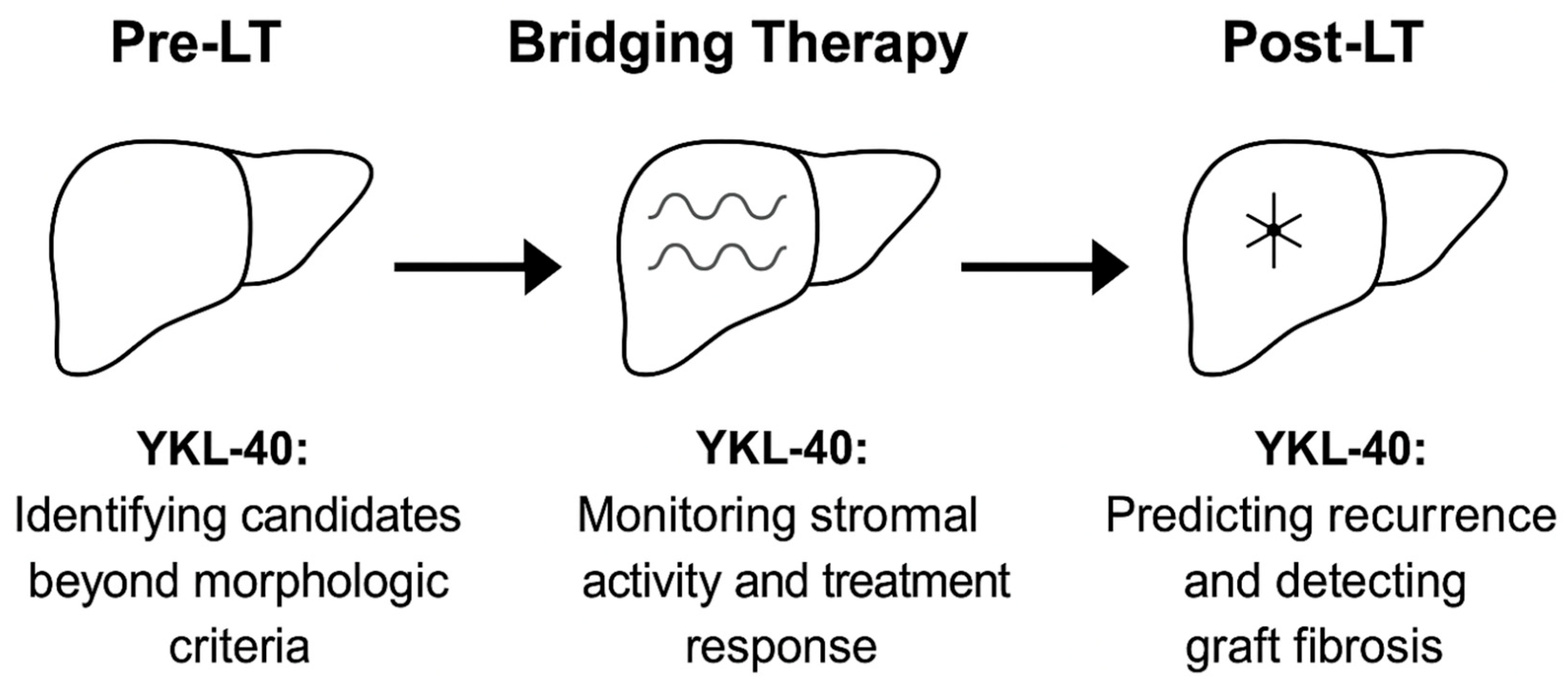
YKL-40 provides a way to interrogate the biological state of the host rather than the morphology of the tumor [100]. In practice, this can sharpen decision-making at two critical junctures. First, in candidates who appear to meet morphologic thresholds, high YKL-40 may uncover a stromal environment already primed for recurrence, suggesting that eligibility should be reconsidered or that intensified bridging therapy is warranted before listing. Second, in patients undergoing downstaging, declining YKL-40 values can provide reassurance that the fibrotic and angiogenic drive has slowed, strengthening the case to proceed to transplantation. Data from TACE cohorts reinforce these scenarios, where serum YKL-40 predicted overall survival independently of AFP and retained value in AFP-negative phenotypes, which are precisely the cases where current allocation models are least informative [101]. Figure 6 provides a visual summary of how YKL-40 contributes to decision-making at different stages of the LT pathway, from pre-transplant eligibility to post-transplant surveillance.

4.2. Refining Pre-Transplant Risk Stratification

Once eligibility is confirmed, the next challenge is to refine biological risk before committing scarce donor organs [102,103]. Conventional stratification relies on imaging and response to bridging therapy, yet these assessments can miss the stromal activity that sustains recurrence beneath apparently stable radiology [104,105].
YKL-40 provides a complementary perspective by signaling microenvironmental activity that is otherwise invisible. Elevated levels correlate with larger tumor burden, vascular invasion, and reduced survival across HCC cohorts [106,107]. Trajectories of YKL-40 during bridging therapy add actionable value. Persistently high concentrations indicate that stromal activity continues and that recurrence risk is greater. Declining levels indicate that biological activity has slowed and provide stronger reassurance to proceed with transplantation.
The mechanistic basis for these associations has been described in experimental models, but their clinical implications can be summarized more directly. Table 4 illustrates how YKL-40 related pathways map onto outcomes that matter in the transplant setting, converting biological activity into markers of recurrence risk and treatment response.

4.3. Post-Transplant Recurrence Prediction and Integration into Biomarker Panels

Even with stringent selection criteria, 10–20% of LT recipients experience HCC recurrence, which emphasizes the limits of morphology-based models [108,109]. Recent studies now provide LT-specific evidence. In a large transplant cohort, elevated peri-transplant YKL-40 was linked with shorter overall and recurrence-free survival. The association was strongest in patients with myosteatosis, a systemic frailty phenotype that is invisible to conventional tumor markers [110]. These results identify YKL-40 as a clinically meaningful signal for peri-transplant risk stratification, particularly in patients who are AFP- or DCP-negative and therefore lack reliable tumor-derived markers [93].
The value of YKL-40 is not confined to recurrence alone. It also shows potential as a biomarker of graft fibrosis. In recipients with recurrent hepatitis C, serial measurements distinguished rapid from slow fibrosis progressors and tracked with histologic progression [111]. Genetic findings reinforce this plausibility. A functional promoter polymorphism in the YKL-40 gene was associated with higher circulating levels, more severe rejection, and faster fibrosis progression after transplantation [112]. These observations support a dual role for YKL-40 in the post-transplant setting, both as a predictor of oncologic recurrence and as a non-invasive marker of graft injury.
Implementation requires careful attention to context. YKL-40 is not disease specific, and elevations can reflect infection, rejection, or systemic inflammation, all of which are common in the early post-transplant course [94]. Laboratory factors also matter, since assay platforms vary in sensitivity and reproducibility, and recent high-dynamic-range immunoassays have highlighted challenges for cross-center comparison [113]. For the marker to be integrated into practice, multicenter validation is needed to confirm whether its addition to prognostic models such as RETREAT or MoRAL provides meaningful reclassification and supports risk-adapted surveillance strategies [114].

4.4. Future Directions

Future investigations will need to focus on translating YKL-40 from a descriptive biomarker into a clinically actionable tool across the spectrum of virus-associated liver disease. At earlier stages, longitudinal monitoring of YKL-40 offers a strategy to capture ongoing stromal remodeling and to assess fibrosis regression during antiviral or antifibrotic therapy. Large, prospective cohorts with standardized protocols are required to define biologically meaningful fluctuations, link them to histological and clinical outcomes, and establish its role in risk stratification and therapeutic planning.
As liver architecture becomes more disrupted, YKL-40 has the potential to signal transition toward decompensation before overt clinical manifestations arise. When incorporated into multivariable frameworks alongside established clinical variables, this biomarker could refine the prediction of complications such as variceal bleeding, ascites, and hepatic encephalopathy. Aligning surveillance and preventive interventions with molecular disease activity rather than static scores would enable timelier and more efficient use of resources.
Persistent stromal activation also contributes to tumor initiation, progression, and recurrence, even when viral replication is well controlled. Future efforts should explore integrating YKL-40 into composite biomarker signatures that merge stromal, immune, and tumor-derived signals with advanced imaging and circulating markers such as ctDNA and microRNAs. Such models could enhance early detection, recurrence prediction, and post-treatment monitoring, particularly for AFP-negative phenotypes and patients under surveillance following curative-intent therapies.
LT represents the final point of this continuum. Embedding YKL-40 within multidimensional predictive frameworks that combine biological, radiological, and clinical data would improve candidate selection, guide decisions on bridging therapy, and tailor post-transplant surveillance for recurrence and graft fibrosis. Harmonized multicenter registries are essential to establish dynamic thresholds, standardize assays, and validate performance across diverse patient populations.
Advances in computational science provide the tools needed to operationalize these concepts. Machine learning and artificial intelligence frameworks are well suited to model the nonlinear interactions between stromal activation, immune modulation, and tumor biology. By integrating real-time biomarker data with imaging, clinical variables, and tumor characteristics, these systems can generate continuously updated, individualized risk estimates. Such adaptive models move beyond static cut-offs to deliver risk-adapted care pathways, aligning surveillance intensity, bridging therapy, and adjuvant strategies with evolving tumor-host interactions. Through this integration, YKL-40 could progress from a descriptive read-out to a functional driver of precision hepatology and transplantation.
Therapeutic implications are also emerging as YKL-40 moves beyond its role as a biomarker. Experimental studies have shown that neutralizing YKL-40 with monoclonal antibodies can reduce angiogenesis, fibrosis, and tumor progression by interrupting IL-13Rα2-mediated signaling pathways [115,116]. In particular, the IL-13Rα2/TMEM219 complex has been identified as a critical mediator of YKL-40-driven MAPK and PI3K/AKT activation, linking this pathway to fibroblast and stellate cell activation [117]. Inhibition of this receptor complex could therefore prevent downstream profibrotic signaling, reducing HSC proliferation and collagen deposition. These strategies are conceptually similar to antifibrotic therapies targeting TGF-β or LOXL2 but uniquely focus on disrupting the feed-forward loop between YKL-40 and ECM remodeling. While no clinical trials have yet evaluated these approaches in viral hepatitis or LT, preclinical data provide a strong rationale for testing YKL-40 blockade as a novel therapeutic avenue to slow fibrosis progression, reduce HCC recurrence risk, and improve graft outcomes after transplantation.

5. Conclusions

The value of YKL-40 lies less in isolated performance metrics and more in its ability to signal a transition from morphology-driven assessment toward biologically adaptive models of care in liver transplantation. While current evidence shows consistent associations with fibrosis stage, tumor behavior, and graft outcomes, its adoption in clinical pathways remains limited. Progress will depend on robust validation in multicenter cohorts, harmonization of assay platforms, and clear definition of dynamic thresholds that can be interpreted across different disease stages and treatment states. Prospective integration of YKL-40 into transplant registries and biomarker-guided surveillance trials will be essential to determine whether it adds value beyond established scores and imaging. The next step is not to test YKL-40 in isolation but to embed it into algorithmic frameworks that combine stromal, tumor, and host signals. Such multidimensional approaches can support risk-adaptive strategies and bring precision medicine in transplantation closer to practice.

Author Contributions

J.P.S., D.L., D.R., and I.L. participated in the conceptualization of this manuscript; J.P.S., D.L., D.R., S.J., D.M., T.F.K., N.P., and I.L. performed the literature review and data analysis; J.P.S., D.L., D.R., and I.L. designed the manuscript’s original draft; S.J., D.M., T.F.K., N.P., L.K.B., I.A., I.B.K., Z.S., G.E., M.A., P.O.Z., and F.J. reviewed and edited the manuscript original draft; J.P.S., D.L., D.R., and I.L. performed manuscript supervision and project administration. All authors have read and agreed to the published version of the manuscript.

Funding

The APC was funded by Clinical Hospital Merkur.

Institutional Review Board Statement

Not applicable.

Informed Consent Statement

Not applicable.

Data Availability Statement

No new data were created or analyzed in this study. Data sharing is not applicable to this article.

Conflicts of Interest

The authors declare no conflict of interest.

Abbreviations

The following abbreviations are used in this manuscript:
ACTA2Alpha-smooth muscle actin
AFPAlpha-fetoprotein
AKTProtein kinase B
ALTAlanine aminotransferase
αvβ3Integrin alpha-V beta-3
αvβ5Integrin alpha-V beta-5
cccDNACovalently closed circular DNA
CCL2C-C motif chemokine ligand 2
CCNE1Cyclin E1
CD8+ T cellsCluster of differentiation 8 positive T lymphocytes
cfDNACell-free DNA
CHBChronic hepatitis B
CHCChronic hepatitis C
COL1A1Collagen type I alpha 1 chain
ctDNACirculating tumor DNA
CTGFConnective tissue growth factor
CTLCytotoxic T lymphocyte
CXCL10Interferon gamma-induced protein 10
DAADirect-acting antiviral
DCPDes-gamma-carboxy prothrombin
DNADeoxyribonucleic acid
ECMExtracellular matrix
EMTEpithelial–mesenchymal transition
ERKExtracellular signal-regulated kinase
EVsExtracellular vesicles
EV cargoExtracellular vesicle cargo
FIB-4Fibrosis-4 Index
GPC3Glypican-3
IFNInterferon
IHCImmunohistochemistry
HBsAgHepatitis B surface antigen
HBVHepatitis B virus
HBxHepatitis B X protein
HCCHepatocellular carcinoma
HCVHepatitis C virus
HDVHepatitis D virus
HSCHepatic stellate cell
IL-1βInterleukin-1 beta
IL-6Interleukin-6
IL-10Interleukin-10
IL-13Rα2Interleukin-13 receptor subunit alpha-2
IRF3Interferon regulatory factor 3
LAG-3Lymphocyte-activation gene 3
L-HDAgLarge hepatitis D antigen
LOXL2Lysyl oxidase-like 2
LTLiver transplantation
M2Type 2 macrophage polarization
MAVSMitochondrial antiviral signaling protein
miRNAMicroRNA
MMPMatrix metalloproteinase
MoRALModel to predict tumor Recurrence After Living-donor liver transplantation
MRDMinimal (molecular) residual disease
MVIMicrovascular invasion
NADPHNicotinamide adenine dinucleotide
NF-κBNuclear factor kappa-light-chain-enhancer of activated B cells
NRNot reported
NS5ANon-structural protein 5A
OPNOsteopontin
PDGFRBPlatelet-derived growth factor receptor beta
PIVKA-IIProtein induced by vitamin K absence or antagonist-II
Pre-LTPre-liver transplantation
Post-LTPost-liver transplantation
SMAD2/3Mothers against decapentaplegic homologs 2 and 3
STAT3Signal transducer and activator of transcription 3
SVRSustained virologic response
SWI/SNFSwitch/sucrose non-fermentable
PD-1Programmed cell death protein 1
RIG-IRetinoic acid-inducible gene I
RNARibonucleic acid
TACETransarterial chemoembolization
TERTTelomerase reverse transcriptase
TGF-βTransforming growth factor-beta
TIMP1Tissue inhibitor of metalloproteinases-1
TLRToll-like receptor
TNF-αTumor necrosis factor-alpha
TregsRegulatory T cells
UCSFUniversity of California, San Francisco
VEGFVascular endothelial growth factor
VETViable enhancing tumor
Wnt/β-cateninWingless-related integration site/β-catenin
YKL-40Chitinase-3-like protein 1

References

  1. Suzuki, H.; Mishra, S.; Paul, S.; Hoshida, Y. Molecular and immune landscape of hepatocellular carcinoma for therapeutic development. J. Liver Cancer 2025, 25, 9–18. [Google Scholar] [CrossRef] [PubMed]
  2. Capasso, M.; Cossiga, V.; Guarino, M.; Ranieri, L.; Morisco, F. The Role of Hepatitis Viruses as Drivers of Hepatocancerogenesis. Cancers 2024, 16, 1505. [Google Scholar] [CrossRef]
  3. Farci, P.; Niro, G.A.; Zamboni, F.; Diaz, G. Hepatitis D Virus and Hepatocellular Carcinoma. Viruses 2021, 13, 830. [Google Scholar] [CrossRef]
  4. Jiang, J.; Shiels, M.S.; Rivera, D.; Ghany, M.G.; Engels, E.A.; O’Brien, T.R. Trends in hepatocellular carcinoma and viral hepatitis treatment in older Americans. PLoS ONE 2024, 19, e0307746. [Google Scholar] [CrossRef]
  5. Shen, C.; Jiang, X.; Li, M.; Luo, Y. Hepatitis Virus and Hepatocellular Carcinoma: Recent Advances. Cancers 2023, 15, 533. [Google Scholar] [CrossRef]
  6. Shan, L.; Wang, F.; Xue, W.; Zhai, D.; Liu, J.; Lv, X. New insights into fibrotic signaling in hepatocellular carcinoma. Front. Oncol. 2023, 13, 1196298. [Google Scholar] [CrossRef]
  7. Kim, J.W.; Kim, Y.J. The evidence-based multifaceted roles of hepatic stellate cells in liver diseases: A concise review. Life Sci. 2024, 344, 122547. [Google Scholar] [CrossRef]
  8. Liu, Y.; Jiang, W.; Li, X.; Zhao, H.; Wang, S. The Diagnostic Performance of AFP, AFP-L3, DCP, CA199, and Their Combination for Primary Liver Cancer. J. Hepatocell. Carcinoma 2025, 12, 513–526. [Google Scholar] [CrossRef]
  9. Zhang, J.; Li, R.; Tan, X.; Wang, C. Targeting Glypican-3 for Liver Cancer Therapy: Clinical Applications and Detection Methods. J. Clin. Transl. Hepatol. 2025, 13, 665–681. [Google Scholar] [CrossRef] [PubMed]
  10. Liu, L.; Niu, K.; Yang, Z.; Song, J.; Wei, D.; Zhang, R.; Tao, K. Osteopontin: An indispensable component in common liver, pancreatic, and biliary related disease. J. Cancer Res. Clin. Oncol. 2024, 150, 508. [Google Scholar] [CrossRef] [PubMed]
  11. Chang, Y.; Jeong, S.W.; Jang, J.Y.; Eun, H.; Lee, Y.S.; Song, D.S.; Yu, S.J.; Lee, S.H.; Kim, W.; Lee, H.W.; et al. The diagnostic value of circulating tumor DNA in hepatitis B virus induced hepatocellular carcinoma: A systematic review and meta-analysis. J. Liver Cancer 2022, 22, 167–177. [Google Scholar] [CrossRef]
  12. Ramalingam, P.S.; Zhang, L.; Hussain, M.S.; Khan, G.; Mawkili, W.; Hanbashi, A.; Gupta, G.; Balakrishnan, P.; Arumugam, S. Non-coding RNAs as key regulators in hepatitis B virus-related hepatocellular carcinoma. Front. Immunol. 2025, 16, 1602252, Correction in: Front. Immunol. 2025, 16, 1653985. [Google Scholar] [CrossRef]
  13. Kouroumalis, E.; Tsomidis, I.; Voumvouraki, A. Extracellular Vesicles in Viral Liver Diseases. Viruses 2024, 16, 1785. [Google Scholar] [CrossRef]
  14. Patel, K.; Sebastiani, G. Limitations of non-invasive tests for assessment of liver fibrosis. JHEP Rep. 2020, 2, 100067. [Google Scholar] [CrossRef] [PubMed]
  15. Zhao, H.; Huang, M.; Jiang, L. Potential Roles and Future Perspectives of Chitinase 3-like 1 in Macrophage Polarization and the Development of Diseases. Int. J. Mol. Sci. 2023, 24, 16149. [Google Scholar] [CrossRef] [PubMed]
  16. Fan, Y.; Meng, Y.; Hu, X.; Liu, J.; Qin, X. Uncovering novel mechanisms of chitinase-3-like protein 1 in driving inflammation-associated cancers. Cancer Cell. Int. 2024, 24, 268. [Google Scholar] [CrossRef]
  17. Boonstra, A.; Sari, G. HBV cccDNA: The Molecular Reservoir of Hepatitis B Persistence and Challenges to Achieve Viral Eradication. Biomolecules 2025, 15, 62. [Google Scholar] [CrossRef]
  18. Fisicaro, P.; Barili, V.; Rossi, M.; Montali, I.; Vecchi, A.; Acerbi, G.; Laccabue, D.; Zecca, A.; Penna, A.; Missale, G.; et al. Pathogenetic Mechanisms of T Cell Dysfunction in Chronic HBV Infection and Related Therapeutic Approaches. Front. Immunol. 2020, 11, 849. [Google Scholar] [CrossRef] [PubMed]
  19. Zheng, H.; Xu, B.; Fan, Y.; Tuekprakhon, A.; Stamataki, Z.; Wang, F. The role of immune regulation in HBV infection and hepatocellular carcinogenesis. Front. Immunol. 2025, 16, 1506526. [Google Scholar] [CrossRef]
  20. Costa, J.P.; de Carvalho, A.; Paiva, A.; Borges, O. Insights into Immune Exhaustion in Chronic Hepatitis B: A Review of Checkpoint Receptor Expression. Pharmaceuticals 2024, 17, 964. [Google Scholar] [CrossRef]
  21. You, H.; Wang, X.; Ma, L.; Zhang, F.; Zhang, H.; Wang, Y.; Pan, X.; Zheng, K.; Kong, F.; Tang, R. Insights into the impact of hepatitis B virus on hepatic stellate cell activation. Cell Commun. Signal. 2023, 21, 70. [Google Scholar] [CrossRef]
  22. Liu, Y.; Zheng, Y.; Yang, Y.; Liu, K.; Wu, J.; Gao, P.; Zhang, C. Exosomes in liver fibrosis: The role of modulating hepatic stellate cells and immune cells, and prospects for clinical applications. Front. Immunol. 2023, 14, 1133297. [Google Scholar] [CrossRef]
  23. Cheng, S.; Zou, Y.; Zhang, M.; Bai, S.; Tao, K.; Wu, J.; Shi, Y.; Wu, Y.; Lu, Y.; He, K.; et al. Single-cell RNA sequencing reveals the heterogeneity and intercellular communication of hepatic stellate cells and macrophages during liver fibrosis. MedComm 2023, 4, e378. [Google Scholar] [CrossRef]
  24. Dong, Y.; Liu, Y.; Liu, X.; Ma, H.; Liu, Y.; Lv, G.; Niu, J. Multi-omics profiling of primary hepatic stellate cells from advanced liver fibrosis patients reveals distinctive molecular signatures. J. Gastroenterol. Hepatol. 2023, 38, 1416–1425. [Google Scholar] [CrossRef]
  25. Rockey, D.C.; Du, Q.; Weymouth, N.D.; Shi, Z. Smooth Muscle α-Actin Deficiency Leads to Decreased Liver Fibrosis via Impaired Cytoskeletal Signaling in Hepatic Stellate Cells. Am. J. Pathol. 2019, 189, 2209–2220, Correction in: Am. J. Pathol. 2020, 190, 1580. [Google Scholar] [CrossRef]
  26. Du, K.; Jun, J.H.; Dutta, R.K.; Diehl, A.M. Plasticity, heterogeneity, and multifunctionality of hepatic stellate cells in liver pathophysiology. Hepatol. Commun. 2024, 8, e0411. [Google Scholar] [CrossRef]
  27. Akkız, H.; Gieseler, R.K.; Canbay, A. Liver Fibrosis: From Basic Science towards Clinical Progress, Focusing on the Central Role of Hepatic Stellate Cells. Int. J. Mol. Sci. 2024, 25, 7873. [Google Scholar] [CrossRef] [PubMed]
  28. Radic, J.; Kozik, B.; Nikolic, I.; Kolarov-Bjelobrk, I.; Vasiljevic, T.; Vranjkovic, B.; Despotovic, S. Multiple Roles of LOXL2 in the Progression of Hepatocellular Carcinoma and Its Potential for Therapeutic Targeting. Int. J. Mol. Sci. 2023, 24, 11745. [Google Scholar] [CrossRef] [PubMed]
  29. Ben-Moshe, S.; Veg, T.; Manco, R.; Dan, S.; Papinutti, D.; Lifshitz, A.; Kolodziejczyk, A.A.; Bahar Halpern, K.; Elinav, E.; Itzkovitz, S. The spatiotemporal program of zonal liver regeneration following acute injury. Cell Stem Cell. 2022, 29, 973–989.e10. [Google Scholar] [PubMed]
  30. Dobie, R.; Wilson-Kanamori, J.R.; Henderson, B.E.P.; Smith, J.R.; Matchett, K.P.; Portman, J.R.; Wallenborg, K.; Picelli, S.; Zagorska, A.; Pendem, S.V.; et al. Single-Cell Transcriptomics Uncovers Zonation of Function in the Mesenchyme during Liver Fibrosis. Cell Rep. 2019, 29, 1832–1847.e8. [Google Scholar] [CrossRef]
  31. Bybee, G.; Moeun, Y.; Wang, W.; Kharbanda, K.K.; Poluektova, L.Y.; Kidambi, S.; Osna, N.A.; Ganesan, M. Increased liver stiffness promotes hepatitis B progression by impairing innate immunity in CCl4-induced fibrotic HBV+ transgenic mice. Front. Immunol. 2023, 14, 1166171. [Google Scholar] [CrossRef] [PubMed]
  32. Jiang, Z.; Wang, S.; Jin, J.; Ying, S.; Chen, Z.; Zhu, D.; Xiao, B.; Hu, Y.; Qian, Y.; Cai, T.; et al. The clinical significance of serum chitinase 3-like 1 in hepatitis B-related chronic liver diseases. J. Clin. Lab. Anal. 2020, 34, e23200. [Google Scholar] [CrossRef] [PubMed]
  33. Wang, L.; Liu, T.; Zhou, J.; You, H.; Jia, J. Changes in serum chitinase 3-like 1 levels correlate with changes in liver fibrosis measured by two established quantitative methods in chronic hepatitis B patients following antiviral therapy. Hepatol. Res. 2018, 48, E283–E290. [Google Scholar] [CrossRef]
  34. Lin, H.Y.; Jeon, A.J.; Chen, K.; Lee, C.J.M.; Wu, L.; Chong, S.L.; Anene-Nzelu, C.G.; Foo, R.S.; Chow, P.K. The epigenetic basis of hepatocellular carcinoma—Mechanisms and potential directions for biomarkers and therapeutics. Br. J. Cancer 2025, 132, 869–887. [Google Scholar] [CrossRef]
  35. Zhang, Y.; Cao, W.; Wang, S.; Zhang, L.; Li, X.; Zhang, Z.; Xie, Y.; Li, M. Epigenetic modification of hepatitis B virus infection and related hepatocellular carcinoma. Virulence 2024, 15, 2421231. [Google Scholar] [CrossRef]
  36. Quan, W.; Bello, K.E.; Shueb, R.H.; Mustaffa, N. Circular RNAs in hepatitis B virus-induced hepatocellular carcinoma: A comprehensive review and recent advances. Genes. Dis. 2025, 12, 101605. [Google Scholar] [CrossRef]
  37. Cui, X.; Li, Y.; Xu, H.; Sun, Y.; Jiang, S.; Li, W. Characteristics of Hepatitis B virus integration and mechanism of inducing chromosome translocation. NPJ Genom. Med. 2023, 8, 11. [Google Scholar] [CrossRef]
  38. Hu, G.; Huang, M.X.; Li, W.Y.; Gan, C.J.; Dong, W.X.; Peng, X.M. Liver damage favors the eliminations of HBV integration and clonal hepatocytes in chronic hepatitis B. Hepatol. Int. 2021, 15, 60–70. [Google Scholar] [CrossRef]
  39. Huang, M.; Wang, D.; Huang, J.; Bae, A.N.; Xia, Y.; Zhao, X.; Mortaja, M.; Azin, M.; Collier, M.R.; Semenov, Y.R.; et al. Hepatitis B virus promotes liver cancer by modulating the immune response to environmental carcinogens. Nat. Commun. 2025, 16, 5360. [Google Scholar] [CrossRef] [PubMed]
  40. Xiao, Q.; Liu, Y.; Li, T.; Wang, C.; He, S.; Zhai, L.; Yang, Z.; Zhang, X.; Wu, Y.; Liu, Y. Viral oncogenesis in cancer: From mechanisms to therapeutics. Signal Transduct. Target. Ther. 2025, 10, 151. [Google Scholar] [CrossRef] [PubMed]
  41. Li, D.; Hamadalnil, Y.; Tu, T. Hepatitis B Viral Protein HBx: Roles in Viral Replication and Hepatocarcinogenesis. Viruses 2024, 16, 1361. [Google Scholar] [CrossRef]
  42. Villanueva, R.A.; Loyola, A. The Intrinsically Disordered Region of HBx and Virus-Host Interactions: Uncovering New Therapeutic Approaches for HBV and Cancer. Int. J. Mol. Sci. 2025, 26, 3552. [Google Scholar] [CrossRef] [PubMed]
  43. Zoulim, F.; Chen, P.J.; Dandri, M.; Kennedy, P.T.; Seeger, C. Hepatitis B virus DNA integration: Implications for diagnostics, therapy, and outcome. J. Hepatol. 2024, 81, 1087–1099. [Google Scholar] [CrossRef]
  44. Zhong, J.; Li, Y.; Liu, Y.; Qiao, J.; Wu, Y.; Kong, X.; Qi, M.; Lin, Y.; Yao, Y.; Jin, Y.; et al. Hepatitis B virus X protein (HBx)-mediated immune modulation and prognostic model development in hepatocellular carcinoma. PLoS ONE 2025, 20, e0325363. [Google Scholar] [CrossRef]
  45. Bang, B.R.; Elmasry, S.; Saito, T. Organ system view of the hepatic innate immunity in HCV infection. J. Med. Virol. 2016, 88, 2025–2037. [Google Scholar] [PubMed]
  46. Lee, J.; Ou, J.J. HCV-induced autophagy and innate immunity. Front. Immunol. 2024, 15, 1305157. [Google Scholar] [CrossRef]
  47. Martineau, C.A.; Rivard, N.; Bisaillon, M. From viruses to cancer: Exploring the role of the hepatitis C virus NS3 protein in carcinogenesis. Infect. Agent. Cancer 2024, 19, 40. [Google Scholar] [CrossRef] [PubMed]
  48. Barik, S. Suppression of Innate Immunity by the Hepatitis C Virus (HCV): Revisiting the Specificity of Host-Virus Interactive Pathways. Int. J. Mol. Sci. 2023, 24, 16100. [Google Scholar]
  49. Cheng, D.; Zhu, C.; Liao, F.; Zhao, L.; Shen, L.; Jiang, W. Reciprocal induction of hepatitis C virus replication and stimulation of hepatic profibrogenic cytokine release and cellular viability by YKL-40. Ann. Transl. Med. 2021, 9, 1649. [Google Scholar] [CrossRef]
  50. Bhardwaj, R.; Yester, J.W.; Singh, S.K.; Biswas, D.D.; Surace, M.J.; Waters, M.R.; Hauser, K.F.; Yao, Z.; Boyce, B.F.; Kordula, T. RelB/p50 complexes regulate cytokine-induced YKL-40 expression. J. Immunol. 2015, 194, 2862–2870. [Google Scholar] [CrossRef]
  51. Osuch, S.; Laskus, T.; Perlejewski, K.; Berak, H.; Bukowska-Ośko, I.; Pollak, A.; Zielenkiewicz, M.; Radkowski, M.; Caraballo Cortés, K. CD8+ T-Cell Exhaustion Phenotype in Chronic Hepatitis C Virus Infection Is Associated With Epitope Sequence Variation. Front. Immunol. 2022, 13, 832206. [Google Scholar] [CrossRef]
  52. Bility, M.T.; Nio, K.; Li, F.; McGivern, D.R.; Lemon, S.M.; Feeney, E.R.; Chung, R.T.; Su, L. Chronic hepatitis C infection-induced liver fibrogenesis is associated with M2 macrophage activation. Sci. Rep. 2016, 6, 39520. [Google Scholar] [CrossRef] [PubMed]
  53. Mondelli, M.U.; Ottolini, S.; Oliviero, B.; Mantovani, S.; Cerino, A.; Mele, D.; Varchetta, S. Hepatitis C Virus and the Host: A Mutual Endurance Leaving Indelible Scars in the Host’s Immunity. Int. J. Mol. Sci. 2023, 25, 268. [Google Scholar] [CrossRef] [PubMed]
  54. Yuan, H.; Liu, J.; Xu, R.; Yang, K.; Qu, R.; Liu, S.; Zhang, Y.; Xiang, M. The spatiotemporal heterogeneity of reactive oxygen species in the malignant transformation of viral hepatitis to hepatocellular carcinoma: A new insight. Cell. Mol. Biol. Lett. 2025, 30, 70. [Google Scholar] [CrossRef] [PubMed]
  55. Saitou, Y.; Shiraki, K.; Yamanaka, Y.; Yamaguchi, Y.; Kawakita, T.; Yamamoto, N.; Sugimoto, K.; Murata, K.; Nakano, T. Noninvasive estimation of liver fibrosis and response to interferon therapy by a serum fibrogenesis marker, YKL-40, in patients with HCV-associated liver disease. World J. Gastroenterol. 2005, 11, 476–481. [Google Scholar] [CrossRef]
  56. Wang, L.T.; Wang, S.N.; Chiou, S.S.; Tsai, J.P.; Chai, C.Y.; Tseng, L.W.; Lee, J.C.; Lin, M.H.; Huang, S.K.; Hsu, S.H. HCV Core Protein-ISX Axis Promotes Chronic Liver Disease Progression via Metabolic Remodeling and Immune Suppression. Adv. Sci. 2023, 10, e2300644. [Google Scholar] [CrossRef]
  57. Cao, Y.; Mai, W.; Li, R.; Deng, S.; Li, L.; Zhou, Y.; Qin, Q.; Zhang, Y.; Zhou, X.; Han, M.; et al. Macrophages evoke autophagy of hepatic stellate cells to promote liver fibrosis in NAFLD mice via the PGE2/EP4 pathway. Cell. Mol. Life Sci. 2022, 79, 303. [Google Scholar] [CrossRef]
  58. Du, Q.; Dickinson, A.; Nakuleswaran, P.; Maghami, S.; Alagoda, S.; Hook, A.L.; Ghaemmaghami, A.M. Targeting Macrophage Polarization for Reinstating Homeostasis following Tissue Damage. Int. J. Mol. Sci. 2024, 25, 7278. [Google Scholar] [CrossRef]
  59. Di Stasio, D.; Guida, A.; Romano, A.; Petruzzi, M.; Marrone, A.; Fiori, F.; Lucchese, A. Hepatitis C Virus (HCV) Infection: Pathogenesis, Oral Manifestations, and the Role of Direct-Acting Antiviral Therapy: A Narrative Review. J. Clin. Med. 2024, 13, 4012. [Google Scholar] [CrossRef]
  60. Jiang, Y.; Cai, R.; Huang, Y.; Zhu, L.; Xiao, L.; Wang, C.; Wang, L. Macrophages in organ fibrosis: From pathogenesis to therapeutic targets. Cell Death Discov. 2024, 10, 487. [Google Scholar] [CrossRef]
  61. Zhao, Y.Q.; Deng, X.W.; Xu, G.Q.; Lin, J.; Lu, H.Z.; Chen, J. Mechanical homeostasis imbalance in hepatic stellate cells activation and hepatic fibrosis. Front. Mol. Biosci. 2023, 10, 1183808. [Google Scholar] [CrossRef]
  62. Zhang, N.; Yang, A.; Zhang, W.; Li, H.; Xu, A.; Yan, X.; Han, Q.; Wang, B.; You, H.; Chen, W. Crosstalk of lysyl oxidase-like 1 and lysyl oxidase prolongs their half-lives and regulates liver fibrosis through Notch signal. Hepatol. Commun. 2024, 8, e0391. [Google Scholar] [CrossRef]
  63. Kazemi, A.; Fathy, M.; Jahanian, A.; Khanali, J.; Ostadi, Y.; Babajani, A.; Tayebi, T.; Niknejad, H. The role of MMPs and TIMPs in regenerative medicine: From pathological ECM remodeling to therapeutic applications. Biomed. Pharmacother. 2025, 191, 118457. [Google Scholar] [CrossRef]
  64. Merens, V.; Knetemann, E.; Gürbüz, E.; De Smet, V.; Messaoudi, N.; Reynaert, H.; Verhulst, S.; van Grunsven, L.A. Hepatic stellate cell single cell atlas reveals a highly similar activation process across liver disease aetiologies. JHEP Rep. 2024, 7, 101223. [Google Scholar] [CrossRef]
  65. Su, T.; Yang, Y.; Lai, S.; Jeong, J.; Jung, Y.; McConnell, M.; Utsumi, T.; Iwakiri, Y. Single-Cell Transcriptomics Reveals Zone-Specific Alterations of Liver Sinusoidal Endothelial Cells in Cirrhosis. Cell. Mol. Gastroenterol. Hepatol. 2021, 11, 1139–1161. [Google Scholar] [CrossRef]
  66. Degasperi, E.; Sandmann, L.; Wedemeyer, H.; Lampertico, P.; Delta Cure 2024 Working Group. Hepatitis D Virus Infection: Pathophysiology, Epidemiology and Treatment. Report From the Third Delta Cure Meeting 2024. Liver Int. 2025, 45, e70189. [Google Scholar] [CrossRef]
  67. Gish, R.G.; Wong, R.J.; Di Tanna, G.L.; Kaushik, A.; Kim, C.; Smith, N.J.; Kennedy, P.T.F. Association of hepatitis delta virus with liver morbidity and mortality: A systematic literature review and meta-analysis. Hepatology 2024, 79, 1129–1140. [Google Scholar] [CrossRef] [PubMed]
  68. Joshi, S.S.; Sadler, M.; Patel, N.H.; Osiowy, C.; Fonseca, K.; Coffin, C.S. Systemic cytokine and viral antigen-specific responses in hepatitis D virus RNA positive versus HDV RNA negative patients. Front. Med. 2023, 10, 1125139. [Google Scholar] [CrossRef] [PubMed]
  69. Smirnova, O.A.; Ivanova, O.N.; Mukhtarov, F.; Valuev-Elliston, V.T.; Fedulov, A.P.; Rubtsov, P.M.; Zakirova, N.F.; Kochetkov, S.N.; Bartosch, B.; Ivanov, A.V. Hepatitis Delta Virus Antigens Trigger Oxidative Stress, Activate Antioxidant Nrf2/ARE Pathway, and Induce Unfolded Protein Response. Antioxidants 2023, 12, 974. [Google Scholar] [CrossRef]
  70. Wranke, A.; Heidrich, B.; Deterding, K.; Hupa-Breier, K.L.; Kirschner, J.; Bremer, B.; Cornberg, M.; Wedemeyer, H. Clinical long-term outcome of hepatitis D compared to hepatitis B monoinfection. Hepatol. Int. 2023, 17, 1359–1367. [Google Scholar] [CrossRef] [PubMed]
  71. Kefalakes, H.; Koh, C.; Sidney, J.; Amanakis, G.; Sette, A.; Heller, T.; Rehermann, B. Hepatitis D Virus-Specific CD8+ T Cells Have a Memory-Like Phenotype Associated With Viral Immune Escape in Patients With Chronic Hepatitis D Virus Infection. Gastroenterology 2019, 156, 1805–1819.e9. [Google Scholar] [CrossRef]
  72. Luxenburger, H.; Neumann-Haefelin, C. Liver-resident CD8+ T cells in viral hepatitis: Not always good guys. J. Clin. Investig. 2023, 133, e165033. [Google Scholar] [CrossRef] [PubMed]
  73. Quirino, A.; Marascio, N.; Branda, F.; Ciccozzi, A.; Romano, C.; Locci, C.; Azzena, I.; Pascale, N.; Pavia, G.; Matera, G.; et al. Viral Hepatitis: Host Immune Interaction, Pathogenesis and New Therapeutic Strategies. Pathogens 2024, 13, 766. [Google Scholar] [CrossRef] [PubMed]
  74. Friedman, S.L.; Pinzani, M. Hepatic fibrosis 2022: Unmet needs and a blueprint for the future. Hepatology 2022, 75, 473–488. [Google Scholar] [CrossRef]
  75. Altalbawy, F.M.A.; Zwamel, A.H.; Sanghvi, G.; Roopashree, R.; Kumari, M.; Kashyap, A.; Gayathri, S.; Panigrahi, R. Interactions in hepatic tumor microenvironment: Potential targets and modulations for effective therapy. Pathol. Res. Pract. 2025, 272, 156074. [Google Scholar] [CrossRef]
  76. Lulic, I.; Lulic, D.; Pavicic Saric, J.; Bacak Kocman, I.; Rogic, D. Personalized translational medicine: Investigating YKL-40 as early biomarker for clinical risk stratification in hepatocellular carcinoma recurrence post-liver transplantation. World J. Transplant. 2025, 15, 103036. [Google Scholar] [CrossRef] [PubMed]
  77. Mizoguchi, E.; Sadanaga, T.; Nanni, L.; Wang, S.; Mizoguchi, A. Recently Updated Role of Chitinase 3-like 1 on Various Cell Types as a Major Influencer of Chronic Inflammation. Cells 2024, 13, 678. [Google Scholar] [CrossRef]
  78. Yu, J.E.; Yeo, I.J.; Han, S.B.; Yun, J.; Kim, B.; Yong, Y.J.; Lim, Y.S.; Kim, T.H.; Son, D.J.; Hong, J.T. Significance of chitinase-3-like protein 1 in the pathogenesis of inflammatory diseases and cancer. Exp. Mol. Med. 2024, 56, 1–18. [Google Scholar] [CrossRef] [PubMed]
  79. Nishimura, N.; De Battista, D.; McGivern, D.R.; Engle, R.E.; Tice, A.; Fares-Gusmao, R.; Kabat, J.; Pomerenke, A.; Nguyen, H.; Sato, S.; et al. Chitinase 3-like 1 is a profibrogenic factor overexpressed in the aging liver and in patients with liver cirrhosis. Proc. Natl. Acad. Sci. USA 2021, 118, e2019633118. [Google Scholar] [CrossRef]
  80. Zhang, Y.; Ren, L.; Tian, Y.; Guo, X.; Wei, F.; Zhang, Y. Signaling pathways that activate hepatic stellate cells during liver fibrosis. Front. Med. 2024, 11, 1454980. [Google Scholar] [CrossRef]
  81. Hu, X.; Liu, W.; Liu, J.; Wang, B.; Qin, X. Research advances in serum chitinase-3-like protein 1 in liver fibrosis. Front. Med. 2024, 11, 1372434. [Google Scholar] [CrossRef]
  82. He, C.H.; Lee, C.G.; Dela Cruz, C.S.; Lee, C.M.; Zhou, Y.; Ahangari, F.; Ma, B.; Herzog, E.L.; Rosenberg, S.A.; Li, Y.; et al. Chitinase 3-like 1 regulates cellular and tissue responses via IL-13 receptor α2. Cell Rep. 2013, 4, 830–841. [Google Scholar] [CrossRef]
  83. Shao, R.; Hamel, K.; Petersen, L.; Cao, Q.J.; Arenas, R.B.; Bigelow, C.; Bentley, B.; Yan, W. YKL-40, a secreted glycoprotein, promotes tumor angiogenesis. Oncogene 2009, 28, 4456–4468. [Google Scholar] [CrossRef]
  84. Chang, M.C.; Chen, C.T.; Chiang, P.F.; Chiang, Y.C. The Role of Chitinase-3-like Protein-1 (YKL40) in the Therapy of Cancer and Other Chronic-Inflammation-Related Diseases. Pharmaceuticals 2024, 17, 307. [Google Scholar] [CrossRef]
  85. Zhao, T.; Su, Z.; Li, Y.; Zhang, X.; You, Q. Chitinase-3 like-protein-1 function and its role in diseases. Signal Transduct. Target. Ther. 2020, 5, 201. [Google Scholar] [CrossRef]
  86. Li, H.; Xu, X.; Yan, T. Serum YKL-40 Levels as a Non-Invasive Potential Biomarker for Liver Fibrosis Patients: A Systematic Review and Meta-Analysis. Iran. J. Public Health 2025, 54, 939–950. [Google Scholar] [CrossRef]
  87. Huang, R.; Liu, J.; Wang, J.; Qiu, Y.; Zhu, L.; Li, Y.; Liu, Y.; Zhan, J.; Xue, R.; Jiang, S.; et al. Histological features of chronic hepatitis B patients with normal alanine aminotransferase according to different criteria. Hepatol. Commun. 2024, 8, e0357. [Google Scholar] [CrossRef]
  88. Duan, M.; Xiao, H.; Shi, M.; Xie, Y.; Zhao, P.; Li, S.; Chi, X.; Liu, X.; Zhuang, H. Significant liver histological change is common in HBeAg-positive chronic hepatitis B with normal ALT. BMC Infect. Dis. 2024, 24, 723. [Google Scholar] [CrossRef] [PubMed]
  89. Li, Y.; Li, C.; Zhang, L.; Hu, W.; Luo, H.; Li, J.; Qiu, S.; Zhu, S. Serum CHI3L1 as a diagnostic marker and risk factor for liver fibrosis in HBeAg-negative chronic hepatitis B. Am. J. Transl. Res. 2022, 14, 4090–4096. [Google Scholar] [PubMed]
  90. Kang, Q.; Chen, J.; Luo, H.; Tan, N.; Gao, H.; Zhang, X.; Yu, M.; Liu, D.; Xi, H.; An, Y.; et al. Decrease in Chitinase 3-Like Protein 1 Levels Reflects Improvement in Liver Fibrosis after HCV Eradication. Dis. Markers 2020, 2020, 8539804. [Google Scholar] [CrossRef] [PubMed]
  91. Mangoud, N.O.M.; Ali, S.A.; El Kassas, M.; Soror, S.H. Chitinase 3-like-1, Tolloid-like protein 1, and intergenic gene polymorphisms are predictors for hepatocellular carcinoma development after hepatitis C virus eradication by direct-acting antivirals. IUBMB Life 2021, 73, 474–482. [Google Scholar] [CrossRef] [PubMed]
  92. Bian, B.; Li, L.; Yang, J.; Liu, Y.; Xie, G.; Zheng, Y.; Zeng, L.; Zeng, J.; Shen, L. Prognostic value of YKL-40 in solid tumors: A meta-analysis of 41 cohort studies. Cancer Cell Int. 2019, 19, 259. [Google Scholar] [CrossRef]
  93. Jiang, Y.; Gong, W.; Liu, Y.; Zhou, Z.; Liang, X.; Lin, Q.; Qiu, M.; Lin, B.; Qiu, X.; Yu, H. Serum CHI3L1 Levels Predict Overall Survival of Hepatocellular Carcinoma Patients after Hepatectomy. J. Cancer 2024, 15, 6315–6325. [Google Scholar] [CrossRef] [PubMed]
  94. Blazevic, N.; Rogic, D.; Pelajic, S.; Miler, M.; Glavcic, G.; Ratkajec, V.; Vrkljan, N.; Bakula, D.; Hrabar, D.; Pavic, T. YKL-40 as a biomarker in various inflammatory diseases: A review. Biochem. Med. 2024, 34, 010502. [Google Scholar]
  95. Schultz, N.A.; Johansen, J.S. YKL-40-A Protein in the Field of Translational Medicine: A Role as a Biomarker in Cancer Patients? Cancers 2010, 2, 1453–1491. [Google Scholar] [CrossRef]
  96. Tırnova, I.; Kanmaz, T. Liver transplantation for HCC within and beyond Milan Criteria: Single center experience with literature review. Front. Surg. 2025, 12, 1594361. [Google Scholar] [CrossRef]
  97. Li, X.; Xu, Y.; Ou, Y.; Li, H.; Xu, W. Optimizing Treatment Selection for Early Hepatocellular Carcinoma Based on Tumor Biology, Liver Function, and Patient Status. J. Hepatocell. Carcinoma 2025, 12, 777–790. [Google Scholar] [CrossRef]
  98. Kokudo, T.; Kokudo, N. Evolving Indications for Liver Transplantation for Hepatocellular Carcinoma Following the Milan Criteria. Cancers 2025, 17, 507. [Google Scholar] [CrossRef]
  99. Núñez, K.G.; Sandow, T.; Fort, D.; Patel, J.; Hibino, M.; Carmody, I.; Cohen, A.J.; Thevenot, P. Baseline Alpha-Fetoprotein, Alpha-Fetoprotein-L3, and Des-Gamma-Carboxy Prothrombin Biomarker Status in Bridge to Liver Transplant Outcomes for Hepatocellular Carcinoma. Cancers 2021, 13, 4765. [Google Scholar] [CrossRef]
  100. Kjaergaard, A.D.; Johansen, J.S.; Bojesen, S.E.; Nordestgaard, B.G. Role of inflammatory marker YKL-40 in the diagnosis, prognosis and cause of cardiovascular and liver diseases. Crit. Rev. Clin. Lab. Sci. 2016, 53, 396–408. [Google Scholar] [CrossRef]
  101. Zhu, C.B.; Wang, C.; Chen, L.L.; Ma, G.L.; Zhang, S.C.; Su, L.; Tian, J.J.; Gai, Z.T. Serum YKL-40 independently predicts outcome after transcatheter arterial chemoembolization of hepatocellular carcinoma. PLoS ONE 2012, 7, e44648. [Google Scholar] [CrossRef]
  102. Ogawa, K.; Takada, Y. Role of Pretransplant Treatments for Patients with Hepatocellular Carcinoma Waiting for Liver Transplantation. Cancers 2022, 14, 396. [Google Scholar] [CrossRef] [PubMed]
  103. Tzartzeva, K.; Obi, J.; Rich, N.E.; Parikh, N.D.; Marrero, J.A.; Yopp, A.; Waljee, A.K.; Singal, A.G. Surveillance Imaging and Alpha Fetoprotein for Early Detection of Hepatocellular Carcinoma in Patients With Cirrhosis: A Meta-analysis. Gastroenterology 2018, 154, 1706–1718.e1. [Google Scholar] [CrossRef] [PubMed]
  104. Lv, J.; Li, X.; Mu, R.; Zheng, W.; Yang, P.; Huang, B.; Liu, F.; Liu, X.; Song, Z.; Qin, X.; et al. Comparison of the diagnostic efficacy between imaging features and iodine density values for predicting microvascular invasion in hepatocellular carcinoma. Front. Oncol. 2024, 14, 1437347. [Google Scholar] [CrossRef]
  105. Yoon, J.K.; Choi, J.Y. MRI Imaging Biomarkers for Prognostication of Hepatocellular Carcinoma. J. Korean Soc. Radiol. 2025, 86, 364–380. [Google Scholar] [CrossRef]
  106. Qiu, Q.C.; Wang, L.; Jin, S.S.; Liu, G.F.; Liu, J.; Ma, L.; Mao, R.F.; Ma, Y.Y.; Zhao, N.; Chen, M.; et al. CHI3L1 promotes tumor progression by activating TGF-β signaling pathway in hepatocellular carcinoma. Sci. Rep. 2018, 8, 15029. [Google Scholar] [CrossRef]
  107. D’souza, S.; Lau, K.C.; Coffin, C.S.; Patel, T.R. Molecular mechanisms of viral hepatitis induced hepatocellular carcinoma. World J. Gastroenterol. 2020, 26, 5759–5783. [Google Scholar] [CrossRef]
  108. Bolkvadze, R.; Kashibadze, K.; Nakashidze, M.; Mikeladze, L.; Beridze, D.; Beridze, S. Incidence and Risk Factor of Hepatocellular Carcinoma Recurrence After Liver Transplant: Results From a Single Clinic. Exp. Clin. Transplant. 2024, 22, 256–262. [Google Scholar]
  109. Garas, M.G.; Calzadilla-Bertot, L.; Smith, B.W.; Delriviere, L.; Jaques, B.; Mou, L.; Adams, L.A.; MacQuillan, G.C.; Garas, G.; Jeffrey, G.P.; et al. Hepatocellular carcinoma recurrence after liver transplant: An Australian single-centre study. World J. Transplant. 2025, 15, 99004. [Google Scholar] [CrossRef] [PubMed]
  110. He, C.; Hu, Z.; Lin, Z.; Chen, H.; Cao, C.; Chen, J.; Yang, X.; Li, H.; Shen, W.; Wei, X.; et al. Chitinase-3 like-protein-1, a prognostic biomarker in patients with hepatocellular carcinoma and concomitant myosteatosis. BMC Cancer 2024, 24, 1042. [Google Scholar] [CrossRef]
  111. Pungpapong, S.; Nunes, D.P.; Krishna, M.; Nakhleh, R.; Chambers, K.; Ghabril, M.; Dickson, R.C.; Hughes, C.B.; Steers, J.; Nguyen, J.H.; et al. Serum fibrosis markers can predict rapid fibrosis progression after liver transplantation for hepatitis C. Liver Transpl. 2008, 14, 1294–1302. [Google Scholar] [CrossRef] [PubMed]
  112. Eurich, D.; Neumann, U.P.; Boas-Knoop, S.; Neuhaus, R.; Kiessling, A.; Yahyazadeh, A.; Trautwein, C.; Wasmuth, H.; Puhl, G.; Neuhaus, P.; et al. YKL-40-gene polymorphism affects acute cellular rejection and fibrosis progression after transplantation for hepatitis C virus-induced liver disease. J. Gastroenterol. Hepatol. 2013, 28, 153–160. [Google Scholar] [CrossRef]
  113. Liao, Y.; Peng, S.; Huang, L.; Li, Z.; Hu, J.; Xu, R.; Tang, W.; Zhuang, J. Analytical and Clinical Evaluation of a Chemiluminescent Immunoassay to Detect Serum Chitinase-3-like Protein 1 in HBV-Related Liver Diseases. Int. J. Anal. Chem. 2024, 2024, 6688819. [Google Scholar] [CrossRef]
  114. Li, P.J.; Tabrizian, P.; Daher, D.; Gaviria, F.; Ajmera, V.; Montalvan-Sanchez, E.E.; Gutierrez, J.A.; Zhou, K.; Delebecque, F.; Garcia, N.; et al. A prospective multicenter validation of RETREAT for posttransplantation HCC recurrence prediction. Hepatology 2025, 10, 1097. [Google Scholar] [CrossRef]
  115. Faibish, M.; Francescone, R.; Bentley, B.; Yan, W.; Shao, R. A YKL-40-neutralizing antibody blocks tumor angiogenesis and progression: A potential therapeutic agent in cancers. Mol. Cancer Ther. 2011, 10, 742–751. [Google Scholar] [CrossRef]
  116. Lee, C.M.; He, C.H.; Nour, A.M.; Zhou, Y.; Ma, B.; Park, J.W.; Kim, K.H.; Dela Cruz, C.; Sharma, L.; Nasr, M.L.; et al. IL-13Rα2 uses TMEM219 in chitinase 3-like-1-induced signalling and effector responses. Nat. Commun. 2016, 7, 12752, Correction in: Nat. Commun. 2016, 7, 13541. [Google Scholar] [CrossRef] [PubMed]
  117. Su, P.C.; Chen, C.Y.; Yu, M.H.; Kuo, I.Y.; Yang, P.S.; Hsu, C.H.; Hou, Y.C.; Hsieh, H.T.; Chang, C.P.; Shan, Y.S.; et al. Fully human chitinase-3 like-1 monoclonal antibody inhibits tumor growth, fibrosis, angiogenesis, and immune cell remodeling in lung, pancreatic, and colorectal cancers. Biomed. Pharmacother. 2024, 176, 116825. [Google Scholar] [CrossRef] [PubMed]
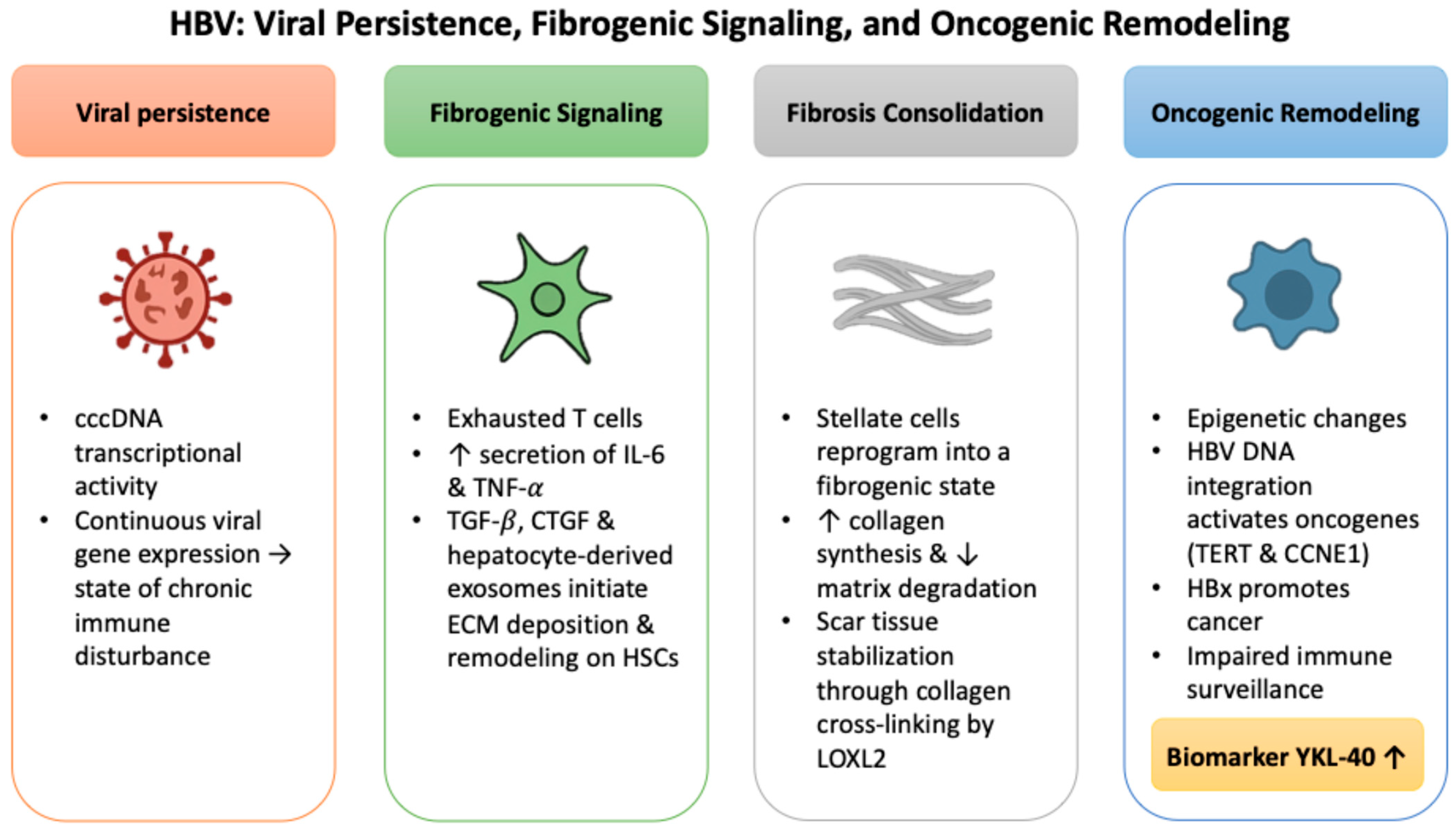
Figure 1. HBV-associated mechanisms of viral persistence, fibrogenic signaling, and oncogenic remodeling converging on ECM remodeling and YKL-40 induction.
Ijms 26 09584 g001
Figure 2. Sequential mechanisms of HCV-driven immune evasion, fibrotic programming, and inflammation-driven transformation leading to YKL-40 induction.
Ijms 26 09584 g002
Figure 3. Intensified immunopathology and accelerated fibrotic progression in HBV/HDV co-infection leading to YKL-40 induction.
Ijms 26 09584 g003
Figure 4. Cross-etiology convergence of HBV, HCV, and HDV on YKL-40 as a biomarker linking fibrosis and oncogenic risk.
Ijms 26 09584 g004
Figure 5. Molecular basis of YKL-40 expression linking viral triggers, cellular sources, and downstream profibrotic signaling.
Ijms 26 09584 g005
Figure 6. Overview of YKL-40 applications in LT, from pre-transplant eligibility to post-transplant surveillance.
Ijms 26 09584 g006
Table 1. Biomarker classes relevant to viral hepatitis-associated liver disease and along the LT pathway: principal biology, clinical readouts, evidence in HBV/HCV/HDV, candidate LT decision points, and key caveats.
Biomarker ClassBiology/Principal SourceWhat the Marker CapturesEvidence in Viral Hepatitis and HCC (HBV/HCV/HDV)Where It Can Inform the LT Pathway *Key Caveats
AFPTumor-derived glycoprotein (oncofetal)Tumor burden/secretory phenotypeWidely used in HBV/HCV HCC; imperfect sensitivity in early/non-secretory tumorsPre-LT: selection models (e.g., AFP-based); Bridging: response tracking; Post-LT: recurrence surveillance adjunctFalse positives (active hepatitis, pregnancy); non-secretors
DCP (PIVKA-II)Abnormal prothrombin (des-γ-carboxylated)Tumor biology; angiogenesis/
vitamin-K axis
Complements AFP in HBV/HCV; prognostic valuePre-LT: MoRAL-type scores; Bridging: biology signal; Post-LT: recurrence risk adjunctAffected by vitamin-K status/
warfarin; assay variability
GPC3Oncofetal proteoglycan (tumor membrane/serum)Tumor presence/
aggressiveness
Overexpressed in viral-related HCC; IHC and serum utilityPre-LT: biology beyond size/number; Bridging: residual activitySerum assays not standardized; not expressed in all HCC
OPNMatricellular protein (hepatocytes, stroma, immune)Inflammation, fibrosis, invasionElevated in HBV/HCV HCC; prognostic associationsPre-LT: risk enrichment; Bridging: limited but plausible; Post-LT: prognosis (exploratory)Non-specific; influenced by systemic
inflammation
ctDNATumor-derived cfDNA (mutational/
epigenetic)
Genomic/epigenetic alterations; MRDHBV-HCC meta-analyses; increasing early-detection/
monitoring data
Pre-LT: biology beyond imaging (VET, MVI risk); Bridging: MRD after downstaging; Post-LT: molecular recurrenceLow tumor fraction in early disease; high-complexity assays
microRNA (cell-free/EV cargo)Regulatory RNAs (hepatocytes, tumor, immune)Pathway dysregulation; injury/oncogenic programsHBV/HCV signatures (e.g., miR-122/21 panels)Pre-LT: risk phenotyping; Post-LT: recurrence/
rejection (emerging)
Normalization, platform and pre-analytics; heterogeneity
EVsVesicles from liver/tumor/immune cellsIntercellular crosstalk; composite cargo (miRNA/protein)Viral hepatitis cohorts; fibrosis/HCC signalsPre-LT/Bridging: disease activity; Post-LT: rejection/
fibrosis (exploratory)
Isolation/
quantification not harmonized; specificity
YKL-40HSCs, macrophages, endothelium, stressed hepatocytesStromal activation/fibrogenesis/angiogenic toneElevated with advanced fibrosis; prognostic in HCC; biologically coherent in HBV/HCVPre-LT: selection beyond morphology; Bridging: trajectory vs. biology; Post-LT: recurrence and graft fibrosis signalsNon-specific (inflammation, infection); age/genetic effects; assay variability
AFP: Alpha-fetoprotein; cfDNA: Cell-free DNA; ctDNA: Circulating tumor DNA; DCP: Des-gamma-carboxy prothrombin; EVs: Extracellular vesicles; EV cargo: Extracellular vesicle cargo; GPC3: Glypican-3; HBV: Hepatitis B virus; HCV: Hepatitis C virus; HDV: Hepatitis D virus; HCC: Hepatocellular carcinoma; IHC: Immunohistochemistry; LT: Liver transplantation; miRNA: MicroRNA; MoRAL: Model to predict tumor Recurrence After Living-donor liver transplantation; MRD: Minimal (molecular) residual disease; MVI: Microvascular invasion; OPN: Osteopontin; PIVKA-II: Protein induced by vitamin K absence or antagonist-II; Pre-LT: Pre-liver transplantation; Post-LT: Post-liver transplantation; VET: Viable enhancing tumor; YKL-40: Chitinase-3-like protein 1. * Pre-LT = candidacy and risk stratification; Bridging = response to downstaging/locoregional therapy; Post-LT = recurrence surveillance and graft injury/fibrosis.
Table 2. Comparative mechanisms of YKL-40 induction and function in HBV and HCV infection: from viral triggers to biomarker translation.
Pathogenic AxisHBV-Associated MechanismsHCV-Associated MechanismsShared Features
Viral triggerHBx activates TGF-β/SMAD and IL-6/STAT3 signalingCore and NS5A proteins induce ROS and NF-κB activationUpregulation of IL-6, TGF-β, and VEGF
FibrogenesiscccDNA persistence sustains HSC activation, even in inactive carriersROS-driven HSC activation and progressive fibrotic remodelingCollagen I and fibronectin accumulation with impaired ECM turnover
Immune modulationExpansion of M2 macrophages and IL-10, dominant immune toleranceT-cell exhaustion with PD-1+ CD8+ subsetsRecruitment of Tregs and MDSCs; promotion of angiogenesis
YKL-40 expressionElevated even at low viremia, correlates with necroinflammatory activityPersists in a subset after SVR, associated with residual fibrosisReflects active stromal remodeling and immunosuppressive microenvironment
Biomarker potentialSensitive marker of fibrogenic activity in HCC-naïve patientsIdentifies a high-risk phenotype post-SVRApplicable to early HCC risk stratification and supportive in pre-transplant decision-making pathways
cccDNA: Covalently closed circular DNA; CD8+: Cluster of differentiation 8-positive; ECM: Extracellular matrix; HCC: Hepatocellular carcinoma; HBV: Hepatitis B virus; HBx: Hepatitis B X protein; HCV: Hepatitis C virus; HSC: Hepatic stellate cell; IL-6: Interleukin-6; IL-10: Interleukin-10; LT: Liver transplantation; M2: M2 macrophage phenotype; MDSCs: Myeloid-derived suppressor cells; NF-κB: Nuclear factor kappa-light-chain-enhancer of activated B cells; NS5A: Non-structural protein 5A; PD-1: Programmed cell death protein 1; ROS: Reactive oxygen species; SMAD: Small mothers against decapentaplegic (SMAD family of proteins); STAT3: Signal transducer and activator of transcription 3; SVR: Sustained virologic response; TGF-β: Transforming growth factor-beta; Tregs: Regulatory T cells; VEGF: Vascular endothelial growth factor; YKL-40: Chitinase-3-like protein 1.
Table 3. Diagnostic performance of YKL-40 in viral liver diseases: receiver operating characteristic (ROC)-based thresholds, sensitivity, and specificity in HBV and HCV cohorts.
Viral EtiologyFirst Author (Year)Cohort/SettingDiagnostic Endpoint *AUCCut-Off (ng/mL)Sensitivity (%)Specificity (%)
HBVJiang (2020) [32]CHB; biopsy-verified fibrosisSignificant fibrosis (F0-F1 vs. F2-F3)0.97068.7595.289.7
HBVWang (2018) [33]CHB; baseline biopsy cohortIshak ≥ F20.8660.98283
Ishak ≥ F3— (NR)73.8 5370
Ishak ≥ F4— (NR)91.9 6967
Ishak ≥ F5— (NR)106.96170
HCVSaitou (2005) [55]CHC; biopsy cohortSignificant fibrosis (F0-F1 vs. F2-F4)0.809186.47881
Cirrhosis (F4 vs. F0-F3)0.795284.88071
CHB: Chronic hepatitis B; CHC: Chronic hepatitis C; HBV: Hepatitis B virus; HCV: Hepatitis C virus; NR: Not reported. * Fibrosis stages according to METAVIR: F0 = no fibrosis; F1 = portal fibrosis without septa; F2 = few septa; F3 = numerous septa without cirrhosis; F4 = cirrhosis. Ishak ≥F2, ≥F3, ≥F4, and ≥F5 represent progressively higher fibrosis thresholds for staging liver disease severity.
Table 4. YKL-40-associated tumor mechanisms and their clinical implications in pre-liver transplant HCC risk stratification.
Tumor-Promoting MechanismYKL-40-Associated EffectClinical Implication
Activation of invasive signalingActivates PI3K/AKT and TGF-β pathways in cancer modelsIndicates high-risk tumor phenotype beyond AFP/morphology
ECM remodelingUpregulates MMP-9 expression, promoting ECM degradationReflects stromal remodeling undetectable by imaging
Loss of epithelial adhesionSuppresses E-cadherin, facilitating epithelial–mesenchymal transition (EMT)Correlates with dedifferentiation and early metastatic potential
AngiogenesisInduces endothelial cell migration and tube formation independently of VEGFMay predict microvascular invasion and post-LT recurrence
Resistance to bridging therapyPersistently elevated levels despite radiologic response suggest stromal or residual tumor activitySupports re-evaluation of treatment response and LT candidacy
AFP: Alpha-fetoprotein; AKT: Protein kinase B; ECM: Extracellular matrix; EMT: Epithelial–mesenchymal transition; E-cadherin: Epithelial cadherin; LT: Liver transplantation; MMP-9: Matrix metalloproteinase-9; PI3K: Phosphoinositide 3-kinase; TGF-β: Transforming growth factor-beta; VEGF: Vascular endothelial growth factor; YKL-40: Chitinase-3-like protein 1.
Disclaimer/Publisher’s Note: The statements, opinions and data contained in all publications are solely those of the individual author(s) and contributor(s) and not of MDPI and/or the editor(s). MDPI and/or the editor(s) disclaim responsibility for any injury to people or property resulting from any ideas, methods, instructions or products referred to in the content.