Article

Porphyromonas gingivalis Lysate Induces TLR-2/4-Dependent NF-κB Activation and Inflammatory Damage in the Human Placental Barrier

by
Sebastián Araneda-Rojas
1,2,
Christian Castillo
1,
Ana Liempi
1,
Alejandro Fernández-Moya
1,
Jesús Guerrero-Muñoz
1,
Sebastián Alfaro
1,
Christian Gallardo
1,
Rocío Arregui
1,
Anilei Hoare
3,
Maria Alejandra Gleisner
1,
Marcela Hernández
2,* and
Ulrike Kemmerling
1,*
1
Instituto de Ciencias Biomédicas (ICBM), Facultad de Medicina, Universidad de Chile, Santiago 8380453, Chile
2
Departamento de Patología y Medicina Oral y Laboratorio de Biología Periodontal, Facultad de Odontología, Universidad de Chile, Santiago 8380544, Chile
3
Laboratory of Oral Microbiota Ecology, Faculty of Dentistry, Universidad Andres Bello, Santiago 8370133, Chile
*
Authors to whom correspondence should be addressed.
Int. J. Mol. Sci. 2025, 26(19), 9558; https://doi.org/10.3390/ijms26199558
Submission received: 11 September 2025 / Revised: 26 September 2025 / Accepted: 29 September 2025 / Published: 30 September 2025
(This article belongs to the Section Molecular Immunology)

Abstract

Periodontitis has been associated with adverse pregnancy outcomes, but the effect of oral pathogens on placental tissue and local immunity remains unclear. In this study, we investigated the response of human placental explants (HPEs) to lysates of Porphyromonas (P.) gingivalis, a keystone periodontal pathogen. Exposure to P. gingivalis induced significant histological damage and extracellular matrix degradation in placental tissue. The lysate activated the canonical NF-κB pathway, as demonstrated by increased phosphorylation of IκBα, particularly in the trophoblast. This activation was predominantly mediated by Toll-like receptor 2 (TLR-2), with partial contribution from TLR-4. Notably, TLR-2 protein levels decreased upon stimulation, while soluble (s) TLR-2 was markedly elevated in culture supernatants, suggesting receptor cleavage as a regulatory mechanism. P. gingivalis also triggered a robust proinflammatory cytokine secretion, including IL-1β, IL-6, IL-8, and TNF-α, with variable dependence on TLR-2 and TLR-4 signaling. These findings reveal that P. gingivalis components elicit a complex innate immune response in the placenta, driven by TLR-mediated NF-κB activation and modulated by sTLR-2. This study provides mechanistic insight into how periodontitis may contribute to placental inflammation and highlights potential pathways linking maternal oral health to pregnancy complications.

Graphical Abstract

1. Introduction

Pregnancy is characterized by a unique immunological context in which the maternal immune system must tolerate the semi-allogeneic fetus while defending against pathogens. The placenta is a vital maternal-fetal interface, facilitating nutrient exchange and immune modulation [1,2]. Moreover, this organ provides signals that regulate the function of the maternal immune system and provide protective mechanisms against various pathogens, including viruses, parasites, and bacteria [3]. Therefore, the interplay between infectious agents and the placenta has significant maternal and fetal health implications, potentially leading to adverse pregnancy outcomes [4,5].
Periodontitis is a chronic inflammatory disease driven by a dysbiotic subgingival biofilm that disrupts host-microbe balance, affecting ~8.5% of adults worldwide [6].
Porphyromonas gingivalis (P. gingivalis), a Gram-negative, strict anaerobic, asaccharolytic coccobacillus, is one of the keystone periodontitis pathobionts in the dysbiotic subgingival biofilm due to its ability to subvert the host immune response and promote systemic inflammation [7]. P. gingivalis presents several virulence factors, including lipopolysaccharides (LPS) with atypical lipid A structures [8], and cysteine proteases, such as gingipains, which degrade epithelial junction proteins and modulate immune signaling [9,10].
These virulence factors can enter the systemic circulation, especially during increased gingival vascular permeability, such as pregnancy [11]. Notably, P. gingivalis DNA has been detected in human placental tissues [12], and its presence has been associated with adverse pregnancy outcomes [13,14,15]. In support of this, gingipain inhibition has been shown to mitigate P. gingivalis-induced preterm birth in experimental murine models [16].
The placenta is a temporary organ that separates the maternal and fetal compartments throughout pregnancy. It provides hormones, gas, nutrition, and waste exchange for the developing fetus, ensuring normal embryo-fetal growth/development [1], and is a key immunological mediator during pregnancy [4]. This organ begins to form approximately a week post-conception upon blastocyst implantation into the maternal endometrium. The trophoblast constitutes the outer blastocyst layer and develops into two main cell lineages: (i) the extravillous trophoblast involved in the embryo’s implantation into the endometrium, and (ii) the villous trophoblast, which is part of the placental barrier [1,17]. In humans, the maternal blood circulates within the intervillous space (IVS) and is in direct contact with the fetal-derived tissues. Thus, the first tissue of the placental barrier is the villous trophoblast, a specialized epithelium composed of two distinct cell types: an outer multinucleated syncytiotrophoblast (STB), which lacks intercellular junctions, and an inner mononuclear cytotrophoblast (CTB), composed of proliferative and germinative layers. A basal lamina separates the trophoblast from the underlying villous stroma (VS), the fetal connective tissue housing fetal capillaries [1,2,17].
The villous trophoblast expresses many pattern recognition receptors (PRRs), including Toll-like receptors (TLRs). TLR-2 and TLR-4 recognize lipoproteins, peptidoglycan, and LPS. Upon activation, these receptors initiate intracellular signaling cascades that converge on the MyD88 adaptor molecule, leading to phosphorylation of IκB, nuclear translocation of NF-κB, and transcription of proinflammatory cytokines such as TNF-α, IL-1β, IL-6, and IL-8 [18,19]. While essential for antimicrobial defense, this inflammatory signaling may be detrimental if dysregulated at the maternal-fetal interface, potentially compromising placental integrity and fetal health [15,20].
Human placental explants (HPEs) are tissue sections maintained in culture media in which the histological architecture, the cell-cell, and cell-ECM crosstalk are preserved. Importantly, HPEs have been used in biomedical research, including the host-pathogen interaction, for several decades [17]. However, no studies have investigated whether P. gingivalis can activate innate immune signaling at the human placental barrier.
Here, we show that P. gigngivalis lysate causes tissue damage and a decrease in TLR-2 protein expression with the release of soluble TLR-2 into the culture media, activation of the canonical NF-κB signaling, and a TLR-2/4 mediated proinflammatory and immunomodulating cytokine response. These findings suggest a mechanistic link between periodontal infection and placental inflammation and might explain adverse pregnancy outcomes in women with periodontitis.

2. Results

2.1. P. gingivalis Lysate Induces Dose-Dependent Histopathological Alterations in HPEs

To determine the effects of P. gingivalis lysate on tissue integrity, HPEs were exposed to increasing concentrations (10–200 µg/mL) of lysate. Histological analysis using H&E staining revealed a concentration-dependent increase in trophoblast damage and detachment, along with marked disorganization of the villous stroma (Figure 1A,D). Quantification of histopathological damage (Table 1) showed significantly elevated scores at 50 µg/mL (* p < 0.05), 100 µg/mL (** p < 0.01), and 200 µg/mL (*** p < 0.001) (Figure 1D) compared to controls. Collagen integrity was also compromised in a dose-dependent manner. Masson’s trichrome staining demonstrated a progressive reduction in collagen staining intensity, with a statistically significant decrease observed at 200 µg/mL (Figure 1B,E). Additionally, analysis of collagen type I and III fibers by Picrosirius Red staining revealed a marked disorganization of collagen type I starting at 10 µg/mL, which became increasingly pronounced at higher concentrations (Figure 1C,F). These findings indicate that P. gingivalis lysate induces significant tissue damage, including extracellular matrix degradation, even at low doses. Based on these findings, a 50 µg/mL concentration of P. gingivalis lysate was selected for subsequent experiments.

2.2. P. gingivalis Lysate Modulates TLR-2 Expression and Induces Soluble TLR-2 Release in HPEs

To evaluate the impact of P. gingivalis lysate on TLR-2 and -4 protein expression, HPEs were incubated for 24 h, in the presence or absence, with 50 µg/mL of bacterial lysate, TLR-2 (Pam3CSK4 (1 µg/mL)), and TLR-4 (E. coli LPS (100 ng/mL)), agonists. Interestingly, Western blot analysis revealed a significant reduction in TLR-2 protein levels compared to unstimulated controls (p ≤ 0.05; Figure 2A). In contrast, TLR-4 expression remained unchanged across treatments (Figure 2B).
Given that TLR-2 can undergo post-translational proteolytic cleavage by metalloproteinases, generating sTLR-2 [21], we assessed its levels in culture supernatants. ELISA analysis showed a marked increase in sTLR-2 release following exposure to P. gingivalis lysate (p ≤ 0.05) compared to controls. A similar effect was observed in the Pam3CSK4-treated group (p ≤ 0.01) (Figure 2C). These findings suggest that P. gingivalis lysate may promote TLR-2 downregulation via cleavage and shedding, modulating the inflammatory response through the receptor availability at the maternal-fetal interface.

2.3. P. gingivalis Activates the Canonical NF-κB Pathway Signaling via TLR-2, but Not TLR-4, in HPEs

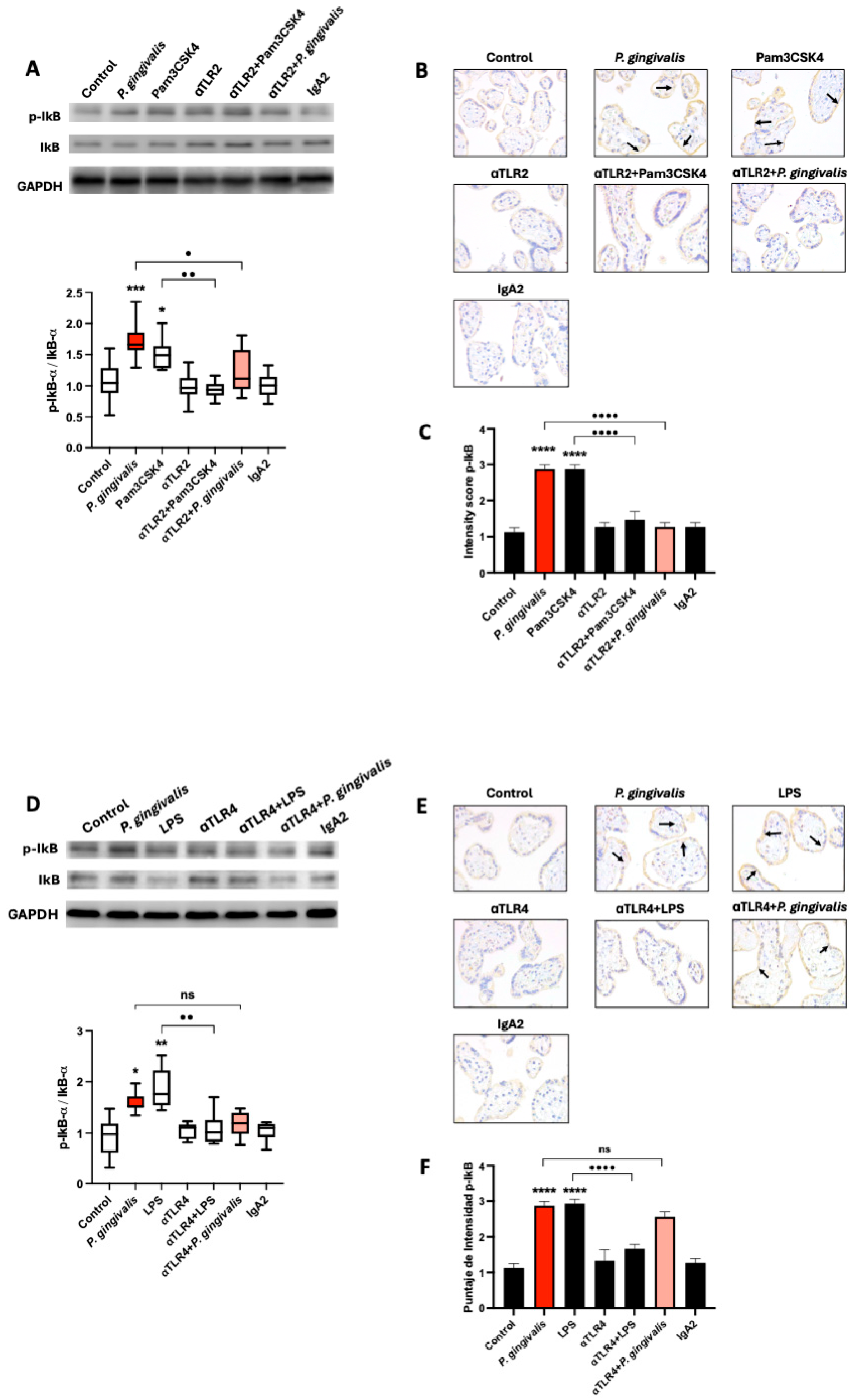
To evaluate the involvement of TLR-2 and TLR-4 in NF-κB pathway activation (Figure 3), HPEs were incubated for 10 min with P. gingivalis lysate (50 µg/mL), Pam3CSK4 (1 µg/mL; TLR-2 agonist) (Figure 3), or E. coli LPS (100 ng/mL; TLR-4 agonist), with or without pre-incubation using neutralizing antibodies against TLR-2 (Figure 3A–C) or TLR-4 (Figure 3D–F), or an IgA2 isotype control. NF-κB activation was assessed by Western blot quantification of the p-IκBα/IκBα ratio and confirmed by immunohistochemistry for p-IκBα.
Exposure to P. gingivalis lysate increased the p-IκBα/IκBα ratio by 48.4% ± 21.9 (p ≤ 0.01), and Pam3CSK4 induced an increase of 50.8% ± 24.9. These activations were significantly inhibited by TLR-2 blockade, which reduced the p-IκBα/IκBα ratio in the P. gingivalis lysate condition (p ≤ 0.05) and in the Pam3CSK4 condition (p ≤ 0.01), relative to their respective ligand-only controls (Figure 3A). Neither anti-TLR-2 nor the IgA2 isotype control induced NF-κB activation alone. Immunohistochemical analysis supported these results. In the trophoblast layer, the p-IκBα staining intensity increased significantly after P. gingivalis exposure and after LPS treatment (both p ≤ 0.0001) (Figure 3B,C). Pre-treatment with anti-TLR-2 significantly reduced this response in both conditions (both p ≤ 0.0001) (Figure 3A–C).
In contrast, blocking TLR-4 had no significant effect on NF-κB activation induced by P. gingivalis, but effectively suppressed the E. coli LPS-induced response (Figure 3D–F). E coli LPS increased the p-IκBα/IκBα ratio (p ≤ 0.0001), which was reduced (p ≤ 0.0001) in the presence of anti-TLR-4 antibodies (Figure 3D) and confirmed by immunohistochemistry (Figure 3E,F).
These results confirm that P. gingivalis lysate activates the canonical NF-κB pathway in HPEs primarily via TLR-2, suggesting that the trophoblast is a key immune-responsive tissue at the maternal-fetal interface.

2.4. P. gingivalis Lysate Induces a TLR-2/4-Dependent Proinflammatory Cytokine Response in HPEs

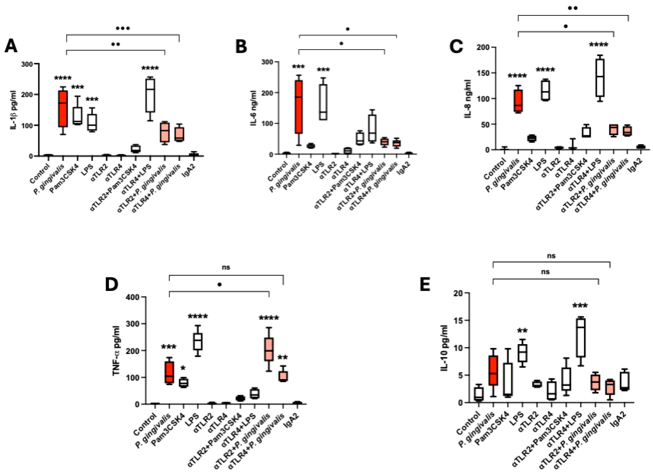
To evaluate the inflammatory response of placental tissue to P. gingivalis lysate, HPEs were incubated for 24 h with bacterial lysate (50 µg/mL), Pam3CSK4 (TLR-2 agonist), or E. coli LPS (TLR-4 agonist). Neutralizing antibodies against TLR-2 and TLR-4 were used to investigate receptor involvement. Cytokine levels in supernatants were quantified by multiplex flow cytometry (Figure 4).
Stimulation with P. gingivalis lysate significantly increased IL-1β secretion (p ≤ 0.0001) compared to unstimulated controls. Similar increases were observed with Pam3CSK4 (p ≤ 0.001) and LPS (p ≤ 0.001). This response by P. gingivalis lysate was reduced through TLR-2 (p ≤ 0.01) and TLR-4 (p ≤ 0.001) blockade (Figure 4A). IL-6 levels were also markedly elevated by the lysate (p ≤ 0.001) and LPS (p ≤ 0.001), but not by Pam3CSK4. Neutralization of TLR-2 and TLR-4 significantly reduced IL-6 levels by P. gingivalis lysate (p ≤ 0.05) (Figure 4B). Similarly, IL-8 secretion increased significantly in response to P. gingivalis (p ≤ 0.0001) and LPS (p ≤ 0.0001), while Pam3CSK4 had no effect. TLR-2 and TLR-4 blockade significantly reduced IL-8 production by P. gingivalis lysate (Figure 4C). TNF-α secretion was significantly increased by P. gingivalis lysate (p ≤ 0.001), Pam3CSK4 (p ≤ 0.05), and LPS (p ≤ 0.0001) compared to unstimulated controls. Notably, TLR-2 blockade did not reduce TNF-α secretion; instead, it increased it further (p ≤ 0.05) compared to the lysate-only condition, suggesting a possible compensatory mechanism. TLR-4 inhibition had no significant effect (Figure 4D). IL-10, in contrast, showed a modest but significant increase in response to LPS (p ≤ 0.01) compared to unstimulated controls, while P. gingivalis lysate and Pam3CSK4 had no effect. Neither TLR-2 nor TLR-4 blockade altered IL-10 levels significantly (Figure 4E). Control experiments using the IgA2 isotype antibody confirmed that cytokine responses were specifically dependent on TLR signaling, as no significant changes were observed in this condition.
These data demonstrate that P. gingivalis lysate induces a potent proinflammatory cytokine profile in human placental explants, mediated primarily through TLR-2 and TLR-4, with distinct roles depending on the cytokine involved.

3. Discussion

This study demonstrates that P. gingivalis lysate elicits a robust proinflammatory response in HPEs, mediated primarily through TLR-2 and TLR-4. This activation leads to canonical NF-κB signaling, release of inflammatory cytokines, and tissue-level changes characteristic of placental damage. Importantly, our work represents, to our knowledge, the first experimental analysis directly assessing the response of the human placental barrier to P. gingivalis components using villous explants that preserve histological architecture and trophoblast–stroma interactions. While previous studies focused mainly on detecting bacterial DNA or antigens in placental tissue [12,22] or relied on animal models [16], our findings provide novel mechanistic insight into innate immune activation at the placental barrier and establish a potential link between maternal periodontitis and adverse pregnancy outcomes.
We used P. gingivalis ATCC 33277®, a well-characterized laboratory strain that expresses major virulence factors, including LPS, gingipains, fimbriae, and peptidylarginine deiminase (PPAD) [7,9,10,23,24]. This strain is widely employed in immunological and pathogenicity studies due to its genetic stability and ability to elicit consistent host responses, minimizing variability inherent to clinical isolates [10]. Exposure to bacterial lysates allowed us to mimic the effects of microbial products without the confounding influence of live infection, thereby isolating the contribution of bacterial components to placental immune activation, making them suitable for in vitro immunomodulatory studies [25,26].
Our findings show that P. gingivalis lysate induces significant tissue injury in HPEs, including trophoblast detachment and stromal disorganization, even at low concentrations (10 µg/mL). Collagen degradation was evident at higher concentrations (≥50 µg/mL), as demonstrated by reduced Masson’s trichrome and Picrosirius Red staining. These findings are consistent with the known proteolytic effects of gingipains on extracellular matrix components [27,28], which may compromise the placental barrier. Moreover, gingipains indirectly contribute to tissue damage by activating latent host matrix metalloproteinases (MMPs) and the inactivation of host proteinase inhibitors [28]. P. gingivalis itself has been shown to induce MMPs in human gingival fibroblasts [29]. The histopathological changes are consistent with the alterations induced by other relevant pathogens that can be transmitted congenitally, such as Toxoplasma gondii [17,30].
Although P. gingivalis primarily signals through TLR-2, our results reveal that stimulation with P. gingivalis lysate significantly reduced membrane-bound TLR-2 protein levels in HPEs, while concurrently increasing soluble TLR-2 (sTLR-2) concentrations in the culture supernatants. This inverse relationship suggests that TLR-2 may undergo proteolytic cleavage or alternative splicing, releasing its soluble form as a regulatory mechanism to inflammatory stimuli [21]. sTLR-2 has been shown to function as a decoy receptor, binding pathogen-associated molecular patterns (PAMPs) and dampening TLR-2-mediated proinflammatory signaling [31]. The observed modulation may result from receptor shedding, likely mediated by bacterial gingipains or host-derived metalloproteinases, a mechanism previously described in other infectious contexts [21]. In contrast, TLR-4 protein expression remained unchanged, underscoring a preferential targeting of TLR-2 in placental tissue. This modulation of surface PRRs may shift the threshold for innate immune activation, potentially influencing the delicate balance between immune tolerance and inflammation during pregnancy.
Activation of Toll-like receptors by microbial components typically triggers intracellular signaling cascades, most notably the canonical NF-κB pathway, which governs the transcription of numerous proinflammatory genes and is a central mediator of innate immune responses [19,32]. Our study observed that P. gingivalis lysate and the TLR-2 agonist Pam3CSK4 triggered phosphorylation of IκBα, indicating activation of this pathway. These results were confirmed by immunoblotting and immunohistochemistry, which showed increased phosphorylation of IκBα predominantly in trophoblast cells. Functional assays demonstrated that TLR-2 blockade significantly reduced P. gingivalis-induced NF-κB activation, whereas TLR-4 inhibition did not affect this response, despite effectively suppressing NF-κB activation induced by canonical LPS stimulation. These findings identify TLR-2 as the primary sensor of P. gingivalis in placental tissues, consistent with previous studies demonstrating that TLR-2 is dominant in recognizing Gram-negative oral pathogens at mucosal and epithelial barriers [18]. Moreover, our observations align with previous reports demonstrating that P. gingivalis LPS induces NF-κB activation via IκBα degradation in human aortic endothelial cells [33] and enhances p-IκBα expression in co-cultures of human gingival fibroblasts and THP-1 monocytes [29]. Notably, NF-κB activation within the placenta has been associated with pathological inflammation, including preterm labor and chorioamnionitis, conditions in which increased numbers of NF-κB–positive immune cells have been identified in placental tissues [30,34].
The strong upregulation of IL-1β, IL-6, IL-8, and TNF-α in HPE supernatants following P. gingivalis lysate exposure further confirmed the inflammatory response. These cytokines play key roles in leukocyte recruitment, vascular permeability, and local inflammation, while their dysregulation has been implicated in preterm labor and other obstetric complications [15,20]. Neutralizing TLR-2 and TLR-4 effectively reduced IL-1β, IL-6, and IL-8 secretion, confirming their receptor-mediated origin. However, TNF-α secretion was paradoxically increased upon TLR-2 blockade, suggesting a compensatory or regulatory mechanism, possibly involving alternative PRRs or feedback loops. Interestingly, gingipains have been demonstrated to affect cytokine signaling networks and modulate the production of proinflammatory mediators, including the here studied IL-1β, IL-6, IL-8, and TNF-α [28], explaining the observed tissue damage. IL-10, on the other hand, was modestly increased but not modulated by TLR blockade, suggesting it may represent a parallel regulatory mechanism insufficient to counterbalance the strong proinflammatory signals.
Nevertheless, some limitations should be considered when interpreting our results. First, the explants were obtained from term placentas, which may not fully reflect the immunological conditions of the first trimester, a critical period for infection susceptibility, implantation, and early pregnancy maintenance. Second, while HPEs explants preserve tissue architecture and local cell–cell interactions, they do not capture the systemic contributions of maternal and fetal immune responses, such as leukocyte trafficking or endocrine regulation, which are known to modulate placental immunity in vivo [17]. Future studies should integrate models using earlier gestational tissues together with in vivo or organ-on-chip approaches that incorporate maternal and fetal compartments to better recapitulate the complexity of host–pathogen interactions at the maternal–fetal interface.

4. Materials and Methods

4.1. Bacterial Strains and Lysate Preparation

P. gingivalis (ATCC 33277®, Manassas, VA, USA) [35] was cultured anaerobically on blood agar with hemin/menadione and in BHI broth (Oxoid, Basingstoke, UK). Bacteria were harvested, washed, and lysed by freeze-thaw cycles and sonication. Lysate sterility was confirmed, and protein was quantified by BCA assay (Thermo Fisher Scientifi Inc., Waltham, MA, USA). LPS content was analyzed by TRIzol (Thermo Fisher Scientifi Inc.®, Waltham, MA, USA) extraction and SDS-PAGE with silver staining. Coomassie-stained SDS-PAGE (Thermo Fisher Scientifi Inc.®, Waltham, MA, USA), verified protein integrity, and gingipain activity were assessed via DL-BAPNA assay (Sigma Aldrich®, Burlington, MA, USA) [36] (Supplementary File S1).

4.2. Human Placental Explants (HPEs) Collection and Culture

Term placentas (≥38 weeks) from healthy donors undergoing elective cesarean at Hospital San José were collected with informed consent and ethical approval (Universidad de Chile, No. 042-2022). Cases with fetal or maternal pathology were excluded. Villous tissue was obtained from the central part of central cotyledons, processed within 30 min post-delivery, dissected into 0.5 cm3 fragments, and cultured in RPMI 1640 (Gibco, New York, NY, USA) with 5% FBS and 1% penicillin-streptomycin at 37 °C in 5% CO2 [17].

4.3. Experimental Design and Treatments

HPEs were exposed to P. gingivalis lysates at concentrations ranging from 10 to 200 µg/mL for 24 h to determine the optimal stimulation condition. We selected 50 µg/mL lysate concentration for subsequent experiments since this is the minimal concentration that induces histopathological damage in the trophoblast and villous stroma. Control explants were incubated with equivalent volumes of PBS. To investigate the protein expression of Toll-like receptors (TLRs), HPEs were pretreated with neutralizing antibodies against TLR-2 and TLR-4 (5 µg/mL, Invivogen®, San Diego, CA, USA) for 30 min before lysate stimulation. As positive controls, Pam3CSK4 (1 µg/mL; (Sigma Aldrich®, Burlington, MA, USA)) and E. coli LPS (100 ng/mL (Sigma Aldrich®, Burlington, MA, USA)) were employed to activate TLR-2 and TLR-4 signaling pathways, respectively. For NF-κB activation, HPEs were incubated for 10 min in the presence and absence of 50 µg/mL lysate. This signaling pathway is activated after 10 min stimulus in HPEs, as shown by us previously [19].

4.4. Histological and Immunohistochemical Analysis

HPEs were fixed in 4% paraformaldehyde, paraffin-embedded, sectioned (3–5 µm), and stained with H&E for histology or with Masson’s trichrome and Picrosirius Red for collagen (dyes were acquired from Merck®, Darmstadt, Germany). Immunohistochemistry was performed using antibodies against TLR-2, TLR-4, and NF-κB p65 (Cell Signaling®, Danvers, MA, USA) after antigen retrieval. Detection was via HRP-conjugated secondary antibodies and DAB (Cell Signaling®, Danvers, MA, USA). Staining intensity and immunopositivity were quantified using ImageJ 1.54p, and histological damage was scored semi-quantitatively across ten random fields according to the scores depicted in Table 1 [37].

4.5. Western Blot Analysis

HPEs were homogenized in lysis buffer with protease inhibitors, sonicated, and centrifuged. Supernatants were quantified via BCA assay. Proteins (50 µg) were denatured, separated by SDS-PAGE, and transferred to PVDF (Thermo Fisher Scientifi Inc.®, Waltham, MA, USA) membranes. Western blotting was performed for TLR-2, TLR-4, phospho-IκBα, total IκBα, and GAPDH using specific antibodies (Cell Signaling®, Danvers, MA, USA). Detection used HRP-conjugated secondary antibodies and chemiluminescence (Cell Signaling®, Danvers, MA, USA). Band intensities were analyzed with ImageJ 1.54p and expressed relative to GAPDH [19,38].

4.6. Cytokine Quantification by ELISA and Cytometric Bead Array

HPE culture supernatants were cleared by centrifugation (500× g, 20 min, 4 °C). IL-1β, IL-6, IL-8, IL-10, and TNF-α levels were measured using BD Cytometric Bead Array (BD Bioscience, Franklin Lakes, NJ, USA) and analyzed with FACSuite™ (BD Bioscience, Franklin Lakes, NJ, USA) and FCAP Array (BD Bioscience, Franklin Lakes, NJ, USA). Soluble TLR-2 was quantified by ELISA (Invitrogen®; detection limit 0.32 ng/mL). Cytokine levels were normalized to the total protein content in the supernatant. No significant differences in protein concentration were observed across experimental conditions.

4.7. Statistical Analysis

Data are presented as mean ± standard deviation (SD). Statistical comparisons between groups were performed using one-way ANOVA followed by Tukey’s post hoc test. Differences were considered statistically significant at p < 0.05. GraphPad Prism 9.0 software was used for all analyses.

5. Conclusions

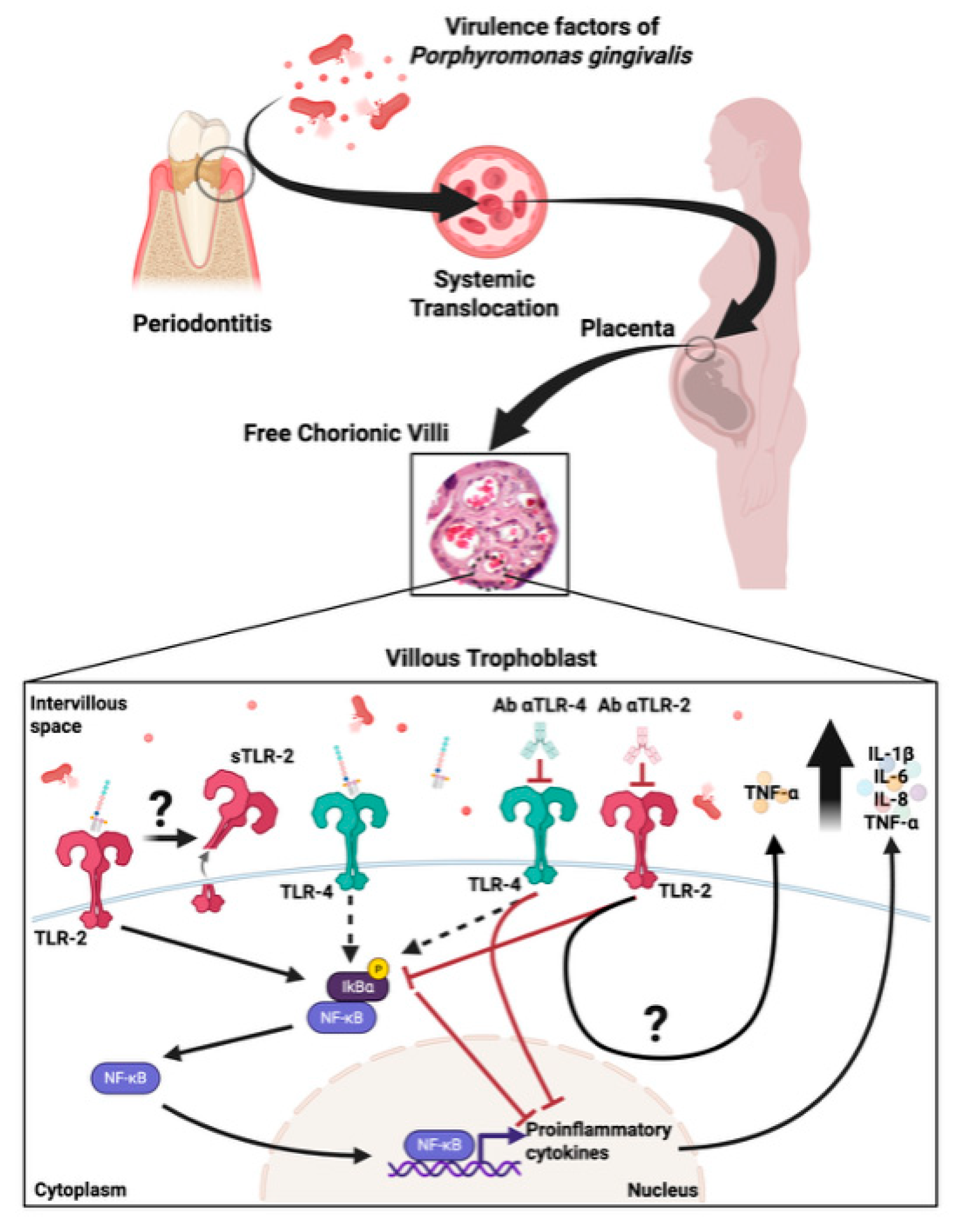
Our findings show that P. gingivalis lysate creates a proinflammatory placental environment through TLR-2/4–mediated NF-κB activation and cytokine release, potentially compromising placental integrity and contributing to adverse pregnancy outcomes. Our results highlight the placental barrier as an active immune site (Figure 5). Future studies should confirm these effects in vivo and explore how periodontal treatment may mitigate related obstetric risks.

Supplementary Materials

The following supporting information can be downloaded at: https://www.mdpi.com/article/10.3390/ijms26199558/s1.

Author Contributions

S.A.-R.: Conception, design, acquisition, analysis, and interpretation, and drafted the manuscript. A.L.: Conception, design, acquisition, analysis, and interpretation. A.F.-M.: Conception, design, acquisition, analysis, and interpretation. J.G.-M.: Acquisition, analysis, and interpretation. S.A.: Acquisition, analysis, and interpretation. C.G.: Conception, design, acquisition, analysis, and interpretation. M.A.G.: Acquisition, analysis, and interpretation. R.A.: Acquisition, analysis, and interpretation. A.H.: Acquisition, analysis, and interpretation. C.C.: Conception, design, acquisition, analysis, and interpretation. M.H.: Conception, design, acquisition, analysis, interpretation, and drafted the manuscript. U.K.: Conception, design, acquisition, analysis, interpretation, and drafted the manuscript. All authors have read and agreed to the published version of the manuscript.

Funding

U.K., C.C., and M.H. received grants from the National Fund for Scientific and Technological Development (FONDECYT; grant numbers 1220105, 11220310, 1200098, respectively). S.A. was supported by a Ph.D. scholarship from the same agency (Beca ANID 21221794).

Institutional Review Board Statement

The study was conducted per the Declaration of Helsinki and approved by the Institutional Ethics Committee of Facultad de Medicina, Universidad de Chile (Number 042-2022, 25 May 2022).

Informed Consent Statement

Informed consent was obtained from all subjects who donated their placenta in the study. No clinical data is being published.

Data Availability Statement

All data supporting the findings are available within the paper and its Supplementary Information.

Acknowledgments

We acknowledge all the women who donate their placentas. While preparing this manuscript/study, the authors used Grammarly to edit the language. The authors have reviewed and edited the output and take full responsibility for the content of this publication.

Conflicts of Interest

The authors declare no conflicts of interest. The funders had no role in the study’s design; in the collection, analyses, or interpretation of data; in the writing of the manuscript; or in the decision to publish the results.

Abbreviations

The following abbreviations are used in this manuscript
HPEsHuman placental explants
P. gingivalisPorphyromonas gingivalis
NF-kBNuclear Factor kappa B
TLRsToll-like Receptors
TLR-2Toll-like Receptor-2
TLR-4Toll-like Receptor-4
sTLR2Soluble TLR-2
IL1βInterleukin-1 beta
IL-6Interleukin-6
IL-8Interleukin-8
IL-10Interleukin-10
TNF-αTumor necrosis Factor alpha
DNADeoxyribonucleic acid
LPSLipopolysaccharides
IVSIntervillous space
STBSyncytiotrophoblast
CTBCytotrophoblast
VSVillous stroma
PRRsPattern recognition receptors
PAMPsPathogen-associated molecular patterns
SDStandard deviation
Pam3CSK4N-Palmitoyl-S-[2,3-bis(palmitoyloxy)-(2RS)-propyl]- [R]-cysteinyl-[S]- seryl-[S]-lysyl-[S]-lysyl-[S]-lysyl-[S]-lysine
E. coliEscherichia coli
p-IκBαPhospho-I-kappa-B-alpha
IκBα I-kappa-B-alpha
IgA2Immunoglobulin A2
PPADPeptidylarginine deiminase
ATCCAmerican Type Culture Collection
BHIBrain Heart Infusion
SDS-PAGESodium dodecyl sulfate polyacrylamide gel electrophoresis
DL-BAPNANα-Benzoyl-DL-arginine 4-nitroanilide hydrochloride
FBSFetal bovine serum
CO2Carbon dioxide
H&EHematoxylin and eosin
HRPHorseradish peroxidase
DABDiaminobenzidine
BCABicinchoninic acid
PVDFPolyvinylidene fluoride
GAPDHGlyceraldehyde-3-phosphate Dehydrogenase

References

  1. Baergen, R.N.; Burton, G.J.; Kaplan, C.G. (Eds.) Benirschke’s Pathology of the Human Placenta; Springer International Publishing: Cham, Switzerland, 2022; ISBN 978-3-030-84724-1. [Google Scholar]
  2. Christian, C.; Ana, L.; Alejandro, F.-M.; Jesús, G.-M.; Sebastian, A.; Gabriela, C.; Sebastián, A.; Christian, G.; Juan Diego, M.; Marioly, M.; et al. Congenital Chagas Disease: The Importance of Trypanosoma cruzi-Placenta Interactions. Placenta 2025. [Google Scholar] [CrossRef]
  3. Ding, J.; Maxwell, A.; Adzibolosu, N.; Hu, A.; You, Y.; Liao, A.; Mor, G. Mechanisms of Immune Regulation by The Placenta: Role of Type I Interferon and Interferon Stimulated Genes Signaling During Pregnancy. Immunol. Rev. 2022, 308, 9–24. [Google Scholar] [CrossRef] [PubMed]
  4. Mor, G.; Aldo, P.; Alvero, A.B. The Unique Immunological and Microbial Aspects of Pregnancy. Nat. Rev. Immunol. 2017, 17, 469–482. [Google Scholar] [CrossRef]
  5. Ander, S.E.; Diamond, M.S.; Coyne, C.B. Immune Responses at the Maternal-Fetal Interface. Sci. Immunol. 2019, 4, eaat6114. [Google Scholar] [CrossRef]
  6. Wu, L.; Zhang, S.-Q.; Zhao, L.; Ren, Z.-H.; Hu, C.-Y. Global, Regional, and National Burden of Periodontitis from 1990 to 2019: Results from the Global Burden of Disease Study 2019. J. Periodontol. 2022, 93, 1445–1454. [Google Scholar] [CrossRef]
  7. Hajishengallis, G.; Liang, S.; Payne, M.A.; Hashim, A.; Jotwani, R.; Eskan, M.A.; McIntosh, M.L.; Alsam, A.; Kirkwood, K.L.; Lambris, J.D.; et al. Low-Abundance Biofilm Species Orchestrates Inflammatory Periodontal Disease through the Commensal Microbiota and Complement. Cell Host Microbe 2011, 10, 497–506. [Google Scholar] [CrossRef] [PubMed]
  8. Herath, T.D.K.; Darveau, R.P.; Seneviratne, C.J.; Wang, C.-Y.; Wang, Y.; Jin, L. Tetra- and Penta-Acylated Lipid A Structures of Porphyromonas gingivalis LPS Differentially Activate TLR4-Mediated NF-κB Signal Transduction Cascade and Immuno-Inflammatory Response in Human Gingival Fibroblasts. PLoS ONE 2013, 8, e58496. [Google Scholar] [CrossRef] [PubMed]
  9. How, K.Y.; Song, K.P.; Chan, K.G. Porphyromonas gingivalis: An Overview of Periodontopathic Pathogen below the Gum Line. Front. Microbiol. 2016, 7, 53. [Google Scholar] [CrossRef] [PubMed]
  10. Widziolek, M.; Mieszkowska, A.; Marcinkowska, M.; Salamaga, B.; Folkert, J.; Rakus, K.; Chadzinska, M.; Potempa, J.; Stafford, G.P.; Prajsnar, T.K.; et al. Gingipains Protect Porphyromonas gingivalis from Macrophage-Mediated Phagocytic Clearance. PLoS Pathog. 2025, 21, e1012821. [Google Scholar] [CrossRef]
  11. Nannan, M.; Xiaoping, L.; Ying, J. Periodontal Disease in Pregnancy and Adverse Pregnancy Outcomes: Progress in Related Mechanisms and Management Strategies. Front. Med. 2022, 9, 963956. [Google Scholar] [CrossRef]
  12. Vanterpool, S.F.; Been, J.V.; Houben, M.L.; Nikkels, P.G.J.; De Krijger, R.R.; Zimmermann, L.J.I.; Kramer, B.W.; Progulske-Fox, A.; Reyes, L. Porphyromonas gingivalis within Placental Villous Mesenchyme and Umbilical Cord Stroma Is Associated with Adverse Pregnancy Outcome. PLoS ONE 2016, 11, e0146157. [Google Scholar] [CrossRef] [PubMed]
  13. Bobetsis, Y.A.; Ide, M.; Gürsoy, M.; Madianos, P.N. Periodontal Diseases and Adverse Pregnancy Outcomes. Present and Future. Periodontology 2000 2020, 83, 154–174. [Google Scholar] [CrossRef] [PubMed]
  14. Reyes, L.; Phillips, P.; Wolfe, B.; Golos, T.G.; Walkenhorst, M.; Progulske-Fox, A.; Brown, M. Porphyromonas gingivalis and Adverse Pregnancy Outcome. J. Oral Microbiol. 2017, 10, 1374153. [Google Scholar] [CrossRef]
  15. Chopra, A.; Radhakrishnan, R.; Sharma, M. Porphyromonas gingivalis and Adverse Pregnancy Outcomes: A Review on Its Intricate Pathogenic Mechanisms. Crit. Rev. Microbiol. 2020, 46, 213–236. [Google Scholar] [CrossRef]
  16. Takii, R.; Kadowaki, T.; Tsukuba, T.; Yamamoto, K. Inhibition of Gingipains Prevents Porphyromonas gingivalis-Induced Preterm Birth and Fetal Death in Pregnant Mice. Eur. J. Pharmacol. 2018, 824, 48–56. [Google Scholar] [CrossRef]
  17. Castillo, C.; Díaz-Luján, C.; Liempi, A.; Fretes, R.; Kemmerling, U. Mammalian Placental Explants: A Tool for Studying Host-Parasite Interactions and Placental Biology. Placenta 2024, 166, 62–70. [Google Scholar] [CrossRef]
  18. Koga, K.; Izumi, G.; Mor, G.; Fujii, T.; Osuga, Y. Toll-like Receptors at the Maternal-Fetal Interface in Normal Pregnancy and Pregnancy Complications. Am. J. Reprod. Immunol. 2014, 72, 192–205. [Google Scholar] [CrossRef]
  19. Liempi, A.; Castillo, C.; Medina, L.; Rojas, M.; Maya, J.D.; Parraguez, V.H.; Kemmerling, U. Ex Vivo Infection of Human Placental Explants with Trypanosoma cruzi and Toxoplasma gondii: Differential Activation of NF Kappa B Signaling Pathways. Acta Trop. 2019, 199, 105153. [Google Scholar] [CrossRef]
  20. Kim, Y.M.; Romero, R.; Oh, S.Y.; Kim, C.J.; Kilburn, B.A.; Armant, D.R.; Nien, J.K.; Gomez, R.; Mazor, M.; Saito, S.; et al. Toll-like Receptor 4: A Potential Link between “Danger Signals,” the Innate Immune System, and Preeclampsia? Am. J. Obs. Gynecol. 2005, 193, 921–927. [Google Scholar] [CrossRef]
  21. Langjahr, P.; Díaz-Jiménez, D.; De la Fuente, M.; Rubio, E.; Golenbock, D.; Bronfman, F.C.; Quera, R.; González, M.-J.; Hermoso, M.A. Metalloproteinase-Dependent TLR2 Ectodomain Shedding Is Involved in Soluble Toll-like Receptor 2 (sTLR2) Production. PLoS ONE 2014, 9, e104624. [Google Scholar] [CrossRef] [PubMed]
  22. Ye, C.; Katagiri, S.; Miyasaka, N.; Kobayashi, H.; Khemwong, T.; Nagasawa, T.; Izumi, Y. The Periodontopathic Bacteria in Placenta, Saliva and Subgingival Plaque of Threatened Preterm Labor and Preterm Low Birth Weight Cases: A Longitudinal Study in Japanese Pregnant Women. Clin. Oral Investig. 2020, 24, 4261–4270. [Google Scholar] [CrossRef]
  23. Hajishengallis, G. Immunomicrobial Pathogenesis of Periodontitis: Keystones, Pathobionts, and Host Response. Trends Immunol. 2014, 35, 3–11. [Google Scholar] [CrossRef]
  24. Wielento, A.; Bereta, G.P.; Łagosz-Ćwik, K.B.; Eick, S.; Lamont, R.J.; Grabiec, A.M.; Potempa, J. TLR2 Activation by Porphyromonas gingivalis Requires Both PPAD Activity and Fimbriae. Front. Immunol. 2022, 13, 823685. [Google Scholar] [CrossRef]
  25. Kaczynska, A.; Klosinska, M.; Janeczek, K.; Zarobkiewicz, M.; Emeryk, A. Promising Immunomodulatory Effects of Bacterial Lysates in Allergic Diseases. Front. Immunol. 2022, 13, 907149. [Google Scholar] [CrossRef]
  26. Di Gioacchino, M.; Santilli, F.; Pession, A. Is There a Role for Immunostimulant Bacterial Lysates in the Management of Respiratory Tract Infection? Biomolecules 2024, 14, 1249. [Google Scholar] [CrossRef]
  27. Dieterle, M.P.; Husari, A.; Steinberg, T.; Wang, X.; Ramminger, I.; Tomakidi, P. From the Matrix to the Nucleus and Back: Mechanobiology in the Light of Health, Pathologies, and Regeneration of Oral Periodontal Tissues. Biomolecules 2021, 11, 824. [Google Scholar] [CrossRef]
  28. Andrian, E.; Grenier, D.; Rouabhia, M. In Vitro Models of Tissue Penetration and Destruction by Porphyromonas gingivalis. Infect. Immun. 2004, 72, 4689–4698. [Google Scholar] [CrossRef]
  29. Jiang, L.; Liu, M.N.; Wang, G.D.; Wu, Q.; Zhao, Y.F. Cyclosporin A Suppresses Porphyromonas gingivalis Lipopolysaccharide Induced Matrix Metalloproteinases Activities in the Co-Culture of Human Gingival Fibroblasts and Monocyte Cell Line THP-1. Growth Factors 2020, 38, 65–74. [Google Scholar] [CrossRef]
  30. Rosini, A.M.; Teixeira, S.C.; Milian, I.C.B.; Silva, R.J.; de Souza, G.; Luz, L.C.; Gomes, A.O.; Mineo, J.R.; Mineo, T.W.P.; Ferro, E.A.V.; et al. LPS-Mediated Activation of TLR4 Controls Toxoplasma Gondii Growth in Human Trophoblast Cell (BeWo) and Human Villous Explants in a Dependent-Manner of TRIF, MyD88, NF-κB and Cytokines. Tissue Cell 2022, 78, 101907. [Google Scholar] [CrossRef]
  31. LeBouder, E.; Rey-Nores, J.E.; Rushmere, N.K.; Grigorov, M.; Lawn, S.D.; Affolter, M.; Griffin, G.E.; Ferrara, P.; Schiffrin, E.J.; Morgan, B.P.; et al. Soluble Forms of Toll-like Receptor (TLR)2 Capable of Modulating TLR2 Signaling Are Present in Human Plasma and Breast Milk. J. Immunol. 2003, 171, 6680–6689. [Google Scholar] [CrossRef]
  32. Napetschnig, J.; Wu, H. Molecular Basis of NF-κB Signaling. Annu. Rev. Biophys. 2013, 42, 443–468. [Google Scholar] [CrossRef]
  33. Liu, B.; Wang, J.; Cheng, L.; Liang, J. Role of JNK and NF-κB Pathways in Porphyromonas gingivalis LPS-Induced Vascular Cell Adhesion Molecule-1 Expression in Human Aortic Endothelial Cells. Mol. Med. Rep. 2013, 8, 1594–1600. [Google Scholar] [CrossRef] [PubMed]
  34. Kramer, B.W.; Kaemmerer, U.; Kapp, M.; Herbst, D.; Marx, A.; Berg, D.; Groneck, P.A.; Speer, C.P. Decreased Expression of Angiogenic Factors in Placentas with Chorioamnionitis after Preterm Birth. Pediatr. Res. 2005, 58, 607–612. [Google Scholar] [CrossRef] [PubMed]
  35. ATCC 33277. Available online: https://www.atcc.org/products/33277?matchtype=&network=x&device=c&adposition=&keyword=&gad_source=1&gad_campaignid=17725151935&gclid=EAIaIQobChMIv5H5gfb_jwMVEZaDBx274SyWEAAYAiAAEgJ5UfD_BwE (accessed on 28 September 2025).
  36. Veloso, P.; Fernández, A.; Astorga, J.; González-Quintanilla, D.; Castro, A.; Escobar, A.; Hoare, A.; Hernández, M. Lipopolysaccharide from Porphyromonas gingivalis, but Not from Porphyromonas Endodontalis, Induces Macrophage M1 Profile. Int. J. Mol. Sci. 2022, 23, 10011. [Google Scholar] [CrossRef]
  37. Fernández-Moya, A.; Oviedo, B.; Liempi, A.I.; Guerrero-Muñoz, J.; Rivas, C.; Arregui, R.; Araneda, S.; Cornet-Gomez, A.; Maya, J.D.; Müller, M.; et al. Trypanosoma cruzi-Derived Exovesicles Contribute to Parasite Infection, Tissue Damage, and Apoptotic Cell Death during Ex Vivo Infection of Human Placental Explants. Front. Cell. Infect. Microbiol. 2024, 14, 1437339. [Google Scholar] [CrossRef]
  38. Castillo, C.; Muñoz, L.; Carrillo, I.; Liempi, A.; Gallardo, C.; Galanti, N.; Maya, J.D.; Kemmerling, U. Ex Vivo Infection of Human Placental Chorionic Villi Explants with Trypanosoma cruzi and Toxoplasma gondii Induces Different Toll-like Receptor Expression and Cytokine/Chemokine Profiles. Am. J. Reprod. Immunol. 2017, 78, e12660. [Google Scholar] [CrossRef]
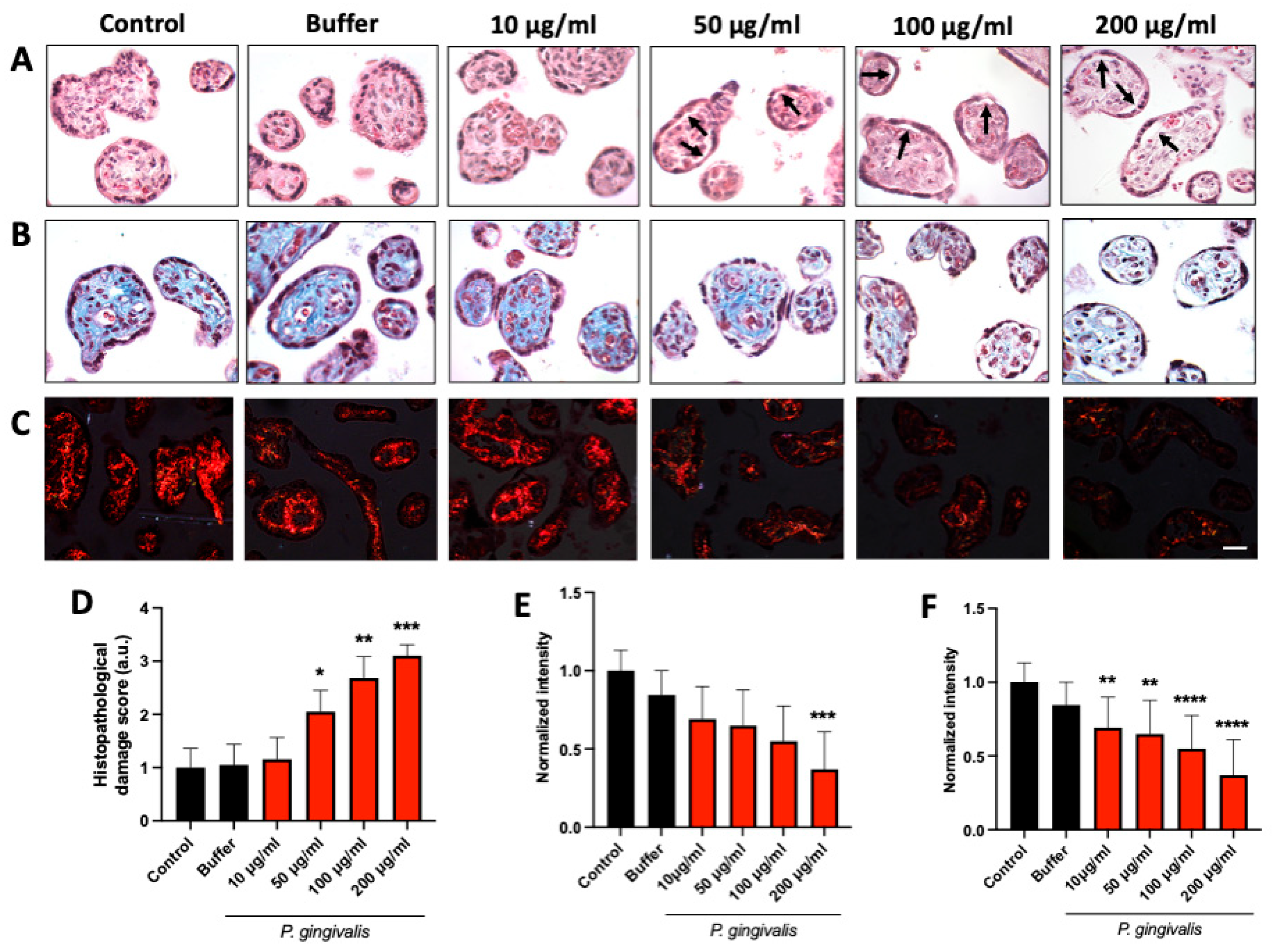
Figure 1. P. gingivalis lysate induces histopathological alterations and collagen disorganization in HPEs: HPEs were incubated for 24 h with increasing concentrations (10–200 µg/mL) of P. gingivalis lysate. H&E staining (A) revealed progressive villous damage, including trophoblast detachment (arrows) and stromal disorganization. Masson’s trichrome (B) and Picrosirius Red (C) staining showed reduced collagen deposition and disarray of collagen fibers, respectively. Semi-quantitative scoring confirmed a dose-dependent increase in histopathological damage (D), with significant effects at ≥50 µg/mL. Quantification of collagen content from Masson’s (E) and Picrosirius Red staining (F) demonstrated a significant loss of matrix integrity at higher lysate concentrations. Scale bar: 50 μm. Data represent mean ± SD from n = 3 independent placentas. * p < 0.05; ** p < 0.01; *** p < 0.001; **** p < 0.0001 versus untreated controls.
Ijms 26 09558 g001
Figure 2. P. gingivalis lysate decreases TLR-2 protein levels and increases soluble TLR-2 in HPEs: HPEs were treated for 24 h with 50 µg/mL of bacterial lysate, Pam3CSK4 (1 µg/mL), or E. coli LPS (100 ng/mL). Western blot analysis (A,B) showed a significant reduction in TLR-2 protein levels (49.6% ± 42.7; * p ≤ 0.05) in lysate-treated explants, while TLR-4 expression remained unchanged. ELISA analysis of culture supernatants (C) revealed a marked increase in soluble TLR-2 (sTLR-2) levels in the lysate and Pam3CSK4-treated groups, suggesting receptor cleavage or release. Data represent mean ± SD from three independent experiments. * p < 0.05; ** p < 0.01 versus untreated controls.
Ijms 26 09558 g002
Figure 3. TLR-2, but not TLR-4, mediates P. gingivalis-induced canonical NF-κB activation in HPEs: HPEs were pre-incubated with anti-TLR-2 or anti-TLR-4 antibodies (5 µg/mL) or IgA2 isotype control for 30 min, followed by stimulation with P. gingivalis lysate (50 µg/mL), Pam3CSK4 (1 µg/mL), or LPS (100 ng/mL) for 10 min. Western blot analysis (A) demonstrated that both stimuli significantly increased the p-IκBα/total IκBα ratio, which was attenuated by TLR-2 blockade (20.0% ± 30.0 and 10.0% ± 10.0, respectively). Immunohistochemistry (B) localized p-IκBα expression predominantly to trophoblast cells, with quantification (C) confirming that TLR-2 inhibition significantly reduced p-IκBα immunoreactivity. Western blot analysis (D) revealed that TLR-4 neutralization significantly reduced LPS-induced NF-κB activation but did not significantly affect lysate-stimulated explants. Immunohistochemistry (E) and corresponding quantification (F) confirmed strong nuclear p-IκBα expression in trophoblasts (arrows) in both LPS and P. gingivalis conditions, with only the LPS response being TLR-4 dependent. Data represent mean ± SD from three independent experiments. ns: not significant; * p < 0.05; ** p < 0.01; *** p < 0.001; **** p < 0.0001 versus untreated controls.
Ijms 26 09558 g003
Figure 4. P. gingivalis lysate triggers a TLR-2/4-dependent cytokine response in HPEs: HPEs were incubated for 24 h with P. gingivalis lysate (50 µg/mL), Pam3CSK4 (1 µg/mL), or LPS (100 ng/mL), with or without pre-treatment with neutralizing antibodies against TLR-2 or TLR-4. Cytokines in supernatants were quantified by multiplex flow cytometry. P. gingivalis significantly increased IL-1β ((A); 4529.74% ± 6498.62), IL-6 ((B); 11.532.27% ± 9622.82), IL-8 ((C); 4736.04% ± 2681.8), and TNF-α ((D); 4183.22% ± 4374.07), with responses attenuated by TLR-2 and TLR-4 inhibition except for TNF-α, which paradoxically increased after TLR-2 blockade. P. gingivalis modestly elevated IL-10 levels (E) but was unaffected by receptor inhibition. Data represent mean ± SD from three to four independent placentas. * p < 0.05; ** p < 0.01; *** p < 0.001; **** p < 0.0001.
Ijms 26 09558 g004
Figure 5. Proposed model of TLR-mediated placental immune response to P. gingivalis lysate: Schematic summary of the proposed mechanism by which P. gingivalis lysate activates innate immune responses in HPEs. Upon stimulation, bacterial components interact with trophoblast-expressed TLR-2 and, to a lesser extent, TLR-4, triggering canonical NF-κB signaling and proinflammatory cytokine production (IL-1β, IL-6, IL-8, TNF-α). Concurrently, TLR-2 surface expression is reduced, and soluble TLR-2 (sTLR-2) is released into the extracellular space, potentially acting as a decoy receptor to modulate the inflammatory response. This local activation may contribute to placental barrier dysfunction and adverse pregnancy outcomes such as preterm birth or fetal growth restriction. The figure encapsulates the placental innate immune dynamics in response to oral pathogen-derived products at the maternal-fetal interface.
Ijms 26 09558 g005
Table 1. Scores for tissue damage analysis.
ScoreHistopathology Damage
1Attached trophoblast, intact fetal connective tissue
2Slight trophoblast detachment and/or fetal connective tissue disorganization
3Almost complete trophoblast detachment and/or fetal connective tissue disorganization
4Complete trophoblast detachment and disorganization or destruction of the fetal connective tissue
Disclaimer/Publisher’s Note: The statements, opinions and data contained in all publications are solely those of the individual author(s) and contributor(s) and not of MDPI and/or the editor(s). MDPI and/or the editor(s) disclaim responsibility for any injury to people or property resulting from any ideas, methods, instructions or products referred to in the content.