Article

Detection of Targetable Genetic Abnormalities in Neuroblastoma Circulating Tumour DNA

by
Marina Danilenko
1,†,
Sharanya Nath
1,2,3,†,
Jack Baines
2,
Freya Gordon
1,4,
Swathi Merugu
1,5,
Lisa M. Allinson
1,
Aaron Potts
2,
Bethany Collins
2,
Angharad Goodman
2,
Samuel E. Kidman
2,
Ciaron McAnulty
2,
David Jamieson
6 and
Deborah A. Tweddle
1,7,*
1
Wolfson Childhood Cancer Research Centre, Translational & Clinical Research Institute, Newcastle University Centre for Cancer, Herschel Building, Newcastle upon Tyne NE1 7RU, UK
2
Newcastle Genetics Lab., Newcastle upon Tyne Hospitals NHS Foundation Trust, Newcastle upon Tyne NE1 4EP, UK
3
Division of Hematology, Medical Oncology and Palliative Care, Department of Medicine, School of Medicine and Public Health, University of Wisconsin, Madison, WI 53726, USA
4
Childhood Brain Tumour Research Group, Developmental Biology and Cancer, Great Ormond Street Institute of Child Health, University College London, London WC1N 1EH, UK
5
The Immune Monitoring Facility, Ludwig Centre for Cancer Immunotherapy, Memorial Sloan Kettering Cancer Center, New York, NY 10021, USA
6
Translational & Clinical Research Institute, Newcastle University Centre for Cancer, Paul O’Gorman Building, Newcastle upon Tyne NE2 4HH, UK
7
Department of Paediatric Oncology, Great North Children’s Hospital, Newcastle NE1 4LP, UK
*
Author to whom correspondence should be addressed.
These authors contributed equally to this manuscript and are to be considered joint first authors.
Int. J. Mol. Sci. 2025, 26(19), 9466; https://doi.org/10.3390/ijms26199466
Submission received: 12 August 2025 / Revised: 15 September 2025 / Accepted: 18 September 2025 / Published: 27 September 2025
(This article belongs to the Special Issue 25th Anniversary of IJMS: Updates and Advances in Molecular Oncology)

Abstract

Neuroblastoma (NB) is an aggressive childhood cancer requiring intensive multimodal therapies in high-risk (HRNB) patients. Currently, invasive surgical biopsies are required to classify NB risk group and assign treatment based on the tumour genetic profile. Circulating tumour DNA (ctDNA) obtained from blood samples can be used to identify tumour biomarkers. Here we applied targeted next-generation sequencing (tNGS) using a panel of 42 genes to analyse 32 NB ctDNA samples for the presence of single-nucleotide variants and copy number changes from 28 patients in all NB risk groups. In two additional ctDNA samples, droplet digital PCR was used to detect hotspot ALK variants. Pathogenic mutations with a variant allele frequency (VAF) > 1% were identified in 13/32 (41%) ctDNA samples. ALK and PTPN11 were the most frequent, each being detected in 4/32 (13%) samples, together with oncogene amplifications. Targeted NGS of ctDNA detected actionable variants, including those absent in the diagnostic primary tumour due to spatial and temporal heterogeneity. Our findings confirm the usefulness of ctDNA in detecting genetic abnormalities in NB.

1. Introduction

Neuroblastoma (NB) is the most frequent extracranial solid malignancy in children below the age of 5 years [1]. Around 100 new cases of NB are diagnosed in the UK each year, and about half of these are high-risk neuroblastoma (HRNB) with a 5-year event-free survival (EFS) below 50%.
Diagnosis and risk group assignment for NB requires an invasive tumour biopsy, which may be associated with considerable morbidity, particularly in patients below the age of 18 months [2]. An alternative non-invasive means of performing genetic studies in patients with solid tumours is through liquid biopsy sampling to assess circulating tumour cells and circulating tumour DNA (ctDNA) [3,4]. Liquid biopsies are not only safer in young patients but also provide knowledge of intra-tumoural heterogeneity [4].
Cell-free DNA (cfDNA) includes circulating short fragments of double-stranded DNA found in peripheral blood. Circulating tumour DNA (ctDNA) is the fraction of cfDNA originating from tumour tissue in cancer patients and generally constitutes less than 0.1–10% of the total cfDNA present in peripheral blood [5]. Recent studies of NB [6,7,8] and other cancers [9,10] have established that genome-wide DNA sequencing from blood plasma is a promising method for assessing tumour burden. These studies demonstrate, that in metastatic cancer, ctDNA in plasma reflects a composite genetic profile of all existing tumours in a patient. The half-life of ctDNA ranges from 16 to 150 min [10] and helps to record real time intra-tumoural events.
ctDNA analyses offer the potential to characterise NB tumour biology, which is not possible by studying the primary tumour alone. The purpose of this study was to detect and characterise the genetic abnormalities in NB ctDNA collected from patients across different risk groups using targeted next-generation sequencing (tNGS), and in a subset droplet digital PCR (ddPCR), and to compare the findings with matched primary tumour data where available.

2. Results

2.1. Pathogenic Variants Identified Following ctDNA Variant Filtering

On average, the pipeline called n = 120 raw variants per sample. After stringent filtering (see Section 4), 12/32 ctDNA samples had 15 driver variants in total (Table 1). Among these, five were pathogenic ALK variants, three were pathogenic TP53 variants and four pathogenic PTPN11 variants (Table 1). In total, 10/15 identified ctDNA variants were predicted by in silico tools to have a detrimental impact on protein function and were recorded in the COSMIC database (Table 1) [11]. The remaining variants were classified either as ‘likely pathogenic’ or ‘variants of uncertain significance’ (VUSs), as all of them had low ‘rare exome variant ensemble learner’ (REVEL) [12] scores (≤0.7) and were not reported in the literature and/or had no COSMIC presence. Finally, two out of three germline DNA samples and two out of four cell-line control DNA samples had driver variants. Previously reported findings were confirmed in the cell lines (Table 1).

2.2. A High Proportion of Variants Were Identified in NB ctDNA but Not in NB Tumours

Twenty-two ctDNA samples had matching tumour NGS data available, and thus 16 variants could be compared between liquid biopsies and NB tumours (Figure 1). Strikingly, only nine variants were identified in both ctDNA and matching tumours. The other seven variants were either present in ctDNA only (five mutations: two in ALK, one in PTPN11, one in NRAS and one in CREBBP) or only identified in the NB tumours (one mutation in ATM and one in CHK2) (Figure 1).

2.3. Spatial and Temporal Heterogeneity of Heterozygous ALK Mutations

ALK was found to be the most commonly mutated gene in this study. A total of five driver ALK variants were observed in four ctDNA samples (Figure 1). Three hotspot kinase domain mutations (R1275Q, F1174L and I1170N) were observed (Figure 2A). R1275Q was present in two ctDNA samples (ctD4 and ctD22), and F1174L was present in two ctDNA samples (ctD4 and ctD13). I1170N was present in one ctDNA sample (ctD10) (Table 1).
The most striking observation to emerge from the ALK variant data was from sample ctD4. The Venn diagram (Figure 2B) shows that two variants, encoding R1275Q and F1174L, were detected in the plasma sample from diagnosis, with VAFs of 2.85% and 28.5%, respectively. However, in the diagnostic primary tumour only the R1275Q variant was detected by Sanger sequencing and Agena Mass Array [13] (VAF: 32.0%) (Figure 2B). In a separate plasma sample analysed by ddPCR at the end of treatment, neither R1275Q nor F1174 variants were detected. Figure 2C shows ddPCR results for F1174L at the end of treatment and relapse; results for R1275Q are not included. In a metastatic relapse sample and a plasma relapse sample, 3 months later only the F1174L mutation was detected by targeted NGS from SMPaeds data on the metastatic tumour [14,15] (VAF: 46.1%) and by digital-droplet PCR (ddPCR) (VAF: 98%) on the plasma (Figure 2B,C). R1275Q was not detected in either tumour or plasma at relapse. This patient did not receive an ALK inhibitor at any stage of treatment. This data reflects the presence of spatial and temporal intra-tumoural heterogeneity. It also shows that ctDNA is more representative of malignant clones promoting NB relapse than the primary tumour.

2.4. Pathogenic, Homozygous Somatic TP53 Mutations

In this study four TP53 pathogenic variants with high VAFs were observed in the DNA binding domain of the TP53 gene (Table 1 and Figure 3A). Three were found in ctDNA and one in a germline DNA sample (Table 1). Observed protein changes were as follows: H179Q in ctD11 and gD1 and R280S in ctD20 and ctD21. Figure 3B shows a homozygous TP53 variant detected in the diagnosis plasma sample (ctD20), post-chemotherapy primary tumour and relapse plasma (ctD21) samples. The lower VAF for the post-chemotherapy tumour reflects the lower tumour cell content (estimated: 30%, calculated: 19%). Homozygosity was confirmed by 17p loss of heterozygosity (Table A1). In another diagnostic case (ctD11), with a homozygous somatic TP53 mutation (VAF: 84.9%) caused by concomitant 17p loss (Table A1), the matching diagnostic germline sample had a VAF of 1.4%, suggesting that the gDNA was contaminated with tumour DNA when collected (Table 1).

2.5. Heterozygous, Pathogenic PTPN11 Variants

Pathogenic PTPN11 variants (RAS-MAPK pathway gene family) were observed in five samples (Table 1). A pathogenic germline variant, PTPN11 N308D, in both ctD15 and gD2 (germline DNA) was identified in a patient with Noonan syndrome and low-risk NB [16,17,18] with VAFs of 35.8% and 43.9%. In the other three cases, activating PTPN11 mutations were detected within the N terminal domain: G60A (VAF: 3.6%) in the case with a concomitant KRAS and ALK mutation (ctD10) and in the other two cases at the known hotspots E76Q (VAF: 18%) and D61Y (VAF: 4.34%).

2.6. Oncogene Amplifications Detected in ctDNA

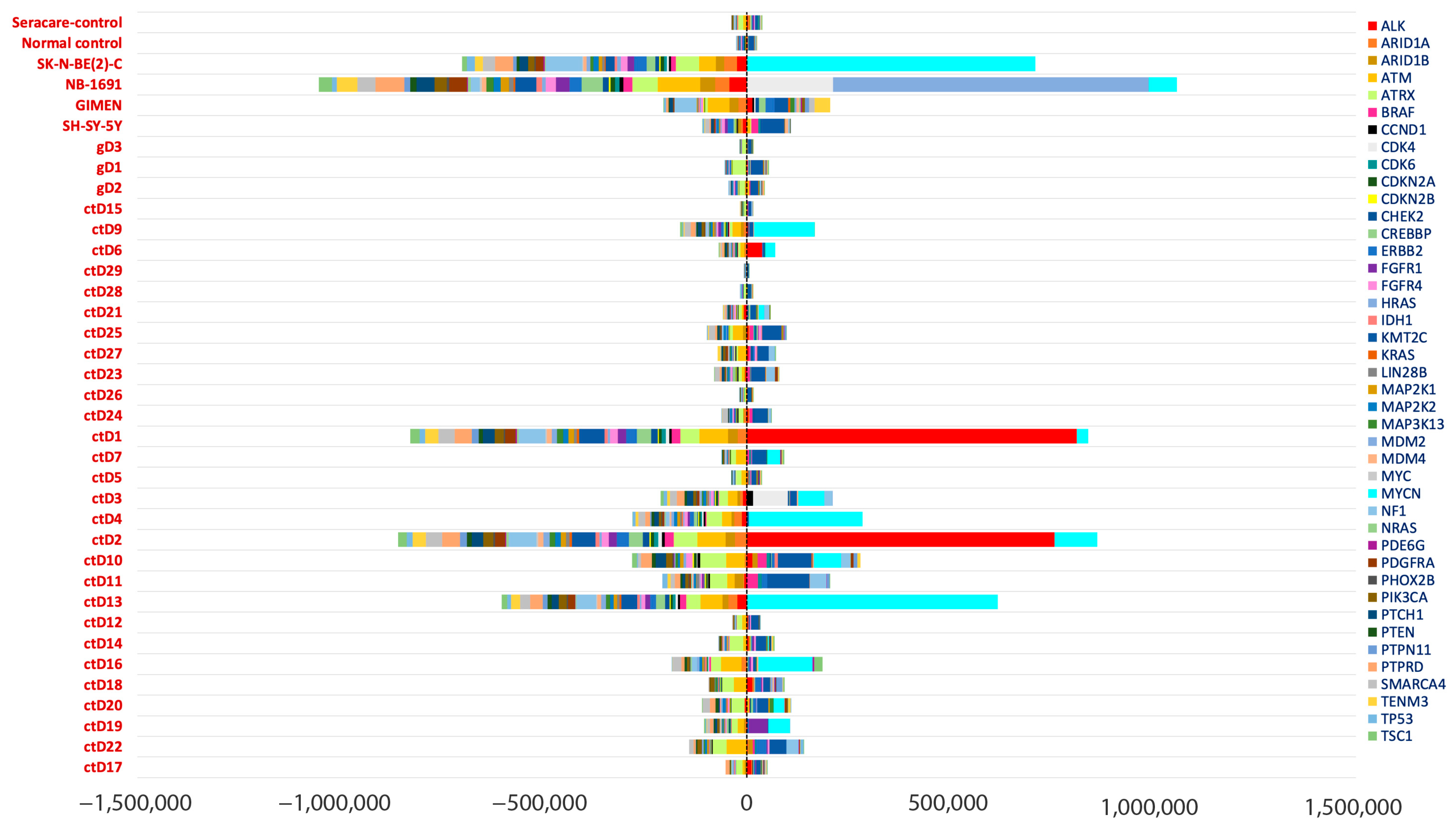
Higher values for coverage were observed in regions corresponding to known oncogene amplification sites (compared to other genes) (Figure 4).
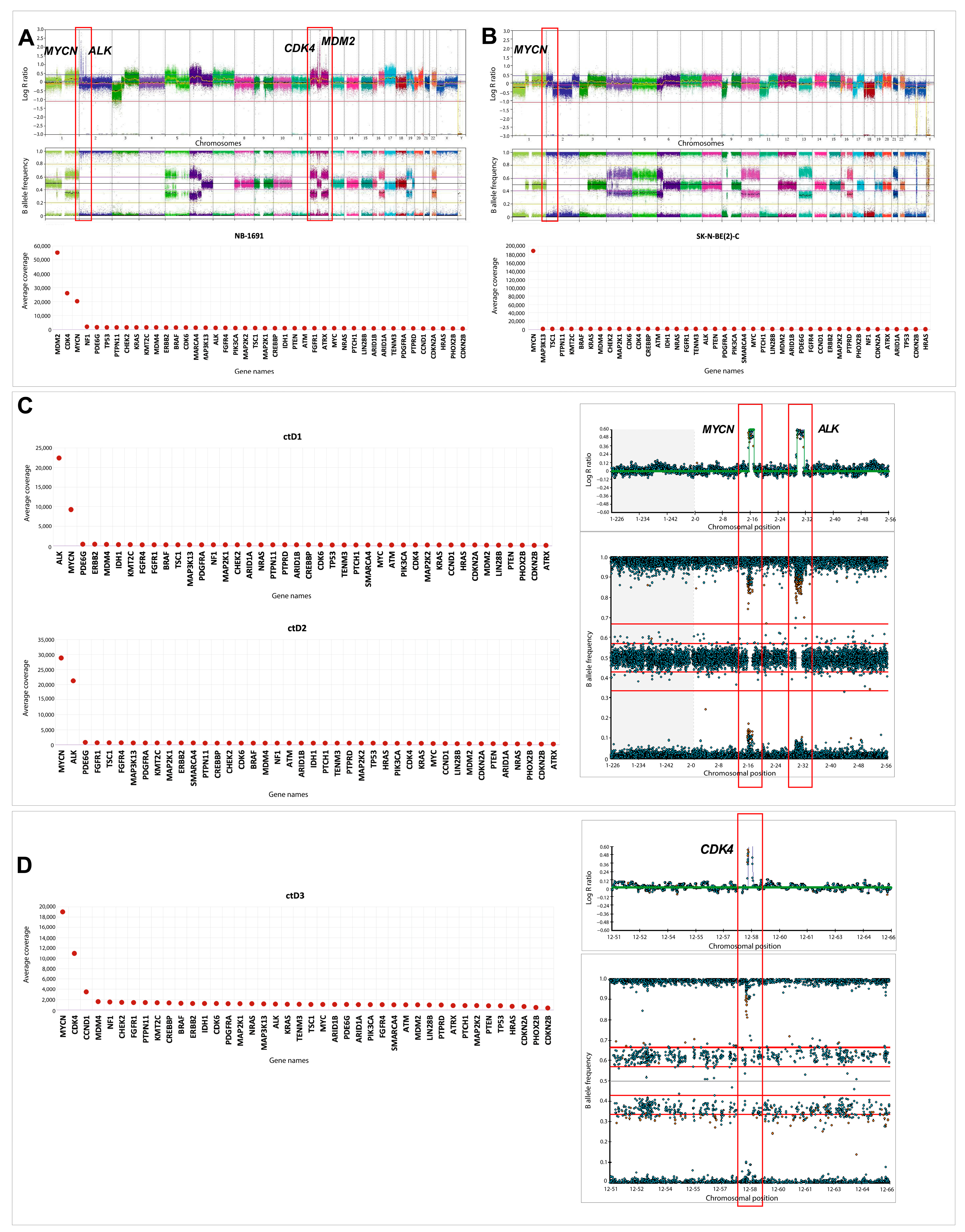
MYCN amplification was detected in 14/32 ctDNA samples and confirmed in 2 known MYCN amplified cell lines (Figure 4 and Figure 5A,B). This was consistent with the amplification status detected by Illumina CytoSNP-850k arrays (Table A1). Furthermore, co-amplification of ALK and MYCN was observed in the ctDNA samples ctD1 and ctD2, taken from the same patient at diagnosis and relapse, and was consistent with SNP array results in the primary tumour and ctDNA [19] (Figure 5C). CDK4 amplification, previously associated with a poor prognosis in NB patients [20,21], was detected in ctD3 and confirmed in the NB-1691 cell line (Figure 5A,D). The presence of MDM2 amplification was also confirmed in the NB-1691 cell line (Figure 5A) and the presence of CCND1 amplification in ctD3 (Figure 5D). However, neither ALK amplification in NB-1691 cells [22] nor FGFR1 amplification in ctD19, which were evident on SNP arrays (Table A1), were detected in ctDNA (Figure 4 and Figure 5).

3. Discussion

The use of liquid biopsy in the management of patients with cancer, either alongside traditional histopathology or as the primary method of obtaining clinically relevant biomarker data, affords clinicians access to real-time tumour monitoring and the ability to detect druggable targets as a tumour evolves. NB frequently metastasises to bone and bone marrow, making liquid biopsy particularly useful for patients with metastatic NB. CfDNA can be extracted from as little as 0.5–2 mL of plasma [23,24,25] and is a viable alternative to invasive biopsies, which increase morbidity in young patients.
Using a targeted gene panel and sequencing ctDNA from plasma samples, we aimed to detect driver events and oncogenic amplifications in ctDNA and to compare these with tumour biopsy samples, where available. Results from this study showed that the bioinformatics pipeline was able to detect all clinically relevant variants, including those with low VAFs (>1%), in ctDNA. Strikingly, 7/16 identified driver ctDNA variants for which tumour NGS data was available, were only present in ctDNA and not in NB tissue. This can be explained by the intra-tumoural heterogeneity of NB when the variants were absent in the biopsied tumour regions while present in ctDNA representing a pool of variants from multiple tumour regions [15,26]. All the variants detected only in ctDNA were at a VAF of <5% and thus likely to be sub-clonal or due to low ctDNA content in the blood. A threshold of 5% has been assigned for most clinical NGS reporting on the bulk tumour, and thus variants with a VAF < 5% could be omitted during data analyses. In two cases, DNA from the NB tumour contained CHK2 (VAF = 3%) and ATM (VAF = 72%) mutations, which were absent in paired ctDNA at the same time point. This could be due to low ctDNA shedding into the blood or variants being sub-clonal. Tumour-only mutations have been previously reported in paired tumour and ctDNA studies and are typically explained by low levels of ctDNA in the patient’s blood [15,26].
Importantly, an ALK variant (F1174L) not detected in the diagnostic primary tumour biopsy using the Agena Mass Spectrometer assay, which detects mutations at a VAF of 1.3% [13], was present in diagnostic ctDNA (ctD4), the metastatic relapse biopsy and relapse ctDNA (Figure 2B). This confirms that ctDNA is more reflective of the global genomic variation in a tumour as it overcomes the problem of intra-tumoural heterogeneity missed by a single-site biopsy. In ctD4 two ALK mutations were identified in diagnostic ctDNA (R1275Q and F1174L), in line with other ctDNA studies reporting several ALK mutations in a single ctDNA sample [15,26,27,28,29]. This case demonstrates both spatial and temporal heterogeneity with different ALK mutations detected in the diagnostic primary tumour (R1275Q) and relapsed metastatic tumour (F1174L), as well clonal evolution with loss of the R1275Q mutation between diagnostic and relapse ctDNA, together with an increase in the VAF of the F1174L mutation. The clonal evolution occurred in the absence of selective pressure from patient treatment with an ALK inhibitor.
In keeping with other studies [30,31], pathogenic ALK mutations were the commonest mutations in this study, while PTPN11 mutations were the second most frequent (Table 1 and Figure 1). A study by Ackermann et al. [32] sequenced NB genomes, where they also found PTPN11 to be commonly mutated. This highlights the importance of developing treatments for PTPN11 mutated NB, the efficacy of which could be tracked by serial ctDNA monitoring. The Ackermann study reported ALK-RAS pathway mutations to be associated with poor outcomes in all NB risk groups when associated with telomere maintenance mechanisms. In our study, ctD10 harboured two RAS-MAPK (KRAS-G12V and PTPN11-G60A) mutations and an ALK-I1170L mutation, in addition to MYCN amplification (a telomere maintenance mechanism) (Table 1). We also detected four ctDNA samples (ctD10, ctD16, ctD20 and ctD21) having RAS-MAPK pathway or TP53 mutations alongside MYCN amplification and hence falling into a very poor prognostic group.
The bioinformatic pipeline was able to detect MYCN, ALK, CDK4 and MDM2 amplifications. However, ALK amplification in NB-1691 cells [22] and FGFR1 amplification in ctD19 were not detected, most likely due to only partial amplification of these oncogenes in regions not covered by the gene panel. Furthermore, the tNGS approach failed to detect other copy number events, as the gene panel was limited to 42 known target genes and did not cover the entire genome (Table A2). Whole-exome sequencing (WES) or low-coverage WGS would be required to detect all copy number changes in these samples
Liquid biopsy-based analysis of ctDNA offers several advantages in the management of NB. Firstly, it is a non-invasive procedure that can be performed longitudinally, allowing for monitoring of disease progression and treatment response over time, as well as early relapse detection. Secondly, it provides a comprehensive assessment of tumour heterogeneity, enabling detection of genetic alterations that may not be captured by single-site tumour biopsies. Thirdly, liquid biopsy can identify actionable variants, guiding personalised treatment decisions.
While our study highlights the potential of ctDNA monitoring in NB, it is limited by its size and the use of mainly diagnostic samples with longitudinal samples in only three cases. The longitudinal testing of plasma samples provides more information about NB progression over time, uncovering possible mechanisms of treatment resistance and relapse, as recently reported by studies of ALK mutation tracking in ctDNA collected from NB patients treated with ALK inhibitors [26,28,29]. Previous studies reported both reductions in the VAFs of ALK mutations in longitudinal ctDNA sampling during successful patient treatment with ALK inhibitors [7,15,26,28,29,33,34] but also the emergence of resistance mutations, including both bypass mutations, particularly of the RAS-MAPK pathway [26,28,29,34], as well as compound ALK mutations [29]. Importantly, one study also reported loss of ALK mutations in some patients who relapsed during treatment with the third-generation ALK inhibitor lorlatinib, due to selection for ALK wild-type clones by lorlatinib treatment [29]. Furthermore, we have previously reported selection for ALK wild-type clones in tumour DNA from two patients between diagnosis and relapse in the absence of ALK inhibitor treatment [13]. These studies emphasise the importance of serial ctDNA monitoring for ALK mutations in patients with NB, particularly when treating patients with ALK inhibitors.
In conclusion, our study confirms the potential of liquid biopsy, specifically ctDNA analysis, to study NB in a non-invasive way.

4. Materials and Methods

4.1. Plasma and Cell-Line Samples

Plasma and germline DNA samples were obtained from VIVO biobank (formerly the Children’s Cancer and Leukaemia Group (CCLG) Tissue Bank) (study number 2021 BS 03). The NGS study included n = 41 samples, of which n = 32 were ctDNA samples from NB patient plasma, including n = 3 paired samples (2 paired diagnosis and relapse and 1 paired blood and bone marrow plasma), n = 3 germline DNA, n = 4 control NB cell-line DNA (SH-SY-5Y, NB-1691, SK-N-BE(2)-C and GIMEN) and n = 2 control samples, one a synthetic control (Seraseq ctDNA. Complete AF0.1%, Seracare) (Seracare, Milford, MA, USA) and the other a normal genomic DNA control (Table A1). Two additional plasma samples (from the end of treatment and three months later at relapse) from the ctD4 patient were obtained and used for ddPCR. In total, 28 NB cases were studied: 4 low risk, 3 intermediate risk and 21 high risk (Figure 1 and Table A1).

4.2. DNA Extraction

DNA from 4 cell lines and ctDNA from 0.5 mL plasma were extracted using EZ1 Advanced XL (QIAGEN, Hilden, Germany) kits: the EZ1 DNA 200 μL Blood Kit for plasma samples and the EZ1 DNA Tissue Kit for cell-line pellets, according to the manufacturer’s instructions. cfDNA content was assessed using the Agilent TapeStation 4200 (Agilent, Santa Clara, CA, USA with the cell-free DNA ScreenTape assay. Sample ctD8 was excluded from further analysis, as the cfDNA concentration was only 6%, below the threshold value of 20%. The library preparation method also incorporated unique molecular indices (UMIs) ligated to the ends of DNA fragments. The UMI code is a sequence of short nucleotides that uniquely identifies each molecule in a sample library, enabling greater precision during sequencing and error correction. The prepared libraries were quality assessed using Bioanalyzer 2400 High Sensitivity D1000 chip assays (Agilent, Santa Clara, CA, USA) by quantifying fragments between 35 and 1000 bp. The size interval used for library preparation was approximately between 200 and 400 bp, where the cfDNA fragment peak formed. The prepared libraries were also quantified using the Qubit Fluorometer 2.0—double-stranded DNA high-sensitivity assay (ThermoFisher, Waltham, MA, USA).

4.3. Library Preparation and Next-Generation Sequencing

Sequencing library preparation for ctDNA samples was performed according to the manufacturer’s instructions using a QIAseq Targeted Custom DNA Panel of 42 genes (Table A2). The gene panel was compiled according to recommendations of the SIOPEN Biology Group [35]. Targeted NGS (Illumina NextSeq 550 System (Illumina, Inc., San Diego, CA, USA)) was performed on n = 31 patient cfDNA samples, n = 4 cell-line DNA samples, n = 3 germline DNA samples and n = 2 control DNA samples. Raw reads from the NextSeq550 were aligned and variant calling was performed using CLC Genomics Workbench 22 (CLC Bio, version 22.0, Aarhus, Denmark). An analysis of the UMI coverage per gene was carried out across all samples.

4.4. Variant Filtering

The mean read depth was calculated to be 2228x after excluding the read depth of over- and underrepresented amplified genes and genes on the X chromosome, consistent with the manufacturers’ specifications of a mean read depth of 2500x for 20 samples on a mid-output flow cell with the QiaSeq kit (QIAGEN). Variants with coverage < 100× and frequencies < 0.5% were filtered out. CLC Genomics workbench tools software was used to remove sequencing artefacts, PCR duplicates and recurrent variants. The resulting variants were then annotated using the Ensembl Variant Effect Predictor (VEP) [36] with Genome Aggregation Database (gnomAD r2.1, exomes only [37]) population frequencies, ‘Sorting Intolerant from Tolerant’ (SIFT 5.2.2) [38] algorithm scores, ‘Polymorphism Phenotyping v2’ (PolyPhen 2.2.2) [39] scores (range from 0 to 1; 0 = most likely benign, 1 = most likely damaging), ‘rare exome variant ensemble learner’ (REVEL) [12] scores (range from 0 to 1; 0 = likely benign, 1 = likely pathogenic) and COSMIC IDs [11]. Using a standard approach, the pipeline filtered out germline variants by removing the SNPs found in the ‘dbSNP common’ database [40] (dbSNP build 151, with ≥1% minor allele frequency (MAF) present in any of the five super-populations). Further stringent filtering for germline variants was performed manually by annotating the variants with gnomAD frequencies using VEP, excluding variants occurring in different population groups (gnomAD r2.1, exomes-only database) with a frequency of more than 1.0%. Furthermore, germline data was used from WGS from blood samples corresponding to ctD19, ctD20, ctD23, ctD25, ctD26 and ctD28 sequenced at 30x coverage (NHS England Whole Genome Sequencing (WGS) service). In these cases, the final filtered variants generated were subjected to a comparative analysis against the germline variants identified in their matched WGS data.

4.5. Coverage Analysis

An analysis of the UMI coverage per gene was carried out across all samples and graphs were plotted. The UMI coverage was assessed using two metrices, relative UMI coverage (average coverage) and UMI read depth, which helped evaluate the reliability of the tNGS panel in covering all the targeted regions. To validate the coverage data to correctly detect gene amplification status, the results, wherever possible, were compared with SNP array data generated from Illumina CytoSNP-850k arrays using Illumina BlueFuse multi v4.4 software or Nexus Copy Number v10.0 software (Bionano Genomics Inc., San Diego, CA, USA).

4.6. Classification of Somatic Variants

Classification of filtered variants was determined by considering both the biological and clinical relevance of the data [5]. The potential pathogenicity of variants was determined using SIFT [38], PolyPhen-2 [39], REVEL [12] scores and COSMIC [11] presence. Variants not classified previously in COSMIC or from a literature review were not discarded but retained for further investigation. The list of somatic driver variants was also compared with those obtained from analysis of the primary or metastatic tumours using WGS (n = 9) or a different targeted or whole-exome sequencing platform (n = 13). This included NGS from Stratified Medicine Paediatrics (SMPaeds) testing [14] or tNGS using a SIOPEN 38 gene panel [35] where available, together with results of ALK sequencing using Sanger sequencing or Agena Mass Spectrometer arrays [13] (Figure 1).

4.7. ddPCR

High-sensitivity testing for the ALK c.3522C>A p.(F1174L) and c3824G>A p.(R1275Q) variants was performed by droplet digital PCR (ddPCR) using commercially designed primers and probes obtained from Bio-Rad (assay ID numbers: dHsaMDV2516768 ALKp.R1275Q and dHsaMDV2010083ALK p.F1174L c.3522C>A). Cell-free DNA extracted from peripheral blood was amplified by standard PCR methods and fragmented by restriction digest using HaeIII. PCR products were then separated using the Bio-Rad QX-200 ddPCR system following standard Bio-Rad protocols. Analysis was performed using QX Manager Software Standard Edition v2.1 obtained from BioRad (Hercules, CA, USA). The sequence for the ALK F1174L c.3522C>A primer is hg19|chr2:29443634-29443756, and for the ALK p.R1275Q primer the sequence is hg19|chr2:29432603-29432725.

4.8. Statistical Analyses

Target-region coverage analysis was carried out for all samples using the CLC Genomics Workbench tools. The resulting numerical data per targeted region per gene was used to calculate a z-score for the determination of CNVs from coverage data [41].
Z = x μ σ
where x = raw score, μ = mean of data array and σ = standard deviation of the data array.

Author Contributions

Conceptualisation, D.A.T., C.M. and M.D.; methodology, S.N. and J.B.; software, S.E.K., S.N. and J.B.; validation, L.M.A., A.G. and A.P.; formal analysis, S.N., J.B., M.D. and B.C.; investigation, S.N., M.D., J.B. and B.C.; resources, S.M. and D.J.; data curation, D.A.T., S.N. and J.B.; writing—original draft preparation, M.D. and S.N.; writing—review and editing, D.A.T., M.D. and F.G.; visualisation, M.D., S.N. and J.B.; supervision, D.A.T. and C.M.; project administration, J.B. and S.N.; funding acquisition, D.A.T., D.J., S.M., S.N. and M.D. All authors have read and agreed to the published version of the manuscript.

Funding

VIVO biobank is funded by Cancer Research UK (C8594/A23804) in partnership with Blood Cancer UK (CRCPSC-Dec21\100002). This study was supported by the Sir Bobby Robson Foundation, Little Princess Trust (CCLGA 2016 08), Children’s Cancer North, a CR UK Centre grant to Newcastle Cancer Centre (CTRQQR-2021\100003), Action Medical Research/Great Ormond Street Hospital Charity (GN2390), a Horizon Europe-Mission Cancer THRIVE grant (grant reference: 101136622) to David Jamieson, a Newcastle University Vice Chancellor’s International scholarship to Sharanya Nath and a Newcastle University Faculty Fellowship to Marina Danilenko.

Institutional Review Board Statement

The samples used were all provided by VIVO biobank, which is a national ethically approved biobank in the UK for children and young people with cancer. The ethical approval number is 21/EM/0130. The study was approved by East Midlands and Deby REC on 10 August 2023.

Informed Consent Statement

Informed consent was obtained from all subjects involved in the study.

Data Availability Statement

Data is contained within the article. Sequencing data has been deposited in NCBI SRA (BioProject accession number PRJNA928778).

Acknowledgments

We thank Newcastle Genetics Lab, Newcastle, UK, for providing WGS and NGS and SNP array data and Jane Lisgo, Leigh Betts and the QIAGEN team (Nick Dobie, Daniel Hand and Brennan Martin) for technical support. We thank June Biedler for SH-SY-5Y cells, Barbara Spengler for SK-N-BE(2)-C cells, Clinton Stewart for NB1691 cells and Mirco Ponzoni for GIMEN cells. We thank VIVO Biobank (formerly the Children’s Cancer & Leukaemia Group Tissue Bank) for the provision of plasma and DNA samples and all patients and families donating to the biobank. WGS was carried out by the NHS Genomic Medicine Service.

Conflicts of Interest

The authors declare no conflicts of interest.

Appendix A

Appendix A.1

Table A1. Samples used in the ctDNA study with existing SNP array data from the primary tumour. ctD8 was excluded from further analysis, as the cfDNA concentration was below the threshold value of 20%. a,b,c = paired ctDNA samples; a,b were obtained from the same patient at diagnosis and relapse and c was obtained from ctDNA from the same patient from blood and bone marrow at diagnosis. d,e,f = ctDNA and matched gDNA pairs. Abbreviations and symbols: NB—Neuroblastoma; Int—Intermediate risk; Int ** = localised, non-MNA, >18 months of age, unfavourable histology; PB—Peripheral blood; BM—Bone marrow; R—Relapse; D—Diagnosis; * denotes cases where SNP array performed on ctDNA [19]; CN-LOH = copy neutral loss of heterozygosity; MNA—MYCN amplified; non-MNA—Non-MYCN amplified.
Sample NameSample TypeNB Risk Group and MYCN StatusTime PointSNP Array Data
GainLoss
a ctD1Plasma (PB)High
MNA
R* 1q, 17q, 18q.
ALK amplification
* 1p, 5p, 18p.
a ctD2Plasma (PB)High
MNA
DALK amplification
11q, 17q.
1p, 10q.
ctD3Plasma (PB)High
MNA
D1q13.1q32.1, 2p16.1p25.3, CDK4 amplification, CCND1 amplification 6p21.3q27, 12p13.3q24.3, 14q11.2q32.12.X, 11, 9, 10, 4, 5p.
ctD4Plasma (PB)High
MNA
D17q.1p, 3p, 10, 15q.
ctD5Plasma (PB)High
Non-MNA
D11pq, 17q, 18.11q.
ctD6Plasma (PB)High
MNA
D2p23.1(ALK), 17q.1p, 9, 11.
ctD7Plasma (PB)High
MNA
D17q22q25.3.1p31.2p36.33.
ctD9Plasma (PB)High
MNA
D16q, 17q, 21.1p, 3, 4q, 5pq, 6, 8, 14, 15q, 16q, 17p,
CN-LOH: 9, 10, 11, 19, X.
ctD10Plasma (PB)High
MNA
D17p12p11.2, 17q12q22.3, 5, 4q, 9, 10, 11, 13q, 21q.
d ctD11Plasma (PB)High
Non-MNA
D6p, 7p, 13q, 21 & 12.6p, 6q, 8p, 8q, 11, 17p.
ctD12Plasma (PB)High
Non-MNA
D2p, 6pq, 7p, 11p, 12q, 16q, 17q, 22q, 13 & 18.9p, 11q.
ctD13Plasma (PB)High
MNA
D17q.1p, 3p.
ctD14Plasma (PB)Low
Non-MNA
DWhole-chromosome abnormalities only
e ctD15Plasma (PB)Low
Non-MNA
DWhole-chromosome abnormalities only
ctD16Plasma (PB)High
MNA
D1q, 2p, 9q, 11q, 17q, 18p.1p, 3p, 11q, X.
f ctD17Plasma (PB)Int **
Non-MNA
D1q, 2p, 5p, 11q, 16q, 17q, 20q.3p, 5q, 6p, 6q, 9p (PTPRD), 9q, Y.
ctD18Plasma (PB)High
Non-MNA
D1q, 2p (MYCN), 12q, 17q,3p, 3q, 11q, 19q,
20q.22q, Xq21.1 (ATRX).
ctD19Plasma (PB)High
MNA
D2, 4, 7, 8, 8p11.22p11.21, 12, 15, 17p11.2q25.3, 18, 20, 21, 22, FGFR1 amplification.10
b ctD20Plasma (PB)High
MNA
D3, 11, 14, 20q13.2q13.33, 21, 22.1, 2, 8p23.3q21.3, 9, 10p15.3q21.1, 13, 16q11.2q24.3, 18, 17p.
b ctD21Plasma (PB)High
MNA
R3, 11, 14, 20q13 2q13.33, 21, 22.1, 2, 8p23, 3q21.3, 9, 10p15, 3q21.1, 13, 16q11, 2q24.3, 18, 17p.
ctD22Plasma (PB)High
Non-MNA
D1q21.1q42.2,6, 7, 8p23.3p22,10p15.3q11.21, 13, 17, 18, 20, 21.1q42.2q44, 3, 9p24.1 (PTPRD), 11, 11q14.1q22.3, 15q13.2q26.3, 19.
ctD23Plasma (PB)High
Non-MNA
D4p16, 3q21.23, 7, 13q11q31.3, 13q32, 1q34, 17, 18.1q44, 2q44, 2q21, 3q37.3, 3, 5q15q35.3, 9p24, 3p22.3, 9p21, 3q34.3, 11, 14, 16, 19, 22, X, LOH: 2q21.3q37.3, 5q15q35.3, 11, 16, 22.
ctD24Plasma (PB)High
Non-MNA
D6p, 7pq, 13q, 17pq, 18, 21.6q, 7p, 17p.
ctD25Plasma (PB)High
Non-MNA
D1q, 3q, 6p, 3q, 7, 11q, 1q, 12, 16q,17q, 18.3p26, 3p12.2, 3p12, 2q29, 4, 6q22, 33q27, 8, 11q13, 3q25, 14, 17p13, 3q21.31,
19p13, 3p13.11.
ctD26Plasma (PB)Low
Non-MNA
D1, 2, 7, 12, 17.No loss
ctD27Plasma (PB)Int
Non-MNA
D2, 5, 7, 13, 17, 22, X.3, 4, 11, 14, 21 LOH: 3, 14.
ctD28Plasma (PB)Int **
Non-MNA
DTetraploid.3, 4, 14, 15, 16.
ctD29Plasma (PB)Low
Non-MNA
DNo gains.2, 3, 4, 11, 12, 14, 15, 16, 19, 21, X
CN-LOH: 11.
c ctD30Plasma (BM)High
Non-MNA
D* 2p, 6p, 7, 17q, 18.3q, 5, 6q,
9p, 10, 11q, 17p, 19.
c ctD31Plasma (PB)High
Non-MNA
D* 2p, 6p, 7, 17q, 18.3q, 5, 6q,
9p, 10, 11q, 17p, 19.
ctD32Plasma (PB)High
MNA
R* 1q, 2p, 11q, 17q,19q, 3, 5, 6, 7, 8, 12, 13, 14, 16, 20, 21.* 1p, 11q, 19p, 22q.
d gD1DNAHighD--
e gD2DNALowD--
f gD3DNAInt **D--
SH-SY-5YDNAHigh
Non-MNA
R1q, 7, 17q.14p, 22q.
GIMENDNAHigh
Non-MNA
R2p.1p, 6q, 11q.
NB-1691DNAHigh
MNA
RALK, CDK4, MDM2 amp
3q, 5p, 6p, 12, 17.
3p.
SK-N-BE-(2)CDNAHigh
MNA
R2p.1p, 17, 18, X, 11p, 9p, 3p.
Control1Synthetic plasma----
Control2Normal control DNA----

Appendix A.2

Table A2. Gene list for the 42-gene QIASeq custom tNGS panel used in this study.
Variant GeneChromosomal LocationTranscriptFunction
ALK2p23.1NM_004304.4Tyrosine kinase
ARID1A1p36.11NM_006015.4Chromatin remodelling factor
ARID1B6q25.3NM_020732.3Chromatin remodelling factor
ATM11q22.3NM_000051.3Serine/threonine kinase
ATRXXq21.1NM_000489.3Chromatin remodelling factor
BRAF7q34NM_004333.4Protein kinase
CCND111q13.3NM_053056.2Cyclin dependent protein kinase
CDK412q14.1NM_000075.3Cyclin dependent protein kinase
CDK67q21.2NM_001259.6Cyclin-dependent protein kinase
CDKN2A9p21.3NM_001195132.1Cyclin-dependent protein kinase
CDKN2B9p21.3NM_004936.3Cyclin-dependent protein kinase
CHEK222q22.1NM_007194.4Protein kinase
CREBBP16p13.3NM_004380.3Acetyltransferase
ERBB217q12NM_004448.2Tyrosine kinase
FGFR18p11.23NM_001174067.1Tyrosine kinase growth factor
FGFR45q35.2NM_002011.3Growth factor receptor tyrosine kinase
HRAS11p15.5NM_005343.2GTPase
IDH12q34NM_005896.2Catalytic isozyme
KMT2C7q36.1NM_170606.3Histone methyltransferase
KRAS12p12.1NM_033360.2GTPase
LIN28B6q16-6q21NM_001004317.3RNA-binding protein
MAP2K115q22.31NM_002755.3MAP kinase
MAP2K219p13.3NM_030662.3Tyrosine kinase
MAP3K133q27.3NM_001242314.1Serine/threonine kinase
MDM212q15NM_002392.4TP53 regulator (negative)
MDM41q32.1NM_002393.4TP53 regulator (negative)
MYC8q24.21NM_002467.4Transcription factor
MYCN2p24.3NM_005378.4Transcription factor
NF117q11.2NM_001042492.2Tumour suppressor
NRAS1p13.2NM_002524.4GTPase
PDE6G17q25.3NM_002602.3Phosphodiesterase
PDGFRA4q12NM_006206.4Tyrosine kinase
PHOX2B4p13NM_003924.3Transcription factor
PIK3CA3q26.32NM_006218.2Lipid kinase
PTCH19q22.32NM_000264.3Receptor for Hedgehog genes
PTEN10q23.31NM_000314.4Phosphatase
PTPN1112q24.13NM_002834.3Protein tyrosine phosphatase
PTPRD9p23NM_002839.3Tyrosine phosphatase
SMARCA419p13.2NM_003072.5Chromatin regulator
TENM34q34.3NM_001080477.1Neuronal development protein
TP5317p13.1NM_000546.6Tumour suppressor
TSC15q32-q33NM_000368.5Tumour suppressor

References

  1. Tolbert, V.P.; Matthay, K.K. Neuroblastoma: Clinical and biological approach to risk stratification and treatment. Cell Tissue Res. 2018, 372, 195–209. [Google Scholar] [CrossRef]
  2. Matthay, K.K.; Maris, J.M.; Schleiermacher, G.; Nakagawara, A.; Mackall, C.L.; Diller, L.; Weiss, W.A. Neuroblastoma. Nat. Rev. Dis. Primers 2016, 2, 16078. [Google Scholar] [CrossRef] [PubMed]
  3. Lone, S.N.; Nisar, S.; Masoodi, T.; Singh, M.; Rizwan, A.; Hashem, S.; El-Rifai, W.; Bedognetti, D.; Batra, S.K.; Haris, M.; et al. Liquid biopsy: A step closer to transform diagnosis, prognosis and future of cancer treatments. Mol. Cancer 2022, 21, 79. [Google Scholar] [CrossRef] [PubMed]
  4. Russano, M.; Napolitano, A.; Ribelli, G.; Iuliani, M.; Simonetti, S.; Citarella, F.; Pantano, F.; Dell’Aquila, E.; Anesi, C.; Silvestris, N.; et al. Liquid biopsy and tumor heterogeneity in metastatic solid tumors: The potentiality of blood samples. J. Exp. Clin. Cancer Res. 2020, 39, 95, Erratum in J. Exp. Clin. Cancer Res. 2020, 39, 120. [Google Scholar] [CrossRef]
  5. Diehl, F.; Schmidt, K.; Choti, M.A.; Romans, K.; Goodman, S.; Li, M.; Thornton, K.; Agrawal, N.; Sokoll, L.; Szabo, S.A.; et al. Circulating mutant DNA to assess tumor dynamics. Nat. Med. 2008, 14, 985–990. [Google Scholar] [CrossRef]
  6. Lodrini, M.; Graef, J.; Thole-Kliesch, T.M.; Astrahantseff, K.; Sprüssel, A.; Grimaldi, M.; Peitz, C.; Linke, R.B.; Hollander, J.F.; Lankes, E.; et al. Targeted Analysis of Cell-free Circulating Tumor DNA is Suitable for Early Relapse and Actionable Target Detection in Patients with Neuroblastoma. Clin. Cancer Res. 2022, 28, 1809–1820. [Google Scholar] [CrossRef]
  7. Kahana-Edwin, S.; Cain, L.E.; McCowage, G.; Darmanian, A.; Wright, D.; Mullins, A.; Saletta, F.; Karpelowsky, J. Neuroblastoma Molecular Risk-Stratification of DNA Copy Number and ALK Genotyping via Cell-Free Circulating Tumor DNA Profiling. Cancers 2021, 13, 3365. [Google Scholar] [CrossRef]
  8. Lodrini, M.; Wünschel, J.; Thole-Kliesch, T.M.; Grimaldi, M.; Sprüssel, A.; Linke, R.B.; Hollander, J.F.; Tiburtius, D.; Künkele, A.; Schulte, J.H.; et al. Circulating Cell-Free DNA Assessment in Biofluids from Children with Neuroblastoma Demonstrates Feasibility and Potential for Minimally Invasive Molecular Diagnostics. Cancers 2022, 14, 2080. [Google Scholar] [CrossRef]
  9. Chan, K.C.; Jiang, P.; Zheng, Y.W.; Liao, G.J.; Sun, H.; Wong, J.; Siu, S.S.; Chan, W.C.; Chan, S.L.; Chan, A.T.; et al. Cancer genome scanning in plasma: Detection of tumor-associated copy number aberrations, single-nucleotide variants, and tumoral heterogeneity by massively parallel sequencing. Clin. Chem. 2013, 59, 211–224. [Google Scholar] [CrossRef]
  10. Leary, R.J.; Sausen, M.; Kinde, I.; Papadopoulos, N.; Carpten, J.D.; Craig, D.; O’Shaughnessy, J.; Kinzler, K.W.; Parmigiani, G.; Vogelstein, B.; et al. Detection of chromosomal alterations in the circulation of cancer patients with whole-genome sequencing. Sci. Transl. Med. 2012, 4, 162ra54. [Google Scholar] [CrossRef] [PubMed]
  11. Tate, J.G.; Bamford, S.; Jubb, H.C.; Sondka, Z.; Beare, D.M.; Bindal, N.; Boutselakis, H.; Cole, C.G.; Creatore, C.; Dawson, E.; et al. COSMIC: The Catalogue Of Somatic Mutations In Cancer. Nucleic Acids Res. 2019, 47, D941–D947. [Google Scholar] [CrossRef]
  12. Ioannidis, N.M.; Rothstein, J.H.; Pejaver, V.; Middha, S.; McDonnell, S.K.; Baheti, S.; Musolf, A.; Li, Q.; Holzinger, E.; Karyadi, D.; et al. REVEL: An Ensemble Method for Predicting the Pathogenicity of Rare Missense Variants. Am. J. Hum. Genet. 2016, 99, 877–885. [Google Scholar] [CrossRef]
  13. Allinson, L.M.; Potts, A.; Goodman, A.; Bown, N.; Bashton, M.; Thompson, D.; Basta, N.O.; Gabriel, A.S.; McCorkindale, M.; Ng, A.; et al. Loss of ALK hotspot mutations in relapsed neuroblastoma. Genes Chromosomes Cancer 2022, 61, 747–753. [Google Scholar] [CrossRef]
  14. George, S.L.; Izquierdo, E.; Campbell, J.; Koutroumanidou, E.; Proszek, P.; Jamal, S.; Hughes, D.; Yuan, L.; Marshall, L.V.; Carceller, F.; et al. A tailored molecular profiling programme for children with cancer to identify clinically actionable genetic alterations. Eur. J. Cancer 2019, 121, 224–235. [Google Scholar] [CrossRef] [PubMed]
  15. George, S.L.; Lynn, C.; Stankunaite, R.; Hughes, D.; Sauer, C.M.; Chalker, J.; Waqar Ahmed, S.; Oostveen, M.; Proszek, P.Z.; Yuan, L.; et al. Stratified Medicine Pediatrics: Cell-Free DNA and Serial Tumor Sequencing Identifies Subtype-Specific Cancer Evolution and Epigenetic States. Cancer Discov. 2025, 15, 717–732. [Google Scholar] [CrossRef] [PubMed]
  16. Jongmans, M.C.J.; van der Burgt, I.; Hoogerbrugge, P.M.; Noordam, K.; Yntema, H.G.; Nillesen, W.M.; Kuiper, R.P.; Ligtenberg, M.J.; van Kessel, A.G.; van Krieken, J.H.; et al. Cancer risk in patients with Noonan syndrome carrying a PTPN11 mutation. Eur. J. Hum. Genet. 2011, 19, 870–874. [Google Scholar] [CrossRef]
  17. Mutesa, L.; Pierquin, G.; Janin, N.; Segers, K.; Thomée, C.; Provenzi, M.; Bours, V. Germline PTPN11 missense mutation in a case of Noonan syndrome associated with mediastinal and retroperitoneal neuroblastic tumors. Cancer Genet. Cytogenet. 2008, 182, 40–42. [Google Scholar] [CrossRef]
  18. Ijiri, R.; Tanaka, Y.; Keisuke, K.; Masuno, M.; Imaizumi, K. A case of Noonan’s syndrome with possible associated neuroblastoma. Pediatr. Radiol. 2000, 30, 432–433. [Google Scholar] [CrossRef] [PubMed]
  19. Merugu, S.; Chen, L.; Gavens, E.; Gabra, H.; Brougham, M.; Makin, G.; Ng, A.; Murphy, D.; Gabriel, A.S.; Robinson, M.L.; et al. Detection of Circulating and Disseminated Neuroblastoma Cells Using the ImageStream Flow Cytometer for Use as Predictive and Pharmacodynamic Biomarkers. Clin. Cancer Res. 2020, 26, 122–134. [Google Scholar] [CrossRef]
  20. Amoroso, L.; Ognibene, M.; Morini, M.; Conte, M.; Di Cataldo, A.; Tondo, A.; D’Angelo, P.; Castellano, A.; Garaventa, A.; Lasorsa, V.A.; et al. Genomic coamplification of CDK4/MDM2/FRS2 is associated with very poor prognosis and atypical clinical features in neuroblastoma patients. Genes Chromosomes Cancer 2020, 59, 277–285. [Google Scholar] [CrossRef]
  21. Rihani, A.; Vandesompele, J.; Speleman, F.; Van Maerken, T. Inhibition of CDK4/6 as a novel therapeutic option for neuroblastoma. Cancer Cell Int. 2015, 15, 76. [Google Scholar] [CrossRef] [PubMed]
  22. Tucker, E.R.; Jiménez, I.; Chen, L.; Bellini, A.; Gorrini, C.; Calton, E.; Gao, Q.; Che, H.; Poon, E.; Jamin, Y.; et al. Combination Therapies Targeting ALK-aberrant Neuroblastoma in Preclinical Models. Clin. Cancer Res. 2023, 29, 1317–1331. [Google Scholar] [CrossRef] [PubMed]
  23. Bagley, S.J.; Nabavizadeh, S.A.; Mays, J.J.; Till, J.E.; Ware, J.B.; Levy, S.; Sarchiapone, W.; Hussain, J.; Prior, T.; Guiry, S.; et al. Clinical Utility of Plasma Cell-Free DNA in Adult Patients with Newly Diagnosed Glioblastoma: A Pilot Prospective Study. Clin. Cancer Res. 2020, 26, 397–407. [Google Scholar] [CrossRef]
  24. Fleischhacker, M.; Schmidt, B.; Weickmann, S.; Fersching, D.M.; Leszinski, G.S.; Siegele, B.; Stötzer, O.J.; Nagel, D.; Holdenrieder, S. Methods for isolation of cell-free plasma DNA strongly affect DNA yield. Clin. Chim. Acta 2011, 412, 2085–2088. [Google Scholar] [CrossRef]
  25. Page, K.; Guttery, D.S.; Zahra, N.; Primrose, L.; Elshaw, S.R.; Pringle, J.H.; Blighe, K.; Marchese, S.D.; Hills, A.; Woodley, L.; et al. Influence of plasma processing on recovery and analysis of circulating nucleic acids. PLoS ONE 2013, 8, e77963. [Google Scholar] [CrossRef]
  26. Bosse, K.R.; Giudice, A.M.; Lane, M.V.; McIntyre, B.; Schürch, P.M.; Pascual-Pasto, G.; Buongervino, S.N.; Suresh, S.; Fitzsimmons, A.; Hyman, A.; et al. Serial Profiling of Circulating Tumor DNA Identifies Dynamic Evolution of Clinically Actionable Genomic Alterations in High-Risk Neuroblastoma. Cancer Discov. 2022, 12, 2800–2819. [Google Scholar] [CrossRef] [PubMed]
  27. Chicard, M.; Colmet-Daage, L.; Clement, N.; Danzon, A.; Bohec, M.; Bernard, V.; Baulande, S.; Bellini, A.; Deveau, P.; Pierron, G.; et al. Whole-Exome Sequencing of Cell-Free DNA Reveals Temporo-spatial Heterogeneity and Identifies Treatment-Resistant Clones in Neuroblastoma. Clin. Cancer Res. 2018, 24, 939–949. [Google Scholar] [CrossRef]
  28. Bobin, C.; Iddir, Y.; Butterworth, C.; Masliah-Planchon, J.; Saint-Charles, A.; Bellini, A.; Bhalshankar, J.; Pierron, G.; Combaret, V.; Attignon, V.; et al. Sequential Analysis of cfDNA Reveals Clonal Evolution in Patients with Neuroblastoma Receiving ALK-Targeted Therapy. Clin. Cancer Res. 2024, 30, 3316–3328. [Google Scholar] [CrossRef]
  29. Berko, E.R.; Witek, G.M.; Matkar, S.; Petrova, Z.O.; Wu, M.A.; Smith, C.M.; Daniels, A.; Kalna, J.; Kennedy, A.; Gostuski, I.; et al. Circulating tumor DNA reveals mechanisms of lorlatinib resistance in patients with relapsed/refractory ALK-driven neuroblastoma. Nat. Commun. 2023, 14, 2601. [Google Scholar] [CrossRef]
  30. Mossé, Y.P.; Laudenslager, M.; Longo, L.; Cole, K.A.; Wood, A.; Attiyeh, E.F.; Laquaglia, M.J.; Sennett, R.; Lynch, J.E.; Perri, P.; et al. Identification of ALK as a major familial neuroblastoma predisposition gene. Nature 2008, 455, 930–935. [Google Scholar] [CrossRef]
  31. Bresler, S.C.; Weiser, D.A.; Huwe, P.J.; Park, J.H.; Krytska, K.; Ryles, H.; Laudenslager, M.; Rappaport, E.F.; Wood, A.C.; McGrady, P.W.; et al. ALK mutations confer differential oncogenic activation and sensitivity to ALK inhibition therapy in neuroblastoma. Cancer Cell 2014, 26, 682–694. [Google Scholar] [CrossRef] [PubMed]
  32. Ackermann, S.; Cartolano, M.; Hero, B.; Welte, A.; Kahlert, Y.; Roderwieser, A.; Bartenhagen, C.; Walter, E.; Gecht, J.; Kerschke, L.; et al. A mechanistic classification of clinical phenotypes in neuroblastoma. Science 2018, 362, 1165–1170. [Google Scholar] [CrossRef]
  33. Rahmqvist, I.; Engström, E.; Mellström, E.; Ibrahim, R.R.; Pujol-Calderón, F.; Dahlstrand Rudin, A.; Ordqvist Redfors, A.; Rostamzadeh, N.; Di Rienzo, R.; Franssila, W.; et al. Personalized circulating tumor DNA analysis for sensitive disease monitoring and detection of relapse in neuroblastoma. Biomark. Res. 2024, 12, 148. [Google Scholar] [CrossRef]
  34. Ek, T.; Ibrahim, R.R.; Vogt, H.; Georgantzi, K.; Träger, C.; Gaarder, J.; Djos, A.; Rahmqvist, I.; Mellström, E.; Pujol-Calderón, F.; et al. Long-Lasting Response to Lorlatinib in Patients with ALK-Driven Relapsed or Refractory Neuroblastoma Monitored with Circulating Tumor DNA Analysis. Cancer Res. Commun. 2024, 4, 2553–2564. [Google Scholar] [CrossRef]
  35. Chen, L.; Humphreys, A.; Turnbull, L.; Bellini, A.; Schleiermacher, G.; Salwen, H.; Cohn, S.L.; Bown, N.; Tweddle, D.A. Identification of different ALK mutations in a pair of neuroblastoma cell lines established at diagnosis and relapse. Oncotarget 2016, 7, 87301–87311. [Google Scholar] [CrossRef] [PubMed]
  36. McLaren, W.; Gil, L.; Hunt, S.E.; Riat, H.S.; Ritchie, G.R.; Thormann, A.; Flicek, P.; Cunningham, F. The Ensembl Variant Effect Predictor. Genome Biol. 2016, 17, 122. [Google Scholar] [CrossRef]
  37. Karczewski, K.J.; Francioli, L.C.; Tiao, G.; Cummings, B.B.; Alföldi, J.; Wang, Q.; Collins, R.L.; Laricchia, K.M.; Ganna, A.; Birnbaum, D.P.; et al. The mutational constraint spectrum quantified from variation in 141,456 humans. Nature 2020, 581, 434–443, Erratum in Nature 2021, 590, E53. [Google Scholar]
  38. Ng, P.C.; Henikoff, S. SIFT: Predicting amino acid changes that affect protein function. Nucleic Acids Res. 2003, 31, 3812–3814. [Google Scholar] [CrossRef]
  39. Adzhubei, I.; Jordan, D.M.; Sunyaev, S.R. Predicting functional effect of human missense mutations using PolyPhen-2. Curr. Protoc. Hum. Genet. 2013, 76, 7.20. 1–7.20. 41. [Google Scholar] [CrossRef]
  40. Sherry, S.T.; Ward, M.H.; Kholodov, M.; Baker, J.; Phan, L.; Smigielski, E.M.; Sirotkin, K. dbSNP: The NCBI database of genetic variation. Nucleic Acids Res. 2001, 29, 308–311. [Google Scholar] [CrossRef] [PubMed]
  41. Eijkelenboom, A.; Tops, B.B.J.; van den Berg, A.; van den Brule, A.J.C.; Dinjens, W.N.M.; Dubbink, H.J.; Ter Elst, A.; Geurts-Giele, W.R.R.; Groenen, P.; Groenendijk, F.H.; et al. Recommendations for the clinical interpretation and reporting of copy number gains using gene panel NGS analysis in routine diagnostics. Virchows Arch. 2019, 474, 673–680. [Google Scholar] [CrossRef] [PubMed]
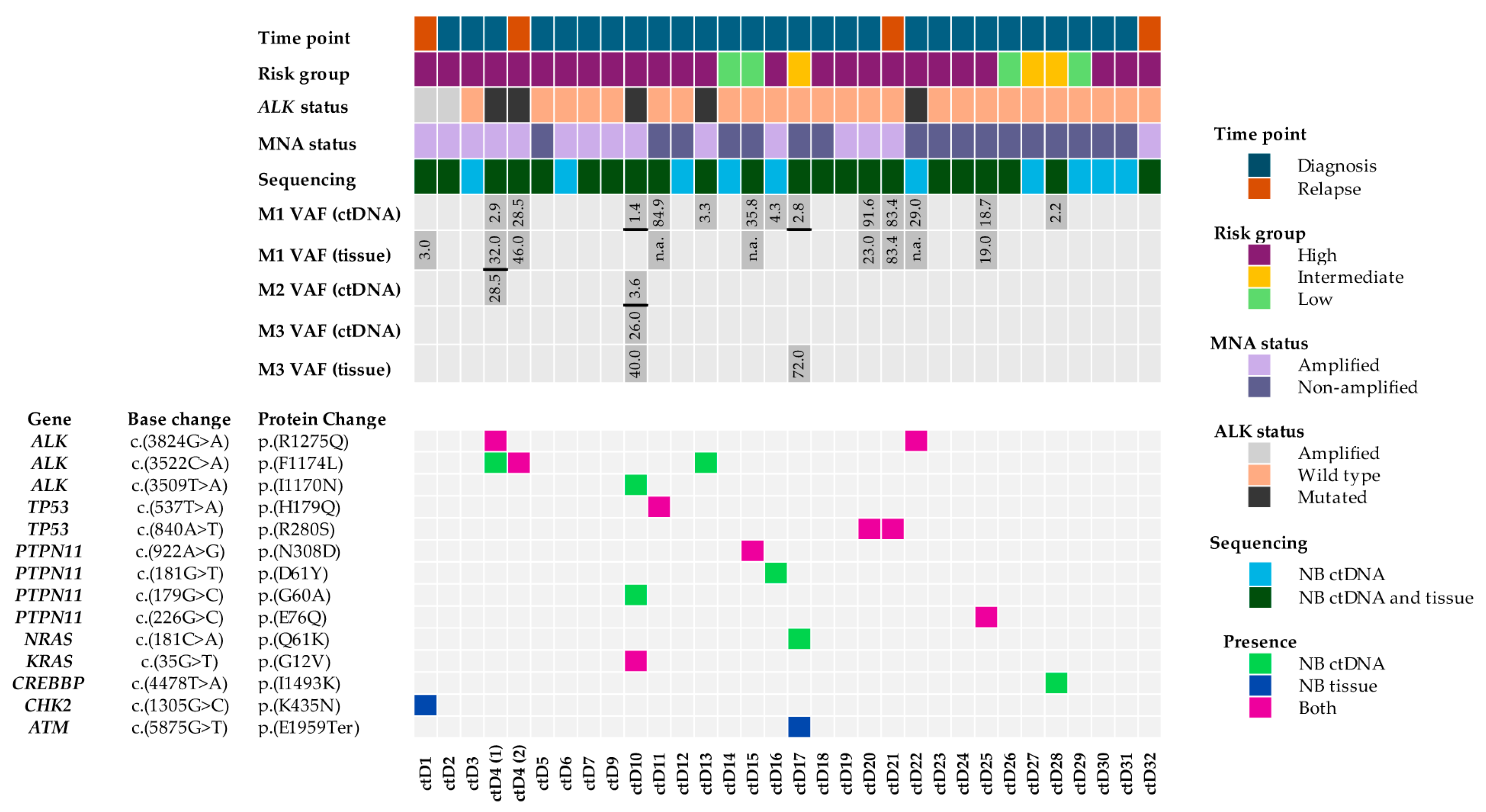
Figure 1. Oncoplot showing comparison of sequencing data between matched NB ctDNA and NB tumours. ctDNA (1–32) is displayed on the x-axes. ctD8 was excluded from further analysis, as the cfDNA concentration was below the threshold value of 20%. Information about patient cohort, VAFs (variant allelic frequencies) and the identified variants are displayed on the y-axes. Three samples had more than one mutation, and hence there are VAFs for M1, M2 and M3 displayed (with M1 being the top one, M2 the middle one and M3 the bottom one on the oncoplot). VAFs from different variants are separated by black lines. ctD4 is included twice, as an additional relapse sample was obtained from this patient and assessed by ddPCR. Samples 20 and 21 are paired diagnosis and relapse samples from the same patient.
Ijms 26 09466 g001
Figure 2. Spatial and temporal heterogeneity of ALK variants. (A) Distribution of pathogenic ALK positions across the tyrosine kinase (TKD) domain within ctDNA. Numbers in red rectangles reflect three cases identified with ALK F1174 mutations, no cases with an ALK F1245 mutation and two cases with an ALK R1275 mutation. (B) Venn diagram showing the spatial and temporal heterogeneity of activating ALK variants in the tumour and plasma obtained from patient ctD4. (C) ALK ddPCR on ctDNA samples collected at the end of treatment when the patient was in remission and 3 months later at relapse. Grey droplets are those with no DNA attached, while green droplets represent the wild-type ALK allele. F1174L variant (visualised as blue droplets) was not detected at the end of treatment but was identified at relapse.
Ijms 26 09466 g002
Figure 3. Longitudinal analysis of a homozygous TP53 variant detected in NB ctDNA and the primary tumour from the same patient. (A) Position of the TP53 variant within the DNA binding domain. (B) Venn diagram depicting the TP53 variant detected across three samples: the diagnostic plasma sample, the post-chemotherapy resected primary tumour and the relapse plasma sample. WGS—whole-genome sequencing.
Ijms 26 09466 g003
Figure 4. Copy number aberrations detected in NB ctDNA. Bar graph shows inverse z-scores for each target region amplified compared to all other regions in the same sample. The regions with a significant copy number change (amplification or gain) are highlighted with longer bins in the positive quadrant. Numerical coverage data could not be retrieved for ctD30, ctD31 and ctD32, so these samples were omitted. ctD1 and ctD2 and ctD20 and 21 are paired samples at diagnosis and relapse, respectively.
Ijms 26 09466 g004
Figure 5. Amplification of MYCN (MNA), ALK and MDM2 oncogenes. The 2D line graph depicts the relative coverage of all 42 targeted genes compared to the respective SNP array data from primary tumours or ctDNA. (A) NB-1691 cell line: MYCN, CDK4 and MDM2 amplification, but not ALK, were detected by relative coverage analyses compared with the SNP array. (B) MNA in SK-N-BE(2)-C cell line. (C) ALK amplification and MNA detected in ctD1 and ctD2 paired samples. (D) MNA, CDK4 and CCND1 amplification in ctD3.
Ijms 26 09466 g005
Table 1. Pathogenic variants detected in ctDNA (ctD), germline DNA (gD) and cell-line DNA samples. All variants were identified as missense variants, apart from NF1, which was identified as a splice donor. Abbreviations: Del—deleterious, POD—possibly damaging, PRD—probably damaging, B—benign, Chr—chromosomal. REVEL score > 0.7 = pathogenic. 1 = SHSY5Y cell line, 2 = SKNBE(2c) cell line. a,b = Identifies ctDNA and matched gDNA pairs. c = Identifies paired samples from the same patient at diagnosis and relapse.
SampleVAFVariant GeneChr. LocationBase ChangeProtein ChangeCOSMIC IDSIFT ScorePolyphen REVEL
ScoreScore
ctD42.85ALKchr2 (p23.2)c.(3824G>A)p.(R1275Q)COSV66555567Del (0)PRD (1)0.885
ctD428.5ALKchr2 (p23.2)c.(3522C>A)p.(F1174L)COSV66556325Del (0.03)POD (0.641)0.752
ctD101.38ALKchr2 (p23.2)c.(3509T>A)p.(I1170N)COSV66589132Del (0)PRD (1)0.906
ctD1026.16KRASchr12 (p12.1)c.(35G>T)p.(G12V)COSV55497419Del (0)PRD (0.972)0.91
ctD103.61PTPN11chr12 (q24.13)c.(179G>C)p.(G60A)COSV61006397Del (0.01)PRD (0.953)0.907
a ctD1184.88TP53chr17 (p13.1)c.(537T>A)p.(H179G)COSV52673406Del (0)PRD (0.98)0.79
a gD11.4TP53chr17 (p13.1)c.(537T>A)p.(H179Q)COSV52669519Del (0)PRD (0.98)0.79
ctD133.33ALKchr2 (p23.2)c.(3522C>A)p.(F1174L)COSV66556325Del (0.03)POD (0.641)0.752
b ctD1535.8PTPN11chr12 (q24.13)c.(922A>G)p.(N308D)COSV61006575Del (0.03)B (0.134)0.838
b gD243.9PTPN11chr12 (q24.13)c.(922A>G)p.(N308D)COSV61006575Del (0.03)B (0.134)0.838
ctD164.34PTPN11chr12 (q24.13)c.(181G>T)p.(D61Y)COSV61004841Del (0)PRD (0.997)0.933
ctD172.26NRASchr1 (p13.2)c.(181C>A)p.(Q61K)COSV54736310Del (0.01)POD (0.709)N/A
c ctD2091.64TP53chr17 (p13.1)c.(840A>T)p.(R280S)COSV52782181Del (0.03)POD (0.843)0.878
c ctD2183.4TP53chr17 (p13.1)c.(840A>T)p.(R280S)COSV52801834Del (0.03)POD (0.843)0.878
ctD2229.03ALKchr2 (p23.2)c.(3824G>A)p.(R1275Q)COSV66555567Del (0)PRD (1)0.885
ctD2518.7PTPN11chr12 (q24.13)c.(226G>C)p.(E76Q)COSV61004751Del (0.01)PRD (0.979)0.733
ctD282.2CREBBPchr16 (p13.3)c.(4478T>A)p.(I1493K)COSV52129182Del (0)PRD (0.991)0.929
144.4KRASchr12 (p12.1)c.(35G>T)p.(G12V)COSV55497419Del (0)PRD (0.972)0.91
150.9SMARCA4chr19 (p13.2)c.(2917C>T)p.(R973W)COSV60787034Del (0)PRD (1)0.86
149.6ALKchr2 (p23.2)c.(3522C>A)p.(F1174L)COSV66555460Del (0.03)POD (0.641)0.752
299.1TP53chr17 (p13.1)c.(404G>T)p.(C135F)COSV52680475Del (0)PRD (1)0.96
2100NF1chr17 (q11.2)c.(1989_2001del)p.(G663fs)N/AN/AN/AN/A
Disclaimer/Publisher’s Note: The statements, opinions and data contained in all publications are solely those of the individual author(s) and contributor(s) and not of MDPI and/or the editor(s). MDPI and/or the editor(s) disclaim responsibility for any injury to people or property resulting from any ideas, methods, instructions or products referred to in the content.