Review

Targeting Mitochondrial Function in Plasmodium falciparum: Insight into Antimalarial Drugs and the Emerging Role of Saccharomyces cerevisiae as a Model System

1
Department of Experimental Medicine, University of Salento, I-73100 Lecce, Italy
2
Parasites and Microbes Research and Training Center, University of Science, Techniques and Technologies, Point G, Bamako P.O. Box 1805, Mali
3
R-Evolution Worldwide Srl Impresa Sociale, I-73024 Maglie, Italy
*
Author to whom correspondence should be addressed.
These authors contributed equally to this work.
Int. J. Mol. Sci. 2025, 26(18), 9150; https://doi.org/10.3390/ijms26189150
Submission received: 21 August 2025 / Revised: 16 September 2025 / Accepted: 17 September 2025 / Published: 19 September 2025
(This article belongs to the Special Issue 25th Anniversary of IJMS: Updates and Advances in Macromolecules)

Abstract

Malaria remains a major global health threat, particularly in low- and middle-income countries, where children under five and pregnant women are most vulnerable. Despite notable progress in reducing malaria-related morbidity and mortality, the rise of drug-resistant Plasmodium falciparum strains continues to undermine eradication efforts. In this context, the parasite’s mitochondrion has emerged as a promising target for novel antimalarial therapies due to its essential role in parasite viability throughout all life cycle stages and its marked structural and biochemical differences from the human counterpart. This review highlights recent advances in the development of compounds targeting mitochondrial function in P. falciparum and discusses the utility of Saccharomyces cerevisiae as a powerful model organism for antimalarial drug discovery. Owing to its shared eukaryotic features, genetic tractability, and capacity for heterologous expression of parasite mitochondrial proteins, S. cerevisiae offers a cost-effective and experimentally accessible platform for elucidating drug mechanisms and accelerating therapeutic development.

1. Introduction

According to the latest World Health Organization (WHO) report, an estimated 263 million malaria cases occurred globally in 2023, across 83 endemic countries. This disease was responsible for approximately 627,000 deaths, with the majority of cases occurring among pregnant women and children under five years of age in low- and middle-income countries [1].
Malaria is caused by protozoan parasites of the genus Plasmodium, with Plasmodium falciparum being the most prevalent and virulent species [2]. Although artemisinin-based combination therapies (ACTs) remain the frontline treatment recommended by the WHO, the emergence and spread of artemisinin (ART)-resistant P. falciparum strains highlight the urgent need for novel therapeutic targets, drugs, and treatment strategies.
The mitochondrion of P. falciparum has attracted increasing scientific interest as an antimalarial target due to its central role in parasite viability and development and its significant divergence from the host organelle, enabling selective therapeutic intervention. Functionally, this organelle supports a wide range of essential cellular processes in the parasite, including ATP production, regulation of redox homeostasis, and the biosynthesis of pyrimidines and heme [3].
Despite stage-specific fluctuations in its metabolic activity throughout the parasite life cycle in the human host, from the liver stages, through the intraerythrocytic asexual stages, to the sexual (gametocyte) stages, the mitochondrion remains an indispensable organelle across all of these phases. Consequently, the increasing focus on this organelle perfectly aligns with the recent paradigm shift in antimalarial drug discovery, which prioritizes the development of multi-stage, mechanism-driven therapies [4].
While current antimalarial drugs primarily act on the asexual blood stages responsible for clinical disease manifestations, there is a pressing demand to identify next-generation antimalarials with broader activity profiles that are able not only to treat the active infection but also to prevent and block the transmission, supporting long-term malaria eradication efforts [5].
In this evolving research landscape, Saccharomyces cerevisiae has emerged as a robust eukaryotic model for mitochondria-targeted antimalarial studies. With its genetic tractability, metabolic versatility, and conservation of core mitochondrial pathways yeast provides an ideal platform for in-depth functional studies and high-throughput screening (HTS) of compounds that disrupt essential mitochondrial processes in P. falciparum. The yeast system not only facilitates the identification of specific mitochondrial molecular targets but also allows for the assessment of stage-selective drug activity by simulating the distinct metabolic states associated with different phases of the P. falciparum life cycle in the human host. Indeed, by modulating growth conditions, such as oxygen levels and carbon-source utilization, yeast metabolism shifts between glycolytic and respiratory states, similarly to what occurs in the parasite’s various life stages.
This review provides a comprehensive overview of the most promising newly discovered or developed mitochondrial-targeting antimalarial compounds, focusing on their molecular mechanisms and stage-specific effects. Additionally, the role of S. cerevisiae is explored as a versatile tool for both drug discovery and mechanistic characterization in the context of mitochondrial inhibition.

2. Role of Mitochondria in the Parasite Life Cycle

2.1. General Overview of the Parasite Life Cycle

P. falciparum has a complex life cycle involving both humans and Anopheles mosquitoes. Transmission begins when a mosquito injects sporozoites into the human bloodstream. These migrate to the liver, infect hepatocytes, and multiply during the pre-erythrocytic stage (liver stage), producing thousands of merozoites.
Merozoites then enter the bloodstream and invade red blood cells (RBCs), starting the erythrocytic stage, where they replicate, cause cell lysis, and release new merozoites. Some differentiate into gametocytes, which are taken up by another mosquito during a blood meal. Inside the mosquito, they develop into sporozoites that reach the salivary glands, ready to infect a new host [6] (Figure 1).

2.2. Mitochondrial Metabolism of the Parasite in the Liver Stage

Studying P. falciparum during its liver stage is particularly difficult, as this phase is asymptomatic and inaccessible in natural human infections. Consequently, much of our understanding comes from rodent malaria models (Plasmodium yoelii and Plasmodium berghei), engrafted mice, and limited in vitro systems using human hepatocytes [8]. After entering hepatocytes, sporozoites mature over 6–7 days, undergoing extensive genome and organelle replication without cytokinesis, leading to a multinucleated schizont. Cytokinesis occurs at the end of this stage, producing thousands of merozoites that enter the bloodstream.
During liver-stage development, the parasite’s mitochondrion increases in size and complexity, displaying tubular cristae characteristic of active, metabolically adaptive organelles [9,10]. However, the metabolic role of mitochondria in this stage remains only partially understood.
To investigate this, Tarun et al. performed transcriptomic and proteomic analyses on P. yoelii liver stages. Their study revealed increased expression of genes involved in the tricarboxylic acid (TCA) cycle and mitochondrial electron transport chain (mETC), suggesting enhanced mitochondrial activity to meet biosynthetic and energetic demands [11].
Further insights came from integrating genetic screens of barcoded P. berghei mutants with transcriptomic data, identifying nine TCA-cycle-related genes as essential for liver-stage development such as the ones encoding for aconitase and α-ketoglutarate dehydrogenase [12,13]. Glucose likely feeds into glycolysis and the TCA cycle, driving ATP production and biosynthesis [14].
Collectively, these findings suggest that during liver-stage growth, Plasmodium adopts a hybrid metabolic strategy—scavenging nutrients such as glucose and amino acids from host hepatocytes, while synthesizing other molecules like fatty acids to support its rapid proliferation.

2.3. Mitochondrial Metabolism of the Parasite in the Asexual Blood Stages

During the 48 h asexual intraerythrocytic cycle of P. falciparum (comprising ring, trophozoite, schizont, and merozoite phases) the parasite’s single mitochondrion undergoes morphological changes, enlarging and branching, yet it remains acristate in all stages [15,16].
The absence of cristae in the mitochondrion suggests a limited capacity for oxidative phosphorylation, implying that the parasite relies predominantly on host-derived glucose and glycolytic pathways for energy production, a metabolic adaptation that has been documented in multiple studies [17,18].
Although the mitochondrion plays a limited role in ATP production during the asexual blood stages of P. falciparum, it remains essential for parasite survival. There are conflicting views regarding the operation of the TCA cycle during the asexual blood stages. Some studies suggest that it functions in a branched manner, with intermediates such as 2-oxoglutarate being diverted mainly for biosynthetic purposes rather than for complete oxidative metabolism. In contrast, other evidence points to a canonical, cyclical function of the pathway. This controversy stems from differences in experimental design, isotope-labeling interpretations, and the parasite’s context-dependent metabolic flexibility.
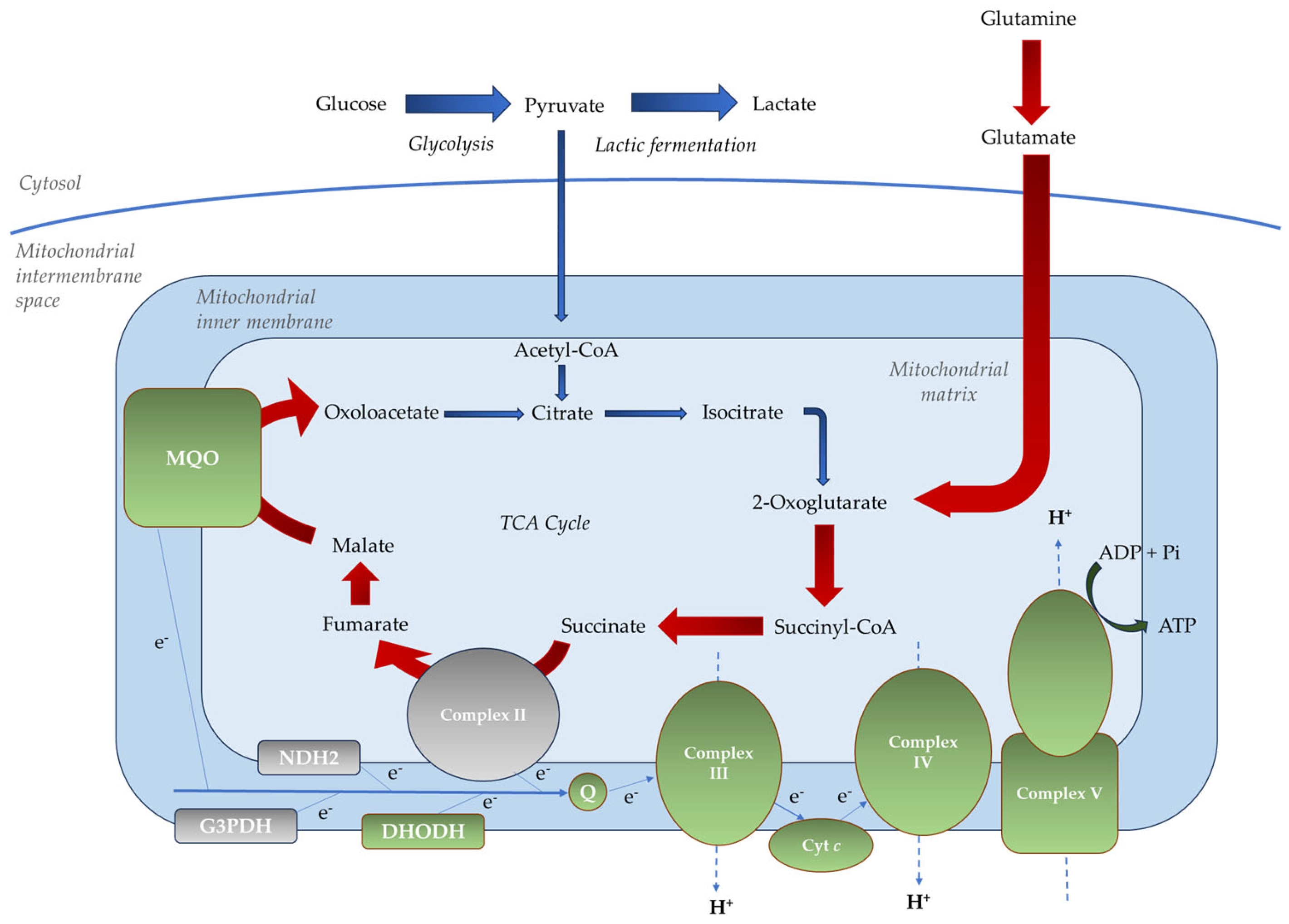
Early metabolomic studies proposed that the TCA cycle in the parasite might not operate as a conventional, unidirectional pathway. Instead, it appeared to function in a branched manner, diverging at the intermediate 2-oxoglutarate, as follows: a reductive branch proceeding in reverse from 2-oxoglutarate through isocitrate, citrate, and oxaloacetate to malate, and an oxidative branch proceeding from 2-oxoglutarate through succinyl-CoA, succinate, and fumarate to malate [19]. However, more recent studies have shown that P. falciparum expresses all of the enzymes required for a complete, canonical TCA cycle during its intraerythrocytic development [17]. This cycle is sustained by carbon skeletons derived from host glucose and glutamine. Specifically, a minor flux from oxaloacetate to isocitrate is maintained through the conversion of pyruvate to acetyl-CoA via the branched-chain ketoacid dehydrogenase complex (PfBCKDH). In contrast, a major flux from 2-oxoglutarate to malate is driven by carbon sourced from glutamine [17,20,21] (Figure 2).
Overall, the parasite demonstrates remarkable metabolic plasticity, regulating nutrient fluxes (glucose, amino acids, and lipids) and rerouting central carbon metabolism to adapt to fluctuating host microenvironments. This flexibility enables P. falciparum to balance energy generation with biosynthetic demands, ensuring survival and replication in response to variations in host cell resources, oxygen availability, and immune pressures.
Genetic knockout studies indicate that most TCA cycle enzymes are not essential for P. falciparum survival during the asexual blood stages, reinforcing the idea that the cycle has a limited role in energy production [22]. Instead, the TCA cycle likely serves biosynthetic and redox functions, contributing to mETC activity, de novo pyrimidine synthesis, purine salvage, and heme biosynthesis by supplying key intermediates such as 2-oxoglutarate, succinyl-CoA, and oxaloacetate [23].
The P. falciparum mETC includes the following:
  • Non-proton motive quinone reductases, such as dihydroorotate dehydrogenase (PfDHODH), malate-quinone oxidoreductase (PfMQO), glycerol 3-phosphate dehydrogenase (PfG3PDH), type II NADH dehydrogenase (PfNDH2, alternative complex I), and succinate dehydrogenase (PfSDH, complex II);
  • Proton motive respiratory complexes, including ubiquinol-cytochrome c oxidoreductase (complex III), cytochrome c oxidase (complex IV), and ATP synthase (complex V) [24] (Figure 2).
Although all these components are expressed during the asexual blood stages, not all are essential. Pfndh2 gene deletion via CRISPR/Cas9 did not affect intraerythrocytic development [25], consistent with findings in P. berghei [26]. Similarly, knockout studies targeting pfsdh revealed that its absence did not impair blood-stage parasite growth [27,28].
In contrast, complexes III, IV, and V are essential. Complex III is critical for reoxidizing ubiquinol to support the activity of PfDHODH, which is indispensable for de novo pyrimidine biosynthesis, a pathway essential to the parasite, as it cannot salvage pyrimidines [21,22]. Complexes III and IV also maintain the mitochondrial membrane potential (Δψm) necessary for metabolite transport and mitochondrial function [29]. Although Complex V is not required for ATP production during the asexual blood stages [30], genetic disruption of its subunits has proven unsuccessful, suggesting it plays a vital role independent of oxidative phosphorylation [31].

2.4. Mitochondrial Metabolism of the Parasite in the Sexual Blood Stages

During each asexual replication cycle, a small fraction (<10%) of P. falciparum ring-stage parasites commit to sexual development, a process called gametocytogenesis [32]. Gametocytes mature through five stages (I–V) over 10–12 days. Mature stage V gametocytes are taken up by mosquitoes, where they differentiate into male (microgametes) and female (macrogametes) gametes in the mosquito midgut [32,33,34].
Gametocyte development involves a programmed remodeling of central carbon metabolism. Unlike asexual blood stages, which rely mainly on anaerobic glycolysis, gametocytes shift toward oxidative metabolism, increasing mitochondrial dependence [17].
This metabolic shift reflects the changing energy demands and environmental conditions faced by gametocytes. While asexual stages rapidly generate ATP through glycolysis in the glucose-rich bloodstream, gametocytes require more efficient and sustained energy production to support prolonged maturation and survival within both the human host and mosquito vector. Hence, the switch to mitochondrial oxidative phosphorylation provides a higher ATP yield and supports biosynthetic pathways crucial for gametocyte development and transmission [17].
These changes are supported by upregulation of the TCA cycle and oxidative phosphorylation genes [17,21], as well as by mitochondrial enlargement into complex multi-lobed structures with tubular cristae [16,35]. This structural adaptation facilitates increased mETC activity and aligns mitochondrial function with the metabolic demands of gametocytes, making mitochondrial morphological changes both a consequence and a facilitator of the metabolic reprogramming.
Metabolomic analyses show gametocytes actively run the TCA cycle using glucose (primary) and glutamine (secondary) as carbon sources, opposite to the asexual stage pattern [17,28] (Figure 3).
Disruption of TCA cycle genes impairs gametocyte development or mosquito-stage transmission [17,21,36]. Indeed, deletion of P. falciparum aconitase (pfaco) results in parasites that arrest at stage III, failing to produce mature gametocytes. Similarly, deletion of α-ketoglutarate dehydrogenase (pfkdh) yields morphologically normal gametocytes that cannot form oocysts when ingested by mosquitoes. Deletion of isocitrate dehydrogenase (pfidh) blocks development beyond stages III/IV [28,36]. Moreover, while several mETC components are dispensable in asexual stages, they are essential in gametocytes. Disruption of genes such as pfndh2 and pfsdh leads to defective gametocyte development.

3. Antimalarial Agents That Target Mitochondrial Function

Atovaquone is the first and only clinically approved antimalarial specifically targeting the Plasmodium mitochondrion. As a ubiquinone analog, it binds to the cytochrome b subunit of complex III, disrupting the mETC and compromising parasite energy metabolism. Chemically classified as a hydroxynaphthoquinone, atovaquone specifically targets the quinol oxidation (Qo) site of complex III [37].
When administered as a fixed-dose combination with proguanil (commercially known as Malarone), atovaquone is widely used for both the treatment of uncomplicated malaria in adults and children and for chemoprophylaxis in travelers. Its excellent safety profile makes it suitable for vulnerable populations, including pregnant women and children, and for mass drug administration campaigns [38]. However, rapid emergence of atovaquone-resistant parasites threatens its long-term efficacy, underscoring the urgent need to develop and clinically validate new mitochondrial-targeting antimalarials.
While resistance to atovaquone arises from mutations in cytochrome b, additional mechanisms of resistance have been reported for other mitochondrial targets. For example, resistance to PfDHODH inhibitors, such as DSM265, can result from point mutations in the pfdhodh gene that reduce drug-binding affinity, or from gene amplification events that increase PfDHODH expression [39]. Moreover, metabolic compensation, such as the upregulation of alternative metabolic pathways or enhanced utilization of host-derived metabolites, can support parasite survival despite inhibition of mitochondrial function, as observed in some ART-resistant parasites [40]. Understanding these adaptive mechanisms is critical for the development of next-generation antimalarials with durable efficacy.

3.1. Agents Targeting the mETC

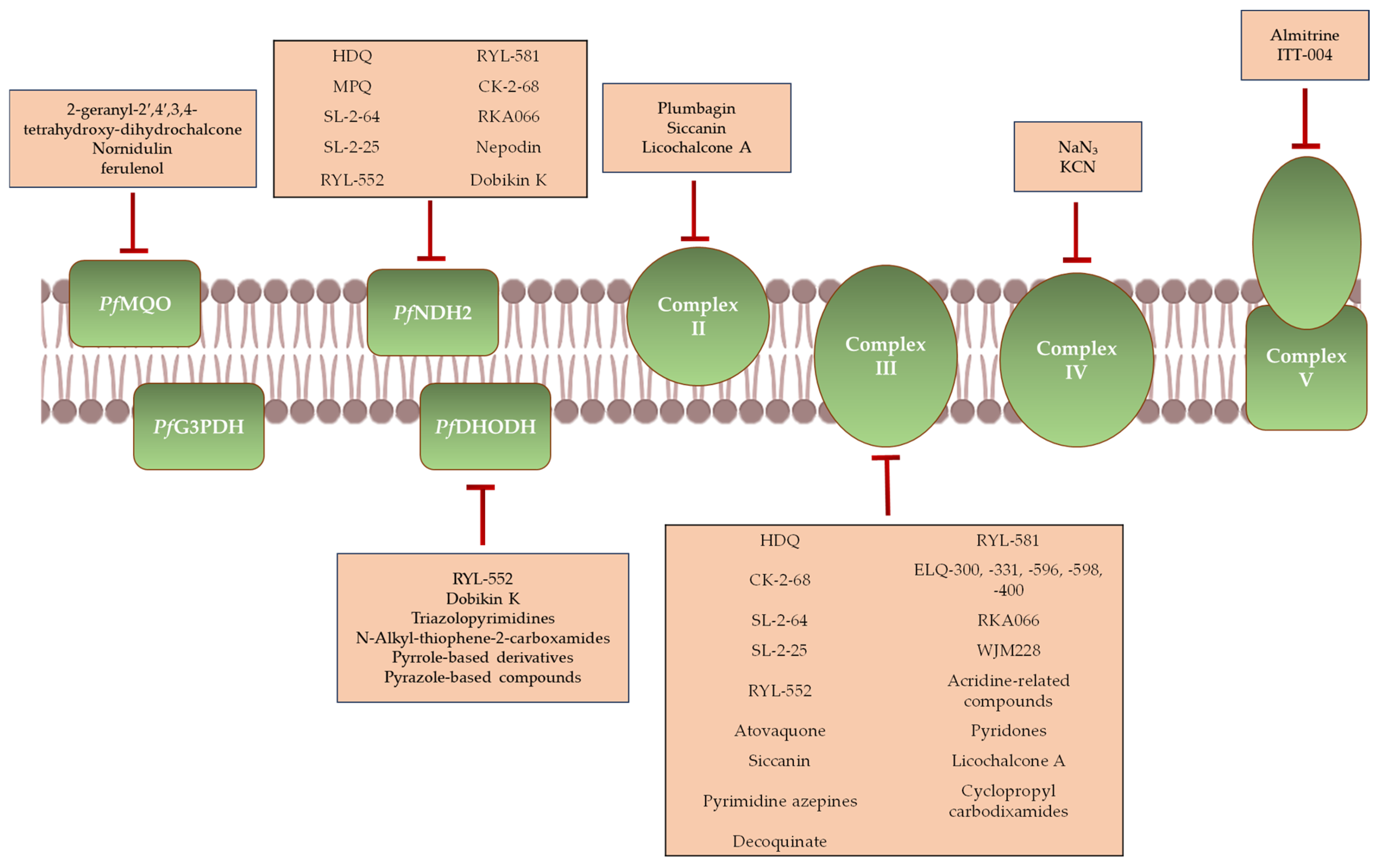
The majority of antimalarial compounds target mETC complexes and dehydrogenases (Figure 4).

3.1.1. Inhibitors of PfNDH2

PfNDH2 is a single-subunit mitochondrial enzyme that functions as an alternative to the multi-subunit complex I in humans. It catalyzes the transfer of electrons from NADH to ubiquinone, thus exhibiting NADH—ubiquinone oxidoreductase activity. Its structural divergence from the human enzyme makes it an attractive target for antimalarial drug development [41].
Several endochin-like quinolones (ELQs) have shown potent inhibitory activity against PfNDH2, with all compounds evaluated in vitro and some also assessed in vivo. Among them, HDQ and its derivatives (CK-2-68, SL-2-64, and SL-2-25) are particularly promising. Initially considered to be PfNDH2-specific, these compounds were later found to also inhibit complex III, indicating a dual-target mechanism [25,42,43].
HDQ exhibits moderate-to-strong in vitro activity (IC50 = 70 nM for PfNDH2; 19 nM for complex III) but is limited by high lipophilicity (CLogP = 6.5) [42,44]. Structural modifications aimed at improving pharmacokinetics led to derivatives such as CK-2-68, which maintains potent PfNDH2 inhibition with reduced activity against complex III, though its lipophilicity remains similar. SL-2-25 offers balanced inhibition of both enzymes and a lower lipophilicity (CLogP = 4.2), while SL-2-64 shows exceptional potency against PfNDH2 (IC50 = 4 nM) and favorable physicochemical properties [38,42].
Another notable class is the RYL series, characterized by a multitarget profile. RYL-552 was first described as a non-competitive PfNDH2 inhibitor (IC50 = 3.73 nM) and later shown to also bind the Qo and quinone reduction (Qi) sites of complex III, with potential activity against PfDHODH [41,45,46]. In murine models, RYL-552 effectively cleared parasitemia at various doses (10–90 mg/kg). Its optimized analog, RYL-581, exhibits markedly improved potency (EC50 = 0.056 nM) and in vivo efficacy, outperforming atovaquone and leading to complete parasite clearance in infected mice [41]. RKA066 is another dual-site inhibitor, active on both PfNDH2 and the Qi site of complex III [47].
In contrast to these multitarget inhibitors, a yeast-based screen identified MPQ as a selective PfNDH2 inhibitor, highlighting its potential for higher target specificity [43].
Natural products have also been explored. Nepodin, a naphthol compound, selectively inhibits PfNDH2 in vitro and significantly suppresses parasitemia in vivo [48]. More recently, Dobikin K, a sesquiterpenoid, was found to inhibit PfNDH2 in a concentration-dependent manner and disrupt the Δψm in P. falciparum [49].

3.1.2. Inhibitors of PfSDH

PfSDH exhibits notable structural differences compared to its mammalian counterpart. While the mammalian enzyme comprises four subunits (Fp, Ip, CybL, and CybS) only the flavoprotein (Fp) and iron–sulfur cluster protein (Ip) subunits have been identified in P. falciparum, with no apparent homologs for the membrane anchor subunits [50,51]. This divergence underscores PfSDH as a potential target for selective antimalarial therapy.
The antibiotic Siccanin has emerged as a potent in vitro PfSDH inhibitor, showing nanomolar activity against the enzyme and low micromolar inhibition of complex III. Notably, Siccanin exhibits no detectable effect on mammalian complexes at high concentrations, supporting its selectivity for the parasite [51].
Other small molecules have also demonstrated PfSDH inhibitory activity. Plumbagin, a hydroxynaphthoquinone, selectively targets PfSDH and shows promising in vitro antimalarial effects, although its in vivo efficacy remains limited [52,53].
Licochalcone A, a natural chalcone, inhibits both PfSDH and complex III in vitro, with a stronger effect on complex III [54,55].

3.1.3. Inhibitors of Complex III

Complex III is a well-established antimalarial target. Inhibitors of this complex typically act at the Qo or Qi sites of the cytochrome b subunit by mimicking ubiquinone/ubiquinol. Their selectivity for P. falciparum arises from species-specific differences in the coenzyme Q (CoQ-binding site residues [56]. These compounds span several chemical classes, from ELQs to cyclopropyl carboxamides.
ELQs primarily target complex III, though many also inhibit other components of the mETC. Among Qi site inhibitors, ELQ-300 is a standout candidate, with nanomolar potency (IC50 = 0.56 nM), high selectivity (>18,000-fold over human complex III), and favorable safety and metabolic profiles [57,58,59,60]. Due to poor solubility and low oral bioavailability, the prodrug ELQ-331 was developed, demonstrating improved pharmacokinetics and progressing toward once-weekly prophylactic use [59]. ELQ-596, a biphenyl analog, improves potency against resistant P. falciparum strains and its prodrug, ELQ-598, was optimized for better ADME (absorption, distribution, metabolism, and excretion) properties [61]. Targeting the Qo site, compounds such as WJM228 have shown potent activity [58,62,63]. ELQ-400 binds both Qo and Qi sites, exhibiting strong dual-site inhibition [64,65].
Acridine-related compounds also demonstrate significant activity against complex III. WR 249685 is a potent Qo site inhibitor (IC50 = 3 nM) with higher selectivity than atovaquone [57]. T111, the most potent acridone reported, is thought to act on both Qo and Qi sites [66,67].
Pyridones are validated scaffolds for complex III inhibition. GW844520 and its successor GSK932121 inhibit the Qi site, showing high potency and preclinical efficacy. However, their clinical development was halted due to cardiac and off-target toxicity in mammals, respectively [42,47,57,68,69].
Another promising class includes pyrimidine azepines. PyAz90 has demonstrated strong in vitro and ex vivo activity as a novel Qo site inhibitor, with partial overlap with atovaquone’s binding region but distinct molecular interactions [70].
Cyclopropyl carboxamides were identified through a Seahorse XFe96 metabolic screening of the MMV (Medicines for Malaria Venture) Pathogen Box. Among these, MMV024397 emerged as a potent Qo-site inhibitor, with reduced efficacy observed in atovaquone-resistant P. falciparum strains, supporting its proposed mechanism of action [56]. Further screening identified several structurally related analogs, including WJM280, W466, and W499, all active in the low nanomolar to submicromolar range without detectable cytotoxicity [71].
The quinolone decoquinate is another significant inhibitor of complex III, likely targeting the Qo site, with strong efficacy in vitro and in vivo, as well as limited cross-resistance with atovaquone [42,72].

3.1.4. Inhibitors of Complex IV

Complex IV has recently emerged as a promising but underexplored antimalarial target. In P. falciparum, it is susceptible to classical inhibitors like sodium azide (NaN3) and potassium cyanide (KCN). However, their lack of selectivity between parasite and host enzymes leads to inhibition of human mitochondrial respiration, significantly limiting their therapeutic potential [73,74]. This highlights the need for parasite-selective inhibitors with improved safety profiles.

3.1.5. Inhibitors of Complex V

Few inhibitors of complex V have been identified to date. Among them, almitrine, originally developed as a respiratory stimulant, has shown moderate in vitro antimalarial activity, with IC50 values of 13.2 ± 5.6 µM (chloroquine-resistant FCM 29) and 12.6 ± 3.6 µM (chloroquine-sensitive L-3) [55,75].
Another example is ITT-004, a derivative of Basic Blue 3. While the parent compound is highly potent but cytotoxic, ITT-004 maintains antimalarial efficacy with significantly reduced cytotoxicity in vitro and in vivo [76].

3.1.6. Inhibitors of PfDHODH

PfDHODH links the mETC to de novo pyrimidine biosynthesis by catalyzing dihydroorotate to orotate, the pathway’s rate-limiting step. Given pyrimidines’ essential role in nucleic acid, phospholipid, and glycoprotein synthesis, PfDHODH is a validated and druggable target.
DSM265, a triazolopyrimidine and the first PfDHODH inhibitor in clinical development, shows potent in vitro activity (IC50 = 0.024 µM), excellent selectivity (>4000-fold vs. human DHODH; IC50 > 100 µM), and favorable pharmacokinetics in preclinical models [77,78,79]. Phase Ia/Ib trials support its potential for single-dose treatment and weekly chemoprophylaxis, and it has advanced to Phase IIa studies as both monotherapy and in combination therapy in adults [80]. DSM421, a second-generation compound of the same chemical class, has slightly lower potency (IC50 = 0.053 µM) but superior pharmacokinetic (PK) properties, including better solubility, plasma exposure, and reduced clearance [77].
Using HTS and medicinal chemistry, Booker et al. identified N-alkyl-5-(1H-350 benzimidazol-1-yl)-thiophene-2-carboxamides as selective PfDHODH inhibitors [81,82]. Among these, Genz-667348 demonstrated potent inhibition (IC50 = 0.022 µM) but was halted at the late lead stage due to off-target toxicity [42,79]. However, its analog Genz-669178, with similar potency (IC50 = 0.04 µM), showed improved safety and PK, and was effective in multiple murine malaria models [42].
Palmer et al. reported two pyrrole-based PfDHODH inhibitors, DSM705 and DSM873, with IC50 values of 0.095 µM and 0.069 µM, respectively. Both showed strong in vivo efficacy and favorable PK in rodents and SCID mice, supporting continued development [83,84].
Building on pyrrole scaffold, pyrazole-based derivatives were explored, leading to DSM1465—a highly potent and selective inhibitor (IC50 = 0.015 µM) that outperformed DSM265 by 7-fold in vitro. DSM1465 also showed robust efficacy in SCID mouse models and has a predicted human half-life of 115 h, suggesting the potential for single-dose chemoprevention lasting up to 30 days [85].

3.1.7. Inhibitors of PfMQO

MQO’s absence in humans makes it a unique antimalarial target.
Nornidulin, a fungal metabolite, inhibited PfMQO (IC50 = 51 µM) and suppressed P. falciparum proliferation (IC50 = 44.6 µM) without mammalian cytotoxicity [86].
A screen of 113 known mETC inhibitors identified ferulenol as the most potent (IC50 = 57 nM), acting as an uncompetitive inhibitor forming a dead-end complex with PfMQO [87].
Hidayati et al. isolated 2-geranyl-2′,4′,3,4-tetrahydroxy-dihydrochalcone from Artocarpus altilis, which showed potent antimalarial activity (IC50 = 0.17 µM), low cytotoxicity (CC50 = 19.7 µM), and a high selectivity index (115.8) [88].

3.2. Other Mitochondrial Targets

Beyond PfMQO and PfSDH, other TCA enzymes are attractive drug targets.
PfACO is selectively inhibited by sodium fluoroacetate (NaFAc), causing citrate accumulation and depletion of downstream TCA metabolites in P. falciparum [17,89].
PfFH, a class I fumarate hydratase, is potently inhibited by mercaptosuccinic acid (Ki in the nanomolar range). The human enzyme, a class II hydratase, is structurally unrelated, making PfFH a highly selective target [90].
Other mitochondria-targeting antimalarials act through indirect mechanisms. ART, for instance, is thought to disrupt mitochondrial function by causing membrane depolarization without directly targeting classical mETC components [91]. The Plasmodium heme biosynthesis pathway also plays a role: excess 5-aminolevulinic acid (ALA) leads to reactive oxygen species (ROS) production and parasite growth inhibition. When combined with sodium ferrous citrate (SFC), ALA shows synergistic antimalarial activity, curing 50% of P.-yoelii-infected mice and providing long-term protection, highlighting its potential for combination therapies [92,93].

4. Potential Therapeutic Applications of Mitochondria-Targeting Agents Across Different Stages of the Parasite Life Cycle

Historically, malaria drug development has concentrated on the asexual blood stage of Plasmodium parasites, the clinically relevant phase responsible for symptoms such as fever, anemia, cerebral malaria, and metabolic acidosis. These complications result from the rupture of infected erythrocytes, triggering inflammation and the release of parasitic by-products. This stage has remained the primary target for antimalarial therapies due to its direct involvement in disease pathology and accessibility to systemic drugs. Consequently, most approved antimalarials act on intraerythrocytic parasites to reduce parasite load and alleviate symptoms. However, the emergence of P. falciparum strains resistant to frontline treatments, including ACTs, underscores the limitations of this single-stage focus. Resistance has rapidly developed to all blood-stage antimalarials, threatening treatment efficacy and global control efforts. To address these challenges and support eradication goals, research is shifting toward developing next-generation drugs with novel mechanisms and multi-stage activity aimed not only at treating acute infection but also at preventing infection and blocking transmission.
To support this approach, the global malaria research community, introduced a framework of target candidate profiles (TCPs) and target product profiles [94]. These TCPs classify compounds based on activity against specific Plasmodium life stages. For P. falciparum, the following three are especially relevant: TCP-1—targets asexual blood stages (symptomatic); TCP-4—targets liver stages (asymptomatic); and TCP-5—targets sexual stages responsible for transmission.
Each stage represents a potential bottleneck in the parasite life cycle and, thus, a valuable target for intervention. Importantly, all human-infecting stages of Plasmodium depend to varying extents on mitochondrial functions essential for survival, including energy metabolism, pyrimidine and heme biosynthesis, and redox balance. The mitochondrion, therefore, presents a promising target for the development of multi-stage antimalarial agents.
Table 1 summarizes the mitochondrial targets discussed in Section 3, along with the corresponding life stages affected in the human host. This helps clarify the relationship between each molecular target and its stage-specific antiplasmodial activity.
Among mETC components, complex III is a well-established antimalarial target, essential across all three developmental stages of Plasmodium within the human host.
In the asexual blood stage, complex III is crucial for ubiquinone recycling, supporting PfDHODH-mediated de novo pyrimidine biosynthesis, a process vital for parasite survival [28,30]. In liver and sexual stages, where mitochondrial respiration appears coupled to ATP production, complex III supports electron transport as part of core mETC function.
All known complex III inhibitors consistently demonstrate strong activity against asexual blood stages, highlighting the complex’s essentiality. Several compounds also inhibit liver and sexual stages, indicating broader utility, though in the absence of data, potential multi-stage activity cannot be ruled out.
Atovaquone, the only approved mitochondrial-targeting antimalarial, is effective at all intra-host stages: it treats symptomatic blood-stage infection, prevents liver-stage development, and blocks transmission by targeting gametocytes. It shows activity across gametocyte maturation, with IC50 values ranging from 24 µM for stages II–III to 5.31–50.4 µM for stages IV–V, indicating reduced potency in later stages [7].
This aligns with the general trend of greater drug susceptibility in early gametocytes (stages I–III).
Several antimalarial compounds targeting complex III have shown efficacy across the three key stages of P. falciparum development in humans. Among them, ELQ-300, ELQ-331, and ELQ-400 have demonstrated potent activity against liver, asexual blood, and transmission stages [59,64,65,96]. In murine models, ELQ-400 prevented both transmission and liver-stage infection at single oral doses as low as 0.08–0.1 mg/kg, and effectively cleared blood-stage parasites at 1 mg/kg [98].
The pyrimidine azepine compound PyAz90 has also exhibited multi-stage potential. Although it showed only moderate activity in vitro against P. falciparum gametocytes (EC50 > 10 μM), it significantly reduced exflagellation center formation by up to 80% at 10 μM, suggesting a transmission-blocking effect [70,101].
Similarly, compounds from the cyclopropyl carboxamide class have demonstrated broad activity across liver, blood, and sexual stages. Decoquinate, identified through a large-scale screen of 1037 compounds, emerged as a highly potent complex III inhibitor against liver stages, with an IC50 of 2.6 nM. It has also shown efficacy against asexual and sexual blood stages in both in vitro and in vivo settings [72].
Beyond complex III, other mitochondrial targets such as PfNDH2 and PfSDH are required for transmission and may play roles in liver-stage development, but they appear non-essential in asexual blood stages [26,27,35,108].
Accordingly, MPQ, a specific PfNDH2 inhibitor, is ineffective against asexual parasites, and its activity in other stages remains uncharacterized. Nepodin, another PfNDH2 inhibitor, has been associated with reduced parasitemia in vivo [48], though this effect may stem from off-target mechanisms. Similarly, plumbagin, a PfSDH inhibitor, shows limited efficacy against asexual stages, and data for liver or gametocyte stages are lacking.
Complex IV, along with complex III, contributes to maintaining the Δψm, suggesting it may be essential across all parasite stages. However, known inhibitors such as NaN3 and KCN are broad-spectrum complex IV inhibitors with poor specificity, and their effects have only been observed in asexual forms [73]. Experimental evidence for their activity against liver or sexual stages is currently absent.
The function of complex V varies with the parasite stage. In liver stages, glucose import suggests possible ATP production via oxidative phosphorylation, although direct experimental support is lacking [14]. In contrast, during the asexual blood stage, mitochondrial ATP generation is minimal [30], yet complex V appears essential, possibly due to alternative functions, as gene disruption attempts in P. falciparum have failed [31]. This is supported by observations on almitrine, which exhibits moderate in vitro activity against asexual blood stages, and ITT-004, which reduces parasitemia both in vitro and in vivo. However, no data are currently available on their efficacy in liver or sexual stages. In gametocytes, where energy metabolism supports the extensive remodeling needed for maturation and transmission, the role of complex V remains unexplored.
PfDHODH, a key enzyme in pyrimidine biosynthesis, is essential in liver and asexual blood stages, but not in gametocytes [30]. DSM265, a selective PfDHODH inhibitor, shows potent activity against liver stages and slows asexual-stage parasite growth, but does not clear mature gametocytes [102]. Other PfDHODH inhibitors (Table 1) also demonstrate efficacy primarily against asexual stages, with occasional activity in liver stages; none show confirmed activity in sexual stages.
The TCA cycle appears largely dispensable during the asexual blood stage, with six out of eight enzymes being non-essential. The essentiality of PfMQO and PfFH remains debated; although early studies suggested they could not be knocked out [28], more recent CRISPR-based evidence indicates they may be dispensable [22]. In contrast, the TCA cycle is more critical in gametocyte development. Disruption of PfACO arrests gametocyte maturation at stage III and its inhibitor NaFAc has no effect on asexual stages but significantly impairs sexual-stage development [17,28,36]. Specific inhibitors of PfMQO (such as 2-geranyl-2′,4′,3,4-tetrahydroxy-dihydrochalcone, nornidulin, and ferulenol) and PfFH (e.g., mercaptosuccinic acid) show activity against asexual stages, though their effects in liver or sexual stages remain untested.

5. The Yeast S. cerevisiae as a Model for Malaria Research: From General Considerations to Mitochondrial Pharmacological Targeting

5.1. General Advantages of S. cerevisiae as a Model for P. falciparum Research

This section outlines the rationale for using S. cerevisiae as a robust and versatile model organism in malaria research, particularly for studying P. falciparum molecular biology and identifying parasite-specific genetic and metabolic pathways.
As unicellular eukaryotes, S. cerevisiae and P. falciparum share key cellular features, including a membrane-bound nucleus, Golgi apparatus, endoplasmic reticulum, and mitochondria. Despite evolutionary divergence, their mitochondria retain similar structural and functional characteristics (double membranes, internal compartmentalization, and an electron transport chain), supporting roles in ATP production, metabolism, oxidative stress response and biosynthesis of essential metabolites. These similarities are discussed in greater detail in Section 5.2.
One major advantage of yeast is its rapid, cost-effective growth in simple media. With a generation time of approximately 90–120 min, S. cerevisiae thrives in media containing basic nutrients [109]. In contrast, P. falciparum requires complex, stage-specific culture conditions, including RPMI 1640 supplemented with glucose, hypoxanthine, and reduced glutathione [110]. The parasite also depends exclusively on glucose, is unable to metabolize alternative sugars, and is highly sensitive to vitamin antimetabolites [111]. Additionally, its in vitro maintenance necessitates a low-oxygen (microaerophilic) environment to replicate erythrocytic conditions.
Yeast’s genetic tractability is another key asset. Its well-characterized genome, short life cycle, and ability to exist in haploid form allow for efficient gene manipulation, mutagenesis, and knockout studies [112]. These properties are invaluable for dissecting gene function and enable heterologous expression of P. falciparum proteins, particularly mitochondrial proteins with yeast orthologs, which is essential for investigating mechanisms of drug action and resistance.
Moreover, S. cerevisiae displays remarkable metabolic plasticity. As a facultative anaerobe, it can switch between fermentation and mitochondrial respiration depending on oxygen availability and carbon source. Under fermentable conditions like glucose, it favors fermentation in low oxygen; when grown on non-fermentable carbon sources (e.g., ethanol or acetate), it relies solely on oxidative metabolism [113,114]. This flexibility mirrors the metabolic transitions in P. falciparum, which shifts from glycolysis in its asexual blood stages to oxidative metabolism during liver-stage development and gametocytogenesis.
This metabolic adaptability is also relevant for understanding drug resistance. For instance, ART resistance in P. falciparum involves not only mutations in the pfkelch13 gene but also significant metabolic reprogramming. Resistant parasites show increased glycolytic activity and altered TCA cycle flux, enhancing glutathione production to mitigate oxidative stress. The mitochondrial citrate/oxoglutarate carrier (PfCOCP) supports this by exporting key metabolites for cytosolic glutathione synthesis and NADPH generation. Simultaneously, downregulation of mETC components indicates a shift toward alternative energy pathways [40].
Finally, S. cerevisiae is a powerful tool for antimalarial drug discovery, particularly in the development of HTS platforms. Genetically engineered yeast strains enable the rapid testing of large chemical libraries, significantly accelerating the identification of candidate compounds [115,116]. For example, Frame et al. engineered yeast to express the P. falciparum purine transporter PfENT1, identifying nine inhibitors that blocked [3H]-adenosine uptake and parasite growth [117]. Similarly, Sweeney et al. constructed a yeast strain dependent on the parasite’s phosphate transporter PfPiT, leading to the identification of two active compounds from a 21-compound library [118].

5.2. Functional and Structural Analogies and Divergences Between the Mitochondrion of P. falciparum and S. cerevisiae

Despite species-specific differences, the mitochondria of S. cerevisiae and P. falciparum share significant structural and functional similarities, making yeast a useful heterologous system for studying mitochondrial proteins from the parasite. These similarities enable functional complementation studies to investigate potential drug targets and stage-specific roles of mitochondrial proteins in P. falciparum.
A key point of comparison is the mETC. Both organisms lack the canonical multi-subunit complex I, instead relying solely on type II NADH dehydrogenases. In yeast, three such enzymes exist, Ndi1, Nde1, and Nde2, with Ndi1 and Nde1 being functionally dominant and homologous to the parasite single NADH dehydrogenase, PfNDH2 [43]. Notably, Ndi1 and PfNDH2 are both oriented toward the mitochondrial matrix and oxidize NADH from the TCA cycle, whereas Nde1 and Nde2 face the intermembrane space and process cytosolic NADH [119].
Complex II in both species includes conserved catalytic subunits (Sdh1 and Sdh2 in Saccharomyces, and PfSDHA and PfSDHB in Plasmodium), responsible for transferring electrons from succinate to ubiquinone. However, P. falciparum exhibits a divergent PfSDHC subunit (Sdh3 in yeast) and lacks a direct homolog of Sdh4. Instead, it incorporates four unique accessory proteins (PfC2AP1-4), which are absent in yeast and likely contribute to complex stability [16]. The parasite’s complex II also supports bidirectional activity, catalyzing both succinate oxidation and reverse fumarate reduction, reflecting its metabolic flexibility [53].
Complex III in P. falciparum (~730 kDa) is slightly larger than that of yeast (~670 kDa) and comprises 12 subunits compared to yeast’s 10. Despite this, it retains approximately 40% sequence homology [16,29,120]. The three catalytic subunits (cytochrome b, cytochrome c1, and the Rieske iron–sulfur protein) are conserved. Importantly, the Qo site of cytochrome b, a known drug-binding region, is highly conserved. While yeast-specific subunits like Core 1, Core 2, and Qcr10 are absent in P. falciparum, the parasite contains orthologs of Qcr6–9 and unique proteins such as PfMPPα/β and apicomplexan-specific PfC3AP1–3 [16].
Complex IV in P. falciparum is notably larger (~570 kDa) than in yeast (~200 kDa), due to its expanded subunit composition—22 subunits versus yeast’s 12–13 [121,122]. While both species share core catalytic subunits (Cox1, Cox2, and Cox3) along with several non-catalytic components, P. falciparum possesses lineage-specific subunits, including PfCOX13, PfCOX14, PfCOX16, PfCOX18, PfCOX19, PfCOX24, PfCOX30, and five unique proteins (PfC4AP1–5) absent in yeast [16].
Complex V is also largely conserved in its catalytic architecture. Both species share core F1 subunits (ATPα, β, γ, δ, and ε) and structural components of the peripheral stalk, such as OSCP and subunits b and d. The proton-translocating Fo domain also shows conservation, including subunit a, the c-ring, ATP8, ATPi/j, and ATPk. However, P. falciparum expresses several unique accessory subunits (PfATPTG2, G3–G4, G6, G9–G13, G15, and G17) not found in yeast, while yeast-specific subunits like ATPe, ATPf, and ATPg are absent in the parasite [16,123,124].
A notable divergence lies in the DHODH enzymes. Yeast expresses a cytosolic, fumarate-dependent type 1A Dhodh that does not require ubiquinone, while P. falciparum utilizes a mitochondrial, ubiquinone-dependent PfDHODH. This difference precludes functional complementation of the parasite enzyme by the yeast counterpart [125].
However, recent advances in genetic engineering could provide potential strategies to overcome this limitation. For instance, heterologous expression of PfDHODH in yeast strains lacking endogenous Dhodh could be optimized by fusing a yeast-specific mitochondrial targeting sequence to the parasite enzyme, to facilitate the correct mitochondrial localization of PfDHODH within yeast cells. Additionally, the design of chimeric proteins combining domains from yeast and P. falciparum DHODHs could enhance structural compatibility and enzymatic functionality in the yeast cellular environment. Such chimeras might retain the parasite-specific catalytic site while adopting yeast-specific regions that promote proper folding, stability, and interactions with other mitochondrial proteins. These approaches have been successfully applied in other cross-species complementation studies and represent promising avenues for functional expression and drug screening applications in yeast-based models [126,127].
TCA cycle enzymes also illustrate key divergences. For example, while yeast mitochondrial malate dehydrogenase (Mdh1) and PfMQO both catalyze the conversion of malate to oxaloacetate, they differ in location and cofactor used. Mdh1 operates in the matrix using NAD+ as an electron acceptor, whereas PfMQO is membrane-bound and uses quinone, reducing it to quinol [128]. Similarly, FH differs in class and catalytic mechanism. S. cerevisiae expresses a class II Fh, oxygen-stable and lacking Fe–S clusters, while P. falciparum encodes a class I PfFH that is oxygen-sensitive and contains [4Fe–4S] clusters critical for radical-based catalysis. These differences prevent functional complementation between the two enzymes [90].
Several yeast mitochondrial carriers also show potential orthology with parasite proteins. For example, Yhm2, involved in oxidative stress response in yeast, has a likely counterpart in P. falciparum (PlasmoDB ID PF3D7_1223800), possibly playing a similar protective role against oxidative damage [40,129,130].

5.3. S. cerevisiae as a Model Organism to Study the Function of P. falciparum Mitochondrial Proteins

Understanding the roles of individual P. falciparum proteins is crucial for uncovering metabolic pathways essential to parasite survival and for identifying new antimalarial targets. S. cerevisiae is a valuable model for the functional characterization of P. falciparum mitochondrial proteins due to its genetic manipulability and capacity for mitochondrial transformation. This system supports detailed analysis of protein localization, regulatory interactions, and function within a conserved eukaryotic mitochondrial environment.
Many P. falciparum mitochondrial proteins share sequence or structural homology with yeast counterparts, enabling comparative functional genomics. Functional complementation assays, expressing parasite genes in yeast strains lacking the corresponding endogenous proteins, can reveal conserved biochemical mechanisms and highlight potential therapeutic targets. This approach has been applied in several studies. Salcedo-Sora et al. characterized PfCHA, a putative mitochondrial Ca2+/H+ antiporter involved in calcium homeostasis. Expression of PfCHA in a Δvcx1 yeast mutant restored calcium sequestration following external calcium challenge, monitored through an apoaequorin-based bioluminescent assay for cytosolic Ca2+, Mg2+, and Mn2+ fluxes [131]. Although PfCHA localizes to the mitochondria in P. falciparum, in yeast it targeted the vacuolar membrane but retained functionality, demonstrating the flexibility of the yeast system for studying membrane transport proteins despite differences in subcellular targeting.
Jenkins et al. examined PfCOQ10, the P. falciparum orthologue of the yeast ubiquinone-binding protein Coq10, essential for mitochondrial respiration. Despite sharing only ~27% sequence identity, PfCOQ10 preserved key residues for ubiquinone binding. Its expression in a Δcoq10 yeast strain rescued growth on glycerol, a non-fermentable carbon source. Western blotting confirmed mitochondrial localization, while a truncated version lacking the mitochondrial leader sequence failed to complement the defect, highlighting the importance of correct mitochondrial import [132].
Chellappan et al. investigated PfPHB1 and PfPHB2, two P. falciparum prohibitins, highly conserved inner mitochondrial membrane proteins involved in mitochondrial DNA organization, respiratory complex assembly, and overall mitochondrial integrity. Structural modeling with Protein homology/analogY recognition engine (Phyre2) revealed strong similarity to homologs in other species, including conserved interaction motifs. Yeast two-hybrid assays confirmed interaction between PfPHB1 and PfPHB2, suggesting that similar prohibitin complexes may form in the parasite mitochondrion [133].

5.4. S. cerevisiae as a Model Organism for the Identification and Characterization of Molecular Targets of Promising Antimalarial Compounds

Given the functional and structural similarities in mitochondrial proteins between yeast and the malaria parasite highlighted in the Section 5.2, along with conserved mitochondrial pathways—such as oxidative phosphorylation, Δψm maintenance, protein import, and redox homeostasis—enable S. cerevisiae to make a meaningful impact on accelerating antimalarial drug discovery. Specifically, S. cerevisiae is a powerful tool for identifying molecular targets of antimalarial compounds, particularly through site-directed mutagenesis to pinpoint amino acid residues critical for compound binding. This approach, combined with computational docking and molecular modeling, provides insights into binding conformations and affinities, aiding drug design and optimization.
Song et al. used yeast to investigate the target of ELQ-400, leveraging the conserved Qo and Qi sites of cytochrome b between yeast and apicomplexans [64]. Yeast strains carrying Plasmodium-like mutations in these sites were engineered to assess ELQ-400 sensitivity. The compound strongly inhibited the yeast complex III (IC50 ≈ 88 nM), with mutations in either site significantly altering sensitivity. Plasmodium-like substitutions in the Qi site increased sensitivity up to seven-fold, while Qo site mutations enhanced it up to 17-fold. ELQ-400 demonstrated a distinct binding mode compared to other Qi inhibitors, as evidenced by the absence of cross-resistance in Qi mutants. Docking analyses showed that ELQ-400 mimics ubiquinol binding at the Qi site, with key residues like M221 contributing to stabilization. At the Qo site, binding is supported by a hydrogen bond with Rieske iron–sulfur protein-H181 and π–π interactions with Y279; the Y279S mutation moderately reduced sensitivity [64]. These findings confirm ELQ-400 targets both Qo and Qi sites, suggesting a dual inhibition mechanism effective against resistant Plasmodium strains.
Since the atomic structure of complex III remains unresolved, the yeast complex III crystal structure (PDB ID: 3CX5) is commonly used as a reference. Based on this, Calit et al. generated a 3D model of complex III based on the yeast structure to investigate the interaction of PyAz90 with the atovaquone binding site [70]. Docking revealed π–π interactions between PyAz90 and complex III residues F123 and F264, located near conserved residues Y268 and V259 within the atovaquone binding pocket [134]. These results indicate that PyAz90 and atovaquone bind overlapping regions of complex III, but engage distinct residues [70].

5.5. S. cerevisiae as a Model Organism to Characterize the Mechanism of Action and/or Resistance of Antimalarial Compounds

5.5.1. Plasmodione

As part of the PlasmoPrim project, Mounkoro et al. investigated the mitochondrial and redox-related effects of Plasmodione (PD) using S. cerevisiae as a model [135]. The AD1–9 yeast strain, lacking several membrane transporters and thus more sensitive to xenobiotics, revealed that PD selectively inhibited respiratory growth at low micromolar concentrations, without affecting fermentative growth up to 80 μM. Despite this growth inhibition, mitochondrial respiration remained intact, as shown by normal oxygen consumption and uncoupling responses [135].
Further analysis indicated that oxidative stress plays a central role in PD toxicity. Yeast mutants deficient in oxidative defense genes such as CTT1, SOD1, and SOD2 were more sensitive, particularly the Δsod2 strain. PD also inactivated yeast aconitase, a mitochondrial Fe–S enzyme susceptible to ROS, while leaving fumarase activity unaffected, indicating selective oxidative damage. Biochemical assays with isolated yeast mitochondria showed that PD and its metabolites (PD-bzol and PDO) act as electron acceptors for mitochondrial NADH dehydrogenases, with PD-bzol and PDO generating more ROS, supporting the involvement of redox cycling in PD toxicity. Unlike atovaquone, PD and most of its metabolites had little effect on the complex III [135].
Subsequent work identified mitochondrial flavoenzymes as key players in PD bioactivation. Mutations in SDH1, encoding the Fp subunit of the yeast succinate dehydrogenase (Sdh), conferred resistance by disrupting the FAD-binding site and reducing enzyme Sdh activity, likely impairing production of toxic metabolites. Mutations in LPD1 and LIP2, affecting pyruvate and α-ketoglutarate dehydrogenase complexes, also conferred resistance, emphasizing the role of NADH-dependent redox cycling in PD activation. Biochemical evidence confirmed mitochondrial NADH dehydrogenases as major drivers of redox cycling, with Sdh catalyzing early FAD-dependent reactions [136]. These findings are relevant to Plasmodium, which expresses a PfSDH complex. The PfSDHA subunit, homologous to yeast Sdh1, is essential during late gametocyte development—stages where PD is particularly active. Transcript data show increased pfsdha and pfndh2 expression in schizonts and gametocytes, consistent with stage-specific PD sensitivity. However, the presence of alternative flavoenzymes or residual PfSDH activity may enable partial PD activation in early stages [136].
Overall, yeast studies highlight the central role of mitochondrial flavoproteins in mediating PD’s redox cycling and antimalarial activity.

5.5.2. Proguanil

Within the same project, S. cerevisiae was used to investigate the mechanism of proguanil, a biguanide antimalarial used in combination with atovaquone in Malarone. Despite its long-standing clinical use, the mode of action of proguanil remains unclear. It does not inhibit the mETC directly, and its activity was hypothesized to involve disruption of the parasite’s alternative Δψm maintenance mechanism, involving the adenine nucleotide translocator (ANT), complex V, and phosphate carriers [30].
Since atovaquone inhibits complex III [35], the synergy with proguanil was thought to result from blocking both mETC-dependent and -independent Δψm pathways. Using AD1–9 yeast grown on non-fermentable media, Mounkoro et al. confirmed that proguanil acts synergistically with complex III inhibitors, including atovaquone, azoxystrobin, and ELQ-271, while showing antagonism with Plasmodione [137]. This synergy was lost in the atovaquone-resistant cytochrome b Y279C mutant, confirming that complex III inhibition is necessary for proguanil potentiation. Alone, proguanil had no effect on respiration, nor did it enhance atovaquone-induced inhibition [137].
Unlike complex III inhibitors, proguanil was cytotoxic rather than cytostatic. Rho0 mutants, lacking mitochondrial DNA and respiratory complexes, showed slightly reduced sensitivity, suggesting that neither the mETC nor Δψm maintenance is its primary target. Resistant mutants carried mutations near PUF3, a regulator of mitochondrial mRNA and CoQ biosynthesis. Deleting COQ5 reduced proguanil sensitivity, while supplementation with decylubiquinone increased it. These findings indicate that CoQ levels and mitochondrial membrane properties affect drug accumulation and toxicity. The synergy with atovaquone may arise from increased mitochondrial accumulation of proguanil rather than direct Δψm disruption [137].

5.5.3. ARTs

Despite widespread clinical use, the mechanisms of action of ART and its derivatives (ARTs) remain incompletely understood. Studies using S. cerevisiae have uncovered multiple, context-dependent effects of ART and its active derivative dihydroartemisinin (DHA) [138].
Both compounds inhibit yeast growth on non-fermentable media at low concentrations, but only DHA inhibits growth on fermentable media at moderate concentrations.
One mechanism shared by both drugs is a mitochondria-targeted, heme- and Sod1-independent effect, observed at 2–8 μM during respiratory growth. In isolated mitochondria, ARTs caused rapid, reversible depolarization of the mitochondrial membrane, independent of ROS. This effect likely involves activation by electrons from the mETC that reduce the endoperoxide bridge of ARTs, generating free radicals [138]. Supporting this, deletion of the NADH dehydrogenases genes NDI1 and NDE1 confers resistance, while overexpression increases sensitivity [139].
A second mechanism involves heme-mediated toxicity. Heme catalyzes ARTs activation, producing ROS that cause broad oxidative damage. While both ART and DHA react with heme, DHA is more potent, likely due to greater chemical reactivity. DHA thus retains activity under both respiratory and fermentative conditions, while ART efficacy is limited to the former [138].
A third mechanism, observed only at high ART concentrations (200–400 μM), involves Sod1-suppressible, ROS-dependent mitochondrial toxicity. Δsod1 mutants are highly sensitive under fermentative conditions, showing increased ROS and mitochondrial depolarization. This effect is abolished under hypoxia or in respiratory-deficient mutants and is mitigated by antioxidants or metal ions. DHA exhibits this activity too, though it is usually masked by its stronger heme-mediated effects [138].
Laleve et al. further explored ARTs effects using in vitro, in organello, and in cellulo models [140]. ARTs were shown to restore respiratory growth in a yeast strain with defective complex III assembly (BCS1 mutation), likely by interfering with cytochrome c1 maturation and restoring a balanced complex assembly. Fluorescently labeled DHA bound reduced cytochrome c, suggesting activation requires reduced iron and highlighting c-type cytochromes as potential mitochondrial targets [140].

5.5.4. Primaquine

Primaquine (PQ), one of the few antimalarials effective against all three Plasmodium life cycle stages, has an incompletely defined mode of action. Using S. cerevisiae mutants, Laleve et al. found that the Δsod2 strain was highly sensitive to PQ, and that antioxidant treatment or overexpression of mitochondrial oxidative stress genes AIM32 and MCR1 reduced this sensitivity. Overexpression of SOD1 had no effect, confirming the importance of mitochondrial, rather than cytosolic, antioxidant defense [141].
Fe–S cluster-containing proteins were identified as PQ targets. PQ treatment selectively inactivated yeast aconitase under respiratory conditions, when ROS levels are elevated. Rho0 mutants were resistant, implicating mitochondrial ROS as key mediators of PQ cytotoxicity. These findings suggest that PQ exploits endogenous ROS to damage mitochondrial targets and inhibit cell growth [141].
Given that PfACO is essential in P. falciparum gametocytes and that Fe–S enzymes are vulnerable to oxidative inactivation, PQ likely exerts its antimalarial activity by targeting redox-sensitive mitochondrial proteins in metabolically active parasite stages.

6. Conclusions and Future Perspectives

Over the past two decades, remarkable progress has been made in reducing the number of malaria cases and deaths worldwide, largely driven by the introduction of innovative approaches and strategies for malaria control and elimination. Yet, despite these intensive, large-scale efforts, malaria remains a major global public health challenge, imposing a significant social and economic burden on endemic countries and undermining their health system resilience and sustainable development [1].
Adding complexity to this scenario is the emergence of Plasmodium strains resistant to all currently available frontline antimalarial drugs, including ACTs. This highlights the urgent need to identify new pharmacologically relevant molecular targets, develop a robust pipeline of promising candidates, and introduce novel treatments. Ideally, next-generation antimalarial drugs should not only prevent clinical disease onset and relieve symptoms but also block transmission to others, a crucial step toward the ultimate goal of malaria eradication.
This review offers a comprehensive overview of the most promising mitochondrial-targeting antimalarial compounds identified in recent years, underscoring the mitochondrion as a particularly attractive target for novel agents with potential multi-stage activity. Although mitochondrial metabolism varies across the different stages of P. falciparum development (hepatic, intraerythrocytic, and gametocytic), it remains essential throughout, enabling the discovery of compounds that function as chemoprophylaxis, treatment, and transmission-blocking agents.
The future outlook in this field is highly encouraging: integrating functional genomics, HTS, and advanced bioinformatics could accelerate the identification of new targets and deepen our understanding of still poorly characterized resistance mechanisms.
In this context, yeast has proven to be an extremely valuable experimental model for identifying and characterizing molecular targets, dissecting drug mechanisms of action, and predicting potential resistance pathways—even for drugs already in clinical use [135,137,138,139,141].
Yeast’s genetic versatility, combined with the conservation of many mitochondrial metabolic pathways with Plasmodium, enables the design of targeted and informative experiments, thereby facilitating early-stage discovery and optimization of new mitochondria-targeting antimalarial agents. Furthermore, developing genetically engineered yeast models that more accurately replicate parasite mitochondrial metabolism could enhance the translational relevance of experimental findings.
Ultimately, a deeper understanding of mitochondrial involvement in drug response may pave the way for more effective combination therapies capable of overcoming emerging drug resistance and making a tangible contribution to malaria elimination efforts.

Author Contributions

Conceptualization, S.G., G.A., and A.F.; methodology, S.G., G.A., and A.F.; writing—original draft preparation, S.G., G.A., P.L., K.K., A.D., D.S., V.Z., and A.F.; writing—review and editing, S.G., G.A., P.L., K.K., A.D., D.S., V.Z., and A.F.; supervision, A.F. and V.Z. All authors have read and agreed to the published version of the manuscript.

Funding

This research was funded by the Italian National Recovery and Resilience Plan (PNRR), Mission 4, Component 2—Investment 3.3 “Introduction of innovative PhD programs addressing the innovation needs of enterprises and promoting the employment of researchers by companies”—Fellowship ID 14230.

Institutional Review Board Statement

Not applicable.

Data Availability Statement

Not applicable.

Conflicts of Interest

The authors declare no conflicts of interest.

Abbreviations

The following abbreviations are used in this manuscript:
ABSAsexual blood stages
ACTsArtemisinin-based combination therapies
ADMEAbsorption, distribution, metabolism, and excretion
ALA5-Aminolevulinic acid
ANTAdenine nucleotide translocator
ARTArtemisinin
ARTs Artemisinin and its derivatives
CLogPCalculated logarithm of the partition coefficient
CoQCoenzyme Q
CRISPRClustered Regularly Interspaced Short Palindromic Repeats
CTT1Cytosolic catalase
CybLCytochrome b large
CybSCytochrome b small
Cyt cCytochrome c
DHADihydroartemisinin
EC50Half-maximal effective concentration
ELQsEndochin-like quinolones
FpFlavoprotein
G3PDHGlycerol 3-phosphate dehydrogenase
GSKGlaxoSmithKline
HTSHigh-throughput screening
IC50Half-maximal inhibitory concentration
IpIron–sulfur cluster protein
KiInhibitory constant
LSsLiver stages
Mdh1Mitochondrial malate dehydrogenase
mETCMitochondrial electron transport chain
MMVMedicines for Malaria Venture
NaFAcSodium fluoroacetate
NDNot detected
PDPlasmodione
PfACOPlasmodium falciparum aconitase
PfBCKDHPlasmodium falciparum branched chain ketoacid dehydrogenase complex
PfCHAPlasmodium falciparum Ca2+/H+ antiporter
PfCOCPPlasmodium falciparum citrate/oxoglutarate carrier protein
PfDHODHPlasmodium falciparum dihydroorotate dehydrogenase
PfENT1Plasmodium falciparum purine transporter
PfFHPlasmodium falciparum fumarate hydratase
PfG3PDHPlasmodium falciparum glycerol 3-phosphate dehydrogenase
PfIDHPlasmodium falciparum isocitrate dehydrogenase
PfKDHPlasmodium falciparum α-ketoglutarate dehydrogenase
PfMQOPlasmodium falciparum malate-quinone oxidoreductase
PfNDH2Plasmodium falciparum type II NADH dehydrogenase
PfPiTPlasmodium falciparum phosphate transporter
PfSDHPlasmodium falciparum succinate dehydrogenase
PHBProhibitin
Phyre2Protein homology/analogY recognition engine
PiInorganic phosphate
PKPharmacokinetic
PQPrimaquine
RBCsRed blood cells
ROSReactive oxygen species
SBSSexual blood stages
SCIDSevere combined immunodeficiency
SFCSodium ferrous citrate
SOD1Cytosolic superoxide dismutase
SOD2Mitochondrial superoxide dismutase
TCATricarboxylic acid
TCPsTarget candidate profiles
WHOWorld Health Organization
ΔψmMitochondrial membrane potential

References

  1. World Malaria Report 2024; World Health Organization: Geneva, Switzerland, 2024; ISBN 9789240086173.
  2. Crabb, B.S.; Cowman, A.; Crabb, B. Plasmodium falciparum Virulence Determinants Unveiled. Genome Biol. 2002, 3, 25. [Google Scholar] [CrossRef] [PubMed]
  3. Vaidya, A.B.; Mather, M.W. Mitochondrial Evolution and Functions in Malaria Parasites. Annu. Rev. Microbiol. 2009, 63, 249–267. [Google Scholar] [CrossRef] [PubMed]
  4. Luo, A.-P.; Giannangelo, C.; Siddiqui, G.; Creek, D.J. Promising Antimalarial Hits from Phenotypic Screens: A Review of Recently-Described Multi-Stage Actives and Their Modes of Action. Front. Cell Infect. Microbiol. 2023, 13, 1308193. [Google Scholar] [CrossRef]
  5. Birkholtz, L.M.; Alano, P.; Leroy, D. Transmission-Blocking Drugs for Malaria Elimination. Trends Parasitol. 2022, 38, 390–403. [Google Scholar] [CrossRef] [PubMed]
  6. Venugopal, K.; Hentzschel, F.; Valkiūnas, G.; Marti, M. Plasmodium Asexual Growth and Sexual Development in the Haematopoietic Niche of the Host. Nat. Rev. Microbiol. 2020, 18, 177–189. [Google Scholar] [CrossRef]
  7. Appetecchia, F.; Fabbrizi, E.; Fiorentino, F.; Consalvi, S.; Biava, M.; Poce, G.; Rotili, D. Transmission-Blocking Strategies for Malaria Eradication: Recent Advances in Small-Molecule Drug Development. Pharmaceuticals 2024, 17, 962. [Google Scholar] [CrossRef]
  8. Goswami, D.; Minkah, N.K.; Kappe, S.H.I. Malaria Parasite Liver Stages. J. Hepatol. 2022, 76, 735–737. [Google Scholar] [CrossRef]
  9. Vaughan, A.M.; Kappe, S.H.I. Malaria Parasite Liver Infection and Exoerythrocytic Biology. Cold Spring Harb. Perspect. Med. 2017, 7, a025486. [Google Scholar] [CrossRef]
  10. Meis, J.F.G.M.; Rijntjes, J.M.; Verhave, J.-P.; Ponnudurai, T.; Hollingdale, M.R.; Smith, J.E.; Sinden, R.E.; Jap, P.H.K.; Meuwissen, J.H.; Yap, S.H. Fine Structure of the Malaria Parasite Plasmodium falciparum in Human Hepatocytes in Vitro. Cell Tissue Res. 1986, 244, 345–350. [Google Scholar] [CrossRef]
  11. Tarun, A.S.; Peng, X.; Dumpit, R.F.; Ogata, Y.; Silva-Rivera, H.; Camargo, N.; Daly, T.M.; Bergman, L.W.; Kappe, S.H.I. A Combined Transcriptome and Proteome Survey of Malaria Parasite Liver Stages. Proc. Natl. Acad. Sci. USA 2008, 105, 305–310. [Google Scholar] [CrossRef]
  12. Stanway, R.R.; Bushell, E.; Chiappino-Pepe, A.; Roques, M.; Sanderson, T.; Franke-Fayard, B.; Caldelari, R.; Golomingi, M.; Nyonda, M.; Pandey, V.; et al. Genome-Scale Identification of Essential Metabolic Processes for Targeting the Plasmodium Liver Stage. Cell 2019, 179, 1112–1128.e26. [Google Scholar] [CrossRef]
  13. Suryavanshi, A.; Chandrashekarmath, A.; Pandey, N.; Balaram, H. Metabolic Flexibility and Essentiality of the Tricarboxylic Acid Cycle in Plasmodium. ACS Infect. Dis. 2025, 11, 335–349. [Google Scholar] [CrossRef]
  14. Lahree, A.; Mello-Vieira, J.; Mota, M.M. The Nutrient Games—Plasmodium Metabolism during Hepatic Development. Trends Parasitol. 2023, 39, 445–460. [Google Scholar] [CrossRef] [PubMed]
  15. Cobbold, S.A.; McConville, M.J. The Plasmodium Tricarboxylic Acid Cycle and Mitochondrial Metabolism. In Encyclopedia of Malaria; Springer: New York, NY, USA, 2014; pp. 1–18. [Google Scholar]
  16. Evers, F.; Cabrera-Orefice, A.; Elurbe, D.M.; Lindert, M.K.; Boltryk, S.D.; Voss, T.S.; Huynen, M.A.; Brandt, U.; Kooij, T.W.A. Composition and Stage Dynamics of Mitochondrial Complexes in Plasmodium falciparum. Nat. Commun. 2021, 12, 3820. [Google Scholar] [CrossRef] [PubMed]
  17. MacRae, J.I.; Dixon, M.W.A.; Dearnley, M.K.; Chua, H.H.; Chambers, J.M.; Kenny, S.; Bottova, I.; Tilley, L.; McConville, M.J. Mitochondrial Metabolism of Sexual and Asexual Blood Stages of the Malaria Parasite Plasmodium falciparum. BMC Biol. 2013, 11, 67. [Google Scholar] [CrossRef] [PubMed]
  18. Van Niekerk, D.D.; du Toit, F.; Green, K.; Palm, D.; Snoep, J.L. A Detailed Kinetic Model of Glycolysis in Plasmodium falciparum-Infected Red Blood Cells for Antimalarial Drug Target Identification. J. Biol. Chem. 2023, 299, 105111. [Google Scholar] [CrossRef]
  19. Ginsburg, H. Malaria Parasite Stands Out. Nature 2010, 466, 702–703. [Google Scholar] [CrossRef]
  20. Oppenheim, R.D.; Creek, D.J.; Macrae, J.I.; Modrzynska, K.K.; Pino, P.; Limenitakis, J.; Polonais, V.; Seeber, F.; Barrett, M.P.; Billker, O.; et al. BCKDH: The Missing Link in Apicomplexan Mitochondrial Metabolism Is Required for Full Virulence of Toxoplasma Gondii and Plasmodium berghei. PLoS Pathog. 2014, 10, e1004263. [Google Scholar] [CrossRef]
  21. Srivastava, A.; Philip, N.; Hughes, K.R.; Georgiou, K.; MacRae, J.I.; Barrett, M.P.; Creek, D.J.; McConville, M.J.; Waters, A.P. Stage-Specific Changes in Plasmodium Metabolism Required for Differentiation and Adaptation to Different Host and Vector Environments. PLoS Pathog. 2016, 12, e1006094. [Google Scholar] [CrossRef]
  22. Rajaram, K.; Tewari, S.G.; Wallqvist, A.; Prigge, S.T. Metabolic Changes Accompanying the Loss of Fumarate Hydratase and Malate–Quinone Oxidoreductase in the Asexual Blood Stage of Plasmodium falciparum. J. Biol. Chem. 2022, 298, 101897. [Google Scholar] [CrossRef]
  23. Mogi, T.; Kita, K. Diversity in Mitochondrial Metabolic Pathways in Parasitic Protists Plasmodium and Cryptosporidium. Parasitol. Int. 2010, 59, 305–312. [Google Scholar] [CrossRef] [PubMed]
  24. Shibeshi, M.A.; Kifle, Z.D.; Atnafie, S.A. Antimalarial Drug Resistance and Novel Targets for Antimalarial Drug Discovery. Infect. Drug Resist. 2020, 13, 4047–4060. [Google Scholar] [CrossRef] [PubMed]
  25. Ke, H.; Ganesan, S.M.; Dass, S.; Morrisey, J.M.; Pou, S.; Nilsen, A.; Riscoe, M.K.; Mather, M.W.; Vaidya, A.B. Mitochondrial Type II NADH Dehydrogenase of Plasmodium falciparum (PfNDH2) Is Dispensable in the Asexual Blood Stages. PLoS ONE 2019, 14, 0214023. [Google Scholar] [CrossRef]
  26. Boysen, K.E.; Matuschewski, K. Arrested Oocyst Maturation in Plasmodium Parasites Lacking Type II NADH:Ubiquinone Dehydrogenase. J. Biol. Chem. 2011, 286, 32661–32671. [Google Scholar] [CrossRef]
  27. Hino, A.; Hirai, M.; Tanaka, T.Q.; Watanabe, Y.I.; Matsuoka, H.; Kita, K. Critical Roles of the Mitochondrial Complex II in Oocyst Formation of Rodent Malaria Parasite Plasmodium berghei. J. Biochem. 2012, 152, 259–268. [Google Scholar] [CrossRef]
  28. Ke, H.; Lewis, I.A.; Morrisey, J.M.; McLean, K.J.; Ganesan, S.M.; Painter, H.J.; Mather, M.W.; Jacobs-Lorena, M.; Llinás, M.; Vaidya, A.B. Genetic Investigation of Tricarboxylic Acid Metabolism during the Plasmodium falciparum Life Cycle. Cell Rep. 2015, 11, 164–174. [Google Scholar] [CrossRef]
  29. Sheokand, P.K.; Pradhan, S.; Maclean, A.E.; Mühleip, A.; Sheiner, L. Plasmodium falciparum Mitochondrial Complex III, the Target of Atovaquone, Is Essential for Progression to the Transmissible Sexual Stages. Int. J. Mol. Sci. 2024, 25, 9239. [Google Scholar] [CrossRef]
  30. Painter, H.J.; Morrisey, J.M.; Mather, M.W.; Vaidya, A.B. Specific Role of Mitochondrial Electron Transport in Blood-Stage Plasmodium falciparum. Nature 2007, 446, 88–91. [Google Scholar] [CrossRef]
  31. Nina, P.B.; Morrisey, J.M.; Ganesan, S.M.; Ke, H.; Pershing, A.M.; Mather, M.W.; Vaidya, A.B. ATP Synthase Complex of Plasmodium falciparum: Dimeric Assembly in Mitochondrial Membranes and Resistance to Genetic Disruption. J. Biol. Chem. 2011, 286, 41312–41322. [Google Scholar] [CrossRef]
  32. Josling, G.A.; Llinás, M. Sexual Development in Plasmodium Parasites: Knowing When It’s Time to Commit. Nat. Rev. Microbiol. 2015, 13, 573–587. [Google Scholar] [CrossRef]
  33. Farfour, E.; Charlotte, F.; Settegrana, C.; Miyara, M.; Buffet, P. The Extravascular Compartment of the Bone Marrow: A Niche for Plasmodium falciparum Gametocyte Maturation? Malar. J. 2012, 11, 285. [Google Scholar] [CrossRef]
  34. Aguilar, R.; Magallon-Tejada, A.; Achtman, A.H.; Moraleda, C.; Joice, R.; Cisteró, P.; Li Wai Suen, C.S.N.; Nhabomba, A.; Macete, E.; Mueller, I.; et al. Molecular Evidence for the Localization of Plasmodium falciparum Immature Gametocytes in Bone Marrow. Blood 2014, 123, 959–966. [Google Scholar] [CrossRef]
  35. Sturm, A.; Mollard, V.; Cozijnsen, A.; Goodman, C.D.; McFadden, G.I. Mitochondrial ATP Synthase Is Dispensable in Blood-Stage Plasmodium berghei Rodent Malaria but Essential in the Mosquito Phase. Proc. Natl. Acad. Sci. USA 2015, 112, 10216–10223. [Google Scholar] [CrossRef] [PubMed]
  36. Yang, M.; Ke, H.; Morrisey, J.M.; Mather, M.W.; Vaidya, A.B. Genetic Disruption of Isocitrate Dehydrogenase Arrests the Full Development of Sexual Stage Parasites in Plasmodium falciparum. bioRxiv 2022. [Google Scholar] [CrossRef]
  37. Goodman, C.D.; Buchanan, H.D.; McFadden, G.I. Is the Mitochondrion a Good Malaria Drug Target? Trends Parasitol. 2017, 33, 185–193. [Google Scholar] [CrossRef] [PubMed]
  38. Nixon, G.L.; Pidathala, C.; Shone, A.E.; Antoine, T.; Fisher, N.; O’Neill, P.M.; Ward, S.A.; Biagini, G.A. Targeting the Mitochondrial Electron Transport Chain of Plasmodium falciparum: New Strategies towards the Development of Improved Antimalarials for the Elimination Era. Future Med. Chem. 2013, 5, 1573–1591. [Google Scholar] [CrossRef]
  39. Phillips, M.A.; Lotharius, J.; Marsh, K.; White, J.; Dayan, A.; White, K.L.; Njoroge, J.W.; El Mazouni, F.; Lao, Y.; Kokkonda, S.; et al. A long-duration dihydroorotate dehydrogenase inhibitor (DSM265) for prevention and treatment of malaria. Sci. Transl. Med. 2015, 7, 296ra111. [Google Scholar] [CrossRef]
  40. Bonive-Boscan, A.D.; Acosta, H.; Rojas, A. Metabolic Changes That Allow Plasmodium falciparum Artemisinin-Resistant Parasites to Tolerate Oxidative Stress. Front. Parasitol. 2024, 3, 1461641. [Google Scholar] [CrossRef]
  41. Yang, Y.; Tang, T.; Li, X.; Michel, T.; Ling, L.; Huang, Z.; Mulaka, M.; Wu, Y.; Gao, H.; Wang, L.; et al. Design, Synthesis, and Biological Evaluation of Multiple Targeting Antimalarials. Acta Pharm. Sin. B 2021, 11, 2900–2913. [Google Scholar] [CrossRef]
  42. Burnell, E.S. Drugs Targeting Mitochondrial Functions. In Antimalarial Agents: Design and Mechanism of Action; Elsevier: Amsterdam, The Netherlands, 2020; pp. 375–402. ISBN 9780081012109. [Google Scholar]
  43. Cao, Y.; Sun, C.; Wen, H.; Wang, M.; Zhu, P.; Zhong, M.; Li, J.; Chen, X.; Tang, Y.; Wang, J.; et al. A Yeast-Based Drug Discovery Platform To Identify Plasmodium falciparum Type II NADH Dehydrogenase Inhibitors. Antimicrob. Agents Chemother. 2021, 65, e02470-20. [Google Scholar] [CrossRef]
  44. Ward, S.A.; Fisher, N.; Hill, A.; Mbekeani, A.; Shone, A.; Nixon, G.; Stocks, P.; Gibbons, P.; Amewu, R.; Hong, D.W.; et al. A Novel Drug for Uncomplicated Malaria: Targeted High Throughput Screening (HTS) against the Type II NADH:Ubiquinone Oxidoreductase (PfNdh2) of Plasmodium falciparum. Malar. J. 2010, 9, I14. [Google Scholar] [CrossRef]
  45. Yang, Y.; Yu, Y.; Li, X.; Li, J.; Wu, Y.; Yu, J.; Ge, J.; Huang, Z.; Jiang, L.; Rao, Y.; et al. Target Elucidation by Cocrystal Structures of NADH-Ubiquinone Oxidoreductase of Plasmodium falciparum (PfNDH2) with Small Molecule To Eliminate Drug-Resistant Malaria. J. Med. Chem. 2017, 60, 1994–2005. [Google Scholar] [CrossRef]
  46. Xie, T.; Wu, Z.; Gu, J.; Guo, R.; Yan, X.; Duan, H.; Liu, X.; Liu, W.; Liang, L.; Wan, H.; et al. The Global Motion Affecting Electron Transfer in: Plasmodium falciparum Type II NADH Dehydrogenases: A Novel Non-Competitive Mechanism for Quinoline Ketone Derivative Inhibitors. Phys. Chem. Chem. Phys. 2019, 21, 1805–18118. [Google Scholar] [CrossRef] [PubMed]
  47. Amporndanai, K.; Pinthong, N.; O’Neill, P.M.; Hong, W.D.; Amewu, R.K.; Pidathala, C.; Berry, N.G.; Leung, S.C.; Ward, S.A.; Biagini, G.A.; et al. Targeting the Ubiquinol-Reduction (Qi) Site of the Mitochondrial Cytochrome Bc1 Complex for the Development of Next Generation Quinolone Antimalarials. Biology 2022, 11, 1109. [Google Scholar] [CrossRef] [PubMed]
  48. Lee, K.H.; Rhee, K.H. Antimalarial Activity of Nepodin Isolated from Rumex Crispus. Arch. Pharm. Res. 2013, 36, 430–435. [Google Scholar] [CrossRef] [PubMed]
  49. Sun, H.; Liu, B.C.; He, L.F.; Xiao, C.J.; Jiang, B.; Shen, L. Dobinin K Displays Antiplasmodial Activity through Disruption of Plasmodium falciparum Mitochondria and Generation of Reactive Oxygen Species. Molecules 2024, 29, 4759. [Google Scholar] [CrossRef]
  50. Harada, S.; Inaoka, D.K.; Ohmori, J.; Kita, K. Diversity of Parasite Complex II. Biochim. Biophys. Acta Bioenerg. 2013, 1827, 658–667. [Google Scholar] [CrossRef]
  51. Komatsuya, K.; Sakura, T.; Shiomi, K.; Ōmura, S.; Hikosaka, K.; Nozaki, T.; Kita, K.; Inaoka, D.K. Siccanin Is a Dual-Target Inhibitor of Plasmodium falciparum Mitochondrial Complex II and Complex III. Pharmaceuticals 2022, 15, 903. [Google Scholar] [CrossRef]
  52. Sumsakul, W.; Plengsuriyakarn, T.; Chaijaroenkul, W.; Viyanant, V.; Karbwang, J.; Na-Bangchang, K. Antimalarial Activity of Plumbagin in Vitro and in Animal Models. BMC Complement. Altern. Med. 2014, 14, 15. [Google Scholar] [CrossRef]
  53. Kita, K.; Hirawake, H.; Miyadera, H.; Amino, H.; Takeo, S. Role of Complex II in Anaerobic Respiration of the Parasite Mitochondria from Ascaris suum and Plasmodium falciparum. Biochim. Biophys. Acta Bioenerg. 2002, 1553, 123–139. [Google Scholar] [CrossRef]
  54. Mi-Ichi, F.; Miyadera, H.; Kobayashi, T.; Takamiya, S.; Waki, S.; Iwata, S.; Shibata, S.; Kita, K. Parasite Mitochondria as a Target of Chemotherapy: Inhibitory Effect of Licochalcone A on the Plasmodium falciparum Respiratory Chain. Ann. N. Y. Acad. Sci. 2005, 1056, 46–54. [Google Scholar] [CrossRef]
  55. Lunev, S.; Batista, F.A.; Bosch, S.S.; Wrenger, C.; Groves, M.R. Identification and Validation of Novel Drug Targets for the Treatment of Plasmodium falciparum Malaria: New Insights. In Current Topics in Malaria; InTech: Vienna, Austria, 2016. [Google Scholar]
  56. Duffy, S.; Sleebs, B.E.; Avery, V.M. An Adaptable, Fit-for-Purpose Screening Approach with High-Throughput Capability to Determine Speed of Action and Stage Specificity of Anti-Malarial Compounds. Antimicrob. Agents Chemother. 2024, 68, e0074624. [Google Scholar] [CrossRef]
  57. Nixon, G.L.; Ward, S.A.; O’Neill, P.M.; Biagini, G.A. Inhibitors of the Plasmodium Mitochondrial Respiratory Chain. In Encyclopedia of Malaria; Springer: New York, NY, USA, 2014; pp. 1–18. [Google Scholar]
  58. Probst, A.S.; Paton, D.G.; Appetecchia, F.; Bopp, S.; Adams, K.L.; Rinvee, T.A.; Pou, S.; Winter, R.; Du, E.W.; Yahiya, S.; et al. In Vivo Screen of Plasmodium Targets for Mosquito-Based Malaria Control. Nature 2025, 643, 785–793. [Google Scholar] [CrossRef] [PubMed]
  59. Frueh, L.; Li, Y.; Mather, M.W.; Li, Q.; Pou, S.; Nilsen, A.; Winter, R.W.; Forquer, I.P.; Pershing, A.M.; Xie, L.H.; et al. Alkoxycarbonate Ester Prodrugs of Preclinical Drug Candidate ELQ-300 for Prophylaxis and Treatment of Malaria. ACS Infect. Dis. 2017, 3, 728–735. [Google Scholar] [CrossRef] [PubMed]
  60. Nilsen, A.; LaCrue, A.N.; White, K.L.; Forquer, I.P.; Cross, R.M.; Marfurt, J.; Mather, M.W.; Delves, M.J.; Shackleford, D.M.; Saenz, F.E.; et al. Quinolone-3-Diarylethers: A New Class of Antimalarial Drug. Sci. Transl. Med. 2013, 5, 177ra37. [Google Scholar] [CrossRef] [PubMed]
  61. Pou, S.; Winter, R.W.; Dodean, R.A.; Liebman, K.; Li, Y.; Mather, M.W.; Nepal, B.; Nilsen, A.; Handford, M.J.; Riscoe, T.M.; et al. 3-Position Biaryl Endochin-like Quinolones with Enhanced Antimalarial Performance. ACS Infect. Dis. 2024, 10, 2419–2442. [Google Scholar] [CrossRef]
  62. Nguyen, W.; Dans, M.G.; Currie, I.; Awalt, J.K.; Bailey, B.L.; Lumb, C.; Ngo, A.; Favuzza, P.; Palandri, J.; Ramesh, S.; et al. 7-N-Substituted-3-Oxadiazole Quinolones with Potent Antimalarial Activity Target the Cytochrome Bc1 Complex. ACS Infect. Dis. 2023, 9, 668–691. [Google Scholar] [CrossRef]
  63. Beteck, R.M.; Smit, F.J.; Haynes, R.K.; N’Da, D.D. Recent Progress in the Development of Anti-Malarial Quinolones. Malar. J. 2014, 13, 339. [Google Scholar] [CrossRef]
  64. Song, Z.; Iorga, B.I.; Mounkoro, P.; Fisher, N.; Meunier, B. The Antimalarial Compound ELQ-400 Is an Unusual Inhibitor of the Bc1 Complex, Targeting Both Qo and Qi Sites. FEBS Lett. 2018, 592, 1346–1356. [Google Scholar] [CrossRef]
  65. Van Schalkwyk, D.A.; Riscoe, M.K.; Pou, S.; Winter, R.W.; Nilsen, A.; Duffey, M.; Moon, R.W.; Sutherland, C.J. Novel Endochin-Like Quinolones Exhibit Potent In Vitro Activity against Plasmodium knowlesi but Do Not Synergize with Proguanil. Antimicrob. Agents Chemother. 2020, 64, e02549-19. [Google Scholar] [CrossRef]
  66. Kancharla, P.; Dodean, R.A.; Li, Y.; Pou, S.; Pybus, B.; Melendez, V.; Read, L.; Bane, C.E.; Vesely, B.; Kreishman-Deitrick, M.; et al. Lead Optimization of Second-Generation Acridones as Broad-Spectrum Antimalarials. J. Med. Chem. 2020, 63, 6179–6202. [Google Scholar] [CrossRef] [PubMed]
  67. Alday, P.H.; McConnell, E.V.; Zarella, J.M.B.; Dodean, R.A.; Kancharla, P.; Kelly, J.X.; Doggett, J.S. Acridones Are Highly Potent Inhibitors of Toxoplasma Gondii Tachyzoites. ACS Infect. Dis. 2021, 7, 1877–1884. [Google Scholar] [CrossRef] [PubMed]
  68. Yeates, C.L.; Batchelor, J.F.; Capon, E.C.; Cheesman, N.J.; Fry, M.; Hudson, A.T.; Pudney, M.; Trimming, H.; Woolven, J.; Bueno, J.M.; et al. Synthesis and Structure–Activity Relationships of 4-Pyridones as Potential Antimalarials. J. Med. Chem. 2008, 51, 2845–2852. [Google Scholar] [CrossRef] [PubMed]
  69. Bueno, J.M.; Manzano, P.; García, M.C.; Chicharro, J.; Puente, M.; Lorenzo, M.; García, A.; Ferrer, S.; Gómez, R.M.; Fraile, M.T.; et al. Potent Antimalarial 4-Pyridones with Improved Physico-Chemical Properties. Bioorg. Med. Chem. Lett. 2011, 21, 5214–5218. [Google Scholar] [CrossRef]
  70. Calit, J.; Prajapati, S.K.; Benavente, E.D.; Araújo, J.E.; Deng, B.; Miura, K.; Annunciato, Y.; Moura, I.M.R.; Usui, M.; Medeiros, J.F.; et al. Pyrimidine Azepine Targets the Plasmodium Bc1 Complex and Displays Multistage Antimalarial Activity. JACS Au 2024, 4, 3942–3952. [Google Scholar] [CrossRef]
  71. Awalt, J.K.; Su, W.; Nguyen, W.; Loi, K.; Jarman, K.E.; Penington, J.S.; Ramesh, S.; Fairhurst, K.J.; Yeo, T.; Park, H.; et al. Exploration and Characterization of the Antimalarial Activity of Cyclopropyl Carboxamides That Target the Mitochondrial Protein, Cytochrome b. Eur. J. Med. Chem. 2024, 280, 116921. [Google Scholar] [CrossRef]
  72. Cruz, F.P.D.; Martin, C.; Buchholz, K.; Lafuente-Monasterio, M.J.; Rodrigues, T.; Sönnichsen, B.; Moreira, R.; Gamo, F.J.; Marti, M.; Mota, M.M.; et al. Drug Screen Targeted at Plasmodium Liver Stages Identifies a Potent Multistage Antimalarial Drug. J. Infect. Dis. 2012, 205, 1278–1286. [Google Scholar] [CrossRef]
  73. Gomez-Lorenzo, M.G.; Rodríguez-Alejandre, A.; Moliner-Cubel, S.; Martínez-Hoyos, M.; Bahamontes-Rosa, N.; Gonzalez del Rio, R.; Ródenas, C.; Fuente, J.; Lavandera, J.L.; García-Bustos, J.F.; et al. Functional Screening of Selective Mitochondrial Inhibitors of Plasmodium. Int. J. Parasitol. Drugs Drug Resist. 2018, 8, 295–303. [Google Scholar] [CrossRef]
  74. Uyemura, S.A.; Luo, S.; Vieira, M.; Moreno, S.N.J.; Docampo, R. Oxidative Phosphorylation and Rotenone-Insensitive Malate- and NADH-Quinone Oxidoreductases in Plasmodium yoelii yoelii Mitochondria in Situ. J. Biol. Chem. 2004, 279, 385–393. [Google Scholar] [CrossRef]
  75. BASCO, L.K.; LE BRAS, J. In Vitro Activity of Mitochondrial ATP Synthetase Inhibitors Against Plasmodium falciparum. J. Eukaryot. Microbiol. 1994, 41, 179–183. [Google Scholar] [CrossRef]
  76. Tougan, T.; Takahashi, K.; Ikegami-Kawai, M.; Horiuchi, M.; Mori, S.; Hosoi, M.; Horii, T.; Ihara, M.; Tsubuki, M. In Vitro and in Vivo Characterization of Anti-Malarial Acylphenoxazine Derivatives Prepared from Basic Blue 3. Malar. J. 2019, 18, 237. [Google Scholar] [CrossRef]
  77. Phillips, M.A.; White, K.L.; Kokkonda, S.; Deng, X.; White, J.; Mazouni, F.E.; Marsh, K.; Tomchick, D.R.; Manjalanagara, K.; Rudra, K.R.; et al. A Triazolopyrimidine-Based Dihydroorotate Dehydrogenase Inhibitor with Improved Drug-like Properties for Treatment and Prevention of Malaria. ACS Infect. Dis. 2016, 2, 945–957. [Google Scholar] [CrossRef]
  78. Minnow, Y.V.T.; Schramm, V.L. Purine and Pyrimidine Pathways as Antimalarial Targets. In Malaria—Recent Advances and New Perspectives; IntechOpen: Rijeka, Croatia, 2023. [Google Scholar] [CrossRef]
  79. Calic, P.P.S.; Mansouri, M.; Scammells, P.J.; McGowan, S. Driving Antimalarial Design through Understanding of Target Mechanism. Biochem. Soc. Trans. 2020, 48, 2067–2078. [Google Scholar] [CrossRef] [PubMed]
  80. McCarthy, J.S.; Rückle, T.; Elliott, S.L.; Ballard, E.; Collins, K.A.; Marquart, L.; Griffin, P.; Chalon, S.; Möhrle, J.J. A Single-Dose Combination Study with the Experimental Antimalarials Artefenomel and DSM265 To Determine Safety and Antimalarial Activity against Blood-Stage Plasmodium falciparum in Healthy Volunteers. Antimicrob. Agents Chemother. 2019, 64, e01371-19. [Google Scholar] [CrossRef] [PubMed]
  81. Booker, M.L.; Bastos, C.M.; Kramer, M.L.; Barker, R.H.; Skerlj, R.; Sidhu, A.B.; Deng, X.; Celatka, C.; Cortese, J.F.; Guerrero Bravo, J.E.; et al. Novel Inhibitors of Plasmodium falciparum Dihydroorotate Dehydrogenase with Anti-Malarial Activity in the Mouse Model. J. Biol. Chem. 2010, 285, 33054–33064. [Google Scholar] [CrossRef] [PubMed]
  82. Sharma, M.; Lolli, M.L.; Vyas, V.K. A Comprehensive Review of Synthetic Strategies and SAR Studies for the Discovery of PfDHODH Inhibitors as Antimalarial Agents. Part 2: Non-DSM Compounds. Bioorg. Chem. 2024, 153, 107754. [Google Scholar] [CrossRef]
  83. Palmer, M.J.; Deng, X.; Watts, S.; Krilov, G.; Gerasyuto, A.; Kokkonda, S.; El Mazouni, F.; White, J.; White, K.L.; Striepen, J.; et al. Potent Antimalarials with Development Potential Identified by Structure-Guided Computational Optimization of a Pyrrole-Based Dihydroorotate Dehydrogenase Inhibitor Series. J. Med. Chem. 2021, 64, 6085–6136. [Google Scholar] [CrossRef]
  84. Sharma, M.; Pandey, V.; Poli, G.; Tuccinardi, T.; Lolli, M.L.; Vyas, V.K. A Comprehensive Review of Synthetic Strategies and SAR Studies for the Discovery of PfDHODH Inhibitors as Antimalarial Agents. Part 1: Triazolopyrimidine, Isoxazolopyrimidine and Pyrrole-Based (DSM) Compounds. Bioorg. Chem. 2024, 146, 107249. [Google Scholar] [CrossRef]
  85. Nie, Z.; Bonnert, R.; Tsien, J.; Deng, X.; Higgs, C.; Mazouni, F.E.; Zhang, X.; Li, R.; Ho, N.; Feher, V.; et al. Structure-Based Discovery and Development of Highly Potent Dihydroorotate Dehydrogenase Inhibitors for Malaria Chemoprevention. J. Med. Chem. 2025, 68, 590–637. [Google Scholar] [CrossRef]
  86. Cahyono, A.W.; Fitri, L.E.; Winarsih, S.; Prabandari, E.E.; Waluyo, D.; Pramisandi, A.; Chrisnayanti, E.; Dewi, D.; Siska, E.; Nurlaila, N.; et al. Nornidulin, A New Inhibitor of Plasmodium falciparum Malate: Quinone Oxidoreductase (PfMQO) from Indonesian Aspergillus Sp. BioMCC f.T.8501. Pharmaceuticals 2023, 16, 268. [Google Scholar] [CrossRef]
  87. Hartuti, E.D.; Inaoka, D.K.; Komatsuya, K.; Miyazaki, Y.; Miller, R.J.; Xinying, W.; Sadikin, M.; Prabandari, E.E.; Waluyo, D.; Kuroda, M.; et al. Biochemical Studies of Membrane Bound Plasmodium falciparum Mitochondrial L-Malate:Quinone Oxidoreductase, a Potential Drug Target. Biochim. Biophys. Acta Bioenerg. 2018, 1859, 191–200. [Google Scholar] [CrossRef]
  88. Hidayati, A.R.; Melinda; Ilmi, H.; Sakura, T.; Sakaguchi, M.; Ohmori, J.; Hartuti, E.D.; Tumewu, L.; Inaoka, D.K.; Tanjung, M.; et al. Effect of Geranylated Dihydrochalcone from Artocarpus Altilis Leaves Extract on Plasmodium falciparum Ultrastructural Changes and Mitochondrial Malate: Quinone Oxidoreductase. Int. J. Parasitol. Drugs Drug Resist. 2023, 21, 40–50. [Google Scholar] [CrossRef] [PubMed]
  89. Lunev, S.; Dömling, A.; Wrenger, C. Antimalarial Drug Discovery: Structural Insights; University of Groningen: Groningen, The Netherlands, 2018; ISBN 9789403407784. [Google Scholar]
  90. Jayaraman, V.; Suryavanshi, A.; Kalale, P.; Kunala, J.; Balaram, H. Biochemical Characterization and Essentiality of Plasmodium Fumarate Hydratase. J. Biol. Chem. 2018, 293, 5878–5894. [Google Scholar] [CrossRef] [PubMed]
  91. Antoine, T.; Fisher, N.; Amewu, R.; O’Neill, P.M.; Ward, S.A.; Biagini, G.A. Rapid Kill of Malaria Parasites by Artemisinin and Semi-Synthetic Endoperoxides Involves ROS-Dependent Depolarization of the Membrane Potential. J. Antimicrob. Chemother. 2014, 69, 1005–1016. [Google Scholar] [CrossRef] [PubMed]
  92. Suzuki, S.; Hikosaka, K.; Balogun, E.O.; Komatsuya, K.; Niikura, M.; Kobayashi, F.; Takahashi, K.; Tanaka, T.; Nakajima, M.; Kita, K. In Vivo Curative and Protective Potential of Orally Administered 5-Aminolevulinic Acid plus Ferrous Ion against Malaria. Antimicrob. Agents Chemother. 2015, 59, 6960–6967. [Google Scholar] [CrossRef]
  93. Hikosaka, K.; Komatsuya, K.; Suzuki, S.; Kita, K. Mitochondria of Malaria Parasites as a Drug Target. In An Overview of Tropical Diseases; InTech: Rijeka, Croatia, 2015. [Google Scholar]
  94. Burrows, J.N.; Duparc, S.; Gutteridge, W.E.; Hooft van Huijsduijnen, R.; Kaszubska, W.; Macintyre, F.; Mazzuri, S.; Möhrle, J.J.; Wells, T.N.C. New Developments in Anti-Malarial Target Candidate and Product Profiles. Malar. J. 2017, 16, 26. [Google Scholar] [CrossRef]
  95. Biagini, G.A.; Fisher, N.; Shone, A.E.; Mubaraki, M.A.; Srivastava, A.; Hill, A.; Antoine, T.; Warman, A.J.; Davies, J.; Pidathala, C.; et al. Generation of Quinolone Antimalarials Targeting the Plasmodium falciparum Mitochondrial Respiratory Chain for the Treatment and Prophylaxis of Malaria. Proc. Natl. Acad. Sci. USA 2012, 109, 8298–8303. [Google Scholar] [CrossRef]
  96. Stickles, A.M.; Ting, L.M.; Morrisey, J.M.; Li, Y.; Mather, M.W.; Meermeier, E.; Pershing, A.M.; Forquer, I.P.; Miley, G.P.; Pou, S.; et al. Inhibition of Cytochrome Bc1 as a Strategy for Single-Dose, Multi-Stage Antimalarial Therapy. Am. J. Trop. Med. Hyg. 2015, 92, 1195–1201. [Google Scholar] [CrossRef]
  97. Smilkstein, M.J.; Pou, S.; Krollenbrock, A.; Bleyle, L.A.; Dodean, R.A.; Frueh, L.; Hinrichs, D.J.; Li, Y.; Martinson, T.; Munar, M.Y.; et al. ELQ-331 as a Prototype for Extremely Durable Chemoprotection against Malaria. Malar. J. 2019, 18, 291. [Google Scholar] [CrossRef]
  98. Poonam; Gupta, Y.; Gupta, N.; Singh, S.; Wu, L.; Chhikara, B.S.; Rawat, M.; Rathi, B. Multistage Inhibitors of the Malaria Parasite: Emerging Hope for Chemoprotection and Malaria Eradication. Med. Res. Rev. 2018, 38, 1511–1535. [Google Scholar] [CrossRef]
  99. Dodean, R.A.; Li, Y.; Zhang, X.; Caridha, D.; Madejczyk, M.S.; Jin, X.; Dennis, W.E.; Chetree, R.; Kudyba, K.; McEnearney, S.; et al. Development of Next-Generation Antimalarial Acridones with Radical Cure Potential. J. Med. Chem. 2025, 68, 8817–8840. [Google Scholar] [CrossRef] [PubMed]
  100. Biamonte, M.A.; Wanner, J.; Roch, K.G. Le Recent Advances in Malaria Drug Discovery. Bioorg. Med. Chem. Lett. 2013, 23, 2829–2843. [Google Scholar] [CrossRef] [PubMed]
  101. Dorjsuren, D.; Eastman, R.T.; Wicht, K.J.; Jansen, D.; Talley, D.C.; Sigmon, B.A.; Zakharov, A.V.; Roncal, N.; Girvin, A.T.; Antonova-Koch, Y.; et al. Chemoprotective Antimalarials Identified through Quantitative High-Throughput Screening of Plasmodium Blood and Liver Stage Parasites. Sci. Rep. 2021, 11, 2121. [Google Scholar] [CrossRef] [PubMed]
  102. Collins, K.A.; Rückle, T.; Elliott, S.; Marquart, L.; Ballard, E.; Chalon, S.; Griffin, P.; Möhrle, J.J.; McCarthy, J.S. DSM265 at 400 Milligrams Clears Asexual Stage Parasites but Not Mature Gametocytes from the Blood of Healthy Subjects Experimentally Infected with Plasmodium falciparum. Antimicrob. Agents Chemother. 2019, 63, e01837-18. [Google Scholar] [CrossRef]
  103. Sulyok, M.; Rückle, T.; Roth, A.; Mürbeth, R.E.; Chalon, S.; Kerr, N.; Samec, S.S.; Gobeau, N.; Calle, C.L.; Ibáñez, J.; et al. DSM265 for Plasmodium falciparum Chemoprophylaxis: A Randomised, Double Blinded, Phase 1 Trial with Controlled Human Malaria Infection. Lancet Infect. Dis. 2017, 17, 636–644. [Google Scholar] [CrossRef]
  104. Murphy, S.C.; Duke, E.R.; Shipman, K.J.; Jensen, R.L.; Fong, Y.; Ferguson, S.; Janes, H.E.; Gillespie, K.; Seilie, A.M.; Hanron, A.E.; et al. A Randomized Trial Evaluating the Prophylactic Activity of DSM265 Against Preerythrocytic Plasmodium falciparum Infection During Controlled Human Malarial Infection by Mosquito Bites and Direct Venous Inoculation. J. Infect. Dis. 2018, 217, 693–702. [Google Scholar] [CrossRef]
  105. Llanos-Cuentas, A.; Casapia, M.; Chuquiyauri, R.; Hinojosa, J.-C.; Kerr, N.; Rosario, M.; Toovey, S.; Arch, R.H.; Phillips, M.A.; Rozenberg, F.D.; et al. Antimalarial Activity of Single-Dose DSM265, a Novel Plasmodium Dihydroorotate Dehydrogenase Inhibitor, in Patients with Uncomplicated Plasmodium falciparum or Plasmodium vivax Malaria Infection: A Proof-of-Concept, Open-Label, Phase 2a Study. Lancet Infect. Dis. 2018, 18, 874–883. [Google Scholar] [CrossRef]
  106. WWARN Gametocyte Study Group. Gametocyte Carriage in Uncomplicated Plasmodium falciparum Malaria Following Treatment with Artemisinin Combination Therapy: A Systematic Review and Meta-Analysis of Individual Patient Data. BMC Med. 2016, 14, 79. [Google Scholar] [CrossRef]
  107. Tilley, L.; Straimer, J.; Gnädig, N.F.; Ralph, S.A.; Fidock, D.A. Artemisinin Action and Resistance in Plasmodium falciparum. Trends Parasitol. 2016, 32, 682–696. [Google Scholar] [CrossRef]
  108. Jacot, D.; Waller, R.F.; Soldati-Favre, D.; MacPherson, D.A.; MacRae, J.I. Apicomplexan Energy Metabolism: Carbon Source Promiscuity and the Quiescence Hyperbole. Trends Parasitol. 2016, 32, 56–70. [Google Scholar] [CrossRef]
  109. Bamforth, C.W. Brewing Materials and Processes: A Practical Approach to Beer Excellence; Elsevier: Amsterdam, The Netherlands, 2016; Volume 1, ISBN 9780127999548. [Google Scholar]
  110. Schuster, F.L. Cultivation of Plasmodium spp. Clin. Microbiol. Rev. 2002, 15, 355–364. [Google Scholar] [CrossRef]
  111. Géry, A.; Basco, L.K.; Heutte, N.; Guillamin, M.; N’Guyen, H.M.T.; Richard, E.; Garon, D.; De Pécoulas, P.E. Long-Term in Vitro Cultivation of Plasmodium falciparum in a Novel Cell Culture Device. Am. J. Trop. Med. Hyg. 2019, 100, 822–827. [Google Scholar] [CrossRef]
  112. Fischer, G.; Liti, G.; Llorente, B. The Budding Yeast Life Cycle: More Complex than Anticipated? Yeast 2021, 38, 5–11. [Google Scholar] [CrossRef] [PubMed]
  113. Rodrigues, F.; Ludovico, P.; Leão, C. Sugar Metabolism in Yeasts: An Overview of Aerobic and Anaerobic Glucose Catabolism. In Biodiversity and Ecophysiology of Yeasts; Springer: Berlin/Heidelberg, Germany, 2006; pp. 101–121. [Google Scholar]
  114. Assalve, G.; Lunetti, P.; Zara, V.; Ferramosca, A. Ctp1 and Yhm2: Two Mitochondrial Citrate Transporters to Support Metabolic Flexibility of Saccharomyces cerevisiae. Int. J. Mol. Sci. 2024, 25, 1870. [Google Scholar] [CrossRef] [PubMed]
  115. Assalve, G.; Lunetti, P.; Zara, V.; Ferramosca, A. In Vivo Antioxidant Activity of Common Dietary Flavonoids: Insights from the Yeast Model Saccharomyces cerevisiae. Antioxidants 2024, 13, 1103. [Google Scholar] [CrossRef] [PubMed]
  116. Norcliffe, J.L.; Alvarez-Ruiz, E.; Martin-Plaza, J.J.; Steel, P.G.; Denny, P.W. The Utility of Yeast as a Tool for Cell-Based, Target-Directed High-Throughput Screening. Parasitology 2014, 141, 8–16. [Google Scholar] [CrossRef]
  117. Frame, I.J.; Deniskin, R.; Rinderspacher, A.; Katz, F.; Deng, S.-X.; Moir, R.D.; Adjalley, S.H.; Coburn-Flynn, O.; Fidock, D.A.; Willis, I.M.; et al. Yeast-Based High-Throughput Screen Identifies Plasmodium falciparum Equilibrative Nucleoside Transporter 1 Inhibitors That Kill Malaria Parasites. ACS Chem. Biol. 2015, 10, 775–783. [Google Scholar] [CrossRef]
  118. Sweeney, J.M.; Willis, I.M.; Akabas, M.H. Yeast-Based Assay to Identify Inhibitors of the Malaria Parasite Sodium Phosphate Uptake Transporter as Potential Novel Antimalarial Drugs. Int. J. Parasitol. Drugs Drug Resist. 2024, 26, 100567. [Google Scholar] [CrossRef]
  119. Meunier, B.; Fisher, N.; Ransac, S.; Mazat, J.-P.; Brasseur, G. Respiratory Complex III Dysfunction in Humans and the Use of Yeast as a Model Organism to Study Mitochondrial Myopathy and Associated Diseases. Biochim. Biophys. Acta Bioenerg. 2013, 1827, 1346–1361. [Google Scholar] [CrossRef]
  120. Zara, V.; De Blasi, G.; Ferramosca, A. Assembly of the Multi-Subunit Cytochrome Bc1 Complex in the Yeast Saccharomyces cerevisiae. Int. J. Mol. Sci. 2022, 23, 10537. [Google Scholar] [CrossRef]
  121. Maclean, A.E.; Hayward, J.A.; Huet, D.; van Dooren, G.G.; Sheiner, L. The Mystery of Massive Mitochondrial Complexes: The Apicomplexan Respiratory Chain. Trends Parasitol. 2022, 38, 1041–1052. [Google Scholar] [CrossRef] [PubMed]
  122. Hartley, A.M.; Lukoyanova, N.; Zhang, Y.; Cabrera-Orefice, A.; Arnold, S.; Meunier, B.; Pinotsis, N.; Maréchal, A. Structure of Yeast Cytochrome c Oxidase in a Supercomplex with Cytochrome Bc1. Nat. Struct. Mol. Biol. 2019, 26, 78–83. [Google Scholar] [CrossRef] [PubMed]
  123. Jonckheere, A.I.; Smeitink, J.A.M.; Rodenburg, R.J.T. Mitochondrial ATP Synthase: Architecture, Function and Pathology. J. Inherit. Metab. Dis. 2012, 35, 211–225. [Google Scholar] [CrossRef] [PubMed]
  124. Salunke, R.; Mourier, T.; Banerjee, M.; Pain, A.; Shanmugam, D. Highly Diverged Novel Subunit Composition of Apicomplexan F-Type ATP Synthase Identified from Toxoplasma Gondii. PLoS Biol. 2018, 16, e2006128. [Google Scholar] [CrossRef]
  125. Ganesan, S.M.; Morrisey, J.M.; Ke, H.; Painter, H.J.; Laroiya, K.; Phillips, M.A.; Rathod, P.K.; Mather, M.W.; Vaidya, A.B. Yeast Dihydroorotate Dehydrogenase as a New Selectable Marker for Plasmodium falciparum Transfection. Mol. Biochem. Parasitol. 2011, 177, 29–34. [Google Scholar] [CrossRef]
  126. Hara, K.; Inada, Y.; Ono, T.; Kuroda, K.; Yasuda-Kamatani, Y.; Ishiguro, M.; Tanaka, T.; Misaka, T.; Abe, K.; Ueda, M. Chimeric yeast G-protein α subunit harboring a 37-residue C-terminal gustducin-specific sequence is functional in Saccharomyces cerevisiae. Biosci. Biotechnol. Biochem. 2012, 76, 512–516. [Google Scholar] [CrossRef]
  127. Sanyal, S.; Kouznetsova, A.; Ström, L.; Björkegren, C. A system for inducible mitochondria-specific protein degradation in vivo. Nat. Commun. 2024, 15, 1454. [Google Scholar] [CrossRef]
  128. Ito, T.; Kajita, S.; Fujii, M.; Shinohara, Y. Plasmodium Parasite Malate-Quinone Oxidoreductase Functionally Complements a Yeast Deletion Mutant of Mitochondrial Malate Dehydrogenase. Microbiol. Spectr. 2023, 11, e0016823. [Google Scholar] [CrossRef]
  129. Ferramosca, A.; Zara, V. Mitochondrial Carriers and Substrates Transport Network: A Lesson from Saccharomyces cerevisiae. Int. J. Mol. Sci. 2021, 22, 8496. [Google Scholar] [CrossRef]
  130. De Blasi, G.; Lunetti, P.; Zara, V.; Ferramosca, A. Mitochondrial Citrate Transporters Ctp1-Yhm2 and Respiratory Chain: A Coordinated Functional Connection in Saccharomyces cerevisiae Metabolism. Int. J. Biol. Macromol. 2024, 270, 132364. [Google Scholar] [CrossRef]
  131. Salcedo-Sora, J.E.; Ward, S.A.; Biagini, G.A. A Yeast Expression System for Functional and Pharmacological Studies of the Malaria Parasite Ca2+/H+ Antiporter. Malar. J. 2012, 11, 254. [Google Scholar] [CrossRef]
  132. Jenkins, B.J.; Daly, T.M.; Morrisey, J.M.; Mather, M.W.; Vaidya, A.B.; Bergman, L.W. Characterization of a falciparum Orthologue of the Yeast Ubiquinone-Binding Protein, Coq10p. PLoS ONE 2016, 11, e0152197. [Google Scholar] [CrossRef]
  133. Chellappan, S.; Roy, S.; Nagmoti, J.M.; Tabassum, W.; Hoti, S.L.; Bhattacharyya, M.K.; Nina, P.B. Functional Studies of Plasmodium falciparum’s Prohibitin1 and Prohibitin 2 in Yeast. Indian J. Med. Microbiol. 2020, 38, 213–215. [Google Scholar] [CrossRef] [PubMed]
  134. Birth, D.; Kao, W.-C.; Hunte, C. Structural Analysis of Atovaquone-Inhibited Cytochrome Bc1 Complex Reveals the Molecular Basis of Antimalarial Drug Action. Nat. Commun. 2014, 5, 4029. [Google Scholar] [CrossRef] [PubMed]
  135. Mounkoro, P.; Michel, T.; Blandin, S.; Golinelli-Cohen, M.-P.; Davioud-Charvet, E.; Meunier, B. Investigating the Mode of Action of the Redox-Active Antimalarial Drug Plasmodione Using the Yeast Model. Free Radic. Biol. Med. 2019, 141, 269–278. [Google Scholar] [CrossRef] [PubMed]
  136. Mounkoro, P.; Michel, T.; Golinelli-Cohen, M.-P.; Blandin, S.; Davioud-Charvet, E.; Meunier, B. A Role for the Succinate Dehydrogenase in the Mode of Action of the Redox-Active Antimalarial Drug, Plasmodione. Free Radic. Biol. Med. 2021, 162, 533–541. [Google Scholar] [CrossRef]
  137. Mounkoro, P.; Michel, T.; Meunier, B. Revisiting the Mode of Action of the Antimalarial Proguanil Using the Yeast Model. Biochem. Biophys. Res. Commun. 2021, 534, 94–98. [Google Scholar] [CrossRef]
  138. Sun, C.; Zhou, B. The Antimalarial Drug Artemisinin Induces an Additional, Sod1-Supressible Anti-Mitochondrial Action in Yeast. Biochim. Biophys. Acta Mol. Cell Res. 2017, 1864, 1285–1294. [Google Scholar] [CrossRef]
  139. Sun, C.; Zhou, B. The Molecular and Cellular Action Properties of Artemisinins: What Has Yeast Told Us? Microb. Cell 2016, 3, 196–205. [Google Scholar] [CrossRef]
  140. Laleve, A.; Panozzo, C.; Kühl, I.; Bourand-Plantefol, A.; Ostojic, J.; Sissoko, A.; Tribouillard-Tanvier, D.; Cornu, D.; Burg, A.; Meunier, B.; et al. Artemisinin and Its Derivatives Target Mitochondrial C-Type Cytochromes in Yeast and Human Cells. Biochim. Biophys. Acta Mol. Cell Res. 2020, 1867, 118661. [Google Scholar] [CrossRef]
  141. Laleve, A.; Vallières, C.; Golinelli-Cohen, M.-P.; Bouton, C.; Song, Z.; Pawlik, G.; Tindall, S.M.; Avery, S.V.; Clain, J.; Meunier, B. The Antimalarial Drug Primaquine Targets Fe–S Cluster Proteins and Yeast Respiratory Growth. Redox Biol. 2016, 7, 21–29. [Google Scholar] [CrossRef]
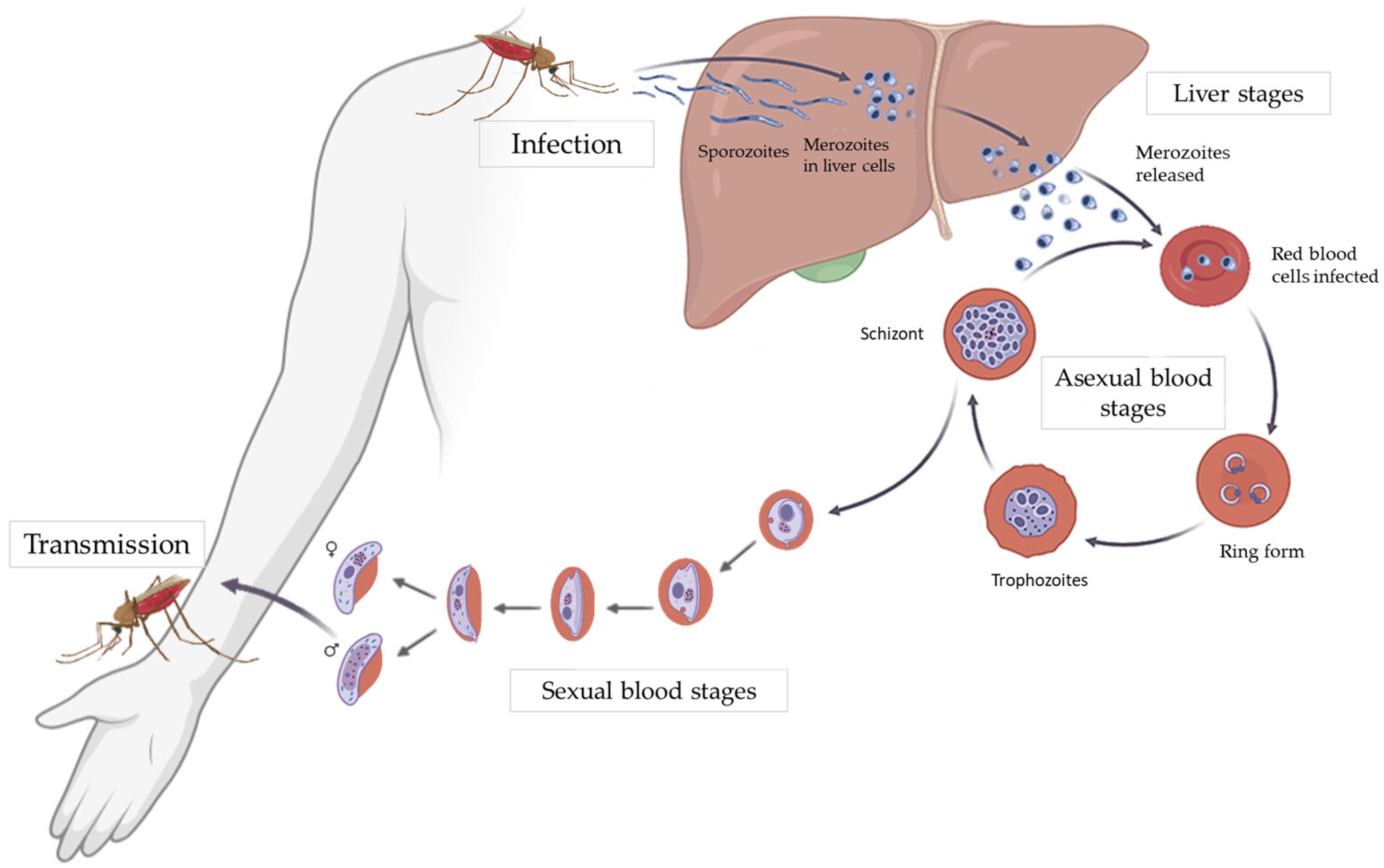
Figure 1. Plasmodium falciparum life cycle. Adapted from [7], licensed under CC BY 4.0.
Ijms 26 09150 g001
Figure 2. The main carbon and mitochondrial metabolic pathways in P. falciparum during the asexual blood stages. In these stages, most glucose is metabolized through glycolysis to produce lactate, with only a small proportion of pyruvate being converted to acetyl-CoA to fuel the tricarboxylic acid cycle (TCA) cycle. Indeed, glucose generates a minor carbon flux from oxaloacetate to isocitrate. Conversely, glutamate produces a major flux from 2-oxoglutarate to malate. Blue arrows indicate glucose metabolism, while red arrows represent glutamine metabolism. Thicker arrows indicate the major carbon flux, while thinner ones represent the minor flux. Essential components of the mitochondrial electron transport chain (mETC) are shown in green, and dispensable ones in gray. Cyt c, cytochrome c; DHODH, dihydroorotate dehydrogenase; G3PDH, glycerol 3-phosphate dehydrogenase; MQO, malate-quinone oxidoreductase; NDH2, type II NADH dehydrogenase; Pi, inorganic phosphate.
Ijms 26 09150 g002
Figure 3. The main carbon and mitochondrial metabolic pathways in P. falciparum during the sexual blood stages. In these stages, a switch to a predominant glycolytic flux into a highly active TCA metabolism occurs, accompanied by a reduced carbon flux from glutamate. Blue arrows indicate glucose metabolism, while red arrows represent glutamine metabolism. Thicker arrows indicate the major carbon flux, while thinner ones represent the minor flux. Essential components of the mETC are shown in green.
Ijms 26 09150 g003
Figure 4. Antimalarial compounds acting on distinct components of the mETC. HDQ, MPQ, CK-2-68, SL-2-64, SL-2-25, RYL-552, RYL-581, RKA066, WJM228, ELQ-300, ELQ-331, ELQ-596, ELQ-598, and ELQ-400 are endochin-like quinolones. Atovaquone, Nepodin, and Plumbagin are naphthoquinones. Siccanin and Dobikin K are terpene compounds.
Ijms 26 09150 g004
Table 1. Specific molecular targets of the mitochondrial inhibitors discussed in Section 3 and the related Plasmodium life stages affected in the human host. ABSs, asexual blood stages; ALA/SFC, 5-aminolevulinic acid/sodium ferrous citrate; ART, artemisinin; LSs, liver stages; ND, not detected; PfACO, Plasmodium falciparum aconitase; PfDHODH, Plasmodium falciparum dihydroorotate dehydrogenase; PfFH, Plasmodium falciparum fumarate hydratase; PfMQO, Plasmodium falciparum malate-quinone oxidoreductase; PfNADH2, Plasmodium falciparum type II NADH dehydrogenase; ROS, reactive oxygen species; SBS, sexual blood stages; (+), active; (−), inactive.
CompoundsMitochondrial TargetPlasmodium Life Cycle
Stage Target
References
LSABSSBS
Endochin-like quinolones
Hydroxy-2-dodecyl-4-(1H)-quinolone (HDQ)PfNADH2
complex III (Qi site)
ND(+)ND[25,42,43,44]
7-Chloro-3-methyl-2-{4-[4-(trifluoromethoxy)benzyl]phenyl}quinolin-4(1H)-one (CK-2-68)PfNADH2
complex III (Qo site)
(+)(+)(+)[25,38,42,43,95]
7-Fluoro-3-methyl-2-{6-[4-(trifluoromethoxy)phenyl]pyridin-3-yl}quinolin-4(1H)-one (SL-2-64)PfNADH2
complex III
ND(+)ND[38,42]
3-Methyl-2-{6-[4-(trifluoromethoxy)phenyl]pyridin-3-yl}quinolin-4(1H)-one (SL-2-25)PfNADH2
complex III
(+)(+)ND[38,42,95]
5-Fluoro-3-methyl-2-[4-[[4-(trifluoromethoxy)phenyl]methyl]phenyl]-1H-quinolin-4-one (RYL-552)PfNADH2
complex III (Qo site, Qi site) PfDHODH
ND(+)ND[41,45,46]
5-Fluoro-3-methyl-2-[4-[[4-(trifluoromethoxy)phenyl]methyl]phenyl]-1-(N-ethyl-N-propylamino)-4(1H)-quinolone (RYL-581)PfNADH2
complex III (Qo site, Qi site)
ND(+)ND[41]
1-Methyl-2-pentyl-4(1H)-quinolinone (MPQ)PfNADH2ND(−)ND[43]
RKA066PfNADH2
complex III (Qisite)
ND(+)ND[47]
6-Chloro-7-methoxy-2-methyl-3-{4-[4-(trifluoromethyl) phenoxy]phenyl}quinolin-4(1H)-one (ELQ-300) complex III (Qi site)(+)(+)(+)[57,58,59,96]
[6-Chloro-7-methoxy-2-methyl-3-{4-[4-(trifluoromethyl) phenoxy]phenyl}quinolin-4-yl]oxymethyl ethyl carbonate (ELQ-331)complex III (Qi site)(+)(+)(+)[58,59,97]
6-Chloro-7-methoxy-2-methyl-3-[4-[4-(trifluoromethoxy)phenyl]phenyl]-1H-quinolin-4-one (ELQ-596)complex III (Qi site)ND(+)ND[61]
[6-Chloro-7-methoxy-2-methyl-3-[4-[4-(trifluoromethoxy)phenyl]phenyl]quinolin-4-yl]oxymethyl ethyl carbonate (ELQ-598)complex III (Qi site)(+)(+)ND[61]
WJM228complex III (Qo site)(+)(+)(+)[62]
ELQ-400complex III (Qo site, Qi site)(+)(+)(+)[58,64,65,98]
Acridine-related compounds
WR 249685complex IIIND(+)ND[57]
T111complex III (Qo site, Qi site)(+)(+)(+)[66,67,99]
Naphthoquinones
3-[4-(4-Chlorophenyl)cyclohexyl]-4-hydroxynaphthalene-1,2-dione (Atovaquone) complex III (Qo site)(+)(+)(+)[7]
1-(1,8-Dihydroxy-3-methylnaphthalen-2-yl) ethanone (Nepodin)PfNDH2ND(+)ND[48]
5-Hydroxy-2-methyl-1,4-naphthoquinone (Plumbagin)complex IIND(+/−)ND [52,53,55]
Pyridones
GW844520complex III (Qi site)(+)(+)ND[42,47,69,100]
GSK932121complex III (Qi site)ND(+)ND[42,47,57]
Pyrimidine Azepines
PyAz90complex III (Qo site)(+)(+)(+)[70,101]
Cyclopropyl carboxamides
MMV024397complex III (Qo site)(+)(+)(+)[56,71]
WJM280 complex III (Qo site)(+)(+)(+)[71]
W466complex III (Qo site)(+)(+)(+)[71]
W499complex III (Qo site)(+)(+)(+)[71]
Terpene compounds
Dobikin KPfNADH2
PfDHODH
ND(+)ND[49]
(4aS,6aS,11bR,13aS,13bS)-4,4,6a,9-Tetramethyl-1,2,3,4,4a,5,6,6a,11b,13b-decahydrobenzo[a]furo [2,3,4-mn]xanthen-11-ol (Siccanin)complex II
complex III
ND(+)ND[51]
Chalcones
(E)-3-[4-hydroxy-2-methoxy-5-(2-methylbut-3-en-2-yl)phenyl]-1-(4-hydroxyphenyl)prop-2-en-1-one (Licochalcone A)complex II
complex III
ND(+)ND[54,55]
2-geranyl-2′,4′,3,4-tetrahydroxy-dihydrochalconePfMQOND(+)ND[88]
Triazolopyrimidines
DSM265PfDHODH(+)(+)(−)[77,78,79,80,102,103,104,105]
DSM421PfDHODH(+)(+)ND[77]
N-Alkyl-thiophene-2-carboxamides
Genz-667348PfDHODHND(+)ND[42,79,82]
Genz-669178PfDHODHND(+)ND[42]
Pyrrole-based derivatives
DSM705PfDHODH(+)(+)ND[83,84]
DSM873PfDHODH(+)(+)ND[83,84]
Pyrazole-based compounds
DSM1465PfDHODH(+)(+)ND[85]
Other compounds
Ethyl-6-decyloxy-7-ethoxy-4-hydroxyquinoline-3-carboxylate (Decoquinate)complex III (Qo site)(+)(+)(+)[42,72]
Sodium azide (NaN3)complex IVND(+/−)ND[73]
Potassium cyanide (KCN)complex IVND(+/−)ND[73,74]
Almitrinecomplex VND(+)ND[55,75]
ITT-004complex VND(+)ND[76]
2,4,7-Trichloro-3,8-dihydroxy-1,9-dimethyl-6-(1-methyl-1-propen-1-yl)-11H-dibenzo[b,e][1,4]dioxepin-11-one (Nornidulin)PfMQOND(+)ND[86]
4-Hydroxy-3-[(2E,6E)-3,7,11-trimethyldodeca-2,6,10-trienyl]chromen-2-one (ferulenol)PfMQOND(+)ND[87]
Sodium fluoroacetate (NaFAc)PfACOND(−)(+)[17,89]
Mercaptosuccinic acidPfFHND(+)ND[90]
ALA/SFCoxidative stressND(+)ND[92,93,94]
ARTmembrane depolarization
other unexplored mechanisms
(−)(+)(+)[106,107]
Disclaimer/Publisher’s Note: The statements, opinions and data contained in all publications are solely those of the individual author(s) and contributor(s) and not of MDPI and/or the editor(s). MDPI and/or the editor(s) disclaim responsibility for any injury to people or property resulting from any ideas, methods, instructions or products referred to in the content.