Article

Mechanism of Regulation of NaCl Homeostasis in the Distal Colon During Obesity

by
Balasubramanian Palaniappan
1,*,
John Crutchley
2,
Raja Singh Paulraj
3,
Alip Borthakur
1 and
Subha Arthur
1
1
Department of Biomedical Sciences, Joan C. Edwards School of Medicine, Marshall University, Huntington, WV 27501, USA
2
Marshall Institute for Interdisciplinary Research, Marshall University, Huntington, WV 25703, USA
3
Joan C. Edwards School of Medicine, Marshall University, Huntington, WV 27501, USA
*
Author to whom correspondence should be addressed.
Int. J. Mol. Sci. 2025, 26(18), 9139; https://doi.org/10.3390/ijms26189139
Submission received: 12 August 2025 / Revised: 11 September 2025 / Accepted: 17 September 2025 / Published: 19 September 2025

Abstract

Obesity is characterized by low-grade chronic inflammation, similar to the pathophysiology of inflammatory bowel disease (IBD) and colon cancer. IBD, which includes Crohn’s disease and ulcerative colitis, is becoming increasingly common in obese individuals. Our previous research documented that both IBD and obesity involve disrupted NaCl homeostasis in the small intestine. The present study investigated how obesity affects NaCl homeostasis in the distal colon, using the Zucker (Leprfa) rat as a genetic model of obesity. The functional and molecular alterations in NaCl homeostasis were evaluated through radioactive uptakes, RT-qPCR, and Western blot studies. We found a significant reduction in Cl absorption via Cl/HCO3 exchanger, Downregulated in Adenoma (DRA) in the distal colon of obese rats compared to lean controls. This reduction was due to a decrease in the maximum transport capacity (Vmax) of DRA, with no change in the affinity of the exchanger for chloride. DRA mRNA and protein levels were also downregulated in obese animals. In contrast, Na absorption via Na+/H+ exchanger and its expression remained unchanged. These findings are the first to demonstrate that DRA is significantly impaired in the distal colon due to obesity. This suggests that net NaCl absorption in the distal colon is compromised in obesity, potentially increasing the risk for IBD and colon cancer.

1. Introduction

The global prevalence of obesity has reached epidemic levels, with nearly a three-fold increase since 1975. According to the World Health Organization, by 2050, an estimated 60% of males and 50% of females will be obese [1,2,3]. Obesity results from excessive body fat accumulation, driven by a combination of etiological factors such as high-calorie food consumption, sedentary lifestyles, obesogenic environmental influences, and genetic predispositions (mono and polygenic) [4,5,6]. Obesity significantly elevates the risk of numerous health complications, including type 2 diabetes, hypertension, metabolic syndrome, cardiovascular disease, inflammatory bowel disease (IBD), such as Crohn’s disease (CD) and Ulcerative colitis (UC), and colon cancers [7,8,9]. In obesity, disrupted glucose and NaCl homeostasis directly contribute to the pathogenesis of diabetes and hypertension [10]. Furthermore, electrolyte disturbances, though often underappreciated, play a pivotal role in metabolic dysfunction and systemic dysfunctions [11]. Therefore, elucidating the complex relationship between obesity and electrolyte disturbances is essential for developing comprehensive and effective treatment strategies.
The mammalian colon’s primary role is electrolyte (Na+ and Cl) and water absorption. This is facilitated by the coupled activity of the Na+/H+ exchanger NHE3 (sodium: proton exchanger-3 encoded by the SLC9A3 gene) and the Cl/HCO3 exchanger DRA (Downregulated in Adenoma encoded by the SLC26A3 gene), with DRA predominantly expressed in the distal colon [12,13,14,15,16]. The regulation of electrolyte and acid/base transport by DRA and other transporters in colonic epithelial cells is vital for maintaining luminal pH, mucus secretion, and gut microbiome homeostasis, thereby preserving epithelial barrier integrity. Disruption of acid/base transport, especially reduced DRA expression, leads to increased intracellular alkalinity, a characteristic feature associated with the onset and progression of colitis-associated colon cancer (CAC) [17,18,19,20,21,22,23,24,25,26,27,28,29,30]. Obesity is a well-established risk factor and plays a crucial role in the early onset of colorectal cancer (CRC) [31]. Many studies have consistently documented the association between obesity and CRC. Further, loss of functional DRA leads to diarrheal diseases like inflammatory bowel disease (IBD) and congenital chloride diarrhea (CLD). Diarrhea, a key symptom in IBD, is also a prevalent symptom among obese patients and has been shown to result from disrupted intestinal epithelial transporter function that impacts fluid balance [32,33,34,35,36,37,38]. Moreover, there is a significant overlap between IBD and obesity, with 15–40% of IBD patients being overweight and 15–40% obese [39,40,41,42].
Our previous research demonstrated that in small intestinal epithelial cells from a monogenic rat model of obesity and obese humans, glucose and sodium absorption (via sodium-dependent glucose transporter, SGLT1) and chloride absorption via DRA are increased. This upregulation leads to enhanced glucose and NaCl absorption, providing a basis for the dysregulation of glucose and NaCl homeostasis observed in diabetes and hypertension associated with obesity [10]. Given DRA’s significant role in the intestine and the rising prevalence of IBD among obese individuals, it is crucial to explore whether DRA function is altered in the colon in the context of obesity. Since obesity is a known risk factor for colon cancer and DRA is involved in colon cancer development, understanding DRA regulation in the colon under obese conditions is essential. Therefore, this study aims to investigate DRA’s role and regulation in the colon in relation to obesity.

2. Results

2.1. Cl/HCO3 Exchange (DRA) Activity in the Colon During Obesity

Lean and obese Zucker rat demonstrated Cl/HCO3 exchange activity in the distal colon (Figure 1). However, obese Zucker rats showed a significant reduction in Cl/HCO3 exchange activity (1315 ± 71.2 pmol/mg protein/min; n = 6; p < 0.0001) compared to lean Zucker rats (2424 ± 165.6 pmol/mg protein/min) (Figure 1). These results suggest that obesity impairs Cl/HCO3 exchange activity in the distal colon.

2.2. Na+/H+ Exchange (NHE3) Activity in the Distal Colon in Obesity

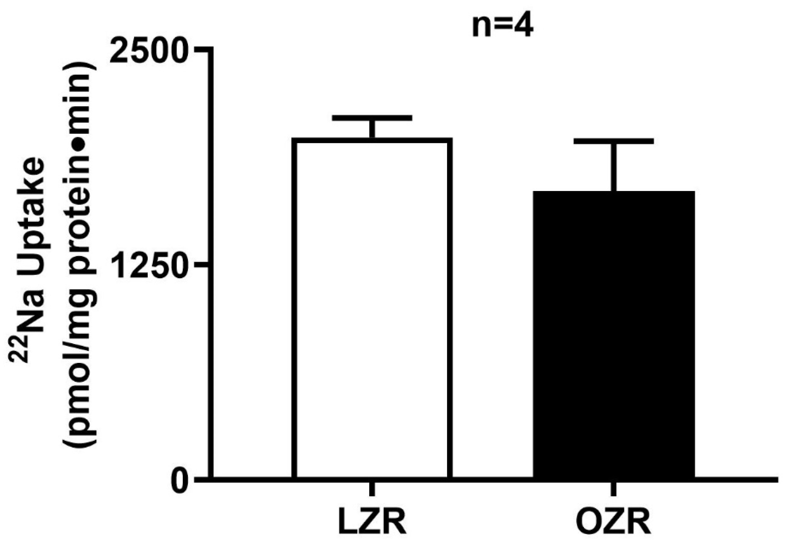
Na+/H+ exchange activity was observed in the distal colon AMVs of both lean and obese Zucker rats. However, as shown in Figure 2, Na+/H+ exchange activity remained unchanged between obese and lean Zucker rats (1678 ± 290.3 pmol/mg protein/min in obese Zucker rats; 1989 ± 113.9 in lean Zucker rats, n = 4).
Data shown in Figure 1 and Figure 2 together indicate that though sodium absorption is unaltered in obesity, Cl absorption is dysregulated, which indicates that the NaCl homeostasis is indeed affected in the distal colon in obesity.

2.3. Kinetic Studies of Cl/HCO3 Exchange in the Distal Colon in Obesity

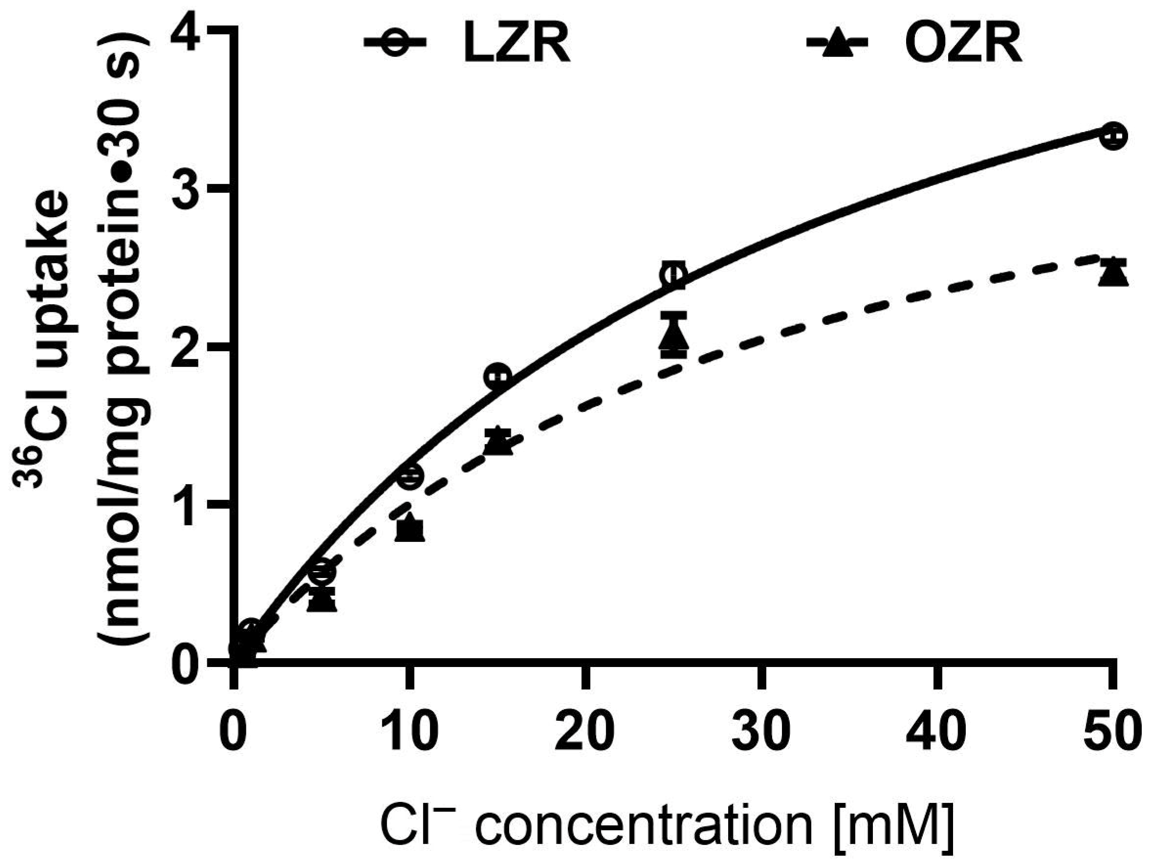
Kinetic studies were conducted to elucidate the mechanism of Cl/HCO3 exchange inhibition in the distal colon of obese Zucker rats. Figure 3 illustrates the extracellular concentration-dependent uptake of 36Cl at 30 s in both lean and obese rats. Kinetic parameters (Table 1) revealed no significant difference in the Michaelis constant (1/Km) for Cl uptake between obese (Km: 32.1 ± 5.7 mM) and lean (Km: 35.7 ± 3.2 mM) rats (n = 3), indicating no change in affinity. However, the maximal rate of uptake (Vmax) was significantly lower in obese rats (4.2 ± 0.4 nmol/mg protein/min) compared to lean rats (5.8 ± 0.3 nmol/mg protein/min; n = 3, p < 0.05). These results suggest that in the distal colon in obesity, inhibition of Cl/HCO3 exchange primarily occurs through a reduction in the maximal transport capacity (Vmax), without affecting the affinity of the exchanger for chloride.

2.4. RT-qPCR Analysis of DRA mRNA Expression in the Obese Zucker Rat

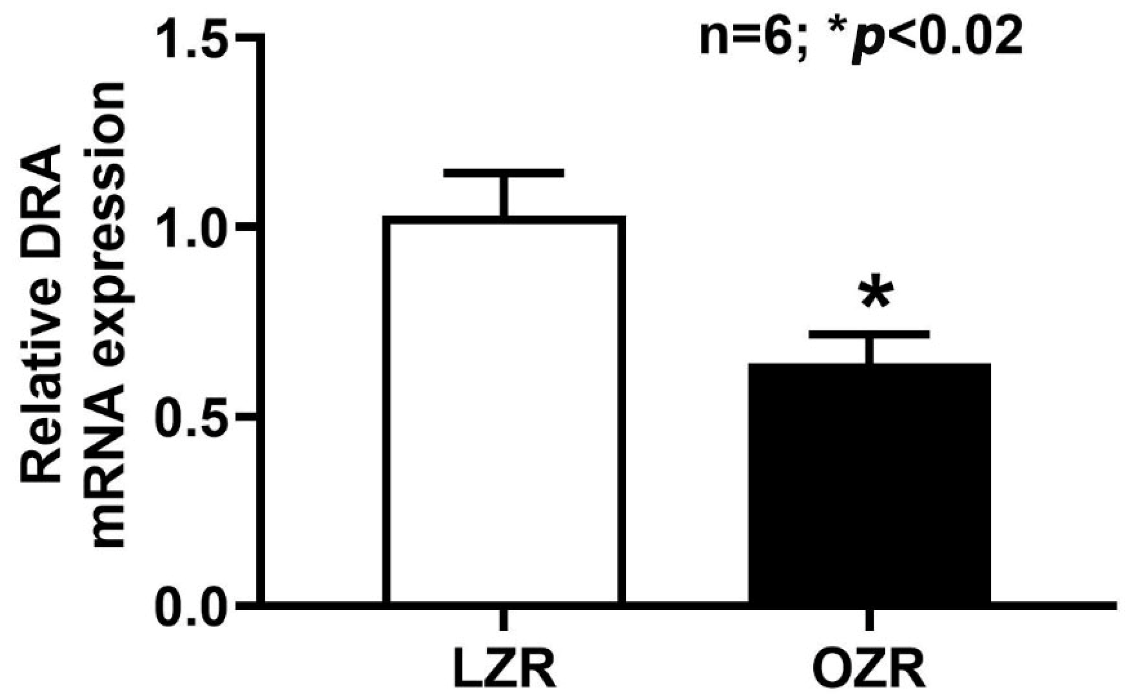
To investigate the molecular basis for the observed reduction in Cl/HCO3 exchange, we examined DRA mRNA expression levels in distal colonocytes isolated from lean and obese Zucker rats using reverse transcription-quantitative polymerase chain reaction (RT-qPCR). The results, presented in Figure 4, demonstrate a decrease in DRA mRNA levels in the distal colon of obese Zucker rats compared to their lean counterparts. This finding suggests that the transcription of the DRA gene is downregulated, or reduced, in the distal colon of obese Zucker rats.

2.5. Western Blot Analysis

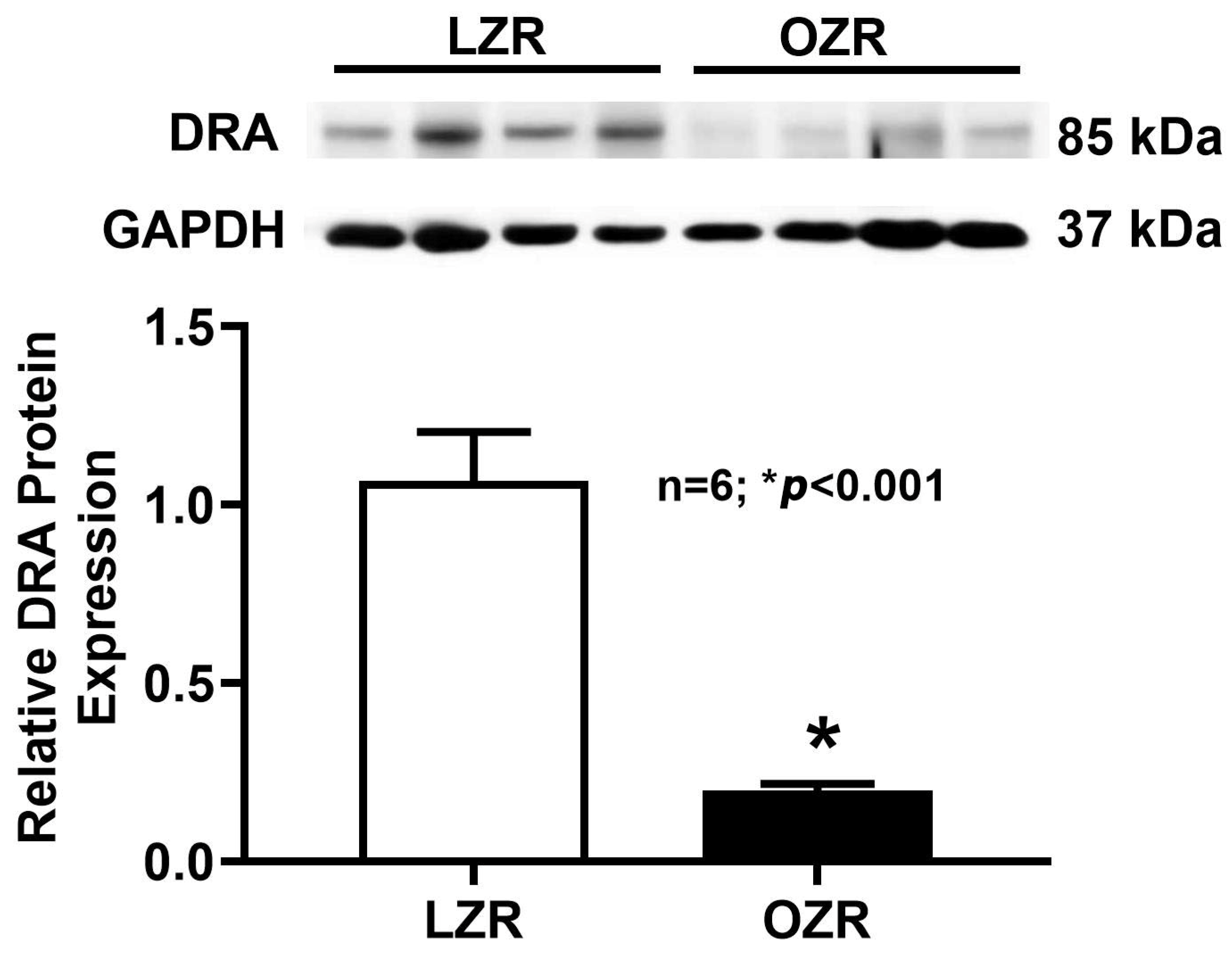
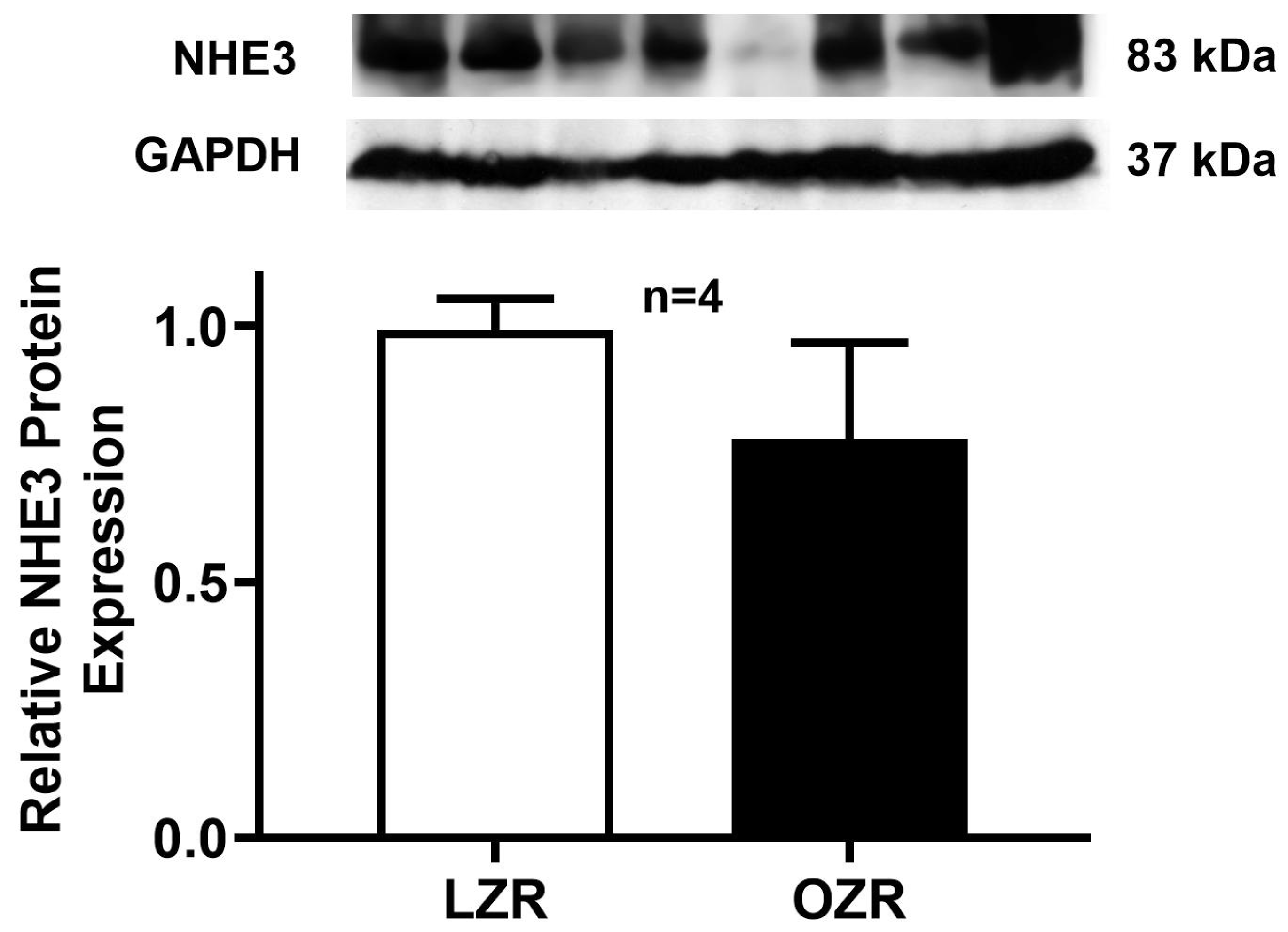
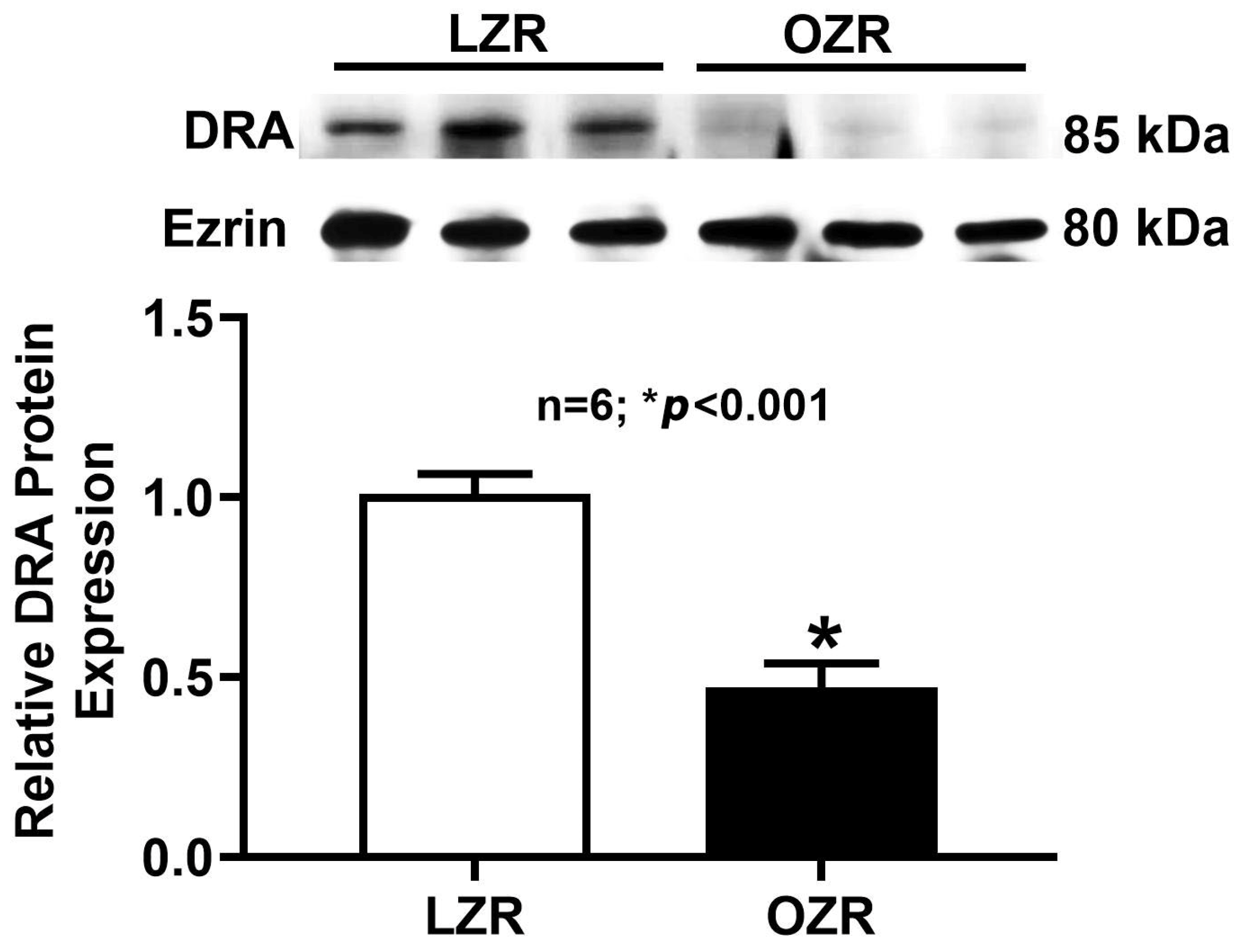
Although RT-qPCR analysis showed a decrease in DRA mRNA levels in obese rats, it is important to directly assess protein levels. Since the amount of mRNA does not always directly translate to the quantity of the functional protein. As shown in Figure 5, Western blot results for DRA protein expression in whole cell lysates revealed a significant decrease in immunoreactive DRA protein in the distal colonocytes of obese Zucker rats compared to lean Zucker rats. Moreover, consistent with our functional studies, NHE3 protein expression (Figure 6) remained unchanged between the lean and obese Zucker rat distal colonocytes. This finding confirmed that the observed reduction in DRA mRNA directly translates to a decrease in the total amount of DRA protein within the cells.
We further examined DRA protein expression at the level of the AM, since AM is the cellular location where DRA facilitates its transport function. As shown in Figure 7, a decrease in AM-localized DRA protein was also observed in the distal colon of obese Zucker rats compared to lean Zucker rats. This suggests that the reduced cellular DRA protein is reflected in a decrease in the amount of DRA protein at the functional membrane surface.

3. Discussion

This study provides compelling new evidence that chloride/bicarbonate (Cl/HCO3) exchange activity mediated by DRA in the distal colon of obese Zucker rats is significantly diminished in obesity. Interestingly, Na+/H+ exchange (NHE3) functional activity and its expression in the distal colon remained unchanged between lean and obese animals. Our findings reveal that the mechanism underlying this obesity-associated inhibition in chloride absorption is due to a reduction in the number of functional Cl/HCO3 exchangers in the apical membrane of the colonocytes. Kinetic studies demonstrated that the affinity of the exchanger for chloride was unaltered, suggesting that the observed decrease in Cl/HCO3 exchange activity is not due to a change in the individual exchanger’s affinity properties, but a consequence of reduced DRA protein expression. This reduction, in turn, was due to the downregulation of de novo DRA mRNA synthesis in the distal colon of obese Zucker rats. Combining the total cellular and AM Western blot data, along with the kinetic parameters and RT-qPCR data, this study provides a comprehensive understanding of the mechanism underlying the inhibition of Cl/HCO3 exchange activity in the distal colon of obese Zucker rats and suggests that net NaCl absorption in the distal colon is impaired during obesity.
The etiology of obesity is complex and includes genetic factors that predispose an individual to gain weight very early in their life. The genetic causes of obesity include monogenic obesity that arises due to mutations in a single gene, specifically mutations in the leptin receptor. Patients with leptin receptor mutations experience an extremely early onset of obesity, which has a direct effect on hyperphagia, insulin sensitivity, glucose intolerance, metabolic disorders, and recurring infections [43]. Obese Zucker rats that mimic monogenic human obesity manifest similar clinical complications that have been reported in patients with leptin receptor mutations. Hence, the present study, designed with the obese Zucker rats, is an ideal model to study physiological alterations induced by obesity.
Obesity is a risk factor for gastrointestinal (GI) diseases [44]. Obesity is known to cause GI disease symptoms, including upper abdominal pain, gastroesophageal reflux, and diarrhea [45]. Population-based studies reveal that among the IBD patients, 15–40% are overweight and 20–40% are obese [39,40,41,42]. A study of the US adult population showed that chronic diarrhea correlates with obesity and that the risk of diarrhea increases with the severity of obesity [37,46]. The overall prevalence of chronic diarrhea among individuals with obesity in US population was 8.18% [32,37]. In obesity and IBD, disrupted NaCl homeostasis is a common symptom that directly contributes to the pathogenesis of hypertension and diarrhea [10,47].
NaCl homeostasis is maintained in the mammalian intestinal epithelial cells by the dual operation of the Na+/H+ exchanger NHE3 and the Cl/HCO3 exchanger DRA. The current study demonstrated that in obese Zucker rats, NaCl homeostasis in the distal colon is indeed dysregulated due to the downregulation of DRA and not NHE3. Two other chloride transporters in addition to DRA, namely Putative Anion Transporter-1 (PAT-1) and Cystic Fibrosis Transmembrane Conductance Regulator (CFTR) have also been implicated in the maintenance of NaCl homeostasis in the intestine. However, preliminary studies done in this Zucker model of obesity showed that the mRNA levels of these compensatory mechanisms were unchanged in the distal colon, indicating their nonparticipation in dysregulated NaCl homeostasis in the colon in obesity. Since this study shows that DRA is the only Cl transporter mechanism that is affected, loss of its function in the colon in obesity would suggest its clinical significance in disrupting luminal acid/base imbalance, causing defective mucus secretion and impaired epithelial barrier integrity, which would ultimately lead to diarrhea, a common symptom observed among IBD as well as in obesity patients. Research into diarrheal conditions such as IBD and CLD has consistently shown that altered DRA expression or mutations in the DRA gene result in decreased chloride absorption [48,49,50,51]. Furthermore, studies on DRA-deficient animals, which exhibit a phenotype similar to human CLD, confirm DRA’s crucial role in chloride absorption [14,17]. The dysregulation of DRA’s functional expression is also known to contribute to gut microbiome dysbiosis and disrupt mucosal immune homeostasis through epithelial-immune cell crosstalk, ultimately leading to IBD-related diarrheal diseases [13,16,52,53,54,55]. All these studies demonstrate the importance and implications of DRA in the pathophysiology of diarrheal diseases. The current study demonstrates that DRA is indeed a vital player in the pathophysiology of diarrhea, but among obese populations.
The data obtained from this study provides a different narrative compared to what was published earlier from the same animal model, but from a different intestinal region. In the small intestine, Na and glucose absorption via sodium-glucose co-transport (SGLT1) was found to be significantly increased. We also observed higher activity and expression of the DRA transporter in small intestinal villus cells, contradicting to our present findings in the colon in obesity. Notably, NHE3 activity and expression remained unchanged in the small intestine, indicating its non-participation in the increased Na and Cl absorption in the small intestine in obesity [10], similar to what was seen in the present study in the colon. These two studies indicate that DRA is differentially regulated along the intestine in obesity highlighting the significance of its dysregulation in the pathogenesis of obesity induced gut disorders that need to be explored in future studies.
As discussed earlier, obesity is a major risk factor for IBD and CRC. In addition, DRA expression is known to be downregulated in these conditions. The downregulation of DRA, as seen in this study, provides an important link between obesity and the higher incidence of CRC and IBD among obese patients. Moreover, CRC is the second-leading cause of cancer deaths worldwide and the most common obesity-related cancer in the US [56], and the downregulation of DRA in the colon appears to be a critical predisposing factor for the development of CRC among the obese population. Evidence shows that in IBD and CRC patients, mRNA and protein levels of DRA in the colonic apical membrane were decreased [21,27,57]. Many reports have implicated the role of proinflammatory cytokines (TNFα, INFγ, interleukins (ILs)), nitric oxide (NO), eicosanoids (arachidonic acid-PGE1,2, etc.), and their activated inflammatory signaling pathways (NF-κB, JAK-STAT, retinoic acid receptor-β/hepatocyte nuclear factor-1β, etc.), post-transcriptional (e.g., miRNA: miR-223-3p, miR-494) and post-translational regulators in the impaired DRA-mediated ion transport in inflamed colonic mucosa [58,59,60,61,62,63,64,65,66,67,68,69,70,71,72,73,74]. Multiple clinical and animal studies have found that proinflammatory immune cells are disturbed in obesity [75,76,77,78,79,80,81]. We speculate that inflammatory mediators and their downstream signaling pathways might be responsible for the deregulation of DRA-mediated electrolyte homeostasis in the obese distal colon. The schematic diagram below summarizes our current study.
Ijms 26 09139 i001

4. Materials and Methods

4.1. Chemicals

Except where indicated, all the chemicals used in this study were purchased from Sigma-Aldrich (Milwaukee, WI, USA).

4.2. Animal Models

Eighteen-week-old male obese (Leprfa) Zucker rats (Strain 185) and lean Zucker rats (Strain 186) were obtained from Charles River Laboratories (Wilmington, MA, USA) as models of genetic obesity and lean controls, respectively. Animals were housed under a 12 h light/dark cycle with ad libitum access to food and water. The animal studies were performed in compliance with the ethical and procedural regulations of the Marshall University Institutional Animal Care and Use Committee (IACUC Protocol #777).

4.3. Colonocyte Isolation and Apical Membrane Vesicle Preparation

Distal colons were excised from lean and obese Zucker rats and rinsed twice with warm saline. Colonocytes were then isolated using a calcium chelation method [82]. Briefly, the distal colon was everted, filled with calcium-free Ringer solution (in mM: 127 NaCl, 10 HEPES, 5 KCl, 5 Na-pyruvate, 5 EDTA, 1 MgCl2, and 5 glucose; gassed with 95% O2 and 5% CO2, pH 7.4), and ligated at both ends. The colon was incubated in this solution at 37 °C for 20 min with gentle shaking to facilitate cell detachment. Following incubation, the colon was removed, and the resulting cell suspension was centrifuged at 4500× g for 2 min to pellet the colonocytes. Isolated colonocytes were immediately used for uptake studies or flash-frozen in liquid nitrogen and stored at −80 °C until needed. Apical membrane (AM) vesicles (AMVs) were prepared from stored colonocytes using the magnesium precipitation and differential centrifugation method, as previously described [83,84].

4.4. Uptake Studies in Intact Ileal Villus Cells and AMV

Chloride-bicarbonate (Cl/HCO3) exchange activity in AMVs was measured using a rapid-filtration technique, as previously described [10,67]. AMVs were suspended in a vesicle medium containing 5 mM N-methyl-D-glucamine (NMG) gluconate and 50 mM HEPES-Tris (pH 7.5), supplemented with either 100 mM KHCO3 (gassed with 5% CO2 and 95% N2) or 50 mM potassium gluconate (gassed with 100% N2). The reaction was initiated by adding 5 μL of AMV suspension to 95 μL of a reaction mixture containing 5 mM NMG 36Cl (American Radiolabeled Chemicals, St. Louis, MO, USA) 15 mM potassium gluconate, and 50 mM MES-Tris (pH 5.5), with or without 1 mM 4,4′-diisothiocyanatostilbene-2,2′-disulfonic acid disodium salt (DIDS), a Cl/HCO3 exchange inhibitor. The reaction was terminated after 60 s by adding 5 mL of ice-cold stop solution (50 mM HEPES-Tris, pH 7.5, 0.10 mM MgSO4, 50 mM potassium gluconate, and 100 mM NMG gluconate). The mixture was then filtered through 0.45 μm Millipore (HAWP) filter and washed twice with 5 mL of ice-cold stop solution. Filters were dissolved in 4 mL of scintillation fluid (Ecoscint; National Diagnostics, Atlanta, GA, USA), and radioactivity was measured using a Perkin Elmer Tri-Carb LSC 4910TR Scintillation Counter. Cl/HCO3 exchange activity was calculated as the DIDS-sensitive, HCO3 dependent [36Cl] uptake.
Kinetic parameters for Cl/HCO3 exchange were determined using intact colonocytes isolated from lean and obese Zucker rats. Briefly, 100 mg of isolated colonocytes were suspended in a buffer containing 5 mM NMG gluconate and 50 mM HEPES-Tris (pH 7.5), supplemented with either 100 mM KHCO3 or 100 mM potassium gluconate. The reaction was initiated by adding 10 μL of colonocyte suspension to 90 μL of a reaction medium containing 5 mM NMG [36Cl], 150 mM potassium gluconate, 50 mM MES-Tris (pH 5.5), and varying concentrations of HCl (0.5, 1, 5, 15, 25, and 50 mM), with or without 1 mM DIDS. After a 30 s incubation, the reaction was terminated by adding an ice-cold stop solution (50 mM HEPES-Tris, pH 7.5, 0.10 mM MgSO4, 50 mM potassium gluconate, and 100 mM NMG gluconate). The mixture was then filtered through 0.65 μm Millipore (MCE MilliporeSigma, Burlington, MA, USA) filters and processed as described above. Michaelis-Menten kinetics were determined using nonlinear regression analysis of the uptake data in GraphPad Prism 9 (GraphPad Software, La Jolla, CA, USA).
For Na+/H+ exchange activity studies, AMV of the distal colon was suspended in a vesicle medium consisting of either 50 mM Tris-MES (pH 5.5) or 50 mM Tris-HEPES (pH 7.5), along with 300 mM mannitol. A 5 μL aliquot of this suspension was then incubated in a 95 μL reaction medium (300 mM mannitol, 50 mM Tris-HEPES (pH 7.5), and 1 mM 22Na+ (Eckert & Ziegler, Atlanta, GA, USA), with or without 1 mM amiloride. The uptake was terminated after 60 s by adding an ice-cold stop solution containing 300 mM mannitol and 50 mM Tris-HEPES (pH 7.5). This was followed by a rapid filtration process and radioactivity determination as described above. Na+/H+ exchange activity was calculated as the amiloride-sensitive, pH-dependent [22Na+] uptake.

4.5. Real-Time Quantitative PCR (RT-qPCR) for DRA

Total RNA was isolated from colonocytes of lean and obese Zucker rats using the RNeasy Mini Kit (Qiagen, Germantown, MD, USA) according to the manufacturer’s protocol. The RNA was reverse-transcribed and amplified with a Brilliant SYBR Green RT-qPCR Master Mix Kit (Qiagen One-Step RT-PCR Kit: Ref:210212, Germantown, MD, USA). Rat DRA was amplified using gene-specific primers custom-designed by Thermo Fisher Scientific (Hillsboro, OR, USA), with GAPDH amplified as an internal control. The sequences of the rat primers used in this study are as follows: DRA: forward primer: TACTGTCTCCCAGAACAGGACT; reverse primer: CGCTTCTTTCTGGCTGTTAGC, and for GAPDH: forward: AGGTCGGTGTGAACGGATTT and reverse: CCACTTTGTCACAAGAGAAGGC.

4.6. Western Blotting

Western blot analyses were performed on whole cell and AM protein extracts from lean and obese Zucker rat colonocytes, as previously described [85]. Proteins were extracted using RIPA buffer (50 mM Tris-HCl pH 7.4, 1% Igepal, 150 mM NaCl, 1 mM EDTA, 1 mM PMSF, 1 mM Na3VO4, 1 mM NaF) supplemented with protease inhibitor cocktail (SAFC Biosciences, Lenexa, KS, USA). The extracted protein was mixed with sample buffer and separated on 8% (custom-made) polyacrylamide gels. Separated proteins were then transferred to BioTrace PVDF membranes, blocked, and incubated overnight at 4 °C with the following primary antibodies (1:1000 dilution): mouse anti-DRA (SC-376187, Santa Cruz Biotechnology, Inc., Dallas, TX, USA), rabbit anti-Ezrin (ab231907, Abcam, Waltham, MA, USA) for AM, and rabbit anti-GAPDH for whole cell lysates. Membranes were then incubated with horseradish peroxidase-conjugated secondary antibodies (1:10,000 dilution): goat anti-mouse IgG (1706516, Bio-Rad Laboratories, 2000 Alfred Nobel Dr., Hercules, CA, USA) for DRA and mouse anti-rabbit IgG (sc-2357, Santa Cruz, CA, USA) for Ezrin and GAPDH, for 1 h at room temperature. Chemiluminescence was detected using ECL Detection Reagent (GE Healthcare, Chicago, IL, USA) and autoradiography. DRA protein density was quantified using ImageJ software J 1.53 t.

4.7. Protein Estimation

Protein quantification for both uptakes and Western blot studies was performed using the DCTM protein assay kit (Bio-Rad, Berkeley, CA, USA) following the Lowry method, according to the manufacturer’s protocol.

4.8. Statistical Analysis

All results are presented as means ± SEM, calculated using GraphPad Prism 9. The ‘n’ value represents the number of independent experiments, with each experiment performed using cells isolated from a different animal. For uptake experiments, each ‘n’ also indicates the average of triplicate measurements. Statistical significance was determined using a two-tailed Student’s t-test in GraphPad Prism 9. A p-value of <0.05 was considered statistically significant.

5. Conclusions

Our study shows that DRA is downregulated in the distal colon during obesity, in contrast to what was seen in the small intestine, highlighting regional differences in DRA regulation in the gut during obesity. This downregulation in the colon may contribute directly or indirectly to early CRC development, beyond just NaCl imbalance. Therefore, further research is needed to define and understand the consequences of DRA downregulation in the colon in obesity in the context of the pathophysiology of obesity-associated colon disorders such as colorectal cancer.

Author Contributions

Conceptualization, B.P.; Methodology, B.P., J.C. and A.B.; Animal Euthanization and Tissue Procurement, B.P. and R.S.P.; Formal Analysis, B.P.; Investigation, B.P., J.C. and A.B.; Data Curation, B.P.; Writing—Original Draft Preparation, B.P.; Writing—Review and Editing, B.P., A.B. and S.A.; Supervision, B.P.; Project Administration, B.P.; Funding Acquisition, B.P. All authors have read and agreed to the published version of the manuscript.

Funding

This work was supported by the NIH grant National Institute of General Medical Sciences (P20 GM 121299-05-6775) to B. Palaniappan (research reported in this publication was supported by the National Institute of General Medical Sciences of the National Institutes of Health under Award Number P20GM121299 of an Institutional grant). The content is solely the responsibility of the authors and does not necessarily represent the official views of the National Institutes of Health.

Institutional Review Board Statement

The animal studies were approved by the Marshall University Institutional Animal Care and Use Committee, adhering to their ethical and procedural regulations.(protocol code Protocol # 777 and date of approval: 21 January 2022).

Informed Consent Statement

Not applicable.

Data Availability Statement

The authors confirm that the data supporting the findings of this study are available within the article.

Acknowledgments

We also gratefully acknowledge U. Sundaram (Joan C. Edwards School of Medicine, Marshall University), who provided the radioactive isotopes 22Na+ and 36Cl. The in vivo animal studies were conducted under IACUC protocol #777 of U. Sundaram, and the PD/PI of the Appalachian Center for Cellular Transport in Obesity Related Disorders, COBRE project (P20GM121299).

Conflicts of Interest

The authors declare no conflicts of interest. The funding sponsors had no role in the design of the study; in the collection, analyses, or interpretation of data; in the writing of the manuscript; or in the decision to publish the results.

Abbreviations

IBDInflammatory Bowel Disease
CDCrohn’s Disease
UCUlcerative colitis
NaClSodium chloride
DRADownregulated in Adenoma
NHE3Sodium proton exchanger 3
SGLT1Sodium-dependent glucose co-transport 1
CFTRCystic Fibrosis Transmembrane Conductance Regulator
PAT-1Putative Anion Transporter-1
CACColitis-associated colon cancer
CRCColorectal cancer
CLDCongenital chloride diarrhea
DIDS4,4′-diisothiocyanatostilbene-2,2′-disulfonic acid disodium salt
LZRLean Zucker Rat
OZRObese Zucker Rat
AMVApical membrane vesicle
GIGastrointestinal
TNFαTumor Necrosis Factor alpha
INFγInterferon gamma
ILInterleukins
NONitric oxide
NF-kBNuclear factor kappa B
miRNAmicro ribonucleic acid

References

  1. Blüher, M. Obesity: Global epidemiology and pathogenesis. Nat. Rev. Endocrinol. 2019, 15, 288–298. [Google Scholar] [CrossRef] [PubMed]
  2. Koliaki, C.; Dalamaga, M.; Liatis, S. Update on the Obesity Epidemic: After the Sudden Rise, Is the Upward Trajectory Beginning to Flatten? Curr. Obes. Rep. 2023, 12, 514–527, Erratum in Curr. Obes. Rep. 2023, 12, 528. [Google Scholar] [CrossRef] [PubMed]
  3. Ward, Z.J.; Bleich, S.N.; Cradock, A.L.; Barrett, J.L.; Giles, C.M.; Flax, C.; Long, M.W.; Gortmaker, S.L. Projected US state-level prevalence of adult obesity and severe obesity. N. Engl. J. Med. 2019, 381, 2440–2450. [Google Scholar] [CrossRef]
  4. Khera, A.V.; Chaffin, M.; Wade, K.H.; Zahid, S.; Brancale, J.; Xia, R.; Distefano, M.; Senol-Cosar, O.; Haas, M.E.; Bick, A. Polygenic prediction of weight and obesity trajectories from birth to adulthood. Cell 2019, 177, 587–596.e589. [Google Scholar] [CrossRef]
  5. Farooqi, I.S. Monogenic obesity syndromes provide insights into the hypothalamic regulation of appetite and associated behaviors. Biol. Psychiatry 2022, 91, 856–859. [Google Scholar] [CrossRef] [PubMed]
  6. Loos, R.J.; Yeo, G.S. The genetics of obesity: From discovery to biology. Nat. Rev. Genet. 2022, 23, 120–133. [Google Scholar] [CrossRef]
  7. Kopelman, P.G. Obesity as a medical problem. Nature 2000, 404, 635–643. [Google Scholar] [CrossRef]
  8. Tarasiuk, A.; Mosińska, P.; Fichna, J. The mechanisms linking obesity to colon cancer: An overview. Obes. Res. Clin. Pract. 2018, 12, 251–259. [Google Scholar] [CrossRef]
  9. Gibson, T.M.; Park, Y.; Robien, K.; Shiels, M.S.; Black, A.; Sampson, J.N.; Purdue, M.P.; Freeman, L.E.B.; Andreotti, G.; Weinstein, S.J. Body mass index and risk of second obesity-associated cancers after colorectal cancer: A pooled analysis of prospective cohort studies. J. Clin. Oncol. 2014, 32, 4004. [Google Scholar] [CrossRef]
  10. Palaniappan, B.; Arthur, S.; Sundaram, V.L.; Butts, M.; Sundaram, S.; Mani, K.; Singh, S.; Nepal, N.; Sundaram, U. Inhibition of intestinal villus cell Na/K-ATPase mediates altered glucose and NaCl absorption in obesity-associated diabetes and hypertension. FASEB J. Off. Publ. Fed. Am. Soc. Exp. Biol. 2019, 33, 9323–9333. [Google Scholar] [CrossRef]
  11. Crintea, I.N.; Cindrea, A.C.; Mederle, O.A.; Trebuian, C.I.; Timar, R. Electrolyte Imbalances and Metabolic Emergencies in Obesity: Mechanisms and Clinical Implications. Diseases 2025, 13, 69. [Google Scholar] [CrossRef]
  12. Alper, S.L.; Sharma, A.K. The SLC26 gene family of anion transporters and channels. Mol. Asp. Med. 2013, 34, 494–515. [Google Scholar] [CrossRef] [PubMed]
  13. Priyamvada, S.; Gomes, R.; Gill, R.K.; Saksena, S.; Alrefai, W.A.; Dudeja, P.K. Mechanisms Underlying Dysregulation of Electrolyte Absorption in Inflammatory Bowel Disease–Associated Diarrhea. Inflamm. Bowel Dis. 2015, 21, 2926–2935. [Google Scholar] [CrossRef]
  14. Ishiguro, H. HCO3 secretion by SLC 26A3 and mucosal defence in the colon. Acta Physiol. 2014, 211, 17–19. [Google Scholar] [CrossRef] [PubMed]
  15. Talbot, C.; Lytle, C. Segregation of Na/H exchanger-3 and Cl/HCO3 exchanger SLC26A3 (DRA) in rodent cecum and colon. Am. J. Physiol.-Gastrointest. Liver Physiol. 2010, 299, G358–G367. [Google Scholar] [CrossRef] [PubMed]
  16. Hayashi, H.; Nagai, H.; Ohba, K.-I.; Soleimani, M.; Suzuki, Y. Segmental differences in Slc26a3-dependent Cl absorption and HCO3 secretion in the mouse large intestine in vitro in Ussing chambers. J. Physiol. Sci. 2021, 71, 5. [Google Scholar] [CrossRef] [PubMed]
  17. Grivennikov, S.I. Inflammation and colorectal cancer: Colitis-associated neoplasia. Semin. Immunopathol. 2013, 35, 229–244. [Google Scholar] [CrossRef]
  18. Simpson, J.E.; Schweinfest, C.W.; Shull, G.E.; Gawenis, L.R.; Walker, N.M.; Boyle, K.T.; Soleimani, M.; Clarke, L.L. PAT-1 (Slc26a6) is the predominant apical membrane Cl/HCO3 exchanger in the upper villous epithelium of the murine duodenum. Am. J. Physiol. Gastrointest. Liver Physiol. 2007, 292, G1079–G1088. [Google Scholar] [CrossRef]
  19. Xiao, F.; Yu, Q.; Li, J.; Johansson, M.; Singh, A.; Xia, W.; Riederer, B.; Engelhardt, R.; Montrose, M.; Soleimani, M. Slc26a3 deficiency is associated with loss of colonic HCO3 secretion, absence of a firm mucus layer and barrier impairment in mice. Acta Physiol. 2014, 211, 161–175. [Google Scholar] [CrossRef]
  20. Webb, B.A.; Chimenti, M.; Jacobson, M.P.; Barber, D.L. Dysregulated pH: A perfect storm for cancer progression. Nat. Rev. Cancer 2011, 11, 671–677. [Google Scholar] [CrossRef]
  21. Yang, H.; Jiang, W.; Furth, E.E.; Wen, X.; Katz, J.P.; Sellon, R.K.; Silberg, D.G.; Antalis, T.M.; Schweinfest, C.W.; Wu, G.D. Intestinal inflammation reduces expression of DRA, a transporter responsible for congenital chloride diarrhea. Am. J. Physiol. Gastrointest. Liver Physiol. 1998, 275, G1445–G1453. [Google Scholar] [CrossRef]
  22. Lohi, H.; Makela, S.; Pulkkinen, K.; Hoglund, P.; Karjalainen-Lindsberg, M.-L.; Puolakkainen, P.; Kere, J. Upregulation of CFTR expression but not SLC26A3 and SLC9A3 in ulcerative colitis. Am. J. Physiol. Gastrointest. Liver Physiol. 2002, 283, G567–G575. [Google Scholar] [CrossRef]
  23. Asano, K.; Matsushita, T.; Umeno, J.; Hosono, N.; Takahashi, A.; Kawaguchi, T.; Matsumoto, T.; Matsui, T.; Kakuta, Y.; Kinouchi, Y. A genome-wide association study identifies three new susceptibility loci for ulcerative colitis in the Japanese population. Nat. Genet. 2009, 41, 1325–1329. [Google Scholar] [CrossRef]
  24. Schweinfest, C.W.; Henderson, K.W.; Suster, S.; Kondoh, N.; Papas, T.S. Identification of a colon mucosa gene that is down-regulated in colon adenomas and adenocarcinomas. Proc. Natl. Acad. Sci. USA 1993, 90, 4166–4170. [Google Scholar] [CrossRef] [PubMed]
  25. Antalis, T.M.; Reeder, J.A.; Gotley, D.C.; Byeon, M.K.; Walsh, M.D.; Henderson, K.W.; Papas, T.S.; Schweinfest, C.W. Down-regulation of the down-regulated in adenoma (DRA) gene correlates with colon tumor progression. Clin. Cancer Res. 1998, 4, 1857–1863. [Google Scholar]
  26. Zhang, N.; Heruth, D.P.; Wu, W.; Zhang, L.Q.; Nsumu, M.N.; Shortt, K.; Li, K.; Jiang, X.; Wang, B.; Friesen, C. Functional characterization of SLC26A3 c. 392C>G (p. P131R) mutation in intestinal barrier function using CRISPR/CAS9-created cell models. Cell Biosci. 2019, 9, 40. [Google Scholar] [CrossRef]
  27. Arthur, S.; Palaniappan, B.; Sundaram, U. Mo1798 Unique Regulation of Apical Membrane Cl: HCO3 Exchange (DRA) in Chronic Colitis Associated Cancer in Rats. Gastroenterology 2013, 144, S-666. [Google Scholar] [CrossRef]
  28. Rubin, D.C.; Shaker, A.; Levin, M.S. Chronic intestinal inflammation: Inflammatory bowel disease and colitis-associated colon cancer. Front. Immunol. 2012, 3, 107. [Google Scholar] [CrossRef] [PubMed]
  29. Eaden, J.; Abrams, K.; Mayberry, J. The risk of colorectal cancer in ulcerative colitis: A meta-analysis. Gut 2001, 48, 526–535. [Google Scholar] [CrossRef]
  30. Canavan, C.; Abrams, K.; Mayberry, J. Meta-analysis: Colorectal and small bowel cancer risk in patients with Crohn’s disease. Aliment. Pharmacol. Ther. 2006, 23, 1097–1104. [Google Scholar] [CrossRef]
  31. Xu, P.; Tao, Z.; Yang, H.; Zhang, C. Obesity and early-onset colorectal cancer risk: Emerging clinical evidence and biological mechanisms. Front. Oncol. 2024, 14, 1366544. [Google Scholar] [CrossRef]
  32. Han, K.; Wang, X.; Niu, X.; Li, T.; Linghu, E. Prevalence and associated factors of chronic diarrhea among adults with obesity in the United States: Evidence from the National Health and Nutrition Examination Survey 2005 to 2010. Obes. Res. Clin. Pract. 2024, 18, 328–335. [Google Scholar] [CrossRef]
  33. Han, K.; Wang, X.; Wang, Y.; Niu, X.; Xiang, J.; Ru, N.; Jia, C.; Sun, H.; He, Z.; Feng, Y.; et al. Prevalence of chronic diarrhea and its association with obesity in a Chinese community-based population. China Med. J. 2024, 138, 1587–1594. [Google Scholar] [CrossRef] [PubMed]
  34. Hihnala, S.; Höglund, P.; Lammi, L.; Kokkonen, J.; Örmälä, T.; Holmberg, C. Long-term clinical outcome in patients with congenital chloride diarrhea. J. Pediatr. Gastroenterol. Nutr. 2006, 42, 369–375. [Google Scholar] [CrossRef]
  35. McCoy, J.; Miller, M.R.; Watson, M.; Crowley, E.; Woolfson, J.P. Paediatric obesity and Crohn’s disease: A descriptive review of disease phenotype and clinical course. Paediatr. Child Health 2024, 29, 158–162. [Google Scholar] [CrossRef]
  36. Zhuang, Y.; Li, L.; Sun, J.; Zhang, Y.; Dai, F. Association of body roundness index with chronic diarrhea and constipation, NHANES 2005–2010. J. Health Popul. Nutr. 2025, 44, 50. [Google Scholar] [CrossRef]
  37. Ballou, S.; Singh, P.; Rangan, V.; Iturrino, J.; Nee, J.; Lembo, A. Obesity is associated with significantly increased risk for diarrhoea after controlling for demographic, dietary and medical factors: A cross-sectional analysis of the 2009–2010 National Health and Nutrition Examination Survey. Aliment. Pharmacol. Ther. 2019, 50, 1019–1024. [Google Scholar] [CrossRef]
  38. Kobayashi, M.; Pattarathitwat, P.; Pongprajakand, A.; Kongkaew, S. Association of normal weight obesity with lifestyle and dietary habits in young Thai women: A cross-sectional study. Obes. Pillars 2023, 5, 100055. [Google Scholar] [CrossRef] [PubMed]
  39. Khakoo, N.S.; Ioannou, S.; Khakoo, N.S.; Vedantam, S.; Pearlman, M. Impact of Obesity on Inflammatory Bowel Disease. Curr. Gastroenterol. Rep. 2022, 24, 26–36. [Google Scholar] [CrossRef] [PubMed]
  40. Nguyen, N.H.; Ohno-Machado, L.; Sandborn, W.J.; Singh, S. Obesity is independently associated with higher annual burden and costs of hospitalization in patients with inflammatory bowel diseases. Clin. Gastroenterol. Hepatol. 2019, 17, 709–718.e707. [Google Scholar] [CrossRef]
  41. Johnson, A.M.; Loftus, E.V. Impact of obesity on the management of inflammatory bowel disease. Gastroenterol. Hepatol. 2020, 16, 350. [Google Scholar]
  42. Singh, S.; Dulai, P.S.; Zarrinpar, A.; Ramamoorthy, S.; Sandborn, W.J. Obesity in IBD: Epidemiology, pathogenesis, disease course and treatment outcomes. Nat. Rev. Gastroenterol. Hepatol. 2017, 14, 110. [Google Scholar] [CrossRef]
  43. Nunziata, A.; Funcke, J.-B.; Borck, G.; Von Schnurbein, J.; Brandt, S.; Lennerz, B.; Moepps, B.; Gierschik, P.; Fischer-Posovszky, P.; Wabitsch, M. Functional and phenotypic characteristics of human leptin receptor mutations. J. Endocr. Soc. 2019, 3, 27–41. [Google Scholar] [CrossRef]
  44. Camilleri, M.; Malhi, H.; Acosta, A. Gastrointestinal complications of obesity. Gastroenterology 2017, 152, 1656–1670. [Google Scholar] [CrossRef]
  45. Eslick, G. Gastrointestinal symptoms and obesity: A meta-analysis. Obes. Rev. 2012, 13, 469–479. [Google Scholar] [CrossRef] [PubMed]
  46. Ciccocioppo, R.; De Giorgio, R. Letter: Diarrhoea in obese patients-a new nosographic entity? Aliment. Pharmacol. Ther. 2020, 51, 405. [Google Scholar] [CrossRef]
  47. Anbazhagan, A.N.; Priyamvada, S.; Alrefai, W.A.; Dudeja, P.K. Pathophysiology of IBD associated diarrhea. Tissue Barriers 2018, 6, e1463897. [Google Scholar] [CrossRef]
  48. Kere, J. Overview of the SLC26 family and associated diseases. In Proceedings of the Novartis Foundation Symposium 273, Epithelial Anion Transport in Health and Disease: The Role of the SLC26 Transporters Family, London, UK, 1–3 March 2005; John Wiley: Chichester, UK; Hoboken, NJ, USA, 2006; pp. 2–18. [Google Scholar]
  49. Kere, J.; Lohi, H.; Höglund, P. Genetic Disorders of Membrane Transport III. Congenital chloride diarrhea. Am. J. Physiol. 1999, 276, G7–G13. [Google Scholar] [CrossRef] [PubMed]
  50. Mäkelä, S.; Kere, J.; Holmberg, C.; Höglund, P. SLC26A3 mutations in congenital chloride diarrhea. Hum. Mutat. 2002, 20, 425–438. [Google Scholar] [CrossRef]
  51. Schweinfest, C.W.; Spyropoulos, D.D.; Henderson, K.W.; Kim, J.H.; Chapman, J.M.; Barone, S.; Worrell, R.T.; Wang, Z.; Soleimani, M. slc26a3 (dra)-deficient mice display chloride-losing diarrhea, enhanced colonic proliferation, and distinct up-regulation of ion transporters in the colon. J. Biol. Chem. 2006, 281, 37962–37971. [Google Scholar] [CrossRef] [PubMed]
  52. Jayawardena, D.; Priyamvada, S.; Kageyama, T.; White, Z.; Kumar, A.; Griggs, T.F.; Majumder, A.; Akram, R.; Anbazhagan, A.N.; Sano, T.; et al. Loss of SLC26A3 Results in Colonic Mucosal Immune Dysregulation via Epithelial-Immune Cell Crosstalk. Cell. Mol. Gastroenterol. Hepatol. 2023, 15, 903–919. [Google Scholar] [CrossRef]
  53. Kumar, A.; Jayawardena, D.; Priyamvada, S.; Anbazhagan, A.N.; Chatterjee, I.; Saksena, S.; Dudeja, P.K. SLC26A3 (DRA, the Congenital Chloride Diarrhea Gene): A Novel Therapeutic Target for Diarrheal Diseases. Cell. Mol. Gastroenterol. Hepatol. 2024, 19, 101452. [Google Scholar] [CrossRef] [PubMed]
  54. Kumar, A.; Priyamvada, S.; Ge, Y.; Jayawardena, D.; Singhal, M.; Anbazhagan, A.N.; Chatterjee, I.; Dayal, A.; Patel, M.; Zadeh, K. A novel role of SLC26A3 in the maintenance of intestinal epithelial barrier integrity. Gastroenterology 2021, 160, 1240–1255.e1243. [Google Scholar] [CrossRef]
  55. Yu, Q. Slc26a3 (DRA) in the gut: Expression, function, regulation, role in infectious diarrhea and inflammatory bowel disease. Inflamm. Bowel Dis. 2021, 27, 575–584. [Google Scholar] [CrossRef]
  56. Bray, F.; Ferlay, J.; Soerjomataram, I.; Siegel, R.; Torre, L.; Jemal, A. Erratum: Global cancer statistics 2018: GLOBOCAN estimates of incidence and mortality worldwide for 36 cancers in 185 countries. CA A Cancer J. Clin. 2020, 70, 313. [Google Scholar]
  57. Hosseini, S.T.; Nemati, F. Identification of GUCA2A and COL3A1 as prognostic biomarkers in colorectal cancer by integrating analysis of RNA-Seq data and qRT-PCR validation. Sci. Rep. 2023, 13, 17086. [Google Scholar] [CrossRef]
  58. Barmeyer, C.; Amasheh, S.; Tavalali, S.; Mankertz, J.; Zeitz, M.; Fromm, M.; Schulzke, J.-D. IL-1β and TNFα regulate sodium absorption in rat distal colon. Biochem. Biophys. Res. Commun. 2004, 317, 500–507. [Google Scholar] [CrossRef] [PubMed]
  59. Al-Ghadban, S.; Kaissi, S.; Homaidan, F.R.; Naim, H.Y.; El-Sabban, M.E. Cross-talk between intestinal epithelial cells and immune cells in inflammatory bowel disease. Sci. Rep. 2016, 6, 29783. [Google Scholar] [CrossRef] [PubMed]
  60. Sugi, K.; Musch, M.W.; Chang, E.B.; Field, M. Inhibition of Na+, K+-ATPase by interferon γ down-regulates intestinal epithelial transport and barrier function. Gastroenterology 2001, 120, 1393–1403. [Google Scholar] [CrossRef]
  61. Rocha, F.; Musch, M.W.; Lishanskiy, L.; Bookstein, C.; Sugi, K.; Xie, Y.; Chang, E.B. IFN-γ downregulates expression of Na+/H+ exchangers NHE2 and NHE3 in rat intestine and human Caco-2/bbe cells. Am. J. Physiol.-Cell Physiol. 2001, 280, C1224–C1232. [Google Scholar] [CrossRef]
  62. Howe, K.; Gauldie, J.; McKay, D.M. TGF-β effects on epithelial ion transport and barrier: Reduced Cl secretion blocked by a p38 MAPK inhibitor. Am. J. Physiol.-Cell Physiol. 2002, 283, C1667–C1674. [Google Scholar] [CrossRef] [PubMed]
  63. Greenwood-Van Meerveld, B.; Tyler, K.; Keith, J.C. Recombinant human interleukin-11 modulates ion transport and mucosal inflammation in the small intestine and colon. Lab. Investig. 2000, 80, 1269–1280. [Google Scholar] [CrossRef] [PubMed]
  64. Cross, R.K.; Wilson, K.T. Nitric oxide in inflammatory bowel disease. Inflamm. Bowel Dis. 2003, 9, 179–189. [Google Scholar] [CrossRef] [PubMed]
  65. Kumar, A.; Chatterjee, I.; Gujral, T.; Alakkam, A.; Coffing, H.; Anbazhagan, A.N.; Borthakur, A.; Saksena, S.; Gill, R.K.; Alrefai, W.A.; et al. Activation of Nuclear Factor-κB by Tumor Necrosis Factor in Intestinal Epithelial Cells and Mouse Intestinal Epithelia Reduces Expression of the Chloride Transporter SLC26A3. Gastroenterology 2017, 153, 1338–1350.e1333. [Google Scholar] [CrossRef] [PubMed]
  66. Manoharan, P.; Coon, S.; Baseler, W.; Sundaram, S.; Kekuda, R.; Sundaram, U. Prostaglandins, not the leukotrienes, regulate Cl/HCO3 exchange (DRA, SLC26A3) in villus cells in the chronically inflamed rabbit ileum. Biochim. Biophys. Acta (BBA)-Biomembr. 2013, 1828, 179–186. [Google Scholar] [CrossRef]
  67. Arthur, S.; Palaniappan, B.; Afroz, S.; Sundaram, U. Unique Regulation of Coupled NaCl Absorption by Inducible Nitric Oxide in a Spontaneous SAMP1/YitFc Mouse Model of Chronic Intestinal Inflammation. Inflamm. Bowel Dis. 2021, 27, 1804–1812. [Google Scholar] [CrossRef]
  68. Rahman, M.M.; Borthakur, A.; Afroz, S.; Arthur, S.; Sundaram, U. Unique Regulation of Intestinal Villus Epithelial Cl/HCO3 Exchange by Cyclooxygenase Pathway Metabolites of Arachidonic Acid in a Mouse Model of Spontaneous Ileitis. Int. J. Mol. Sci. 2021, 22, 4171. [Google Scholar] [CrossRef]
  69. Rahman, M.M.; Afroz, S.; Arthur, S.; Sundaram, U. Mast cell mediated regulation of small intestinal chloride malabsorption in SAMP1/YitFc mouse model of spontaneous chronic Ileitis. Cells 2021, 10, 697. [Google Scholar] [CrossRef]
  70. Ding, X.; Li, D.; Li, M.; Wang, H.; He, Q.; Wang, Y.; Yu, H.; Tian, D.; Yu, Q. SLC26A3 (DRA) prevents TNF-alpha-induced barrier dysfunction and dextran sulfate sodium-induced acute colitis. Lab. Investig. J. Tech. Methods Pathol. 2018, 98, 462–476. [Google Scholar] [CrossRef]
  71. Saksena, S.; Singla, A.; Goyal, S.; Katyal, S.; Bansal, N.; Gill, R.K.; Alrefai, W.A.; Ramaswamy, K.; Dudeja, P.K. Mechanisms of transcriptional modulation of the human anion exchanger SLC26A3 gene expression by IFN-γ. Am. J. Physiol.-Gastrointest. Liver Physiol. 2010, 298, G159–G166. [Google Scholar] [CrossRef]
  72. Priyamvada, S.; Anbazhagan, A.N.; Gujral, T.; Borthakur, A.; Saksena, S.; Gill, R.K.; Alrefai, W.A.; Dudeja, P.K. All-trans-retinoic acid increases SLC26A3 DRA (down-regulated in adenoma) expression in intestinal epithelial cells via HNF-1β. J. Biol. Chem. 2015, 290, 15066–15077. [Google Scholar] [CrossRef]
  73. Van der Goten, J.; Vanhove, W.; Lemaire, K.; Van Lommel, L.; Machiels, K.; Wollants, W.-J.; De Preter, V.; De Hertogh, G.; Ferrante, M.; Van Assche, G. Integrated miRNA and mRNA expression profiling in inflamed colon of patients with ulcerative colitis. PLoS ONE 2014, 9, e116117. [Google Scholar] [CrossRef]
  74. Anbazhagan, A.N.; Priyamvada, S.; Kumar, A.; Maher, D.B.; Borthakur, A.; Alrefai, W.A.; Malakooti, J.; Kwon, J.H.; Dudeja, P.K. Translational repression of SLC26A3 by miR-494 in intestinal epithelial cells. Am. J. Physiol.-Gastrointest. Liver Physiol. 2014, 306, G123–G131. [Google Scholar] [CrossRef]
  75. Schmidt, F.M.; Weschenfelder, J.; Sander, C.; Minkwitz, J.; Thormann, J.; Chittka, T.; Mergl, R.; Kirkby, K.C.; Faßhauer, M.; Stumvoll, M. Inflammatory cytokines in general and central obesity and modulating effects of physical activity. PLoS ONE 2015, 10, e0121971. [Google Scholar] [CrossRef]
  76. Winer, D.A.; Luck, H.; Tsai, S.; Winer, S. The intestinal immune system in obesity and insulin resistance. Cell Metab. 2016, 23, 413–426. [Google Scholar] [CrossRef]
  77. Shemtov, S.J.; Emani, R.; Bielska, O.; Covarrubias, A.J.; Verdin, E.; Andersen, J.K.; Winer, D.A. The intestinal immune system and gut barrier function in obesity and ageing. FEBS J. 2023, 290, 4163–4186. [Google Scholar] [CrossRef]
  78. Khan, S.; Luck, H.; Winer, S.; Winer, D.A. Emerging concepts in intestinal immune control of obesity-related metabolic disease. Nat. Commun. 2021, 12, 2598. [Google Scholar] [CrossRef]
  79. Kaur, K.K.; Allahbadia, G.; Singh, M. Intestinal Immune System in the Regulation of Obesity and Metabolic Syndrome-Therapeutic Implications-A Systematic Review. EC Clin. Exp. Anat. 2020, 3, 7–22. [Google Scholar]
  80. Spyrou, N.; Vallianou, N.; Kadillari, J.; Dalamaga, M. The interplay of obesity, gut microbiome and diet in the immune check point inhibitors therapy era. Semin. Cancer Biol. 2021, 73, 356–376. [Google Scholar] [CrossRef] [PubMed]
  81. Acciarino, A.; Diwakarla, S.; Handreck, J.; Bergola, C.; Sahakian, L.; McQuade, R.M. The role of the gastrointestinal barrier in obesity-associated systemic inflammation. Obes. Rev. 2023, 25, e13673. [Google Scholar] [CrossRef] [PubMed]
  82. Yde, J.; Keely, S.; Wu, Q.; Borg, J.F.; Lajczak, N.; O’Dwyer, A.; Dalsgaard, P.; Fenton, R.A.; Moeller, H.B. Characterization of AQPs in mouse, rat, and human colon and their selective regulation by bile acids. Front. Nutr. 2016, 3, 46. [Google Scholar] [CrossRef] [PubMed]
  83. Sundaram, U.; Wisel, S.; Rajendren, V.; West, A. Mechanism of inhibition of Na+-glucose cotransport in the chronically inflamed rabbit ileum. Am. J. Physiol.-Gastrointest. Liver Physiol. 1997, 273, G913–G919. [Google Scholar] [CrossRef] [PubMed]
  84. Sundaram, U.; West, A.B. Effect of chronic inflammation on electrolyte transport in rabbit ileal villus and crypt cells. Am. J. Physiol. Gastrointest. Liver Physiol. 1997, 272, G732–G741. [Google Scholar] [CrossRef] [PubMed]
  85. Palaniappan, B.; Sundaram, S.; Arthur, S.; Afroz, S.; Sundaram, U. Inducible nitric oxide regulates na-glucose Co-transport in a spontaneous SAMP1/YitFc mouse model of chronic ileitis. Nutrients 2020, 12, 3116. [Google Scholar] [CrossRef]
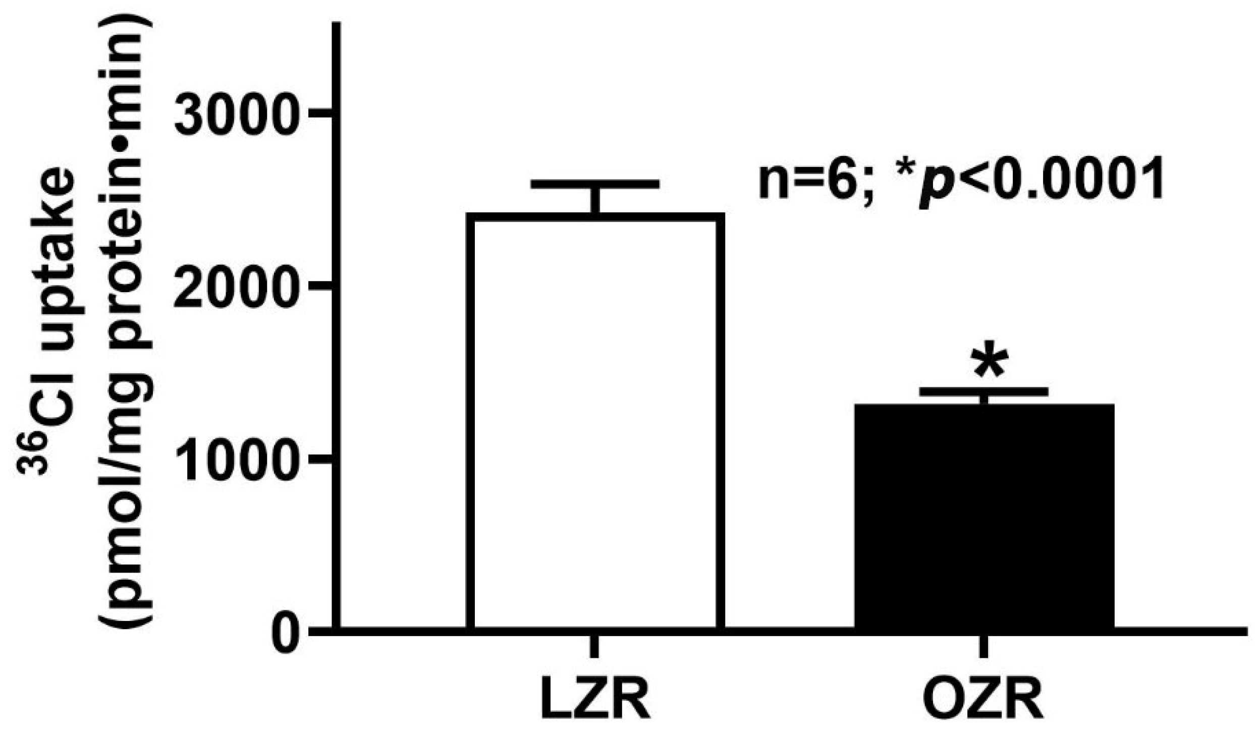
Figure 1. Cl/HCO3 exchange activity in the obese Zucker rat distal colon. DIDS (4,4’-diisothiocyanatostilbene-2,2′-disulfonic acid disodium salt)-sensitive HCO3 dependent 36Cl uptake was significantly inhibited in the AMV of the distal colon of the obese Zucker rat compared with the lean (n = 6; * p < 0.0001). LZR: Lean Zucker Rat, OZR: Obese Zucker Rat, AMV: Apical Membrane Vesicle.
Ijms 26 09139 g001
Figure 2. Na+/H+ exchange activity in the distal colon of the obese Zucker rat. Na+/H+ exchange activity was determined by pH-dependent amiloride-sensitive 22Na uptake, was comparable in the AMV of both the obese and lean Zucker rat distal colon (n = 4). This indicates Na+/H+ exchange was unaffected in obese animals. LZR: Lean Zucker Rat, OZR: Obese Zucker Rat, AMV: Apical Membrane Vesicle.
Ijms 26 09139 g002
Figure 3. Kinetic studies of Cl/HCO3 exchange in the distal colon of the obese Zucker rat. 36Cl uptake at all concentrations was measured over 30 s, as the initial rate of uptake for Cl/HCO3 in AMV was linear for at least 60 s. As the extra vesicular chloride concentration increased, bicarbonate-dependent 36Cl uptake was also stimulated and then saturated under all conditions. The kinetic parameters from these studies are shown in Table 1; the maximal velocity (Vmax; nmol/mg protein · 30 s) is significantly reduced in the distal colonocytes of obese Zucker rats, whereas there is no change in the affinity of the exchangers (1/Km; mM) for chloride between the two groups. LZR: Lean Zucker rat; OZR: Obese Zucker rat (n = 3).
Ijms 26 09139 g003
Figure 4. DRA mRNA levels in the distal colonocytes of the obese Zucker rats. DRA mRNA levels were present in both lean and obese Zucker rat distal colon. However, in obesity, DRA mRNA levels were moderately downregulated in the distal colon (n = 6, * p < 0.02).
Ijms 26 09139 g004
Figure 5. DRA protein expression in the distal colonocytes lysate of obese Zucker rats. Western blot studies revealed that the total cellular DRA protein expression of the distal colonocytes was significantly decreased in obese Zucker rats compared to lean rats (n = 6; * p < 0.001).
Ijms 26 09139 g005
Figure 6. NHE3 protein expression in the distal colonocytes of the obese Zucker Rat. Western blot studies revealed that the total cellular NHE3 protein expression was unaltered between the distal colonocytes of obese and lean Zucker rats.
Ijms 26 09139 g006
Figure 7. Apical membrane DRA protein expression in the distal colon of obese Zucker rats. Western blot studies revealed that the DRA protein expression in the apical membrane of the distal colonocytes from obese Zucker rats was significantly decreased compared to lean Zucker rats (n = 6; * p < 0.001).
Ijms 26 09139 g007
Table 1. Kinetic parameters of Cl/HCO3 exchange in the distal colonocytes of obese Zucker rats.
Km (mM)Vmax
(nmol/mg Protein·30 s)
Lean Zucker rat35.7 ± 3.25.8 ± 0.3
Obese Zucker rat32.1 ± 5.74.2 ± 0.4 *
n = 3, * p < 0.05
Disclaimer/Publisher’s Note: The statements, opinions and data contained in all publications are solely those of the individual author(s) and contributor(s) and not of MDPI and/or the editor(s). MDPI and/or the editor(s) disclaim responsibility for any injury to people or property resulting from any ideas, methods, instructions or products referred to in the content.