Article

Differential Expression of Galectin-1 and Galectin-9 in Immune-Mediated Inflammatory Diseases

by
Cristina Valero-Martínez
1,
Marisa Pardines-Ortiz
1,
Nuria Montes
1,
Esteban Dauden
2,
Benjamín Fernández-Gutierrez
3,
Esther García-Planella
4,
Fernando Gomollón García
5,
Jordi Gratacós
6,
Jose Javier Pérez-Venegas
7,
Antonio Julía
8,
Sara Marsal
8,
Amalia Lamana
1,
Rosario García-Vicuña
1,
Isidoro González-Alvaro
1,*,† and
Ana Triguero-Martínez
1,*,†
1
Rheumatology Department, Hospital Universitario La Princesa, Instituto de Investigación Sanitaria La Princesa (IIS-Princesa), 28006 Madrid, Spain
2
Dermatology Department, Hospital Universitario La Princesa, Instituto de Investigación Sanitaria La Princesa (IIS-Princesa), 28006 Madrid, Spain
3
Rheumatology Department, Hospital Clínico San Carlos, 28040 Madrid, Spain
4
Gastroenterology Department, Hospital de la Santa Creu i Sant Pau, 08041 Barcelona, Spain
5
Gastroenterology Department, Hospital Clínico Universitario Lozano Blesa IIS Aragón, CIBEREHD, 50009 Zaragoza, Spain
6
Department of Rheumatology, Parc Tauli Hospital Universitari, I3PT-CERCA, Medicina UAB, 08208 Sabadell, Spain
7
Rheumatology Service, Hospital Universitario Virgen Macarena, 41009 Sevilla, Spain
8
Rheumatology Department, Hospital Universitari Vall d’Hebron, 08035 Barcelona, Spain
*
Authors to whom correspondence should be addressed.
These authors contributed equally to this work.
Int. J. Mol. Sci. 2025, 26(18), 9087; https://doi.org/10.3390/ijms26189087
Submission received: 11 August 2025 / Revised: 11 September 2025 / Accepted: 16 September 2025 / Published: 18 September 2025
(This article belongs to the Special Issue Galectins (Gals), 2nd Edition)

Abstract

Galectin-1 and -9 (Gal1/9) are essential mediators of immune-inflammatory responses, which makes these proteins potential biomarkers for immune-mediated diseases (IMIDs), such as rheumatoid arthritis (RA), psoriasis (PS), psoriatic arthritis (PsA), inflammatory bowel disease, and systemic lupus erythematosus (SLE). Our aim was to evaluate plasma Gal1/9 differences between IMID patients and healthy donors (HD). We analyzed 980 plasma samples divided into two analytical cohorts (600 discovery group and 380 validation group). Generalized linear models estimated Gal1/9 levels, adjusting for sex, age, storage time, and plate variability. In the overall IMID group, plasma Gal1 levels were comparable to those of HD, while Gal9 levels were significantly elevated. Levels varied across individual diseases: SLE patients consistently showed the highest Gal1/9 levels compared to both HD and other IMIDs, and RA patients had elevated Gal9 levels versus HD. Both Gal1 and Gal9 plasma levels correlated positively with higher disease activity, and Gal1 was higher in patients with longer disease duration. After adjustment for these confounders, SLE and RA patients maintained the highest Gal9 levels compared to HD. Our study demonstrates that Gal1 and Gal9 are differentially expressed across IMIDs, with particularly elevated levels in SLE, and both galectins are associated with disease activity.

1. Introduction

Immune-mediated inflammatory diseases (IMIDs) are a broad group of disorders characterized by abnormal immune responses that lead to inflammation, tissue damage, and, in many cases, long-term complications. Examples of IMIDs are rheumatoid arthritis (RA), psoriasis (PS), psoriatic arthritis (PsA), spondyloarthritis (SpA), inflammatory bowel disease (IBD), and systemic lupus erythematosus (SLE). The prevalence of IMIDs in Western society is around 5–7% [1]. IMIDs are complex, heterogeneous diseases with an etiology that remains unclear, likely due to their molecular complexity and polygenic nature and the presence of environmental risk factors such as smoking, overweight, advanced age, and infections. There is an overlap of risk loci between the different IMIDs, especially for those loci with immune-related genes, suggesting that IMIDs share common signaling pathways [2].
Although multiple factors may interact to promote the development of IMIDs, it is generally believed that most of these disorders arise due to a deficiency in immune tolerance or an imbalance between activation and termination of the immune response [3]. IMIDs share a common hallmark: an immune cell-mediated inflammation that affects various organs and systems [1], resulting in diverse clinical manifestations. However, the presence of overlapping clinical features, especially early in the disease course and in conditions such as inflammatory arthropathies, poses a challenge to precise classification. Additionally, delayed diagnosis in IMIDs is well known to be associated with worse clinical outcomes. In these diseases, the therapeutic goal is to achieve long-term and sustained control of inflammation to prevent tissue damage [4]. As a result, strong evidence in RA [5,6], PsA [7,8], and SpA [9] recommends implementing a treat-to-target strategy and supports the initiation of immunomodulatory therapy as soon as the diagnosis is established. Thus, the identification of additional accurate biomarkers for early diagnosis and poor prognosis remains essential.
Galectins are β-galactoside-binding animal lectins expressed in many tissues and organs, which show their highest expression in the immune system, where they regulate its homeostasis [10,11]. Galectins are characterized by shared consensus amino acid sequences in the carbohydrate-recognition domain (CRD) [10]. The variety of binding partners and the widespread distribution of galectins enable them to participate in multiple biological functions, including the regulation of the immune system [12]. In this regard, galectins play a key role as immune regulators, capable of amplifying or dampening immune and inflammatory responses. These responses depend on factors such as the inflammatory stimulus and the microenvironment or the target cell [13,14]. Emerging literature indicates that galectins are involved in the pathogenic mechanisms and development of IMIDs, including RA, SLE, IBD, or PS [15,16]. These characteristics make these proteins attractive therapeutic targets and potential biomarkers for IMIDs.
Galectin 1 (Gal1) acts as a dual regulator of the immune system, playing protective or anti-inflammatory roles in some contexts (autoimmune diseases, allergies, atherosclerosis) while exacerbating pathological processes in others (cancer, infections, osteoarthritis) [12,16,17]. Gal1 suppresses pro-inflammatory cytokine production; modulates monocyte, macrophage, and dendritic cell activity; limits neutrophil and eosinophil migration; and promotes regulatory T cell function. Through these mechanisms, Gal1 mitigates Th1- and Th17-mediated responses, drives the immune response toward a Th2-type profile, and supports the resolution of inflammation [13,16,17].
Galectin-9 (Gal9), as Gal1, acts as an immunomodulator that regulates both adaptive and innate immune responses. In T cells, Gal9 binds to receptors such as TIM-3, promoting apoptosis of pathogenic Th1/Th17 pathways, expanding Tregs, and therefore limiting effector T cell responses [18]. Conversely, intracellular Gal9 can enhance TCR signaling and support Th17 differentiation, potentially exacerbating autoimmune pathology.
Beyond T cells, Gal9 also inhibits TLR7/TLR9-mediated activation of plasmacytoid dendritic cells and B cells, thereby reducing type I interferon secretion and autoantibody production [19,20].
Both galectins are differentially expressed depending on the inflammatory milieu, thereby influencing leukocyte recruitment, activation, and polarization toward pro- or anti-inflammatory phenotypes [21]. Their pleiotropic actions are highly context- and tissue-dependent, and they are currently being investigated as therapeutic targets for modulating immune responses in multiple IMIDs [13,21]. Our group previously validated that Gal1 levels were elevated in the serum of patients with early RA compared to healthy donors (HD) [22]. In the present study, our objective was to explore whether Gal1 levels are also elevated in other IMIDs, beyond RA, compared to HD, thereby validating our previous findings in RA [22]. Furthermore, Gal9 was included in this study because of its functional similarity to Gal1 and the previously observed increase in serum levels in RA and SLE [21]. These approaches may help clarify whether the elevation of Gal1 and Gal9 reflects a general regulatory mechanism in IMIDs or supports their potential utility as diagnostic biomarkers, particularly in RA.

2. Results

2.1. Study Population

We analyzed a total of 980 plasma samples divided into two analytical cohorts: 600 samples in the discovery group (group 1) and 380 in the validation group (group 2). Of these, 150 were from healthy donors (HD) and 830 from patients with IMIDs, including 150 with RA, 165 with PsA, 165 with PS, 150 with SLE, 100 with UC, and 100 with CD. The main characteristics of these populations are presented in Table 1. Overall, there were more women in the SLE and RA patient groups, and the sex distribution of these patients was more balanced in group 1. Patients with PsA, and especially those with RA, were older than those in other disease groups, and their age was evenly distributed between group 1 and 2. Samples were collected at baseline diagnosis from 10 patients, while only 55 patients (6.6%) had early-stage disease (<2 years since onset). The majority (n = 650/830; 78.3%) exhibited long-standing disease (>5 years of duration), particularly among patients with RA, SLE, and PS. In group 1, patients with PS and CD exhibited longer disease duration compared to those in group 2, while no significant differences were observed for the other IMIDs. Moderate-to-high DA was observed in nearly a quarter of the patients at the time of sample collection (n = 191; 23%), with significantly higher activity observed in RA patients (n = 77; 41%) compared to those with other IMIDs. Most patients with SLE, RA, and IBD were receiving conventional synthetic Disease-Modifying Antirheumatic Drug therapy, while the use of biological therapy was substantially lower overall. However, it was moderate for RA patients (n = 73; 48.6%), exceeding that for other IMIDs.

2.2. Gal1 and Gal9 Plasma Levels in Different IMIDs

Plasma Gal1 levels in the overall IMID group were comparable to those of HD, both in group 1 (22.6 [17.2–31.05] vs. 23.5 [18.9–28.2]; p = 0.816) and in group 2 (14.8 [20.5–27.0] vs. 18.4 [16.1–26.4]; p = 0.626). In addition, as shown in Figure 1A,B, compared to HD, patients with SLE showed a non-statistically significant increase in Gal1 levels in both groups (p = 0.008 group 1 and p = 0.03 group 2), and these levels were also higher compared to RA, PsA, UC, and CD in both groups (Supplementary Table S1). In group 1, Gal1 levels were higher in patients with CD and PS compared to HD and lower in those with PsA. They were also greater in PS and RA (p < 0.001) than in PsA; however, those findings were not replicated in group 2. In contrast, other IMIDs such as UC and RA did not show significant differences in Gal1 levels vs HD. Among IBD subtypes, CD patients in group 1 exhibited higher Gal1 levels than UC patients (p < 0.001), although this difference was not observed in group 2.
Unlike Gal1, plasma Gal9 levels were significantly higher in the overall IMID group compared to HD in both cohorts (group 1 (5.3 [3.6–7.5] vs. 3.8 [2.9–4.8]; p < 0.001) and group 2 (4.8 [3.5–6.6] vs. 4.1 [3.0–5.0]; p = 0.004), despite considerable overlap in values. When analyzing individual IMIDs, patients with SLE, followed by RA, consistently showed significantly higher Gal9 levels than HD in both groups (p < 0.001 for both) (Figure 1C,D). As observed with Gal1, SLE patients exhibited the highest Gal9 levels among all IMIDs studied (Supplementary Table S2).
On the other hand, PsA patients showed only a trend toward increased levels vs. HD (p = 0.06 in group 1; p = 0.04 in group 2) and had significantly lower levels than RA patients (p < 0.001 in group 1; p = 0.004 in group 2). Similar to Gal1, Gal9 levels in PsA patients were also lower than in PS patients in group 1, although this difference did not reach statistical significance. Among the other IMIDs, CD and PS patients had significantly higher Gal9 levels than HD only in group 1 (p < 0.001), with no significant differences observed in group 2.

2.3. Association of Gal1 and Gal9 Plasma Levels with Disease Activity and Disease Duration

Given the broad variability in Gal1 and Gal9 levels across IMIDs, we hypothesized that disease duration or DA could contribute to this heterogeneity. To assess this contribution, DA was standardized into a common categorical variable using disease-specific activity indices to define the categories in each disease, as described in the Methods section.
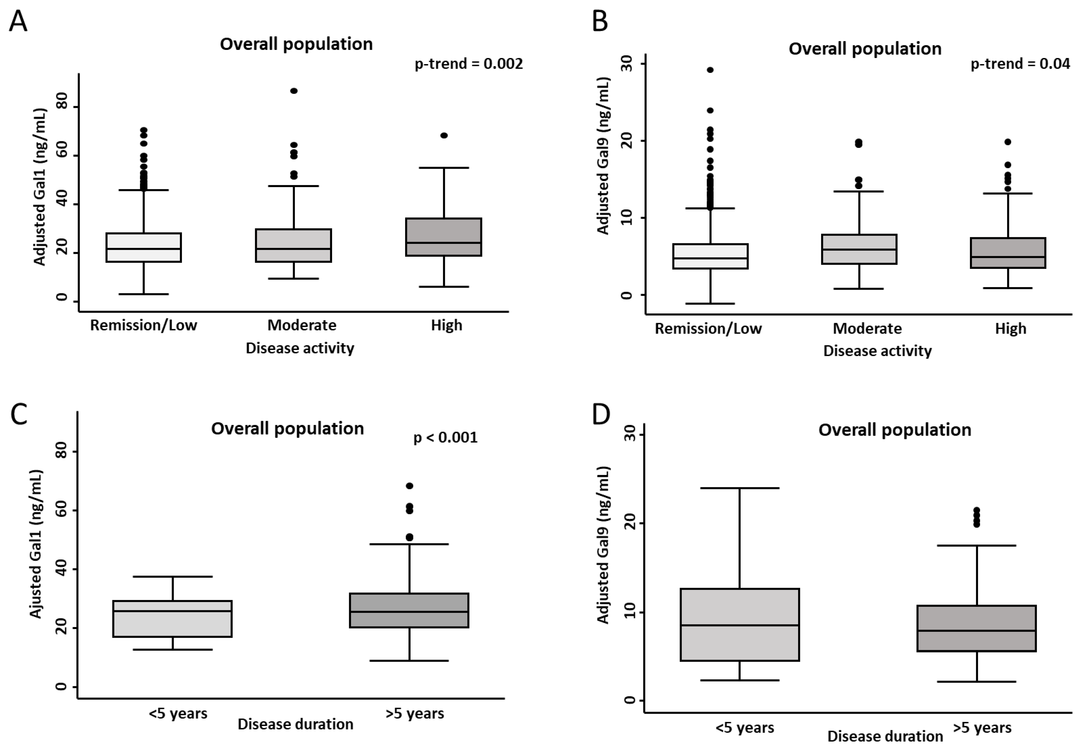
When analyzing DA in the overall population, we observed a significant positive trend toward higher plasma levels of Gal1 (p = 0.002) and Gal9 (p = 0.041) across increasing DA categories. This suggests a progressive rise in galectin levels with worsening DA (Figure 2A,B). Among the different IMIDs, patients with moderate and high DA exhibited significantly higher Gal1 levels in RA (p = 0.0005), while a non-significant trend was observed in UC (p = 0.01) (Supplementary Figure S1). Regarding Gal9, non-significant trends toward elevated levels in patients with increased DA were found in SLE (p = 0.0306), PS (p = 0.0094), and RA (p = 0.0240) (Supplementary Figure S2).
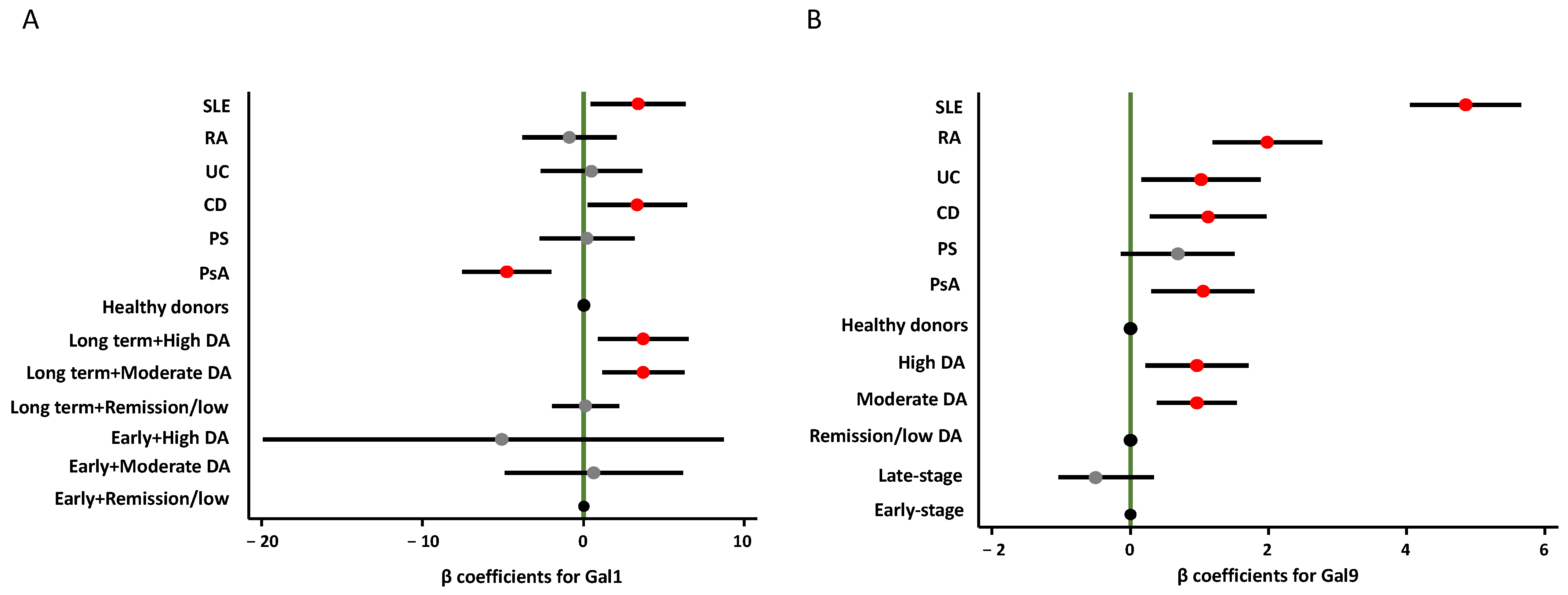
Additionally, we compared patients with early-stage (<5 years) and late-stage (≥5 years) IMIDs (Figure 2C,D), detecting significantly higher Gal1 plasma levels in those with longer disease duration (p < 0.001). However, this pattern was not evident for Gal9. As shown in Figure 3, the best multivariable model for Gal1, which included diagnosis, DA, and disease duration, revealed a significant interaction between the latter two variables. Notably, only patients with long-standing disease and moderate to high DA exhibited significantly elevated plasma Gal1 levels, compared to early-stage patients in remission or with low DA (Figure 3, left panel and Supplementary Table S3; p = 0.004 and p = 0.010, respectively). However, among patients with early-stage disease, no relevant association was observed between Gal1 plasma levels and DA. In contrast, Gal9 levels were not significantly influenced by disease duration, but patients with moderate and high DA showed significantly elevated Gal9 plasma levels compared to those in remission or with low DA (Figure 3, right panel, and Supplementary Table S4; p = 0.001 and p = 0.011, respectively).
After adjusting for DA and stratifying by disease duration, patients with PsA showed significantly lower Gal1 plasma levels (p = 0.001), while those with CD (p = 0.034) and SLE (p = 0.023) exhibited significantly higher levels compared to HD (Figure 3, left panel; Supplementary Table S3). In contrast, all IMIDs except PS displayed elevated Gal9 levels relative to HD (Figure 3, right panel; Supplementary Table S4).

3. Discussion

The main strength of this study lies in demonstrating that plasma levels of Gal1 and Gal9 are dysregulated in patients with IMIDs, through an assessment within the same population and across two independent cohorts. Our findings suggest that plasma levels of Gal1 and particularly Gal9 are generally elevated in IMIDs compared to HD, with galectin levels influenced by disease activity in both cases and by disease duration in the case of Gal1. These results reinforce a potential role for galectins in the pathophysiology of IMIDs as key regulators of immune and inflammatory responses [13,14].
It is worth noting that SLE was the only IMID in which both Gal1 and Gal9 levels were consistently elevated compared to HD across both cohorts. Furthermore, these results remained significant after adjustment by disease duration and disease activity. Gal9 was also increased in RA, whereas findings for other IMIDs versus HD were inconsistent between the two study populations. However, after adjustment by disease activity level, all IMIDs except PS showed increased Gal9 plasma levels.
The differential levels of Gal9 and Gal1 observed across IMIDs can be interpreted in light of their dual immunoregulatory roles and the predominant immune pathways in each condition [21]. Both Gal1 and Gal9 share key anti-inflammatory properties at the level of adaptive immunity, particularly by inhibiting Th1/Th17 responses (decreased IFNγ, TNF-α, and IL-17), promoting expansion of Foxp3+ regulatory T cells, and attenuating pathogenic T cell activity [12,13,16,17,19]. However, their effects diverge in humoral and innate immunity. Gal9 inhibits plasmablast differentiation and autoantibody generation, whereas Gal1 favors a Th2-skewed response. At the innate immunity level, Gal1 predominantly exerts anti-inflammatory effects (e.g., neutrophil apoptosis, M2 macrophage polarization), while Gal9 enhances granulocyte survival, activation, and pro-inflammatory cytokine release [12,13,16,17,19,21]. Overall, their anti-inflammatory mechanisms appear to dominate over their pro-inflammatory properties, consistent with the protective effects observed in murine models of SLE and RA, where administration of either galectin ameliorated disease activity [23,24,25,26]. In this context, the particularly high Gal9 levels observed in our SLE and RA patients compared to other IMID patients could reflect its additional inhibitory role in humoral responses, beyond T cell modulation. Conversely, the highest Gal1 levels observed in SLE, but not in other IMIDs, may be interpreted as a consequence of its anti-inflammatory function in the innate immunity, which plays a central role in SLE pathogenesis.
To the best of our knowledge, our study provides the first description in the literature of plasma galectin levels across all these IMIDs within the same population. Previous studies focused on individual IMIDs had already reported increased plasma levels of Gal1 in patients compared to controls in RA [21,22], IBD [27,28], and PS [29]. However, no evidence on Gal1 levels was available for SLE or PsA. Regarding Gal9, other publications had also reported elevated plasma levels in patients with RA [30] and SLE [31,32] compared to controls, in accordance with our study. In PS, Chagan-Yatusan et al. [33] found elevated levels of Gal9 compared to HD, whereas Nofal et al. [34] described similar Gal9 levels, in line with our results. In contrast, our PsA patients had a trend to higher Gal9 levels than the controls. In this case, no previous studies had assessed Gal9 levels in PsA compared to HD. In IBD, a study found no association between higher Gal9 levels and the disease [35], and similarly, in our cohort, elevated Gal9 levels were only observed in patients with IBD after adjusting for disease activity.
Once it was established that these galectins could be elevated in certain IMIDs, our study aimed to identify differences between some of them. Remarkably, the highest levels of both Gal1 and, especially, Gal9 were observed in SLE patients compared to those with other IMIDs, a finding that was replicated in both cohorts.
Interestingly, PsA patients exhibited significantly lower Gal9 levels compared to RA patients in both cohorts; however, lower Gal1 levels were observed in the discovery cohort, but this result was not replicated in the validation cohort. Previous evidence from our group had suggested that Gal1 levels in both serum and synovial fluid were higher in early RA compared to SpA [36]. More recently, and in line with our findings, Bably et al. [37] reported higher serum Gal1 levels in RA compared to PS and PsA, with no differences between PS and PsA. In our study, Gal1 and Gal9 levels were lower in PsA than in PS; although this finding could not be validated in both cohorts. No reports were found in the literature comparing Gal9 levels between PsA and PS; however, Gal9 has been investigated in eczematous inflammatory skin conditions, showing elevated levels in atopic dermatitis compared to PS [34].
Although we cannot draw firm conclusions at this stage, these findings may be useful in distinguishing certain clinical patterns in some IMIDS—for example, in cases of undifferentiated arthritis, where SLE, RA, PsA, and osteoarthritis should be considered in the differential diagnosis. In our analysis in IBD, we were also unable to establish consistent differences in Gal1 or Gal9 levels between CD and UC. Previous studies in IBD have reported no differences in Gal1 and Gal9 plasma levels between CD and UC [27,38], nor in the mRNA expression of Galectins-1, -2, -3, -4, and -9 [39].
Given the anti-inflammatory properties of Gal1 and Gal9 [12,13,14,15,16], their elevated levels in IMIDs may represent a mixed regulatory loop mechanism aimed at controlling or promoting inflammation, which could be associated with disease activity or severity. Supporting this interpretation, our study revealed that Gal9 plasma levels were increased in patients with moderate or high DA compared to those in remission or low DA. Regarding Gal1, this association could only be demonstrated in late-stage patients. Interestingly, in RA patients, Mendez-Huergo et al. [40], in a population mostly including long-standing RA, reported correlations between Gal1 plasma levels and DA assessed with both the ESR and the DAS-28 indices. Conversely, our previous observations from longitudinal studies on early RA revealed no clear correlation between serum Gal1 levels and DA [22], in agreement with results reported by Bably et al. [37]. Altogether, these results suggest that increased Gal1 levels could be associated with severity in long-term RA patients rather than being a disease activity biomarker in early arthritis. Considering Gal9 in RA, substantial evidence has shown that its plasma levels positively correlate with increased disease activity and severity, as reflected by higher disease scores, elevated acute-phase reactants, greater treatment burden, and longer disease duration [30,41,42,43,44,45]. Moreover, Gal9 levels have been shown to decrease in response to effective immunosuppressive therapy [43,46].
In the case of SLE, our data confirmed an association between Gal9 plasma levels (not confirmed for Gal1) and higher DA, consistent with previous studies using the SLEDAI-2K index or the type I IFN gene signature [21,47,48,49,50,51]. These studies also reported correlations with disease severity—reflected by organ involvement and damage—and with treatment response, as Gal9 levels decreased with clinical improvement. On the contrary, urinary Gal-9 levels do not differentiate active from inactive lupus and are similar to those of controls [47,48].
As for SLE, patients with PS exhibited a trend to higher plasma Gal9 levels correlating with PS activity, which were not observed for Gal1; however, other studies found no differences in Gal1 or Gal9 levels [29,34]. Our cohort exhibited heterogeneous results in the association between IBD and disease activity, and the limited number of patients in the moderate-to-high activity subgroup precludes drawing definitive conclusions. Previous studies have reported a correlation of IBD flares and histological colonic inflammation with both serum levels and mRNA expression of Gal1 [27,38,52], whereas others have found no significant differences in Gal1 serum levels between active disease and remission [27]. As for Gal9, Cibor et al. [35] found no association between its serum levels and disease activity in either CD or UC.
Some limitations of this study must be considered: (a) the cross-sectional design does not allow us to determine whether galectin levels vary over time during patient follow-up, nor does it allow us to confirm whether Gal1 remains stable over time in other IMIDs, as previously observed in RA and SpA [22,36]; (b) there is a lack of clinical and therapeutic data, such as obesity, cardiovascular disease, and cancer, which have been associated with elevated galectin levels [53,54,55,56] and could improve adjustment of Gal1 and Gal9 plasma levels; (c) the study population was limited to Caucasian individuals, which may affect the generalizability of Gal1 and Gal9 as potential biomarkers for IMIDs. For instance, in Gal9 studies, results reported in Asian cohorts were similar to those obtained in our study, both in RA [30,41,42,43,46] and in SLE [31]. In SLE, our Gal9 results have been also replicated in other different cohorts (Egyptian, Indian, or Turkish [32,48,49]); (d) the use of a unified categorical disease activity variable across diverse IMIDs. While widely accepted cut-offs were applied for each index, the constructs differ substantially (e.g., joint counts, skin involvement, serology), so “moderate” or “high” activity may not reflect equivalent inflammatory burden. This common construct may partly explain the heterogeneity across activity categories, but it enabled the inclusion of all IMIDs and provided sufficient power to detect differences related to disease activity; (e) most patients had long-standing disease (>10 years), and the lower proportion with early disease may have limited the sensitivity and statistical power to detect associations, despite applying a 5-year cut-off to distinguish early from later disease; finally, we cannot rule out the influence of the smaller sample size in the validation cohort, which may have limited the ability to detect subtle differences in galectin levels.

4. Methods

4.1. Patients and Samples

In this study, we requested plasma samples from patients diagnosed with six different IMIDs, including RA, PsA, SLE, IBD (ulcerative colitis [UC], Crohn’s disease [CD]), and PS, as well as HD from different sources. The samples from 830 patients were provided by Hospital Universitario Vall d’Hebron Biobank, collected by the Immune Mediated Inflammatory Disease Consortium (IMIDC) between June 2007 and December 2010. HD samples from 150 individuals were collected from IIS-IP Biobank and Spanish National DNA Bank Carlos III (Salamanca University). Samples were collected during patient follow-up after the initial diagnosis and were sequentially assigned to one of two groups based on their order of arrival at our center: a discovery group (1) and a subsequent validation group (2). Galectin determination was conducted in batches at two different time points in each group (2022 and 2024). We included patients with a confirmed diagnosis of a single IMID, excluding those with more than one IMID. Diagnostic and inclusion criteria for each IMID were as follows:
  • RA: American College of Rheumatology (ACR) classification criteria for RA [57].
  • PsA: Classification Criteria for Psoriatic Arthritis (CASPAR) [58].
  • PS: diagnosis based on the dermatologist clinical criteria.
  • IBD: diagnosed according to the standard Lennard-Jones criteria for CD and UC [59].
  • SLE: 1982 revised ACR criteria for SLE classification [60].

4.2. Measurement of Gal1 and Gal9 Plasma Levels in HD and IMID Population

Gal1 and Gal9 were measured in plasma samples from all patients and HD by using the Human Galectin-1 Quantikine ELISA Kit DGAL10 (R&D Systems; Minneapolis, MN, USA), and the Human Galectin-9 Quantikine ELISA Kit DGAL90 (R&D Systems; Minneapolis, MN, USA) according to the manufacturer’s instructions. All samples were tested in duplicate in each plate, and, to minimize the influence of inter-assay variability, samples from the different IMIDs were included in every plate. Once the procedure for each ELISA was performed, plates were immediately read in a spectrophotometer (Innogenetics Diagnóstica y Terapéutica S.A.U, Barcelona, Spain) Absorbance was measured at 450 nm with correction at 620 nm.

4.3. Statistical Analysis

The time of sample collection; demographic, laboratory, and disease-related data; and treatment patterns were provided by Hospital Universitario Vall d’Hebron Biobank. Differences in demographic and clinical characteristics between the discovery and validation populations, as well as across different diseases, were analyzed. For plasma levels of Gal1 and Gal9, their variations across different IMID diagnoses and their relationship with disease duration and moderate or high disease activity (DA) were analyzed. To allow comparative analyses across the cohort, a unified DA variable was created, classifying each patient into one of three activity levels (remission/low, moderate, and high DA). DA was assessed using validated indices specific to each condition, applying the following cut-off values to categorize remission/low, moderate, and high DA, respectively: DAS-28 ≥ 3.2 and ≥5.1 for PsA and RA [61]; PASI ≥ 7 and ≥15 for PS [62]; SLEDAI ≥ 6 and ≥10 for SLE [63]; Harvey–Bradshaw Index ≥ 8 and ≥16 for CD [64], and Lichtiger score ≥ 9 and ≥15 for UC [65]. To avoid missing data at multivariable analysis exploring factors that can impact DA, HD were included in the remission/low activity group.
Most quantitative variables showed a non-normal distribution and were therefore presented as median and interquartile range (IQR). The Mann–Whitney U test or Kruskal–Wallis test was used to assess significant differences in Gal1 and Gal9 plasma levels according to clinical or laboratory variables and across different diagnostic groups. Bonferroni correction was applied to adjust for multiple comparisons, with p-values considered statistically significant if below 0.008. Correlations between continuous variables were analyzed using Spearman’s rho test. In a previous study, we demonstrated that Gal1 levels can be influenced by sample storage time, age, sex, and inter-assay variability (plate) [22]. Accordingly, generalized linear models (GLMs) were independently fitted for Gal1 and Gal9 within each group, using the respective galectin as the dependent variable and including the aforementioned covariates to obtain more accurate adjusted estimates. GLMs were used to evaluate differences between IMID diagnosis and HD or other IMIDs, DA, or duration of disease. Forest plots were generated to visualize the associations of Gal1 and Gal9 with DA, incorporating interaction terms to assess the modifying effect of disease duration. Statistical analyses were performed using Stata 14.0 for Windows (StataCorp LP, College Station, TX, USA).

5. Conclusions

Our study revealed that plasma levels of Gal1 and Gal9 are differentially regulated across various IMIDs, with particularly elevated levels observed in SLE patients compared to other IMIDs. Additionally, Gal1 and Gal9 may serve as biomarkers for monitoring disease activity, with Gal1 potentially influenced by disease duration. While our data validate the utility of Gal9 in RA and SLE, we also highlight the need to further explore the role of Gal1 in both diseases.

Supplementary Materials

The following supporting information can be downloaded at https://www.mdpi.com/article/10.3390/ijms26189087/s1.

Author Contributions

Conception or design of the work: C.V.-M. and A.T.-M.; acquisition of data: IMID Consortium; analysis and interpretation of data: All authors; drafting of the manuscript: C.V.-M., A.T.-M., I.G.-A. and R.G.-V.; critical revision of the manuscript for important intellectual content: all authors; statistical analysis: C.V.-M., A.T.-M. and I.G.-A.; review and approval of the final version of the manuscript: all authors; supervision: A.T.-M., I.G.-A. and R.G.-V. All authors have read and agreed to the published version of the manuscript.

Funding

This work was supported by grants RD24/0007/0006, PI21/00526, and PI24/00138 to IG-A from Ministerio de Ciencia e Innovación (Instituto de Salud Carlos III, ISCIII), co-funded by European Regional Development Fund (ERDF) “A way to make Europe” and the project SPIDeRR funded by the Horizon Europe program under grant agreement 101080711.

Institutional Review Board Statement

The present study was approved by the Research Ethics Committee of Hospital Universitario de La Princesa (PI- 4529; 20 May 2021) and it complied with principles expressed in the Helsinki Declaration of 1983 and successive actualizations. To participate in the study it was necessary to sign a written informed consent and to be older than 18 years old. The Banco Nacional de ADN Carlos III (ISCIII B.0000716), the BIOBANCO HOSPITAL UNIVERSITARIO VALL D’HEBRON (ISCIII B.0000618) and the IIS-IP Biobank (ISCIII B.0000763) provided the samples and data from patients included in this study. All samples were processed following standard operating procedures with the appropriate approval of the Ethics and Scientific Committees.

Informed Consent Statement

All participants included were over 18 years old and signed a written informed consent.

Data Availability Statement

The original contributions presented in this study are included in the article/Supplementary Material. Further inquiries can be directed to the corresponding authors.

Acknowledgments

We thank Manuel Gomez Gutierrez, for his excellent editing assistance. During the preparation of this manuscript, the authors used ChatGPT (GPT-4-turbo, OpenAI) for the purposes of assisting in text editing and improving the clarity and consistency of the English language. The authors have reviewed and edited the output and take full responsibility for the content of this publication. IMID Consortium: Eduardo Fonseca, Jesús Rodríguez, Patricia Carreira, Valle García, José A Pinto-Tasende, Lluís Puig, Elena Ricart, Francisco Blanco, Ricardo Blanco, Víctor Martínez Taboada, Emilia Fernández, Pablo Unamuno, Raimon Sanmartí, Ana Gutiérrez, Àlex Olivé, José Luís López Estebaranz, Juan Carlos Torre-Alonso, José Luis Andreu, David Moreno Ramírez, Mª Ángeles Aguirre Zamorano, Pablo de la Cueva, Pilar Nos Mateu, Paloma Vela, Francisco Vanaclocha, Héctor Corominas, San-tiago Muñoz, Joan Miquel Nolla, Enrique Herrera, Carlos González, José Luis Marenco de la Fuente, Maribel Vera, Alba Erra, Daniel Roig, Antonio Zea, María Esteve Comas, Carles Tomàs, Pedro Zarco, José María Pego, Cristina Saro, Antonio González, Mercedes Freire, Alicia García, Elvira Díez, Georgina Salvador, César Díaz, Simón Sánchez, Alfredo Willisch Dominguez, José Antonio Mosquera, Julio Ramírez, Esther Rodríguez Almaraz, Núria Palau, Raül Tortosa, Mireia López, Andrea Pluma, Adrià Aterido.

Conflicts of Interest

The authors declare no conflicts of interest. The funders had no role in the design of the study; in the collection, analyses, or interpretation of data; in the writing of the manuscript; or in the decision to publish the results.

Abbreviations

The following abbreviations are used in this manuscript:
CDCrohn’s disease
DAdisease activity
DAS-28disease activity score 28 (joints)
ELISAenzyme-linked immunosorbent assay
GLMsgeneralized linear models
HDhealthy donors
IBDinflammatory bowel disease
IMIDsimmune-mediated inflammatory diseases
PASIPsoriasis Area and Severity Index
PsApsoriatic arthritis
PSpsoriasis
RArheumatoid arthritis
SLEsystemic lupus erythematosus
SLEDAISystemic Lupus Erythematosus Disease Activity Index
UCulcerative colitis

References

  1. El-Gabalawy, H.; Guenther, L.C.; Bernstein, C.N. Epidemiology of immune-mediated inflammatory diseases: Incidence, prevalence, natural history, and comorbidities. J. Rheumatol. Suppl. 2010, 85, 2–10. [Google Scholar] [CrossRef]
  2. Degboe, Y.; Vastert, S.J.; Prakken, B.J.; McInnes, I.B. How does age determine the development of human immune-mediated arthritis? Nat. Rev. Rheumatol. 2022, 18, 501–512. [Google Scholar] [CrossRef]
  3. Theofilopoulos, A.N.; Kono, D.H.; Baccala, R. The multiple pathways to autoimmunity. Nat. Immunol. 2017, 18, 716–724. [Google Scholar] [CrossRef]
  4. Kuek, A.; Hazleman, B.L.; Ostör, A.J. Immune-mediated inflammatory diseases (IMIDs) and biologic therapy: A medical revolution. Postgrad. Med. J. 2007, 83, 251–260. [Google Scholar] [CrossRef]
  5. Stoffer, M.A.; Schoels, M.M.; Smolen, J.S.; Aletaha, D.; Breedveld, F.C.; Burmester, G.; Bykerk, V.; Dougados, M.; Emery, P.; Haraoui, B.; et al. Evidence for treating rheumatoid arthritis to target: Results of a systematic literature search. Ann. Rheum. Dis. 2010, 69, 638–643. [Google Scholar] [CrossRef]
  6. Burgers, L.E.; Raza, K.; van der Helm-van Mil, A.H. Window of opportunity in rheumatoid arthritis—Definitions and supporting evidence: From old to new perspectives. RMD Open 2019, 5, e000870. [Google Scholar] [CrossRef]
  7. Haroon, M.; Gallagher, P.; FitzGerald, O. Diagnostic delay of more than 6 months contributes to poor radiographic and functional outcome in psoriatic arthritis. Ann. Rheum. Dis. 2015, 74, 1045–1050. [Google Scholar] [CrossRef] [PubMed]
  8. Snoeck Henkemans, S.V.J.; de Jong, P.H.P.; Luime, J.J.; Kok, M.R.; Tchetverikov, I.; Korswagen, L.A.; van der Kooij, S.M.; van Oosterhout, M.; Baudoin, P.; Bijsterbosch, J.; et al. Window of opportunity in psoriatic arthritis: The earlier the better? RMD Open 2024, 10, e004062. [Google Scholar] [CrossRef] [PubMed]
  9. Mauro, D.; Forte, G.; Poddubnyy, D.; Ciccia, F. The Role of Early Treatment in the Management of Axial Spondyloarthritis: Challenges and Opportunities. Rheumatol. Ther. 2024, 11, 19–34. [Google Scholar] [CrossRef] [PubMed]
  10. Barondes, S.H.; Cooper, D.N.; Gitt, M.A.; Leffler, H. Galectins. Structure and function of a large family of animal lectins. J. Biol. Chem. 1994, 269, 20807–20810. [Google Scholar] [CrossRef] [PubMed]
  11. Hirabayashi, J.; Kasai, K. The family of metazoan metal-independent beta-galactoside-binding lectins: Structure, function and molecular evolution. Glycobiology 1993, 3, 297–304. [Google Scholar] [CrossRef]
  12. Mansour, A.A.; Krautter, F.; Zhi, Z.; Iqbal, A.J.; Recio, C. The interplay of galectins-1, -3, and -9 in the immune-inflammatory response underlying cardiovascular and metabolic disease. Cardiovasc. Diabetol. 2022, 21, 253. [Google Scholar] [CrossRef]
  13. Cutine, A.M.; Bach, C.A.; Veigas, F.; Merlo, J.P.; Laporte, L.; Manselle Cocco, M.N.; Massaro, M.; Sarbia, N.; Perrotta, R.M.; Mahmoud, Y.D.; et al. Tissue-specific control of galectin-1-driven circuits during inflammatory responses. Glycobiology 2021, 31, 891–907. [Google Scholar] [CrossRef]
  14. Cummings, R.D.; Liu, F.T.; Rabinovich, G.A.; Stowell, S.R.; Vasta, G.R. Galectins. In Essentials of Glycobiology [Internet], 4th ed; Varki, A., Cummings, R.D., Esko, J.D., Stanley, P., Hart, G.W., Aebi, M., Mohnen, D., Kinoshita, T., Packer, N.H., Prestegard, J.H., et al., Eds.; Cold Spring Harbor Laboratory Press: Cold Spring Harbor, NY, USA, 2022. Available online: http://www.ncbi.nlm.nih.gov/books/NBK579987/ (accessed on 21 November 2022).
  15. He, Y.S.; Hu, Y.Q.; Xiang, K.; Chen, Y.; Feng, Y.T.; Yin, K.J.; Huang, J.X.; Wang, J.; Wu, Z.D.; Wang, G.H.; et al. Therapeutic Potential of Galectin-1 and Galectin-3 in Autoimmune Diseases. Curr. Pharm. Des. 2022, 28, 36–45. [Google Scholar] [CrossRef] [PubMed]
  16. Yu, X.; Qian, J.; Ding, L.; Yin, S.; Zhou, L.; Zheng, S. Galectin-1: A Traditionally Immunosuppressive Protein Displays Context-Dependent Capacities. Int. J. Mol. Sci. 2023, 24, 6501. [Google Scholar] [CrossRef]
  17. Liu, F.T.; Stowell, S.R. The role of galectins in immunity and infection. Nat. Rev. Immunol. 2023, 23, 479–494. [Google Scholar] [CrossRef] [PubMed]
  18. Seki, M.; Oomizu, S.; Sakata, K.M.; Sakata, A.; Arikawa, T.; Watanabe, K.; Ito, K.; Takeshita, K.; Niki, T.; Saita, N. Galectin-9 suppresses the generation of Th17, promotes the induction of regulatory T cells, and regulates experimental autoimmune arthritis. Clin. Immunol. 2008, 127, 78–88. [Google Scholar] [CrossRef]
  19. Gossink, E.M.; Coffer, P.J.; Cutilli, A.; Lindemans, C.A. Immunomodulation by galectin-9: Distinct role in T cell populations, current therapeutic avenues and future potential. Cell Immunol. 2025, 407, 104890. [Google Scholar] [CrossRef]
  20. Chen, H.Y.; Wu, Y.F.; Chou, F.C.; Wu, Y.H.; Yeh, L.T.; Lin, K.I.; Liu, F.T.; Sytwu, H.K. Intracellular Galectin-9 Enhances Proximal TCR Signaling and Potentiates Autoimmune Diseases. J. Immunol. 2020, 204, 1158–1172. [Google Scholar] [CrossRef] [PubMed]
  21. Xu, W.D.; Huang, Q.; Huang, A.F. Emerging role of galectin family in inflammatory autoimmune diseases. Autoimmun. Rev. 2021, 20, 102847. [Google Scholar] [CrossRef]
  22. Triguero-Martínez, A.; de la Fuente, H.; Montes, N.; Ortiz, A.M.; Roy-Vallejo, E.; Castañeda, S.; González-Alvaro, I.; Lamana, A. Validation of galectin-1 as potential diagnostic biomarker of early rheumatoid arthritis. Sci. Rep. 2020, 10, 17799. [Google Scholar] [CrossRef]
  23. Rabinovich, G.A.; Daly, G.; Dreja, H.; Tailor, H.; Riera, C.M.; Hirabayashi, J.; Chernajovsky, Y. Recombinant galectin-1 and its genetic delivery suppress collagen-induced arthritis via T cell apoptosis. J. Exp. Med. 1999, 190, 385–398. [Google Scholar] [CrossRef] [PubMed]
  24. Huang, Y.J.; Shiau, A.L.; Chen, S.Y.; Chen, Y.L.; Wang, C.R.; Tsai, C.Y.; Chang, M.Y.; Li, Y.T.; Leu, C.H.; Wu, C.L. Multivalent structure of galectin-1-nanogold complex serves as potential therapeutics for rheumatoid arthritis by enhancing receptor clustering. Eur. Cell Mater. 2012, 23, 170–181; discussion 181. [Google Scholar] [CrossRef] [PubMed]
  25. Seki, M.; Sakata, K.M.; Oomizu, S.; Arikawa, T.; Sakata, A.; Ueno, M.; Nobumoto, A.; Niki, T.; Saita, N.; Ito, K.; et al. Beneficial effect of galectin 9 on rheumatoid arthritis by induction of apoptosis of synovial fibroblasts. Arthritis Rheum. 2007, 56, 3968–3976. [Google Scholar] [CrossRef]
  26. Panda, S.K.; Facchinetti, V.; Voynova, E.; Hanabuchi, S.; Karnell, J.L.; Hanna, R.N. Galectin-9 inhibits TLR7-mediated autoimmunity in murine lupus models. J. Clin. Investig. 2018, 128, 1873–1887. [Google Scholar] [CrossRef]
  27. Yu, T.B.; Dodd, S.; Yu, L.G.; Subramanian, S. Serum galectins as potential biomarkers of inflammatory bowel diseases. PLoS ONE 2020, 15, e0227306. [Google Scholar] [CrossRef]
  28. Alahmed, A.; Al-Rubaee, E. New insight for ulcerative colitis diagnosis via serum netrin-1 and galectin-1 biomarkers. Hum. Antibodies. 2025. [Google Scholar] [CrossRef]
  29. Nowowiejska, J.; Baran, A.; Hermanowicz, J.M.; Sieklucka, B.; Pawlak, D.; Flisiak, I. Evaluation of Plasma Concentrations of Galectins-1, 2 and 12 in Psoriasis and Their Clinical Implications. Biomolecules 2023, 13, 1472. [Google Scholar] [CrossRef] [PubMed]
  30. Wang, Y.; Song, L.; Sun, J.; Sui, Y.; Li, D.; Li, G.; Liu, J.; Shu, Q. Expression of Galectin-9 and correlation with disease activity and vascular endothelial growth factor in rheumatoid arthritis. Clin. Exp. Rheumatol. 2020, 38, 654–661. [Google Scholar] [PubMed]
  31. Matsuoka, N.; Fujita, Y.; Temmoku, J.; Furuya, M.Y.; Asano, T.; Sato, S.; Matsumoto, H.; Kobayashi, H.; Watanabe, H.; Suzuki, E.; et al. Galectin-9 as a biomarker for disease activity in systemic lupus erythematosus. PLoS ONE 2020, 15, e0227069. [Google Scholar] [CrossRef]
  32. Abdelgalil, A.H.; Elsharaby, R.M.; Abd ElGany, S.E.; Ibrahim, W.S. EXPRESS: Assessment of serum level of galectin-9 in systemic lupus erythematosus patients in Tanta university hospitals. J. Investig. Med. 2025. [Google Scholar] [CrossRef]
  33. Chagan-Yasutan, H.; He, N.; Arlud, S.; Fang, J.; Hattori, T. The elevation of plasma galectin-9 levels in patients with psoriasis and its associations with inflammatory and immune checkpoint molecules in skin tissues. Hum. Immunol. 2024, 85, 110741. [Google Scholar] [CrossRef]
  34. Nofal, E.; Eldesoky, F.; Nofal, A.; Abdelshafy, A.; Zedan, A. Serum galectin-9 levels in atopic dermatitis, psoriasis and allergic contact dermatitis: A cross-sectional study. Indian J. Dermatol. Venereol. Leprol. 2019, 85, 195–196. [Google Scholar] [CrossRef]
  35. Cibor, D.; Szczeklik, K.; Brzozowski, B.; Mach, T.; Owczarek, D. Serum galectin 3, galectin 9 and galectin 3-binding proteins in patients with active and inactive inflammatory bowel disease. J. Physiol. Pharmacol. 2019, 70, 95–104. [Google Scholar] [CrossRef]
  36. Triguero-Martínez, A.; Roy-Vallejo, E.; Tomero, E.G.; Montes, N.; de la Fuente, H.; Ortiz, A.M.; Castañeda, S.; Lamana, A.; González-Álvaro, I. Galectin-1: A Potential Biomarker Differentiating between Early Rheumatoid Arthritis and Spondyloarthritis. J. Clin. Med. 2022, 11, 6313. [Google Scholar] [CrossRef]
  37. El Bably, M.M.; Abdel Aziz, D.M.; Adly, N.N.; Ali, M.A.; Khedr, A.S.; El Leithy, S.A. Galectin-1 in Psoriatic arthritis, Psoriasis, Rheumatoid arthritis and its relation with disease activity and skin lesion. Egypt. J. Immunol. 2023, 30, 73–82. [Google Scholar] [CrossRef] [PubMed]
  38. Kessel, C.; Lavric, M.; Weinhage, T.; Brueckner, M.; de Roock, S.; Däbritz, J.; Weber, J.; Vastert, S.J.; Foell, D. Serum biomarkers confirming stable remission in inflammatory bowel disease. Sci. Rep. 2021, 11, 6690. [Google Scholar] [CrossRef] [PubMed]
  39. Papa Gobbi, R.; De Francesco, N.; Bondar, C.; Muglia, C.; Chirdo, F.; Rumbo, M.; Rocca, A.; Toscano, M.A.; Sambuelli, A.; Rabinovich, G.A.; et al. A galectin-specific signature in the gut delineates Crohn’s disease and ulcerative colitis from other human inflammatory intestinal disorders. Biofactors 2016, 42, 93–105. [Google Scholar] [CrossRef]
  40. Mendez-Huergo, S.P.; Hockl, P.F.; Stupirski, J.C.; Maller, S.M.; Morosi, L.G.; Pinto, N.A.; Berón, A.M.; Musuruana, J.L.; Nasswetter, G.G.; Cavallasca, J.A.; et al. Clinical Relevance of Galectin-1 and Galectin-3 in Rheumatoid Arthritis Patients: Differential Regulation and Correlation With Disease Activity. Front. Immunol. 2019, 9, 3057. [Google Scholar] [CrossRef] [PubMed]
  41. Fujita, Y.; Asano, T.; Matsuoka, N.; Temmoku, J.; Sato, S.; Matsumoto, H.; Furuya, M.Y.; Suzuki, E.; Watanabe, H.; Kawakami, A.; et al. A Differential regulation and correlation between galectin-9 and anti-CCP antibody (ACPA) in rheumatoid arthritis patients. Arthritis Res. Ther. 2020, 22, 80. [Google Scholar] [CrossRef]
  42. Guo, J.; Ai, X.; Jia, B.; Zhong, X.; Liu, L.; Hu, Q.; Xie, J.; Hong, X.; Chen, Y.; Liu, D. Galectin-9 as an indicator of functional limitations and radiographic joint damage in patients with rheumatoid arthritis. Front. Immunol. 2024, 15, 1419676. [Google Scholar] [CrossRef]
  43. Sun, J.; Sui, Y.; Wang, Y.; Song, L.; Li, D.; Li, G.; Liu, J.; Shu, Q. Galectin-9 expression correlates with therapeutic effect in rheumatoid arthritis. Sci. Rep. 2021, 11, 5562. [Google Scholar] [CrossRef]
  44. Wiersma, V.R.; Clarke, A.; Pouwels, S.D.; Perry, E.; Abdullah, T.M.; Kelly, C.; Soyza, A.; Hutchinson, D.; Eggleton, P.; Bremer, E. Galectin-9 Is a Possible Promoter of Immunopathology in Rheumatoid Arthritis by Activation of Peptidyl Arginine Deiminase 4 (PAD-4) in Granulocytes. Int. J. Mol. Sci. 2019, 20, 4046. [Google Scholar] [CrossRef]
  45. Nielsen, M.A.; Køster, D.; Mehta, A.Y.; Stengaard-Pedersen, K.; Busson, P.; Junker, P.; Hørslev-Petersen, K.; Hetland, M.L.; Østergaard, M.; Hvid, M.; et al. Increased Galectin-9 Levels Correlate with Disease Activity in Patients with DMARD-Naïve Rheumatoid Arthritis and Modulate the Secretion of MCP-1 and IL-6 from Synovial Fibroblasts. Cells 2023, 12, 327. [Google Scholar] [CrossRef] [PubMed]
  46. Jia, Q.; Che, Q.; Zhang, X.; Chen, J.; Ren, C.; Wu, Y.; Liang, W.; Zhang, X.; Li, Y.; Li, Z.; et al. Knockdown of Galectin-9 alleviates rheumatoid arthritis through suppressing TNF-α-induced activation of fibroblast-like synoviocytes. Biochem. Pharmacol. 2024, 220, 115994. [Google Scholar] [CrossRef] [PubMed]
  47. van den Hoogen, L.L.; van Roon, J.A.G.; Mertens, J.S.; Wienke, J.; Lopes, A.P.; de Jager, W.; Rossato, M.; Pandit, A.; Wichers, C.G.K.; van Wijk, F.; et al. Galectin-9 is an easy to measure biomarker for the interferon signature in systemic lupus erythematosus and antiphospholipid syndrome. Ann. Rheum. Dis. 2018, 77, 1810–1814. [Google Scholar] [CrossRef]
  48. Mehta, P.; Singh, P.; Aggarwal, A. Serum and urinary galectin-9 and C-X-C motif chemokine ligand 10. Lupus 2022, 31, 482–487. [Google Scholar] [CrossRef]
  49. Mirioğlu, Ş.; Çinar, S.; Uludağ, Ö.; Gürel, E.; Varelci, S.; Özlük, Y.; Kiliçaslan, I.; Yalçinkaya, Y.; Yazici, H.; Gül, A.; et al. Serum and urine interferon-inducible protein 10, galectin-9, and SIGLEC-1 as biomarkers of disease activity in systemic lupus erythematosus. Turk. J. Med. Sci. 2024, 54, 391–400. [Google Scholar] [CrossRef]
  50. Gensous, N.; Vagner, D.; Barnetche, T.; Duffau, P.; Lazaro, E.; Richez, C.; Blanco, P. Galectin 9, CXCL-10 and tumor necrosis factor receptor type II as biomarkers of disease activity in systemic lupus erythematosus. Jt. Bone Spine. 2022, 89, 105311. [Google Scholar] [CrossRef]
  51. Enocsson, H.; Wetterö, J.; Eloranta, M.L.; Gullstrand, B.; Svanberg, C.; Larsson, M.; Bengtsson, A.A.; Rönnblom, L.; Sjöwall, C. Comparison of Surrogate Markers of the Type I Interferon Response and Their Ability to Mirror Disease Activity in Systemic Lupus Erythematosus. Front. Immunol. 2021, 12, 688753. [Google Scholar] [CrossRef] [PubMed]
  52. Chen, P.; Zhou, G.; Lin, J.; Li, L.; Zeng, Z.; Chen, M.; Zhang, S. Serum Biomarkers for Inflammatory Bowel Disease. Front. Med. 2020, 7, 123. [Google Scholar] [CrossRef]
  53. Rodrigues, C.F.; Santos, F.A.; Amorim, L.A.A.; da Silva, A.L.C.; Marques, L.G.A.; Rocha, B.A.M. Galectin-9 is a target for the treatment of cancer: A patent review. Int. J. Biol. Macromol. 2024, 254, 127768. [Google Scholar] [CrossRef]
  54. Chou, R.H.; Huang, S.S.; Kuo, C.S.; Wang, S.C.; Tsai, Y.L.; Lu, Y.W.; Chang, C.C.; Huang, P.H.; Lin, S.J. Galectin-1 is associated with the severity of coronary artery disease and adverse cardiovascular events in patients undergoing coronary angiography. Sci. Rep. 2020, 10, 20683. [Google Scholar] [CrossRef]
  55. Zhu, R.; Liu, C.; Tang, H.; Zeng, Q.; Wang, X.; Zhu, Z.; Liu, Y.; Mao, X.; Zhong, Y. Serum Galectin-9 Levels Are Associated with Coronary Artery Disease in Chinese Individuals. Mediat. Inflamm. 2015, 2015, 457167. [Google Scholar] [CrossRef] [PubMed]
  56. Gurel Cayir, E.; Demir, L.; Varol, U.; Atahan, M.K.; Salman, T.; Oflazoglu, U.; Yildiz, Y.; Taskaynatan, H.; Saray, S.; Kucukzeybek, Y.; et al. Preliminary study of serum Galectin-1 in breast cancer carcinogenesis [Izmir Oncology Group (IZOG) study]. J. BUON 2020, 25, 675–680. [Google Scholar] [PubMed]
  57. Arnett, F.C.; Edworthy, S.M.; Bloch, D.A.; McShane, D.J.; Fries, J.F.; Cooper, N.S.; Healey, L.A.; Kaplan, S.R.; Liang, M.H.; Luthra, H.S.; et al. The American Rheumatism Association 1987 revised criteria for the classification of rheumatoid arthritis. Arthritis Rheum. 1988, 31, 315–324. [Google Scholar] [CrossRef] [PubMed]
  58. Taylor, W.; Gladman, D.; Helliwell, P.; Marchesoni, A.; Mease, P.; Mielants, H.; CASPAR Study Group. Classification criteria for psoriatic arthritis: Development of new criteria from a large international study. Arthritis Rheum. 2006, 54, 2665–2673. [Google Scholar] [CrossRef]
  59. Lennard-Jones, J.E. Classification of inflammatory bowel disease. Scand. J. Gastroenterol. Suppl. 1989, 170, 2–6; discussion 16-9. [Google Scholar] [CrossRef]
  60. Tan, E.M.; Cohen, A.S.; Fries, J.F.; Masi, A.T.; McShane, D.J.; Rothfield, N.F.; Challer, J.G.; Talal, N.; Winchester, R.J. The 1982 revised criteria for the classification of systemic lupus erythematosus. Arthritis Rheum. 1982, 25, 1271–1277. [Google Scholar] [CrossRef]
  61. Van Riel, P.L.; Renskers, L. The Disease Activity Score (DAS) and the Disease Activity Score using 28 joint counts (DAS28) in the management of rheumatoid arthritis. Clin. Exp. Rheumatol. 2016, 34 (Suppl. 101), S40–S44. [Google Scholar]
  62. Llamas-Velasco, M.; de la Cueva, P.; Notario, J.; Martínez-Pilar, L.; Martorell, A.; Moreno-Ramírez, D. Moderate Psoriasis: A Proposed Definition. Actas Dermosifiliogr. 2017, 108, 911–917, (English, Spanish). [Google Scholar] [CrossRef] [PubMed]
  63. Mosca, M.; Bombardieri, S. Assessing remission in systemic lupus erythematosus. Clin. Exp. Rheumatol. 2006, 24 (Suppl. 43), S-99–S-104. [Google Scholar] [PubMed]
  64. Best, W.R. Predicting the Crohn’s disease activity index from the Harvey-Bradshaw Index. Inflamm. Bowel Dis. 2006, 12, 304–310. [Google Scholar] [CrossRef]
  65. Schoepfer, A.M.; Beglinger, C.; Straumann, A.; Safroneeva, E.; Romero, Y.; Armstrong, D.; Schmidt, C.; Trummler, M.; Pittet, V.; Vavricka, S.R. Fecal calprotectin more accurately reflects endoscopic activity of ulcerative colitis than the Lichtiger Index, C-reactive protein, platelets, hemoglobin, and blood leukocytes. Inflamm. Bowel Dis. 2013, 19, 332–341. [Google Scholar] [CrossRef] [PubMed]
Figure 1. Gal1 and Gal9 plasma levels across IMIDs and HD in the discovery (A,C) and validation (B,D) populations. Values of Gal1 and Gal9 plasma levels were adjusted for frozen storage time, age, sex, and inter-assay variability (plate). Data are shown as box plots in which the line in the box represents the median, and the box edges represent the interquartile range. Whiskers represent the 5th and 95th percentiles. Dots indicate outliers. The Mann–Whitney test was used to determine significant differences with respect to HD. Due to multiple comparisons, the level of significance was set at p < 0.008 (* p > 0.008 to <0.05; # p < 0.008). Abbreviations: CD: Crohn’s disease; Gal1: galectin 1; Gal9: galectin 9; HD: healthy donors; IMIDs: immune-mediated inflammatory diseases; PS: psoriasis; PsA: psoriatic arthritis; RA: rheumatoid arthritis. SLE: systemic lupus erythematosus; UC: ulcerative colitis.
Ijms 26 09087 g001
Figure 2. Gal1 and Gal9 plasma levels according to disease activity category (A,B) and disease duration (C,D) in patients with immune-mediated inflammatory diseases. Box plots show the interquartile range (IQR; box edges = 25th to 75th percentile, midline = median), and whiskers represent the 5th and 95th percentiles. Dots indicate outliers. Values for Gal1 and Gal9 plasma levels were predicted from multivariable linear regression models adjusted for frozen storage time, age, sex, and inter-assay variability (plate). Statistical significance was determined by Cuzick’s test (panels A,B) and the Mann–Whitney test (panels C,D); significance level was set at p < 0.05. Abbreviations: CD: Crohn’s disease; Gal1: galectin 1; Gal9: galectin 9.
Ijms 26 09087 g002
Figure 3. Variables associated with Gal1 and Gal9 plasma levels. Data represent de β coefficients (dots) and the 95% confidence interval (black line) obtained in multivariable linear regression models assessing the association between plasma levels of Gal1 (A) and Gal9 (B) and the following variables: diagnosis (HD (refence), SLE, RA, UC, CD, PS, PsA), disease activity (high, moderate, remission/low), and disease duration (early vs. late-stage). Black dots represent the reference condition of each variable, gray dots represent non-significant conditions, and red dots significant conditions. (See Supplementary Tables S3 and S4 and Methods section for further information). Abbreviations: CD: Crohn’s disease; DA: disease activity; Gal1: galectin-1; Gal9: galectin-9; PS: psoriasis; PsA: psoriatic arthritis; RA: rheumatoid arthritis. SLE: systemic lupus erythematosus; UC: ulcerative colitis.
Ijms 26 09087 g003
Table 1. Characteristics of patients diagnosed with IMIDs and controls.
DiagnosisTotal Patients (n = 830)Age
(Median; IQR)
Sex
(Female)
(n; %)
Duration of Disease (Years; Median: IQR)Moderate/
High Disease Activity ** (n; %)
csDMARD
(n; %)
bDMARD
(n; %)
HD15044 (30–75)87 (58)
Group 110035 (28–48)57 (57)
Group 25048 (44–53)30 (60)
PsA16552.5 (42.5–63)85 (51.5)10 (5–17)42 (25.4)92 (55.7)41 (24.8)
Group 110050 (42–63)50 (50)11 (5–17)22 (22)56 (56) 31 (31)
Group 26554 (43–61)35 (53.8)10 (5–16)20 (30.7)36 (65.4)10 (15.3)
Psoriasis16544.5 (33–59)82 (49.6)15 (6–25)39 (23.6)31 (18.2)42 (25.4)
Group 110049 (37–60)50 (50)17.5 (9–27.5)24 (24)26 (26)33 (33)
Group 26540 (28–53)32 (49.2)11 (4–22)15 (23)5 (7.6)9 (13.8)
CD10039 (33–49)46 (46)12 (6–18)6 (6)79 (79)11 (11)
Group 15042 (36–52)25 (50)16.5 (11–21)4 (8)38 (76)11 (22)
Group 25036 (30–44)21 (42)7.5 (4–13)2 (4)41 (82)0 (0)
UC10043.5 (35–57.5)51 (51)10 (7–16)4 (4)72 (72)7 (7)
Group 15039.5 (32–53)25 (50)10 (7–14)4 (4)42 (84)3 (6)
Group 25050.5 (39–60)26 (52)12 (8–17)0 (0)30 (60)4 (8)
RA/sero+ *150/6263 (54–71)90 (60)13 (8–19)77 (41)105 (70)73 (48.6)
Group 110062 (54–71)50 (50)13 (8–18)53 (53)88 (88)63 (63)
Group 25064 (56–72)40 (80)12.5 (8–23)24 (48)14 (28)10 (20)
SLE15044 (33–52)99 (66)14 (8–19)23 (15.3)97 (64.6)4 (4.6)
Group 110042.5 (32–51)50 (50)13 (7.5–18)17 (17)62 (62)3 (3)
Group 25047 (38–52)49 (98)15.5 (9–21)6 (12)35 (70)1 (2)
Group 1: discovery cohort; Group 2: validation cohort. * Sero+: seropositive for both rheumatoid factor and anti-citrullinated peptide antibodies. ** Moderate-high disease activity was evaluated from assessments with and specific index for each IMID according to the following cut-offs: DAS-28 > 3.2 for PsA and RA, PASI > 10 for PS, SLEDAI > 6 in SLE, Harvey-Bradshaw > 8 in Crohn’s disease, and Lichtiger score > 8 in ulcerative colitis. Abbreviations: CD: Crohn’s disease; bDMARD: biological disease-modifying antirheumatic drug; csDMARD: conventional synthetic DMARD; HD: healthy donors; IQR: interquartile range; n: number; PsA: psoriatic arthritis; RA: rheumatoid arthritis; SLE: systemic lupus erythematosus; UC: ulcerative colitis.
Disclaimer/Publisher’s Note: The statements, opinions and data contained in all publications are solely those of the individual author(s) and contributor(s) and not of MDPI and/or the editor(s). MDPI and/or the editor(s) disclaim responsibility for any injury to people or property resulting from any ideas, methods, instructions or products referred to in the content.