Review

Roles of Neutrophils in Autoimmune Diseases and Cancers

by
Anjali Bhargav
1,
Vinay Kumar
2 and
Neeraj Kumar Rai
3,*
1
Department of Clinical Immunology and Rheumatology, Sanjay Gandhi Post Graduate Institute of Medical Sciences, Lucknow 226014, India
2
Hershey Medical Center, Heart and Vascular Institute, Pennsylvania State University, Hershey, PA 17033, USA
3
Nora Eccles Harrison Cardiovascular Research and Training Institute, University of Utah, Salt Lake City, UT 84112, USA
*
Author to whom correspondence should be addressed.
Int. J. Mol. Sci. 2025, 26(18), 9040; https://doi.org/10.3390/ijms26189040
Submission received: 25 July 2025 / Revised: 3 September 2025 / Accepted: 16 September 2025 / Published: 17 September 2025

Abstract

Neutrophils, a first-line defender, has a multifaceted presence in chronic inflammation, autoimmune pathology, and tumor progression. The microenvironmental cues facilitate functional plasticity and phenotypic heterogeneity to neutrophils that enable both their protective and pathogenic roles. Autoimmune diseases including systemic lupus erythematosus (SLE), rheumatoid arthritis (RA), and juvenile idiopathic arthritis (JIA) display the presence of dysregulated subsets of neutrophil, such as low-density granulocytes (LDGs) that promote proinflammation and contribute to tissue damage via NETosis and type I interferon-mediated signaling. In cancer, particularly tumors, they exhibit tumor-associated neutrophils (TANs) which may polarize either towards anti-tumorigenic ‘N1’ or pro-tumorigenic ‘N2’ phenotypes based on available modulators such as TGF-β and leucine-driven epigenetic modifications. The development in neutrophil biology has introduced several novel therapeutic strategies that allow NET targeting, inhibition of chemokine receptors like CXCR2, and exploration of neutrophil-derived biomarkers for diagnosis and disease monitoring. Such findings encourage the importance of neutrophils as both effectors and therapeutic targets in inflammatory and neoplastic conditions.

1. Introduction

Neutrophils are granulocytes, the most abundant myeloid cell type in the vascular system. Their differentiation and maturation initiate in the bone marrow before entering the blood circulation. This process is regulated through an intricate pathway via several stages of hematopoietic stem cells (HSCs) that exhibit signature gene expression. During development, three neutrophil subsets reside in bone marrow; a proliferative precursor of neutrophils named pre-neutrophils (preNeus), an intermediate neutrophil (immature Neus), and mature neutrophils population [1]. The preNeus are early-stage neutrophils that do not respond to infection and inflammation. At the same time, immature Neus enter the bloodstream late and respond to the immune response less efficiently than mature Neus. Mature neutrophils lose self-renewal ability before entering blood circulation and exhibit an immune response against infection. They also play a decisive role in excluding pathogens in hosts through their antimicrobial functions, such as phagocytosis, degranulation, NET (neutrophil extracellular traps) development, and ROS (reactive oxygen species) generation (Figure 1).
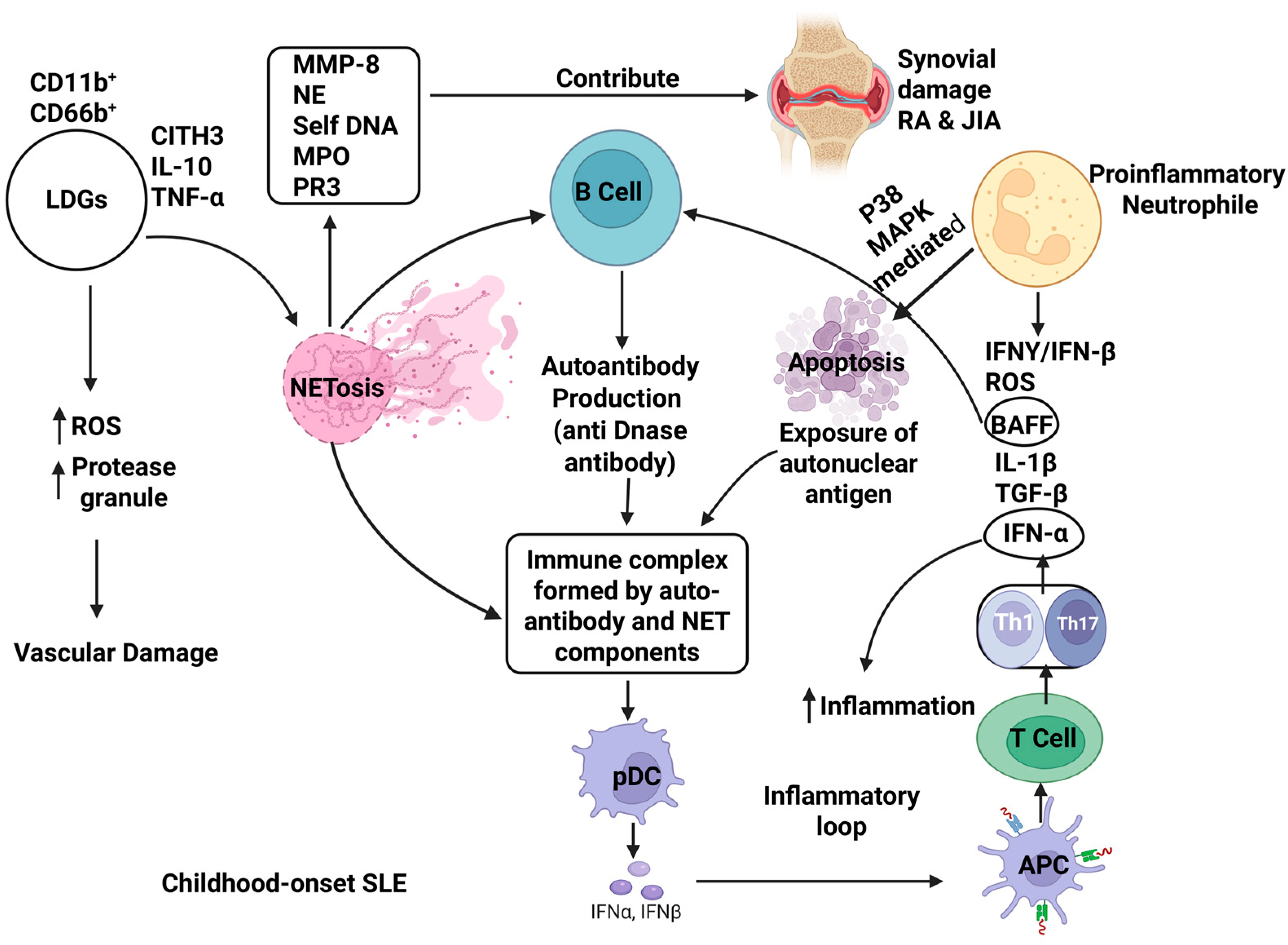
Apart from their function in innate immunity, neutrophils also regulate the adaptive immunity effectively by migrating to lymphoid organs, presenting antigens, and modulating the lymphocyte responses [2,3]. Neutrophils’ profound role in innate and adaptive immunity exerts its dynamic and plastic properties with distinct subsets. However, neutrophils do not contribute exclusively to providing immunity against antigens. It may shift toward promoting chronic inflammation and immune dysregulation through several mechanisms, like NET formation (NETosis) and dysfunctional neutrophil subsets. NETosis has an identified role in host defense, but dysregulated NETosis exposes modified self-antigens, stimulates type I interferon production, and plasmacytoid dendritic cells, with activation that further activates APC (antigen-presenting cells) and T cells mediated by an inflammatory loop (Figure 2) [4]. This phenomenon drives the pathogenesis of autoimmune conditions like systemic lupus erythematosus (SLE) [5], rheumatoid arthritis (RA) [6], and type 1 diabetes (T1D) [7]. Moreover, patients with autoimmune disease often exhibit elevated low-density neutrophils (LDNs) and low-density granulocytes (LDGs) subsets with increased antigen-presenting capacity, NETosis, and proinflammatory activity (Figure 2) [7]. Along with these effector activities, hyperactive or dysregulated neutrophil subsets may exacerbate autoimmune responses and tissue damage. Besides NETs, calprotectin and neutrophil serine proteinases are other neutrophil-based biomarkers that are correlated to autoimmunity. Neutrophils are also able to recruit and activate a variety of immune and nonimmune cells that contribute to regulating inflammation [8].
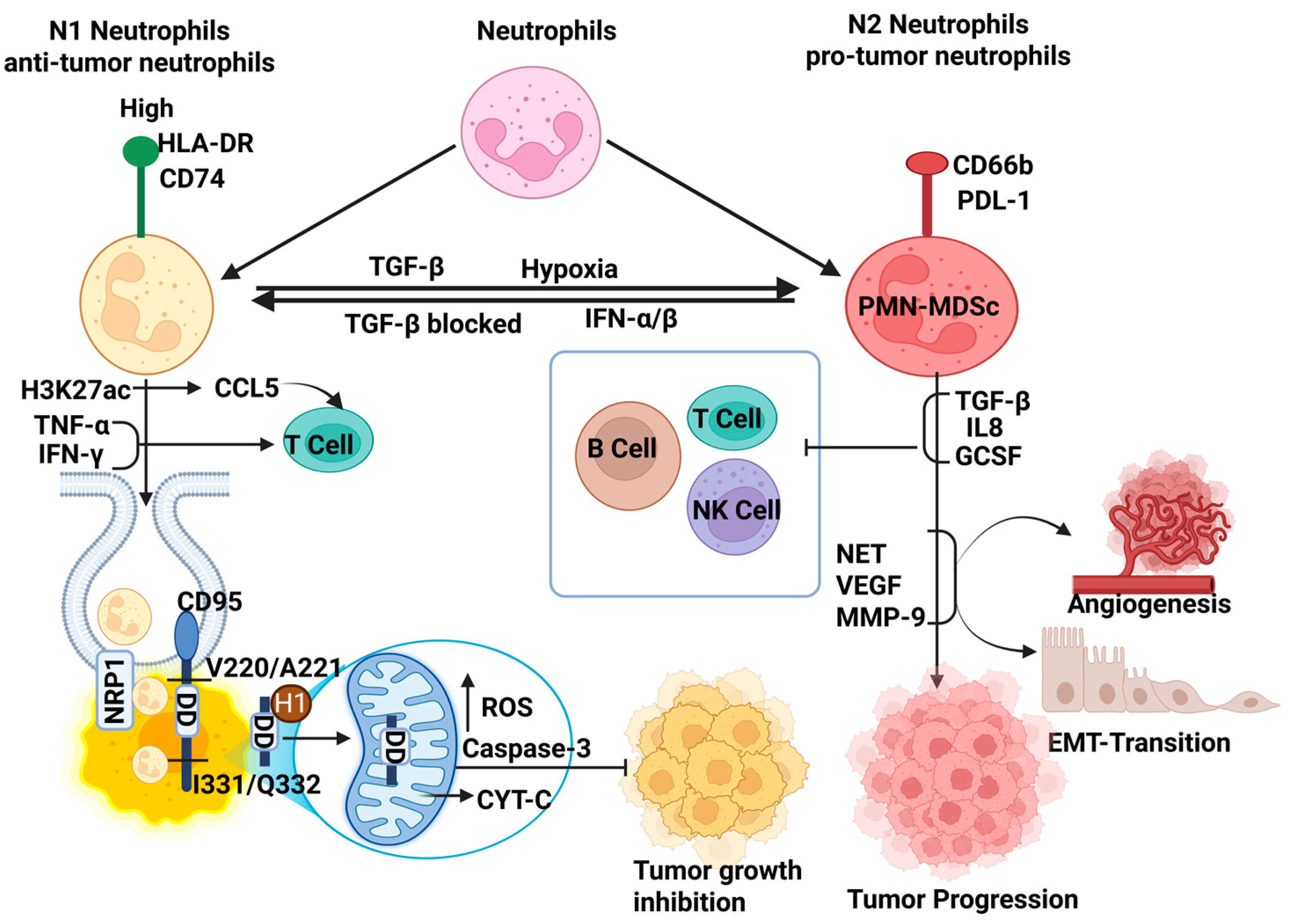
Interestingly, while neutrophils exhibit antigen clearance and protect host immunity, altered signaling within the tumor microenvironment (TME) reprograms them to adopt such a function and phenotype that encourages tumor-promoting activities. Dual and often contradictory roles of neutrophils are reported; for example, ROS [9] or tumor necrosis factor-related apoptosis-inducing ligand (TRAIL)-mediated apoptosis can stimulate T cell responses [10] that exhibit an anticancer effect (Figure 2). Conversely, tumor-associated neutrophils (TANs) [11] and pro-tumor phenotype [12] of neutrophils support tumor progression by promoting angiogenesis, suppressing cytotoxic T cells, and facilitating metastasis via extracellular matrix remodeling and epithelial–mesenchymal transition (EMT) (Figure 3).
Recent studies have identified distinct pro-and anti-tumor neutrophil phenotypes that can transform themselves among each other under the influence of different local cues associated with specific signaling pathways and functional markers (Table 1). Influencing factors that enable such pro-tumor to anti-tumor transitions and vice versa are not limited to prognosis through distinct neutrophil biomarkers but also have potential therapeutic targets. Along with neutrophils, NETs themselves secrete various molecules, like self-DNA and other signaling factors that enable the crosstalk with other immune cells, such as T cells, B cells, natural killer (NK) cells, and plasmacytoid dendritic cells (pDCs), which help them to modulate adaptive immune responses based on NET immunogenicity. Therefore, inhibiting NET formation, modulating neutrophil recruitment, or reprogramming neutrophil phenotypes are active areas of therapeutic exploration in both autoimmune and cancer [13,14].

2. Neutrophil Biology: Maturation, Subtypes, and Functional Plasticity

The developing neutrophil biology uncovers the extended subsets that exhibit distinct morphology, functionality, and gene expression. This diverse neutrophil population impacts the immune system distinctly based on regulating inflammatory responses and adaptability to cellular microenvironment. For instance, a long time ago, Pillay J et al. identified a subset of human neutrophils with unique phenotypes and functionality, characterized by hyper-segmented nuclear morphology. These neutrophils were comparatively more mature than normal blood neutrophils. They respond to severe inflammation even when T cell activity is suppressed. These mature neutrophils display a distinctive surface marker profile (CD62Ldim/CD16bright/CD11bbright/CD54bright) whereas short-lived classic neutrophils show (CD62Ldim/CD16bright/CD11bbright/CD54dim) [18]. CD54 (ICAM) neutrophils show increased reverse trans-endothelial migration and NETosis under the influence of inflammatory signals, contributing to chronic systemic inflammation. CD54high or CD54bright exhibit a more proinflammatory feature than CD54dim and present their dysregulation [19]. Another surface marker, CD11b, represents active adhesion and migration, expressed by myeloid cells including neutrophil, macrophage, and monocytes. It resides stably within secretory granules of neutrophils and is expressed at a minimal level on the surface along with other neutrophil markers such as CD16 [20]. The elevated ROS and chronic inflammation upregulate CD11b expression on normal neutrophils and highly express on dysregulated subsets of neutrophils (Figure 1) [21].
Beyond the surface marker subsets, neutrophils are classified into functionally distinct populations; for example, classic neutrophils, low-density neutrophils (LDNs), tumor-associated neutrophils (TANs), and G-CSF-primed neutrophils. The classic neutrophils are known for their innate immune response against microbial infections. They eliminate pathogens via phagocytosis of bacterial cells, liberation of antimicrobial peptides, and ROS production. LDNs are a neutrophil subtype observed in the peripheral blood of patients diagnosed with conditions like cancer, autoimmune diseases, sepsis, and other inflammatory disorders. Their size and density are less than those of classic neutrophils. These cells are known for suppressing T cell responses and tumor promotion. Importantly, LDN’s population coexists with immature MDSCs (myeloid-derived suppressor cells; PMN-MDSCs) and mature cells [22]. CD11b and CD16 are common markers of LDNs (Figure 1). CD36, CD41, CD61, and CD226 upregulated on LDNs associated with non-small-cell lung cancer (NSCLC) [23] and CD10, CD16, and CD45high present a mature phenotype with T cell-suppressive role in blood cancer [24]. Elevated NETosis is one of the functional features of LDNs or low-density granulocytes (LDGs) in SLE and severe inflammation. LDGs are also referred to as LDNs, a proinflammatory neutrophil subset that co-exists with peripheral blood mononuclear cells and separate during density gradient centrifugation in various immune-mediated diseases [25].
TANs (tumor-associated neutrophils) are a neutrophil population that accumulate within the tumors and exhibit tumor progression, possibly by compromising immune response and enabling angiogenesis. It contributes to immune evasion by regulatory T cell (Treg) expansion and arginase-1 and other immunosuppressive molecules expression [26]. High levels of CCL2, CXCL2, CCL8, IL-8/CXCL8, and CXCL16 may characterize it. Another subset of TANs exhibits anti-tumor features, characterized by TNFα, CCL3, and ICAM-1. Anti-tumor TANs produce ROS, hydrogen peroxide (H2O2), Nitric oxide (NO), and neutrophil elastase (NE) that induce oxidative stress and membrane damage. Further, stressed cells go through Fas ligand-receptor (Fas ligand expressed on neutrophil binds to receptor on tumor cell)-based, ADCC (Antibody-dependent cell-mediated cytotoxicity), TRAIL pathway- and CTL (Cytotoxic T lymphocyte)-based apoptosis in tumor cells [5,27]. G-CSF-primed neutrophils have been noticed as a response to G-CSF (Granulocyte colony-stimulating factor; a cytokine) treatment as it can stimulate neutrophil production from bone marrow. It may help in neutrophil recovery by increasing their population while in an emergency, under the regulation of IL-17 [28]. G-CSF-primed neutrophils are featured with over-expression of adhesion molecules, and activation markers of phagocytosis [29]. Overall, the shape of neutrophils, their adaptation to maturation state, surrounding microenvironment, and disease context significantly influence the neutrophil behaviors. This remarkable heterogeneity and functional plasticity underlie their diverse role in both autoimmune diseases and cancer.

3. Neutrophils in Autoimmune Diseases

3.1. Systemic Lupus Erythematosus (SLE)

Dysregulated neutrophil subsets play a key role in pathogenesis of systemic lupus erythematosus due to their dysfunction such as ROS generation and granule proteases release that results in vascular tissue damage and trigger NET formation simultaneously. NETs expose neoepitopes, to which immune cells bind, these may nuclear autoantigens such as double-stranded DNA and modified histones. These antigen-specific peptides bind to B cell receptors (BCR) and stimulate the autoantibody production, including anti-dsDNA and anti-histone antibodies, followed by B cell activation, regulated by B cell-activating factor (BAFF) and interferons (IFNs) that sustain autoimmunity. In maintaining homeostasis, neutrophils undergo spontaneous apoptosis, but this process can be accelerated. Enhanced neutrophil apoptosis increases the apoptotic burden, generating antinuclear autoantibodies. Dysregulated NETs released by neutrophils also secrete proinflammatory mediators that recruit other immune cells to contribute tissue damage and a prominent subset of neutrophils in chronic inflammation called proinflammatory neutrophils [4,30]. Through the diverse subsets of neutrophils, LDGs expand significantly in SLE and are characterized by a distinct proinflammatory phenotype. Recently, Saisorn et al. reported highly CD66b-expressing LDGs that upregulate adhesion, NET formation, and apoptosis during lupus pathogenesis. In addition, the serum levels of IL-10 and TNF-α are significantly elevated in the patients suffering from childhood-onset SLE. Additionally, the presence of extracellular traps (ETs) and serum citrullinated histone H3 (CitH3) in LDGs compared to healthy controls may serve as practical biomarkers for disease severity. NETs act as potent autoantigens, capable of triggering autoantibody production and driving a robust type I interferon response that defines the feature of childhood-onset SLE immunopathology (Figure 2) [31]. Similarly, another study explains that LDGs are characterized by spontaneous NETosis, upregulated expression of type I interferon (IFN)-stimulated genes, and metabolic dysregulation, including impaired redox capacity. This dysregulation exacerbates ROS-mediated tissue damage and accelerates disease severity [32,33]. For example, LDGs show increased interferon-stimulated protein level and altered phosphorylation of cytoskeletal regulators in SLE that dysregulate cellular trafficking and encourage retention within lung and vascular tissues. Biomechanically, LDGs exhibit rougher cell surfaces, and membrane perturbations are independent of immune complex exposure, suggesting that intrinsic cellular abnormalities and microvascular injury can be used as a retention marker in future [34].
Functionally, LDGs contribute to vasculopathy and sustained IFN production by stimulating plasmacytoid dendritic cells. Compounding this problem, impaired NET clearance due to the presence of anti-DNase I autoantibodies leads to an accumulation of extracellular DNA, worsening tissue injury [35] (Figure 2). A particularly concerning subset includes immature neutrophils (CD10 CD16), which are elevated in active SLE. These developmentally arrested cells exhibit poor phagocytosis but secrete inflammatory cytokines and undergo spontaneous NETosis. They can influence adaptive immune responses by modulating T cell activity [36]. Conversely, aged neutrophils express high CD62L, gather at the site of chronic inflammation, and cause T cell dysregulation, impaired migration, upregulated adhesion, and NETs, amplifying the inflammatory loops and vascular injury consecutively [37]. Moreover, excess B cell-activating factor (BAFF) release stimulated through proinflammatory neutrophils encourage abnormal B cell activation and autoantibody production. This process may induce B cell dysregulation through immune complex formation and IFNγ-mediated signaling [38]. These findings underscore the critical role of neutrophil subset diversity, including LDGs, immature, and aged neutrophils, in the immunopathogenesis of SLE. These subsets act as mediators of inflammation and represent valuable biomarkers and potential therapeutic subjects for monitoring disease.

3.2. Rheumatoid Arthritis (RA)

Neutrophils exhibit both significant heterogeneity as well as functional plasticity under the influence of cytokine factors that impact local anatomy in rheumatoid arthritis. Although proinflammatory subsets of neutrophil predominate the RA affected area, where ROS release was associated with pathology earlier, recent studies suggest their dual function. Here, lower ROS levels aggravate the inflammation through Th1/Th17 responses in the early stages of immune activation while elevated ROS levels in later stages are implicated in driving synovial tissue damage, ROS complex that present stage-specific pathogenesis of RA [39]. A subset of LDNs, elevated in RA, may be characterized by enhanced NETosis, an aberrant release of NETs composed of self-DNA, myeloperoxidase (MPO), and proteinase 3 (PR3) that directly contributes to synovial tissue damage (Figure 2). Inhibition of NETosis reduces the severity of disease through inducing potential anti-inflammatory pathways. However, NET and LDN inhibitors, Baricitinib, Tofacitinib, and Upadacitinib, have been reported to have a positive effect on inflammation in a Phase 3 clinical trial (Table 2) [40,41]. LDGs in RA also exhibit reduced apoptosis and diminished responsiveness to TNF-α, resulting in impaired chemotaxis, phagocytosis, ROS generation, and NET formation compared to NDNs. These functional deficits contribute to chronic inflammation and immune dysregulation [42]. Type I interferon (IFN-α)-regulated neutrophils further enhance RA pathogenesis. They show increased ROS production, delayed apoptosis via p38 MAPK, and chemokine reprogramming through the STAT signaling axis, functions distinct from neutrophils under normal physiological conditions [43]. A transcriptomic study has identified the miR-183C cluster as a dominant IFN-response gene signature across neutrophil subsets in RA. This microRNA cluster appears activated by circulating NETs and neutrophil-derived debris, potentially through the cGAS-STING pathway, which is known to amplify inflammatory cascades. This study strongly suggests that miR-183C could serve both as a therapeutic target (via inhibition) and a diagnostic/prognostic biomarker for rheumatoid arthritis [44]. In summary, the expanding knowledge of neutrophil heterogeneity and functional reprogramming in RA, while shaping disease outcomes, modulates neutrophil activity while preserving essential host defense mechanisms.

3.3. Juvenile Idiopathic Arthritis (JIA)

In juvenile idiopathic arthritis (JIA), certain neutrophils called LDNs, along with genes related to neutrophil activation, are found at higher levels in the blood. This increase leads to the release of effector molecules like myeloperoxidase (MPO), neutrophil elastase (NE), and MMP8, which can contribute to joint damage, similar to what is seen in RA. Studies also show that LDNs in JIA have impaired suppressive immune response, which may lead to ongoing inflammation. Interestingly, unlike in lupus (SLE), specific markers on the surface of neutrophils such as CD62L, CD66b, and CD11b are linked to lower levels of joint inflammation. This suggests that neutrophils in JIA may behave differently from those in other autoimmune diseases. However, the expression profile of these neutrophil subsets still requires validation as a differential marker of JIA, from other inflammatory conditions [55,56].
Further evidence highlights a sex-specific immune signature in systemic JIA (sJIA). Immature neutrophils and activation-associated genes are more prominent in female sJIA patients, indicating sexual dimorphism in neutrophil biology. Treatment with IL-1 receptor antagonists such as anakinra has proven effective in modulating these immature neutrophils [57]. In oligoarticular JIA, neutrophils in synovial fluid acquire an aged, activated phenotype. These cells show increased levels of CD206 and CD14 markers, usually found on monocytes, and lower levels of CD62L. This change in their surface markers is linked to reduced phagocytic ability and lower production of ROS, both of which are important for clearing cellular debris. As a result, these defects may contribute to ongoing joint inflammation and poor immune regulation in JIA. Increased CD206+ neutrophils with impaired phagocytosis and oxidative burst could represent a novel target for therapeutic intervention in persistent disease [58]. Additionally, functionally activated, proinflammatory neutrophils expressing elevated levels of IL-1β and TNF-α are implicated in driving synovial inflammation. These neutrophils may also influence adaptive immune responses, further contributing to disease severity and progression [59].
Evidence from autoimmune disease brings out plasticity of neutrophils contributing pathogenesis, which is equally evident in cancer. In autoimmunity, subsets like LDGs, immature, and aged neutrophils amplify inflammation via excessive NETosis, ROS generation, and autoantigen exposure. Similarly, tumor-associated neutrophils as well as stromal cues reprogram the neutrophils into proinflammatory and immunosuppressive phenotypes in oncology that enhance immunity and promote angiogenesis or metastasis, respectively. Despite opposite outcomes, for immune hyperactivation in autoimmune diseases versus suppression in cancer, both share common mechanisms, including NET signaling, type I interferon responses, metabolic remodeling, and immune cell crosstalk, under neutrophil-dependent immunity.

4. Neutrophils Heterogeneity in Cancer

4.1. Anti-Tumorigenic Neutrophils

N1 tumor-associated neutrophils (N1-TANs) are a specialized group of neutrophils that play an important role in fighting tumors within the TME. Unlike other neutrophil subsets, N1-TANs have strong tumor-killing (cytotoxic) abilities. They produce high levels of proinflammatory molecules such as TNF-α and ICAM-1, which help stimulate immune responses. Additionally, N1-TANs effectively induce T cell activation, further strengthening the host anti-tumor defense. They are typically induced in milieu where type I interferons are high and TGF-β is low, or under TGF-β blockade, to favor their anti-tumorigenic polarization [60]. Recent single-cell transcriptomic analyses have identified a unique subset of anti-tumor neutrophils in the TME marked by high HLA-DR and CD74 expression, enabling antigen presentation. This unique feature of these neutrophils can be influenced by leucine-driven epigenetic changes, especially an increase in a specific histone modification called H3K27ac. This modification turns on genes involved in antigen presentation and triggers the release of chemokines like CCL5, which helps attract T cells to the tumor site. In addition, these neutrophils produce inflammatory cytokines such as TNF-α and IFN-γ, which boost the immune response and support eliminating tumor cells through programmed cell death (apoptosis). Their presence correlates with improved patient survival, and they are distinguished from pro-tumor neutrophils across several metastatic cancers, including non-small-cell lung cancer (NSCLC), colorectal cancer, head and neck squamous cell carcinoma, melanoma, and triple-negative breast cancer (TNBC) [61]. Moreover, programmed cell death, a mitochondria-targeted tumor killing, has been highlighted recently which showed that anti-tumor neutrophils internalize into cancer cells via neuropilin-1 (NRP1)-mediated endocytosis by proteolytic cleavage of CD95/Fas receptor at specific sites (V220/A221 and I331/Q332) through effective neutrophil elastase (ELANE) and liberating a C-terminal death domain fragment (DDELANE). Further, this fragment binds to highly expressed histone H1 isoforms (e.g., H1.0, H1.2), and forms a DDELANE–H1 complex that translocate to the mitochondria. Consequently, it triggers mitochondrial dysfunction, elevated ROS production, DNA damage (γH2AX), and activation of the intrinsic apoptotic cascade (e.g., caspase-3, cleaved PARP), which enables H1-targeted selective killing of cancer cells. Additionally, ELANE-mediated tumor cell death releases tumor antigens, enhancing dendritic cell activation and promoting CD8+ T cell priming, which target distant metastases through a systemic anti-tumor immune response (Figure 3) [62]. Although N1-TANs share only a small portion of the total neutrophil population in tumors like NSCLC and colorectal cancer, their role as antigen-presenting cells and in activating T cells highlights their importance. These functions remark on the subset’s dynamic and inducible nature as their potential targets for neutrophil-based immunotherapies across various tumor types.

4.2. Pro-Tumorigenic Neutrophils

Pro-tumor neutrophils are commonly identified as N2-tumor-associated neutrophils (N2-TANs) or polymorphonuclear myeloid-derived suppressor cells (PMN-MDSCs) and the activated PMN-MDSC subset in cancer. Unlike anti-tumor neutrophils, pro-tumor neutrophils express low HLA-DR and CD74; rather, they exhibit Programmed death-ligand 1 (PDL-1), CD66b, and CD15, which contribute to immunosuppression in the tumor microenvironment [63]. A study revealed the elevated expression of immune suppressor genes (such as S100a8, S100a9, Mmp8, and Cybb) and inflammation (including Il1b, Socs3, and Ptgs2) as a result of pro-tumor neutrophils. Notably, CD14high neutrophils is an undisguisable tumor marker and exhibits the strongest suppressive activity. It suppresses T, B, and NK cell functions and promotes tumor progression through both immune and nonimmune mechanisms. The gene signature of tumor-infiltrating PMN-MDSCs shared similarity among mice and human PMN-MDSCs but found underscores to clinical outcomes in cancer patients [64]. The TME is profoundly affected by TGF-β, hypoxia, and tumor-derived cytokines, including IL-8 and G-CSF, which promote neutrophil polarization towards a pro-tumorigenic phenotype. Recently, a single-cell transcriptomic study across multiple cancer types highlighted vascular endothelial growth factor (VEGFA) and SPP1 (osteopontin) secretion as distinct transcriptional signatures. Pre-metastatic niches are nurtured through NET, VEGF, and MMP-9 secreted from N2-TANs that promote angiogenesis, epithelial-to-mesenchymal transition (EMT), and tumor cell intravasation. It suppresses T cell proliferation and cytotoxicity by upregulating arginase-1, ROS, and expressing immune checkpoint molecules like PD-L1, which implies immunosuppression as a hallmark (Figure 3) [61]. For example, breast ductal carcinoma in situ (DCIS) tissues have been shown to harbor more neutrophils than normal breast tissues. As these lesions transition into invasive ductal carcinomas, the TME becomes enriched in PD-L1-expressing neutrophils, regulatory T cells (Tregs), and dysfunctional TCR clonotypes, reflecting a deeply immune-suppressed landscape [65]. Similarly, in late-stage head and neck cancers, infiltrating neutrophils in TME are likely to be aged and possess suppressive characteristics, in contrast to those found at earlier tumor stages and mediate anti-tumor responses, initially [66].

4.3. Phenotypes Transition in TME: Anti-Tumorigenic (N1) and Pro-Tumorigenic (N2)

The tumor microenvironment is inclusion of local cues, abundant with cytokines, immune cells and other molecules that may regulate neutrophil polarization among anti-tumor (N1) and pro-tumor (N2) neutrophils. Adaptation into another subset provides plasticity in their function that determines the cancer outcomes as either progression or suppression by affecting related processes such as metastasis, angiogenesis, and immune regulation. For example, TANs can be polarized into either an anti-tumor ‘N1’ phenotype by enhancing immune activation and tumor control or a pro-tumor ‘N2’ phenotype by promoting immune suppression and tumor growth.
Transforming growth factor-beta (TGF-β) is reported as a switch to neutrophil phenotypic shift. TGF-β supports the pro-tumor N2 polarization by promoting angiogenesis through VEGF secretion, enhancing metastatic potential, and suppressing immune activation by upregulating PD-L1 expression, ultimately establishing an immunosuppressive tumor niche [61].
Blocking TGF-β signaling or introducing acute inflammatory stimuli can reprogram TANs from the N2 to the anti-tumor N1 phenotype (Figure 3). This shift may be marked by upregulation of immune activation genes, increased ROS-mediated cytotoxicity, enhanced recruitment, and activation of CD8+ T cells and natural killer (NK) cells. Restoration of antigen presentation led to direct tumor cell killing [15].

5. Therapeutic Targeting of Neutrophils

Advances in understanding neutrophil function, diversity, and their interactions with other immune cells have opened new possibilities for treating autoimmune diseases and cancers. Therapies are now being developed to reduce tissue damage by targeting NETosis in autoimmune conditions and to block neutrophil-driven inflammation in tumors using chemokine receptor inhibitors like CXCR2 antagonists. Many of these strategies are in preclinical or early clinical trials, showing promise in controlling autoimmune flares and improving cancer immunotherapy. Also, biomarkers such as low-density neutrophils (LDNs) and NET components are being studied for their potential in diagnosis, disease monitoring, and treatment feedback. Modulating the pathological subsets of neutrophil, without impairing host defense, may allow discovering neutrophil-targeted immunomodulation in both autoimmune and oncologic contexts.
The therapeutic landscape for neutrophil-targeted interventions reveals a contrary context across autoimmune diseases and cancer. In autoimmune diseases, strategies focus on dampening hyperactivated neutrophils through PAD4 inhibitors to prevent NETosis, DNase therapies to clear existing NETs, and JAK/STAT or IL-6 receptor blockade to reduce proinflammatory subsets like low-density neutrophils (Table 2). On the other hand, cancer therapeutics employ CXCR1/2 antagonists to block neutrophil recruitment and prevent their conversion to immunosuppressive myeloid-derived suppressor cells, to restore anti-tumor immunity (Table 2). Notably, agents such as JAK inhibitors show efficacy across both contexts, suggesting shared pathological mechanisms and opportunities for drug repurposing [67]. The future challenge lies in developing specific approaches that selectively target pathological neutrophil functions while preserving essential host defense roles. This leaves an important question on how biomarker-driven therapeutic strategies can be developed that may allow prediction and selective modulation of neutrophil phenotypes to switch them from pathogenic to homeostatic neutrophil, utilizing their plasticity, without compromising their fundamental antimicrobial and tissue repair functions.

6. Conclusions

Recent discoveries on neutrophil diversity, plasticity, and crosstalk with other immune cells contribute significantly in reshaping their multifaceted role in cancer and autoimmune diseases. Once recognized as an early responder, short-lived cells, neutrophils, are now known for their dynamic functions, either immuno-protective or pathogenic. Selective modulation in autoimmune diseases potentially minimizes chronic inflammation through targeting neutrophil-derived mediators like ROS, NETs, or BAFF. In cancer, reprogramming pro-tumor neutrophils to anti-tumor TANs presents a promising avenue for immunotherapy enhancement. Focus on identifying and validating neutrophil-based biomarkers (e.g., LDGs, NET components) for early diagnosis, disease monitoring, and predicting treatment response would be the future of this field.

Author Contributions

A.B. and N.K.R. jointly conceived the study, A.B. and N.K.R. designed the study, A.B. and N.K.R. writing—review and editing, V.K. draft editing, N.K.R. offered important intellectual input, provided critical resources, and critically reviewed the study design. All authors have read and agreed to the published version of the manuscript.

Funding

This research received no external funding.

Data Availability Statement

No new experimental data were created.

Conflicts of Interest

The authors declare no conflicts of interest.

Abbreviations

The following abbreviations are used in this manuscript:
HSCsHematopoietic stem cells
preNeusPre-neutrophils
NETNeutrophil extracellular traps
NETosisNET formation
ROSReactive oxygen species
pDCPlasmacytoid dendritic cells
APCAntigen-presenting cells
LDGLow-density granulocytes
LDNsLow-density neutrophils
SLESystemic lupus erythematosus
RARheumatoid arthritis
T1DType 1 diabetes
TMETumor microenvironment
TRAILTumor necrosis factor-related apoptosis-inducing ligand
TANsTumor-associated neutrophils
EMTEpithelial–mesenchymal transition
NK CellsNatural killer cells
MDSCsMyeloid-derived suppressor cells
NSCLCNon-small-cell lung cancer
T regRegulatory T cell
H2O2Hydrogen peroxide
NONitric oxide
NENeutrophil elastase
ADCCAntibody-dependent cell-mediated cytotoxicity
CTLCytotoxic T lymphocyte
G-CSFGranulocyte colony-stimulating factor
BCRB cell receptors
TCRT cell receptors
BAFFB cell-activating factor
IFNsInterferons
ETsExtracellular traps
CitH3Citrullinated histone H3
MPOMyeloperoxidase
MMP-9Matrix metalloproteinase-9
PR3Proteinase 3
JIAJuvenile idiopathic arthritis
sJIASystemic JIA
TNBCTriple-negative breast cancer
NPR1Neuropilin-1
PMN-MDSCsPolymorphonuclear myeloid-derived suppressor cells
N1Anti-tumorigenic Neutrophil
N2Pro-tumorigenic Neutrophil
VEGFAVascular endothelial growth factor
DCISDuctal carcinoma in situ
PD-L1Programmed death ligand-1
TGF-βTransforming growth factor-beta
TNF-αTumor Necrosis Factors-α

References

  1. Evrard, M.; Kwok, I.W.H.; Chong, S.Z.; Teng, K.W.W.; Becht, E.; Chen, J.; Sieow, J.L.; Penny, H.L.; Ching, G.C.; Devi, S.; et al. Developmental Analysis of Bone Marrow Neutrophils Reveals Populations Specialized in Expansion, Trafficking, and Effector Functions. Immunity 2018, 48, 364–379.e8. [Google Scholar] [CrossRef] [PubMed]
  2. Rosales, C. Neutrophils at the crossroads of innate and adaptive immunity. J. Leukoc. Biol. 2020, 108, 377–396. [Google Scholar] [CrossRef]
  3. Nauseef, W.M.; Borregaard, N. Neutrophils at work. Nat. Immunol. 2014, 15, 602–611. [Google Scholar] [CrossRef]
  4. Zeng, L.; Xiang, W.; Xiao, W.; Wu, Y.; Sun, L. The emerging role of neutrophil extracellular traps in autoimmune and autoinflammatory diseases. Medcomm 2025, 6, e70101. [Google Scholar] [CrossRef]
  5. Salemme, R.; Peralta, L.N.; Meka, S.H.; Pushpanathan, N.; Alexander, J.J. The Role of NETosis in Systemic Lupus Erythematosus. J. Cell Immunol. 2019, 1, 33–42. [Google Scholar] [CrossRef] [PubMed]
  6. Song, W.; Ye, J.; Pan, N.; Tan, C.; Herrmann, M. Neutrophil Extracellular Traps Tied to Rheumatoid Arthritis: Points to Ponder. Front. Immunol. 2020, 11, 578129. [Google Scholar] [CrossRef]
  7. Fu, X.; Liu, H.; Huang, G.; Dai, S.-S. The emerging role of neutrophils in autoimmune-associated disorders: Effector, predictor, and therapeutic targets. MedComm 2021, 2, 402–413. [Google Scholar] [CrossRef]
  8. Carnevale, S.; Di Ceglie, I.; Grieco, G.; Rigatelli, A.; Bonavita, E.; Jaillon, S. Neutrophil diversity in inflammation and cancer. Front. Immunol. 2023, 14, 1180810. [Google Scholar] [CrossRef] [PubMed]
  9. Yan, J.; Kloecker, G.; Fleming, C.; Bousamra, M.; Hansen, R.; Hu, X.; Ding, C.; Cai, Y.; Xiang, D.; Donninger, H.; et al. Human polymorphonuclear neutrophils specifically recognize and kill cancerous cells. OncoImmunology 2014, 3, e950163. [Google Scholar] [CrossRef]
  10. Kundu, M.; Greer, Y.E.; Lobanov, A.; Ridnour, L.; Donahue, R.N.; Ng, Y.; Ratnayake, S.; Voeller, D.; Weltz, S.; Chen, Q.; et al. TRAIL-induced cytokine production via NFKB2 pathway promotes neutrophil chemotaxis and immune suppression in triple negative breast cancers. bioRxiv 2024. [Google Scholar] [CrossRef]
  11. Zhang, S.; Sun, L.; Zuo, J.; Feng, D. Tumor associated neutrophils governs tumor progression through an IL-10/STAT3/PD-L1 feedback signaling loop in lung cancer. Transl. Oncol. 2024, 40, 101866. [Google Scholar] [CrossRef] [PubMed]
  12. Silvestre-Roig, C.; Kalafati, L.; Chavakis, T. Neutrophils are shaped by the tumor microenvironment: Novel possibilities for targeting neutrophils in cancer. Signal Transduct. Target. Ther. 2024, 9, 77. [Google Scholar] [CrossRef] [PubMed]
  13. Gao, F.; Peng, H.; Gou, R.; Zhou, Y.; Ren, S.; Li, F. Exploring neutrophil extracellular traps: Mechanisms of immune regulation and future therapeutic potential. Exp. Hematol. Oncol. 2025, 14, 80. [Google Scholar] [CrossRef] [PubMed]
  14. Zhou, Y.; Shen, G.; Zhou, X.; Li, J. Therapeutic potential of tumor-associated neutrophils: Dual role and phenotypic plasticity. Signal Transduct. Target. Ther. 2025, 10, 178. [Google Scholar] [CrossRef]
  15. Zhang, M.; Qin, H.; Wu, Y.; Gao, Q. Complex role of neutrophils in the tumor microenvironment: An avenue for novel immunotherapies. Cancer Biol. Med. 2024, 21, 849–863. [Google Scholar] [CrossRef]
  16. Shaul, M.E.; Levy, L.; Sun, J.; Mishalian, I.; Singhal, S.; Kapoor, V.; Horng, W.; Fridlender, G.; Albelda, S.M.; Fridlender, Z.G. Tumor-associated neutrophils display a distinct N1 profile following TGFβ modulation: A transcriptomics analysis of pro- vs. antitumor TANs. OncoImmunology 2016, 5, e1232221. [Google Scholar] [CrossRef]
  17. Wang, Y.; Wang, L.; Wang, Z.; Guo, Y.; Zhang, X.; Wan, B. Targeting tumor-associated neutrophils: A promising strategy in lung cancer immunotherapy. Cancer Pathog. Ther. 2025, 3, E01–E07. Available online: https://mednexus.org/doi/10.1016/j.cpt.2025.02.004 (accessed on 15 August 2025). [CrossRef]
  18. Pillay, J.; Kamp, V.M.; van Hoffen, E.; Visser, T.; Tak, T.; Lammers, J.-W.; Ulfman, L.H.; Leenen, L.P.; Pickkers, P.; Koenderman, L. A subset of neutrophils in human systemic inflammation inhibits T cell responses through Mac-1. J. Clin. Investig. 2012, 122, 327–336. [Google Scholar] [CrossRef]
  19. Sengupta, S.; Caldwell, C.C.; Nomellini, V. Distinct Neutrophil Populations in the Spleen During PICS. Front. Immunol. 2020, 11, 804. [Google Scholar] [CrossRef]
  20. Weirich, E.; Rabin, R.L.; Maldonado, Y.; Benitz, W.; Modler, S.; Herzenberg, L.A. Neutrophil CD11b expression as a diagnostic marker for early-onset neonatal infection. J. Pediatr. 1998, 132 Pt 1, 445–451. [Google Scholar] [CrossRef] [PubMed]
  21. O’DEa, M.I.; Kelly, L.; McKenna, E.; Melo, A.M.; Ni Bhroin, M.; Hurley, T.; Byrne, A.T.; Colleran, G.; Vavasseur, C.; El-Khuffash, A.; et al. Dysregulated Monocyte and Neutrophil Functional Phenotype in Infants with Neonatal Encephalopathy Requiring Therapeutic Hypothermia. Front. Pediatr. 2021, 8, 598724. [Google Scholar] [CrossRef] [PubMed]
  22. Sagiv, J.Y.; Michaeli, J.; Assi, S.; Mishalian, I.; Kisos, H.; Levy, L.; Damti, P.; Lumbroso, D.; Polyansky, L.; Sionov, R.V.; et al. Phenotypic diversity and plasticity in circulating neutrophil subpopulations in cancer. Cell Rep. 2015, 10, 562–573. [Google Scholar] [CrossRef]
  23. Valadez-Cosmes, P.; Maitz, K.; Kindler, O.; Raftopoulou, S.; Kienzl, M.; Santiso, A.; Mihalic, Z.N.; Brcic, L.; Lindenmann, J.; Fediuk, M.; et al. Identification of Novel Low-Density Neutrophil Markers Through Unbiased High-Dimensional Flow Cytometry Screening in Non-Small Cell Lung Cancer Patients. Front. Immunol. 2021, 12, 703846. [Google Scholar] [CrossRef]
  24. Vanhaver, C.; Nana, F.A.; Delhez, N.; Luyckx, M.; Hirsch, T.; Bayard, A.; Houbion, C.; Dauguet, N.; Brochier, A.; van der Bruggen, P.; et al. Immunosuppressive low-density neutrophils in the blood of cancer patients display a mature phenotype. Life Sci. Alliance 2024, 7, e202302332. [Google Scholar] [CrossRef]
  25. Ning, X.; Wang, W.-M.; Jin, H.-Z.; Fang, W. Low-Density Granulocytes in Immune-Mediated Inflammatory Diseases. J. Immunol. Res. 2022, 2022, 1622160. [Google Scholar] [CrossRef]
  26. Mishalian, I.; Bayuh, R.; Levy, L.; Zolotarov, L.; Michaeli, J.; Fridlender, Z.G. Tumor-associated neutrophils (TAN) develop pro-tumorigenic properties during tumor progression. Cancer Immunol. Immunother. 2013, 62, 1745–1756. [Google Scholar] [CrossRef]
  27. Ye, Z.; Cheng, P.; Huang, Q.; Hu, J.; Huang, L.; Hu, G. Immunocytes interact directly with cancer cells in the tumor microenvironment: One coin with two sides and future perspectives. Front. Immunol. 2024, 15, 1388176. [Google Scholar] [CrossRef]
  28. Zhang, F.; Xia, Y.; Su, J.; Quan, F.; Zhou, H.; Li, Q.; Feng, Q.; Lin, C.; Wang, D.; Jiang, Z. Neutrophil diversity and function in health and disease. Signal Transduct. Target. Ther. 2024, 9, 343. [Google Scholar] [CrossRef]
  29. Wenisch, C.; Patruta, S.; Daxböck, F.; Krause, R.; Hörl, W. Effect of age on human neutrophil function. J. Leukoc. Biol. 2000, 67, 40–45. [Google Scholar] [CrossRef] [PubMed]
  30. Wigerblad, G.; Kaplan, M.J. Neutrophil extracellular traps in systemic autoimmune and autoinflammatory diseases. Nat. Rev. Immunol. 2022, 23, 274–288. [Google Scholar] [CrossRef] [PubMed]
  31. Saisorn, W.; Santiworakul, C.; Phuengmaung, P.; Siripen, N.; Rianthavorn, P.; Leelahavanichkul, A. Extracellular traps in peripheral blood mononuclear cell fraction in childhood-onset systemic lupus erythematosus. Sci. Rep. 2024, 14, 23177. [Google Scholar] [CrossRef] [PubMed]
  32. Henning, S.; Reimers, T.; Abdulahad, W.; Fierro, J.J.; der Meer, B.D.-V.; Bootsma, H.; Horvath, B.; de Leeuw, K.; Westra, J. Low-density granulocytes and neutrophil extracellular trap formation are increased in incomplete systemic lupus erythematosus. Rheumatology 2025, 64, 1234–1242. [Google Scholar] [CrossRef]
  33. Tay, S.H.; Celhar, T.; Fairhurst, A. Low-Density Neutrophils in Systemic Lupus Erythematosus. Arthritis Rheumatol. 2020, 72, 1587–1595. [Google Scholar] [CrossRef] [PubMed]
  34. Bashant, K.R.; Aponte, A.M.; Randazzo, D.; Sangsari, P.R.; Wood, A.J.; A Bibby, J.; E West, E.; Vassallo, A.; Manna, Z.G.; Playford, M.P.; et al. Proteomic, biomechanical and functional analyses define neutrophil heterogeneity in systemic lupus erythematosus. Ann. Rheum. Dis. 2021, 80, 209–218. [Google Scholar] [CrossRef]
  35. Denny, M.F.; Yalavarthi, S.; Zhao, W.; Thacker, S.G.; Anderson, M.; Sandy, A.R.; McCune, W.J.; Kaplan, M.J. A Distinct Subset of Proinflammatory Neutrophils Isolated from Patients with Systemic Lupus Erythematosus Induces Vascular Damage and Synthesizes Type I IFNs. J. Immunol. 2010, 184, 3284–3297, Correction in J. Immunol. 2010, 185, 3779. [Google Scholar] [CrossRef]
  36. Marini, O.; Costa, S.; Bevilacqua, D.; Calzetti, F.; Tamassia, N.; Spina, C.; De Sabata, D.; Tinazzi, E.; Lunardi, C.; Scupoli, M.T.; et al. Mature CD10+ and immature CD10− neutrophils present in G-CSF–treated donors display opposite effects on T cells. Blood 2017, 129, 1343–1356. [Google Scholar] [CrossRef]
  37. Herrero-Cervera, A.; Soehnlein, O.; Kenne, E. Neutrophils in chronic inflammatory diseases. Cell. Mol. Immunol. 2022, 19, 177–191. [Google Scholar] [CrossRef] [PubMed]
  38. Wang, T.; Vasconcellos, A.; Marken, J.; Skopelja-Gardner, S.; Lood, C.; Giltiay, N.V. Immune complex–driven neutrophil activation and BAFF release: A link to B cell responses in SLE. Lupus Sci. Med. 2022, 9, e000709. [Google Scholar] [CrossRef]
  39. Chen, T.; Zhou, Z.; Liu, Y.; Xu, J.; Zhu, C.; Sun, R.; Hu, H.; Liu, Y.; Dai, L.; Holmdahl, R.; et al. Neutrophils with low production of reactive oxygen species are activated during immune priming and promote development of arthritis. Redox Biol. 2024, 78, 103401. [Google Scholar] [CrossRef]
  40. Fousert, E.; Toes, R.; Desai, J. Neutrophil Extracellular Traps (NETs) Take the Central Stage in Driving Autoimmune Responses. Cells 2020, 9, 915. [Google Scholar] [CrossRef]
  41. Yang, F.; Luo, X.; Luo, G.; Zhai, Z.; Zhuang, J.; He, J.; Han, J.; Zhang, Y.; Zhuang, L.; Sun, E.; et al. Inhibition of NET formation by polydatin protects against collagen-induced arthritis. Int. Immunopharmacol. 2019, 77, 105919. [Google Scholar] [CrossRef]
  42. Wright, H.L.; A Makki, F.; Moots, R.J.; Edwards, S.W. Low-density granulocytes: Functionally distinct, immature neutrophils in rheumatoid arthritis with altered properties and defective TNF signalling. J. Leukoc. Biol. 2017, 101, 599–611. [Google Scholar] [CrossRef]
  43. Glennon-Alty, L.; Moots, R.J.; Edwards, S.W.; Wright, H.L. Type I interferon regulates cytokine-delayed neutrophil apoptosis, reactive oxygen species production and chemokine expression. Clin. Exp. Immunol. 2021, 203, 151–159. [Google Scholar] [CrossRef]
  44. Alarcon, M.F.; Abdullah, G.A.; Beggs, J.A.; Kynoch, I.; Sellin, A.; Cross, A.; Haldenby, S.; Antczak, P.; Gutiérrez, E.C.; Wright, H.L. Complexity of the neutrophil transcriptome in early and severe rheumatoid arthritis: A role for microRNAs? J. Leukoc. Biol. 2025, 117, qiaf090. [Google Scholar] [CrossRef] [PubMed]
  45. Hasni, S.A.; Gupta, S.; Davis, M.; Poncio, E.; Temesgen-Oyelakin, Y.; Carlucci, P.M.; Wang, X.; Naqi, M.; Playford, M.P.; Goel, R.R.; et al. Phase 1 double-blind randomized safety trial of the Janus kinase inhibitor tofacitinib in systemic lupus erythematosus. Nat. Commun. 2021, 12, 3391. [Google Scholar] [CrossRef]
  46. E Rysenga, C.; May-Zhang, L.; Zahavi, M.; Knight, J.S.; Ali, R.A. Taxifolin inhibits NETosis through activation of Nrf2 and provides protective effects in models of lupus and antiphospholipid syndrome. Rheumatology 2023, 63, 2006–2015. [Google Scholar] [CrossRef]
  47. Stabach, P.R.; Sims, D.; Gomez-Bañuelos, E.; Zehentmeier, S.; Dammen-Brower, K.; Bernhisel, A.; Kujawski, S.; Lopez, S.G.; Petri, M.; Goldman, D.W.; et al. A dual-acting DNASE1/DNASE1L3 biologic prevents autoimmunity and death in genetic and induced lupus models. J. Clin. Investig. 2024, 9, e177003. [Google Scholar] [CrossRef]
  48. Sun, F.; Wang, H.J.; Liu, Z.; Geng, S.; Wang, H.T.; Wang, X.; Li, T.; Morel, L.; Wan, W.; Lu, L.; et al. Safety and efficacy of metformin in systemic lupus erythematosus: A multicentre, randomised, double-blind, placebo-controlled trial. Lancet Rheumatol. 2020, 2, e210–e216. [Google Scholar] [CrossRef] [PubMed]
  49. Díaz-Planellas, S.; Katsifis-Nezis, D.; Fanouriakis, A. Clinical Trials of Interferon Inhibitors in Systemic Lupus Erythematosus and Preliminary Real-World Efficacy of Anifrolumab. Mediterr. J. Rheumatol. 2024, 35 (Suppl. S2), 381–391. [Google Scholar] [CrossRef] [PubMed]
  50. Gajendran, C.; Fukui, S.; Sadhu, N.M.; Zainuddin, M.; Rajagopal, S.; Gosu, R.; Gutch, S.; Fukui, S.; Sheehy, C.E.; Chu, L.; et al. Alleviation of arthritis through prevention of neutrophil extracellular traps by an orally available inhibitor of protein arginine deiminase 4. Sci. Rep. 2023, 13, 3189. [Google Scholar] [CrossRef] [PubMed]
  51. Tanaka, Y.; Yamanaka, H.; Toyoizumi, S.; Hirose, T.; Takeuchi, T. Laboratory markers predicting tofacitinib efficacy in Japanese patients with rheumatoid arthritis: A pooled analysis of Phase 2/3 randomised controlled clinical trials. Mod. Rheumatol. 2024, 35, 417–424. [Google Scholar] [CrossRef]
  52. Guo, C.; Sharp, A.; Gurel, B.; Crespo, M.; Figueiredo, I.; Jain, S.; Vogl, U.; Rekowski, J.; Rouhifard, M.; Gallagher, L.; et al. Targeting myeloid chemotaxis to reverse prostate cancer therapy resistance. Nature 2023, 623, 1053–1061. [Google Scholar] [CrossRef]
  53. Lazennec, G.; Rajarathnam, K.; Richmond, A. CXCR2 chemokine receptor—A master regulator in cancer and physiology. Trends Mol. Med. 2024, 30, 37–55. [Google Scholar] [CrossRef]
  54. Korbecki, J.; Bosiacki, M.; Pilarczyk, M.; Kot, M.; Defort, P.; Walaszek, I.; Chlubek, D.; Baranowska-Bosiacka, I. The CXCL1-CXCR2 Axis as a Component of Therapy Resistance, a Source of Side Effects in Cancer Treatment, and a Therapeutic Target. Cancers 2025, 17, 1674. [Google Scholar] [CrossRef]
  55. Parackova, Z.; Zentsova, I.; Bloomfield, M.; Klocperk, A.; Horvath, R.; Malcova, H.; Cebecauerova, D.; Sediva, A. Expanded population of low-density neutrophils in juvenile idiopathic arthritis. Front. Immunol. 2023, 14, 1229520. [Google Scholar] [CrossRef]
  56. Ramanathan, K.; Glaser, A.; Lythgoe, H.; Ong, J.; Beresford, M.W.; Midgley, A.; Wright, H.L. Neutrophil activation signature in juvenile idiopathic arthritis indicates the presence of low-density granulocytes. Rheumatology 2017, 57, 488–498. [Google Scholar] [CrossRef] [PubMed]
  57. Prada-Medina, C.A.; Peron, J.P.S.; I Nakaya, H. Immature neutrophil signature associated with the sexual dimorphism of systemic juvenile idiopathic arthritis. J. Leukoc. Biol. 2020, 108, 1319–1327. [Google Scholar] [CrossRef]
  58. Arve-Butler, S.; Schmidt, T.; Mossberg, A.; Berthold, E.; Gullstrand, B.; Bengtsson, A.A.; Kahn, F.; Kahn, R. Synovial fluid neutrophils in oligoarticular juvenile idiopathic arthritis have an altered phenotype and impaired effector functions. Arthritis Res. Ther. 2021, 23, 109. [Google Scholar] [CrossRef] [PubMed]
  59. Baek, I.-W.; Rhim, J.W.; Park, K.-S.; Kim, K.-J. Blood molecular subtypes to guide precision treatment strategies in systemic juvenile idiopathic arthritis. Arthritis Res. Ther. 2025, 27, 27. [Google Scholar] [CrossRef] [PubMed]
  60. Fridlender, Z.G.; Sun, J.; Kim, S.; Kapoor, V.; Cheng, G.; Ling, L.; Worthen, G.S.; Albelda, S.M. Polarization of tumor-associated neutrophil pheno-type by TGF-beta: “N1” versus “N2” TAN. Cancer Cell 2009, 16, 183–194. [Google Scholar] [CrossRef]
  61. Wu, Y.; Ma, J.; Yang, X.; Nan, F.; Zhang, T.; Ji, S.; Rao, D.; Feng, H.; Gao, K.; Gu, X.; et al. Neutrophil profiling illuminates anti-tumor antigen-presenting potency. Cell 2024, 187, 1422–1439.e24. [Google Scholar] [CrossRef]
  62. Cui, C.; Chakraborty, K.; Tang, X.A.; Zhou, G.; Schoenfelt, K.Q.; Becker, K.M.; Hoffman, A.; Chang, Y.-F.; Blank, A.; Reardon, C.A.; et al. Neutrophil elastase selectively kills cancer cells and attenuates tumorigenesis. Cell 2021, 184, 3163–3177.e21. [Google Scholar] [CrossRef] [PubMed]
  63. Yan, M.; Zheng, M.; Niu, R.; Yang, X.; Tian, S.; Fan, L.; Li, Y.; Zhang, S. Roles of tumor-associated neutrophils in tumor metastasis and its clinical applications. Front. Cell Dev. Biol. 2022, 10, 938289. [Google Scholar] [CrossRef] [PubMed]
  64. Veglia, F.; Hashimoto, A.; Dweep, H.; Sanseviero, E.; De Leo, A.; Tcyganov, E.; Kossenkov, A.; Mulligan, C.; Nam, B.; Masters, G.; et al. Analysis of classical neutrophils and polymorphonuclear myeloid-derived suppressor cells in cancer patients and tumor-bearing mice. J. Exp. Med. 2021, 218, e20201803. [Google Scholar] [CrossRef]
  65. Gil Del Alcazar, C.R.; Huh, S.J.; Ekram, M.B.; Trinh, A.; Liu, L.L.; Beca, F.; Zi, X.; Kwak, M.; Bergholtz, H.; Su, Y.; et al. Immune Escape in Breast Cancer During In Situ to Invasive Carcinoma Transition. Cancer Discov. 2017, 7, 1098–1115. [Google Scholar] [CrossRef]
  66. Pylaeva, E.; Korschunow, G.; Spyra, I.; Bordbari, S.; Siakaeva, E.; Ozel, I.; Domnich, M.; Squire, A.; Hasenberg, A.; Thangavelu, K.; et al. During early stages of cancer, neutrophils initiate anti-tumor immune responses in tumor-draining lymph nodes. Cell Rep. 2022, 40, 111171. [Google Scholar] [CrossRef]
  67. Schwartz, D.M.; Kanno, Y.; Villarino, A.; Ward, M.; Gadina, M.; O’Shea, J.J. JAK inhibition as a therapeutic strategy for immune and inflammatory diseases. Nat. Rev. Drug Discov. 2017, 16, 843–862. [Google Scholar] [CrossRef] [PubMed]
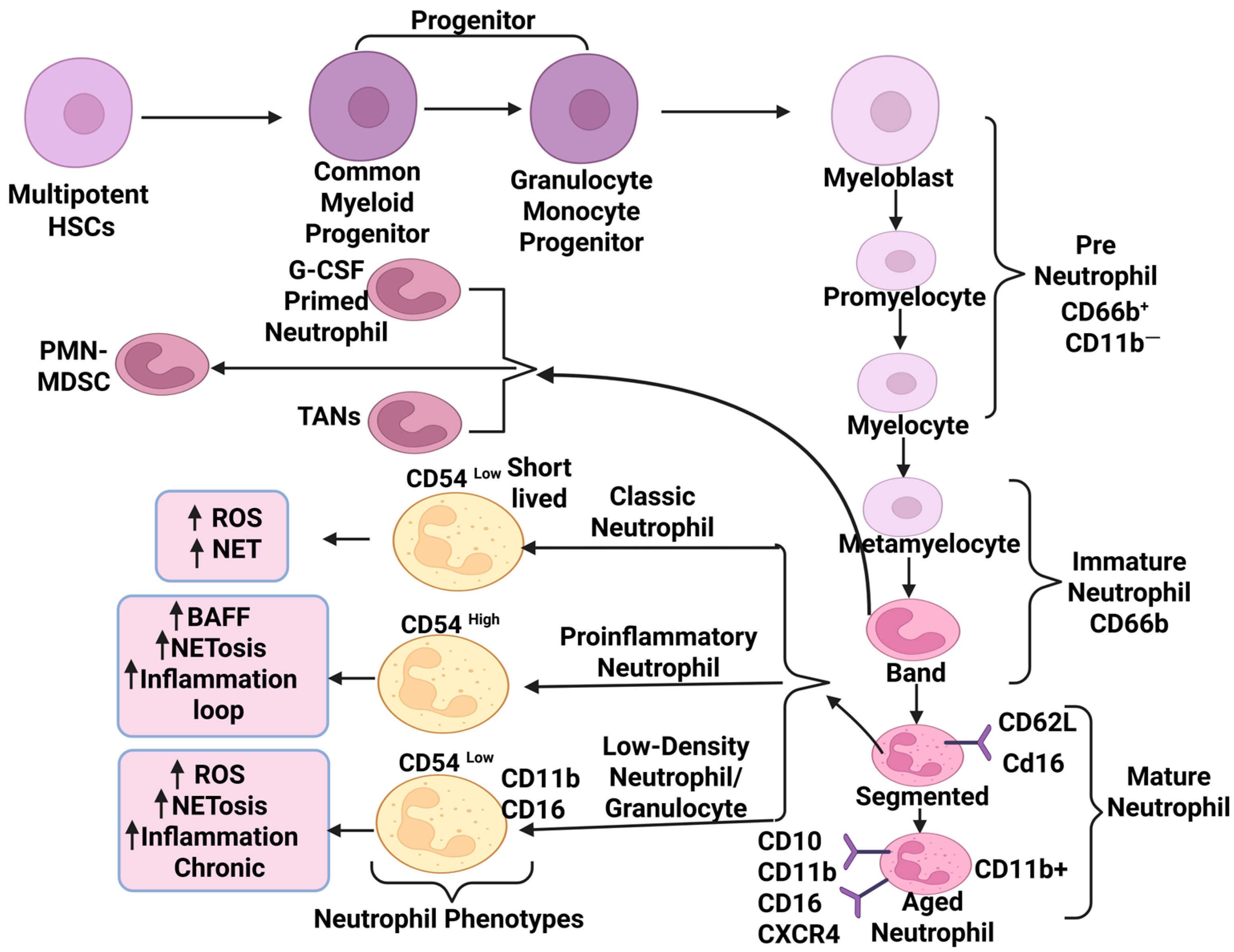
Figure 1. Diagrammatic representation of neutrophil development, differentiation into major subsets, and their later functions involved in autoimmune diseases and cancer. The figure highlights phenotypically and functionally distinct surface marker-defined population of neutrophils, including CD54high mature neutrophils showing elevated (represented as an upright arrow) NETosis and reverse trans-endothelial migration. CD11bhigh aged subsets exhibit increased adhesion, migration, and dysregulated activation under chronic inflammatory conditions. Similarly, low-density neutrophils (LDNs) or low-density granulocytes (LDGs) present immunosuppressive activity (T cell suppression), proinflammatory loop, NETosis, and marker profiles such as CD11b and CD16; CD66b banded immature neutrophil subsets arise into tumor-associated neutrophils (TANs), PMN-MDSC, and G-CSF-primed neutrophils under diseased conditions that display elevated adhesion such as cancer.
Ijms 26 09040 g001
Figure 2. Schematic representation for role of low-density neutrophils and proinflammatory neutrophils in development of chronic inflammation childhood-onset SLE, RA, and JIA. LDGs releasing CitH3, IL-10, TNF-α, mediating high ROS (represented as an upright arrow) induced vascular damage and NETosis. Release of cytoplasmic protein, self-DNA, and cytokines during NETosis; promoting synovial damage in RA and JIA; BAFF-mediated B cell dysregulation; immune complex formation and type I IFN-mediated immune cell (pDC, APC, T cell) crosstalk and inflammatory loop formation through proinflammatory neutrophils. Abbreviations: SLE—systemic lupus erythematosus; RA—rheumatoid arthritis; JIA—juvenile idiopathic arthritis; LDGs—low-density granulocytes; CitH3—citrullinated histone H3; IL-10—Interleukin 10; TNF-α—tumor necrosis factor-α; NETosis—Neutrophil Extracellular Trap formation; type I IFN—interferon (such as IFN-α, IFN-β); ROS—reactive oxygen species; BAFF—B cell-activating factor of the tumor necrosis factor family; pDC—plasmacytoid dendritic cells; APC—antigen-presenting cells.
Ijms 26 09040 g002
Figure 3. Schematic representation of anti-tumorigenic neutrophils and pro-tumorigenic and their transition vice versa. Anti-tumor neutrophils releases cytokines (CCL5, TNFα, IFNγ) that activate T cells and assists NRP1-mediated mitochondria-targeted tumor killing (anti-tumor neutrophils enter cancer cells via NRP1-mediated endocytosis, where effective neutrophil elastase cleaves CD95/Fas at specific sites (V220/A221 and I331/Q332) to release a C-terminal death domain fragment). This binds histone H1 isoforms, forming a complex that translocate to mitochondria, inducing dysfunction, ROS production (represented as an upright arrow), DNA damage, and activation of the intrinsic apoptotic cascade, enabling selective cancer cell killing. Pro-tumor neutrophil release TGF-β, IL-8, GCSF that inhibit immune cells (T, B, NK cells) activation, and promote tumor growth through angiogenesis as well as EMT. Transition among anti-tumor neutrophil to pro-tumor neutrophil under milieu rich in TGF-β and vice versa in TGF-β blockade condition. Abbreviations: NRP1—neuropilin-1; TGF-β—transforming growth factor-beta; IL-8—Interleukin-8; GCSF—Granulocyte colony-stimulating factor; PMN-MDSC—polymorphonuclear myeloid-derived suppressor cells.
Ijms 26 09040 g003
Table 1. Description on transition among phenotypes of tumor-associated neutrophils.
S. No.Transition of TANsKey FactorsPhenotypic MarkersReferences
1.Pro-tumor (N2) to Anti-tumor (N1)Microbial signals, TGF-β blockade, IFN-α/β, acute inflammationIncreased ROS, HLA-DRhigh, CD74high, CD80, CD86, high chemokines recruiting T/NK cells[15,16]
2.Anti-tumor (N1) to Pro-tumor (N2)TGF-β, chronic inflammation, hypoxiaIncreased VEGF, PD-L1, ARG1, HLA-DRlow and CD74low[16,17]
Table 2. Description of neutrophil-targeted, drugs, or therapeutic agents in autoimmune diseases and cancer.
Neutrophil-Targeted Therapeutics in Autoimmune Diseases and Cancer (Clinical Trials)
Drugs and Therapeutic AgentsMechanism of ActionPhase/Status of Clinical TrialOutcome of TherapeuticsReferences
Baricitinib, Tofacitinib, UpadacitinibModulate neutrophil activation and NETosis targeting JAK/STAT pathwayPhase 1–3 trialsReduces NETs, LDNs[45]
ClinicalTrials.gov ID NCT02535689
ClinicalTrials.gov ID NCT05843643
TaxifolinInhibits NETosis (Nrf2)Preclinical/ex vivoProtective in lupus models[46]
Dual-acting DNase1/DNase1L3Degrades NET DNAPreclinicalPromising in SLE models[47]
MetforminInhibits NETosisAdd-on trial (completed)Reduced flares, indirect NETosis effect[48]
AnifrolumabType I Interferon InhibitorApproved, real-world/clinicalReduces NETs and LDNs; rapid efficacy in SLE[49]
JBI-589 Peptidylargininedeiminase 4 (PAD4) inhibitorInhibits PAD4; blocks NETosisPreclinical (animal models, in vitro human/mouse neutrophils)Highly selective PAD4 inhibitor; blocks NET formation and reduces arthritis severity in mouse models; confirmed NET inhibition in vito [50]
TocilizumabIL-6R blockade; modulates neutrophil function/NETsPhase 3, approved Reduces NETosis, neutrophil infiltration, and improves neutrophil function in RA, systemic JIA[51]
REC reference
10/H0904/14
AZD5069Inhibits CXCR2Phase 1/2 (combo with immunotherapy)Inhibits neutrophil recruitment to tumors; reduces intratumoral neutrophil and MDSC infiltration, shown to decrease neutrophil levels and improve response in Prostate, NSCLC, solid tumors[52]
ClinicalTrials.gov ID NCT02583477
Navarixin (SCH-527123, MK-7123)Inhibits CXCR2Phase 1b/2Inhibits CXCR2-mediated neutrophil chemotaxis and tumor infiltration; reduces tumor-promoting neutrophils in NSCLC, solid tumors[53,54]
ClinicalTrials.gov ID NCT03473925
ReparixinInhibits CXCR1/2Phase 2Blocks neutrophil recruitment and MDSC infiltration by antagonizing CXCR1/2 in breast and pancreatic cancer[53,54]
Disclaimer/Publisher’s Note: The statements, opinions and data contained in all publications are solely those of the individual author(s) and contributor(s) and not of MDPI and/or the editor(s). MDPI and/or the editor(s) disclaim responsibility for any injury to people or property resulting from any ideas, methods, instructions or products referred to in the content.