Article

Transcriptomic Analysis Reveals the Regulatory Mechanism of Cold Tolerance in Saussurea involucrata: The Gene Expression and Function Characterization of Dehydrins

1
State Key Laboratory of Bioactive Substance and Function of Natural Medicines, Institute of Medicinal Plant Development, Chinese Academy of Medical Sciences and Peking Union Medical College, Beijing 100193, China
2
Xinjiang Institute of Materia Medica, Urumqi 830010, China
*
Authors to whom correspondence should be addressed.
Int. J. Mol. Sci. 2025, 26(18), 9030; https://doi.org/10.3390/ijms26189030
Submission received: 18 August 2025 / Revised: 4 September 2025 / Accepted: 8 September 2025 / Published: 17 September 2025
(This article belongs to the Section Molecular Plant Sciences)

Abstract

Saussurea involucrata, a rare and endangered medicinal plant of the Asteraceae family, is primarily distributed in high-altitude rocky slopes and meadows at elevations of 2400–4100 m. In nature, this herb endures various abiotic stresses, including intense cold and ultraviolet radiation. In our study, transcriptomic profiles revealed that most of the differentially expressed genes (DEGs) enriched in stress response pathways, such as “response to water”, “response to abscisic acid”, “cold acclimation”, and “response to water deprivation”, were significantly upregulated after low-temperature treatment. In contrast, the majority of genes with lower expression were related to “photosynthesis”, “protein–chromophore linkage”, and “chloroplast thylakoid membrane”. Among them, Kyoto Encyclopedia of Genes and Genomes (KEGG) and Gene Ontology (GO) database analysis revealed that approximately 20 DEGs were identified and annotated as dehydrin genes (DHNs). Quantitative PCR (qPCR) validation also confirmed that these DHNs were upregulated under cold stress. Moreover, SiDHN3, a new dehydrin gene, was cloned by Rapid Amplification of cDNA Ends (RACE). SiDHN3’s heterologous expression in E. coli showed enhanced salt, osmotic, freeze–thaw, and cold stress tolerance. A functional analysis of SiDHN3’s truncated derivatives revealed that the K-segment was critical for its protective function under freeze–thaw and cold stresses. Collectively, our study demonstrated the potential role of various DHNs as a functional protein, enhancing tolerance to cold stress in the high-altitude adaptation of plants.

1. Introduction

In the Asteraceae family, Saussurea involucrata Kar. et Kir. has a long history of ethnic medicinal use with high economic value. Many studies have shown that this herb may have various pharmacological activities, such as anti-inflammatory and analgesic effects, as well as anti-rheumatoid arthritic, anti-obesity, and anti-cancer effects [1,2,3,4,5]. In nature, S. involucrata is a rare and endangered plant with limited resources due to harsh living conditions, excessive harvesting, and immature artificial cultivation techniques. The habitat is mainly distributed in high mountain rocky beaches, meadows, and other areas ranging from 2400 to 4100 m [6]. Low temperatures, low oxygen, and high ultraviolet radiation levels are major unfavorable conditions for S. involucrata survival. Chilled temperatures (0 °C–12 °C) can inhibit the growth and development of most plants [7], while some alpine plants in extreme habitats, such as Saussurea species, have evolved many survival and reproduction strategies for resistance against these low temperatures [8,9]. However, research on the molecular mechanisms of adapting to low temperatures in S. involucrata is limited. At present, only some aquaporin proteins and dehydrins in S. involucrata have been verified in tobacco and tomato for their cold tolerance function [10,11,12,13].
Dehydrins, a subgroup of the late embryogenesis abundant (LEA) protein family, are important in the response to abiotic stress in plants [7,14] and has received widespread attention recently [15,16,17]. When plants encounter low temperatures and drought, DHNs accumulate extensively [18]; for example, DHN5 in barley can be significantly upregulated after low-temperature treatment [19]. In addition, cold-resistant plants exhibited higher expression levels of DHNs than sensitive phenotypes when exposed to low temperatures [20]. In recent years, research progress on cold resistance has shown that dehydrins in plants can protect and restore the activity of lactate dehydrogenase (LDH) and catalase in freeze–thaw conditions [21,22,23], scavenge free radicals by binding to their easily oxidized amino acid sequences [22], and increase the expression levels of ROS-related genes [24,25]. Meanwhile, functional studies have also suggested that dehydrins can respond to hormones such as abscisic acid, salicylic acid, and jasmonic acid to coordinate plant development and stress responses [26]. However, little research has been carried out on dehydrins in alpine plants such as S. involucrata.
In our study, the morphological and physiological profiles of S. involucrata seedlings treated with different low temperatures were investigated. Transcriptomic analysis characterized the classification and expression pattern of dehydrin genes (DHNs) in S. involucrata, determining their responses to varying degrees of low temperature. A new differentially expressed dehydrin gene, SiDHN3, was cloned. Subsequently, a prokaryotic expression system in Escherichia coli was established, expressing a recombinant protein. The protective role of SiDHN3 against multiple abiotic stresses, including freeze–thaw cycles, low-temperature exposure, and high osmotic pressure, was investigated.
Our study lays the groundwork for deciphering the survival strategies of S. involucrata in extreme habitats while providing insights into alpine plant stress response mechanisms. Furthermore, it advances our understanding of dehydrins in extremophytes, paving the way for deeper exploration of the plant dehydrin family.

2. Results

2.1. Morphological Profiles of S. involucrata at Low Temperature

Compared with the control group, there were no significant morphological changes observed in the cold or chilled groups of S. involucrata seedlings (Figure 1A–C), including the length and number of roots or leaves (Figure 1E). In contrast, the fresh weight in the cold group (170.07 ± 47.68 mg) and chilled group (167.80 ± 34.53 mg) treated with low temperatures was significantly reduced compared to the control group (281.07 ± 13.32 mg).

2.2. Physiological Profiles of S. involucrata at Low Temperature

As shown in Figure 1D, malondialdehyde (MDA), soluble protein, and soluble sugar in S. involucrata seedlings in each group were measured. Among them, the MDA (33.35 ± 4.70 nmol/g) and soluble protein content (0.39 ± 0.14%) in the chilled group were significantly higher than those in the control group (21.72 ± 3.38 nmol/g and 0.18 ± 0.07%). However, there was no significant difference in MDA, soluble protein, and soluble sugar between the cold and control groups. The results indicated that plant membrane lipids were subjected to low-temperature stress and underwent peroxidation after 4 °C/0 °C treatment.

2.3. RNA-Seq Profiles of S. involucrata at Low Temperature

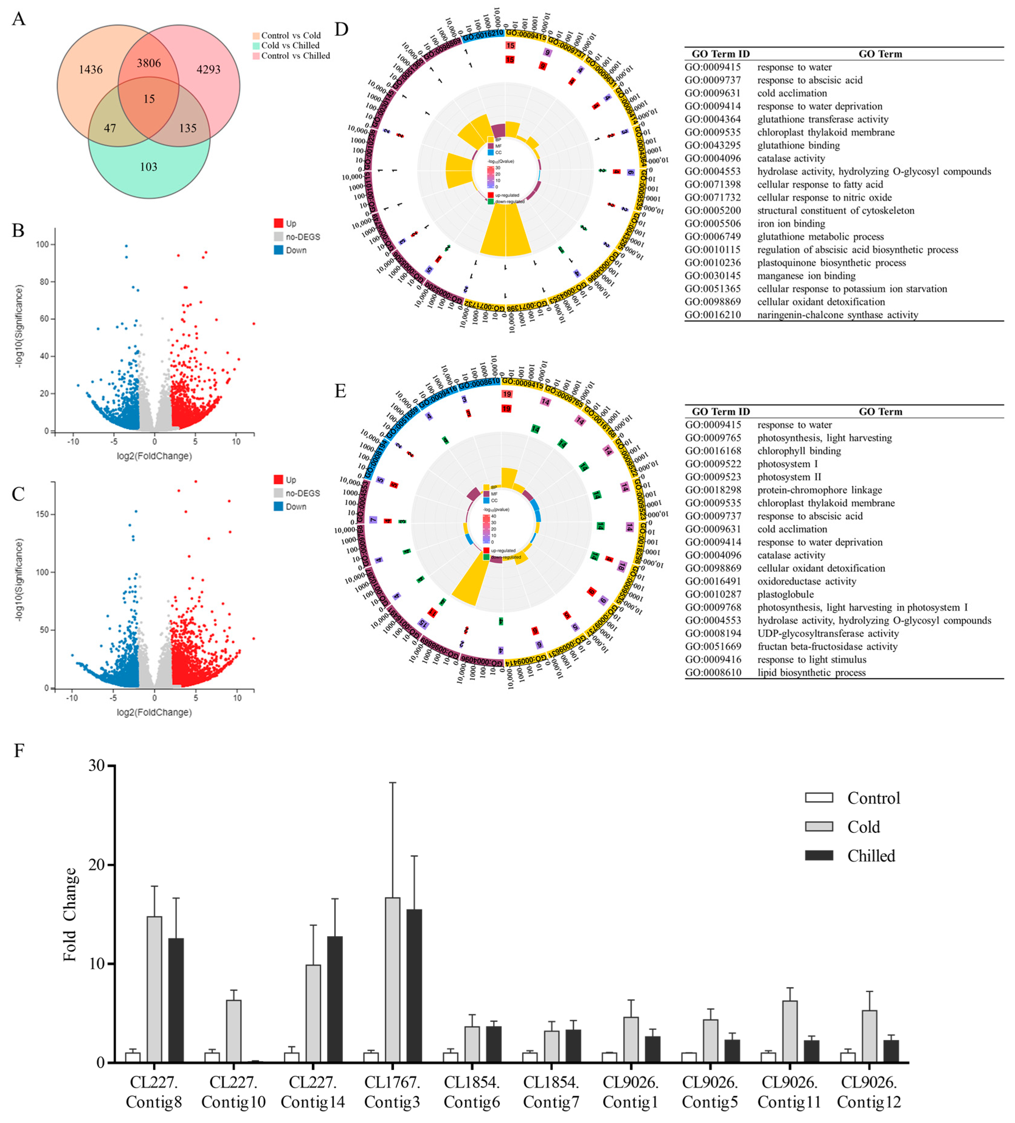
The RNA-seq datasets are available in the BioProject (Accession Number: PRJNA1033840) repository of the National Center for Biotechnology Information (NCBI). Since there were no reference genomes of S. involucrata, the clean reads from all libraries were pooled together and de novo assembled into transcripts. A total of 87,143 unigenes were obtained to analyze sample differences and screen differentially expressed genes (DEGs, |log2(FoldChange)| > 2, q-value < 0.05). A Venn plot (Figure 2A) revealed that there were only 1436 and 4293 DEGs in the cold and chilled groups, indicating various response mechanisms in S. involucrata at different low temperatures. Furthermore, compared with the control group, 3141 and 2163 genes were significantly up- and downregulated in the cold group, and the numbers in the chilled group were 4739 and 3510.

2.4. Annotation of S. involucrata Transcripts

Gene annotation was applied by BLAST against the NCBI Non-redundant Protein Sequence (Nr)/Nucleotide Sequence (Nt), Protein families (Pfam), Swiss-Prot Protein Knowledgebase (Swiss-Prot), Gene Ontology (GO), and Kyoto Encyclopedia of Genes and Genomes (KEGG) databases. In total, there were 72,818 unigenes (83.56% of all unigenes) annotated in at least one database. Among them, a total of 68,624 unigenes (78.75%) were annotated in the Nr database. Subsequently, DEGs with Fragments Per Kilobase of transcript per Million mapped fragments (FPKM) > 100 were annotated to the top 20 terms of GO classification (Figure 2D,E). Compared with the control group, DEGs in the cold group were significantly enriched in “response to water”, “response to abscisic acid”, “cold acclimation”, and “response to water deprivation”. All DEGs in these four pathways were upregulated (Figure 2D). It indicates that the low-temperature response system of S. involucrata has been activated under the condition of 4 °C. Moreover, the GO terms of DEGs in the chilled group were also significantly enriched in the cold-related pathways, including “response to water”, “response to abscisic acid”, “cold acclimation”, etc. (Figure 2E). However, “photosynthesis”, “light harvesting”, “Chlorophyll binding”, and “Photosystem I” were the top five GO terms for DEGs only in the chilled vs. control group, suggesting that S. involucrata photosynthesis was significantly inhibited.

2.5. Expression Analysis of DEGs Related to Cold Stress

Based on all of unigenes, there were 51, 79, and 25 genes annotated to “response to water”, “cold acclimation”, and “response to water deprivation”. Of the 131 unigenes annotated to these three pathways, a total of 20 DEGs were annotated as proteins in the dehydrin subgroup (Table 1). Among them, there were five, two, four, and three unigenes annotated as embryogenic cell protein 40-like(ECP 40-like), dehydrin Xero 1-like, dehydrin Rab18-like, and dehydrin ERD14-like genes assigned with Cynara cardunculus var. scolymus. Additionally, eleven unigenes were annotated to three species of dehydrin (S. involucrata, Artemisia annua, and C. cardunculus var. scolymus). Compared with the control group (Table 1), five ECP 40-like, two Xero 1-like, one Rab18-like, three ERD14-like, and seven dehydrin genes were significantly upregulated in the cold group; four, two, one, three, and eight of these genes were distinctly expressed higher in the chilled group.

2.6. qPCR Analysis of Putative DHNs

As illustrated in Figure 2F, we performed quantitative PCR analysis on 10 selected DHNs putatively. The expression of these DHNs was low in the control group, while it was increased in the cold and chilled group. This result was consistent with the transcriptome profiles, indicating that our experimental data were reliable.

2.7. DHN Gene Expression in Different Periods of Treatment at Multiple Low Temperatures

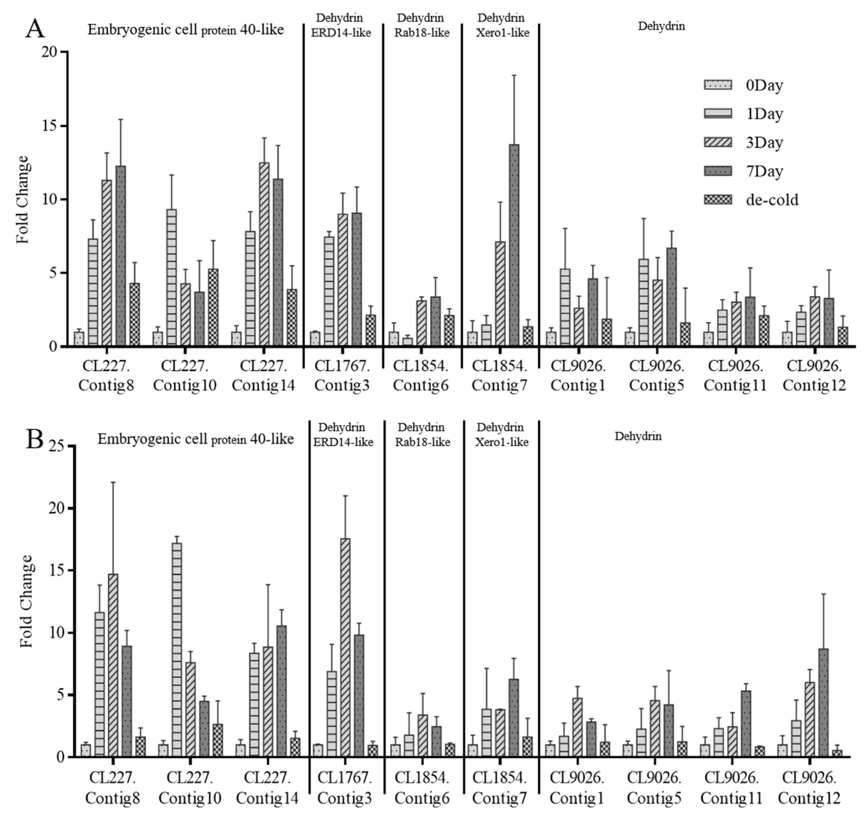
As shown in Figure 3, most of the DHNs were more significantly upregulated in the chilled than in the cold group, except Xero-1 like (CL1854.Contig7) and dehydrin (CL9026.Contig1 and CL9026.Contig5).
As shown in Figure 3A, the expression of each DHNs was detected at 4 °C for day 0 (control), day 1, day 3, day 7, and day 14 (4 °C for day 7 and 20 °C for day 7). Indeed, the highest expression of almost all DHNs appeared on the seventh day, except CL227.Contig10 (on the first day) and CL227.Contig14 (on the third day). However, DHNs expression was different when the temperature was 4 °C/0 °C (day/night). According to Figure 3B, the highest expression of many DHNs appeared on the first or third day such as CL227.Contig10, CL1767.Contig3, CL9026.Contig1, etc. Significantly, all of the DHNs were distinctly downregulated in the day 14 group, suggesting that these genes were induced by low temperatures and reversed under 20 °C.

2.8. Sequencing Analysis of SiDHN3

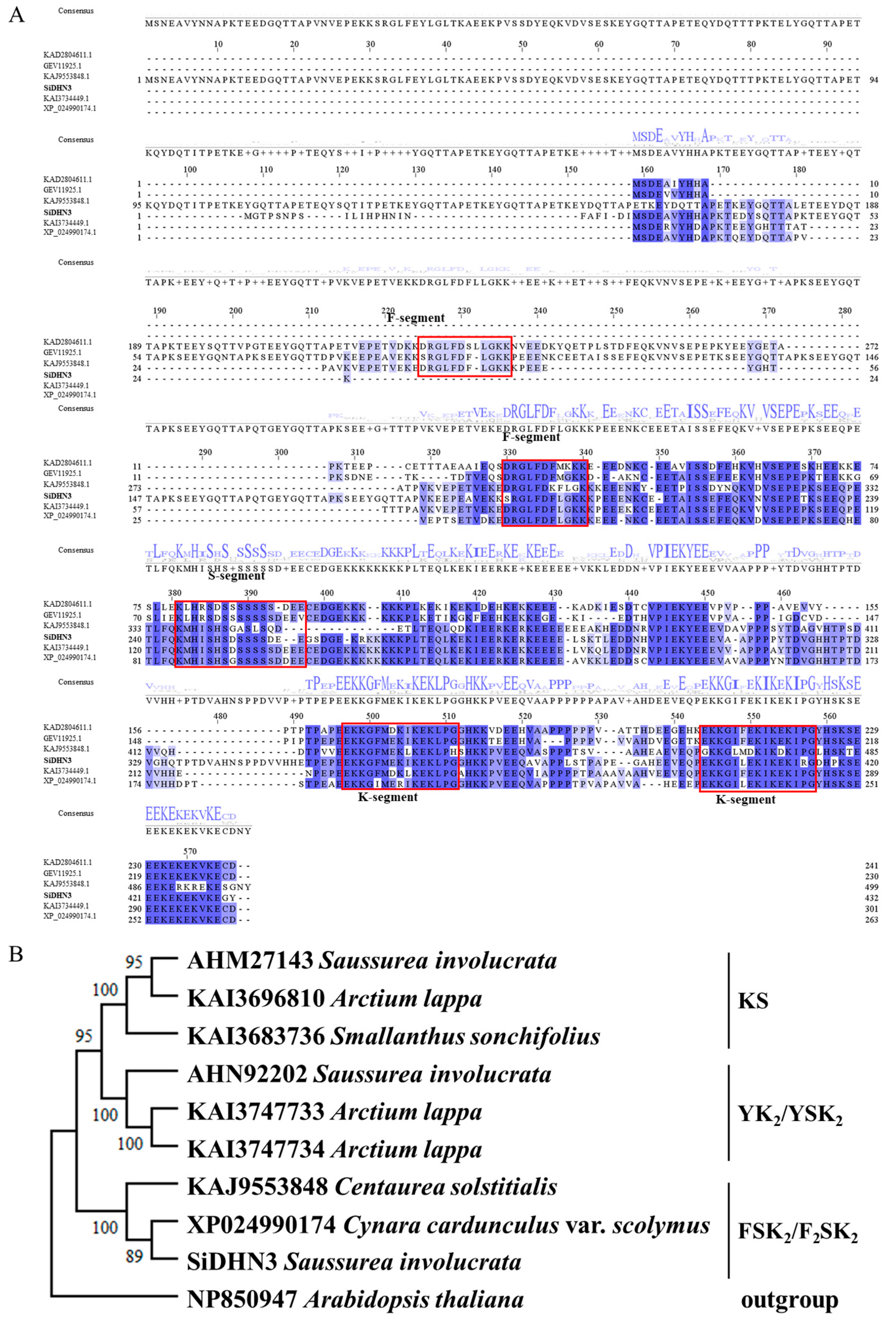
The nucleotide sequence of the SiDHN3 ORF was translated into amino acids using SnapGene 7.1.2, homologous sequences were aligned using Clustal Omega, and the alignment results were visualized in Jalview 2.11.4.0. SiDHN3 was classified as an F2SK2-type dehydrin based on its conserved motifs (Figure 4).

2.9. Expression and Protective Effect of SiDHN3 Proteins on E. coli

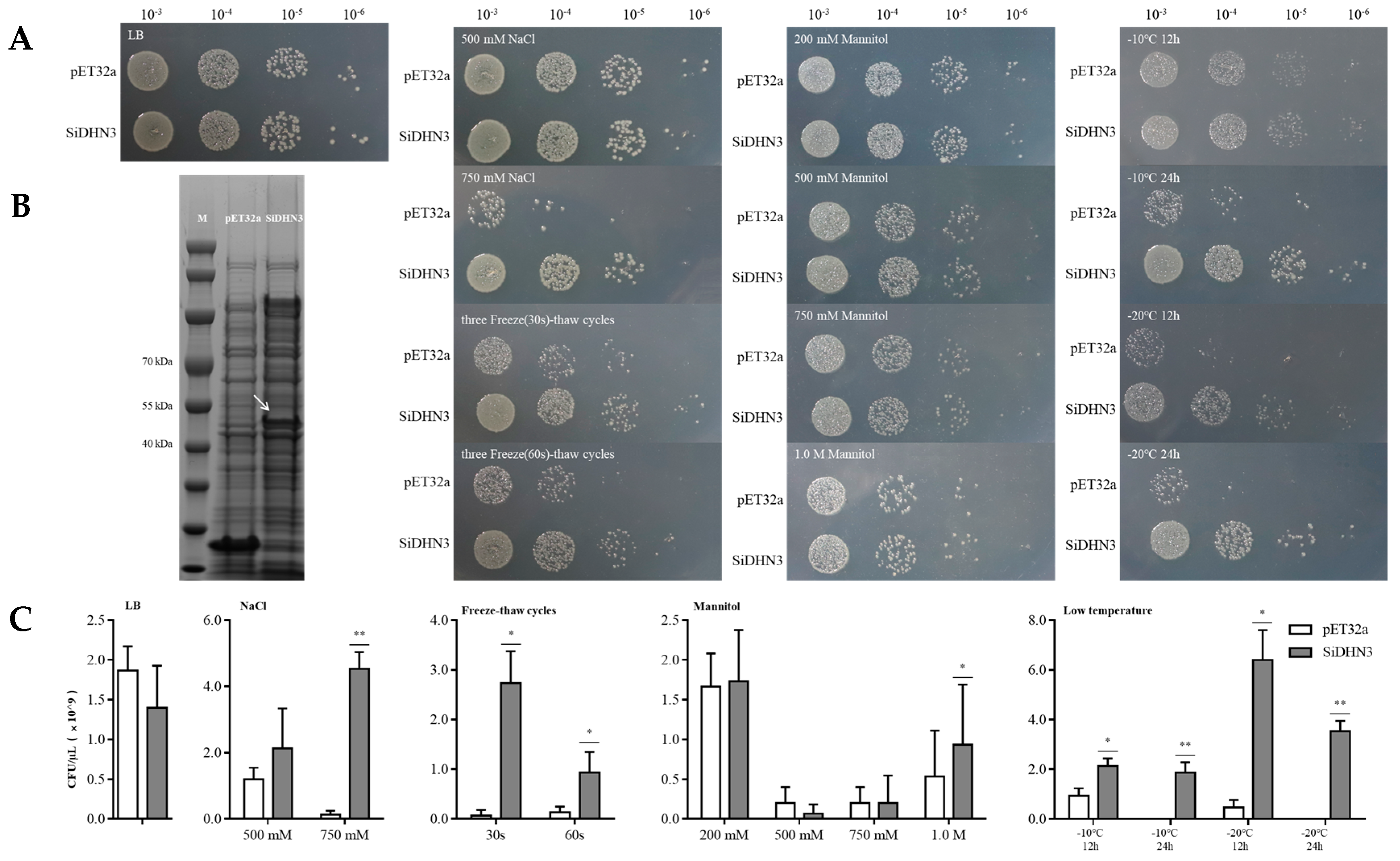
The molecular weight of SiDHN3 was predicted using ExPASy ProtParam to be approximately 48.593 kDa. A prominent ~50 kDa protein band was exclusively detected by SDS-PAGE in E. coli harboring the pET32a-SiDHN3 plasmid, while the empty vector control showed no signal at this position, confirming successful expression (Figure 5B).
To simulate high-salinity and osmotic stress conditions, varying concentrations of NaCl and mannitol were supplemented into LB medium. The results revealed that under high concentrations of 750 mM NaCl and 750 mM mannitol, the accumulation of SiDHN3 exerted a statistically profound enhancement on E. coli survival (p < 0.01). Notably, when exposed to osmotic stress with 1.0 M mannitol in LB medium, E. coli expressing SiDHN3 exhibited a statistically significant survival advantage compared to controls (p < 0.05).
Following three freeze–thaw cycles, SiDHN3 demonstrated a statistically significant cryoprotective effect on E. coli compared to the control group (p < 0.05).
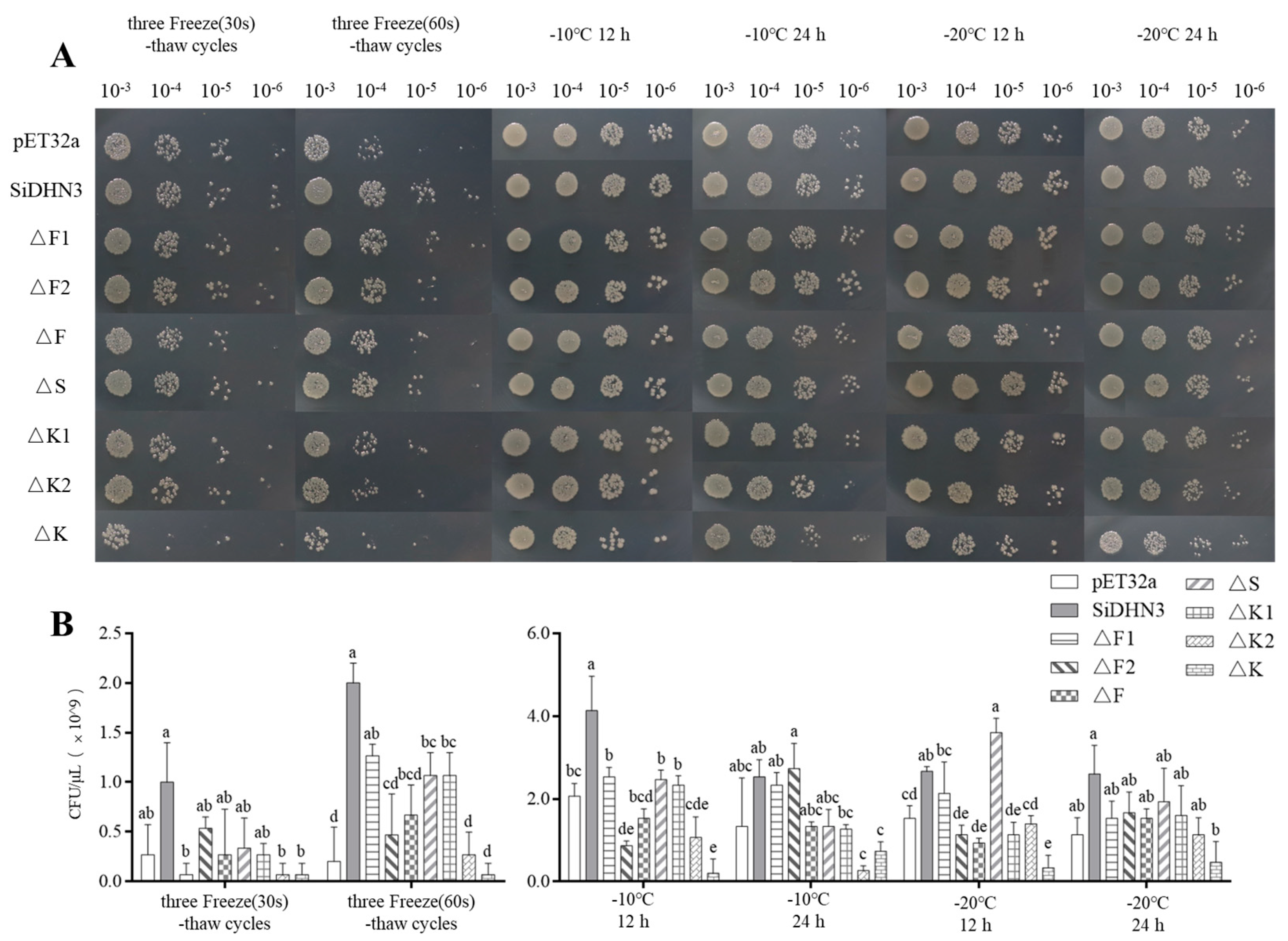
When subjected to low-temperature stress at −10 °C and −20 °C with varying exposure durations (12 h and 24 h), E. coli expressing SiDHN3 demonstrated significant cryoprotective effects. As shown in Figure 5A,C, the growth profiles of E. coli were consistent with the statistical results of CFU, which indicated that SiDHN3 could protect E. coli from various stress conditions.

2.10. Functional Validation of SiDHN3 Protective Domains

The growth profiles of E. coli expressing truncated SiDHN3 variants under freeze–thaw cycles and low-temperature stress are presented in Figure 6. Simultaneous deletion of both K-segments (∆K) abolished the cytoprotective function of SiDHN3, rendering transformed cells susceptible to both freeze–thaw stress and sustained low-temperature exposure. During 60 s freeze–thaw cycles, deletion of either the F-segment (∆F) or S-segment (∆S) significantly compromised (p < 0.05) SiDHN3-mediated protection, though to a lesser extent than ∆K (Figure 6B).
Under 12 h low-temperature treatments, F-segment deletion (∆F) alone reduced E. coli survival (p < 0.05) across tested temperatures (−10 °C to −20 °C), indicating its essential role in freeze–thaw stress mitigation (Figure 6B).

3. Discussion

Plants frequently delay growth as a strategy to adapt to low-temperature stress [27]. This response is particularly pronounced in alpine species; for example, S. leontodontoides exhibits inverse correlations between altitude and vegetative organ biomass [28]. Consistent with these observations, S. involucrata showed significantly reduced fresh weight under low-temperature conditions, though root length, root number, and leaf characteristics remained unaffected. Concurrently, levels of osmoregulatory compounds—specifically MDA and soluble proteins—increased significantly in chill-treated (4 °C/0 °C) plants relative to controls.
Low temperatures typically disrupt water absorption–transpiration equilibrium, inducing cellular dehydration. Consequently, proteins related to water deprivation like dehydrins are known to accumulate rapidly under cold stress and are widely studied [26,29,30]. Supporting this, GO analysis revealed significant enrichment of cold-related terms (“response to water”, “cold acclimation”, and “response to water deprivation”) among DEGs in 4 °C/4 °C and 4 °C/0 °C groups. Twenty of these DEGs encoded annotated dehydrin proteins.
Substantial DHN upregulation during cold exposure is well-documented in pepper (Capsicum spp.), Solanum sogarandinum, and barley (Hordeum vulgare) [18,19,31,32]. Cold-tolerant plants exhibit enhanced DHNs expression [20], exemplified by higher DHN1/DHN2 transcript levels in winter barley cultivars than in spring genotypes [15,19]. Furthermore, in both wheat and barley, highly tolerant varieties exhibit a more rapid and substantial accumulation of dehydrins compared to less tolerant varieties [33]. Critically, the heterologous expression of cold-adapted DHNs enhances low-temperature tolerance in recipient plants [34,35], as demonstrated by improved cold resistance in transgenic tomato and tobacco expressing S. involucrata-derived SiDHN2 [36] or SiDHN [12]. Our data align with this paradigm: DHN expression in S. involucrata was cold-inducible and stress intensity-dependent. Notably, virtually all DHNs were markedly downregulated after 7-day recovery at 20 °C, indicating reversible cold responsiveness, a pattern also observed for RcDHN1-5 in Rhododendron catawbiense [37].
Despite the established importance of dehydrins, functional studies on S. involucrata remain scarce, with only two genes characterized to date. This contrasts sharply with the extensive structural and functional knowledge of dehydrins in other species [30], where they mitigate diverse abiotic stresses including salinity [38,39], drought [40,41], and cold [42].
Here, we isolated the full-length coding sequence of a new cold-induced dehydrin (CL1767.Contig1) via RACE amplification. The protein, designated as SiDHN3, contains two F-segments, one S-segment, and two K-segments, classifying it as the first documented F2SK2-type dehydrin from S. involucrata. Heterologous expression in E. coli significantly enhanced bacterial survival under low temperatures, freeze–thaw cycles, and hyperosmotic stress.
While dehydrins are classically categorized into five types (Kn, SKn, KnS, YnKn, and YnSKn) by conserved domains [43,44], expanding sequence/functional data reveal limitations in predicting properties based solely on Y-/S-/K-segment counts [26]. Their complex regulation necessitates segment-specific functional analyses. The K-segment, a core motif implicated in cellular protection, antioxidation, and protein/membrane stabilization [45,46,47,48,49], mediates macromolecular interactions (e.g., binding acidic phospholipid vesicles and membrane domains) [45,50]. Truncation analysis confirmed the K-segment’s indispensability for SiDHN3-mediated multistress protection in E. coli, with efficacy scaling with the K-segment copy number.
SiDHN3 represents the first cloned F-segment containing dehydrin from S. involucrata. This conserved phenylalanine pair-containing motif [51] remains poorly characterized but may facilitate macromolecule binding and cold-labile enzyme stabilization [52]. Crucially, F-segment deletion significantly reduced freeze–thaw stress protection by SiDHN3 in E. coli assays.
As proteins responsive to multiple stresses, dehydrins play various protective roles in plant cells [53], mechanisms that are closely linked to their structures [54]. Further investigation into the structure and function of SiDHN3 in model organisms is crucial, particularly given its significant promise for application in economic crops. Meanwhile, elucidating the subcellular localization of dehydrins can help clarify their functional mechanisms [55]. For instance, dehydrins localized to the plasma membrane often exhibit membrane cryoprotection functions [56,57]. Additionally, S. involucrata, adapted to high-altitude environments, is a valuable model for studying dehydrins. Genomic studies of its DHNs [58] could clarify their ecological role in adaptation.
In summary, S. involucrata showed significantly reduced fresh weight under low-temperature conditions; transcriptomic profiles revealed that most of the differentially expressed genes (DEGs) enriched in the stress response pathways were significantly upregulated after low-temperature treatment. Among them, KEGG and GO analysis revealed approximately 20 DEGs and annotated them as DHNs. The coding sequence of SiDHN3, a differentially expressed DHNs, was cloned, representing the first characterized F2SK2-type dehydrin in S. involucrata. SiDHN3 significantly enhanced E. coli survival under diverse abiotic stresses, including low temperatures and freeze–thaw cycles. The protective function depended on the K-segment within the structure of SiDHN3, while the F-segment proved critical for freeze–thaw stress tolerance. These findings elucidate molecular adaptations underlying the high-altitude resilience S. involucrata and advances our understanding of dehydrin-mediated abiotic stress protection in plants.

4. Materials and Methods

4.1. Plant Materials

S. involucrata was germinated in 1/2MS medium at 20 °C in a Ningbo Jiangnan DRXM incubator (Ningbo Jiangnan DRXM, Ningbo, China) in our laboratory. Based on the methodology previously described [59], the 30-day-old seedlings were divided into three groups for treatment at different temperatures for 7 days, including the control (20 °C), cold (4 °C), and chilled groups (4 °C/0 °C, day/night).
At the end of 7 days, five repetitions were collected from each group. The fresh weight and length of the leaves and roots were measured, as well as the number of roots and leaves. The same samples were immersed in liquid nitrogen for rapid freezing and were restored at −80 °C for physiological profiles and RNA-seq.

4.2. Determining the Physiological Characteristics of S. involucrata

Referring to the manufacturer’s instructions for the malondialdehyde (MDA) assay kit and plant soluble sugar content test kit (Nanjing Jiancheng Bioengineering Institute, Nanjing, China), as well as the Bradford Protein Assay Kit (Beyotime Biotech, Shanghai, China), the MDA, soluble sugar, and total soluble protein content of S. involucrata were detected.

4.3. Transcriptomic Analysis

Sample processing, library construction, sequencing, assembly, and analysis were completed by BGI Tech Solutions Co., Ltd. (Shenzhen, China). In brief, the raw data was filtered with SOAPnuke (v1.4.0) to obtain clean reads [60]. After removing reads containing adapters, as well as ensuring that the unknown base (“N” base) ratio is more than 5% and the low-quality base ratio is more than 20%, clean reads were assembled by Trinity (v2.0.6) and BUSCO (v5.7.0) to assess the assembly quality [61]. Then, clean reads were mapped to unigenes by Bowtie2 (v2.2.5), and RSEM (v1.2.8) was used to calculate the expression of unigenes [62,63]. Distinct unigenes were annotated with NCBI Nr/Nt, Pfam, Swiss-Prot, GO, and KEGG databases.
PossionDis was performed for between-group differential expression analysis, using |log2(FoldChange)| > 2, q-value < 0.05, and PFKM > 100 as a threshold to identify differentially expressed genes (DEGs). An advanced Circos bar plot was plotted by an online platform (https://www.bioinformatics.com.cn, accessed on 1 November 2023).

4.4. Expression of DHNs from Different Groups

In order to verify the reliability of the RNA-seq data, samples from the same libraries were used for quantitative PCR analyses. The total RNA of each sample was extracted using the Quick RNA isolation Kit (Huayueyang Biotechnology, Beijing, China), and the cDNA library was obtained by using HiScript III All in one RT SuperMix (Vazyme, Nanjing, China). In total, 1 μL of cDNA diluted with 10 μL of ddH2O was used as the template.
Primers for qRT-PCR (Supplementary Table S1) were designed with Primer Premier software (Primer Premier v5.0; Premier Biosoft International, Palo Alto, CA, USA). The expression level of the target genes was determined by qTR-PCR using the ROCHE LightCycler® 480 II system (Roche, Basel, Switzerland) and Taq Pro Universal SYBR qPCR Master Mix (Vazyme, Nanjing, China) in 15 μL reactions. Each reaction consisted of 5 ng of total RNA, 0.3 μL of each primer, and 7.5 μL master mixes. In total, we performed three biological replicates and three technical replicates. The PCR reactions were performed in a thermocycler with the following conditions: 5 min at 95 °C, 45 cycles of 10 s at 95 °C, 10 s at 60 °C, and 10 s at 72 °C. The GAPDH gene was used as a reference [59]. The 2−∆∆Ct method was used for evaluating gene expression [64].

4.5. Expression of DHNs in Different Periods of Treatment at Multiple Low Temperatures

In plants, DHNs can be constantly strengthened with a longer duration in the cold or lower temperature stress. For investigating DHN expression patterns, we performed quantitative PCR analysis on 10 candidate genes in different periods at multiple low temperatures. Here, S. involucrata seedlings were germinated for 30 days. As described in a previous study with slight adjustments [65], for cold treatment, samples from day 0, day 1 (4 °C), day 3 (4 °C), day 7 (4 °C), and day 14 (7 day 4 °C + 7 day 20 °C) groups were collected. For the chilled treatment, the same procedure was carried out at 4 °C/0 °C. Next, qPCR was performed according to the method described in Section 4.4.

4.6. Cloning of Full-Length Coding Sequence of SiDHN3

Total RNA was extracted from S. involucrata using the Quick RNA isolation Kit (Huayueyang Biotechnology, Beijing, China). The 5′- and 3′-cDNA library construction and full-length coding sequence of SiDHN3 were obtained by the 5′- and 3′- rapid amplification of cNDA ends (RACE) using the SMARTerTM RACE cDNA Amplification kit (Clontech, California, CA, USA) according to the manufacturer’s instructions.

4.7. Sequence Analysis of SiDHN3

The open reading frames (ORFs) of SiDHN3 were identified using the ORF Finder tool (https://www.ncbi.nlm.nih.gov/orffinder/, accessed on 3 June 2024). The amino acid sequences were compared with reported plant dehydrin sequences through the NCBI BLAST Program (https://blast.ncbi.nlm.nih.gov/Blast.cgi, accessed on 3 June 2024), and high-similarity protein matches were performed for multiple sequence alignments with Clustal Omega (https://www.ebi.ac.uk/jdispatcher/msa/clustalo, accessed on 30 March 2025). The phylogenetic tree was generated using MEGA 11.0.13 [66].

4.8. Construction and Expression of SiDHN3 and Its Truncated Derivatives

Truncated derivatives (∆F1, ∆F2, ∆S, ∆K1, ∆K2) were generated by sequentially removing segments. Derivative constructs ∆F and ∆K were created by deleting two F-segments and two K-segments, respectively (the sequence design of the SiDHN3-truncated derivative is shown in Supplementary Figure S1). The plasmids were engineered to contain the SiDHN3-truncated derivatives by GenScript (Suzhou, China). The recombinant plasmids (pET32a-SiDHN3, ∆F1, ∆F2, ∆S, ∆K1, ∆K2, ∆F, ∆K, and the control vector pET32a; the pET32a-SiDHN3 plasmid diagram is shown in Supplementary Figure S2) were transformed into an E. coli strain OrigamiB (DE3) (Beyotime, Shanghai, China) according to Beyotime’s protocol. The sequencing results were assembled with Codoncode Aligner (Codoncode, Centerville, MA, USA).

4.9. Abiotic Stress Tolerance Assays

E. coli OrigamiB (DE3) cells carrying pET32a-SiDHN3 plasmids and pET32a (control) were grown in liquid LB medium at 37 °C and 180 rpm until an OD600 of 0.5–0.6 was reached followed by 20 h of induction at 16 °C and 150 rpm with 0.2 mM IPTG. The OD600 of the cell cultures was then adjusted to 0.5, and they were then diluted serially to 10-3, 10-4, 10-5, and 10-6 with LB. Next, 5 μL of each sample was spotted separately on LB plates containing NaCl (500 mM, 750 mM) and mannitol (500 mM, 750 mM, 1.0 M) for salinity and osmotic treatments. For the freeze–thaw test, the OD-adjusted cells were placed in liquid nitrogen for 30 s and 60 s separately and thawed at room temperature; after freeze–thaw repetitions, the cultures were diluted and spotted on the control LB plate. All plates were incubated at 37 °C for 24 h, and colony forming units (CFUs) were counted at 10−5 or 10−6 dilution [58]. For the statistical analysis of colony forming units (CFUs), an independent-samples t-test was performed to compare differences between two groups. A one-way ANOVA followed by Tukey’s post hoc test was used for comparisons among multiple groups.

Supplementary Materials

The following supporting information can be downloaded at: https://www.mdpi.com/article/10.3390/ijms26189030/s1.

Author Contributions

Conceptualization, T.C., X.Z. and X.C.; methodology, T.C., J.Z. and L.Z.; software, T.C.; validation, T.C., X.Z. and L.Z.; formal analysis, T.C.; resources, J.Z., C.L. and A.W.; data curation, L.Z. and T.C.; writing—original draft preparation, X.Z. and T.C.; writing—review and editing, S.G. and J.Z.; project administration, X.Z. and X.C. All authors have read and agreed to the published version of the manuscript.

Funding

This research was funded by the CAMS Innovation Fund for medical Sciences, grant number 2021-I2M-1-031, and the NATCM Demonstration Project on Artificial Breeding of Rare and Endangered Medicinal Herbs, grant number GHC-2024-ZFGM-406.

Institutional Review Board Statement

Not applicable.

Informed Consent Statement

Not applicable.

Data Availability Statement

The raw data supporting the conclusions of this article will be made available by the authors without undue reservation.

Conflicts of Interest

The authors declare no conflicts of interest.

References

  1. Jia, J.M.; Wu, C.F.; Liu, W.; Yu, H.; Hao, Y.; Zheng, J.H.; Ji, Y.R. Antiinflammatory and analgesic activities of the tissue culture of Saussurea involucrata (pharmacology). Biol. Pharm. Bull. 2005, 28, 1612–1614. [Google Scholar] [CrossRef]
  2. Xu, M.H.; Guo, Q.Y.; Wang, S.J.; Wang, N.; Wei, L.R.; Wang, J.B. Anti-rheumatoid arthritic effects of Saussurea involucrata on type II collagen-induced arthritis in rats. Food Funct. 2016, 2, 763–770. [Google Scholar] [CrossRef] [PubMed]
  3. Liou, C.J.; Wu, S.J.; Chen, C.L.; Yeh, K.W.; Chen, C.Y.; Huang, W.C. Acacetin from traditionally used Saussurea involucrata Kar. et Kir. suppressed adipogenesis in 3T3-L1 adipocytes and attenuated lipid accumulation in obese mice. Front. Pharmacol. 2017, 8, 589. [Google Scholar] [CrossRef]
  4. Gong, G.W.; Xie, F.; Zheng, Y.Z.; Hu, W.H.; Qi, B.H.; He, H.; Dong, T.T.; Tsim, K.W. The effect of methanol extract from Saussurea involucrata in the lipopolysaccharide-stimulated inflammation in cultured RAW 264.7 cells. J. Ethnopharmacol. 2020, 251, 112532. [Google Scholar] [CrossRef]
  5. Zhang, Q.; He, L.Y.; Jiang, Q.Q.; Zhu, H.Q.; Kong, D.H.; Zhang, H.; Cheng, Z.Q.; Deng, H.T.; Zheng, Y.X.; Ying, X. Systems Pharmacology-Based Dissection of Anti-Cancer Mechanism of Traditional Chinese Herb Saussurea involucrata. Front. Pharmacol. 2021, 12, 678203. [Google Scholar] [CrossRef]
  6. Yin, L.K. Rare Endangered Endemic Higher Plants in Xinjiang of China; Xinjiang Science and Technology Press: Urumqi, China, 2006; pp. 126–127. [Google Scholar]
  7. Kidokoro, S.; Shinozaki, K.; Yamaguchi-Shinozaki, K. Transcriptional regulatory network of plant cold-stress responses. Trends Plant Sci. 2022, 27, 922–935. [Google Scholar] [CrossRef]
  8. Lu, C.F.; Jian, L.C.; Kuang, T.Y. Freezing hardiness in alpine plants. Chin. Bull. Bot. 1998, 15, 17–22. [Google Scholar] [CrossRef]
  9. Peng, D.L.; Zhang, Z.Q.; Niu, Y.; Yang, Y.; Song, B.; Sun, H.; Li, Z.M. Advances in the studies of reproductive strategies of alpine plants. Biodivers. Sci. 2012, 20, 286–299. [Google Scholar] [CrossRef]
  10. Li, J.; Xia, W.W.; Zang, H.X.; Dai, B.; Zhang, Y.; Feng, Y.J.; Wang, A.W.; Lin, Z.P.; Liu, H.L.; Zhu, J.B. Expression analysis of aquaporin genes in Saussurea involucrata rosette leaves and functional analysis of upregulated SiPIP1;5A under low-temperature stress. Environ. Exp. Bot. 2020, 171, 103958. [Google Scholar] [CrossRef]
  11. Xia, W.W.; Liu, X.Y.; Xin, H.L.; Wu, X.Y.; Liu, R.N.; Li, J.; Zhu, J.B. Saussurea involucrata PIP2;7 improves photosynthesis and drought resistance by decreasing the stomatal density and increasing intracellular osmotic pressure. Environ. Exp. Bot. 2021, 191, 104605. [Google Scholar] [CrossRef]
  12. Guo, X.; Zhang, L.; Zhu, J.B.; Liu, H.L.; Wang, A.Y. Cloning and characterization of SiDHN, a novel dehydrin gene from Saussurea involucrata Kar. et Kir. that enhances cold and drought tolerance in tobacco. Plant Sci. 2017, 256, 160–169. [Google Scholar] [CrossRef]
  13. Guo, X.Y.; Zhang, L.; Wang, X.Z.; Zhang, M.H.; Xi, Y.X.; Wang, A.Y.; Zhu, J.B. Overexpression of Saussurea involucrata dehydrin gene SiDHN promotes cold and drought tolerance in transgenic tomato plants. PLoS ONE 2019, 14, e0225090. [Google Scholar] [CrossRef]
  14. Gusain, S.; Joshi, S.; Joshi, R. Sensing, signalling, and regulatory mechanism of cold-stress tolerance in plants. Plant Physiol. Biochem. 2023, 197, 107646. [Google Scholar] [CrossRef]
  15. Hanin, M.; Brini, F.; Ebel, C.; Toda, Y.; Takeda, S.; Masmoudi, K. Plant dehydrins and stress tolerance versatile proteins for complex mechanisms. Plant Signal. Behav. 2011, 6, 1503–1509. [Google Scholar] [CrossRef]
  16. Yang, Y.Z.; He, M.Y.; Zhu, Z.G.; Li, S.X.; Xu, Y.; Zhang, C.H.; Singer, S.D.; Wang, Y.J. Identification of the dehydrin gene family from grapevine species and analysis of their responsiveness to various forms of abiotic and biotic stress. BMC Plant Biol. 2012, 12, 140. [Google Scholar] [CrossRef]
  17. Sun, Z.P.; Li, S.Y.; Chen, W.Y.; Zhang, J.Q.; Zhang, L.X.; Sun, W.; Wang, Z.L. Plant dehydrins: Expression, regulatory networks, and protective roles in plants challenged by abiotic stress. Int. J. Mol. Sci. 2021, 22, 12619. [Google Scholar] [CrossRef]
  18. Rorat, T.; Grygorowicz, W.J.; Irzykowski, W.; Rey, P. Expression of KS-type dehydrins is primarily regulated by factors related to organ type and leaf developmental stage during vegetative growth. Planta 2004, 218, 878–885. [Google Scholar] [CrossRef] [PubMed]
  19. Zhu, B.; Choi, D.W.; Fenton, R.; Close, T.J. Expression of the barley dehydrin multigene family and the development of freezing tolerance. Mol. Gen. Genet. 2000, 264, 145–153. [Google Scholar] [CrossRef] [PubMed]
  20. Liu, H.; Ouyang, B.; Zhang, J.H.; Wang, T.T.; Li, H.X.; Zhang, Y.Y.; Yu, C.Y.; Ye, Z.B. Differential modulation of photosynthesis, signaling, and transcriptional regulation between tolerant and sensitive tomato genotypes under cold stress. PLoS ONE 2012, 11, e50785. [Google Scholar] [CrossRef] [PubMed]
  21. Hara, M.; Terashima, S.; Kuboi, T. Characterization and cryoprotective activity of cold-responsive dehydrin from Citrus unshiu. J. Plant Physiol. 2001, 158, 1333–1339. [Google Scholar] [CrossRef]
  22. Hara, M.; Terashima, S.; Fukaya, T.; Kuboi, T. Enhancement of cold tolerance and inhibition of lipid peroxidation by citrus dehydrin in transgenic tobacco. Planta 2003, 217, 290–298. [Google Scholar] [CrossRef]
  23. Hara, M.; Monna, S.; Murata, T.; Nakano, T.; Amano, S.; Nachbar, M.; Wätzig, H. The Arabidopsis KS-type dehydrin recovers lactate dehydrogenase activity inhibited by copper with the contribution of His residues. Plant Sci. 2016, 245, 135–142. [Google Scholar] [CrossRef]
  24. Zhang, H.; Zheng, J.X.; Su, H.X.; Xia, K.F.; Jian, S.G.; Zhang, M. Molecular cloning and functional characterization of the Dehydrin (IpDHN) gene from Ipomoea pes-caprae. Front. Plant Sci. 2018, 9, 1454. [Google Scholar] [CrossRef] [PubMed]
  25. Cao, Y.X.; Xiang, X.; Geng, M.T.; You, Q.; Huang, X. Effect of HbDHN1 and HbDHN2 Genes on Abiotic Stress Responses in Arabidopsis. Front. Plant Sci. 2017, 8, 470. [Google Scholar] [CrossRef]
  26. Wang, X.Z.; Xin, X.; Zhang, F.Y.Z.; Yu, Q.Y.; Chen, X.; Pang, Q.Y.; Zhang, A.Q. Advances in biological function and regulation mechanism of plant dehydrins. Plant Physiol. J. 2022, 58, 1617–1628. [Google Scholar] [CrossRef]
  27. Zhang, H.; Zhao, Y.; Zhu, J.K. Thriving under stress: How plants balance growth and the stress response. Dev. Cell 2020, 55, 529–543. [Google Scholar] [CrossRef]
  28. Li, L.; Wang, Y.F.; Guo, W.X.; Ma, W.M.; Jiang, C.L. Response of resource allocation Saussurea leontodontoides during its fruiting stage to the elevation. Chin. J. Plant Ecol. 2020, 44, 1164–1171. [Google Scholar] [CrossRef]
  29. Rorat, T. Plant dehydrins-tissue location, structure and function. Cell. Mol. Biol. Lett. 2006, 11, 536–556. [Google Scholar] [CrossRef] [PubMed]
  30. Tiwari, P.; Chakrabarty, D. Dehydrin in the past four decades: From chaperones to transcription co-regulators in regulating abiotic stress response. Curr. Res. Biotechnol. 2021, 3, 249–259. [Google Scholar] [CrossRef]
  31. Jing, H.; Li, C.; Ma, F.; Ma, J.H.; Khan, A.; Wang, X.; Zhao, L.Y.; Gong, Z.H.; Chen, R.G. Genome-Wide identification, expression diversication of dehydrin gene family and characterization of CaDHN3 in pepper (Capsicum annuum L.). PLoS ONE 2016, 11, e0161073. [Google Scholar] [CrossRef]
  32. Kosová, K.; Klíma, M.; Prášil, I.T.; Vítámvás, P. COR/LEA proteins as indicators of frost tolerance in Triticeae: A comparison of controlled versus field conditions. Plants 2021, 10, 789. [Google Scholar] [CrossRef]
  33. Vítámvás, P.; Kosová, K.; Musilová, J.; Holková, L.; Mařík, P.; Smutná, P.; Klíma, M.; Prášil, I.T. Relationship between dehydrin accumulation and winter survival in winter wheat and barley grown in the field. Front. Plant Sci. 2019, 10, 7. [Google Scholar] [CrossRef]
  34. Ochoa-Alfaro, A.E.; Rodríguez-Kessler, M.; Pérez-Morales, M.B.; Delgado-Sánchez, B.; Cuevas-Velazquez, C.L.; Gómez-Anduro, G.; Jiménez-Bremont, J.F. Functional characterization of an acidic SK3 dehydrin isolated from an Opuntia streptacantha cDNA library. Planta 2011, 235, 565–578. [Google Scholar] [CrossRef]
  35. Shin, S.Y.; Kim, H.Y.; Kim, I.S.; Kim, J.J.; Kim, Y.S.; Yoon, H.S. The dehydrin gene of the Arctic plant Cerastium arcticum, CaDHN, increases tolerance to multiple stresses in Arabidopsis thaliana. Plant Biotechnol. Rep. 2020, 14, 387–395. [Google Scholar] [CrossRef]
  36. Qiu, H.L.; Zhang, L.H.; Liu, C.; He, L.; Wang, A.Y.; Liu, H.L.; Zhu, J.B. Cloning and characterization of a novel dehydrin gene, SiDhn2, from Saussurea involucrata Kar. et Kir. Plant Mol. Biol. 2013, 84, 707–718. [Google Scholar] [CrossRef] [PubMed]
  37. Wei, H.; Yang, Y.; Himmel, M.E.; Tucker, M.P.; Ding, S.Y.; Yang, S.H.; Arora, R. Identification and characterization of five cold stress-related rhododendron dehydrin genes: Spotlight on a FSK-type dehydrin with multiple f-segments. Front. Bioeng. Biotechnol. 2019, 7, 30. [Google Scholar] [CrossRef]
  38. Qin, Y.X.; Qin, F.Y. Dehydrins from wheat x Thinopyrum ponticum amphiploid increase salinity and drought tolerance under their own inducible promoters without growth retardation. Plant Physiol. Biochem. 2016, 99, 142–149. [Google Scholar] [CrossRef]
  39. Luo, D.; Hou, X.M.; Zhang, Y.M.; Meng, Y.C.; Zhang, H.F.; Liu, S.Y.; Wang, X.K.; Chen, R.G. CaDHN5, a dehydrin gene from pepper, plays an important role in salt and osmotic stress responses. Int. J. Mol. Sci. 2019, 20, 1989. [Google Scholar] [CrossRef]
  40. Aduse Poku, S.; Nkachukwu Chukwurah, P.; Aung, H.H.; Nakamura, I. Over-Expression of a Melon Y3SK2-Type LEA Gene Confers Drought and Salt Tolerance in Transgenic Tobacco Plants. Plants 2020, 9, 1749. [Google Scholar] [CrossRef]
  41. Meng, Y.C.; Zhang, H.F.; Pan, X.X.; Chen, N.; Hu, H.F.; Haq, S.; Khan, A.; Chen, R.G. CaDHN3, a pepper (Capsicum annuum L.) dehydrin gene enhances the tolerance against salt and drought stresses by reducing ROS accumulation. Int. J. Mol. Sci. 2021, 22, 3205. [Google Scholar] [CrossRef] [PubMed]
  42. Halder, T.; Upadhyaya, G.; Ray, S. YSK2 type dehydrin (SbDhn1) from Sorghum bicolor showed improved protection under high temperature and osmotic stress condition. Front. Plant Sci. 2017, 8, 918. [Google Scholar] [CrossRef] [PubMed]
  43. Close, T.J. Dehydrins: Emergence of a biochemical role of a family of plant dehydration proteins. Physiol. Plant. 1996, 97, 795–803. [Google Scholar] [CrossRef]
  44. Close, T.J. Dehydrins: A commonalty in the response of plants to dehydration and low temperature. Physiol. Plant. 1997, 100, 291–296. [Google Scholar] [CrossRef]
  45. Alsheikh, M.K.; Heyen, B.J.; Randall, S.K. Ion binding properties of the dehydrin ERD14 are dependent upon phosphorylation. J. Biol. Chem. 2003, 278, 40882–40889. [Google Scholar] [CrossRef]
  46. Drira, M.; Saibi, W.; Brini, F.; Gargouri, A.; Masmoudi, K.; Hanin, M. The K-segments of the wheat dehydrin DHN-5 are essential for the protection of lactate dehydrogenase and β-glucosidase activities in vitro. Mol. Biotechnol. 2013, 54, 643–650. [Google Scholar] [CrossRef] [PubMed]
  47. Liu, Y.; Song, Q.P.; Li, D.X.; Yang, X.H.; Li, D.Q. Multifunctional roles of plant dehydrins in response to environmental stresses. Front. Plant Sci. 2017, 8, 1018. [Google Scholar] [CrossRef] [PubMed]
  48. Malik, A.A.; Veltri, M.; Boddington, K.F.; Singh, K.K.; Graether, S.P. Genome analysis of conserved dehydrin motifs in vascular plants. Front. Plant Sci. 2017, 8, 709. [Google Scholar] [CrossRef]
  49. Petersen, J.; Eriksson, S.K.; Harryson, P.; Pierog, S.; Colby, T.; Bartels, D.; Rohrig, H. The lysine-rich motif of intrinsically disordered stress protein CDeT11-24 from Craterostigma plantagineum is responsible for phosphatidic acid binding and protection of enzymes from damaging effects caused by desiccation. J. Exp. Bot. 2012, 63, 4919–4929. [Google Scholar] [CrossRef]
  50. Koag, M.C.; Fenton, R.D.; Wilkens, S.; Close, T.J. The binding of maize DHN1 to lipid vesicles. Gain of structure and lipid specificity. Plant Physiol. 2003, 131, 309–316. [Google Scholar] [CrossRef]
  51. Strimbeck, G.R. Hiding in plain sight: The F-segment and other conserved features of seed plant SKn dehydrins. Planta 2017, 245, 1061–1066. [Google Scholar] [CrossRef]
  52. Ohkubo, T.; Kameyama, A.; Kamiya, K.; Kondo, M.; Hara, M. F-segments of Arabidopsis dehydrins show cryoprotective activities for lactate dehydrogenase depending on the hydrophobic residues. Phytochemistry 2020, 173, 112300. [Google Scholar] [CrossRef]
  53. Mohanty, D.; Sharma, G.S. Function in disorder: A review on the roles of the disordered dehydrin proteins in conferring stress tolerance. Int. J. Biol. Macromol. 2025, 311, 143672. [Google Scholar] [CrossRef]
  54. Ahari, D.; Sahil, K.; Kaushal, S.; Sharma, A.; Rangan, L.; Swaminathan, R. Structural transitions of dehydrin in response to temperature, the presence of trifluoroethanol and sodium dodecyl sulfate, and its protective role in heat and cold stress. Biochemistry 2025, 64, 3045–3062. [Google Scholar] [CrossRef] [PubMed]
  55. Szlachtowska, Z.; Rurek, M. Plant dehydrins and dehydrin-like proteins: Characterization and participation in abiotic stress response. Front. Plant Sci. 2023, 14, 1213188. [Google Scholar] [CrossRef] [PubMed]
  56. Sheng, J.; Wang, C.; Liu, T.; Yang, T.C.; Zhang, D. The cytoprotective function of NnRab18 dehydrin, a late embryogenesis abundant protein specifically accumulated in seeds of Nelumbo nucifera. Plant Physiol. Biochem. 2025, 110261. [Google Scholar] [CrossRef]
  57. Murray, M.R.; Graether, S.P. Physiological, structural, and functional insights into the cryoprotection of membranes by the dehydrins. Front. Plant Sci. 2022, 13, 886525. [Google Scholar] [CrossRef]
  58. Melgar, A.E.; Rizzo, A.J.; Moyano, L.; Cenizo, R.; Palacios, M.B.; Zelada, A.M. Genome-wide identification and salt stress-expression analysis of the dehydrin gene family in Chenopodium quinoa. Curr. Plant Biol. 2024, 38, 100340. [Google Scholar] [CrossRef]
  59. Li, J.; Liu, H.L.; Xia, W.W.; Mu, J.Q.; Feng, Y.J.; Liu, R.N.; Yan, P.Y.; Wang, A.Y.; Lin, Z.P.; Guo, Y.; et al. De novo transcriptome sequencing and the hypothetical cold response mode of Saussurea involucrata in extreme cold environments. Int. J. Mol. Sci. 2017, 18, 1155. [Google Scholar] [CrossRef]
  60. Chen, Y.X.; Chen, Y.S.; Shi, C.M.; Huang, Z.B.; Zhang, Y.; Li, S.K.; Li, Y.; Ye, J.; Yu, C.; Li, Z.; et al. SOAPnuke: A MapReduce acceleration-supported software for integrated quality control and preprocessing of high-throughput sequencing data. Gigascience 2018, 7, gix120. [Google Scholar] [CrossRef]
  61. Grabherr, M.G.; Haas, B.J.; Yassour, M.; Levine, J.Z.; Thompson, D.A.; Amit, I.; Adiconis, X.; Fan, L.; Raychowdhury, R.; Zeng, Q.D.; et al. Full-length transcriptome assembly from RNA-Seq data without a reference genome. Nat. Biotechnol. 2011, 29, 644–652. [Google Scholar] [CrossRef] [PubMed]
  62. Langmead, B.; Saizberg, S.L. Fast gapped-read alignment with Bowtie 2. Nat. Methods 2012, 9, 357–359. [Google Scholar] [CrossRef]
  63. Li, B.; Dewey, C.N. RSEM: Accurate transcript quantification from RNA-Seq data with or without a reference genome. BMC Bioinform. 2011, 12, 323. [Google Scholar] [CrossRef]
  64. Shan, T.T.; Zhou, L.S.; Li, B.; Chen, X.M.; Guo, S.X.; Wang, A.R.; Tian, L.X.; Liu, J.T. The plant growth-promoting fungus MF23 (Mycena sp.) increases production of Dendrobium officinale (Orchidaceae) by affecting nitrogen uptake and NH4+ assimilation. Front. Plant Sci. 2021, 12, 693561. [Google Scholar] [CrossRef] [PubMed]
  65. Chen, Y.Z.; Li, F.L. Effects of cold-hardening on freezing tolerance and antioxidant enzyme activities in plantlets of Saussurea laniceps Hand.-Mazz. J. Plant Physiol. Mol. Biol. 2005, 31, 437–440. [Google Scholar] [CrossRef]
  66. Waterhouse, A.M.; Procter, J.B.; Martin, D.M.A.; Clamp, M.; Barton, G.J. Jalview Version 2—A multiple sequence alignment editor and analysis workbench. Bioinformatics 2009, 25, 1189–1191. [Google Scholar] [CrossRef] [PubMed]
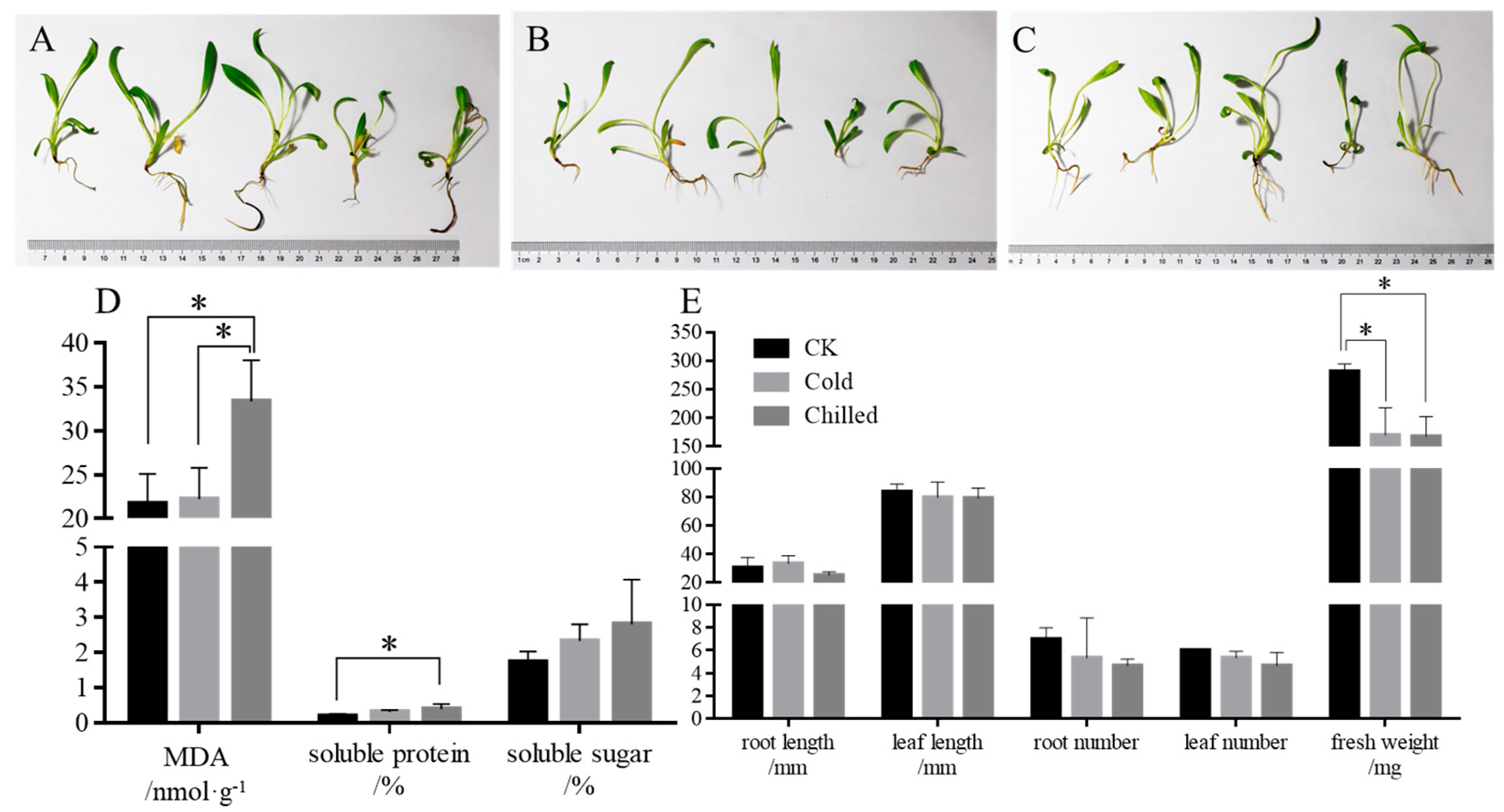
Figure 1. Morphological and physiological profiles of S. involucrata. (AC) The phenotypes of the control (A), cold (B), and chilled (C) group; (D) the content of osmoregulatory substances; *: statistically significant, p < 0.05, N = 3. The color of each bar corresponds to the categories defined in the legend of (E). (E) The growth index of roots, leaves, and fresh weight. *: statistically significant, p < 0.05, N = 5. MDA: malondialdehyde.
Ijms 26 09030 g001
Figure 2. Transcriptome profiles of S. involucrata under low temperatures. (A) Venn diagram of S. involucrata in response to various low-temperature stress treatments. (B) Volcano plot of cold vs. control. (C) Volcano plot of chilled vs. control. (D) Circos bar plot of Gene Ontology (GO) database. classification for DEGs between cold and control. (E) Circos barplot of GO classification for DEGs between chilled and control. (F) Expression analysis of putative DEGs from RNA-seq. N = 3.
Ijms 26 09030 g002
Figure 3. Expression analysis of putative DHNs from S. involucrate in different periods. (A) DHNs expression in seedlings treated at 4 °C/4 °C in different periods. (B) DHNs expression in seedlings treated at 4 °C/0 °C in different periods. N = 3.
Ijms 26 09030 g003
Figure 4. Analysis of SiDHN3 amino acid sequence characteristics. (A) Multiple sequence alignment profile of SiDHN3 amino acid with homologs. (B) Phylogenetic analysis of SiDHN3 and homologous SiDHN amino acid sequences. Blue letters indicate the consensus sequence from the multiple sequence alignment. The red box highlights the identified dehydrin segment.
Ijms 26 09030 g004
Figure 5. Expression analysis and functional validation of SiDHN3. (A) Growth profile of pET32a and pET32a-SiDHN3 strains in various stress environments. (B) Recombinant SiDHN3 expression profiling. (C) CFU of pET32a and pET32a-SiDHN3 strains subjected to various stresses. *: compared with the control group (pET32a), p < 0.05; **: compared with the control group (pET32a), p < 0.01. N = 3. The arrow points to the band corresponding to the target protein, SiDHN3. LB: Luria-Bertani agar. M: ColorMixed Protein Marker 10–180 kDa.
Ijms 26 09030 g005
Figure 6. Functional validation of SiDHN3-truncated derivatives. (A) Growth profile of SiDHN3-truncated derivative strains in various stress environments. (B) CFU of SiDHN3-truncated derivative strains subjected to various stresses. Different lowercase letters indicate significant differences between different habitats at the 0.05 level. ∆F1, ∆F2, ∆S, ∆K1, and ∆K2 are the truncated derivatives that were generated by sequentially removing segments.
Ijms 26 09030 g006
Table 1. Expression of dehydrin genes.
No.Gene idGeneReference Specieslog2 (Cold/Control)log2 (Chilled/Control)
1CL227.Contig10embryogenic cell protein 40-likeC. cardunculus var. scolymus5.455.09
2CL227.Contig45.765.11
3CL227.Contig84.223.95
4CL227.Contig143.883.13
5CL227.Contig62.981.93
6CL1854.Contig4dehydrin Xero 1-like3.634.02
7CL1854.Contig73.584.00
8CL1854.Contig6dehydrin Rab18-like2.912.95
9CL1767.Contig3dehydrin ERD14-like5.406.00
10CL1767.Contig13.624.23
11CL1767.Contig42.733.57
12CL9026.Contig6dehydrinS. involucrata6.015.78
13CL9026.Contig43.093.44
14CL9026.Contig101.703.19
15CL9026.Contig51.543.11
16CL9026.Contig113.073.10
17CL9026.Contig12.142.27
18CL9026.Contig84.886.11
19CL9026.Contig122.342.72
20CL9026.Contig72.111.40
Disclaimer/Publisher’s Note: The statements, opinions and data contained in all publications are solely those of the individual author(s) and contributor(s) and not of MDPI and/or the editor(s). MDPI and/or the editor(s) disclaim responsibility for any injury to people or property resulting from any ideas, methods, instructions or products referred to in the content.