Review

Cold, Hot, and Lethal—The Tumour Microenvironment and the Immunology of Head and Neck Squamous Cell Carcinoma

1
Department of Otorhinolaryngology 3FM CU and UHKV, Charles University, 10034 Prague, Czech Republic
2
Institute of Anatomy of the 1st Faculty of Medicine, Charles University, 12800 Prague, Czech Republic
3
General University Hospital, 12800 Prague, Czech Republic
4
Department of Otorhinolaryngology, Head and Neck Surgery First Faculty of Medicine Charles University and University Hospital Motol, 15000 Prague, Czech Republic
5
Institute of Molecular Genetics of the Czech Academy of Sciences, 14200 Prague, Czech Republic
6
Faculty of Science, Charles University, 12800 Prague, Czech Republic
7
Laboratory of Gene Expression, Institute of Biotechnology of the Czech Academy of Sciences, 25250 Vestec, Czech Republic
*
Author to whom correspondence should be addressed.
Int. J. Mol. Sci. 2025, 26(18), 8844; https://doi.org/10.3390/ijms26188844
Submission received: 19 August 2025 / Revised: 6 September 2025 / Accepted: 8 September 2025 / Published: 11 September 2025
(This article belongs to the Special Issue 25th Anniversary of IJMS: Updates and Advances in Molecular Oncology)

Abstract

Head and neck squamous cell carcinomas (HNSCCs) represent a diverse group of malignancies, both clinically and biologically, with human papillomavirus (HPV) infection playing a significant role. HPV-positive tumours generally tend to have a better prognosis and are driven by oncoproteins E6 and E7. In contrast, HPV-negative tumours typically have a worse prognosis and are often linked to mutations in tumour suppressor genes. HNSCCs exist within a complex environment known as the tumour microenvironment (TME). The TME includes tumour cells, cancer stem cells (CSCs), cancer-associated fibroblasts (CAFs), immune cells, extracellular matrix (ECM), blood vessels, and various signalling molecules. These components support tumour progression, invasion, metastasis, and resistance to treatment. Intercellular signalling within the TME—mediated by cytokines such as IL-6, TGF-b, and galectins—further promotes tumour growth and systemic effects like cachexia. Notably, the TME shares features with granulation tissue during wound healing, supporting the concept of cancer as a chronic, non-resolving wound. Effective therapy must target not only tumour cells but also the dynamic TME.

1. Introduction

Head and neck squamous cell carcinomas (HNSCC) are the sixth most common cancers worldwide, with 50–70% of cases occurring in men aged 60–70 years. They originate from the squamous cell epithelium in the oral and nasal cavities, pharynx, larynx, and skin [1]. HNSCC risk factors include tobacco, alcohol, and human papillomavirus infection (HPV). Based on HPV status, HNSCC can be classified as either HPV-positive or HPV-negative [2]. Information about HPV status, alongside TNM staging (tumour, node, metastasis), serves as the primary prognostic factor for HNSCC. Generally, HPV-positive tumours tend to have a better prognosis. Notably, up to 85% of HPV-positive cases contain HPV-16 or HPV-18.
The pathogenesis of HNSCC primarily involves inhibition of tumour suppressor genes or their proteins (tumour suppressor proteins), particularly p53 and pRb. Their inactivation allows cells to bypass cell-cycle checkpoints, which also usually leads to the accumulation of DNA errors or mutations, and ultimately, these cells can undergo malignant transformation. In HPV-positive tumours, p53 (protein p53) and pRb (Retinoblastoma protein) are inhibited by viral oncoproteins E6 and E7. HPV-positive tumours are also linked with an upregulated production of p16 (protein p16), an immunohistochemical marker routinely used to detect HPV infection. Conversely, in HPV-negative tumours, p53 is inactivated by gene mutation, while pRb is inhibited by cyclin D1 or cyclin D2 and cyclin-dependent kinases (CDK-)4-6 complexes. HPV-negative tumours often exhibit deletion of the CDKN2A gene, which paradoxically correlates with reduced p16 expression and worse prognosis in comparison to HPV-positive tumours [3].
Tumours are highly heterogeneous complexes with structures that vary depending on the specific tumour type. Generally, genetically abnormal malignant cells are surrounded by complex environmental components that constitute what is known as the tumour microenvironment (TME). The TME is crucial for cancer cells; for example, it promotes cancer cell proliferation and migration, which may also contribute to metastatic progression. Moreover, this microenvironment consists of cancer cells, cancer-associated fibroblasts (CAFs), immune cells, endothelial cells, extracellular matrix (ECM), cytokines, chemokines, growth factors, and other elements [4]. These components not only facilitate intercellular tumour communication but also exert systemic effects through molecules such as TNF-α (Tumour Necrosis Factor), TGF-β (Transforming Growth Factor), IL-6, IL-10, and others [5]. Recent studies highlight the increasing expression of proteins that can recognise sugars, such as lectins and their glyco-ligands, in malignancies [6]. These glycoproteins (lectins) can interpret information hidden within structural carbohydrate motifs. Galectins (Gal), which bind beta-galactosides and poly-N-acetyllactosamines, have complex roles: they can regulate pre-mRNA splicing, proliferation, and apoptosis, and facilitate interactions between cells and the ECM. Additionally, galectins are involved in innate immunity. The deregulation of lectin and glyco-ligand (carbohydrate) expression significantly impacts the malignant transformation of the normal squamous epithelium and its differentiation, metastatic potential, and responsiveness to treatment [7]. The so-called glyco-code is a fundamental aspect of tumour biology, encoding biological information through monosaccharide sequences that form oligo- and polysaccharides [8].
With respect to the immunological activity estimated by the spatial distribution of T cells observed in solid tumours, three distinct immune phenotypes are currently recognised [9,10]. The first are inflamed/hot tumours, characterised by abundant intratumoural T cell infiltration. Next are desert/cold tumours, defined by minimal T cell presence. Lastly, there are immune-excluded tumours, in which even rich CD8+ T cells are evident; however, there is an obvious sequestration of these antitumoural lymphocytes within the tumour stroma only.
Over the past decade, pioneering immunotherapeutics, such as anti-CTLA4 (cytotoxic T lymphocyte-associated protein 4) and anti-PD-1 (programmed cell death protein 1) antibodies, have dramatically transformed the landscape of oncology. Immune checkpoint inhibitors (ICIs) have revolutionised cancer treatment. However, durable responses are achieved only in a subtype of treated patients. Primary or acquired resistance to these treatments is frequently seen. Sadly, even if immune cells seem to be abundant in biopsy, tumours can be or become resistant to ICIs due to upregulation of alternative checkpoints (TIM3 (T cell immunoglobulin and mucin-domain containing-3), LAG3 (lymphocyte-activation gene 3), and TIGIT (T cell immunoreceptor with Ig and ITIM domains)) and/or by using non-PD-1/CTLA4-mediated inhibitory pathways [11,12].
In this review, we aim to summarise the immunological factors of TME in HNSCC and elucidate the mechanisms of resistance of these tumours to oncological treatments. Emerging evidence highlights the role of CAFs in orchestrating the immunomodulatory aspects of TMEs, making CAFs the principal contributors to immune evasion and cancer therapy. We believe that research on the TME, and particularly on CAF heterogeneity, may ignite innovative strategies and can even boost the success of existing immunotherapies.

1.1. Tumour Stem Cells

Tumour cell heterogeneity is notable even within a single tumour in one patient, mainly due to diverse gene mutations, particularly so-called driver mutations, which are the primary cause of carcinogenesis initiation. Among these cells, there is a subpopulation that exhibits stem-like properties, known as cancer stem cells (CSCs) [13]. CSCs are poorly differentiated cells that are vital for tissue maintenance and regeneration. They have a distinctive localisation within tumour stroma, surrounded by a specific environment (niche) necessary for their normal function [14]. CSCs are capable of constant cell division, which supports tissue upkeep. Their division is very slow and asymmetric, producing one daughter cell that retains stemness and another that further differentiates. CSCs can actively exclude xenobiotics, including chemotherapeutic agents, from their cytoplasm, often rendering them resistant to conventional cancer therapies [15]. CSCs have been identified even in HNSCC [16,17], and they are implicated in minimal residual disease, tumour recurrence, metastasis, and drug resistance [18]. Somatic mutations accumulating with ageing, combined with impaired DNA repair, drive extensive mutagenesis. As a result, metastatic cells often possess a genetic profile distinct from their primary tumour counterparts. Tumour progression follows a Darwinian evolutionary trajectory, shaped by mutational landscape and microenvironment interactions [19,20]. Therefore, effective oncological therapy should target CSCs directly—either by eliminating their stemness or by modulating the TME to suppress tumour-promoting signals.
So what makes CSCs hot, cold, or lethal? Due to their slow population kinetics, CSCs are naturally resistant to conventional chemotherapy and radiotherapy. Considering novel immunotherapeutic options, immune evasion is a major obstacle for successful cancer treatment in many patients. Why do even these trendy therapeutic modalities fail?
It is well documented that immune privilege is not a general quality of all stem cells [21]. However, it has been confirmed that Lgr5+ stem cells of the bulge hair follicle in skin were entirely resistant to T cell attack, even by fully activated T cells [22]. The immune-privileged status of CSCs is induced and preserved by various mechanisms [23]. Common mechanisms of evasion include impaired antigen presentation caused by mutations or loss of heterozygosity of the major histocompatibility complex class I (MHC-I), which has been implicated in resistance to immune checkpoint inhibitor (ICI) therapy in some cancer types [24]. However, frequent downregulation of MHC-I expression seen in tumours is not always linked to such mutations. Alternatively, MHC-I molecules are in some tumours selectively targeted for lysosomal degradation by an autophagy-dependent mechanism. Notably, inhibition of autophagy, e.g., pharmacologically with the well recognised antimalarial drug chloroquine, restores surface levels of MHC-I and leads to improved antigen presentation [25]. This improves antitumour T cell responses, which can be enhanced therapeutically with anti-PD1 (programmed cell death protein 1 ligand) and anti-CTLA4 antibodies.
Using a model for cutaneous squamous cell carcinoma (SCC), tumours can be effectively challenged by adoptive cytotoxic T cell transfer (ACT)-based immunotherapy [26]. However, it was confirmed that transforming growth factor β (TGF-β)-responding CSCs are superior at resisting therapeutic T cell transfer. Mechanistically, CSCs selectively acquire CD80, a surface ligand previously identified on immune cells, and directly dampen cytotoxic T cell activity. Conversely, upon CTLA4- or TGF-β-blocking immunotherapies or CD80 ablation, CSCs become vulnerable, diminishing tumour relapse after ACT treatment. Therefore, deeper insights into the immunobiology of CSCs are essential in our pursuit to find new therapeutic opportunities that eradicate cancer cells, including CSCs.

1.2. Metastasis

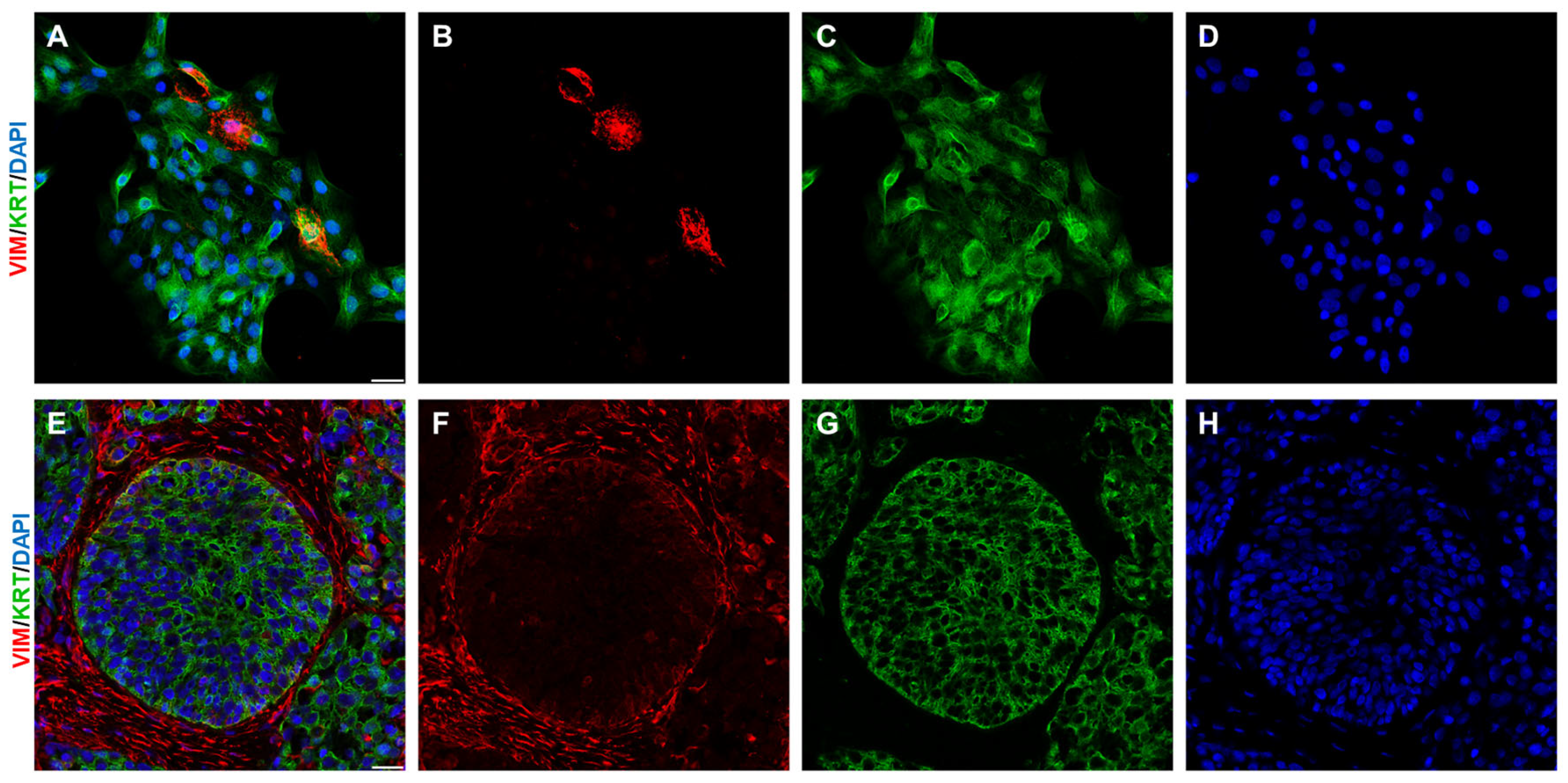
Metastasis is a key characteristic of malignant tumour progression. It is a highly complex process that typically begins with tumour growth, invasion, remodelling of the extracellular matrix, intravasation, and extravasation and ends with the colonisation of distant tissues by cancer cells. A crucial mechanism enabling cancer cells to migrate during metastasis is epithelial–mesenchymal transition (EMT). Colonisation of distant tissue is facilitated by mesenchymal–epithelial transition (MET) [4]. EMT (Figure 1) and MET are opposing processes involved in embryogenesis, regeneration, and metastasis. During EMT, cancer cells gain mesenchymal traits [27]. After undergoing EMT, they lose intercellular adhesions and apico-basal polarity and acquire the ability to migrate and invade, which enables their dissemination to distant tissues where micrometastases form. Metastasis concludes with colonisation, followed by further proliferation of cancer cells, eventually leading to macrometastasis. The course of this process varies depending on the specific type of carcinoma. However, it appears that EMT is not strictly essential for metastasis, as some cancer cells can migrate via amoeboid movement. The main difference between EMT-dependent migration and amoeboid migration is that the latter does not involve enzymatic degradation of the ECM; instead, these cells deform and squeeze through gaps between ECM fibres [28,29]. The exact mechanisms by which tumour cells select their target tissues remain unclear. Nonetheless, factors secreted by cells within the tumour microenvironment seem to contribute to establishing the so-called pre-metastatic niche. This review primarily addresses the issues of the TME. In a historical perspective, Stephen Paget first proposed the famous “seed and soil hypothesis”, highlighting the increased tendency of the stereotypical occurrence of metastasis in certain organs [30]. More recently, the concept of premetastatic niche (PME) was postulated [31] and is noteworthy. The PMN is characterised as a hospitable tissue microenvironment offering a safe harbour for the colonisation of metastatic tumour cells, facilitating their tumour settlement and growth in distant organs. Despite apparent similarities, there is a principal difference. The TME can be gradually crafted by direct contact of cells or via paracrine signalling in situ. To create a PMN, the tumour must act over a long distance; thus, it should already be considered a systemic disease. The exact mechanisms of preferential cancer metastasis to particular body sites remain somewhat elusive, even a century later. Nevertheless, the PMN thus represents a complex microenvironment, with ongoing interplay of numerous cellular and various molecular constituents. Once constituted, the PMN represents a microenvironment with enhanced vascular permeability, prone to angiogenesis, and with notable immunosuppressive and cancer-promoting features [32].
Tumour cells that migrate to form metastasis must be able to survive without a connection to the matrix, since in that situation, many cell types undergo anoikis (a specialised form of apoptosis). Anoikis is an important characteristic of nucleated cells (except leucocytes). For instance, CSCs exhibit resistance to anoikis, which enables their survival within the circulatory system. Multiple underlying mechanisms mediate this unique feature [33,34]. Metastasis represents a critical complication of carcinogenesis, which is often fatal. Therefore, inhibiting metastasis from the primary tumour and inducing its transition into a chronic controlled state offers a promising therapeutic strategy, particularly in patients with inoperable tumours [35].
Figure 1. During EMT, epithelial cells gain mesenchymal traits. This shift in phenotype can be visualised by co-expression of epithelial markers (e.g., keratins (KRT) in C,G) along with mesenchymal markers (e.g., vimentin (VIM) in B,F). In vitro (AD), the epithelial SCC13 squamous cell carcinoma cell line frequently produces vimentin-positive cells (B, vimentin stained in red) on the background of keratin positivity (C, pan-keratin KRT). In vivo (EH, human squamous cell carcinoma tissue), however, such overlap of vimentin (F) and pan-keratin (G) is only rarely seen. Cell nuclei were counterstained by DAPI (D,H). The bar in merged images (A,E) represents 25 mm. The illustration was collated from earlier research listed in Refs. [36,37].
Ijms 26 08844 g001
Metastases are one of the main prognostic markers of HNSCC, mainly spreading to the neck lymph nodes. Neck lymph node metastases reduce survival by 50%. Tumour cells can spread via local invasion and via lymphatic or blood vessels. Metastatic spread in HNSCC is supported by many different mechanisms and interlinked signalling cascades, such as cytokine signalling, involving the activation of TGF-β and CXCR4, which ultimately enhance cell migration and invasion, leading to metastasis. TGF-β can trigger EMT and supports the aggressive phenotype of HNSCC. Moreover, TGF-β controls the activity of genes like cadherin-E or vimentin. These genes are involved in adhesion, movement, and invasion. So the influence of TGF-β can generate tumour cells that are highly mobile and capable of spreading to distant parts of the body [38].
So, what makes metastasis hot, cold, or lethal? There is progressively growing evidence that cancer cells can disseminate from primary lesions much earlier than the classical metastasis models predicted [39]. This is a well known phenomenon in some particular cancer types, namely melanoma. As most morbidity and mortality in solid cancer stems from metastases, it seems critical to prevent the migration of these cells capable of dissemination. This is the theoretical basis of the migrastatic strategy suggested in the last decade [35]. It seems well documented that malignant cells in a mesenchymal-like state can survive using their dormancy programme for extended periods, even years. The mechanisms supporting their dormancy and maintenance of their potential are poorly understood. More recently, it was suggested that, for example, the transcription factor ZFP281 induces transcriptional programmes in disseminated tumour cells that associate with a dormancy signature and phenotype in vivo [40]. Notably, downregulation of ZFP281 in the mouse model leads to a switch to lung metastatic outgrowth. Its human homologue, ZNF281—a zinc-finger transcriptional regulator—has been characterised as an EMT-inducing transcription factor, suggesting its involvement in the regulation of pluripotency, stemness, and cancer [41]. ZNF281 also contributes to organ fibrosis triggered, e.g., by the pro-fibrotic cytokine TGFβ1. Importantly, ZNF281 is also a key regulator of fibroblast activation and myofibroblast differentiation upon fibrotic stimuli. This is a critical step in controlling extracellular matrix composition and remodelling. It seems likely that therapeutic tools regulating these mechanisms of cancer cell dormancy would have a certain therapeutic potential.
If metastasis is not prevented, is there a way to convert its environment into an immunologically “hot” one, with more favourable features for patients? Intralesional interventions for metastases were occasionally used and have proven beneficial in certain patients, even in recent years. Multiple drugs can be injected into metastases, including various chemotherapeutic agents (methotrexate, bleomycin, 5-fluorouracil) [42]. However, due to the immunosuppressive microenvironment established in the metastasis, the release of tumour antigens does not sufficiently elicit systemic immune responses. These procedures are certainly not expected to be curative, but they are frequently associated with favourable effects, e.g., a reduction in the size of the treated and otherwise unresectable metastasis.
Surprisingly, there are well documented benefits of intraleasional administration of drugs which are usually not used in cytotoxic chemotherapy. Among those, rose bengal, a water-soluble xanthine dye historically used in ophthalmology, has more recently been identified as a potential anti-neoplastic agent, particularly in melanoma metastasis treatment [43]. Following intralesional application, tumour cell death stimulates local immune response, and in some patients, it is also associated with tumour necrosis in both adjacent non-injected lesions and occasionally in distant metastases [44]. This suggests a broader effect and involvement of systemic immune response, which was confirmed in studies combining this dye with dendritic cell vaccines [45]. This simple historical dye can also be significantly modified, and loading of rose bengal into vesicle-like constructs of amphiphilic triazine–carbosilane dendrons (dendrimersomes) was tested in the treatment of epidermal carcinomas [46]. This is a remarkable example of drug repurposing and also of a smart redesign of long-known core chemical structures.
More recently, oncolytic viral therapies became another novel category of non-surgical treatment options available [47]. Injected genetically modified viruses (e.g., herpes viruses, coxsackie A21 virus, vaccinia virus) not only induce tumour cell lysis, but their application leads to a remarkable systemic antitumour activity through selective infection of tumour cells. The effect of tumour cell lysis can be further enhanced, e.g., by simultaneous therapy with ICIs. In recent years, there have been numerous oncolytic viral therapies in various stages of development for the treatment of malignant melanoma [48] and other tumours, including SCC [49,50,51]. However, this is still not in routine clinical use; it will be some time before clinicians can engage with viruses used as drugs.
Intriguingly, intratumoral administration of living bacteria can also help ignite an immune response in metastasis. This concept is nothing new, as it has been studied since the nineteenth century [52]. The intralesional application of Mycobacterium bovis (BCG, Bacillus Calmette–Guérin) is a long-known method with some effects in the treatment of several types of tumours [53]. However, intralesional BCG does not usually significantly improve overall survival. Nevertheless, this effect can be enhanced, e.g., by a combination of BCG lysates with thermosensitive hydrogel carriers [53,54]. In patients with metastatic melanoma, intralesional BCG–hydrogel administration was associated with enhanced antigen processing and presentation, maturation of dendritic cells, and a shift towards an M1 phenotype of macrophages and enhanced T cellular responses [55]. This altogether positively correlated with patient survival [53].
Intralesional interventions can also be combined with radiotherapy. Radiotherapy itself can trigger the so-called abscopal effect, where, in some cases, radiotherapy results in a systemic anticancer response clearing even the non-irradiated metastatic lesions at a distance from the primary site of irradiation [56]. The abscopal effect is a rare event in oncology, and it is currently not predictable due to the lack of biomarkers for such a response. Due to the paucity of reliable data, our understanding of the mechanisms eliciting such a response remains vague [57]. There are attempts to combine intralesional therapies with radiation to induce the abscopal effect more regularly. In a mouse model of breast cancer, systemic and abscopal effects were frequently seen when radiotherapy was combined with BCG [58]. This combination seems to reshape the landscape of the immune microenvironment and mitigate leukocyte-like responses by enhancing the infiltration of CD8+ T cells, promoting dendritic cell maturation, reducing the infiltration of immunosuppressive cells, and downregulating the expression of immunosuppressive cytokines. Similarly, following radiotherapy, the intratumoural injection of a genetically attenuated strain of Salmonella coated with antigen-adsorbing cationic polymer nanoparticles extended survival in multiple tumour models in mice, including a model of metastasis and recurrence. The antitumour effects were abrogated by the antibody-mediated depletion of CD8+ T cells, indicating that adaptive immune responses caused systemic tumour regression [59].
It was suggested based on genome-wide CRISPR screening in mice that SFRP2 is a potential stromal regulator of the abscopal effect of radioimmunotherapy. SFRP2 exhibits CAF expression and radioimmunotherapy-mediated upregulation in unirradiated tumours. Serum proteomics reveals that irradiated tumour-secreted PAI-1 triggers the fate transition of distant tumour pericyte cells into SFRP2-high CAFs via the LRP1/p65 axis. Pharmacologically blocking SFRP2 or PAI-1 enhances the abscopal effect in humanised patient-derived xenograft models [60].
However, this is a good example of making the metastasis immunologically “hot” again. Nevertheless, there is an urgent need to acquire more robust data in humans, where the induction of the abscopal effect remains rare and where its mechanisms remain elusive [61].

2. The Tumour Microenvironment and Its Components
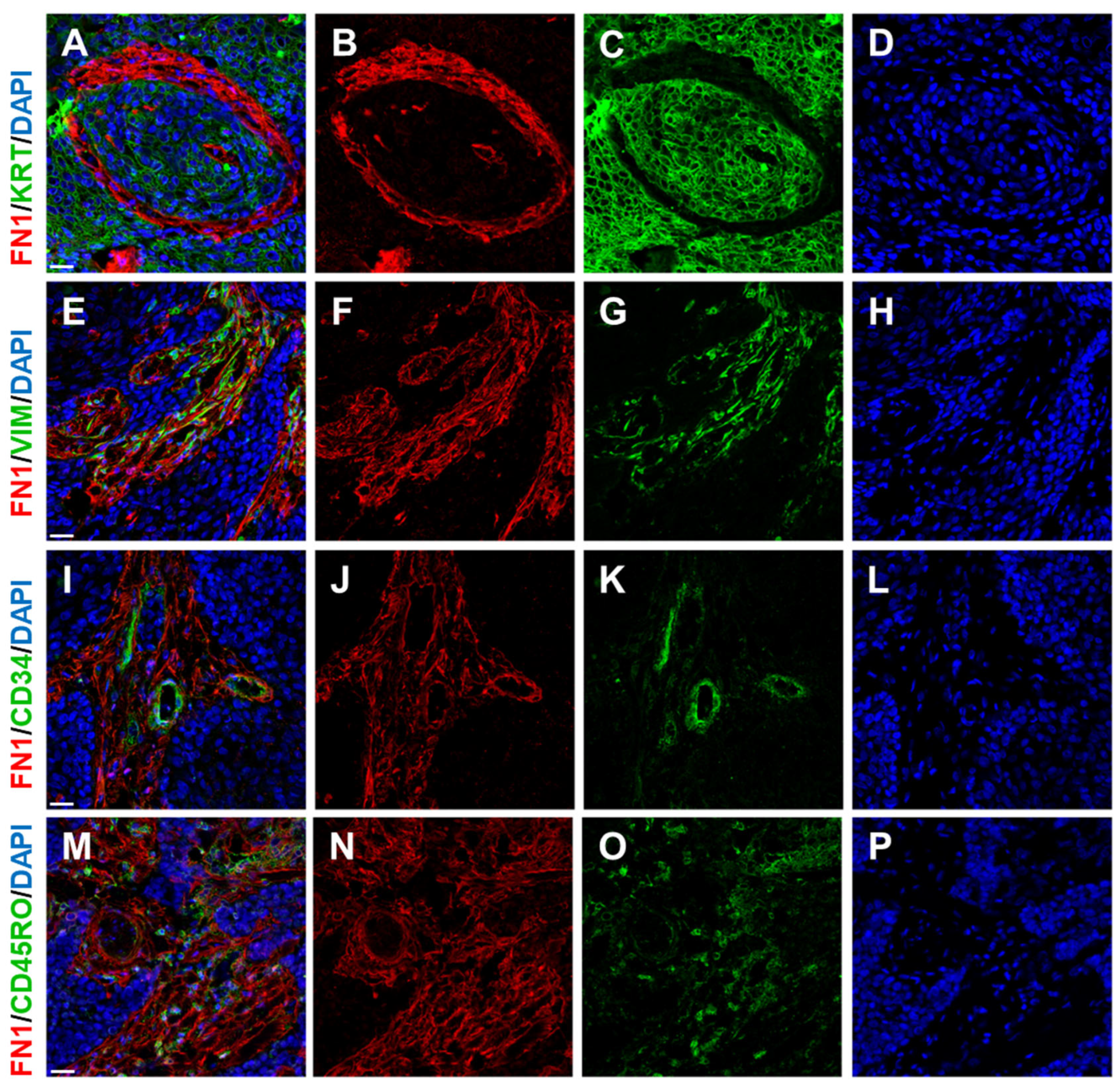
The TME comprises cancer cells, including cancer stem cells, alongside immune cells, vasculature, cancer-associated fibroblasts (CAFs), and the bioactive molecules they secrete (Figure 2) [4].
Figure 2. Human squamous cell carcinoma tissue contains several important components, namely malignant epithelial cells and stromal tissue. The extent of tumour stoma can be visualised by, e.g., fibronectin (FN1 in B,F,J,N) as a representative of its extracellular matrix. Malignant epithelial cells express various keratins (pan-keratin (KRT) in C). The cellular components of tumour stroma mainly represent CAFs expressing vimentin (VIM in G). Delicate stromal neovascularisation can be visualised using, e.g., an antibody against the endothelial marker CD34 (CD34 in K). Scattered immune cells can be visualised by an antibody against CD45RO, a 180 kDa isoform of CD45 (CD45RO in O), which is expressed by most thymocytes, activated memory T cells, granulocytes, and monocytes. Cell nuclei were counterstained by DAPI (D,H,L,P). The bar in merged images (A,E,I,M) represents 25 mm. The illustration was collated from earlier research listed in Refs. [14,62,63].
Ijms 26 08844 g002

2.1. Immune Cells

Immune cells often infiltrate tumours, but they rarely eliminate malignant cells. Tumour cells frequently harbour extensive mutations yet often fail to express recognisable surface antigens, allowing them to evade immune detection. Paradoxically, infiltrating leucocytes often even promote tumour progression. A specific example is tumour-associated macrophages (TAMs). TAMs adopt an M2-like phenotype after being polarised by cells within the tumour ecosystem. They secrete the Interleukins (IL-) IL-4, IL-6, IL-8, and IL-10, TGF-β (Transforming Growth Factor), EGF (Epidermal Growth Factor), and other factors that facilitate tumour growth and cell migration [64]. Myeloid immunosuppressor cells have a similar function [65]. Cytotoxic CD8+ T cells in malignant tumours often express the PD-1 receptor on their surface. Tumour cells express a ligand for this receptor (PD-L1). The binding of the ligand to the receptor functionally exhausts CD8+ lymphocytes and may induce apoptosis. This process leads to immune surveillance failure, promoting tumour growth, invasiveness, and metastasis. Therefore, the use of immunotherapy to treat advanced HNSCC, particularly through immune checkpoint inhibitors, has proven effective in a subset of patients. It has significantly improved the prognosis of many [66,67]. However, it should be noted that treatment is relevant only in patients where the expression of these molecules has been confirmed. Previous chemotherapy may be helpful in these cases [68]. Additionally, PD-1/PD-L1 blockade is associated with the risk of excessive immune activation, which could lead to adverse effects related to inflammation and autoimmune reactions [69]. Immune cells also play a significant role in the pathogenesis and progression of HNSCC. Although immune cells often infiltrate the tumour stroma (especially cytotoxic T lymphocytes), their anticancer functions are considerably impaired due to the active formation of an immunosuppressive TME. This is usually mediated by cytokines such as IL-10, TGF-β, recruitment of Treg lymphocytes, and MDSCs (myeloid-derived suppressor cells), which hinder effective immune activation [70,71].
In the broadest sense, based on immune activity within the TME, we can divide tumours into two groups. The first group consists of so-called hot tumours, characterised by inflammation. These tumours have a high infiltration of immune cells, over-expression of PD-L1, genomic instability, and pre-existing antitumour immune responses. Hot tumours are usually more responsive to immunotherapy, such as ICIs. The opposite group, “cold” tumours, typically show poor response to immunotherapy. This area is particularly promising and may be especially useful in treating malignant tumours [72].
Nevertheless, the immune infiltrate is much more complex, with striking local intratumoral heterogeneity. This is due to genetic and cellular diversity within the invading immune cells. Importantly, this can be a basis for the failure of immunotherapy and an inferior antitumour immune response [73]. It also reflects an intricate network of intercellular regulations and contraregulations among immune cells. For example, regions occupied by increased numbers of CD206-positive macrophages functionally diminish the functions of present T cells. The resulting suboptimal T cell activity in “cold” tumours persists even after immunotherapy. Mechanistically, this effect is attributed to CX3CL1 produced by the cold tumour cells.
Thus, it seems evident that simple quantification of CD8 T cells alone cannot determine the competence of these cells to attack tumour cells. Instead, it was suggested that activated tumour-specific tissue resident memory CD8 T cells can be defined by the co-expression of CD103, CD39, and CD8 in colorectal cancer [74]. The validity of this concept was supported by clinical data, which showed that patients with low numbers of activated TRM cells had a poor prognosis, even with high CD8+ T cell infiltration, and vice versa.
In HNSCCs, combined single-cell RNA sequencing and multiplex immunofluorescence recently identified a new tumour-infiltrating cell (TIL) subtype, CD103+ CD8+ TILs, associated with clinical response. More specifically, CD103+ CD8+ TILs were tumour-reactive T cells, and ICIs targeting PD-1 enhanced CD103+ CD8+ TILs cytotoxicity against tumour growth in vivo [75].
Mutational status should also be considered in immunotherapy outcome prediction. It has been documented that somatic mutation of NFE2L2 leads to Nrf2 activation and radioresistance in HNSCC cells [76]. It is also accompanied by increased recruitment of intratumoral polymorphonuclear myeloid-derived suppressor cells and reduction in M1-polarised macrophages [77]. Interestingly, treatment with the glutaminase inhibitor telaglenastat (CB-839) [78] is capable of overcoming radioresistance. Moreover, it has been hypothesised that it also switches intratumoral myeloid cells to an antitumor phenotype.
Altogether, the tumour ecosystem’s state must be carefully assessed. The overall evaluation appears to benefit from deep molecular profiling and immune phenotyping, and it must be aligned with patient outcome data in the longer term. Of note, approximately 50% of the TME proteome is differentially expressed between cold and hot regions [79].

2.2. Cancer Association Fibroblasts (CAFs)
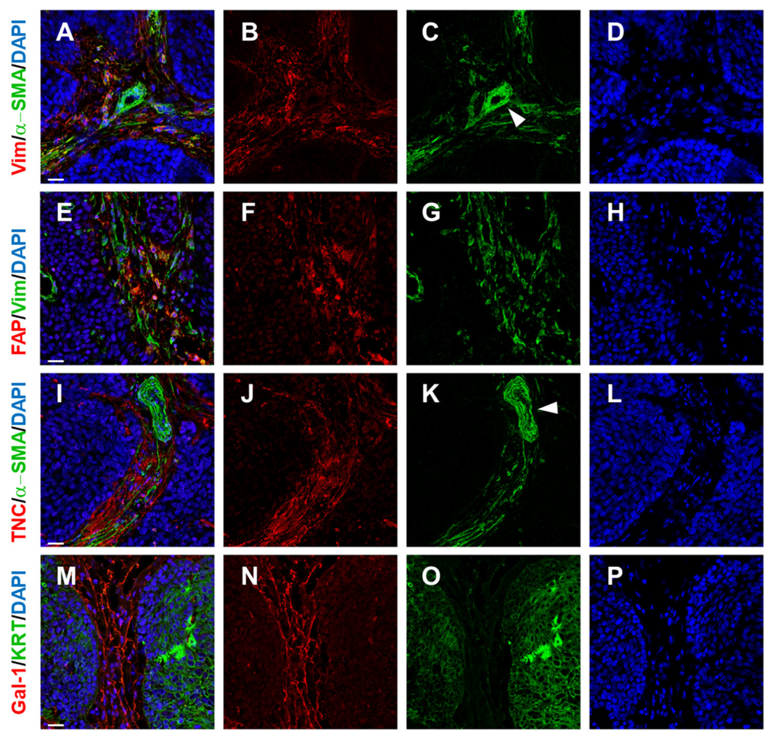
Another important component of the TME is CAFs, whose main functions include ECM production, the synthesis of proteases for ECM biodegradation, and the production of various other bioactive cytokines, chemokines, and growth factors. They often also express α-SMA (α-smooth muscle actin), which is a key feature of myofibroblasts, one of the subpopulations of these cells (Figure 3). Unfortunately, there is no specific feature of CAFs. Genes that are expressed differently in CAFs compared to normal fibroblasts have been identified through transcriptional profiling [80]. The origin of CAFs is not uniform. Local fibroblasts or mesenchymal stem cells are likely primary sources. Notably, they express the topologically appropriate HOX code based on their site of origin in embryogenesis [81,82]. However, there is also evidence that they may originate from, for example, pericytes, endothelial cells, and macrophages [14,83].
Figure 3. Human squamous cell carcinoma tissue and its CAFs (A,E) and ECM (I,M). As typical mesenchymal cells, CAFs express vimentin (Vim in B,G). This marker is not specific to CAFs. Similarly, α-SMA (αSMA in C,K) is also not a universal marker for all fibroblasts in tumour stroma; it is co-expressed with vimentin only in a variable proportion of CAFs (depending on, e.g., tumour type). Of note, α-SMA-positive cells are present in the vascular wall of larger stromal vessels (C,K; vascular structure highlighted by white arrowhead). Similarly, Fibroblast Activation Protein (FAP) is positive in a subset of stromal cells (F). The composition of the extracellular matrix in tumour stroma (I,M) is complex, and the ECM can significantly influence behaviour of malignant cells (pan-keratin KRT staining of malignant cells in O). The ECM contains, e.g., Tenascin C (TNC in J) and also galectin-1 (Gal-1 in N). Cell nuclei were counterstained by DAPI (D,H,L,P). The bar in merged images (A,E,I,M) represents 25 mm. The illustration was collated from earlier research listed in Refs. [7,63].
Ijms 26 08844 g003
Pericytes in tissues retain characteristics of progenitor cells [84] and can differentiate into other cells under normal physiological conditions and into myofibroblasts under pathological fibrotic conditions [85]. It has been suggested that cancer-derived exosomes play an essential role in regulating the transition of pericytes to CAFs. Mechanistically, this exosome-mediated transition is achieved via cargo, including Bone Morphogenetic Proteins (BMP) and PI3K/AKT and MEK/ERK pathway activation [86].
Regarding the endothelium, an increasing number of studies have also implicated the endothelial-to-mesenchymal transition (EndMT) as a principal important topic in cardiac development [87], but also as a potentially disease-driving mechanism in pathologic processes, including organ fibrosis [88] and cancer [81].
It has been documented that the EndMT can be a process initiated by activation of endothelial TGF-β signalling. It has been suggested that it leads to a further increase in TGF-β signalling, thus establishing a positive feedback loop with the EndMT, leading to more EndMT [89]. Such self-intensifying vicious circuits are particularly interesting with respect to cancer progression. Notably, metabolic reprogramming of endothelial cells within the tumour microenvironment also contributes to the EndMT. This can be explained by metabolic modulation. In detail, atypical production of acetate from glucose results in acetylation of the TGF-β receptor ALK5 and SMAD2 and SMAD4, leading to activation and long-term stabilisation of TGF-β signalling [90]. CAFs are potent producers of multiple cytokines in the TME, including TGFb [91]. Besides this, it has been reported that CAF-derived PAI-1 also promotes the EndoMT in cervical SCC. This can also initiate tumour neolymphangiogenesis, which facilitates cancer cell intravasation/extravasation, which in turn promotes lymphatic metastasis [92].
The hypothesis that CAFs may derive from mesenchymal cells is supported by the fact that CAFs in basal cell carcinomas can further differentiate into adipocytes, osteoblasts, or chondrocytes. This suggests that CAFs are multipotent cells, similar to mesenchymal stem cells [93]. In vitro experiments and animal models have indicated that CAFs can also originate from tumour cells through the epithelial-to-mesenchymal transition (EMT) [94,95]. The primary driver of CAF formation from their precursors is TGF-β, with the contribution of Gal-1 [96].
CAFs are not a homogeneous family; rather, they constitute a broad and functionally diverse group. Myofibroblastic CAFs (myCAFs) express α-SMA and actively produce ECM. Occasionally, cells that generate ECM are called matrix-producing CAFs (matCAFs). There is no strict boundary between myCAFs and matCAFs. Fibroblasts known as inflammatory CAFs (iCAFs) promote inflammation, while antigen-presenting CAFs (apCAFs) are active antigen-presenting cells. A subpopulation of CAFs, characterised by high expression of ID genes, is responsive to TGF-β and potentially serves as a precursor for other CAF subpopulations [97,98,99]. CAFs exert a significant paracrine influence on surrounding cells, both normal and tumour cells. CAFs isolated from cutaneous basal cell carcinoma induce keratins 8 and 19, vimentin (a marker of mesenchymal cells), and transcription factor snail in normal keratinocytes in vitro, indicating the low differentiation and EMT of these keratinocytes. Similarly, cancer cells (HNSCC, melanoma) cocultured with normal fibroblasts alter their phenotype to CAFs [3]. However, fibroblast stimulation in this context is usually temporary [100], often explained by epigenetic modifications such as DNA methylation [101]. Some mediators involved in communication between tumour cells and CAFs—such as BMP-4, IGF-2, IL-6, IL-8, CXCL-1, and TGF-β—may be potential therapeutic targets [14]. The impact of CAFs on tumour cells appears to be universal. For instance, CAFs from melanoma not only influence its tumour cells and keratinocytes but may also affect cells from other tumour types, such as glioblastoma or breast cancer. This could hold significance for cancer therapies targeting tumour stroma [37,102].
As already mentioned above, CAFs produce the extracellular matrix (ECM) of the tumour. In addition to commonly found molecules such as collagens and fibronectin, the production of tenascin-C is also important and can be found in both the tumour stroma and the healing wound. Among other things, tenascin-C is responsible for stimulating aggressive growth in many types of tumours [103,104]. The most robust ECM production has been observed in ductal adenocarcinoma of the pancreas, which has a desmoplastic character [105]. This fact may contribute to the high therapeutic resistance of this tumour [106]. Generally, a high number of matCAFs is a sign of a very poor prognosis, no matter the specific type of tumour. Differences in ECM production in comparison to normal tissues influence the mechanical characteristics of cancer tissue, which may also lead to changes in the biological characteristics of the tumour [107].
Collagens are the most structural ECM components of normal and tumour tissues, including HNSCCs [108]. However, CAF-deposited collagen undergoes progressive fibre reorganisation and enzymatic cross-linking in the inflammatory microenvironment [109]. This correlates with tissue stiffening, increased tumour cell invasion, and poor survival [110].
The linearization of collagen can be microscopically visualised as an overall increase in tissue birefringence. Structural patterns of fibrillar collagen organisation are termed “Tumour-Associated Collagen Signatures” and have been used to classify the changes in collagen arrangement that accompany carcinoma progression [109]. This progression is usually most striking at the invasive front of the tumour, where the stiffness of the stroma and cellular mechanosignaling is highest [111]. Intriguingly, the most significant number of infiltrating macrophages and the highest level of TGFβ signalling were found within cells at the invasive front. It seems that the difference in stiffness can also be key to immune infiltration and response [112]. Stiffness can be one of the limiting factors for the access of immune cells to the tumour [113,114], and collagen and collagen degradation products regulate the phenotypes and functions of immune cells via, e.g., collagen-binding endocytotic receptors Endo180 and mannose receptor (CD206) on macrophages [115].
Fibronectin is a glycoprotein with a multitude of functions in normal tissue and in the tumour stroma. It is not a cancer-specific ECM. However, it has been noted that the extracellular matrix protein fibronectin contains a domain that is rarely found in healthy adults and is almost exclusively expressed by newly formed blood vessels in tumours [116]. Similarly, O-glycosylated oncofoetal fibronectin is found in foetal and cancer cells, but usually not in healthy tissues.
It seems that it is not easy to predict the role of fibronecin in tumours [117]. Using in vitro models of HNSCC, it was documented that the ECM type plays a major role in HNSCC cell phenotype, influencing proliferation, migration, and invasion [118]. However, the observed effects of ECMs were cell line-dependent. Fibronectin harbours three repeating units, named type I, II, and III repeats, that contain various binding sites for other molecules, e.g., collagen/gelatin, integrins, and heparin. Polymerisation and pericellular fibronectin assembly associate fibronectin dimers into fibrils, and cell-binding activity enables these fibrils to participate in various physiological and pathological functionalities [119]. As a provisional matrix protein, fibronectin assembly provides a template for the assembly of collagen and integration of other proteins into the tumour ECM, including matrix components involved in TGFβ signalling [119]. In HNSCC, stromal over-expression of fibronectin, determined by immunohistochemistry (Figure 2), was shown to be an independent unfavourable prognostic indicator of overall and disease-free survival [120].
It has been suggested that, in HNSCC, the oncofoetal form of fibronectin is a major and obligate component of the matrix assembled by stromal fibroblasts. In contrast to earlier views, fibronectin was more recently determined to be an independent unfavourable prognostic marker in HNSCC and other cancer types [121]. Mechanistically, cancer cells readily migrate onto fibrillar fibronectin-rich matrices, which is achieved through αvβ6 and α9β1 integrin interaction [120].
Interestingly, the oncofoetal form of fibronectin was demonstrated to be specifically associated with the M2 polarisation of intratumoural macrophages [122]. It has also been suggested that upregulation of the O-glycosylated oncofoetal fibronectin can be linked to the acquisition of a multidrug resistance phenotype in a breast cancer model exposed to chemotherapy [123]. These unusual extracellular matrix glycoproteins can be used as potential therapeutic targets for the treatment of cancer. This underscores the importance of tumour–stroma interaction in the orchestration of the collective mode of migration in HNSCC [118].
Tenascin C is another large matrix glycoprotein with several alternatively spliced forms and binding partners. Tenascin-C protein synthesis is tightly regulated, with widespread protein distribution in embryonic tissues but restricted distribution of tenascin-C in adult tissues. Tenascin-C is also expressed de novo in adults during wound healing [124], inflammatory disorders [125], or in tissue fibrosis [126]. Similar to oncofoetal fibronectin, tenascin C expression is intensely studied in pathologies, including cancer. In HNSCC, intense staining of tenascin C is observed in the tumour stroma (Figure 3), with pronounced immunoreactivity frequently surrounding advancing tumour cell islets [104]. This implies a possible role of Tenascin C in cancer invasion. In a rare genodermatosis known as recessive dystrophic epidermolysis bullosa, a severe genetic disease caused by mutations in the collagen COL7A1 gene, it is associated with fibrotic scarring of the skin, pseudosyndactyly, and an early onset of frequently fatal cutaneous squamous cell carcinoma. In a mouse model post-injury, persistent dermal fibrosis with elevated levels of Collagen I, α-SMA+ myofibroblasts, and Tenascin-C was observed [127]. This altogether makes an excellent environment for highly invasive and frequently fatal SCC [128]. Notably, tenascin C was recently shown to facilitate tumour progression in a carcinogen-induced immunocompetent murine model of oral squamous cell carcinoma by promoting an immune-suppressive stroma [129]. Gal-1 belongs to the family of carbohydrate-binding proteins with a high affinity for β-galactose-containing oligosaccharides [8]. Gal-1 is expressed in many sites under normal and pathological conditions, including SCC (Figure 3). Similar to previous ECM molecules, Gal-1 exerts a timely, precise modulatory role in different stages of wound healing. Conversely, persistent over-expression of Gal-1 enhances angiogenesis and ECM production, leading to keloid scar formation and organ fibrosis, respectively. In tumour stroma, galectin-1 is an essential player in the induction of myCAFs from resident fibroblasts; this effect seems to be TGFβ-independent [96]. Galectins, as a family, are versatile proteins involved in immunomodulation. Evidence suggests that galectins can control the immunoregulatory function of cytokines and chemokines through direct binding. There is evidence that galectin-1 binds, e.g., C-X-C motive chemokine ligand 4 (CXCL4). Regarding immunomodulation, CXCL4 significantly increases the apoptotic activity of Gal-1 on activated CD8+ T cells, while no effect is observed in CD4+ T cells [130]. Of note, Gal-9/CCL5 (Chemokine (C-C motif) ligand 5) heterodimer significantly reduces the Gal-9-induced apoptosis of CD4+ T cells and not of CD8+ T cells. This underscores specific galectin–chemokine interactions and the versatile immunoregulatory function of galectins. In HNSCC, Gal-1 was linked to poor prognosis [63]. Similarly, in gastric cancer, it was suggested that heparanase and Gal-1, which could regulate ECM remodelling and tumour immune microenvironment, contribute to 5-fluorouracil chemoresistance [131].
In conclusion, CAFs play a critical role in HNSCC pathogenesis (e.g., formation of ECM, angiogenesis, and immune and metabolic reprogramming of the TME, impacting metastasis and therapeutic resistance). They possess many molecules and signalling pathways that could be important targets in future oncological therapy [132]. On the other hand, production of CXCL14 in iCAFs is related to better prognosis [98]. Production of inflammation-stimulating cytokines is connected with the change in cell metabolism influenced by hypoxic conditions in tumour stroma and related to the expression of FAP (Fibroblast Activation Protein) in these cells [133].
So, what makes CAFs hot, cold, or lethal? Accumulating evidence has demonstrated that the overall proportion, phenotype, and distribution of CAFs within the tumour microenvironment are important in determining responses to ICIs [134]. This functional clustering of CAFs seems to be a rather universal phenomenon, as evidenced across several types of tumour, including HNSCC [135]. Sadly, heterogeneity in the cellular composition of tumours in individual patients represents a major challenge in modern oncology. CAFs, in the broad sense of the term, have been reported to promote the establishment of an immunologically cold TME [136]. CAFs act both through direct modulation of immune cell phenotypes and indirectly via inhibition of immune cell recruitment and infiltration into the developing tumour [137]. Of note, CAFs of clusters characterised by extracellular matrix proteins and TGFβ signalling are indicative of primary resistance to immunotherapies [134]. In detail, Kieffer and co-workers demonstrated that the abundance of ECM-producing CAFs and TGFβ-regulated CAFs in tumour samples is anticorrelated with CD8+ T cell infiltration but correlated with PD-1+ and CTLA4+ CD4+ T cell content in breast cancer. Moreover, these observations imply the existence of a reciprocal crosstalk between these two CAF clusters and Tregs. TGFβ-regulated CAFs drive T cell exclusion, preventing intratumoral T cells from engaging cancer cells [138]. Collectively, this suggests the development of novel tools for the therapeutic targeting of a specific CAF subpopulation, aiming to enhance clinical responses to existing immunotherapy.

2.3. Blood Vessels

An important part of the TME is also blood vessels, which are formed inside the tumour stroma by a process known as neovascularisation. Neovascularisation and the abundant tumour permeation by capillaries supply nutrients to the tumour. Specifically, targeting new vessels in cancer treatment is one of the most frequently used strategies [14]. Despite abundant neovascularisation, hypoxic areas can be found in solid tumours. These areas are not typically present in healthy tissue. Hypoxia itself promotes resistance of tumour cells to chemotherapy and radiotherapy and promotes tumour progression overall [139].
Overall, low pO2 (partial oxygen pressure) in the tumour microenvironment prior to initiation of therapy indicates a worse prognosis for patients [140]. Some studies suggest that tumour hypoxia is partially related to the amount of haemoglobin in the blood, and thus, tumours in anaemic patients will have lower pO2. Nonetheless, both well and poorly oxygenated tumours can be found in patients with normal haemoglobin levels [141]. Both tumour and nontumour cells in the tumour ecosystem produce factors such as HIF, VEGFA (Vascular Endothelial Growth Factor), IL-6, and IL-8, which stimulate endothelial cell proliferation and thus tumour vascularisation [142,143]. Notably, in HNSCCs, cancer stem cells are localised in perivascular niches and rely on crosstalk with endothelial cells for survival and self-renewal. It was suggested [144] that IL-6 levels in tumour-associated endothelial cells correlate with the tumourigenicity of CSCs, as evidenced in vitro by the p-STAT3 activation, survival, and self-renewal of human CSCs.
There is also evidence acquired in glioblastoma suggesting that hypoxic niches attract and sequester myeloid and cytotoxic T lymphocytes, where they were reprogrammed toward an immunosuppressive state. Mechanistically, it was suggested that chemokine CCL8 and cytokine IL-1β are two hypoxic-niche factors critical for immunosupresive tuning in the tumour mass [145]. On the other hand, the defective wall of tumour capillaries could be exploited for the targeted accumulation of polymeric drugs in the tumour [146].
Tumours are intrinsically heterogeneous, as stated above. The origin of such remarkable cell state diversity remains poorly understood. It was suggested earlier that clinically relevant phenomena such as primary tumour growth and its consequent metastatic dissemination result from distinct cancer cell subpopulations [147]. It was suggested that phenotypic competencies of various tumour cells can be dynamically acquired after exposure to specific niche signals. These cues are associated with a spatially localised perivascular niche. In this context, the phenotype of cancer cells can be acquired through an intercellular communication pathway established by, e.g., endothelial cells. However, this niche is also a shelter for tumour-associated macrophages, which can facilitate cancer progression. In a murine breast cancer model, lymphatic vessel endothelial hyaluronan receptor-1 (LYVE-1)-expressing TAMs form coordinated multicellular nests. In response to IL-6, these TAMs express the immune-suppressive enzyme heme oxygenase-1. Blocking the development of LYVE-1+ TAMs in the mouse model resulted in an increase in CD8+ T cell recruitment to the tumour and enhanced response to chemotherapy [148].
Lastly, there is also a specific subset of CAFs residing in the perivascular niche [134,149]. These CAFs seem to be responsible for the restriction of T cell extravasation and, consequently, also for tumour immune exclusion. In solid tumours, desmoplasia in tumour stroma represents a formidable challenge to immunotherapies and also to endogenous or adoptively transferred T cells. Depletion of FAP+ CAFs in the perivascular niche results in the loss of the structural integrity of the desmoplastic matrix. This reduces myeloid cell accumulation and increases endogenous CD8+ T cell and NK cell infiltration. This makes tumours more susceptible to consequent anti-PD-1 antibody-based therapy [150].

3. Intercellular Signalling in the Tumour Ecosystem
In the context of tumourigenesis, cells within the tumour ecosystem produce cytokines, chemokines, growth factors, and other molecules, such as TGF-β, monocyte chemoattractant protein 1, IL-6 and IL-8, Tumour Necrosis Factor TNF-β, CXCL-1, and VEGF. They promote tumour-stimulating inflammation, tumour cell proliferation and migration, immunosuppression, and metastasis [142].
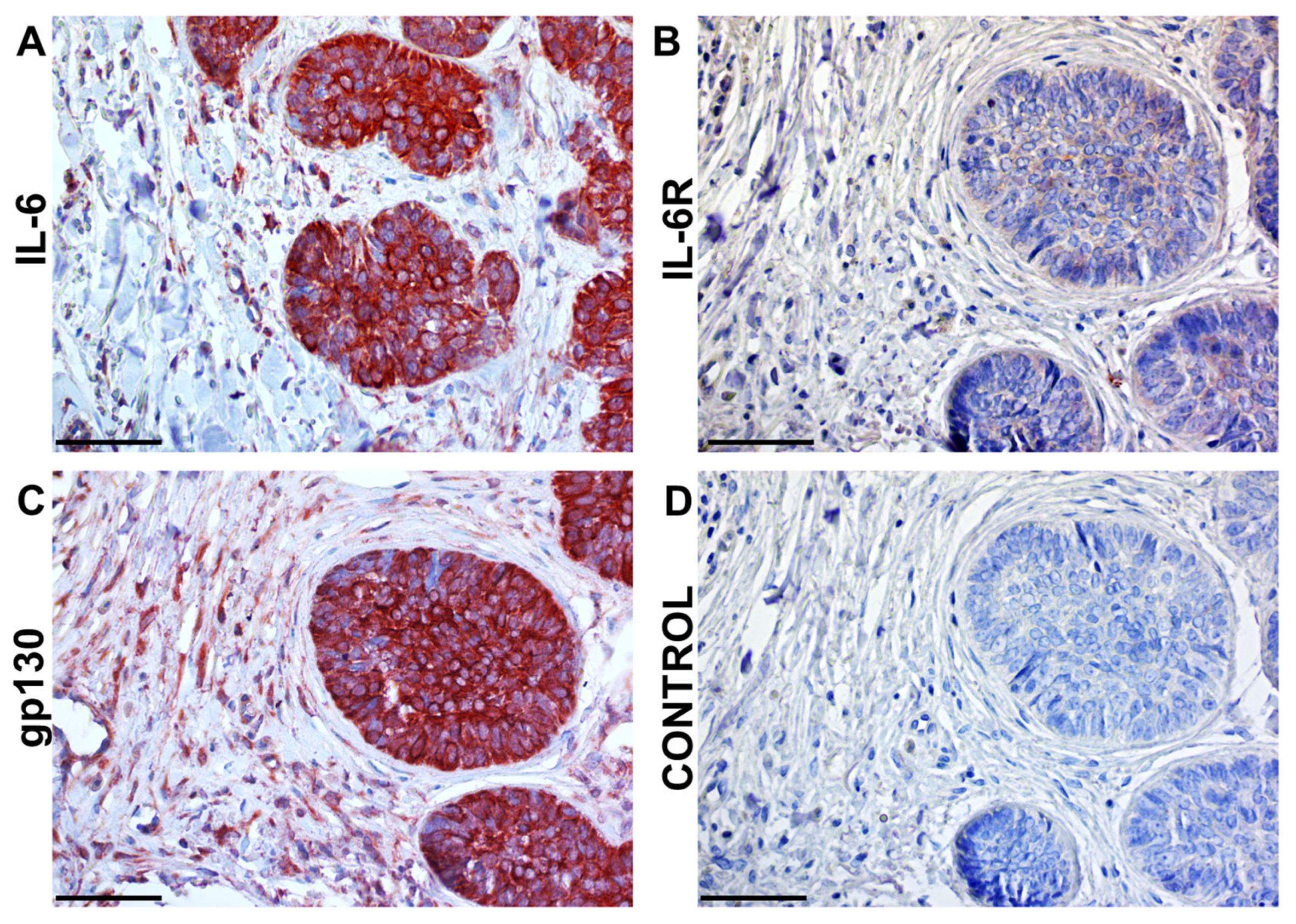
Specifically, IL-6 is a crucial component of the TME (Figure 4) and can be produced by tumour cells themselves or possibly by CAFs and TAMs. A rise in serum IL-6 levels is generally associated with processes such as inflammation, ageing, tumourigenesis, autoimmune disorders, and psychiatric disorders [71]. With ageing, IL-6 levels increase in tissues and the body, thereby promoting the development of chronic inflammation in the elderly. In any case, ageing-associated chronic inflammation is considered one of the leading causes of ageing-related diseases such as neurodegenerative processes, cardiovascular disease, and carcinogenesis [151]. IL-6 has two different modes of action, either through membrane-bound IL-6 receptors or soluble IL-6 receptors. Transmembrane IL-6Rs are associated with the membrane and form complexes with gp130 (Glycoprotein 130). Upon IL-6 binding, IL-6R tyrosine kinase is activated, and STAT3 is stimulated. Transmembrane IL-6Rs are thus involved in anti-inflammatory and pro-carcinogenic signalling. Soluble IL-6R binds IL-6 in the extracellular environment, and subsequently, the whole complex binds to gp130, which is anchored in the membrane (Figure S1). Soluble IL-6Rs promote inflammatory processes [152].
Figure 4. Human squamous cell carcinoma tissue and principal components of the IL-6 signalling cascade. The interleukin IL-6 itself (A) is abundantly produced by cancer cells, as well as in the stroma. However, IL-6R (B) is predominantly detected in the tumour parenchyma. The signal transducer gp130 (C) is broadly present in tumour cells and in stromal populations. Isotype control (D) was used to confirm the specificity of the immunohistochemical reaction using AEC substrate (red). Sections AD were counterstained lightly by Gill’s haematoxillin. The bar represents 100 mm. The illustration was collated from earlier research listed in Ref. [71].
Ijms 26 08844 g004
As described above, the IL-6 signalling cascade offers several candidate targets for therapeutic interventions. In principle, neutralisation of cytokines (e.g., TNFa) via specific antibodies has proved to be a successful strategy in the management of numerous inflammatory and autoimmune conditions [153].
During inflammation, IL-6 levels in the circulation rise dramatically from virtually undetectable in healthy individuals to several-fold higher levels in patients who develop fatal sepsis [154]. Therefore, IL-6 inhibition has also emerged as an attractive therapeutic strategy for translation to the clinic. In principle, the inhibition of IL-6 signalling can be achieved by the application of neutralising antibodies.
However, IL-6 is produced in large quantities by numerous cell types (as evident in Figure 4), and thus, the consumption of neutralising antibodies seems to be a significant hurdle for this strategy. Nevertheless, e.g., the chimeric anti-IL-6 monoclonal antibody siltuximab was approved for patients with idiopathic multicentric Castleman disease [155]. However, the production of IL-6 can be achieved with ease by various anti-inflammatory drugs, including corticoids. This strategy was reported to have some benefits in the management of patients during the COVID-19 pandemic [156,157]. Similarly, the broad expression of gp-130 is high across tissues. Additionally, because many IL-6 family cytokines generally utilise gp130 and gp130 knockout mice are not viable, this increased scepticism towards and hindered the clinical development of gp130 antibodies.
However, small-molecule inhibitors targeting JAKs (Janus Activated Kinases), which act downstream of gp130, represent another FDA-approved approach for JAK/STAT inhibition. Such molecules have recently been used in various indications in the clinic to treat rheumatoid arthritis and other inflammatory diseases [158]. Unfortunately, these JAKs are not exclusively activated by IL-6, so targeting these signalling molecules does not offer selective inhibition of IL-6.
On the contrary, the humanised monoclonal antibodies to IL-6R have reached clinical use. These antibodies prevent the binding of IL-6 to IL-6R. Tocilizumab and sarilumab were approved for the treatment of rheumatoid arthritis [155].
In cancer, the relative scarcity of IL-6 receptors seems to be critical for the precise regulation of the signalling cascade. The amount of IL-6R present at the cell surface determines the responsiveness of the cell to the cytokine and might therefore be decisive in the development of an inflammatory response. It has been shown that IL-6R and gp130 are constitutively internalised independent of IL-6 [159]. This makes the IL-6 receptor, theoretically, a particularly suitable therapeutic target. However, IL-6 receptor blockade has not yielded the anticipated substantial results as an oncological therapy of solid tumours or haematological malignancies [160].
Therefore, new strategies for IL-6 receptor inhibition are in development. For instance, peptides derived from the ABD fragment of streptococcal G protein appear capable of inhibiting both the proliferation and migration of pancreatic ductal adenocarcinoma and melanoma cells [161].
IL-8 is another potent CXC chemokine family member. Following its binding to the receptor, IL-8 (also known as CXCL8) promotes the proliferation, survival, and metastasis of cancer cells and tumour angiogenesis [162]. Together with other chemokines (small chemotactic cytokines), IL-8 shares two types of receptors, CXCR1 and CXCR2. CXCR2 is expressed on a broad spectrum of carcinomas. In SCCs, the expression of both IL-8 and CXCR2 in ESCC was an independent predictor of prognosis [163].
Dual targeting can frequently offer significant benefits in oncology. In experiments where IL-6 and IL-8 were inhibited in melanomas, these interleukins were found to be essential for promoting the activity of CAFs. When IL-6 and IL-8 are deficient in the environment, the invasiveness of melanoma cells is suppressed [164,165].
For example, therapeutic blockade of IL-6 and IL-8 in mice using Tocilizumab (a humanised monoclonal antibody against the α chain of IL-6) and Reparixin (a low-molecular-weight inhibitor of IL-8 signalling) seems to be very effective in suppressing breast cancer metastasis in experiments. Recently, blockade of the IL-6 and IL-8 receptors (IL-6R and IL-8R) with a novel bispecific antibody significantly reduced metastatic burden in multiple preclinical mouse models of cancer [166]. In head and neck squamous cell carcinomas, inhibiting the triple combination of IL-6, IL-8, and CXCL-1 has been shown to be highly effective in reducing tumour cell activity [167].
Another example is MIF (macrophage migration inhibitory factor), which is one of the main pro-inflammatory cytokines. It is produced by several cells and acts as an inhibitor of macrophage migration. At the same time, it promotes macrophage activation, stimulates NO and cytokine production (such as TNF-α, IL-1, and IL-6), and influences cell proliferation and differentiation, which may also contribute to tumourigenesis and metastasis. Evidence of its role in tumourigenesis is shown by the increased MIF mRNA expression found in lymph node metastases from prostate cancer [168]. Nevertheless, it has both positive and negative effects on healing, and its cellular specificity is not fully understood [169]. Furthermore, it also plays an important role as a key enhancer of the effect of oestrogen on wound healing [170]. Oestrogens themselves are crucial regulators of wound repair and healing, especially in the skin. This has been demonstrated through a series of experiments in mice, where the absence of oestrogens led to a significant delay in the healing and repair of skin wounds. More specifically, the contractility of the emerging scar was affected [171].
Macrophage colony-stimulating factor (M-CSF) has become a potentially important factor for targeted cancer therapy. This cytokine is crucial for the differentiation of the monocytic cell line and for encouraging the formation of dense vascular plexuses within tumours. In experiments on murine osteosarcomas, inhibition of M-CSF led to suppression of tumour angiogenesis and lymphangiogenesis [172].
The mentioned systemic therapy using inhibitors of inflammatory molecules causes many side effects. In particular, it adversely affects wound healing, causes immunomodulation, and may lead to further cancer development [170]. Additionally, the ability of tumour cells to develop resistance to oncological drugs plays a significant role. Several preclinical studies have demonstrated that this ability is linked to the activation of cancer-associated NF-κb (Nuclear Factor kappa-light-chain-enhancer of activated B cells). NF-κB inhibits cell death and promotes cell proliferation, angiogenesis, and migration, which likely counteract the cytotoxic effects of chemotherapy and radiotherapy, thereby facilitating resistance development [173]. STAT3 has also been associated with poor responses to cancer therapy. Experimental inhibition of NF-κB and STAT3 in prostate cancer resulted in tumour growth arrest and prevented cancer stem cells from thriving [174]. However, long-term inhibition caused adverse effects such as neutrophilia, liver damage, and acute inflammation, enhanced by IL-1 [175]. IL-1 is crucial for promoting healing by protecting wounds from bacterial infection; nonetheless, its inhibition does not diminish new tissue production [176].
CAFs also produce various factors that influence angiogenesis, including VEGFA, TGF-β, HGF, EGF, and FGFs (Fibroblast Growth Factors). One of the most important factors is VEGFA, which is upregulated in most tumours and plays an indispensable role in their stroma. CAFs also express several receptors, such as Platelet-Derived Growth Factor receptor PDGFR-a and PDGFR-b [177]. They also produce ECM components and enzymes that remodel the ECM, such as NG2 (Neuron-Glial antigen 2), tenascin-C, type I collagen, fibronectin, and matrix metalloproteinases. Nowadays, it is evident that CAFs have an impact on the fate of tumours. The main mechanism of their action is precisely their strong paracrine effect on surrounding cells. The effect of CAFs on tumour cells is to stimulate their proliferation, migration, invasiveness, and EMT, thereby giving tumour cells a stem cell phenotype [178].
Endogenous lectins are proteins distinct from immunoglobulins that specifically recognise saccharide motifs. For example, galectins are highly significant in carcinogenesis and exert complex biological effects, one of their primary functions being the ability to interpret the structure of beta-galactosides [179]. About 15 different galectins have been identified in mammals, 12 of which are present in human cells. Some galectins are broadly distributed across most tissues, while others display more restricted tissue specificity [180]. Notably, they are abundantly expressed in the microenvironment of tumour cells and the granulation tissue during wound healing [181]. Gal-1 and Gal-3 are the most extensively studied galectins in cancer research. Broadly speaking, Galectins 2-15 are involved in tumourigenesis, although their precise roles remain uncertain. Gal-1 expression is regulated by HIF1, which is crucial in shaping the tumour microenvironment [182]. Specifically, upregulation of Gal-1 can significantly influence tumour progression by disrupting cell transformation, proliferation, angiogenesis, cell adhesion, invasiveness, and immunosuppression. Gal-1 is linked to all cancer stages, affecting both tumour cells and stromal cells within the TME [183]. The immunosuppressive effects of Gal-1 involve inhibiting cytotoxic T lymphocytes through binding to their surface via CD4, CD7, CD43, and CD5 [184]. In HNSCC, Gal-1 expression correlates with the presence of αSMA-positive CAFs and is also associated with the expression of several molecules, such as MAP3K2 (Mitogen-Activated Protein Kinase Kinase Kinase 2), TRIM23, PTPLAD1, FUSIP1 (Fused in Sarcoma protein), SLC25A40 (Solute Carrier Family 25 Member 40), Spindlin 1 (SPIN1), and others [63]. Conversely, the interaction of Gal-1 with integrin receptors induces anoikis in tumour cells [185]. Gal-3, produced by the tumour microenvironment, primarily supports cells, including mesenchymal stromal cells. It functions as a regulator of diverse processes crucial for tumourigenesis, such as apoptosis, metastasis, immune surveillance, mRNA splicing, gene expression, and inflammatory responses [186].

4. Similarity Between Cancer and Wound Healing

Several studies indicate similarities between cancer and wound healing (Figure 5). These similarities have been outlined since the 1990s [187]. Specifically, we note the resemblance between the granulation tissue formed during wound healing and the tumour stroma [188,189]. Fibroblasts play a key role in both processes; their activation leads to increased proliferation and enhanced productivity. Indeed, activated fibroblasts produce various growth factors, cytokines, chemotactic molecules, and ECM proteins, supporting the formation of both granulation tissue and tumour stroma [190]. Fibroblasts can also differentiate into myofibroblasts, with the expression of αSMA being a major feature of this change. In a healing wound, myofibroblasts facilitate contraction, thereby reducing the area for re-epithelialisation [191].
Fibroblast-activating protein alpha (FAP-α) and dipeptidyl peptidase IV (DPPIV) are located on the plasma membrane of fibroblasts in abnormally healing wounds or on tumour cell surfaces, where both promote cell invasiveness, tumour growth, and keloid scar formation [188]. It should be noted that FAP-α is not present on normal, healthy adult tissue cells. Therefore, inhibiting FAP-α or DPPIV with antibodies (e.g., sibrotuzumab) is considered a treatment option in oncology. However, studies in small-cell lung cancer and colorectal cancer have yielded disappointing results [178].
Healing is a strictly regulated process that involves four basic phases: hemocoagulation, inflammation, proliferation, and maturation/remodelling [71,192]. At the time of injury, cells at the wound border are activated, and there is stimulation of local migration, proliferation, and differentiation of various cell types. Inflammation plays a vital role in this process, serving as protection against infection and aiding in the clearance of tissue debris [189]. This inflammatory response occurs in both healing wounds and tumour stroma. In healing wounds, inflammation gradually resolves, but in tumours, it does not, and the tumour continues to proliferate [193]. Because of this, tumours are sometimes described as “wounds that never heal” [187]. The subsequent phase is proliferation, characterised by the formation of granulation tissue, which closely resembles tumour stroma in architecture. Within granulation tissue, fibroblasts produce cytokines, chemokines, and growth factors that stimulate angiogenesis and facilitate re-epithelialisation. Both granulation tissue and tumour stroma can encourage poorly differentiated epithelial cells to proliferate. From this perspective, the tumour can be seen as an unending healing process with significant inflammation [157]. Unlike wound healing, platelets—though essential for healing—are not involved in the tumour stroma of solid tumours [71]. Therefore, healing is a tightly regulated process, and its disruption can lead to abnormal outcomes such as hypertrophic or keloid scars [194].
Cells that support healing and the migration of other cells are currently a very intriguing topic. For example, a recent study on tadpole tail regeneration in the Xenopus laevis model used single-cell and spatial transcriptomics to identify a new population of cells that appear only in the early stages of regeneration. These are known as RICs (regeneration-initiating cells). Soon after amputation, RICs emerge at the wound edge and trigger various signalling cascades that regulate the production and breakdown of collagen, laminin, midkine, and thrombospondin. Their activity generally entails remodelling the surrounding ECM and encouraging the migration of cells vital for regeneration. The role of similar supporting cells in the initiation and spread of tumourigenesis in vertebrates is highly likely [189].
So, what are the hot, cold, and lethal aspects of this wound/cancer parallel? Inflammation is an integral part of the healing process. However, if inflammation exceeds the expected duration for any reason, it can result in poor healing [195]. Bacterial populations, especially those forming biofilms, can be one of the possible reasons for such impairment [196]. Numerous studies report the prevalence of biofilm in chronic cutaneous wounds at anywhere between 20 and 100% [197]. In the oral cavity, several chronic conditions associated with bacterial dysbiosis and resulting in poor healing, namely periodontitis [198] and mucosal lichen planus [199], have been found to increase the risk of developing oral SCC. In oral SCC, two prominent oral pathogens, Porphyromonas gingivalis and Fusobacterium nucleatum, can promote tumour progression in mice [200]. It has been observed that these infections lead to the proliferation of oral cancer stem cells [201] and also significantly contribute to the increased invasivity of SCC via promotion of EMT [202]. There is also evidence that the microbiome can adversely interact with cancer therapies [203].
Surprisingly, bacterial colonisation is not always associated with activation of the immune response. In triple-negative breast cancer, it has been shown that Fusobacterium nucleatum not only colonised these tumours but was also found to be responsible for the immunosuppressive tumour microenvironment and tumour metastasis [204]. A similar observation was confirmed in, e.g., colorectal cancer [205]. Once the infection was cleared, e.g., with metronidazole, dead intratumoral bacteria were transformed into immunopotentiators in immunotherapy via the release of pathogen-associated molecular patterns, influencing the subsequent maturation of dendritic cells and subsequent T cell infiltration. Evidence of the multifaceted intratumoural microbiome and its roles is growing exponentially [206,207,208,209].
However, these results should be interpreted cautiously. Several studies pointed out that depletion of gut microbiota with an antibiotic treatment can compromise the anticancer effect of anti-CTLA-4 therapy in mouse models of sarcoma, melanoma, and colon cancer [210]. Similarly, it has been demonstrated that antibiotic administration also compromises the clinical benefits of PD-1-based immunotherapy, both in mice and in humans [211]. Restoration of intestinal microbiota as an adjuvant factor in cancer therapy was investigated in clinical studies [212,213], with somewhat positive outcomes reported. However, it is too early to draw definitive conclusions from these preliminary data acquired on small published cohorts.

5. Systemic Effects of Tumours

The terminal stage of malignant disease is characterised by wasting, cachexia, and psychological changes, including loss of appetite. These life-threatening events seem to be triggered by mediators of cellular communication within the tumour system, particularly a high level of IL-6, which activates STAT3 even in normal cells. This process leads to altered metabolism in white adipose tissue, skeletal muscle, and hepatocytes [214].
Focusing on the brain, an animal experiment showed that IL-6 activates neurons in the area postrema of the brain and, through signalling to other brain regions, causes dopamine deficiency in the nucleus accumbens, leading the animal to become apathetic and refuse food [215]. Similar conditions, such as apathy, depression, and loss of appetite, are also observed in patients with malignant diseases [216,217,218]. In patients with malignant tumours experiencing these symptoms, we assume the same mechanism described above is at work. Patients with malignant tumours often also suffer from a diverse range of paraneoplastic syndromes, which encompass a variety of complications from cutaneous to neurological [192]. These conditions are also triggered by bioactive factors produced by tumours.
In humans, mood disorders are associated with chronic low-grade systemic (sterile) inflammation, with increased plasma levels of pro-inflammatory cytokines like TNF-α and IL-6, which regulate mood behaviour and cognition by influencing neurotransmitter levels [219]. However, the mechanisms underlying this enhanced inflammation are not well understood. In intestinal tumours, the dysbiosis of microbiota can significantly interfere with the immune response and digestion, but also with cortisol regulation and control of neurotransmitters and their metabolism, including serotonin, dopamine, noradrenaline, and gamma-Aminobutyric Acid (GABA) [220]. These neuromediators can not only act locally, but they can also enter circulation. There is a growing body of evidence indicating that modulation of gut microbiota exerts a modulatory influence on depression and exhibits therapeutic promise [221]. Relevant for head and neck cancers, depressive symptoms evaluated in these patients were significantly associated with inflammation. Moreover, both depression and inflammation were associated with early mortality [222].
In many malignant diseases, a broad spectrum of alterations in body metabolism has been documented [223]. In this regard, the liver appears to be a crucial target that warrants closer attention. In recent years, tumour-derived extracellular vesicles were identified as potent mediators of intercellular communication within the cancer microenvironment and also over long distances across the whole organism [224,225]. More closely, exosomes are also crucial in cancer-induced hepatic reprogramming. Of note, this dysregulation of hepatic functions happens commonly even in tumour-free livers of patients who developed extrahepatic metastasis [226]. Liver accumulation of cholesterol, triglycerides, and ceramides is linked to non-alcoholic fatty liver disease [227]. It seems that extracellular vesicles primarily act on Kupffer cells [228] via an IL-1-dependent mechanism. Notably, tumour-originated extracellular vesicles worsen the adverse effects of chemotherapy, including bone marrow suppression and cardiotoxicity. This can be linked to poor tolerance of chemotherapy and a worsened prognosis [226].
Frequently neglected, adipose tissue is a remarkably metabolically active endocrine organ of the human body. It is also a highly relevant topic in cancer. It is well documented that obesity, the abnormal or excessive expansion of white adipose tissue, is a common root for several malignancies [229,230]. Among the adipokines released from this tissue, leptin is recognised as one of the most important mediators of obesity-associated cancers [231]. There is well recognised pro-inflammatory signalling between cancer cells and adipocytes via the insulin/IGF-1 axis and adipokines [232].
Conversely, it is well recognised that exercise can reduce body weight and also reduce obesity-associated cancer risk. Mechanistically, the underlying mechanisms behind the positive effects of exercise remain largely poorly understood, but it was suggested that fibronectin type III domain-containing protein 5 (FNDC5)/Irisin, a hormone released from exercising muscle, could be responsible for this [233]. Further, upon exercise, skeletal muscles react by secreting various types of myokines, namely IL-6, interleukin-8, interleukin-15, brain-derived neurotrophic factor (BDNF), and irisin. These mediators can improve obesity-induced inflammation by stimulating lipolysis of adipose tissues, promoting glucose uptake [234]. This also notably accelerates the browning of white fat. Brown fat tissue is metabolically very different from white adipose tissue in structure and also in function [235].
In a mouse model, it was observed that exposure of tumour-bearing mice to cold conditions markedly inhibited the growth of various types of solid tumours [236]. Mechanistically, it was suggested that upon this cold exposure, brown adipose tissue was activated, which substantially decreased blood glucose and impeded glycolysis-based metabolism in cancer cells. Conversely, feeding on a high-glucose diet under cold exposure restored tumour growth. Notably, in a small pilot human study, mild cold exposure activated brown adipose tissue and mitigated glucose uptake in the tumour tissue [236]. As glucose uptake is essential for cancer glycolysis, this opens the path to novel therapeutic interventions [237].
However, there is another aspect of adipose tissue—the end-stage, cachexia [238]. Cancer cachexia is quite commonly seen in advanced cancer patients. In these individuals, weight loss is a characteristic symptom of cancer cachexia, along with decreased skeletal muscle mass. Despite extensive research, the pathogenesis of cachexia has not been fully understood until now. The onset of cachexia and its gradual further progression are clinically evident. As mentioned earlier, cancer-associated systemic inflammation is one of the critical pathophysiologies of cancer cachexia, resulting in weight loss due to degradation of skeletal muscle and adipose tissue and suppression of appetite. Prevention of weight loss by early nutritional intervention and exercise may also lead to, at least in some patients, continuation of cancer treatment and a more favourable prognosis. In mouse models, inflammatory cytokines such as IL-6 were elevated in mice with cachexia. In mice in which these inflammatory cytokines are suppressed, weight loss did not occur even when cancer progressed [239]. The importance of this aspect in oncology and its relevance to clinical interventions was acknowledged in the Consensus on the definition and diagnosis of sarcopenia (EWGSOP2) and the Global Leadership Initiative on Malnutrition (GLIM) criteria [240,241]. This is of special concern in the elderly, in whom sarcopenia, malnutrition, and cancer cachexia are highly prevalent due to ageing and can adversely impact prognosis in cancer treatment [242]. It is obvious that complex and multifaceted metabolic crosstalk mechanisms represent challenges, but also opportunities, in the treatment of these devastating neoplastic diseases [243].

6. Conclusions

HNSCCs represent a highly heterogeneous group of tumours, both oncologically and biologically, with different etiopathogenesis, often partly based on the presence of HPV infection. HPV-positive tumours tend to have a better prognosis, and their pathogenesis involves the viral oncoproteins E6 and E7. Conversely, HPV-negative tumours usually have a worse prognosis and are often linked to mutations in tumour suppressor genes and reduced p16 expression. These differences significantly influence prognosis and the therapeutic response.
A tumour (including HNSCC) is a complex ecosystem comprising tumour and nontumour cells, with mutual regulation distinct from the body’s normal control systems. Besides primary genetic alterations, the TME plays a vital role in tumour development. The TME includes not only tumour cells but also CSCs, CAFs, immune cells, blood vessels, ECM, and signalling molecules.
CSCs are a key subpopulation with a high capacity for tissue regeneration—including tumour tissue—along with invasiveness and resistance to therapy. They are likely a major cause of recurrence and metastasis. Understanding tumour evolution requires recognising its Darwinian trajectory, driven by selection pressures within the TME.
CAFs play a crucial role in the TME. They are highly heterogeneous and originate from various sources. CAFs can influence the behaviour of surrounding tumour cells and normal cells. They also produce the ECM (e.g., tenascin-C), remodel their environment, and affect angiogenesis and immune responses. Their paracrine effects contribute to tumour progression, EMT, and changes in cell phenotype.
The EMT (epithelial–mesenchymal transition) and its reverse process, MET (mesenchymal–epithelial transition), are vital in the migration and colonisation of distant tissues by tumour cells, which is an important aspect of metastasis—a key process in malignant tumours. Some tumour cells may employ alternative migration mechanisms, such as amoeboid movement. It is important to note that CSCs can survive without attachment to the matrix (they are anoikis-resistant). This facilitates the survival of CSCs in circulation and subsequent metastasis formation.
Intercellular signalling within the TME is essential for the specific functions of cells and mechanisms of tumourigenesis. Molecules like IL-6, IL-8, CXCL1, MIF, or TGF-β are involved in inflammatory, immunoregulatory, and anticancer mechanisms. Cytokines such as IL-6 play a key role in the systemic effects of cancer, including cachexia and neuropsychiatric symptoms. Inhibition of these molecules in preclinical studies shows therapeutic potential; however, transitioning to clinical practice has yet to prove effective.
During tumourigenesis and metastasis, immune cells, especially TAMs, support tumour progression. Targeted oncological therapies inhibiting immune checkpoints (e.g., PD-1/PD-L1) appear promising. However, they are only effective in patients with so-called “hot” tumours. Conversely, they tend to be ineffective in cases of “cold” tumours.
Blood supply through neovascularisation is essential for tumour growth. It is common for hypoxia zones to form within the tumour. Hypoxia stimulates the expression of specific factors, such as HIF and VEGF, which promote angiogenesis and tumour aggressiveness. Furthermore, a hypoxic environment contributes to a poorer response to therapy.
Finally, it is important to note that tumours and wounds share several fundamental similarities—both involve fibroblast activation, ongoing inflammation, granulation tissue formation, and secretion of growth factors. A tumour can be viewed as a state of chronic wound healing that has escaped physiological regulation.
Overall, effective oncological therapy should target not only tumour cells but also the dynamic TME, including CAFs, CSCs, immune cells, and intercellular signals. However, achieving this requires a thorough understanding of the patterns of TME function.

Supplementary Materials

The following supporting information can be downloaded at https://www.mdpi.com/article/10.3390/ijms26188844/s1.

Author Contributions

Conceptualisation, K.S.J. and L.L.; literature research, S.V. and J.N.; clinical oncology data review, M.C. and J.P.; developmental data and healing data review, R.Š.; genetics data review, M.K.; writing—original draft preparation, S.V.; writing—review and editing, K.S.J., R.Š., M.K., and L.L.; visualisation, L.L.; supervision, K.S.J., M.C., and M.K.; funding acquisition, K.S.J., M.K., and R.Š. All authors have read and agreed to the published version of the manuscript.

Funding

This study was supported by the project National Institute for Cancer Research (Programme EXCELES, ID Project No. LX22NPO5102) funded by the European Union–Next Generation EU; the Ministry of Health of the Czech Republic project AZV CR NW24-03-00459; and the Charles University projects COOPERATIO-Onco and -Surgical Disciplines. The ELIXIR CZ research infrastructure (MEYS Grant No LM2023055) is gratefully acknowledged. The project “Centre for Tumour Ecology–Research of the Cancer Microenvironment Supporting Cancer Growth and Spread” (reg. No. CZ.02.1.01/0.0/0.0/16_019/0000785), supported by the Operational Programme Research, Development and Education in the regimen of sustainability, is also appreciated.

Institutional Review Board Statement

Not applicable.

Informed Consent Statement

Not applicable.

Data Availability Statement

No new data were created.

Acknowledgments

Helena Stýblová is appreciated for excellent technical assistance, and Šárka Takáčová for the language revision. During the preparation of this manuscript/study, the author(s) used Biorender for the purposes of producing of Figure 5. The authors have reviewed and edited the output and take full responsibility for the content of this publication.

Conflicts of Interest

The authors declare no conflicts of interest.

Abbreviations

The following abbreviations are used in this manuscript:
a-SMAa-Smooth Muscle Actin
ACTAdoptive Cytotoxic T cell transfer
apCAFsAntigen-Presenting Cancer-Associated Fibroblasts
BMP-4Bone Morphogenetic Protein 4
CAFsCancer-Associated Fibroblasts
CCLChemokine (C-C motif) Ligand
CDCluster of Differentiation
CDK4-6Cyclin-Dependent Kinase 4-6
CDKN2ACyclin-Dependent Kinase inhibitor 2A
CSCsCancer Stem Cells
CTLA-4Cytotoxic T lymphocyte-associated protein 4
CXCLC-X-C motive chemokine ligand
CXCRC-X-C chemokine receptor
DPPIVDipeptidyl-peptidase IV
ECMExtracellular Matrix
EGFEpidermal Growth Factor
EMTEpithelial–Mesenchymal Transition
FAPFibroblast Activation Protein
FGFFibroblast Growth Factor
FUSFused in Sarcoma protein
FUSIP1FUS-Interacting Protein 1
Gal-1Galectin-1
gp130Glycoprotein 130
HGFHepatocyte Growth Factor
HIFHypoxia-Inducible Factor
HNSCCHead and Neck Squamous Cell Carcinoma
HPVHuman Papillomavirus
iCAFsInflammation Cancer-Associated Fibroblasts
ICIsImmune-Checkpoint Inhibitors
IGF-2Insulin-like Growth Factor-2
IL-Interleukin-
JAKsJanus Activated Kinases
MAP3K2Mitogen-Activated Protein Kinase Kinase Kinase 2
matCAFsMatrix Cancer-Associated Fibroblasts
M-CSFMacrophage Colony-Stimulating Factor
MDSCsMyeloid-Derived Suppressor Cells
METMesenchymal–Epithelial Transition
MIFMacrophage Migration Inhibitory Factor
myCAFsMyofibroblastic Cancer-Associated Fibroblasts
NF- κBNuclear Factor kappa-light-chain-enhancer of activated B cells
NG2Neuron-Glial antigen 2
PD1-LProgrammed cell death protein 1 ligand
PD1-RProgrammed cell death protein 1 receptor
PDGFRPlatelet-Derived Growth Factor receptor
pRbRetinoblastoma protein
PTPLAD1Protein Tyrosine Phosphatase-Like A Domain Containing 1
RICsRegeneration-initiating cells
ROCsRegeneration-organising cells
SLC25A40Solute Carrier Family 25 Member 40
SPIN1Spindlin 1
STAT3Signal Transducer and Activator of Transcription 3
TAMsTumour-Associated Macrophages
TGFβTransforming Growth Factor β
TMETumour Microenvironment
TNFaTumour Necrosis Factor α
TNFbTumour Necrosis Factor β
TregRegulatory T cells
TRIM23Tripartite Motive Containing 23
VEGFVascular Endothelial Growth Factor

References

  1. Liu, X.; Gao, X.L.; Liang, X.H.; Tang, Y.L. The Etiologic Spectrum of Head and Neck Squamous Cell Carcinoma in Young Patients. Oncotarget 2016, 7, 66226–66238. [Google Scholar] [CrossRef] [PubMed]
  2. Barsouk, A.; Aluru, J.S.; Rawla, P.; Saginala, K.; Barsouk, A. Epidemiology, Risk Factors, and Prevention of Head and Neck Squamous Cell Carcinoma. Med. Sci. 2023, 11, 42. [Google Scholar] [CrossRef] [PubMed]
  3. Novotný, J.; Bandúrová, V.; Strnad, H.; Chovanec, M.; Hradilová, M.; Šáchová, J.; Šteffl, M.; Grušanović, J.; Kodet, R.; Pačes, V.; et al. Analysis of HPV-Positive and HPV-Negative Head and Neck Squamous Cell Carcinomas and Paired Normal Mucosae Reveals Cyclin D1 Deregulation and Compensatory Effect of Cyclin D2. Cancers 2020, 12, 792. [Google Scholar] [CrossRef] [PubMed]
  4. Birbrair, A. Tumor Microenvironment; Birbrair, A., Ed.; Springer International Publishing: Cham, Germany, 2020; Volume 1225, ISBN 978-3-030-35726-9. [Google Scholar]
  5. Landskron, G.; De La Fuente, M.; Thuwajit, P.; Thuwajit, C.; Hermoso, M.A. Chronic Inflammation and Cytokines in the Tumor Microenvironment. J. Immunol. Res. 2014, 2014, 149185. [Google Scholar] [CrossRef]
  6. Gorelik, E.; Galili, U.; Raz, A. On the Role of Cell Surface Carbohydrates and Their Binding Proteins (Lectins) in Tumor Metastasis. Cancer Metastasis Rev. 2001, 20, 245–277. [Google Scholar] [CrossRef]
  7. Plzák, J.; Smetana, K.; Chovanec, M.; Betka, J. Glycobiology of Head and Neck Squamous Epithelia and Carcinomas. ORL 2005, 67, 61–69. [Google Scholar] [CrossRef]
  8. Solís, D.; Bovin, N.V.; Davis, A.P.; Jiménez-Barbero, J.; Romero, A.; Roy, R.; Smetana, K.; Gabius, H.J. A Guide into Glycosciences: How Chemistry, Biochemistry and Biology Cooperate to Crack the Sugar Code. Biochim. Biophys. Acta Gen. Subj. 2015, 1850, 186–235. [Google Scholar] [CrossRef]
  9. Mellman, I.; Chen, D.S.; Powles, T.; Turley, S.J. The Cancer-Immunity Cycle: Indication, Genotype, and Immunotype. Immunity 2023, 56, 2188–2205. [Google Scholar] [CrossRef]
  10. Joyce, J.A.; Fearon, D.T. T Cell Exclusion, Immune Privilege, and the Tumor Microenvironment. Science 2015, 348, 74–80. [Google Scholar] [CrossRef]
  11. Wang, Y.; Zhang, H.; Liu, C.; Wang, Z.; Wu, W.; Zhang, N.; Zhang, L.; Hu, J.; Luo, P.; Zhang, J.; et al. Immune Checkpoint Modulators in Cancer Immunotherapy: Recent Advances and Emerging Concepts. J. Hematol. Oncol. 2022, 15, 111. [Google Scholar] [CrossRef]
  12. Marin-Acevedo, J.A.; Dholaria, B.; Soyano, A.E.; Knutson, K.L.; Chumsri, S.; Lou, Y. Next Generation of Immune Checkpoint Therapy in Cancer: New Developments and Challenges. J. Hematol. Oncol. 2018, 11, 39. [Google Scholar] [CrossRef]
  13. Muzaffar, A.; Tajudin, A.A.; Syahir, A. A Cutting-Edge Solution to a Gordian Knot? Aptamers Targeting Cancer Stem Cell Markers for Strategic Cancer Therapy. Drug Discov. Today 2025, 30, 104365. [Google Scholar] [CrossRef]
  14. Lacina, L.; Plzak, J.; Kodet, O.; Szabo, P.; Chovanec, M.; Dvorankova, B.; Smetana, K. Cancer Microenvironment: What Can We Learn from the Stem Cell Niche. Int. J. Mol. Sci. 2015, 16, 24094–24110. [Google Scholar] [CrossRef] [PubMed]
  15. Motlík, J.; Klíma, J.; Dvořánková, B.; Smetana, K. Porcine Epidermal Stem Cells as a Biomedical Model for Wound Healing and Normal/Malignant Epithelial Cell Propagation. Theriogenology 2007, 67, 105–111. [Google Scholar] [CrossRef] [PubMed]
  16. Čada, Z.; Bouček, J.; Dvořánková, B.; Chovanec, M.; Plzák, J.; Kodet, R.; Betka, J.; Pinot, G.L.; Gabius, H.J.; Smetana, K. Nucleostemin Expression in Squamous Cell Carcinoma of the Head and Neck. Anticancer Res. 2007, 27, 3279–3284. [Google Scholar]
  17. Fík, Z.; Dvorøánková, B.; Kodet, O.; Bouèek, J.; Betka, J.A.; Betka, J.; André, S.; Gabius, H.J.; Šnajdr, P.; Smetana, K.; et al. Towards Dissecting Molecular Routes of Intercellular Communication in the Tumour Microenvironment: Phenotypic Plasticity of Stem Cell-Associated Markers in Co-Culture (Carcinoma Cell/Fibroblast) Systems. Folia Biol. 2014, 60, 205–212. [Google Scholar] [CrossRef]
  18. Hamburger, A.W.; Salmon, S.E. Primary Bioassay of Human Tumor Stem Cells. Science 1977, 197, 461–463. [Google Scholar] [CrossRef]
  19. Smetana, K.; Lacina, L.; Szabo, P.; Dvoánková, B.; Broẑ, P.; Ŝedo, A. Ageing as an Important Risk Factor for Cancer. Anticancer Res. 2016, 36, 5009–5017. [Google Scholar] [CrossRef]
  20. Lacina, L.; Čoma, M.; Dvořánková, B.; Kodet, O.; Melegová, N.; Gál, P.; Smetana, K. Evolution of Cancer Progression in the Context of Darwinism. Anticancer Res. 2019, 39, 1–16. [Google Scholar] [CrossRef]
  21. Agudo, J. Immune Privilege of Skin Stem Cells: What Do We Know and What Can We Learn? Exp. Dermatol. 2021, 30, 522–528. [Google Scholar] [CrossRef]
  22. Agudo, J.; Park, E.S.; Rose, S.A.; Alibo, E.; Sweeney, R.; Dhainaut, M.; Kobayashi, K.S.; Sachidanandam, R.; Baccarini, A.; Merad, M.; et al. Quiescent Tissue Stem Cells Evade Immune Surveillance. Immunity 2018, 48, 271–285.e5. [Google Scholar] [CrossRef]
  23. Galassi, C.; Musella, M.; Manduca, N.; Maccafeo, E.; Sistigu, A. The Immune Privilege of Cancer Stem Cells: A Key to Understanding Tumor Immune Escape and Therapy Failure. Cells 2021, 10, 2361. [Google Scholar] [CrossRef]
  24. Rodig, S.J.; Gusenleitner, D.; Jackson, D.G.; Gjini, E.; Giobbie-Hurder, A.; Jin, C.; Chang, H.; Lovitch, S.B.; Horak, C.; Weber, J.S.; et al. MHC Proteins Confer Differential Sensitivity to CTLA-4 and PD-1 Blockade in Untreated Metastatic Melanoma. Sci. Transl. Med. 2018, 10, eaar3342. [Google Scholar] [CrossRef] [PubMed]
  25. Yamamoto, K.; Venida, A.; Yano, J.; Biancur, D.E.; Kakiuchi, M.; Gupta, S.; Sohn, A.S.W.; Mukhopadhyay, S.; Lin, E.Y.; Parker, S.J.; et al. Autophagy Promotes Immune Evasion of Pancreatic Cancer by Degrading MHC-I. Nature 2020, 581, 100–105. [Google Scholar] [CrossRef] [PubMed]
  26. Miao, Y.; Yang, H.; Levorse, J.; Yuan, S.; Polak, L.; Sribour, M.; Singh, B.; Rosenblum, M.D.; Fuchs, E. Adaptive Immune Resistance Emerges from Tumor-Initiating Stem Cells. Cell 2019, 177, 1172–1186.e14. [Google Scholar] [CrossRef]
  27. Mani, S.A.; Guo, W.; Liao, M.J.; Eaton, E.N.; Ayyanan, A.; Zhou, A.Y.; Brooks, M.; Reinhard, F.; Zhang, C.C.; Shipitsin, M.; et al. The Epithelial-Mesenchymal Transition Generates Cells with Properties of Stem Cells. Cell 2008, 133, 704–715. [Google Scholar] [CrossRef]
  28. Tolde, O.; Gandalovičová, A.; Křížová, A.; Veselý, P.; Chmelík, R.; Rosel, D.; Brábek, J. Quantitative Phase Imaging Unravels New Insight into Dynamics of Mesenchymal and Amoeboid Cancer Cell Invasion. Sci. Rep. 2018, 8, 12020. [Google Scholar] [CrossRef]
  29. Gandalovičová, A.; Vomastek, T.; Rosel, D.; Brábek, J. Cell Polarity Signaling in the Plasticity of Cancer Cell Invasiveness. Oncotarget 2016, 7, 25022–25049. [Google Scholar] [CrossRef]
  30. Paget, S. THE DISTRIBUTION OF SECONDARY GROWTHS IN CANCER OF THE BREAST. Lancet 1889, 133, 571–573. [Google Scholar] [CrossRef]
  31. Kaplan, R.N.; Riba, R.D.; Zacharoulis, S.; Bramley, A.H.; Vincent, L.; Costa, C.; MacDonald, D.D.; Jin, D.K.; Shido, K.; Kerns, S.A.; et al. VEGFR1-Positive Haematopoietic Bone Marrow Progenitors Initiate the Pre-Metastatic Niche. Nature 2005, 438, 820–827. [Google Scholar] [CrossRef]
  32. Wang, Y.; Jia, J.; Wang, F.; Fang, Y.; Yang, Y.; Zhou, Q.; Yuan, W.; Gu, X.; Hu, J.; Yang, S. Pre-Metastatic Niche: Formation, Characteristics and Therapeutic Implication. Signal Transduct. Target. Ther. 2024, 9, 236. [Google Scholar] [CrossRef]
  33. Shaw, P.; Dey Bhowmik, A.; Gopinatha Pillai, M.S.; Robbins, N.; Dwivedi, S.K.D.; Rao, G. Anoikis Resistance in Cancer: Mechanisms, Therapeutic Strategies, Potential Targets, and Models for Enhanced Understanding. Cancer Lett. 2025, 624, 217750. [Google Scholar] [CrossRef] [PubMed]
  34. Dvoránková, B.; Smetana, K.; Chovanec, M.; Lacina, L.; Stork, J.; Plzáková, Z.; Galovicová, M.; Gabius, H.J. Transient Expression of Keratin 19 Is Induced in Originally Negative Interfollicular Epidermal Cells by Adhesion of Suspended Cells. Int. J. Mol. Med. 2005, 16, 525–531. [Google Scholar] [PubMed]
  35. Gandalovičová, A.; Rosel, D.; Fernandes, M.; Veselý, P.; Heneberg, P.; Čermák, V.; Petruželka, L.; Kumar, S.; Sanz-Moreno, V.; Brábek, J. Migrastatics—Anti-Metastatic and Anti-Invasion Drugs: Promises and Challenges. Trends Cancer 2017, 3, 391–406. [Google Scholar] [CrossRef]
  36. Dvořánková, B.; Smetana, K.; Říhová, B.; Kučera, J.; Mateu, R.; Szabo, P. Cancer-Associated Fibroblasts Are Not Formed from Cancer Cells by Epithelial-to-Mesenchymal Transition in Nu/Nu Mice. Histochem. Cell Biol. 2015, 143, 463–469. [Google Scholar] [CrossRef]
  37. Dvořánková, B.; Szabo, P.; Lacina, L.; Kodet, O.; Matouškové, E.; Smetana, K. Fibroblasts Prepared from Different Types of Malignant Tumors Stimulate Expression of Luminal Marker Keratin 8 in the EM-G3 Breast Cancer Cell Line. Histochem. Cell Biol. 2012, 137, 679–685. [Google Scholar] [CrossRef]
  38. Mastronikolis, N.S.; Delides, A.; Kyrodimos, E.; Piperigkou, Z.; Spyropoulou, D.; Giotakis, E.; Tsiambas, E.; Karamanos, N.K. Insights into Metastatic Roadmap of Head and Neck Cancer Squamous Cell Carcinoma Based on Clinical, Histopathological and Molecular Profiles. Mol. Biol. Rep. 2024, 51, 597. [Google Scholar] [CrossRef]
  39. Klein, C.A. Cancer Progression and the Invisible Phase of Metastatic Colonization. Nat. Rev. Cancer 2020, 20, 681–694. [Google Scholar] [CrossRef]
  40. Nobre, A.R.; Dalla, E.; Yang, J.; Huang, X.; Wullkopf, L.; Risson, E.; Razghandi, P.; Anton, M.L.; Zheng, W.; Seoane, J.A.; et al. ZFP281 Drives a Mesenchymal-like Dormancy Program in Early Disseminated Breast Cancer Cells That Prevents Metastatic Outgrowth in the Lung. Nat. Cancer 2022, 3, 1165–1180. [Google Scholar] [CrossRef]
  41. Laudadio, I.; Bastianelli, A.; Fulci, V.; Carissimi, C.; Colantoni, E.; Palone, F.; Vitali, R.; Lorefice, E.; Cucchiara, S.; Negroni, A.; et al. ZNF281 Promotes Colon Fibroblast Activation in TGFβ1-Induced Gut Fibrosis. Int. J. Mol. Sci. 2022, 23, 10261. [Google Scholar] [CrossRef]
  42. Baeza-Hernández, G.; Cañueto, J. Intralesional Treatments for Invasive Cutaneous Squamous Cell Carcinoma. Cancers 2024, 16, 158. [Google Scholar] [CrossRef]
  43. Lippey, J.; Bousounis, R.; Behrenbruch, C.; McKay, B.; Spillane, J.; Henderson, M.A.; Speakman, D.; Gyorki, D.E. Intralesional PV-10 for in-Transit Melanoma—A Single-Center Experience. J. Surg. Oncol. 2016, 114, 380–384. [Google Scholar] [CrossRef] [PubMed]
  44. Read, T.A.; Smith, A.; Thomas, J.; David, M.; Foote, M.; Wagels, M.; Barbour, A.; Smithers, B.M. Intralesional PV-10 for the Treatment of in-Transit Melanoma Metastases—Results of a Prospective, Non-Randomized, Single Center Study. J. Surg. Oncol. 2018, 117, 579–587. [Google Scholar] [CrossRef] [PubMed]
  45. Zhang, L.; Du, J.; Song, Q.; Zhang, C.; Wu, X. A Novel In Situ Dendritic Cell Vaccine Triggered by Rose Bengal Enhances Adaptive Antitumour Immunity. J. Immunol. Res. 2022, 2022, 1178874. [Google Scholar] [CrossRef]
  46. Sztandera, K.; Gorzkiewicz, M.; Bątal, M.; Arkhipova, V.; Knauer, N.; Sánchez-Nieves, J.; de la Mata, F.J.; Gómez, R.; Apartsin, E.; Klajnert-Maculewicz, B. Triazine–Carbosilane Dendrimersomes Enhance Cellular Uptake and Phototoxic Activity of Rose Bengal in Basal Cell Skin Carcinoma Cells. Int. J. Nanomed. 2022, 17, 1139–1154. [Google Scholar] [CrossRef]
  47. Greig, S.L. Talimogene Laherparepvec: First Global Approval. Drugs 2016, 76, 147–154. [Google Scholar] [CrossRef]
  48. DePalo, D.K.; Zager, J.S. Advances in Intralesional Therapy for Locoregionally Advanced and Metastatic Melanoma: Five Years of Progress. Cancers 2023, 15, 1404. [Google Scholar] [CrossRef]
  49. Pannhausen, J.; Wirtz, J.; Mantwill, K.; Holm, P.-S.; Schwamborn, K.; Jonigk, D.D.; Gschwend, J.E.; Rose, M.; Gaisa, N.T.; Nawroth, R. Oncolytic Virotherapy Provides a Potent Therapy Option for Squamous Bladder Cancer. Sci. Rep. 2025, 15, 13443. [Google Scholar] [CrossRef]
  50. Ogawa, F.; Takaoka, H.; Iwai, S.; Aota, K.; Yura, Y. Combined Oncolytic Virotherapy with Herpes Simplex Virus for Oral Squamous Cell Carcinoma. Anticancer Res. 2008, 28, 3637–3645. [Google Scholar]
  51. Wu, A.; Li, Z.; Wang, Y.; Chen, Y.; Peng, J.; Zhu, M.; Li, Y.; Song, H.; Zhou, D.; Zhang, C.; et al. Recombinant Measles Virus Vaccine RMV-Hu191 Exerts an Oncolytic Effect on Esophageal Squamous Cell Carcinoma via Caspase-3/GSDME-Mediated Pyroptosis. Cell Death Discov. 2023, 9, 171. [Google Scholar] [CrossRef]
  52. Coley, W.B. CONTRIBUTION TO THE KNOWLEDGE OF SARCOMA. Ann. Surg. 1891, 14, 199–220. [Google Scholar] [CrossRef]
  53. Kremenovic, M.; Chan, A.A.; Feng, B.; Bäriswyl, L.; Robatel, S.; Gruber, T.; Tang, L.; Lee, D.J.; Schenk, M. BCG Hydrogel Promotes CTSS-Mediated Antigen Processing and Presentation, Thereby Suppressing Metastasis and Prolonging Survival in Melanoma. J. Immunother. Cancer 2022, 10, e004133. [Google Scholar] [CrossRef] [PubMed]
  54. Chen, Y.; Zhang, L.; Shi, H.; Shen, Z.; Huang, D.; Tang, S.; He, Y.; Wang, G.; Pan, H.; Wang, Z. Thermosensitive Hydrogel Delivery of BCG Lysates and Tumor Antigens: A Novel Strategy for Melanoma Immunoprevention and Therapeutics. Biochem. Biophys. Res. Commun. 2025, 745, 151215. [Google Scholar] [CrossRef] [PubMed]
  55. Lardone, R.D.; Chan, A.A.; Lee, A.F.; Foshag, L.J.; Faries, M.B.; Sieling, P.A.; Lee, D.J. Mycobacterium Bovis Bacillus Calmette–Guérin Alters Melanoma Microenvironment Favoring Antitumor T Cell Responses and Improving M2 Macrophage Function. Front. Immunol. 2017, 8, 965. [Google Scholar] [CrossRef] [PubMed]
  56. Craig, D.J.; Nanavaty, N.S.; Devanaboyina, M.; Stanbery, L.; Hamouda, D.; Edelman, G.; Dworkin, L.; Nemunaitis, J.J. The Abscopal Effect of Radiation Therapy. Future Oncol. 2021, 17, 1683–1694. [Google Scholar]
  57. Rodríguez-Ruiz, M.E.; Vanpouille-Box, C.; Melero, I.; Formenti, S.C.; Demaria, S. Immunological Mechanisms Responsible for Radiation-Induced Abscopal Effect. Trends Immunol. 2018, 39, 644–655. [Google Scholar] [CrossRef]
  58. Yang, H.; Hu, Y.; Kong, D.; Chen, P.; Yang, L. Intralesional Bacillus Calmette–Guérin Injections and Hypo-Fractionated Radiation Synergistically Induce Systemic Antitumor Immune Responses. Int. Immunopharmacol. 2023, 114, 109542. [Google Scholar] [CrossRef]
  59. Wang, W.; Xu, H.; Ye, Q.; Tao, F.; Wheeldon, I.; Yuan, A.; Hu, Y.; Wu, J. Systemic Immune Responses to Irradiated Tumours via the Transport of Antigens to the Tumour Periphery by Injected Flagellate Bacteria. Nat. Biomed. Eng. 2022, 6, 44–53. [Google Scholar] [CrossRef]
  60. Zhang, Y.-P.; Guo, Z.-Q.; Cai, X.-T.; Rong, Z.-X.; Fang, Y.; Chen, J.-Q.; Zhuang, K.-M.; Ruan, M.-J.; Ma, S.-C.; Lin, L.-Y.; et al. PAI-1-Driven SFRP2high Cancer-Associated Fibroblasts Hijack the Abscopal Effect of Radioimmunotherapy. Cancer Cell 2025, 43, 856–874.e9. [Google Scholar] [CrossRef]
  61. Vidovic, D.; Helyer, L.K.; Pasternak, S.; Giacomantonio, C.A. Abscopal Responses in Patients with Metastatic Melanoma Involving Skin and Subcutaneous Tissues Treated with Intralesional IL2 plus BCG. Front. Oncol. 2023, 13, 1160269. [Google Scholar] [CrossRef]
  62. Plzák, J.; Lacina, L.; Chovanec, M.; Dvořánková, B.; Szabo, P.; Čada, Z.; Smetana, K. Epithelial-Stromal Interaction in Squamous Cell Epithelium-Derived Tumors: An Important New Player in the Control of Tumor Biological Properties. Anticancer Res. 2010, 30, 455–462. [Google Scholar]
  63. Valach, J.; Fík, Z.; Strnad, H.; Chovanec, M.; Plzák, J.; Čada, Z.; Szabo, P.; Šáchová, J.; Hroudová, M.; Urbanová, M.; et al. Smooth Muscle Actin-Expressing Stromal Fibroblasts in Head and Neck Squamous Cell Carcinoma: Increased Expression of Galectin-1 and Induction of Poor Prognosis Factors. Int. J. Cancer 2012, 131, 2499–2508. [Google Scholar] [CrossRef]
  64. Liu, L.; Li, Y.; Li, B. Interactions between Cancer Cells and Tumor-Associated Macrophages in Tumor Microenvironment. Biochim. Biophys. Acta (BBA)—Rev. Cancer 2025, 1880, 189344. [Google Scholar] [CrossRef]
  65. Kurt, F.G.O.; Lasser, S.; Arkhypov, I.; Utikal, J.; Umansky, V. Enhancing Immunotherapy Response in Melanoma: Myeloid-Derived Suppressor Cells as a Therapeutic Target. J. Clin. Investig. 2023, 133, e170762. [Google Scholar] [CrossRef]
  66. von der Grün, J.; Rödel, F.; Brandts, C.; Fokas, E.; Guckenberger, M.; Rödel, C.; Balermpas, P. Targeted Therapies and Immune-Checkpoint Inhibition in Head and Neck Squamous Cell Carcinoma: Where Do We Stand Today and Where to Go? Cancers 2019, 11, 472. [Google Scholar] [CrossRef]
  67. Liu, J.; Chen, Z.; Li, Y.; Zhao, W.; Wu, J.B.; Zhang, Z. PD-1/PD-L1 Checkpoint Inhibitors in Tumor Immunotherapy. Front. Pharmacol. 2021, 12, 731798. [Google Scholar] [CrossRef] [PubMed]
  68. Zhang, B.; Song, Y.; Min, Q.; Cheng, W.; Wang, J.; Fu, Y.; Yin, J. The Administration Sequences of Immune Checkpoint Inhibitors and Chemotherapy Cause Discrete Efficacy When Treating Non-Small Cell Lung Cancer: A Retrospective Study. Front. Immunol. 2025, 16, 1579420. [Google Scholar] [CrossRef] [PubMed]
  69. Carter, S.; Wigmore, T. Immunotherapy on ICU: A Narrative Review. Anaesthesia 2025, 80, 299–310. [Google Scholar] [CrossRef] [PubMed]
  70. Curry, J.M.; Sprandio, J.; Cognetti, D.; Luginbuhl, A.; Bar-Ad, V.; Pribitkin, E.; Tuluc, M. Tumor Microenvironment in Head and Neck Squamous Cell Carcinoma. Semin. Oncol. 2014, 41, 217–234. [Google Scholar] [CrossRef]
  71. Španko, M.; Strnadová, K.; Pavlíček, A.J.; Szabo, P.; Kodet, O.; Valach, J.; Dvořánková, B.; Smetana, K.; Lacina, L. Il-6 in the Ecosystem of Head and Neck Cancer: Possible Therapeutic Perspectives. Int. J. Mol. Sci. 2021, 22, 11027. [Google Scholar] [CrossRef]
  72. Wang, L.; Geng, H.; Liu, Y.; Liu, L.; Chen, Y.; Wu, F.; Liu, Z.; Ling, S.; Wang, Y.; Zhou, L. Hot and Cold Tumors: Immunological Features and the Therapeutic Strategies. MedComm 2023, 4, e343. [Google Scholar] [CrossRef]
  73. Tanaka, M.; Lum, L.; Hu, K.H.; Chaudhary, P.; Hughes, S.; Ledezma-Soto, C.; Samad, B.; Superville, D.; Ng, K.; Chumber, A.; et al. Tumor Cell Heterogeneity Drives Spatial Organization of the Intratumoral Immune Response. J. Exp. Med. 2025, 222, e20242282. [Google Scholar] [CrossRef]
  74. Talhouni, S.; Fadhil, W.; Mongan, N.P.; Field, L.; Hunter, K.; Makhsous, S.; Maciel-Guerra, A.; Kaur, N.; Nestarenkaite, A.; Laurinavicius, A.; et al. Activated Tissue Resident Memory T-Cells (CD8+CD103+CD39+) Uniquely Predict Survival in Left Sided “Immune-Hot” Colorectal Cancers. Front. Immunol. 2023, 14, 1057292. [Google Scholar] [CrossRef]
  75. Ren, S.; Lan, T.; Wu, F.; Chen, S.; Jiang, X.; Huo, C.; Li, Z.; Xie, S.; Wu, D.; Wang, R.; et al. Intratumoral CD103+ CD8+ T Cells Predict Response to Neoadjuvant Chemoimmunotherapy in Advanced Head and Neck Squamous Cell Carcinoma. Cancer Commun. 2023, 43, 1143–1163. [Google Scholar] [CrossRef]
  76. Patel, R.; Saab, K.; Luo, L.; Ma, Y.; Osman, R.A.; Williams, N.T.; Everitt, J.; Zelazowski, M.J.; Castro, P.; Decker, W.K.; et al. Nrf2 Hyperactivation as a Driver of Radiotherapy Resistance and Suppressed Antitumor Immunity in Head and Neck Squamous Cell Carcinoma. Clin. Cancer Res. 2025; ahead of print. [Google Scholar] [CrossRef]
  77. Guan, L.; Nambiar, D.K.; Cao, H.; Viswanathan, V.; Kwok, S.; Hui, A.B.; Hou, Y.; Hildebrand, R.; von Eyben, R.; Holmes, B.J.; et al. NFE2L2 Mutations Enhance Radioresistance in Head and Neck Cancer by Modulating Intratumoral Myeloid Cells. Cancer Res. 2023, 83, 861–874. [Google Scholar] [CrossRef]
  78. Cederkvist, H.; Kolan, S.S.; Wik, J.A.; Sener, Z.; Skålhegg, B.S. Identification and Characterization of a Novel Glutaminase Inhibitor. FEBS Open Bio 2022, 12, 163–174. [Google Scholar] [CrossRef]
  79. Grünwald, B.T.; Devisme, A.; Andrieux, G.; Vyas, F.; Aliar, K.; McCloskey, C.W.; Macklin, A.; Jang, G.H.; Denroche, R.; Romero, J.M.; et al. Spatially Confined Sub-Tumor Microenvironments in Pancreatic Cancer. Cell 2021, 184, 5577–5592.e18. [Google Scholar] [CrossRef]
  80. Ma, C.; Yang, C.; Peng, A.; Sun, T.; Ji, X.; Mi, J.; Wei, L.; Shen, S.; Feng, Q. Pan-Cancer Spatially Resolved Single-Cell Analysis Reveals the Crosstalk between Cancer-Associated Fibroblasts and Tumor Microenvironment. Mol. Cancer 2023, 22, 170. [Google Scholar] [CrossRef]
  81. Pfeiferová, L.; Španko, M.; Šáchová, J.; Hradilová, M.; Pienta, K.J.; Valach, J.; Machoň, V.; Výmolová, B.; Šedo, A.; Bušek, P.; et al. The HOX Code of Human Adult Fibroblasts Reflects Their Ectomesenchymal or Mesodermal Origin. Histochem Cell Biol 2025, 163, 38. [Google Scholar] [CrossRef]
  82. Ciszewski, W.M.; Wawro, M.E.; Sacewicz-Hofman, I.; Sobierajska, K. Cytoskeleton Reorganization in EndMT—The Role in Cancer and Fibrotic Diseases. Int. J. Mol. Sci. 2021, 22, 11607. [Google Scholar] [CrossRef]
  83. Jia, H.; Chen, X.; Zhang, L.; Chen, M. Cancer Associated Fibroblasts in Cancer Development and Therapy. J. Hematol. Oncol. 2025, 18, 36. [Google Scholar] [CrossRef]
  84. Farrington-Rock, C.; Crofts, N.J.; Doherty, M.J.; Ashton, B.A.; Griffin-Jones, C.; Canfield, A.E. Chondrogenic and Adipogenic Potential of Microvascular Pericytes. Circulation 2004, 110, 2226–2232. [Google Scholar] [CrossRef]
  85. Lin, S.-L.; Kisseleva, T.; Brenner, D.A.; Duffield, J.S. Pericytes and Perivascular Fibroblasts Are the Primary Source of Collagen-Producing Cells in Obstructive Fibrosis of the Kidney. Am. J. Pathol. 2008, 173, 1617–1627. [Google Scholar] [CrossRef]
  86. Ning, X.; Zhang, H.; Wang, C.; Song, X. Exosomes Released by Gastric Cancer Cells Induce Transition of Pericytes Into Cancer-Associated Fibroblasts. Med. Sci. Monit. 2018, 24, 2350–2359. [Google Scholar] [CrossRef]
  87. Xu, Y.; Kovacic, J.C. Endothelial to Mesenchymal Transition in Health and Disease. Annu. Rev. Physiol. 2023, 85, 245–267. [Google Scholar] [CrossRef]
  88. Di Benedetto, P.; Ruscitti, P.; Berardicurti, O.; Vomero, M.; Navarini, L.; Dolo, V.; Cipriani, P.; Giacomelli, R. Endothelial-to-Mesenchymal Transition in Systemic Sclerosis. Clin. Exp. Immunol. 2021, 205, 12–27. [Google Scholar] [CrossRef]
  89. Schwartz, M.A.; Vestweber, D.; Simons, M. A Unifying Concept in Vascular Health and Disease. Science 2018, 360, 270–271. [Google Scholar] [CrossRef]
  90. Zhu, X.; Wang, Y.; Soaita, I.; Lee, H.-W.; Bae, H.; Boutagy, N.; Bostwick, A.; Zhang, R.-M.; Bowman, C.; Xu, Y.; et al. Acetate Controls Endothelial-to-Mesenchymal Transition. Cell Metab. 2023, 35, 1163–1178.e10. [Google Scholar] [CrossRef]
  91. O’Fee, K.; Burley, A.; Stewart, S.; Wilkins, A. Targeting Cancer-Associated Fibroblasts (CAFs) to Optimize Radiation Responses. Cancer J. 2025, 31, e0776. [Google Scholar] [CrossRef]
  92. Wei, W.-F.; Zhou, H.-L.; Chen, P.-Y.; Huang, X.-L.; Huang, L.; Liang, L.-J.; Guo, C.-H.; Zhou, C.-F.; Yu, L.; Fan, L.-S.; et al. Cancer-Associated Fibroblast-Derived PAI-1 Promotes Lymphatic Metastasis via the Induction of EndoMT in Lymphatic Endothelial Cells. J. Exp. Clin. Cancer Res. 2023, 42, 160. [Google Scholar] [CrossRef]
  93. Szabó, P.; Kolář, M.; Dvořánková, B.; Lacina, L.; Štork, J.; Vlček, Č.; Strnad, H.; Tvrdek, M.; Smetana, K. Mouse 3T3 Fibroblasts under the Influence of Fibroblasts Isolated from Stroma of Human Basal Cell Carcinoma Acquire Properties of Multipotent Stem Cells. Biol. Cell 2011, 103, 233–248. [Google Scholar] [CrossRef]
  94. Petersen, O.W.; Nielsen, H.L.; Gudjonsson, T.; Villadsen, R.; Rank, F.; Niebuhr, E.; Bissell, M.J.; Rønnov-Jessen, L. Epithelial to Mesenchymal Transition in Human Breast Cancer Can Provide a Nonmalignant Stroma. Am. J. Pathol. 2003, 162, 391–402. [Google Scholar] [CrossRef]
  95. Smetana, K.; Dvoránková, B.; Lacina, L.; Cada, Z.; Vonka, V. Human Hair Follicle and Interfollicular Keratinocyte Reactivity to Mouse HPV16-Transformed Cells: An in Vitro Study. Oncol. Rep. 2008, 20, 75–80. [Google Scholar] [CrossRef]
  96. Dvorankova, B.; Szabo, P.; Lacina, L.; Gal, P.; Uhrova, J.; Zima, T.; Kaltner, H.; André, S.; Gabius, H.J.; Sykova, E.; et al. Human Galectins Induce Conversion of Dermal Fibroblasts into Myofibroblasts and Production of Extracellular Matrix: Potential Application in Tissue Engineering and Wound Repair. Cells Tissues Organs 2011, 194, 469–480. [Google Scholar] [CrossRef]
  97. Novotný, J.; Strnadová, K.; Dvořánková, B.; Kocourková, Š.; Jakša, R.; Dundr, P.; Pačes, V.; Smetana, K.; Kolář, M.; Lacina, L. Single-Cell RNA Sequencing Unravels Heterogeneity of the Stromal Niche in Cutaneous Melanoma Heterogeneous Spheroids. Cancers 2020, 12, 3324. [Google Scholar] [CrossRef]
  98. Chen, B.; Chan, W.N.; Xie, F.; Mui, C.W.; Liu, X.; Cheung, A.H.K.; Lung, R.W.M.; Chow, C.; Zhang, Z.; Fang, C.; et al. The Molecular Classification of Cancer-associated Fibroblasts on a Pan-cancer Single-cell Transcriptional Atlas. Clin. Transl. Med. 2023, 13, e1516. [Google Scholar] [CrossRef]
  99. Wan, Y.; Hu, Q.; Sun, K.; Shi, J.; Liu, L.; Zhang, X.; Huang, J.; Gong, C.; Liu, J.; Wang, H.; et al. Heterogenous Cancer-Associated Fibroblasts Related Tumor Microenvironment Marked by CD10/KLF4/TIAM1 Were Identified in Pancreatic Adenocarcinoma by Integrated Transcriptomics. Front. Immunol. 2025, 16, 1557698. [Google Scholar] [CrossRef]
  100. Szabo, P.; Valach, J.; Smetana, K.; Dvoránková, B. Comparative Analysis of IL-8 and CXCL-1 Production by Normal and Cancer Stromal Fibroblasts. Folia Biol. 2013, 59, 134–137. [Google Scholar] [CrossRef]
  101. Jiang, L.; Gonda, T.A.; Gamble, M.V.; Salas, M.; Seshan, V.; Tu, S.; Twaddell, W.S.; Hegyi, P.; Lazar, G.; Steele, I.; et al. Global Hypomethylation of Genomic DNA in Cancer-Associated Myofibroblasts. Cancer Res. 2008, 68, 9900–9908. [Google Scholar] [CrossRef]
  102. Trylcova, J.; Busek, P.; Smetana, K.; Balaziova, E.; Dvorankova, B.; Mifkova, A.; Sedo, A. Effect of Cancer-Associated Fibroblasts on the Migration of Glioma Cells in Vitro. Tumor Biol. 2015, 36, 5873–5879. [Google Scholar] [CrossRef]
  103. Berndt, A.; Richter, P.; Kosmehl, H.; Franz, M. Tenascin-C and Carcinoma Cell Invasion in Oral and Urinary Bladder Cancer. Cell Adh. Migr. 2015, 9, 105–111. [Google Scholar] [CrossRef]
  104. Zivicova, V.; Gal, P.; Mifkova, A.; Novak, S.; Kaltner, H.; Kolar, M.; Strnad, H.; Sachova, J.; Hradilova, M.; Chovanec, M.; et al. Detection of Distinct Changes in Gene-Expression Profiles in Specimens of Tumors and Transition Zones of Tenascin-Positive/-Negative Head and Neck Squamous Cell Carcinoma. Anticancer Res. 2018, 38, 1279–1290. [Google Scholar] [CrossRef]
  105. Novák, Š.; Kolář, M.; Szabó, A.; Vernerová, Z.; Lacina, L.; Strnad, H.; Šáchová, J.; Hradilová, M.; Havránek, J.; Španko, M.; et al. Desmoplastic Crosstalk in Pancreatic Ductal Adenocarcinoma Is Reflected by Different Responses of Panc-1, MIAPaCa-2, PaTu-8902, and CAPAN-2 Cell Lines to Cancer-Associated/Normal Fibroblasts. Cancer Genom. Proteom. 2021, 18, 221–243. [Google Scholar] [CrossRef]
  106. Anastasia, A.; Formenti, L.; Ostano, P.; Minoli, L.; Resovi, A.; Morosi, L.; Fioravanti, C.; Micotti, E.; Matteo, C.; Scanziani, E.; et al. Stroma Gene Signature Predicts Responsiveness to Chemotherapy in Pancreatic Ductal Adenocarcinoma Patient-derived Xenograft Models. Mol. Oncol. 2025, 19, 1075–1091. [Google Scholar] [CrossRef]
  107. Bates, M.E.; Libring, S.; Reinhart-King, C.A. Forces Exerted and Transduced by Cancer-Associated Fibroblasts during Cancer Progression. Biol. Cell 2023, 115, e2200104. [Google Scholar] [CrossRef]
  108. Saint, A.; Van Obberghen-Schilling, E. The Role of the Tumor Matrix Environment in Progression of Head and Neck Cancer. Curr. Opin. Oncol. 2021, 33, 168–174. [Google Scholar] [CrossRef]
  109. Maller, O.; Drain, A.P.; Barrett, A.S.; Borgquist, S.; Ruffell, B.; Zakharevich, I.; Pham, T.T.; Gruosso, T.; Kuasne, H.; Lakins, J.N.; et al. Tumour-Associated Macrophages Drive Stromal Cell-Dependent Collagen Crosslinking and Stiffening to Promote Breast Cancer Aggression. Nat. Mater. 2021, 20, 548–559. [Google Scholar] [CrossRef]
  110. Miéville, A.; Fonta, C.M.; Leo, C.; Christe, L.; Goldhahn, J.; Singer, G.; Vogel, V. Fibronectin Fibers Progressively Lose Their Tension in Invasive Human Breast Carcinoma While Being Tensed in DCIS and Healthy Breast Tissue. Adv. Sci. 2025, 12, e04351. [Google Scholar] [CrossRef]
  111. Provenzano, P.P.; Eliceiri, K.W.; Campbell, J.M.; Inman, D.R.; White, J.G.; Keely, P.J. Collagen Reorganization at the Tumor-Stromal Interface Facilitates Local Invasion. BMC Med. 2006, 4, 38. [Google Scholar] [CrossRef]
  112. Acerbi, I.; Cassereau, L.; Dean, I.; Shi, Q.; Au, A.; Park, C.; Chen, Y.Y.; Liphardt, J.; Hwang, E.S.; Weaver, V.M. Human Breast Cancer Invasion and Aggression Correlates with ECM Stiffening and Immune Cell Infiltration. Integr. Biol. 2015, 7, 1120–1134. [Google Scholar] [CrossRef] [PubMed]
  113. Gordon-Weeks, A.; Yuzhalin, A.E. Cancer Extracellular Matrix Proteins Regulate Tumour Immunity. Cancers 2020, 12, 3331. [Google Scholar] [CrossRef] [PubMed]
  114. Seclì, L.; Fusella, F.; Avalle, L.; Brancaccio, M. The Dark-Side of the Outside: How Extracellular Heat Shock Proteins Promote Cancer. Cell. Mol. Life Sci. 2021, 78, 4069–4083. [Google Scholar] [CrossRef]
  115. Jürgensen, H.J.; van Putten, S.; Nørregaard, K.S.; Bugge, T.H.; Engelholm, L.H.; Behrendt, N.; Madsen, D.H. Cellular Uptake of Collagens and Implications for Immune Cell Regulation in Disease. Cell. Mol. Life Sci. 2020, 77, 3161–3176. [Google Scholar] [CrossRef]
  116. Lieverse, R.I.Y.; Marcus, D.; van der Wiel, A.M.A.; Van Limbergen, E.J.; Theys, J.; Yaromina, A.; Lambin, P.; Dubois, L.J. Human Fibronectin Extra Domain B as a Biomarker for Targeted Therapy in Cancer. Mol. Oncol. 2020, 14, 1555–1568. [Google Scholar] [CrossRef]
  117. Lin, T.-C.; Yang, C.-H.; Cheng, L.-H.; Chang, W.-T.; Lin, Y.-R.; Cheng, H.-C. Fibronectin in Cancer: Friend or Foe. Cells 2019, 9, 27. [Google Scholar] [CrossRef]
  118. Wahbi, W.; Naakka, E.; Tuomainen, K.; Suleymanova, I.; Arpalahti, A.; Miinalainen, I.; Vaananen, J.; Grenman, R.; Monni, O.; Al-Samadi, A.; et al. The Critical Effects of Matrices on Cultured Carcinoma Cells: Human Tumor-Derived Matrix Promotes Cell Invasive Properties. Exp. Cell Res. 2020, 389, 111885. [Google Scholar] [CrossRef]
  119. Barker, T.H.; Engler, A.J. The Provisional Matrix: Setting the Stage for Tissue Repair Outcomes. Matrix Biol. 2017, 60–61, 1–4. [Google Scholar] [CrossRef]
  120. Gopal, S.; Veracini, L.; Grall, D.; Butori, C.; Schaub, S.; Audebert, S.; Camoin, L.; Baudelet, E.; Adwanska, A.; Beghelli-De La Forest Divonne, S.; et al. Fibronectin-Guided Migration of Carcinoma Collectives. Nat. Commun. 2017, 8, 14105. [Google Scholar] [CrossRef]
  121. Rick, J.W.; Chandra, A.; Dalle Ore, C.; Nguyen, A.T.; Yagnik, G.; Aghi, M.K. Fibronectin in Malignancy: Cancer-Specific Alterations, Protumoral Effects, and Therapeutic Implications. Semin. Oncol. 2019, 46, 284–290. [Google Scholar] [CrossRef]
  122. da Costa Santos, M.A.R.; dos Reis, J.S.; do Nascimento Santos, C.A.; da Costa, K.M.; Barcelos, P.M.; de Oliveira Francisco, K.Q.; Barbosa, P.A.G.N.; da Silva, E.D.S.; Freire-de-Lima, C.G.; Morrot, A.; et al. Expression of O-Glycosylated Oncofetal Fibronectin in Alternatively Activated Human Macrophages. Immunol. Res. 2023, 71, 92–104, Correction in Immunol. Res. 2023, 71, 105–106. [Google Scholar] [CrossRef]
  123. Dos Reis, J.S.; da Costa Santos, M.A.R.; da Costa, K.M.; Freire-de-Lima, C.G.; Morrot, A.; Previato, J.O.; Previato, L.M.; da Fonseca, L.M.; Freire-de-Lima, L. Increased Expression of the Pathological O-Glycosylated Form of Oncofetal Fibronectin in the Multidrug Resistance Phenotype of Cancer Cells. Matrix Biol. 2023, 118, 47–68. [Google Scholar] [CrossRef] [PubMed]
  124. Wang, Y.; Wang, G.; Liu, H. Tenascin-C: A Key Regulator in Angiogenesis during Wound Healing. Biomolecules 2022, 12, 1689. [Google Scholar] [CrossRef] [PubMed]
  125. Cai, X.; Han, M.; Lou, F.; Sun, Y.; Yin, Q.; Sun, L.; Wang, Z.; Li, X.; Zhou, H.; Xu, Z.; et al. Tenascin C+ Papillary Fibroblasts Facilitate Neuro-Immune Interaction in a Mouse Model of Psoriasis. Nat. Commun. 2023, 14, 2004. [Google Scholar] [CrossRef]
  126. Bhattacharyya, S.; Midwood, K.S.; Varga, J. Tenascin-C in Fibrosis in Multiple Organs: Translational Implications. Semin. Cell Dev. Biol. 2022, 128, 130–136. [Google Scholar] [CrossRef] [PubMed]
  127. De Gregorio, C.; Ramos-Gonzalez, G.; Morales-Catalán, B.; Ezquer, F.; Ezquer, M. Paw Skin as a Translational Model for Investigating Fibrotic and Inflammatory Wound Healing Defects in Recessive Dystrophic Epidermolysis Bullosa. Int. J. Mol. Sci. 2025, 26, 4281. [Google Scholar] [CrossRef]
  128. Bonamonte, D.; Filoni, A.; De Marco, A.; Lospalluti, L.; Nacchiero, E.; Ronghi, V.; Colagrande, A.; Giudice, G.; Cazzato, G. Squamous Cell Carcinoma in Patients with Inherited Epidermolysis Bullosa: Review of Current Literature. Cells 2022, 11, 1365. [Google Scholar] [CrossRef]
  129. Spenle, C.; Loustau, T.; Murdamoothoo, D.; Erne, W.; Beghelli-De la Forest Divonne, S.; Veber, R.; Petti, L.; Bourdely, P.; Morgelin, M.; Brauchle, E.M.; et al. Tenascin-C Orchestrates an Immune-Suppressive Tumor Microenvironment in Oral Squamous Cell Carcinoma. Cancer Immunol. Res. 2020, 8, 1122–1138. [Google Scholar] [CrossRef]
  130. Sanjurjo, L.; Schulkens, I.A.; Touarin, P.; Heusschen, R.; Aanhane, E.; Castricum, K.C.M.; De Gruijl, T.D.; Nilsson, U.J.; Leffler, H.; Griffioen, A.W.; et al. Chemokines Modulate Glycan Binding and the Immunoregulatory Activity of Galectins. Commun. Biol. 2021, 4, 1415. [Google Scholar] [CrossRef]
  131. Dong, S.; Zhang, S.; Zhao, P.; Lin, G.; Ma, X.; Xu, J.; Zhang, H.; Hu, J.; Zou, C. A Combined Analysis of Bulk and Single-Cell Sequencing Data Reveals That Depleted Extracellular Matrix and Enhanced Immune Processes Co-Contribute to Fluorouracil Beneficial Responses in Gastric Cancer. Front. Immunol. 2022, 13, 999551. [Google Scholar] [CrossRef]
  132. Raudenska, M.; Balvan, J.; Hanelova, K.; Bugajova, M.; Masarik, M. Cancer-Associated Fibroblasts: Mediators of Head and Neck Tumor Microenvironment Remodeling. Biochim. Biophys. Acta Rev. Cancer 2023, 1878, 188940. [Google Scholar] [CrossRef] [PubMed]
  133. Cumming, J.; Maneshi, P.; Dongre, M.; Alsaed, T.; Dehghan-Nayeri, M.J.; Ling, A.; Pietras, K.; Patthey, C.; Öhlund, D. Dissecting FAP+ Cell Diversity in Pancreatic Cancer Uncovers an Interferon-Response Subtype of Cancer-Associated Fibroblasts with Tumor-Restraining Properties. Cancer Res. 2025, 85, 2388–2411. [Google Scholar] [CrossRef] [PubMed]
  134. Kieffer, Y.; Hocine, H.R.; Gentric, G.; Pelon, F.; Bernard, C.; Bourachot, B.; Lameiras, S.; Albergante, L.; Bonneau, C.; Guyard, A.; et al. Single-Cell Analysis Reveals Fibroblast Clusters Linked to Immunotherapy Resistance in Cancer. Cancer Discov. 2020, 10, 1330–1351. [Google Scholar] [CrossRef] [PubMed]
  135. Puram, S.V.; Tirosh, I.; Parikh, A.S.; Patel, A.P.; Yizhak, K.; Gillespie, S.; Rodman, C.; Luo, C.L.; Mroz, E.A.; Emerick, K.S.; et al. Single-Cell Transcriptomic Analysis of Primary and Metastatic Tumor Ecosystems in Head and Neck Cancer. Cell 2017, 171, 1611–1624.e24. [Google Scholar] [CrossRef]
  136. Pradhan, R.N.; Krishnamurty, A.T.; Fletcher, A.L.; Turley, S.J.; Müller, S. A Bird’s Eye View of Fibroblast Heterogeneity: A Pan-Disease, Pan-Cancer Perspective. Immunol. Rev. 2021, 302, 299–320. [Google Scholar] [CrossRef]
  137. Jenkins, L.; Jungwirth, U.; Avgustinova, A.; Iravani, M.; Mills, A.; Haider, S.; Harper, J.; Isacke, C.M. Cancer-Associated Fibroblasts Suppress CD8+ T-Cell Infiltration and Confer Resistance to Immune-Checkpoint Blockade. Cancer Res. 2022, 82, 2904–2917. [Google Scholar] [CrossRef]
  138. Hou, W. Role of TGFβ-Activated Cancer-Associated Fibroblasts in the Resistance to Checkpoint Blockade Immunotherapy. Front. Oncol. 2025, 15, 1602452. [Google Scholar] [CrossRef]
  139. Yun, Z.; Giaccia, A.J. Tumor Deprivation of Oxygen and Tumor Suppressor Gene Function. In Tumor Suppressor Genes; Humana Press: Totowa, NJ, USA, 2003; pp. 485–504. [Google Scholar]
  140. Le, Q.T.; Shi, G.; Cao, H.; Nelson, D.W.; Wang, Y.; Chen, E.Y.; Zhao, S.; Kong, C.; Richardson, D.; O’Byrne, K.J.; et al. Galectin-1: A Link between Tumor Hypoxia and Tumor Immune Privilege. J. Clin. Oncol. 2005, 23, 8932–8941. [Google Scholar] [CrossRef]
  141. Brizel, D.M.; Dodge, R.K.; Clough, R.W.; Dewhirst, M.W. Oxygenation of Head and Neck Cancer: Changes during Radiotherapy and Impact on Treatment Outcome. Radiother. Oncol. 1999, 53, 113–117. [Google Scholar] [CrossRef]
  142. Bremnes, R.M.; Dønnem, T.; Al-Saad, S.; Al-Shibli, K.; Andersen, S.; Sirera, R.; Camps, C.; Marinez, I.; Busund, L.T. The Role of Tumor Stroma in Cancer Progression and Prognosis: Emphasis on Carcinoma-Associated Fibroblasts and Non-Small Cell Lung Cancer. J. Thorac. Oncol. 2011, 6, 209–217. [Google Scholar] [CrossRef]
  143. Comito, G.; Giannoni, E.; Segura, C.P.; Barcellos-De-Souza, P.; Raspollini, M.R.; Baroni, G.; Lanciotti, M.; Serni, S.; Chiarugi, P. Cancer-Associated Fibroblasts and M2-Polarized Macrophages Synergize during Prostate Carcinoma Progression. Oncogene 2014, 33, 2423–2431. [Google Scholar] [CrossRef]
  144. Krishnamurthy, S.; Warner, K.A.; Dong, Z.; Imai, A.; Nör, C.; Ward, B.B.; Helman, J.I.; Taichman, R.S.; Bellile, E.L.; McCauley, L.K.; et al. Endothelial Interleukin-6 Defines the Tumorigenic Potential of Primary Human Cancer Stem Cells. Stem Cells 2014, 32, 2845–2857. [Google Scholar] [CrossRef]
  145. Sattiraju, A.; Kang, S.; Giotti, B.; Chen, Z.; Marallano, V.J.; Brusco, C.; Ramakrishnan, A.; Shen, L.; Tsankov, A.M.; Hambardzumyan, D.; et al. Hypoxic Niches Attract and Sequester Tumor-Associated Macrophages and Cytotoxic T Cells and Reprogram Them for Immunosuppression. Immunity 2023, 56, 1825–1843.e6. [Google Scholar] [CrossRef] [PubMed]
  146. Seymour, L.W.; Ulbrich, K.; Steyger, P.S.; Brereton, M.; Subr, V.; Strohalm, J.; Duncan, R. Tumour Tropism and Anti-Cancer Efficacy of Polymer-Based Doxorubicin Prodrugs in the Treatment of Subcutaneous Murine B16F10 Melanoma. Br. J. Cancer 1994, 70, 636–641. [Google Scholar] [CrossRef] [PubMed]
  147. Karras, P.; Bordeu, I.; Pozniak, J.; Nowosad, A.; Pazzi, C.; Van Raemdonck, N.; Landeloos, E.; Van Herck, Y.; Pedri, D.; Bervoets, G.; et al. A Cellular Hierarchy in Melanoma Uncouples Growth and Metastasis. Nature 2022, 610, 190–198, Correction in Nature 2022, 611, E4. [Google Scholar] [CrossRef] [PubMed]
  148. Anstee, J.E.; Feehan, K.T.; Opzoomer, J.W.; Dean, I.; Muller, H.P.; Bahri, M.; Cheung, T.S.; Liakath-Ali, K.; Liu, Z.; Choy, D.; et al. LYVE-1+ Macrophages Form a Collaborative CCR5-Dependent Perivascular Niche That Influences Chemotherapy Responses in Murine Breast Cancer. Dev. Cell 2023, 58, 1548–1561.e10. [Google Scholar] [CrossRef]
  149. Verginadis, I.I.; Avgousti, H.; Monslow, J.; Skoufos, G.; Chinga, F.; Kim, K.; Leli, N.M.; Karagounis, I.V.; Bell, B.I.; Velalopoulou, A.; et al. A Stromal Integrated Stress Response Activates Perivascular Cancer-Associated Fibroblasts to Drive Angiogenesis and Tumour Progression. Nat. Cell Biol. 2022, 24, 940–953. [Google Scholar] [CrossRef]
  150. Xiao, Z.; Todd, L.; Huang, L.; Noguera-Ortega, E.; Lu, Z.; Huang, L.; Kopp, M.; Li, Y.; Pattada, N.; Zhong, W.; et al. Desmoplastic Stroma Restricts T Cell Extravasation and Mediates Immune Exclusion and Immunosuppression in Solid Tumors. Nat. Commun. 2023, 14, 5110. [Google Scholar] [CrossRef]
  151. Puzianowska-Kuźnicka, M.; Owczarz, M.; Wieczorowska-Tobis, K.; Nadrowski, P.; Chudek, J.; Slusarczyk, P.; Skalska, A.; Jonas, M.; Franek, E.; Mossakowska, M. Interleukin-6 and C-Reactive Protein, Successful Aging, and Mortality: The PolSenior Study. Immun. Ageing 2016, 13, 21. [Google Scholar] [CrossRef]
  152. Wolf, J.; Rose-John, S.; Garbers, C. Interleukin-6 and Its Receptors: A Highly Regulated and Dynamic System. Cytokine 2014, 70, 11–20. [Google Scholar] [CrossRef]
  153. Neurath, L.; Sticherling, M.; Schett, G.; Fagni, F. Targeting Cytokines in Psoriatic Arthritis. Cytokine Growth Factor Rev. 2024, 78, 1–13. [Google Scholar] [CrossRef] [PubMed]
  154. Rose-John, S.; Jenkins, B.J.; Garbers, C.; Moll, J.M.; Scheller, J. Targeting IL-6 Trans-Signalling: Past, Present and Future Prospects. Nat. Rev. Immunol. 2023, 23, 666–681. [Google Scholar] [CrossRef] [PubMed]
  155. Thuya, W.L.; Cao, Y.; Ho, P.C.-L.; Wong, A.L.-A.; Wang, L.; Zhou, J.; Nicot, C.; Goh, B.C. Insights into IL-6/JAK/STAT3 Signaling in the Tumor Microenvironment: Implications for Cancer Therapy. Cytokine Growth Factor Rev. 2025, ahead of print. [Google Scholar] [CrossRef] [PubMed]
  156. Schaefer, E.J.; Dulipsingh, L.; Comite, F.; Jimison, J.; Grajower, M.M.; Lebowitz, N.E.; Lang, M.; Geller, A.S.; Diffenderfer, M.R.; He, L.; et al. Corona Virus Disease-19 Serology, Inflammatory Markers, Hospitalizations, Case Finding, and Aging. PLoS ONE 2021, 16, e0252818. [Google Scholar] [CrossRef]
  157. Gál, P.; Brábek, J.; Holub, M.; Jakubek, M.; Šedo, A.; Lacina, L.; Strnadová, K.; Dubový, P.; Hornychová, H.; Ryška, A.; et al. Autoimmunity, Cancer and COVID-19 Abnormally Activate Wound Healing Pathways: Critical Role of Inflammation. Histochem. Cell Biol. 2022, 158, 415–434. [Google Scholar] [CrossRef]
  158. Virtanen, A.; Spinelli, F.R.; Telliez, J.B.; O’Shea, J.J.; Silvennoinen, O.; Gadina, M. JAK Inhibitor Selectivity: New Opportunities, Better Drugs? Nat. Rev. Rheumatol. 2024, 20, 649–665. [Google Scholar] [CrossRef]
  159. Flynn, C.M.; Kespohl, B.; Daunke, T.; Garbers, Y.; Düsterhöft, S.; Rose-John, S.; Haybaeck, J.; Lokau, J.; Aparicio-Siegmund, S.; Garbers, C. Interleukin-6 Controls Recycling and Degradation, but Not Internalization of Its Receptors. J. Biol. Chem. 2021, 296, 100434. [Google Scholar] [CrossRef]
  160. Locke, F.L.; Neelapu, S.S.; Bartlett, N.L.; Lekakis, L.J.; Jacobson, C.A.; Braunschweig, I.; Oluwole, O.O.; Siddiqi, T.; Lin, Y.; Timmerman, J.M.; et al. Tocilizumab Prophylaxis Following Axicabtagene Ciloleucel in Relapsed or Refractory Large B-Cell Lymphoma. Transpl. Cell Ther. 2024, 30, 1065–1079. [Google Scholar] [CrossRef]
  161. Groza, Y.; Lacina, L.; Kuchař, M.; Rašková Kafková, L.; Zachová, K.; Janoušková, O.; Osička, R.; Černý, J.; Petroková, H.; Mierzwicka, J.M.; et al. Small Protein Blockers of Human IL-6 Receptor Alpha Inhibit Proliferation and Migration of Cancer Cells. Cell Commun. Signal. 2024, 22, 261. [Google Scholar] [CrossRef]
  162. Vandercappellen, J.; Van Damme, J.; Struyf, S. The Role of CXC Chemokines and Their Receptors in Cancer. Cancer Lett. 2008, 267, 226–244. [Google Scholar] [CrossRef]
  163. Ogura, M.; Takeuchi, H.; Kawakubo, H.; Nishi, T.; Fukuda, K.; Nakamura, R.; Takahashi, T.; Wada, N.; Saikawa, Y.; Omori, T.; et al. Clinical Significance of CXCL-8/CXCR-2 Network in Esophageal Squamous Cell Carcinoma. Surgery 2013, 154, 512–520. [Google Scholar] [CrossRef]
  164. Jobe, N.P.; Rösel, D.; Dvořánková, B.; Kodet, O.; Lacina, L.; Mateu, R.; Smetana, K.; Brábek, J. Simultaneous Blocking of IL-6 and IL-8 Is Sufficient to Fully Inhibit CAF-Induced Human Melanoma Cell Invasiveness. Histochem. Cell Biol. 2016, 146, 205–217. [Google Scholar] [CrossRef] [PubMed]
  165. Jayatilaka, H.; Tyle, P.; Chen, J.J.; Kwak, M.; Ju, J.; Kim, H.J.; Lee, J.S.H.; Wu, P.H.; Gilkes, D.M.; Fan, R.; et al. Synergistic IL-6 and IL-8 Paracrine Signalling Pathway Infers a Strategy to Inhibit Tumour Cell Migration. Nat. Commun. 2017, 8, 15584. [Google Scholar] [CrossRef] [PubMed]
  166. Ray, C.M.P.; Yang, H.; Spangler, J.B.; Mac Gabhann, F. Mechanistic Computational Modeling of Monospecific and Bispecific Antibodies Targeting Interleukin-6/8 Receptors. PLoS Comput. Biol. 2024, 20, e1012157. [Google Scholar] [CrossRef] [PubMed]
  167. Kolář, M.; Szabo, P.; Dvořánková, B.; Lacina, L.; Gabius, H.J.; Strnad, H.; Šáchová, J.; Vlček, Č.; Plzák, J.; Chovanec, M.; et al. Upregulation of IL-6, IL-8 and CXCL-1 Production in Dermal Fibroblasts by Normal/Malignant Epithelial Cells in Vitro: Immunohistochemical and Transcriptomic Analyses. Biol. Cell 2012, 104, 738–751. [Google Scholar] [CrossRef]
  168. Nishihira, J. Macrophage Migration Inhibitory Factor (MIF): Its Essential Role in the Immune System and Cell Growth. J. Interferon Cytokine Res. 2000, 20, 751–762. [Google Scholar] [CrossRef]
  169. Gilliver, S.C.; Emmerson, E.; Bernhagen, J.; Hardman, M.J. MIF: A Key Player in Cutaneous Biology and Wound Healing. Exp. Dermatol. 2011, 20, 1–6. [Google Scholar] [CrossRef]
  170. Ashcroft, G.S.; Mills, S.J.; Lei, K.J.; Gibbons, L.; Jeong, M.J.; Taniguchi, M.; Burow, M.; Horan, M.A.; Wahl, S.M.; Nakayama, T. Estrogen Modulates Cutaneous Wound Healing by Downregulating Macrophage Migration Inhibitory Factor. J. Clin. Investig. 2003, 111, 1309–1318. [Google Scholar] [CrossRef]
  171. Calvin, M.; Dyson, M.; Rymer, J.; Young, S.R. The Effects of Ovarian Hormone Deficiency on Wound Contraction in a Rat Model. BJOG 1998, 105, 223–227. [Google Scholar] [CrossRef]
  172. Kubota, Y.; Takubo, K.; Shimizu, T.; Ohno, H.; Kishi, K.; Shibuya, M.; Saya, H.; Suda, T. M-CSF Inhibition Selectively Targets Pathological Angiogenesis and Lymphangiogenesis. J. Exp. Med. 2009, 206, 1089–1102. [Google Scholar] [CrossRef]
  173. Izzo, J.G.; Correa, A.M.; Wu, T.T.; Malhotra, U.; Chao, C.K.S.; Luthra, R.; Ensor, J.; Dekovich, A.; Liao, Z.; Hittelman, W.N.; et al. Pretherapy Nuclear Factor-ΚB Status, Chemoradiation Resistance, and Metastatic Progression in Esophageal Carcinoma. Mol. Cancer Ther. 2006, 5, 2844–2850. [Google Scholar] [CrossRef] [PubMed]
  174. Civenni, G.; Longoni, N.; Costales, P.; Dallavalle, C.; Inclán, C.G.; Albino, D.; Nuñez, L.E.; Morís, F.; Carbone, G.M.; Catapano, C.V. EC-70124, a Novel Glycosylated Indolocarbazole Multikinase Inhibitor, Reverts Tumorigenic and Stem Cell Properties in Prostate Cancer by Inhibiting STAT3 and NF-ΚB. Mol. Cancer Ther. 2016, 15, 806–818. [Google Scholar] [CrossRef] [PubMed]
  175. Greten, F.R.; Arkan, M.C.; Bollrath, J.; Hsu, L.C.; Goode, J.; Miething, C.; Göktuna, S.I.; Neuenhahn, M.; Fierer, J.; Paxian, S.; et al. NF-ΚB Is a Negative Regulator of IL-1β Secretion as Revealed by Genetic and Pharmacological Inhibition of IKKβ. Cell 2007, 130, 918–931. [Google Scholar] [CrossRef] [PubMed]
  176. Graves, D.T.; Nooh, N.; Gillen, T.; Davey, M.; Patel, S.; Cottrell, D.; Amar, S. IL-1 Plays a Critical Role in Oral, But Not Dermal, Wound Healing. J. Immunol. 2001, 167, 5316–5320. [Google Scholar] [CrossRef]
  177. Spaeth, E.L.; Dembinski, J.L.; Sasser, A.K.; Watson, K.; Klopp, A.; Hall, B.; Andreeff, M.; Marini, F. Mesenchymal Stem Cell Transition to Tumor-Associated Fibroblasts Contributes to Fibrovascular Network Expansion and Tumor Progression. PLoS ONE 2009, 4, e4992. [Google Scholar] [CrossRef]
  178. Gál, P.; Varinská, L.; Fáber, L.; Novák, Š.; Szabo, P.; Mitrengová, P.; Mirossay, A.; Mučaji, P.; Smetana, K. How Signaling Molecules Regulate Tumor Microenvironment: Parallels to Wound Repair. Molecules 2017, 22, 1818. [Google Scholar] [CrossRef]
  179. Barondes, S.H.; Castronovo, V.; Cooper, D.N.W.; Cummings, R.D.; Drickamer, K.; Felzi, T.; Gitt, M.A.; Hirabayashi, J.; Hughes, C.; Kasai, K.; et al. Galectins: A Family of Animal β-Galactoside-Binding Lectins. Cell 1994, 76, 597–598. [Google Scholar] [CrossRef]
  180. Yang, R.Y.; Rabinovich, G.A.; Liu, F.T. Galectins: Structure, Function and Therapeutic Potential. Expert Rev. Mol. Med. 2008, 10, e17. [Google Scholar] [CrossRef]
  181. Thijssen, V.L.; Heusschen, R.; Caers, J.; Griffioen, A.W. Galectin Expression in Cancer Diagnosis and Prognosis: A Systematic Review. Biochim. Biophys. Acta Rev. Cancer 2015, 1855, 235–247. [Google Scholar] [CrossRef]
  182. Ito, K.; Stannard, K.; Gabutero, E.; Clark, A.M.; Neo, S.Y.; Onturk, S.; Blanchard, H.; Ralph, S.J. Galectin-1 as a Potent Target for Cancer Therapy: Role in the Tumor Microenvironment. Cancer Metastasis Rev. 2012, 31, 763–778. [Google Scholar] [CrossRef]
  183. Astorgues-Xerri, L.; Riveiro, M.E.; Tijeras-Raballand, A.; Serova, M.; Neuzillet, C.; Albert, S.; Raymond, E.; Faivre, S. Unraveling Galectin-1 as a Novel Therapeutic Target for Cancer. Cancer Treat. Rev. 2014, 40, 307–319. [Google Scholar] [CrossRef]
  184. Perillo, N.L.; Pace, K.E.; Seilhamer, J.J.; Baum, L.G. Apoptosis of T Cells Mediated by Galectin−1. Nature 1995, 378, 736–739. [Google Scholar] [CrossRef]
  185. Sanchez-Ruderisch, H.; Detjen, K.M.; Welzel, M.; André, S.; Fischer, C.; Gabius, H.-J.; Rosewicz, S. Galectin-1 Sensitizes Carcinoma Cells to Anoikis via the Fibronectin Receptor A5β1-Integrin. Cell Death Differ. 2011, 18, 806–816. [Google Scholar] [CrossRef] [PubMed]
  186. Ruvolo, P.P. Galectin 3 as a Guardian of the Tumor Microenvironment. Biochim. Biophys. Acta Mol. Cell Res. 2016, 1863, 427–437. [Google Scholar] [CrossRef] [PubMed]
  187. Dvorak, H.F. Tumors: Wounds That Do Not Heal. Similarities between Tumor Stroma Generation and Wound Healing. N. Engl. J. Med. 1986, 315, 1650–1659. [Google Scholar] [CrossRef] [PubMed]
  188. Busek, P.; Duke-Cohan, J.S.; Sedo, A. Does DPP-IV Inhibition Offer New Avenues for Therapeutic Intervention in Malignant Disease? Cancers 2022, 14, 2072. [Google Scholar] [CrossRef]
  189. Sindelka, R.; Naraine, R.; Abaffy, P.; Zucha, D.; Kraus, D.; Netusil, J.; Smetana, K.; Lacina, L.; Endaya, B.B.; Neuzil, J.; et al. Characterization of Regeneration Initiating Cells during Xenopus Laevis Tail Regeneration. Genome Biol. 2024, 25, 251. [Google Scholar] [CrossRef]
  190. Li, H.; Fan, X.; Houghton, J.M. Tumor Microenvironment: The Role of the Tumor Stroma in Cancer. J. Cell Biochem. 2007, 101, 805–815. [Google Scholar] [CrossRef]
  191. Hinz, B. Formation and Function of the Myofibroblast during Tissue Repair. J. Investig. Dermatol. 2007, 127, 526–537. [Google Scholar] [CrossRef]
  192. Lacina, L.; Kolář, M.; Pfeiferová, L.; Gál, P.; Smetana, K. Wound Healing: Insights into Autoimmunity, Ageing, and Cancer Ecosystems through Inflammation and IL-6 Modulation. Front. Immunol. 2024, 15, 1403570. [Google Scholar] [CrossRef]
  193. Jakovija, A.; Chtanova, T. Skin Immunity in Wound Healing and Cancer. Front. Immunol. 2023, 14, 1060258. [Google Scholar] [CrossRef] [PubMed]
  194. Robles, D.T.; Berg, D. Abnormal Wound Healing: Keloids. Clin. Dermatol. 2007, 25, 26–32. [Google Scholar] [CrossRef] [PubMed]
  195. Živicová, V.; Lacina, L.; Mateu, R.; Smetana, K.; Kavková, R.; Krejcí, E.D.; Grim, M.; Kvasilová, A.; Borský, J.; Strnad, H.; et al. Analysis of Dermal Fibroblasts Isolated from Neonatal and Child Cleft Lip and Adult Skin: Developmental Implications on Reconstructive Surgery. Int. J. Mol. Med. 2017, 40, 1323–1334. [Google Scholar] [CrossRef] [PubMed]
  196. Uberoi, A.; McCready-Vangi, A.; Grice, E.A. The Wound Microbiota: Microbial Mechanisms of Impaired Wound Healing and Infection. Nat. Rev. Microbiol. 2024, 22, 507–521. [Google Scholar] [CrossRef]
  197. Goswami, A.G.; Basu, S.; Banerjee, T.; Shukla, V.K. Biofilm and Wound Healing: From Bench to Bedside. Eur. J. Med. Res. 2023, 28, 157. [Google Scholar] [CrossRef]
  198. Yuan, Q.; Wu, H.; Tan, H.; Wang, X.; Cao, Y.; Chen, G. Oral Microbial Dysbiosis Driven by Periodontitis Facilitates Oral Squamous Cell Carcinoma Progression. Cancers 2025, 17, 2181. [Google Scholar] [CrossRef]
  199. Roberts, S.L.; Bhamra, R.; Ilankovan, V. Malignant Transformation Rate of Erosive Oral Lichen Planus: A Retrospective Study. Br. J. Oral Maxillofac. Surg. 2024, 62, 788–793. [Google Scholar] [CrossRef]
  200. Gallimidi, A.B.; Fischman, S.; Revach, B.; Bulvik, R.; Maliutina, A.; Rubinstein, A.M.; Nussbaum, G.; Elkin, M. Periodontal Pathogens Porphyromonas Gingivalis and Fusobacterium Nucleatum Promote Tumor Progression in an Oral-Specific Chemical Carcinogenesis Model. Oncotarget 2015, 6, 22613–22623. [Google Scholar] [CrossRef]
  201. Saikia, P.J.; Pathak, L.; Mitra, S.; Das, B. The Emerging Role of Oral Microbiota in Oral Cancer Initiation, Progression and Stemness. Front. Immunol. 2023, 14, 1198269. [Google Scholar] [CrossRef]
  202. Zhang, S.; Li, C.; Liu, J.; Geng, F.; Shi, X.; Li, Q.; Lu, Z.; Pan, Y. Fusobacterium Nucleatum Promotes Epithelial-Mesenchymal Transiton through Regulation of the LncRNA MIR4435-2HG/MiR-296-5p/Akt2/SNAI1 Signaling Pathway. FEBS J. 2020, 287, 4032–4047. [Google Scholar] [CrossRef]
  203. Panebianco, C.; Andriulli, A.; Pazienza, V. Pharmacomicrobiomics: Exploiting the Drug-Microbiota Interactions in Anticancer Therapies. Microbiome 2018, 6, 92. [Google Scholar] [CrossRef]
  204. Liu, X.; Sun, M.; Pu, F.; Ren, J.; Qu, X. Transforming Intratumor Bacteria into Immunopotentiators to Reverse Cold Tumors for Enhanced Immuno-Chemodynamic Therapy of Triple-Negative Breast Cancer. J. Am. Chem. Soc. 2023, 145, 26296–26307. [Google Scholar] [CrossRef]
  205. Yu, T.C.; Guo, F.; Yu, Y.; Sun, T.; Ma, D.; Han, J.; Qian, Y.; Kryczek, I.; Sun, D.; Nagarsheth, N.; et al. Fusobacterium Nucleatum Promotes Chemoresistance to Colorectal Cancer by Modulating Autophagy. Cell 2017, 170, 548–563.e16. [Google Scholar] [CrossRef] [PubMed]
  206. Zhang, S.; Huang, J.; Jiang, Z.; Tong, H.; Ma, X.; Liu, Y. Tumor Microbiome: Roles in Tumor Initiation, Progression, and Therapy. Mol. Biomed. 2025, 6, 9. [Google Scholar] [CrossRef] [PubMed]
  207. Liu, J.; Zhang, Y. Intratumor Microbiome in Cancer Progression: Current Developments, Challenges and Future Trends. Biomark. Res. 2022, 10, 37. [Google Scholar] [CrossRef] [PubMed]
  208. Jiang, M.; Yang, Z.; Dai, J.; Wu, T.; Jiao, Z.; Yu, Y.; Ning, K.; Chen, W.; Yang, A. Intratumor Microbiome: Selective Colonization in the Tumor Microenvironment and a Vital Regulator of Tumor Biology. MedComm 2023, 4, e376. [Google Scholar] [CrossRef]
  209. Wong-Rolle, A.; Wei, H.K.; Zhao, C.; Jin, C. Unexpected Guests in the Tumor Microenvironment: Microbiome in Cancer. Protein Cell 2021, 12, 426–435. [Google Scholar] [CrossRef]
  210. Vétizou, M.; Pitt, J.M.; Daillère, R.; Lepage, P.; Waldschmitt, N.; Flament, C.; Rusakiewicz, S.; Routy, B.; Roberti, M.P.; Duong, C.P.M.; et al. Anticancer Immunotherapy by CTLA-4 Blockade Relies on the Gut Microbiota. Science 2015, 350, 1079–1084. [Google Scholar] [CrossRef]
  211. Routy, B.; Le Chatelier, E.; Derosa, L.; Duong, C.P.M.; Alou, M.T.; Daillère, R.; Fluckiger, A.; Messaoudene, M.; Rauber, C.; Roberti, M.P.; et al. Gut Microbiome Influences Efficacy of PD-1-Based Immunotherapy against Epithelial Tumors. Science 2018, 359, 91–97. [Google Scholar] [CrossRef]
  212. Borgers, J.S.W.; Burgers, F.H.; Terveer, E.M.; van Leerdam, M.E.; Korse, C.M.; Kessels, R.; Flohil, C.C.; Blank, C.U.; Schumacher, T.N.; van Dijk, M.; et al. Conversion of Unresponsiveness to Immune Checkpoint Inhibition by Fecal Microbiota Transplantation in Patients with Metastatic Melanoma: Study Protocol for a Randomized Phase Ib/IIa Trial. BMC Cancer 2022, 22, 1366. [Google Scholar] [CrossRef]
  213. Routy, B.; Lenehan, J.G.; Miller, W.H.; Jamal, R.; Messaoudene, M.; Daisley, B.A.; Hes, C.; Al, K.F.; Martinez-Gili, L.; Punčochář, M.; et al. Fecal Microbiota Transplantation plus Anti-PD-1 Immunotherapy in Advanced Melanoma: A Phase I Trial. Nat. Med. 2023, 29, 2121–2132, Correction in Nat. Med. 2024, 30, 604. [Google Scholar] [CrossRef] [PubMed]
  214. Zimmers, T.A.; Fishel, M.L.; Bonetto, A. STAT3 in the Systemic Inflammation of Cancer Cachexia. Semin. Cell Dev. Biol. 2016, 54, 28–41. [Google Scholar] [CrossRef] [PubMed]
  215. Zhu, X.A.; Starosta, S.; Ferrer, M.; Hou, J.; Chevy, Q.; Lucantonio, F.; Muñoz-Castañeda, R.; Zhang, F.; Zang, K.; Zhao, X.; et al. A Neuroimmune Circuit Mediates Cancer Cachexia-Associated Apathy. Science 2025, 388, eadm8857. [Google Scholar] [CrossRef] [PubMed]
  216. Yun, J.-Y.; Jung, J.Y.; Keam, B.; Lee, N.-R.; Kang, J.H.; Kim, Y.J.; Shim, H.-J.; Jung, K.H.; Koh, S.-J.; Ryu, H.; et al. Depression, Performance Status, and Discontinued Treatment Mediate an Association of Curability Belief with Prognosis in Advanced Cancer Patients. Sci. Rep. 2024, 14, 29098. [Google Scholar] [CrossRef]
  217. Emmons, H.A.; Wallace, C.W.; Fordahl, S.C. Interleukin-6 and Tumor Necrosis Factor-α Attenuate Dopamine Release in Mice Fed a High-Fat Diet, but Not Medium or Low-Fat Diets. Nutr. Neurosci. 2023, 26, 864–874. [Google Scholar] [CrossRef]
  218. Felger, J.C. The Role of Dopamine in Inflammation-Associated Depression: Mechanisms and Therapeutic Implications. In Inflammation-Associated Depression: Evidence, Mechanisms and Implications; Springer: Cham, Switzerland, 2016; pp. 199–219. [Google Scholar]
  219. Bauer, M.E.; Teixeira, A.L. Neuroinflammation in Mood Disorders: Role of Regulatory Immune Cells. Neuroimmunomodulation 2021, 28, 99–107. [Google Scholar] [CrossRef]
  220. Alpert, O.; Begun, L.; Issac, T.; Solhkhah, R. The Brain–Gut Axis in Gastrointestinal Cancers. J. Gastrointest. Oncol. 2021, 12, S301–S310. [Google Scholar] [CrossRef]
  221. Dai, H.; Yang, H.; Wang, R.; Wang, X.; Zhang, X. Modulating Gut Microbiota with Dietary Components: A Novel Strategy for Cancer–Depression Comorbidity Management. Nutrients 2025, 17, 1505. [Google Scholar] [CrossRef]
  222. Cash, E.; Albert, C.; Palmer, I.; Polzin, B.; Kabithe, A.; Crawford, D.; Bumpous, J.M.; Sephton, S.E. Depressive Symptoms, Systemic Inflammation, and Survival Among Patients With Head and Neck Cancer. JAMA Otolaryngol.–Head Neck Surg. 2024, 150, 405. [Google Scholar] [CrossRef]
  223. Gonçalves, D.C.; Gomes, S.P.; Seelaender, M. Metabolic, Inflammatory, and Molecular Impact of Cancer Cachexia on the Liver. Int. J. Mol. Sci. 2024, 25, 11945. [Google Scholar] [CrossRef]
  224. Strnadová, K.; Pfeiferová, L.; Přikryl, P.; Dvořánková, B.; Vlčák, E.; Frýdlová, J.; Vokurka, M.; Novotný, J.; Šáchová, J.; Hradilová, M.; et al. Exosomes Produced by Melanoma Cells Significantly Influence the Biological Properties of Normal and Cancer-Associated Fibroblasts. Histochem. Cell Biol. 2022, 157, 153–172. [Google Scholar] [CrossRef] [PubMed]
  225. Vokurka, M.; Lacina, L.; Brábek, J.; Kolář, M.; Ng, Y.Z.; Smetana, K. Cancer-Associated Fibroblasts Influence the Biological Properties of Malignant Tumours via Paracrine Secretion and Exosome Production. Int. J. Mol. Sci. 2022, 23, 964. [Google Scholar] [CrossRef] [PubMed]
  226. Wang, G.; Li, J.; Bojmar, L.; Chen, H.; Li, Z.; Tobias, G.C.; Hu, M.; Homan, E.A.; Lucotti, S.; Zhao, F.; et al. Tumour Extracellular Vesicles and Particles Induce Liver Metabolic Dysfunction. Nature 2023, 618, 374–382. [Google Scholar] [CrossRef] [PubMed]
  227. Tilg, H.; Adolph, T.E.; Dudek, M.; Knolle, P. Non-Alcoholic Fatty Liver Disease: The Interplay between Metabolism, Microbes and Immunity. Nat. Metab. 2021, 3, 1596–1607. [Google Scholar] [CrossRef]
  228. Stienstra, R.; Saudale, F.; Duval, C.; Keshtkar, S.; Groener, J.E.M.; van Rooijen, N.; Staels, B.; Kersten, S.; Müller, M. Kupffer Cells Promote Hepatic Steatosis Via Interleukin-1β–Dependent Suppression of Peroxisome Proliferator-Activated Receptor α Activity. Hepatology 2010, 51, 511–522. [Google Scholar] [CrossRef]
  229. Rubinstein, M.M.; Brown, K.A.; Iyengar, N.M. Targeting Obesity-Related Dysfunction in Hormonally Driven Cancers. Br. J. Cancer 2021, 125, 495–509. [Google Scholar] [CrossRef]
  230. Brown, K.A. Metabolic Pathways in Obesity-Related Breast Cancer. Nat. Rev. Endocrinol. 2021, 17, 350–363. [Google Scholar] [CrossRef]
  231. Caruso, A.; Gelsomino, L.; Panza, S.; Accattatis, F.M.; Naimo, G.D.; Barone, I.; Giordano, C.; Catalano, S.; Andò, S. Leptin: A Heavyweight Player in Obesity-Related Cancers. Biomolecules 2023, 13, 1084. [Google Scholar] [CrossRef]
  232. Pham, D.-V.; Park, P.-H. Tumor Metabolic Reprogramming by Adipokines as a Critical Driver of Obesity-Associated Cancer Progression. Int. J. Mol. Sci. 2021, 22, 1444. [Google Scholar] [CrossRef]
  233. Shi, H.; Hao, X.; Sun, Y.; Zhao, Y.; Wang, Y.; Cao, X.; Gong, Z.; Ji, S.; Lu, J.; Yan, Y.; et al. Exercise-inducible Circulating Extracellular Vesicle Irisin Promotes Browning and the Thermogenic Program in White Adipose Tissue. Acta Physiol. 2024, 240, e14103. [Google Scholar] [CrossRef]
  234. Huang, Q.; Wu, M.; Wu, X.; Zhang, Y.; Xia, Y. Muscle-to-Tumor Crosstalk: The Effect of Exercise-Induced Myokine on Cancer Progression. Biochim. Biophys. Acta (BBA)—Rev. Cancer 2022, 1877, 188761. [Google Scholar] [CrossRef]
  235. Becher, T.; Palanisamy, S.; Kramer, D.J.; Eljalby, M.; Marx, S.J.; Wibmer, A.G.; Butler, S.D.; Jiang, C.S.; Vaughan, R.; Schöder, H.; et al. Brown Adipose Tissue Is Associated with Cardiometabolic Health. Nat. Med. 2021, 27, 58–65. [Google Scholar] [CrossRef] [PubMed]
  236. Seki, T.; Yang, Y.; Sun, X.; Lim, S.; Xie, S.; Guo, Z.; Xiong, W.; Kuroda, M.; Sakaue, H.; Hosaka, K.; et al. Brown-Fat-Mediated Tumour Suppression by Cold-Altered Global Metabolism. Nature 2022, 608, 421–428. [Google Scholar] [CrossRef] [PubMed]
  237. Oliveira, G.L.; Coelho, A.R.; Marques, R.; Oliveira, P.J. Cancer Cell Metabolism: Rewiring the Mitochondrial Hub. Biochim. Biophys. Acta (BBA)—Mol. Basis Dis. 2021, 1867, 166016. [Google Scholar] [CrossRef] [PubMed]
  238. Nishikawa, H.; Goto, M.; Fukunishi, S.; Asai, A.; Nishiguchi, S.; Higuchi, K. Cancer Cachexia: Its Mechanism and Clinical Significance. Int. J. Mol. Sci. 2021, 22, 8491. [Google Scholar] [CrossRef]
  239. Petruzzelli, M.; Schweiger, M.; Schreiber, R.; Campos-Olivas, R.; Tsoli, M.; Allen, J.; Swarbrick, M.; Rose-John, S.; Rincon, M.; Robertson, G.; et al. A Switch from White to Brown Fat Increases Energy Expenditure in Cancer-Associated Cachexia. Cell Metab. 2014, 20, 433–447. [Google Scholar] [CrossRef]
  240. Thompson, J.J.; McGovern, J.; Roxburgh, C.S.D.; Edwards, J.; Dolan, R.D.; McMillan, D.C. The Relationship between LDH and GLIM Criteria for Cancer Cachexia: Systematic Review and Meta-Analysis. Crit. Rev. Oncol. Hematol. 2024, 199, 104378. [Google Scholar] [CrossRef]
  241. Li, L.; Xing, M.; Wang, R.; Ding, X.; Wan, X.; Yu, X. The Predictive Values of Sarcopenia Screening Tools in Preoperative Elderly Patients with Colorectal Cancer: Applying the Diagnostic Criteria of EWGSOP2 and AWGS2019. BMC Geriatr. 2025, 25, 206. [Google Scholar] [CrossRef]
  242. Meza-Valderrama, D.; Marco, E.; Dávalos-Yerovi, V.; Muns, M.D.; Tejero-Sánchez, M.; Duarte, E.; Sánchez-Rodríguez, D. Sarcopenia, Malnutrition, and Cachexia: Adapting Definitions and Terminology of Nutritional Disorders in Older People with Cancer. Nutrients 2021, 13, 761. [Google Scholar] [CrossRef]
  243. Berriel Diaz, M.; Rohm, M.; Herzig, S. Cancer Cachexia: Multilevel Metabolic Dysfunction. Nat. Metab. 2024, 6, 2222–2245. [Google Scholar] [CrossRef]
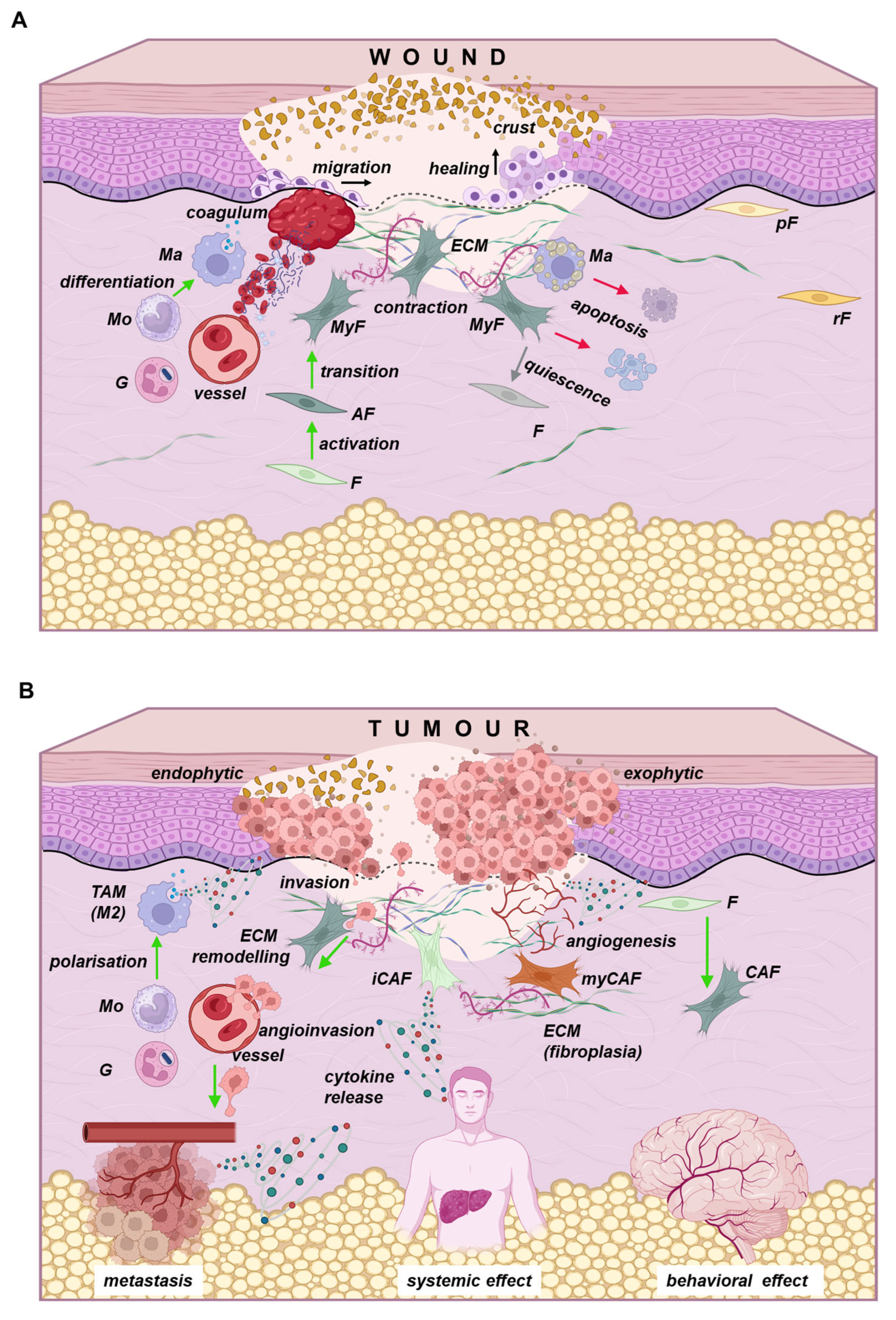
Figure 5. Wound healing (A) and tumour growth (B) share several similar mechanisms. After a traumatic injury (A), a blood coagulum forms, providing a primary seal for the wound site, with dried proteins creating a surface crust. Various immune cells released from the blood, such as granulocytes (G) and monocytes (Mo), which differentiate into macrophages (Ma), contribute to immune surveillance and the removal of tissue debris, including dead cells and extracellular matrix (ECM). Tissues contain different subsets of resident fibroblasts, such as papillary dermis fibroblasts (pF) and reticular dermis fibroblasts (rF). These fibroblasts are generally quiescent cells. Following an injury, local fibroblasts increase their transcriptional and metabolic activity, turning into activated fibroblasts (aF). Eventually, these fibroblasts can transition into myofibroblasts (MyF), which are capable of wound contraction. Once the granulation tissue adequately fills the dermal defect, epithelial cells at the wound’s edge can migrate over the granulation tissue, proliferate to heal the epithelial defect, and reconstitute the epithelial barrier through stratification and differentiation. The tissue then enters a stage of slow remodelling. However, successful healing also initiates negative feedback that reduces overall tissue activity. Unnecessary cells are eliminated by apoptosis or enter a quiescent state. In tumours (B), a similar activation of several cell types occurs; however, this activation is sustained and lacks the negative feedback typical of healing. Subsets of immune cells, including granulocytes (G) and monocytes (Mo), are generally present, but this immune response is ineffective at eliminating malignant cells. Moreover, the phenotype of macrophages shifts towards M2 polarised tumour-associated macrophages (TAMs). Resident fibroblasts are recruited through the paracrine release of various cytokines, transforming into cancer-associated fibroblasts of several categories, such as myofibroblastic CAFs (myCAFs) and pro-inflammatory CAFs (iCAFs). Myofibroblasts can secrete ECM, creating a tumour-promoting niche, which may lead to fibroplasia in some tumours. Conversely, CAFs can also degrade the ECM, making it more permissive for invasion and facilitating the invasion of tumour cells. This process allows tumour cells to traverse the vascular wall and enter circulation, leading to the formation of distant metastases. Primary tumours, along with their microenvironments, release various cytokines. These molecules can act locally through autocrine and paracrine effects; however, they can also have long-distance effects, contributing to the formation of a premetastatic niche and resulting in systemic effects in organs such as the liver, adipose tissue, or brain. These systemic effects can lead to conditions like terminal cachexia and depression. Created in https://BioRender.com (accessed on 19 August 2025).
Ijms 26 08844 g005
Disclaimer/Publisher’s Note: The statements, opinions and data contained in all publications are solely those of the individual author(s) and contributor(s) and not of MDPI and/or the editor(s). MDPI and/or the editor(s) disclaim responsibility for any injury to people or property resulting from any ideas, methods, instructions or products referred to in the content.