Review

Unraveling the Epigenetic Landscape of Mature B Cell Neoplasia: Mechanisms, Biomarkers, and Therapeutic Opportunities

1
Division of Hematology, Department of Translational Medicine, Università del Piemonte Orientale and Azienda Ospedaliero-Universitaria di Alessandria, 15121 Alessandria, Italy
2
Division of Hematology, Department of Translational Medicine, Università del Piemonte Orientale and Azienda Ospedaliero-Universitaria Maggiore della Carità, 28100 Novara, Italy
*
Author to whom correspondence should be addressed.
Int. J. Mol. Sci. 2025, 26(17), 8132; https://doi.org/10.3390/ijms26178132
Submission received: 6 July 2025 / Revised: 12 August 2025 / Accepted: 20 August 2025 / Published: 22 August 2025
(This article belongs to the Special Issue Leukemia and Lymphoma: A Focus on Molecular Genetics Research)

Abstract

Epigenetic regulation is critical to B cell development, guiding gene expression via DNA methylation, histone modifications, chromatin remodeling, and noncoding RNAs. In mature B cell neoplasms, particularly diffuse large B cell lymphoma (DLBCL), follicular lymphoma (FL), and chronic lymphocytic leukemia (CLL), these mechanisms are frequently disrupted. Recurrent mutations in key epigenetic regulators such as EZH2, KMT2D, CREBBP, and TET2 lead to altered chromatin states, repression of tumor suppressor genes, and enhanced oncogenic signaling. Dysregulation of specific microRNAs (e.g., miR-155, miR-21) further contributes to pathogenesis and therapeutic resistance. In DLBCL, hypermethylation of SMAD1 and CREBBP mutations are associated with immune evasion and chemoresistance. In FL, EZH2 gain-of-function and KMT2D loss-of-function mutations alter germinal center B cell programming, while in CLL, DNA hypomethylation patterns reflect the cell of origin and correlate with clinical outcome. Targeted therapies such as the EZH2 inhibitor tazemetostat have demonstrated efficacy in EZH2-mutant FL, while HDAC and BET inhibitors show variable responses across B cell malignancies. The limitations of current epigenetic therapies reflect the complexity of targeting epigenetic dysregulation rather than therapeutic futility. These challenges nonetheless highlight the relevance of epigenetic alterations as biomarkers and therapeutic targets, with potential to improve the management of mature B cell neoplasms.

1. Introduction

Mature B-cell neoplasms are a diverse group of lymphoid cancers arising from antigen-experienced B cells, typically at or beyond the germinal center stage [1,2]. They include malignancies derived from germinal center B cells (e.g., follicular lymphoma, FL and a fraction of diffuse large B cell lymphomas, DLBCLs), and post-GC B cells (e.g., certain DLBCLs), and memory B cells (e.g., chronic lymphocytic leukemia, CLL) [1,2]. Although these tumors often retain key immunophenotypic traits of their normal counterparts, they exhibit profound dysregulation of gene expression due to disruptions in the regulatory networks that control cellular identity and function [3]. This dysregulation is driven by both genetic and epigenetic alterations and leads to aberrant expression patterns, loss of normal differentiation, and malignant features such as uncontrolled growth and immune evasion [4]. For instance, the activated B cell-like (ABC) subtype of DLBCL is marked by constitutive activation of the NF-κB pathway, upon which these tumors depend for survival [5]. Similarly, certain DLBCL subsets rely on sustained BCL6 expression to repress its target genes, highlighting BCL6 as a central transcriptional driver. However, beyond transcription factors (TFs) like NF-κB and BCL6, gene expression is governed by the epigenetic landscape, that includes chromatin architecture shaped by DNA methylation, histone modifications, nucleosome remodeling and related mechanisms [6].
Epigenetics refers to heritable changes in gene function that occur without alterations to the DNA sequence [7]. These chromatin-based regulatory layers encode DNA sequence independent information that is essential for lineage fidelity, cell fate transitions, and the dynamic gene expression programs required for normal B cell development [4]. From naïve B cells to germinal center (GC) cells, and ultimately memory or plasma cells, each stage of B cell maturation demands precise coordination of transcriptional networks, signaling pathways and epigenetic machinery. The inherent plasticity of these processes contributes to the molecular heterogeneity seen in B cell malignancies. Notably, large-scale genomic analyses of follicular lymphoma (FL), DLBCL, and chronic lymphocytic leukemia (CLL), among others, consistently reveal a high mutational burden affecting chromatin modifiers, DNA methylation regulators, and TFs that disrupt normal epigenetic control [8].
Importantly, the recognition of epigenetic dysregulation as a key oncogenic driver has translated into therapeutic advances. Agents that target the epigenome, such as the DNA methylation inhibitors azacitidine and decitabine, have demonstrated significant clinical efficacy in myelodysplastic syndromes (MDS) and acute myeloid leukemia (AML) [9]. These successes provide a proof-of-principle that pharmacologic modulation of epigenetic alterations can produce meaningful therapeutic benefits and offer promising avenues for the treatment of mature B cell neoplasms.
As lymphomas often evolve from or hijack normal epigenetic programs, understanding how these regulatory mechanisms are subverted in a subtype-specific manner is essential. While several reviews have examined epigenetic mechanisms in B-cell malignancies, many are either disease-specific or focus on isolated pathways. Our review presents an integrated analysis across the three most prevalent mature B-cell lymphoid neoplasms, namely DLBCL, FL, and CLL selected for their shared lymphoid origin, overlapping epigenetic features, and the availability of comparable epigenomic data. We explore how aberrant DNA methylation, mutations in chromatin modifiers, and altered histone landscapes contribute to disease pathogenesis, and discuss how these alterations represent potential prognostic biomarkers that are guiding the development of novel epigenetic therapies. Furthermore, by integrating all epigenetic mechanisms with the most recent therapeutic strategies, our review may provide a framework for both research and clinical applications.

2. Epigenetic Mechanisms in B Cell Maturation

B cell maturation is a complex multi-step process strictly regulated by the interplay between different stage-specific TFs and epigenetic complexes, which define specific gene expression programs [10,11]. Epigenetic modifications play a crucial role in B cell maturation by using chemical modifications translated into instructions which can regulate gene expression and influence cell growth, apoptosis, development, differentiation, and immune response. The four main epigenetic modifications include DNA methylation, histone modifications, chromatin remodeling, and noncoding RNAs (Figure 1) [12,13].

2.1. DNA Methylation

DNA methylation involves the covalent addition of a methyl group to cytosines, mostly in the context of CpG dinucleotides. These CpG sites are often clustered in regions known as CpG islands, which are located in the promoters and/or first exons of approximately 60% of human genes [14]. The methylation machinery is represented by DNA methyltransferases (DNMT) (enzymes known as writers), 5-methylcytosine (5mC) binding proteins (readers), and by ten-eleven translocation methylcytosine dioxygenase (TET) enzymes (erasers), responsible for the demethylation of 5mC [15]. Methylation marks are related to enhancers and promoters, affecting heterochromatin and nuclear lamina associated domains, polycomb repressed regions and DNA repeats (Figure 1). The role of TET2 is sustained by Isocitrate Dehydrogenase 1 and 2 (IDH1/2) activity which consists of the conversion of isocitrate in α-ketoglutarate (α-KG), an important cofactor of TET2. Alterations of this enzyme lead to the formation of an oncometabolite taking part in the leukemogenesis process [16].
In the context of B cell maturation, the methylome represents a fundamental element in differentiation programs, activating or repressing cell pathways to define distinct features of each maturation step. Methylation effects profoundly depend upon their localization in the genome. If DNA methylation occurs in CpG rich gene promoters, it is associated with transcriptional silencing. Conversely, methylation of intragenic regions is more commonly linked to gene activation [13]. DNA methylation is rare during early-stage B cells but is rather frequent in mature B cells and is regulated by three classes of methyl transferases: DNMT1, DNMT3A, DNMT3B [17]. DNMT3A and DNMT3B are responsible for de novo methylation. During transit of centroblasts through the GC, DNMT3A expression is markedly downregulated. In contrast, DNMT1 is significantly upregulated, showing a central role in GC development and during replication, maintaining DNA methylation and genomic integrity as documented by the observation that its reduced expression leads to DNA damage in hypomorphic mice [18,19].
Demethylation is mediated by TET and occurs during early B cell stages and in the GC, operating on enhancers and in heterochromatic regions, respectively. Its central role in the maturation program was demonstrated in TET2 and TET3 knockout mice, where the lack of these elements blocks the transition from pro- to pre-B cells in the bone marrow, resulting in decreased expression of IRF4 and an impaired IgK rearrangement [20].

2.2. Histone Modifications

Histone proteins organize DNA in nucleosomes, wrapping around the histone core composed of an octamer of H2A, H2B, H3, and H4 proteins. Nucleosomes are the fundamental element of chromatin and can be present in two different conformations: heterochromatin (condensed state) and euchromatin (accessible state) [21]. The N-terminal tails of histone proteins can undergo post-translational modifications, changing the chromatin properties and consequently the gene expression. The most frequent histone modifications include acetylation, methylation, ubiquitination, phosphorylation, and sumoylation [22]. Each modification is carried out by specific enzymes that introduce or remove chemical groups from terminal histone amino acids, and the combination of these changes represents a “histone code” that will determine chromatin conformation (Figure 1) [23].
Histone acetyltransferases and histone deacetylases (HADCs) are responsible for the addition and removal of an acetyl group, respectively. Acetylation is normally associated with gene expression, and it commonly takes place at the H3K9, H3K18, H3K27, H3K4, and H3K5 lysine residues. Among these epigenetic modifications, bromodomain and extraterminal motif (BET) family proteins act as readers interacting with hyper-acetylated lysine residues and promoting gene transcription. Specifically, they can occupy super-enhancers of oncogenes such as MYC, CCDN2, and MCL-1 [24]. Instead, histone methylation can occur on arginine, lysine, and histidine residues, is mediated by methyltransferases and the most frequently methylated sites are H3K4 (catalyzed by MLL1-5, SET1-A, SET1-B), H3K9 (SUV39-H1), H3K27 (EZH2), H3K36 (SET2, NSD1), and H3K79 (DOT1-L) [25]. Lysine and arginine can be represented in three different methylation statuses (mono-, bi-, and tri-methylated) depending on how many methyl groups are linked to terminal amino acid residues.
During T cell dependent activation, a subset of naïve B cells can be induced to migrate into lymphoid follicles, becoming centroblasts and forming the dark zone of the GC, while another fraction of naïve B cells differentiates into low-affinity plasma cells after immunoglobulin switch recombination. Centroblasts are subjected to rapid clonal expansion and somatic hypermutation mediated by activation-induced cytosine deaminase (AICDA) introducing point mutations at cytosine residues.
EZH2, a component of the polycomb repressive complex2 (PRC2), is responsible for the trimethylation of H3K27 and has a pivotal role in GC formation and antibodies production [26]. EZH2 levels are low during early B cell stages but increase at the time of GC transit. The addition of H3K27me3 at gene promoters, already marked by H3K4me3 in naïve B cells, leads to transcriptional silencing of the IRF4 and PRDM1 genes involved in plasma cell differentiation and of the CDKN1A (p21) and CDKN1B check point regulators of proliferation. EZH2 levels are lower in centrocytes due to the action of the KDM6A-B demethylase, restoring the active state of promoters and inducing differentiation program [27,28].
DOT1L is involved in H3K79 methylation which is associated with active transcription during B cell differentiation, promoting the expression of a pro-proliferative and pro-GC transcriptional program. In vitro studies on B cells with DOT1L deletion revealed an aberrant differentiation and premature acquisition of plasma cells features [29].
The transcriptional repressor BCL-6 cooperates with EZH2 in GC-cells, recruiting SMRT and NCOR (corepressor proteins) and thus binding HDAC3, which mediates H3K27 deacetylation. During this stage, the PAX5, CD19, EBF1, and SPIB genes are expressed [30]. This deacetylation effect is reversible upon CD40 signaling in the GC, promoting SMRT phosphorylation trough ERK signaling and initiating the exit of B cells from the GC [31].
Among these epigenetic modifications, BET proteins are crucial regulators of gene expression that bind acetylated lysine residues on histones. BET proteins act as epigenetic readers, recruiting the positive transcription elongation factor b (p-TEFb) and promoting gene transcription. Specifically, they can occupy super-enhancers of oncogenes such as MYC, CCDN2, MCL-1, and an aberrant acetylation of these sites results in cancer cell proliferation, survival, and oncogenic progression [24,32].

2.3. Chromatin Remodeling

The nucleosome core particle is the basic structural unit of chromatin, consisting of DNA wrapped around a core of histone proteins [33].
The arrangement of nucleosomes and their distribution across chromatin determines how DNA becomes accessible to nuclear macromolecules, which control essential genetic operations including DNA replication, transcription, recombination, and repair (Figure 1) [34,35].
B cell maturation depends on chromatin remodeling for its regulation, especially in the GC where activated B cells undergo clonal expansion, somatic hypermutation (SHM) and class-switch recombination (CSR) [36]. The regulation of these processes depends on the dynamic control of gene accessibility, which is achieved through chromatin remodeling complexes, histone modifications, and changes in 3D genome architecture [37].
The SWI/SNF (BAF) complex through its catalytic subunit BRG1 functions as a crucial chromatin remodeling complex that drives B cell activation and GC development [38]. BRG1 enables B cell proliferation and GC formation through enhancer region targeting, resulting in elevated chromatin accessibility and activation of cell cycle gene transcription. Absence of BRG1 in B cells blocks the start of GC responses while preventing proper class-switched plasma cell development [38]. B cells undergo substantial 3D chromatin rearrangements after antigen recognition to activate essential GC genes including BCL6 and AICDA. The chromatin organization becomes disrupted when histone H1 mutations occur, leading to chromatin decompaction and enabling GC B cells to self-renew more frequently [39].

2.4. Noncoding RNA

In mature B cell neoplasms, gene expression is also controlled by noncoding RNAs that influence multiple regulatory pathways. Both long noncoding RNAs (lncRNAs) and microRNAs (miRNAs) play key roles in epigenetic mechanisms such as genomic imprinting and chromatin remodeling (Figure 1) [40]. These noncoding RNAs act as signals, guides, decoys, or scaffolds, modulating gene expression at transcriptional and post-transcriptional levels. Dysregulation of lncRNAs and miRNAs in B cell lymphomas disrupts genes involved in B cell differentiation while promoting tumorigenic gene expression patterns [41].
lncRNAs are involved in cellular differentiation and tissue development, while miRNAs are involved in B cell maturation and activation in both normal and neoplastic B cells The abnormal regulation of these noncoding RNAs leads to drug resistance in CLL and lymphomas, which makes them useful as biomarkers for diagnosis, prognosis, and therapeutic response [42]. Noncoding RNAs control gene expression during B cell maturation through their action at developmental checkpoints [43].

3. Epigenetic Landscape in B Cell Neoplasia

3.1. Diffuse Large B Cell Lymphoma

DLBCL is the most common lymphoid neoplasm in the adult population, arising from mature B cells, and it is characterized by a heterogeneous genetic and epigenetic landscape which influences morphology, immunophenotype, and therapy response. Two principal subtypes can be distinguished based on the cell of origin: the GC B cell-like (GCB) subtype, which is generally associated with a more favorable prognosis, and the activated B cell-like (ABC) subtype, which is typically linked to inferior clinical outcomes [1,2,44].
The pathogenesis of DLBCL is characterized by the accumulation of genetic aberrations causing an altered structure and expression of important proto-oncogenes and tumor suppressor genes. Genetic alterations include somatically acquired point mutations, gene copy number variation and translocations. Translocations lead to the placement of a regulatory element (promoter or enhancer), often involving IG loci, in proximity to the coding sequence of an oncogene, resulting in its constitutive expression [45]. Among the main aberrations, MYC, BCL2, and BCL6 translocations are assessed by FISH analysis to define the double- or triple- hit status in co-presence of MYC and BCL2 or/and BCL6 alterations [46].

3.1.1. DNA Methylation in DLBCL

Aberrant methylation disrupts the normal regulation of critical genes involved in cell cycle control, apoptosis, and differentiation. Among the key genes affected in DLBCL are MYC, SLIT2, KLF4, and CDKN2A, whose inactivation facilitates uncontrolled proliferation and tumor progression. DLBCL shows methylation heterogenicity which defines different molecular subgroups and clinical outcomes. In particular, six molecular subtypes have been identified based on methylation variability, with high grade methylation variability being associated with shorter survival compared to lower grade variability [47,48,49,50]. Additionally, higher methylation heterogeneity increases the probability of the appearance of DLBCL clones that are epigenetically programmed to better tolerate chemoimmunotherapy. In fact, patients with higher heterogeneity displayed a poorer outcome with a shorter survival after R-CHOP therapy [51]. Consistently, in vitro studies have demonstrated that the hypermethylation of SMAD1, a key transducer of TGF-β signaling, contributes to the development of chemoresistance in DLBCL cells exposed to R-CHOP treatment [52].

3.1.2. Histone Modifications and Chromain Remodeling in DLBCL

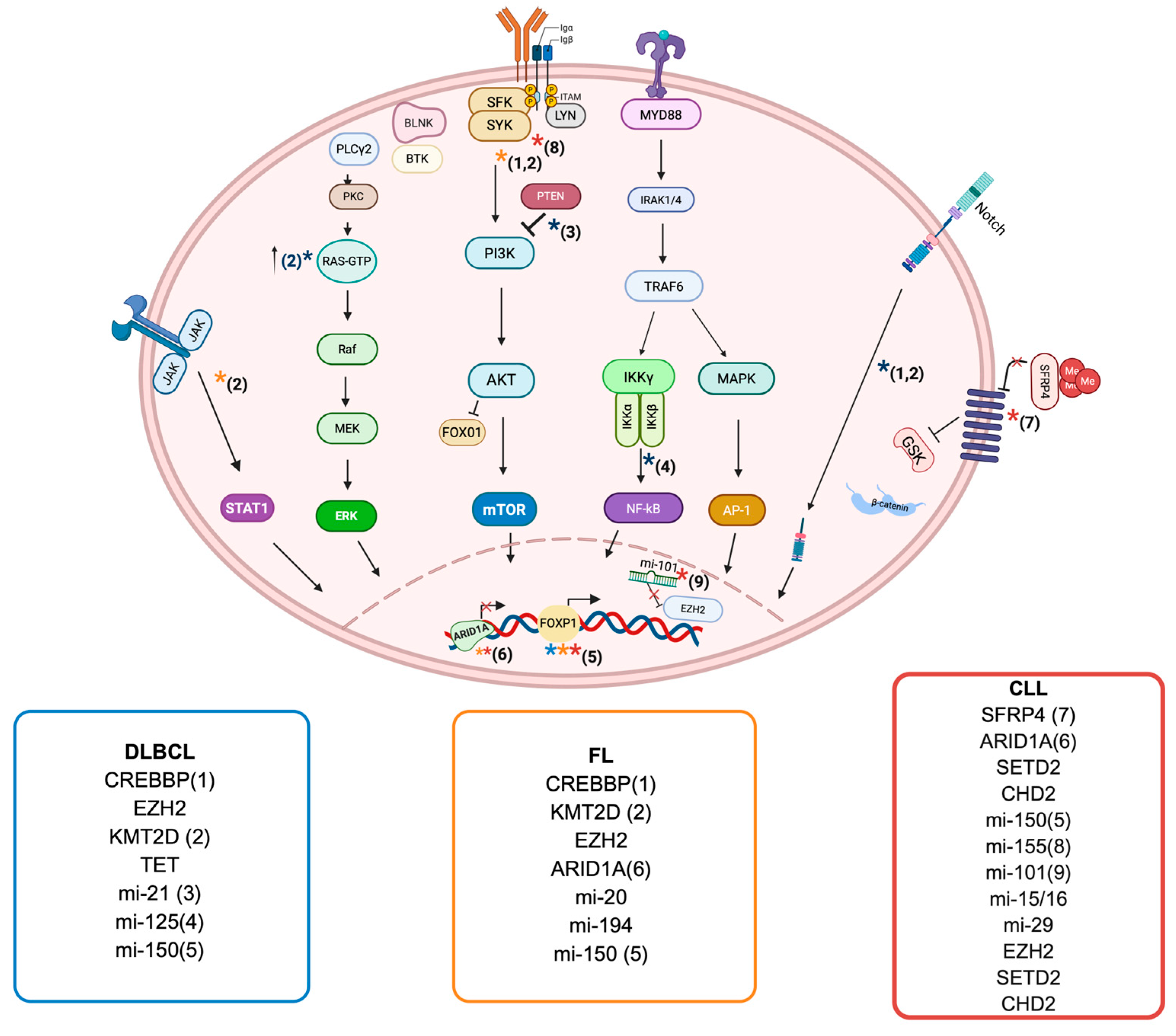
Mutations in genes responsible for histone and chromatin modifications, such as CREBBP, KMT2D, EZH2, and TET2, drive aberrant epigenetic B cell programming and influence DLBCL clinical outcome [53]. Loss-of-function mutations in CREBBP are common in GCB-DLBCL, occur in approximately 30% of cases and lead to reduced acetyltransferase activity. Under normal conditions, CREBBP regulates enhancers that are repressed by BCL6 through cooperation with the co-repressors SMRT and NCOR in the GC [54]. This repressor complex also includes HDAC3, which mediates deacetylation of H3K27. Loss of CREBBP function results in unchecked enhancer repression, altering the expression of key genes, including those encoding MHC class II molecules [54]. Furthermore, CREBBP deficiency contributes to dysregulated B cell signaling and excessive repression of gene transcription. The CREBBP-mediated deacetylation of H3K27 downregulates the expression of FBXW7, a key negative regulator of NOTCH, and thus activates the NOTCH signaling pathway, which plays a critical role in B cell malignancies [55]. CREBBP mutations further disrupt the downstream components of the NOTCH pathway, leading to expression of CCL2 and CSF1, which in turn promote the polarization of tumor-associated macrophages toward the immunosuppressive M2 phenotype [56]. These alterations impair normal immune surveillance, facilitating tumor immune evasion and driving disease progression (Figure 2) [55,56].
EZH2 mutations are found in 6–14% of DLBCL overall and in about 20% of GCB-DLBCL, where they are frequently associated with BCL2 translocations [57]. Under normal conditions, EZH2 cooperates with BCL6 to mediate gene silencing at the promoters of genes involved in plasma cell differentiation and cell cycle regulation [28]. In mouse models, EZH2 knockout leads to accelerated lymphomagenesis [58]. EZH2 gain-of-function mutations, most commonly affecting the Tyr641 residue within the SET domain, drive germinal center hyperplasia and contribute to remodeling of the immune microenvironmen [57]. Unlike normal GC B cells, where cytosine methylation and H3K27me3 marks are usually mutually exclusive, in DLBCL these modifications often overlap, with hypermethylation partly affecting EZH2 target genes [59].
KMT2D, also known as MLL2, encodes a methyltransferase essential for the methylation of lysine 4 on histone H3 (H3K4), a key modification that regulates gene enhancer activity [60]. Loss-of-function mutations in KMT2D are found in approximately 30% of DLBCL [61]. These mutations impair H3K4 methylation, leading to the downregulation of FBXW7, a negative regulator of the NOTCH signaling pathway. The resulting activation of NOTCH signaling drives several oncogenic processes, including enhanced cell survival, proliferation, and inhibition of terminal B cell differentiation [62]. This aberrant signaling further promotes activation of downstream pathways such as RAS, ERK, and MYC/TGF-β1, contributing to tumor growth and immune evasion [62].
The TET family of enzymes plays a key role in active DNA demethylation, particularly at gene enhancers, where this activity is linked to transcriptional activation [63]. TET2 mutations occur in approximately 10% of DLBCL patients and result in abnormal DNA hypermethylation at regulatory elements within the GC [64]. This hypermethylation leads to the silencing of genes involved in GC exit and B cell receptor signaling, contributing to lymphomagenesis. In TET2 knockout mice, loss of TET2 disrupts GC B cell homeostasis and promotes transformation into aggressive lymphomas. These models also show increased formation of G-quadruplexes and R-loops, that represent structures associated with double-strand DNA breaks at immunoglobulin switch regions [65].

3.1.3. Noncoding RNA in DLBCL

miRNA dysregulation plays a key role in the pathogenesis and treatment resistance of DLBCL. High miR-21 expression has been linked to poorer survival through its suppression of FOXO1 and PTEN, activating the PI3K/AKT/mTOR pathway (Figure 2) [66]. High miR-155 levels are linked to poor response to R-CHOP therapy [67]. Overexpression of miR-125a and miR-125b targets TNFAIP3, leading to NF-κB activation and promoting a pro-proliferative and anti-apoptotic state. Similarly, increased let-7a/b/f levels reduce PRDM1 expression, impairing B cell differentiation [68]. Distinct miRNA profiles help distinguish DLBCL from other lymphomas and define disease subtypes. For example, a 27-miRNA signature differentiates Burkitt lymphoma from DLBCL, while panels including miR-28-5p, miR-214-5p, miR-339-3p, and miR-5586-5p are associated with better outcomes [69]. Conversely, reduced expression of miR-370-3p, miR-381-3p, and miR-409-3p in relapsed cases suggests their role in modulating chemosensitivity. Altogether, these findings highlight how miRNA dysregulation disrupts key signaling pathways and drives abnormal cell proliferation, survival, and differentiation in DLBCL.

3.2. Follicular Lymphoma

FL represents the second most prevalent form of lymphoma occurring in adults [70]. FL originates from B cells with the t(14;18) translocation, which triggers BCL2 overexpression but is not sufficient for full transformation [71]. Additional genetic mutations accumulate as these cells persist and cycle through the GC. Crucially, many of these mutations affect epigenetic regulators, leading to widespread changes in chromatin structure, histone modification, and DNA methylation. These epigenetic alterations reshape gene expression and the tumor microenvironment, driving FL development and progression. Understanding these epigenetic mechanisms is essential for identifying new therapeutic targets in FL [72,73,74,75].

3.2.1. Histone Modifications and Chromatin Remodeling in FL

Mutations affecting epigenetic-related genes primarily involve histone methyltransferases like KMT2D and EZH2, as well as histone acetyltransferases such as CREBBP and EP300. Most of these mutations are inactivating, except those in EZH2. These genes are essential for regulating gene expression through the modification of H3K4 by KMT2D and H3K27 by EZH2, CREBBP and EP300 (Figure 2) [8,76].
KMT2D is the most frequently mutated gene in FL, and its loss of function contributes significantly to lymphomagenesis [61]. Loss-of-function mutations in KMT2D significantly influence the transcriptional regulation of immune-related genes and facilitate FL development by modifying H3K4 methylation and the expression of genes associated with B cell activation pathways, which include CD40, JAK-STAT, and B cell receptor signaling [77]. KMT2D mutations compromise global H3K4 methylation in GC B cells, thereby promoting proliferation and lymphomagenesis [61]. Gain-of-function mutations in EZH2 occur in approximately 20–25% of FL cases and were among the first epigenetic alterations identified as critical drivers in the early stages of this lymphoma [27]. These mutations enhance EZH2-mediated trimethylation of H3K27 (H3K27me3), leading to abnormal repression of gene transcription. As a key epigenetic writer active in GC B cells, EZH2 silences regulators of B cell differentiation through H3K27me3 deposition. This results in the silencing of genes involved in cell cycle checkpoints, differentiation, and immune synapse signaling, reducing the dependency of GC B cells on T cell help. Consequently, lymphoma cells evade immune control while promoting their survival and proliferation. EZH2 mutations also shift GC B cell reliance from T cell interactions to increased engagement with follicular dendritic cells, expanding the centrocyte-like population characterized by heightened proliferation and survival [27,78,79].
CREBBP functions as a haploinsufficient tumor suppressor and plays a critical role in both the development and progression of FL [80]. It regulates the expression of genes involved in B cell receptor and CD40 signaling pathways, which are essential for normal B cell activation and differentiation. Inactivating mutations in CREBBP, particularly those affecting its acetyltransferase (KAT) domain, disrupt chromatin modification, leading to abnormal gene expression [56]. These mutations are common in FL and can often be detected years before clinical diagnosis, highlighting their role in early disease development [81].

3.2.2. DNA Methylation in FL

Although DNA methyltransferase genes are not mutated per se in FL, aberrant DNA methylation is a hallmark of the disease [82]. For instance, mutations in H1 linker histone genes impair the binding of DNMT3B, contributing to abnormal methylation patterns [83]. Promoter hypermethylation, particularly at PRC2 target genes, leads to silencing of key tumor suppressors such as CDKN2A, allowing unchecked cellular proliferation [84]. On the other hand, global hypomethylation contributes to genomic instability and activation of oncogenes, further fueling tumorigenesis [85]. A more deregulated DNA methylation landscape has been associated with greater disease aggressiveness. While methylation patterns generally remain stable between pre- and post-transformation FL, relapsed FL often displays increased methylation levels [86].
Mutations of genes encoding subunits of chromatin remodeling complexes such as ARID1A, which are part of the SWI/SNF (BAF) complex, are present in a fraction of FL [72,87]. Mutations in ARID1A result in reduced chromatin accessibility, and loss of ARID1A expression has been associated with a shift towards a memory B cell-like state associated with increased risk of progression to aggressive lymphoma [71,87].
Recent advances underscore the role of epigenetic alterations in improving risk stratification in FL. While the traditional follicular lymphoma international prognostic index (FLIPI) score often overestimates the proportion of high-risk patients at diagnosis, the integration of genetic data in the m7-FLIPI model incorporating mutations in seven genes, including key epigenetic regulators such as EZH2, CREBBP, EP300, MEF2B, and ARID1A, has significantly improved prognostic accuracy [88]. This model effectively distinguishes between high- and low-risk patients and predicts progression within 24 months of first-line R-CHOP therapy, a critical surrogate for overall survival. Notably, mutations in CREBBP and EP300 are associated with high-risk disease, whereas EZH2 and MEF2B mutations correlate with more favorable outcomes. These findings underscore the potential of epigenetic modifier mutations to enhance risk prediction and guide treatment intensity [88].

3.2.3. Noncoding RNA in FL

ncRNAs function as essential elements in the progression and formation of FL. The expression of long noncoding RNAs (lncRNAs) shows the greatest variation between FL classes, with grade 3B displaying the highest levels of unregulated lncRNAs that modulate proliferation and cell cycle [89]. FL also involves miRNAs, with 133 miRNAs showing significant differences between follicular hyperplasia and FL [90]. A unique FL miRNA profile comprises miRNAs associated with improved response to chemotherapy. MiR-20a/b and miR-194, respectively, target CDKN1A and SOCS2, which may affect the survival and proliferation of tumor cells [90]. Both lncRNAs and miRNAs affect cell proliferation, tumor response, and other biological processes in B cell development and lymphomagenesis [91,92]. Musilova and colleagues analyzed miRna profiles in serial biopsies of transformed follicular lymphoma and identified five miRnas enriched during transformation, including miR-150 [93]. Their study revealed a c-MYC/miR-150/FOXP1 regulatory axis, in which c-MYC overexpression suppresses miR-150, resulting in increased FOXP1 expression. FOXP1, a TF essential for B cell development, is associated with the ABC subtype of DLBCL and is linked to poor clinical outcomes in both DLBCL and FL [93].

3.3. Chronic Lymphocytic Leukemia

CLL is a mature B cell malignancy characterized by the clonal expansion and accumulation of CD5+CD19+ B lymphocytes in the blood, lymphoid organs, and bone marrow [1]. It is the most common leukemia in adults, typically following an indolent course but exhibiting significant clinical variability [94]. Advances in targeted therapies, including inhibitors of Bruton tyrosine kinase (BTK) and B cell lymphoma 2 (BCL2), have significantly improved progression-free and overall survival, particularly in patients with high-risk genetic features [95,96]. Despite therapeutic progress, the molecular mechanisms underlying CLL pathogenesis remain only partially understood. Increasing evidence suggests that epigenetic alterations play a central role in shaping disease onset, progression, and response to therapy, highlighting the need for a deeper insight into the epigenetic landscape of CLL.

3.3.1. DNA Methylation in CLL

CLL is characterized by widespread alterations in DNA methylation, most notably global hypomethylation relative to healthy B cells [97]. Interestingly, the global DNA methylation landscape in CLL is remarkably stable over time and largely conserved between circulating resting cells and those in proliferative lymphoid niches [98]. This epigenetic stability across disease compartments supports the notion that aberrant DNA methylation is established early in leukemogenesis and may serve as a foundational event in CLL pathobiology (Figure 2).
Distinct DNA methylation patterns among CLL subtypes reflect epigenetic imprints inherited from their respective cells of origin, influencing both the biological behavior of leukemic cells and the clinical course of the disease [97,99]. Mutated IGHV CLL (M-CLL) retain a DNA methylation pattern resembling that of normal post-GC (memory-like) B cells, whereas unmutated CLL (U-CLL) exhibit a naïve-like methylation signature [100]. Interestingly, these analyses have also identified a third epigenetic subtype of CLL with an intermediate methylation profile, falling between the naïve-like and memory-like patterns. This suggests that the intermediate subtype may arise from a yet unidentified normal B cell subset. These three epigenetically defined CLL subtypes exhibit distinct patterns in terms of somatic mutation landscapes, immunoglobulin heavy chain variable region (IGHV) gene usage, and clinical outcomes [97,100,101]. Naïve-like CLL typically associates with unmutated IGHV genes, frequent usage of IGHV1-69, 11q deletions, 2p16 gains and recurrent NOTCH1 mutations. The intermediate subtype often displays partially mutated IGHV genes, with a higher frequency of IGHV3-21, IGHV1-18, and BCR stereotyped subset #2, alongside elevated rates of SF3B1 and MYD88 mutations. In contrast, memory-like CLL is characterized by mutated IGHV genes, commonly utilizing IGHV4-34 and IGHV3-7 [101]. Importantly, these epigenetic subtypes also correlate with significant differences in time to first treatment and OS [100]. The prognostic significance of this epigenetic classification has been validated in independent studies [100,102,103]. Collectively, these findings support the notion that the cell of origin, defined by its immunogenetic or epigenetic signature, plays a crucial role in shaping the biology and clinical behavior of CLL.
The expression of tumor suppressor genes (TSGs) is frequently silenced through promoter hypermethylation in CLL. The Wnt signaling pathway, which plays a crucial role in normal B cell development and regulation of apoptosis, is often aberrantly activated in CLL [104]. One of the key negative regulators of this pathway, secreted frizzled-related protein 4 (SFRP4), is a member of the SFRP family and is commonly hypermethylated in CLL samples [105]. Hypermethylation of CpG islands in the promoters of SFRP genes, including SFRP4, leads to transcriptional silencing and may contribute to the pathological activation of Wnt signaling in CLL [106]. Among the five SFRP family members, SFRP1 was found to be consistently hypermethylated and downregulated in CLL, suggesting that its epigenetic inactivation is a critical event in leukemogenesis.
Whole-epigenome analyses tracking DNA methylation dynamics throughout the course of treatment have revealed that specific epigenetic features in CLL may serve as predictors of clinical outcomes [107]. Notably, epigenetic silencing of HOXA4 has been associated with reduced therapeutic sensitivity, indicating its potential role in chemoresistance [107]. In one study, ibrutinib treatment induced significant epigenetic and transcriptomic remodeling. Chromatin accessibility shifted toward a more closed state overall, with increased accessibility at ERK-related TF sites and reduced accessibility at BCR-related sites [108]. Most differentially expressed genes were downregulated, including those linked to IL-4, NF-κB, metabolism, and proliferation, while MAPK and RAS signaling genes were upregulated [108]. These results underscore the critical role of epigenetic reprogramming in shaping therapeutic response beyond genetic evolution alone. Further studies are needed to elucidate the role of epigenetic changes in resistance to other targeted agents, including BCL2 inhibitors as well as covalent and non-covalent BTK inhibitors.
A recent analysis of de novo chromatin activation in CLL revealed two distinct and opposing chromatin activation programs: one associated with progressive disease (PD) and the other with indolent disease (ID) [109]. De novo chromatin activation refers to the aberrant opening and activation of chromatin regions that are normally inactive in healthy B cells, enabling inappropriate gene expression that contributes to leukemogenesis [110]. These two programs correspond to underlying molecular pathways that drive disease behavior and progression. A balance score, quantifying the relative activation of PD and ID chromatin signatures, was shown to outperform conventional markers such as IGHV mutation status as an independent predictor of time to first treatment. Notably, the PD signature is enriched for TNF-α/NF-κB and mTOR signaling pathways and is shaped by the lymph node microenvironment, whereas the ID signature appears largely independent of microenvironmental influences. These findings underscore the critical role of epigenetic characterization in advancing our understanding of CLL biology and refining risk stratification.

3.3.2. Histone Modifications and Chromatin Remodeling in CLL

Beyond aberrant DNA methylation, CLL exhibits widespread epigenetic remodeling that includes substantial changes in chromatin accessibility and histone modification patterns [111,112]. While mutations in epigenetic regulators are less frequent compared to other B cell lymphomas, namely FL and DLBCL, alterations in chromatin modification nonetheless play a significant role in CLL pathogenesis [113].
Although some aspects of the CLL chromatin landscape appear to be shaped by epigenetic dynamics inherited from normal B cell maturation, integrative chromatin state analyses have uncovered approximately 500 de novo activated regulatory elements common to CLL cells regardless of their underlying genetic heterogeneity. These elements correspond to enhancers or promoters of genes with established roles in CLL pathogenesis, including TCF4, FMOD, CTLA4, and LEF1 [110,114]. Interestingly, the activation of these regulatory regions appears to be orchestrated by a limited number of TF families, notably NFAT, FOX, and TCF/LEF, whose binding motifs are enriched across CLL-specific chromatin-accessible sites [99,110,114]. Further investigation into the mechanistic roles of these TFs may elucidate their contribution to disease initiation and persistence, and potentially uncover therapeutic vulnerabilities related to aberrant chromatin activation.
Complementary to enhancer-focused studies, additional epigenomic analyses have revealed an unexpected co-occurrence of histone modifications in CLL that are typically considered mutually exclusive in normal cells. This epigenetic anomaly suggests the presence of intra-tumoral heterogeneity at the chromatin level, likely reflecting a mixture of subpopulations with distinct cellular identities and epigenetic states. As with DNA methylation, such chromatin diversity may provide valuable insights into subclonal evolution and disease progression in CLL.
Among the histone-modifying enzymes implicated in CLL is EZH2. Although recurrent EZH2 mutations are common in FL and DLBCL, CLL is distinct in exhibiting overexpression of EZH2 without accompanying genetic alterations [101]. Papakonstantinou et al. demonstrated that EZH2 is overexpressed at both the mRNA and protein levels in a subset of patients with U-CLL, and that EZH2 overexpression correlates with elevated global H3K27me3 levels [115]. Interestingly, U-CLL cases with high EZH2 expression show a lower incidence of TP53 mutations, suggesting a distinct molecular subset within the disease [115]. In vitro studies showed that overexpression of EZH2 enhances leukemic cell viability, while EZH2 knockdown or pharmacological inhibition induces apoptosis, supporting its role as an oncogenic epigenetic regulator [115]. Treatment with EZH2 inhibitors reduces H3K27me3 levels and triggers apoptotic responses in vitro, providing a potential therapeutic angle for targeted intervention.
Analogous to FL and DLBCL, mutations in histone-modifying enzymes are also observed in CLL, though at lower frequency. Alterations in genes such as SETD2, ARID1A, and CHD2 further emphasize the pivotal role of epigenetic dysregulation in CLL pathogenesis [101,116,117]. SETD2, a histone methyltransferase responsible for H3K36 trimethylation, is mutated or deleted in up to 4% of cases and is associated with impaired genomic stability, chemoresistance, and poor clinical outcomes, often co-occurring with TP53 alterations [117,118]. Similarly, loss-of-function mutations in ARID1A, a core component of the SWI/SNF chromatin remodeling complex, may drive proliferation through dysregulation of key cell cycle genes [101]. CHD2, part of the CHD family of chromatin remodelers, is mutated in approximately 5% of cases, especially in mutated-IGHV CLL [116]. Collectively, these findings underscore the broader relevance of histone modification and chromatin remodeling pathways in CLL biology and prognosis, supporting further exploration of epigenetic vulnerabilities as therapeutic targets (Figure 2).

3.3.3. Noncoding RNA in CLL

One of the most frequently observed genetic alterations in CLL is the deletion of the miR-15a/16-1 cluster located at chromosome 13q14, occurring in more than 65% of cases and resulting in the downregulation of miR-15a/16-1 [119,120]. Notably, the first nine nucleotides of the 5′ ends of miR-15/16 are complementary to bases 3287–3279 of the 3′ UTR of BCL2 mRNA, an anti-apoptotic gene found to be overexpressed in nearly all CLL patients. Conversely, in normal physiology, the direct interaction between miR-15/16 and BCL2 leads to repression of BCL2 expression and promotion of apoptosis.
Moreover, miR-34b/c, located on chromosome 11q, functions as a tumor suppressor by negatively regulating protein translation [121]. Its expression is frequently reduced due to promoter hypermethylation, a change that strongly correlates with 11q deletions and contributes to aggressive CLL phenotypes [122]. Similarly, miR-155, encoded within the MIR155HG (BIC) locus, is a conserved microRNA that modulates B cell gene expression at the post-transcriptional level [123,124,125]. Overexpression of miR-155 enhances BCR signaling responsiveness and is often driven by microenvironmental cues. Recent studies show that XPO1 mutations in CLL further contribute to this dysregulation by increasing chromatin accessibility and transcription of MIR155HG and its regulator MYB, leading to elevated miR-155 levels [126]. This upregulation amplifies BCR signaling, promotes CLL cell proliferation, and is associated with shorter time to first treatment (TTFT), independent of IGHV status. Together, these findings underscore the critical roles of miR-34b/c loss and miR-155 overexpression in driving aggressive disease and highlight their potential as therapeutic targets in CLL [126].
Several other miRNAs have also been implicated in CLL pathogenesis. For instance, miR-29 has demonstrated anti-tumor activity by targeting TRAF4, and its expression is modulated by BCR inhibitors, making it a potential therapeutic target [127]. miR-125a-5p and miR-34a-5p have emerged as predictive markers for Richter transformation [128]. miR-150, which is abundantly expressed in CLL cells, has been linked to disease progression through its regulation of GAB1 and FOXP1 [129].

4. Therapeutic Strategies Targeting Epigenetic Alterations

Over the past decade, our understanding of the epigenetic landscape in B cell malignancies has expanded significantly, revealing how aberrant DNA methylation, histone modifications, and dysregulated chromatin architecture contribute to malignant transformation, disease progression, and therapy resistance. These reversible changes offer promising therapeutic targets, enabling modulation of gene expression without modifying the underlying DNA sequence. A summary of the ongoing clinical trials investigating epigenetic drugs (epi-drugs) in mature B cell NHL is presented in Table 1.

4.1. Demethylating Agents

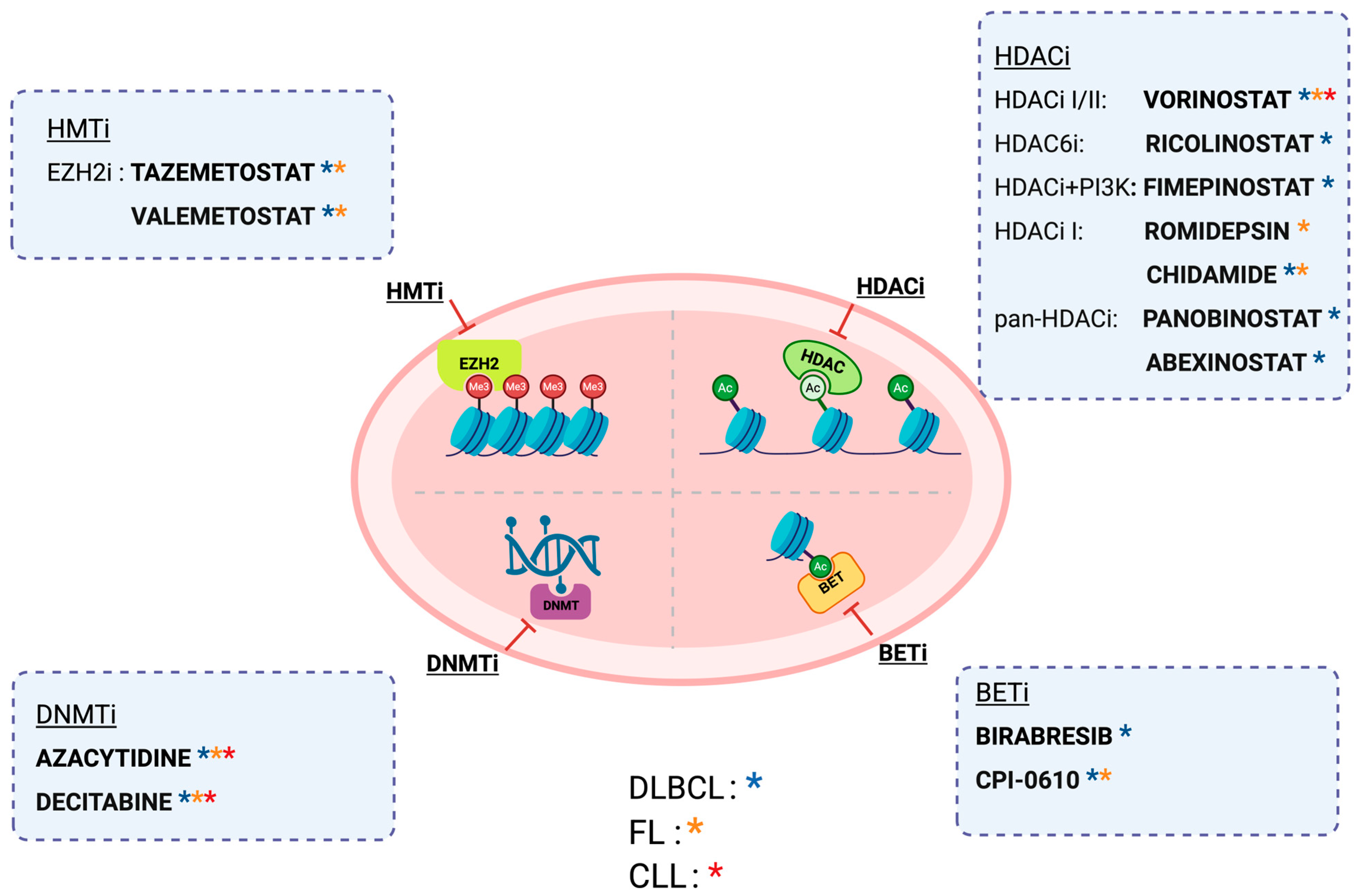
DNMT inhibitors such as 5-azacytidine and decitabine, initially developed for AML, have shown limited efficacy as monotherapy in lymphoid malignancies like DLBCL, FL, and CLL. Clinical trials using these agents alone in B cell malignancies have been largely disappointing, prompting a shift toward combination strategies [130,131,132]. While combinations with agents like HDAC inhibitors (e.g., romidepsin) have demonstrated some success in peripheral T-cell lymphoma (PTCL), other regimens, such as azacytidine with immune checkpoint inhibitors, have failed to show sufficient clinical activity [133,134]. Efforts to enhance efficacy through co-administration with enzyme cytidine deaminase (CDA) inhibitors or novel DNMTis are ongoing, but robust clinical benefits in B cell malignancies remain unproven (Figure 3).

4.2. HMT Inhibitors

The development of HMT inhibitors in B cell malignancies has concentrated on targeting EZH2. Among EZH2 inhibitors, tazemetostat has emerged as the most clinically advanced. Preclinical studies showed that tazemetostat was effective in vitro and in vivo (including in xenograft models) against EZH2-mutant lymphoma. In a phase I trial, tazemetostat demonstrated a favorable safety profile and achieved a 38% overall response rate (ORR) in refractory B-NHL [135]. A subsequent phase II trial in relapsed/refractory (R/R) FL confirmed higher activity in EZH2-mutant patients (ORR 69%) compared to EZH2-wild-type patients (ORR 35%) [136]. These data led the FDA to grant tazemetostat accelerated approval for the treatment of R/R FL with EZH2 mutations. In addition, tazemetostat was evaluated in a small DLBCL cohort in combination with R-CHOP, achieving an ORR of 86% [137]. However, when combined with the PD-L1 inhibitor atezolizumab in R/R DLBCL, no added benefit was observed (Figure 3) [138]. This highlights the ongoing challenge of improving outcomes through combination therapies. Further trials are exploring new combinations and strategies to enhance the efficacy of tazemetostat and other HMT inhibitors, aiming to improve overall survival in patients with these lymphomas.
Other agents have shown more limited efficacy. For instance, GSK126, despite its strong preclinical activity against mutant EZH2, failed to show meaningful clinical responses in a phase I trial in R/R DLBCL, transformed FL, and other NHL, leading to early trial termination [139,140]. Valemetostat, a dual EZH1/2 inhibitor, has shown promise in early-phase studies, achieving an ORR of 53% in B- and T-NHL, with particularly high activity (80% ORR) in T-cell lymphoma [141].

4.3. HDACs Inhibitors

HDACs play a key role in regulating cell proliferation, growth, and angiogenesis, which are often dysregulated in cancer. Overexpression of HDACs promotes tumor development, making HDAC inhibitors (HDACis) a promising anticancer strategy (Figure 3) [142]. HDACis induce apoptosis, cause cell cycle arrest, and can modulate immune responses, leading to therapeutic effects in various lymphomas [143]. Vorinostat, the first FDA-approved HDACi (2006), targets class I and II HDACs and is approved for relapsed/refractory cutaneous T-cell lymphoma (CTCL) [144]. It has demonstrated benefits as monotherapy in FL and in combination regimens with rituximab, fludarabine, or etoposide for relapsed/refractory B-NHL, CLL, and T-NHL. Belinostat, a pan-HDACi, is approved for relapsed/refractory PTCL and has shown activity in CTCL, although with limited efficacy in B-cell lymphomas. Romidepsin, a class I HDACi, is also approved for relapsed/refractory CTCL and PTCL and has demonstrated additive or synergistic effects when combined with gemcitabine, 5-azacitidine, or alisertib in T-cell lymphomas and FL [145]. Mocetinostat showed limited efficacy as monotherapy in relapsed CLL, with most patients tolerating only two treatment cycles, and no responses observed when combined with rituximab [146]. Ricolinostat, a selective HDAC6 inhibitor, exhibited disease stabilization in ~50% of patients with relapsed/refractory B- and T-cell lymphomas in early clinical studies, though no objective responses were recorded; it also showed synergistic effects in preclinical DLBCL models when combined with agents like ibrutinib or carfilzomib [147,148,149]. Fimepinostat, a dual HDAC/PI3K inhibitor, achieved response rates of 64% in MYC-altered DLBCL versus 37% in non-MYC-altered cases when combined with rituximab. Panobinostat showed modest activity in relapsed/refractory DLBCL and limited benefits in Hodgkin lymphoma trials; combination strategies yielded mixed efficacy and notable toxicities [148,150]. Abexinostat, another pan-HDACi, demonstrated promising results in phase II trials, with overall response rates ranging from 31% to 56% in FL, PTCL, and DLBCL. These findings underscore the potential of HDACis in lymphoma therapy, particularly in biomarker-selected or combination treatment contexts [151].

4.4. BET Inhibitor

Early BET inhibitors (BETi) showed promise preclinically, but clinical outcomes have been mixed. Birabresib, the first BETi tested in R/R DLBCL, achieved a CR rate of 47% in 17 patients, though only 18% had durable OR (Figure 3) [152]. Other BETis, including INCB054329 and INCB057643, demonstrated minimal clinical activity with no meaningful responses and significant toxicity, leading to early trial termination. CPI-0610 showed a modest 7% overall response rate (ORR) in B-NHL [153]. A combination of the BETi RO6870810 with venetoclax (± rituximab) in R/R DLBCL achieved an ORR of 38.5%, including 20.5% CR and 17.9% partial response (PR), with stable disease in 15.4% of patients, but without clear evidence of synergistic benefits [154]. These data underscore the challenges in translating BET inhibition into durable clinical responses in B-cell malignancies.

5. Discussion and Conclusions

The last decade has witnessed remarkable progress in our understanding of the epigenetic underpinnings of mature B cell neoplasms. Epigenetic dysregulation including aberrant DNA methylation, histone modifications, chromatin remodeling, and noncoding RNA activity emerges not only as a hallmark of tumor biology but also as a key driver of disease heterogeneity, immune evasion, and therapeutic resistance across DLBCL, FL, and CLL. Mutations in chromatin-modifying enzymes such as EZH2, KMT2D, CREBBP, and TET2 among others, disrupt normal gene expression programs essential for B cell maturation and facilitate malignant transformation. Beyond genetic lesions, many of these epigenetic alterations converge on common oncogenic pathways, leading to the upregulation or downregulation of BCR signaling, NF-κB activity, and apoptosis regulators. The activity of these pathways, whether altered through epigenetic mechanisms or co-existing mutations, can define aggressive disease subsets and influence therapeutic response, underscoring the need to account for their status when designing epigenetic-targeted strategies. Furthermore, the interplay between epigenetic modifiers and transcription factors, along with the contribution of noncoding RNAs, defines disease-specific regulatory networks with clinical relevance. Although current therapeutic strategies targeting the epigenome have shown promise in selected patient subsets, their clinical impact remains variable and often limited as monotherapy. Emerging evidence underscores the crucial role of epigenetic mechanisms in shaping the tumor microenvironment by modulating immune-related processes and fostering immune evasion. Targeting these epigenetic alterations may offer a promising strategy to reprogram the TME and enhance the efficacy of novel immunotherapies (i.e., chimeric antigen receptor (CAR) T-cells and bispecific antibodies) through synergistic combination approaches. These challenges underscore the necessity of developing rational combination regimens, better biomarkers of response, and subtype-specific approaches guided by the molecular and epigenetic context of each neoplasm. The advent of integrative multi-omics and single-cell epigenetic profiling is poised to unravel intra-tumoral heterogeneity and inform precision medicine strategies. While this review focuses on DLBCL, FL, and CLL as representative mature B-cell neoplasms, epigenomic alterations are also critical in other subtypes. In mantle cell lymphoma, mutations in key regulators such as KMT2D, NSD2, SMARCA4, SP140, and KMT2C contribute to disease biology, with epigenomic profiling distinguishing SOX11-positive and SOX11-negative variants of differing aggressiveness [155]. In Hodgkin lymphoma, alterations in BCL6 and CREBBP, along with EBV-driven epigenomic modulation, play major pathogenic roles [156,157]. Ultimately, a deeper mechanistic understanding of the epigenetic architecture governing B cell malignancies holds promising potential, not only for refining prognostic models and improving risk stratification but also for unlocking new therapeutic avenues that can overcome resistance and induce durable remissions. The convergence of epigenetic insight and clinical innovation stands to redefine the therapeutic landscape for patients with mature B cell neoplasms.

Author Contributions

Conceptualization, N.M. and G.G.; writing—original draft preparation, N.M., F.M. and J.G.; writing—review and editing, N.M., F.M., J.G., S.R., R.M. and G.G.; visualization, F.M. and J.G.; supervision G.G. All authors have read and agreed to the published version of the manuscript.

Funding

This work was supported by Molecular bases of disease dissemination in lymphoid malignancies to optimize curative therapeutic strategies, (5 × 1000 No. 21198); the AGING Project—Department of Excellence—DIMET, Università del Piemonte Orientale, Novara, Italy; PNRR-MAD-2022-12375673 (Next Generation EU, M6/C2_CALL 2022), Italian MoH, Rome, Italy; and AIL Novara VCO ODV, Novara, Italy.

Conflicts of Interest

N.M., F.M., J.G. and S.R. declare no conflicts of interest for this specific work. R.M. declares Advisory Board and Speaker’s bureau honoraria from AbbVie, Johnson & Johnson, AstraZeneca, and BeOne. G.G. declares Advisory Board and Speaker’s bureau honoraria from AbbVie, AstraZeneca, BeOne, Hikma, Incyte, Johnson & Johnson, and Lilly.

References

  1. Alaggio, R.; Amador, C.; Anagnostopoulos, I.; Attygalle, A.D.; Araujo, I.B.d.O.; Berti, E.; Bhagat, G.; Borges, A.M.; Boyer, D.; Calaminici, M.; et al. The 5th edition of the World Health Organization Classification of Haematolymphoid Tumours: Lymphoid Neoplasms. Leukemia 2022, 36, 1720–1748. [Google Scholar] [CrossRef] [PubMed]
  2. Campo, E.; Jaffe, E.S.; Cook, J.R.; Quintanilla-Martinez, L.; Swerdlow, S.H.; Anderson, K.C.; Brousset, P.; Cerroni, L.; de Leval, L.; Dirnhofer, S.; et al. The International Consensus Classification of Mature Lymphoid Neoplasms: A report from the Clinical Advisory Committee. Blood 2022, 140, 1229–1253. [Google Scholar] [CrossRef] [PubMed]
  3. Yu, X.; Zhao, H.; Wang, R.; Chen, Y.; Ouyang, X.; Li, W.; Sun, Y.; Peng, A. Cancer epigenetics: From laboratory studies and clinical trials to precision medicine. Cell Death Discov. 2024, 10, 28. [Google Scholar] [CrossRef] [PubMed]
  4. Wu, H.; Deng, Y.; Feng, Y.; Long, D.; Ma, K.; Wang, X.; Zhao, M.; Lu, L.; Lu, Q. Epigenetic regulation in B-cell maturation and its dysregulation in autoimmunity. Cell. Mol. Immunol. 2018, 15, 676–684. [Google Scholar] [CrossRef]
  5. Schmitz, R.; Wright, G.W.; Huang, D.W.; Johnson, C.A.; Phelan, J.D.; Wang, J.Q.; Roulland, S.; Kasbekar, M.; Young, R.M.; Shaffer, A.L.; et al. Genetics and Pathogenesis of Diffuse Large B-Cell Lymphoma. N. Engl. J. Med. 2018, 378, 1396–1407. [Google Scholar] [CrossRef]
  6. Jiang, Y.; Melnick, A. The epigenetic basis of diffuse large B-cell lymphoma. Semin. Hematol. 2015, 52, 86–96. [Google Scholar] [CrossRef]
  7. Allis, C.D.; Jenuwein, T. The molecular hallmarks of epigenetic control. Nat. Rev. Genet. 2016, 17, 487–500. [Google Scholar] [CrossRef]
  8. Yang, H.; Green, M.R. Harnessing lymphoma epigenetics to improve therapies. Blood 2020, 136, 2386–2391. [Google Scholar] [CrossRef]
  9. Stomper, J.; Rotondo, J.C.; Greve, G.; Lübbert, M. Hypomethylating agents (HMA) for the treatment of acute myeloid leukemia and myelodysplastic syndromes: Mechanisms of resistance and novel HMA-based therapies. Leukemia 2021, 35, 1873–1889. [Google Scholar] [CrossRef]
  10. Cooper, M.D. The early history of B cells. Nat. Rev. Immunol. 2015, 15, 191–197. [Google Scholar] [CrossRef]
  11. Matthias, P.; Rolink, A.G. Transcriptional networks in developing and mature B cells. Nat. Rev. Immunol. 2005, 5, 497–508. [Google Scholar] [CrossRef]
  12. Bernstein, B.E.; Meissner, A.; Lander, E.S. The mammalian epigenome. Cell 2007, 128, 669–681. [Google Scholar] [CrossRef]
  13. Chi, P.; Allis, C.D.; Wang, G.G. Covalent histone modifications—Miswritten, misinterpreted and mis-erased in human cancers. Nat. Rev. Cancer 2010, 10, 457–469. [Google Scholar] [CrossRef] [PubMed]
  14. Saxonov, S.; Berg, P.; Brutlag, D.L. A genome-wide analysis of CpG dinucleotides in the human genome distinguishes two distinct classes of promoters. Proc. Natl. Acad. Sci. USA 2006, 103, 1412–1417. [Google Scholar] [CrossRef] [PubMed]
  15. Wu, X.; Zhang, Y. TET-mediated active DNA demethylation: Mechanism, function and beyond. Nat. Rev. Genet. 2017, 18, 517–534. [Google Scholar] [CrossRef]
  16. Solary, E.; Bernard, O.A.; Tefferi, A.; Fuks, F.; Vainchenker, W. The Ten-Eleven Translocation-2 (TET2) gene in hematopoiesis and hematopoietic diseases. Leukemia 2014, 28, 485–496. [Google Scholar] [CrossRef]
  17. Kulis, M.; Merkel, A.; Heath, S.; Queirós, A.C.; Schuyler, R.P.; Castellano, G.; Beekman, R.; Raineri, E.; Esteve, A.; Clot, G.; et al. Whole-genome fingerprint of the DNA methylome during human B cell differentiation. Nat. Genet. 2015, 47, 746–756. [Google Scholar] [CrossRef] [PubMed]
  18. Barwick, B.G.; Scharer, C.D.; Martinez, R.J.; Price, M.J.; Wein, A.N.; Haines, R.R.; Bally, A.P.R.; Kohlmeier, J.E.; Boss, J.M. B cell activation and plasma cell differentiation are inhibited by de novo DNA methylation. Nat. Commun. 2018, 9, 1900. [Google Scholar] [CrossRef]
  19. Shaknovich, R.; Cerchietti, L.; Tsikitas, L.; Kormaksson, M.; De, S.; Figueroa, M.E.; Ballon, G.; Yang, S.N.; Weinhold, N.; Reimers, M.; et al. DNA methyltransferase 1 and DNA methylation patterning contribute to germinal center B-cell differentiation. Blood 2011, 118, 3559–3569. [Google Scholar] [CrossRef]
  20. Lio, C.W.; Zhang, J.; González-Avalos, E.; Hogan, P.G.; Chang, X.; Rao, A. Tet2 and Tet3 cooperate with B-lineage transcription factors to regulate DNA modification and chromatin accessibility. Elife 2016, 5, e18290. [Google Scholar] [CrossRef]
  21. Kornberg, R.D.; Lorch, Y. Twenty-five years of the nucleosome, fundamental particle of the eukaryote chromosome. Cell 1999, 98, 285–294. [Google Scholar] [CrossRef]
  22. Zhao, Z.; Shilatifard, A. Epigenetic modifications of histones in cancer. Genome Biol. 2019, 20, 245. [Google Scholar] [CrossRef]
  23. Audia, J.E.; Campbell, R.M. Histone Modifications and Cancer. Cold Spring Harb. Perspect. Biol. 2016, 8, a019521. [Google Scholar] [CrossRef]
  24. Cheung, K.L.; Kim, C.; Zhou, M.M. The Functions of BET Proteins in Gene Transcription of Biology and Diseases. Front. Mol. Biosci. 2021, 8, 728777. [Google Scholar] [CrossRef]
  25. Greer, E.L.; Shi, Y. Histone methylation: A dynamic mark in health, disease and inheritance. Nat. Rev. Genet. 2012, 13, 343–357. [Google Scholar] [CrossRef]
  26. Liu, Y.; Yang, Q. The roles of EZH2 in cancer and its inhibitors. Med. Oncol. 2023, 40, 167. [Google Scholar] [CrossRef]
  27. Béguelin, W.; Popovic, R.; Teater, M.; Jiang, Y.; Bunting, K.L.; Rosen, M.; Shen, H.; Yang, S.N.; Wang, L.; Ezponda, T.; et al. EZH2 is required for germinal center formation and somatic EZH2 mutations promote lymphoid transformation. Cancer Cell 2013, 23, 677–692. [Google Scholar] [CrossRef]
  28. Béguelin, W.; Rivas, M.A.; Calvo Fernández, M.T.; Teater, M.; Purwada, A.; Redmond, D.; Shen, H.; Challman, M.F.; Elemento, O.; Singh, A.; et al. EZH2 enables germinal centre formation through epigenetic silencing of CDKN1A and an Rb-E2F1 feedback loop. Nat. Commun. 2017, 8, 877. [Google Scholar] [CrossRef] [PubMed]
  29. Aslam, M.A.; Alemdehy, M.F.; Kwesi-Maliepaard, E.M.; Muhaimin, F.I.; Caganova, M.; Pardieck, I.N.; van den Brand, T.; van Welsem, T.; de Rink, I.; Song, J.Y.; et al. Histone methyltransferase DOT1L controls state-specific identity during B cell differentiation. EMBO Rep. 2021, 22, e51184. [Google Scholar] [CrossRef] [PubMed]
  30. Cobaleda, C.; Schebesta, A.; Delogu, A.; Busslinger, M. Pax5: The guardian of B cell identity and function. Nat. Immunol. 2007, 8, 463–470. [Google Scholar] [CrossRef] [PubMed]
  31. Hatzi, K.; Melnick, A. Breaking bad in the germinal center: How deregulation of BCL6 contributes to lymphomagenesis. Trends Mol. Med. 2014, 20, 343–352. [Google Scholar] [CrossRef] [PubMed]
  32. Mochizuki, K.; Nishiyama, A.; Jang, M.K.; Dey, A.; Ghosh, A.; Tamura, T.; Natsume, H.; Yao, H.; Ozato, K. The bromodomain protein Brd4 stimulates G1 gene transcription and promotes progression to S phase. J. Biol. Chem. 2008, 283, 9040–9048. [Google Scholar] [CrossRef]
  33. McGinty, R.K.; Tan, S. Histone, Nucleosome, and Chromatin Structure. In Fundamentals of Chromatin; Workman, J.L., Abmayr, S.M., Eds.; Springer: New York, NY, USA, 2014; pp. 1–28. [Google Scholar]
  34. Bell, O.; Tiwari, V.K.; Thomä, N.H.; Schübeler, D. Determinants and dynamics of genome accessibility. Nat. Rev. Genet. 2011, 12, 554–564. [Google Scholar] [CrossRef]
  35. Klemm, S.L.; Shipony, Z.; Greenleaf, W.J. Chromatin accessibility and the regulatory epigenome. Nat. Rev. Genet. 2019, 20, 207–220. [Google Scholar] [CrossRef]
  36. Choi, J.; Jeon, S.; Choi, S.; Park, K.; Seong, R.H. The SWI/SNF chromatin remodeling complex regulates germinal center formation by repressing Blimp-1 expression. Proc. Natl. Acad. Sci. USA 2015, 112, E718–E727. [Google Scholar] [CrossRef]
  37. Vilarrasa-Blasi, R.; Soler-Vila, P.; Verdaguer-Dot, N.; Russiñol, N.; Di Stefano, M.; Chapaprieta, V.; Clot, G.; Farabella, I.; Cuscó, P.; Kulis, M.; et al. Dynamics of genome architecture and chromatin function during human B cell differentiation and neoplastic transformation. Nat. Commun. 2021, 12, 651. [Google Scholar] [CrossRef]
  38. Schmiedel, D.; Hezroni, H.; Hamburg, A.; Shulman, Z. Brg1 Supports B Cell Proliferation and Germinal Center Formation Through Enhancer Activation. Front. Immunol. 2021, 12, 705848. [Google Scholar] [CrossRef] [PubMed]
  39. Wu, S.; Yin, Y.; Wang, X. The epigenetic regulation of the germinal center response. Biochim. Biophys. Acta Gene Regul. Mech. 2022, 1865, 194828. [Google Scholar] [CrossRef]
  40. Sun, W.; Yang, Y.; Xu, C.; Guo, J. Regulatory mechanisms of long noncoding RNAs on gene expression in cancers. Cancer Genet. 2017, 216, 105–110. [Google Scholar] [CrossRef]
  41. Koues, O.I.; Oltz, E.M.; Payton, J.E. Short-Circuiting Gene Regulatory Networks: Origins of B Cell Lymphoma. Trends Genet. 2015, 31, 720–731. [Google Scholar] [CrossRef] [PubMed]
  42. Mohammadian, M.; Salimi, M.; Farhadihosseinabadi, B.; Noorazar, L.; Dehghani Ghorbi, M.; Mohammadali, F.; Mirfakhraie, R.; Roshandel, E.; Hajifathali, A. B-Cell Chronic Lymphocytic Leukemia and B-Cell Lymphomas: The Key Role of Micro and Long Noncoding RNAs. J. Clin. Pharm. Ther. 2024, 2024, 3091760. [Google Scholar] [CrossRef]
  43. Attaway, M.; Chwat-Edelstein, T.; Vuong, B.Q. Regulatory Non-Coding RNAs Modulate Transcriptional Activation During B Cell Development. Front. Genet. 2021, 12, 678084. [Google Scholar] [CrossRef]
  44. Shi, Y.; Xu, Y.; Shen, H.; Jin, J.; Tong, H.; Xie, W. Advances in biology, diagnosis and treatment of DLBCL. Ann. Hematol. 2024, 103, 3315–3334. [Google Scholar] [CrossRef] [PubMed]
  45. Pasqualucci, L.; Dalla-Favera, R. Genetics of diffuse large B-cell lymphoma. Blood 2018, 131, 2307–2319. [Google Scholar] [CrossRef]
  46. Melchardt, T.; Egle, A.; Greil, R. How I treat diffuse large B-cell lymphoma. ESMO Open 2023, 8, 100750. [Google Scholar] [CrossRef]
  47. Caloian, A.D.; Cristian, M.; Calin, E.; Pricop, A.R.; Mociu, S.I.; Seicaru, L.; Deacu, S.; Ciufu, N.; Suceveanu, A.I.; Suceveanu, A.P.; et al. Epigenetic Symphony in Diffuse Large B-Cell Lymphoma: Orchestrating the Tumor Microenvironment. Biomedicines 2025, 13, 853. [Google Scholar] [CrossRef]
  48. Chapuy, B.; Stewart, C.; Dunford, A.J.; Kim, J.; Kamburov, A.; Redd, R.A.; Lawrence, M.S.; Roemer, M.G.M.; Li, A.J.; Ziepert, M.; et al. Molecular subtypes of diffuse large B cell lymphoma are associated with distinct pathogenic mechanisms and outcomes. Nat. Med. 2018, 24, 679–690. [Google Scholar] [CrossRef]
  49. Wright, G.W.; Huang, D.W.; Phelan, J.D.; Coulibaly, Z.A.; Roulland, S.; Young, R.M.; Wang, J.Q.; Schmitz, R.; Morin, R.D.; Tang, J.; et al. A Probabilistic Classification Tool for Genetic Subtypes of Diffuse Large B Cell Lymphoma with Therapeutic Implications. Cancer Cell 2020, 37, 551–568.e14. [Google Scholar] [CrossRef]
  50. Moia, R.; Talotta, D.; Terzi Di Bergamo, L.; Almasri, M.; Dondolin, R.; Salehi, M.; Cosentino, C.; Soscia, R.; Della Starza, I.; Bruscaggin, A.; et al. Molecular clustering on ctDNA improves the prognostic stratification of patients with DLBCL compared with ctDNA levels. Blood Adv. 2025, 9, 1692–1701. [Google Scholar] [CrossRef] [PubMed]
  51. Pan, H.; Jiang, Y.; Boi, M.; Tabbò, F.; Redmond, D.; Nie, K.; Ladetto, M.; Chiappella, A.; Cerchietti, L.; Shaknovich, R.; et al. Epigenomic evolution in diffuse large B-cell lymphomas. Nat. Commun. 2015, 6, 6921. [Google Scholar] [CrossRef] [PubMed]
  52. Clozel, T.; Yang, S.; Elstrom, R.L.; Tam, W.; Martin, P.; Kormaksson, M.; Banerjee, S.; Vasanthakumar, A.; Culjkovic, B.; Scott, D.W.; et al. Mechanism-based epigenetic chemosensitization therapy of diffuse large B-cell lymphoma. Cancer Discov. 2013, 3, 1002–1019. [Google Scholar] [CrossRef]
  53. Bakhshi, T.J.; Georgel, P.T. Genetic and epigenetic determinants of diffuse large B-cell lymphoma. Blood Cancer J. 2020, 10, 123. [Google Scholar] [CrossRef] [PubMed]
  54. Hashwah, H.; Schmid, C.A.; Kasser, S.; Bertram, K.; Stelling, A.; Manz, M.G.; Müller, A. Inactivation of CREBBP expands the germinal center B cell compartment, down-regulates MHCII expression and promotes DLBCL growth. Proc. Natl. Acad. Sci. USA 2017, 114, 9701–9706. [Google Scholar] [CrossRef] [PubMed]
  55. Huang, Y.H.; Cai, K.; Xu, P.P.; Wang, L.; Huang, C.X.; Fang, Y.; Cheng, S.; Sun, X.J.; Liu, F.; Huang, J.Y.; et al. CREBBP/EP300 mutations promoted tumor progression in diffuse large B-cell lymphoma through altering tumor-associated macrophage polarization via FBXW7-NOTCH-CCL2/CSF1 axis. Signal Transduct. Target. Ther. 2021, 6, 10. [Google Scholar] [CrossRef]
  56. Pasqualucci, L.; Dominguez-Sola, D.; Chiarenza, A.; Fabbri, G.; Grunn, A.; Trifonov, V.; Kasper, L.H.; Lerach, S.; Tang, H.; Ma, J.; et al. Inactivating mutations of acetyltransferase genes in B-cell lymphoma. Nature 2011, 471, 189–195. [Google Scholar] [CrossRef]
  57. Morin, R.D.; Johnson, N.A.; Severson, T.M.; Mungall, A.J.; An, J.; Goya, R.; Paul, J.E.; Boyle, M.; Woolcock, B.W.; Kuchenbauer, F.; et al. Somatic mutations altering EZH2 (Tyr641) in follicular and diffuse large B-cell lymphomas of germinal-center origin. Nat. Genet. 2010, 42, 181–185. [Google Scholar] [CrossRef]
  58. Caganova, M.; Carrisi, C.; Varano, G.; Mainoldi, F.; Zanardi, F.; Germain, P.L.; George, L.; Alberghini, F.; Ferrarini, L.; Talukder, A.K.; et al. Germinal center dysregulation by histone methyltransferase EZH2 promotes lymphomagenesis. J. Clin. Investig. 2013, 123, 5009–5022. [Google Scholar] [CrossRef]
  59. Velichutina, I.; Shaknovich, R.; Geng, H.; Johnson, N.A.; Gascoyne, R.D.; Melnick, A.M.; Elemento, O. EZH2-mediated epigenetic silencing in germinal center B cells contributes to proliferation and lymphomagenesis. Blood 2010, 116, 5247–5255. [Google Scholar] [CrossRef] [PubMed]
  60. Froimchuk, E.; Jang, Y.; Ge, K. Histone H3 lysine 4 methyltransferase KMT2D. Gene 2017, 627, 337–342. [Google Scholar] [CrossRef]
  61. Zhang, J.; Dominguez-Sola, D.; Hussein, S.; Lee, J.E.; Holmes, A.B.; Bansal, M.; Vlasevska, S.; Mo, T.; Tang, H.; Basso, K.; et al. Disruption of KMT2D perturbs germinal center B cell development and promotes lymphomagenesis. Nat. Med. 2015, 21, 1190–1198. [Google Scholar] [CrossRef]
  62. Liu, Q.X.; Zhu, Y.; Yi, H.M.; Shen, Y.G.; Wang, L.; Cheng, S.; Xu, P.P.; Xu, H.M.; Zhou, L.T.; Huang, Y.H.; et al. KMT2D mutations promoted tumor progression in diffuse large B-cell lymphoma through altering tumor-induced regulatory T cell trafficking via FBXW7-NOTCH-MYC/TGF-β1 axis. Int. J. Biol. Sci. 2024, 20, 3972–3985. [Google Scholar] [CrossRef]
  63. Bowman, R.L.; Levine, R.L. TET2 in Normal and Malignant Hematopoiesis. Cold Spring Harb. Perspect. Med. 2017, 7, a026518. [Google Scholar] [CrossRef] [PubMed]
  64. Rosikiewicz, W.; Chen, X.; Dominguez, P.M.; Ghamlouch, H.; Aoufouchi, S.; Bernard, O.A.; Melnick, A.; Li, S. TET2 deficiency reprograms the germinal center B cell epigenome and silences genes linked to lymphomagenesis. Sci. Adv. 2020, 6, eaay5872. [Google Scholar] [CrossRef] [PubMed]
  65. Shukla, V.; Samaniego-Castruita, D.; Dong, Z.; González-Avalos, E.; Yan, Q.; Sarma, K.; Rao, A. TET deficiency perturbs mature B cell homeostasis and promotes oncogenesis associated with accumulation of G-quadruplex and R-loop structures. Nat. Immunol. 2022, 23, 99–108. [Google Scholar] [CrossRef] [PubMed]
  66. Go, H.; Jang, J.Y.; Kim, P.J.; Kim, Y.G.; Nam, S.J.; Paik, J.H.; Kim, T.M.; Heo, D.S.; Kim, C.W.; Jeon, Y.K. MicroRNA-21 plays an oncogenic role by targeting FOXO1 and activating the PI3K/AKT pathway in diffuse large B-cell lymphoma. Oncotarget 2015, 6, 15035–15049. [Google Scholar] [CrossRef]
  67. Due, H.; Schönherz, A.A.; Ryø, L.; Primo, M.N.; Jespersen, D.S.; Thomsen, E.A.; Roug, A.S.; Xiao, M.; Tan, X.; Pang, Y.; et al. MicroRNA-155 controls vincristine sensitivity and predicts superior clinical outcome in diffuse large B-cell lymphoma. Blood Adv. 2019, 3, 1185–1196. [Google Scholar] [CrossRef]
  68. Kim, S.W.; Ramasamy, K.; Bouamar, H.; Lin, A.P.; Jiang, D.; Aguiar, R.C. MicroRNAs miR-125a and miR-125b constitutively activate the NF-κB pathway by targeting the tumor necrosis factor alpha-induced protein 3 (TNFAIP3, A20). Proc. Natl. Acad. Sci. USA 2012, 109, 7865–7870. [Google Scholar] [CrossRef]
  69. Larrabeiti-Etxebarria, A.; Bilbao-Aldaiturriaga, N.; Arzuaga-Mendez, J.; Martin-Arruti, M.; Cozzuto, L.; Gaafar, A.; Ruiz-Diaz, I.; Guerra, I.; Martin-Guerrero, I.; Lopez-Lopez, E.; et al. microRNA sequencing for biomarker detection in the diagnosis, classification and prognosis of Diffuse Large B Cell Lymphoma. Sci. Rep. 2023, 13, 12159. [Google Scholar] [CrossRef]
  70. Swerdlow, S.H.; Campo, E.; Pileri, S.A.; Harris, N.L.; Stein, H.; Siebert, R.; Advani, R.; Ghielmini, M.; Salles, G.A.; Zelenetz, A.D.; et al. The 2016 revision of the World Health Organization classification of lymphoid neoplasms. Blood 2016, 127, 2375–2390. [Google Scholar] [CrossRef]
  71. Maher, N.; Mouhssine, S.; Matti, B.F.; Alwan, A.F.; Gaidano, G. Molecular Mechanisms in the Transformation from Indolent to Aggressive B Cell Malignancies. Cancers 2025, 17, 907. [Google Scholar] [CrossRef]
  72. Green, M.R. Chromatin modifying gene mutations in follicular lymphoma. Blood 2018, 131, 595–604. [Google Scholar] [CrossRef]
  73. Hellmuth, J.C.; Louissaint, A., Jr.; Szczepanowski, M.; Haebe, S.; Pastore, A.; Alig, S.; Staiger, A.M.; Hartmann, S.; Kridel, R.; Ducar, M.D.; et al. Duodenal-type and nodal follicular lymphomas differ by their immune microenvironment rather than their mutation profiles. Blood 2018, 132, 1695–1702. [Google Scholar] [CrossRef]
  74. Green, M.R.; Gentles, A.J.; Nair, R.V.; Irish, J.M.; Kihira, S.; Liu, C.L.; Kela, I.; Hopmans, E.S.; Myklebust, J.H.; Ji, H.; et al. Hierarchy in somatic mutations arising during genomic evolution and progression of follicular lymphoma. Blood 2013, 121, 1604–1611. [Google Scholar] [CrossRef]
  75. Okosun, J.; Bödör, C.; Wang, J.; Araf, S.; Yang, C.Y.; Pan, C.; Boller, S.; Cittaro, D.; Bozek, M.; Iqbal, S.; et al. Integrated genomic analysis identifies recurrent mutations and evolution patterns driving the initiation and progression of follicular lymphoma. Nat. Genet. 2014, 46, 176–181. [Google Scholar] [CrossRef]
  76. Béguelin, W.; Teater, M.; Meydan, C.; Hoehn, K.B.; Phillip, J.M.; Soshnev, A.A.; Venturutti, L.; Rivas, M.A.; Calvo-Fernández, M.T.; Gutierrez, J.; et al. Mutant EZH2 Induces a Pre-malignant Lymphoma Niche by Reprogramming the Immune Response. Cancer Cell 2020, 37, 655–673.e11. [Google Scholar] [CrossRef] [PubMed]
  77. Ortega-Molina, A.; Boss, I.; Pan, H.; Jiang, Y.; Hu, D.; Gao, X.; Shaknovich, R.; Shilatifard, A.; Melnick, A.M.; Wendel, H.-G. Abstract LB-064: Characterization of the tumor suppressor function of the lysine-specific methyltransferase KMT2D in follicular lymphoma. Cancer Res. 2015, 75, LB-064. [Google Scholar] [CrossRef]
  78. Margueron, R.; Li, G.; Sarma, K.; Blais, A.; Zavadil, J.; Woodcock, C.L.; Dynlacht, B.D.; Reinberg, D. Ezh1 and Ezh2 maintain repressive chromatin through different mechanisms. Mol. Cell 2008, 32, 503–518. [Google Scholar] [CrossRef]
  79. Shen, X.; Liu, Y.; Hsu, Y.J.; Fujiwara, Y.; Kim, J.; Mao, X.; Yuan, G.C.; Orkin, S.H. EZH1 mediates methylation on histone H3 lysine 27 and complements EZH2 in maintaining stem cell identity and executing pluripotency. Mol. Cell 2008, 32, 491–502. [Google Scholar] [CrossRef]
  80. Zhang, J.; Vlasevska, S.; Wells, V.A.; Nataraj, S.; Holmes, A.B.; Duval, R.; Meyer, S.N.; Mo, T.; Basso, K.; Brindle, P.K.; et al. The CREBBP Acetyltransferase Is a Haploinsufficient Tumor Suppressor in B-cell Lymphoma. Cancer Discov. 2017, 7, 322–337. [Google Scholar] [CrossRef] [PubMed]
  81. Schroers-Martin, J.G.; Soo, J.; Brisou, G.; Scherer, F.; Kurtz, D.M.; Sworder, B.J.; Khodadoust, M.S.; Jin, M.C.; Bru, A.; Liu, C.L.; et al. Tracing Founder Mutations in Circulating and Tissue-Resident Follicular Lymphoma Precursors. Cancer Discov. 2023, 13, 1310–1323. [Google Scholar] [CrossRef]
  82. Korfi, K.; Ali, S.; Heward, J.A.; Fitzgibbon, J. Follicular lymphoma, a B cell malignancy addicted to epigenetic mutations. Epigenetics 2017, 12, 370–377. [Google Scholar] [CrossRef] [PubMed]
  83. Takeshima, H.; Suetake, I.; Tajima, S. Mouse Dnmt3a preferentially methylates linker DNA and is inhibited by histone H1. J. Mol. Biol. 2008, 383, 810–821. [Google Scholar] [CrossRef]
  84. Alhejaily, A.; Day, A.G.; Feilotter, H.E.; Baetz, T.; Lebrun, D.P. Inactivation of the CDKN2A tumor-suppressor gene by deletion or methylation is common at diagnosis in follicular lymphoma and associated with poor clinical outcome. Clin. Cancer Res. 2014, 20, 1676–1686. [Google Scholar] [CrossRef]
  85. De, S.; Shaknovich, R.; Riester, M.; Elemento, O.; Geng, H.; Kormaksson, M.; Jiang, Y.; Woolcock, B.; Johnson, N.; Polo, J.M.; et al. Aberration in DNA methylation in B-cell lymphomas has a complex origin and increases with disease severity. PLoS Genet. 2013, 9, e1003137. [Google Scholar] [CrossRef]
  86. Kretzmer, H.; Bernhart, S.H.; Wang, W.; Haake, A.; Weniger, M.A.; Bergmann, A.K.; Betts, M.J.; Carrillo-de-Santa-Pau, E.; Doose, G.; Gutwein, J.; et al. DNA methylome analysis in Burkitt and follicular lymphomas identifies differentially methylated regions linked to somatic mutation and transcriptional control. Nat. Genet. 2015, 47, 1316–1325. [Google Scholar] [CrossRef]
  87. Barisic, D.; Chin, C.; Meydan, C.; Teater, M.; Tsialta, I.; Mlynarczyk, C.; Chadburn, A.; Xia, M.; Steidl, C.; Scott, D.W.; et al. SWI/SNF Chromatin Remodeling Complex Orchestrates Sequential Binding of Key Transcription Factors in B Cells and Restricts Aggressive Lymphoma. Blood 2023, 142, 718. [Google Scholar] [CrossRef]
  88. Pastore, A.; Jurinovic, V.; Kridel, R.; Hoster, E.; Staiger, A.M.; Szczepanowski, M.; Pott, C.; Kopp, N.; Murakami, M.; Horn, H.; et al. Integration of gene mutations in risk prognostication for patients receiving first-line immunochemotherapy for follicular lymphoma: A retrospective analysis of a prospective clinical trial and validation in a population-based registry. Lancet Oncol. 2015, 16, 1111–1122. [Google Scholar] [CrossRef]
  89. Roisman, A.; Castellano, G.; Navarro, A.; Gonzalez-Farre, B.; Pérez-Galan, P.; Esteve-Codina, A.; Dabad, M.; Heath, S.; Gut, M.; Bosio, M.; et al. Differential expression of long non-coding RNAs are related to proliferation and histological diversity in follicular lymphomas. Br. J. Haematol. 2019, 184, 373–383. [Google Scholar] [CrossRef]
  90. Wang, W.; Corrigan-Cummins, M.; Hudson, J.; Maric, I.; Simakova, O.; Neelapu, S.S.; Kwak, L.W.; Janik, J.E.; Gause, B.; Jaffe, E.S.; et al. MicroRNA profiling of follicular lymphoma identifies microRNAs related to cell proliferation and tumor response. Haematologica 2012, 97, 586–594. [Google Scholar] [CrossRef]
  91. Artemaki, P.I.; Letsos, P.A.; Zoupa, I.C.; Katsaraki, K.; Karousi, P.; Papageorgiou, S.G.; Pappa, V.; Scorilas, A.; Kontos, C.K. The Multifaceted Role and Utility of MicroRNAs in Indolent B-Cell Non-Hodgkin Lymphomas. Biomedicines 2021, 9, 333. [Google Scholar] [CrossRef] [PubMed]
  92. Li, J.; Zou, J.; Wan, X.; Sun, C.; Peng, F.; Chu, Z.; Hu, Y. The Role of Noncoding RNAs in B-Cell Lymphoma. Front. Oncol. 2020, 10, 577890. [Google Scholar] [CrossRef]
  93. Musilova, K.; Devan, J.; Cerna, K.; Seda, V.; Pavlasova, G.; Sharma, S.; Oppelt, J.; Pytlik, R.; Prochazka, V.; Prouzova, Z.; et al. miR-150 downregulation contributes to the high-grade transformation of follicular lymphoma by upregulating FOXP1 levels. Blood 2018, 132, 2389–2400. [Google Scholar] [CrossRef] [PubMed]
  94. Hallek, M. Chronic Lymphocytic Leukemia: 2025 Update on the Epidemiology, Pathogenesis, Diagnosis, and Therapy. Am. J. Hematol. 2025, 100, 450–480. [Google Scholar] [CrossRef] [PubMed]
  95. Moia, R.; Gaidano, G. Prognostication in chronic lymphocytic leukemia. Semin. Hematol. 2024, 61, 83–90. [Google Scholar] [CrossRef]
  96. Mouhssine, S.; Maher, N.; Kogila, S.; Cerchione, C.; Martinelli, G.; Gaidano, G. Current Therapeutic Sequencing in Chronic Lymphocytic Leukemia. Hematol. Rep. 2024, 16, 270–282. [Google Scholar] [CrossRef] [PubMed]
  97. Kulis, M.; Heath, S.; Bibikova, M.; Queirós, A.C.; Navarro, A.; Clot, G.; Martínez-Trillos, A.; Castellano, G.; Brun-Heath, I.; Pinyol, M.; et al. Epigenomic analysis detects widespread gene-body DNA hypomethylation in chronic lymphocytic leukemia. Nat. Genet. 2012, 44, 1236–1242. [Google Scholar] [CrossRef]
  98. Cahill, N.; Bergh, A.C.; Kanduri, M.; Göransson-Kultima, H.; Mansouri, L.; Isaksson, A.; Ryan, F.; Smedby, K.E.; Juliusson, G.; Sundström, C.; et al. 450K-array analysis of chronic lymphocytic leukemia cells reveals global DNA methylation to be relatively stable over time and similar in resting and proliferative compartments. Leukemia 2013, 27, 150–158. [Google Scholar] [CrossRef]
  99. Oakes, C.C.; Seifert, M.; Assenov, Y.; Gu, L.; Przekopowitz, M.; Ruppert, A.S.; Wang, Q.; Imbusch, C.D.; Serva, A.; Koser, S.D.; et al. DNA methylation dynamics during B cell maturation underlie a continuum of disease phenotypes in chronic lymphocytic leukemia. Nat. Genet. 2016, 48, 253–264. [Google Scholar] [CrossRef]
  100. Queirós, A.C.; Villamor, N.; Clot, G.; Martinez-Trillos, A.; Kulis, M.; Navarro, A.; Penas, E.M.; Jayne, S.; Majid, A.; Richter, J.; et al. A B-cell epigenetic signature defines three biologic subgroups of chronic lymphocytic leukemia with clinical impact. Leukemia 2015, 29, 598–605. [Google Scholar] [CrossRef]
  101. Puente, X.S.; Beà, S.; Valdés-Mas, R.; Villamor, N.; Gutiérrez-Abril, J.; Martín-Subero, J.I.; Munar, M.; Rubio-Pérez, C.; Jares, P.; Aymerich, M.; et al. Non-coding recurrent mutations in chronic lymphocytic leukaemia. Nature 2015, 526, 519–524. [Google Scholar] [CrossRef]
  102. Bhoi, S.; Ljungström, V.; Baliakas, P.; Mattsson, M.; Smedby, K.E.; Juliusson, G.; Rosenquist, R.; Mansouri, L. Prognostic impact of epigenetic classification in chronic lymphocytic leukemia: The case of subset #2. Epigenetics 2016, 11, 449–455. [Google Scholar] [CrossRef]
  103. Giacopelli, B.; Zhao, Q.; Ruppert, A.S.; Agyeman, A.; Weigel, C.; Wu, Y.Z.; Gerber, M.M.; Rabe, K.G.; Larson, M.C.; Lu, J.; et al. Developmental subtypes assessed by DNA methylation-iPLEX forecast the natural history of chronic lymphocytic leukemia. Blood 2019, 134, 688–698. [Google Scholar] [CrossRef]
  104. Zhang, Y.; Wang, X. Targeting the Wnt/β-catenin signaling pathway in cancer. J. Hematol. Oncol. 2020, 13, 165. [Google Scholar] [CrossRef]
  105. Pawar, N.M.; Rao, P. Secreted frizzled related protein 4 (sFRP4) update: A brief review. Cell Signal 2018, 45, 63–70. [Google Scholar] [CrossRef] [PubMed]
  106. Liu, T.H.; Raval, A.; Chen, S.S.; Matkovic, J.J.; Byrd, J.C.; Plass, C. CpG island methylation and expression of the secreted frizzled-related protein gene family in chronic lymphocytic leukemia. Cancer Res. 2006, 66, 653–658. [Google Scholar] [CrossRef] [PubMed]
  107. Barrow, T.M.; Nakjang, S.; Lafta, F.; Bilotkach, K.; Woodhouse, L.; Junge, G.; Tudhope, S.J.; Wallis, J.P.; Marr, H.; Marshall, S.; et al. Epigenome-wide analysis reveals functional modulators of drug sensitivity and post-treatment survival in chronic lymphocytic leukaemia. Br. J. Cancer 2021, 124, 474–483. [Google Scholar] [CrossRef]
  108. Terzi Di Bergamo, L.; Forestieri, G.; Loh, J.W.; Singh, A.; Spina, V.; Jauk, F.; Zucchetto, A.; Condoluci, A.; Bruscaggin, A.; Pini, K.; et al. Adaptation of Chronic Lymphocytic Leukemia to Ibrutinib Is Mediated By Epigenetic Plasticity of Residual Disease and By-Pass Signaling Via MAPK Pathway. Blood 2024, 144, 73. [Google Scholar] [CrossRef]
  109. Chapaprieta, V.; Maiques-Diaz, A.; Nadeu, F.; Clot, G.; Massoni-Badosa, R.; Mozas, P.; Mateos-Jaimez, J.; Vidal, A.; Charalampopoulou, S.; Duran-Ferrer, M.; et al. Dual biological role and clinical impact of de novo chromatin activation in chronic lymphocytic leukemia. Blood 2025, 145, 2473–2487. [Google Scholar] [CrossRef] [PubMed]
  110. Beekman, R.; Chapaprieta, V.; Russiñol, N.; Vilarrasa-Blasi, R.; Verdaguer-Dot, N.; Martens, J.H.A.; Duran-Ferrer, M.; Kulis, M.; Serra, F.; Javierre, B.M.; et al. The reference epigenome and regulatory chromatin landscape of chronic lymphocytic leukemia. Nat. Med. 2018, 24, 868–880. [Google Scholar] [CrossRef]
  111. Mansouri, L.; Wierzbinska, J.A.; Plass, C.; Rosenquist, R. Epigenetic deregulation in chronic lymphocytic leukemia: Clinical and biological impact. Semin. Cancer Biol. 2018, 51, 1–11. [Google Scholar] [CrossRef]
  112. Nadeu, F.; Diaz-Navarro, A.; Delgado, J.; Puente, X.S.; Campo, E. Genomic and Epigenomic Alterations in Chronic Lymphocytic Leukemia. Annu. Rev. Pathol. 2020, 15, 149–177. [Google Scholar] [CrossRef]
  113. Rosenthal, A.C.; Munoz, J.L.; Villasboas, J.C. Clinical advances in epigenetic therapies for lymphoma. Clin. Epigenetics 2023, 15, 39. [Google Scholar] [CrossRef]
  114. Mallm, J.P.; Iskar, M.; Ishaque, N.; Klett, L.C.; Kugler, S.J.; Muino, J.M.; Teif, V.B.; Poos, A.M.; Großmann, S.; Erdel, F.; et al. Linking aberrant chromatin features in chronic lymphocytic leukemia to transcription factor networks. Mol. Syst. Biol. 2019, 15, e8339. [Google Scholar] [CrossRef] [PubMed]
  115. Papakonstantinou, N.; Ntoufa, S.; Chartomatsidou, E.; Kotta, K.; Agathangelidis, A.; Giassafaki, L.; Karamanli, T.; Bele, P.; Moysiadis, T.; Baliakas, P.; et al. The histone methyltransferase EZH2 as a novel prosurvival factor in clinically aggressive chronic lymphocytic leukemia. Oncotarget 2016, 7, 35946. [Google Scholar] [CrossRef]
  116. Rodríguez, D.; Bretones, G.; Quesada, V.; Villamor, N.; Arango, J.R.; López-Guillermo, A.; Ramsay, A.J.; Baumann, T.; Quirós, P.M.; Navarro, A.; et al. Mutations in CHD2 cause defective association with active chromatin in chronic lymphocytic leukemia. Blood 2015, 126, 195–202. [Google Scholar] [CrossRef] [PubMed]
  117. Parker, H.; Rose-Zerilli, M.J.J.; Larrayoz, M.; Clifford, R.; Edelmann, J.; Blakemore, S.; Gibson, J.; Wang, J.; Ljungström, V.; Wojdacz, T.K.; et al. Genomic disruption of the histone methyltransferase SETD2 in chronic lymphocytic leukaemia. Leukemia 2016, 30, 2179–2186. [Google Scholar] [CrossRef] [PubMed]
  118. Pfister, S.X.; Ahrabi, S.; Zalmas, L.P.; Sarkar, S.; Aymard, F.; Bachrati, C.Z.; Helleday, T.; Legube, G.; La Thangue, N.B.; Porter, A.C.; et al. SETD2-dependent histone H3K36 trimethylation is required for homologous recombination repair and genome stability. Cell Rep. 2014, 7, 2006–2018. [Google Scholar] [CrossRef] [PubMed]
  119. Cimmino, A.; Calin, G.A.; Fabbri, M.; Iorio, M.V.; Ferracin, M.; Shimizu, M.; Wojcik, S.E.; Aqeilan, R.I.; Zupo, S.; Dono, M.; et al. miR-15 and miR-16 induce apoptosis by targeting BCL2. Proc. Natl. Acad. Sci. USA 2005, 102, 13944–13949. [Google Scholar] [CrossRef]
  120. Pekarsky, Y.; Croce, C.M. Role of miR-15/16 in CLL. Cell Death Differ. 2015, 22, 6–11. [Google Scholar] [CrossRef]
  121. Corney, D.C.; Flesken-Nikitin, A.; Godwin, A.K.; Wang, W.; Nikitin, A.Y. MicroRNA-34b and MicroRNA-34c are targets of p53 and cooperate in control of cell proliferation and adhesion-independent growth. Cancer Res. 2007, 67, 8433–8438. [Google Scholar] [CrossRef]
  122. Deneberg, S.; Kanduri, M.; Ali, D.; Bengtzen, S.; Karimi, M.; Qu, Y.; Kimby, E.; Mansouri, L.; Rosenquist, R.; Lennartsson, A.; et al. microRNA-34b/c on chromosome 11q23 is aberrantly methylated in chronic lymphocytic leukemia. Epigenetics 2014, 9, 910–917. [Google Scholar] [CrossRef]
  123. Eis, P.S.; Tam, W.; Sun, L.; Chadburn, A.; Li, Z.; Gomez, M.F.; Lund, E.; Dahlberg, J.E. Accumulation of miR-155 and BIC RNA in human B cell lymphomas. Proc. Natl. Acad. Sci. USA 2005, 102, 3627–3632. [Google Scholar] [CrossRef]
  124. Cui, B.; Chen, L.; Zhang, S.; Mraz, M.; Fecteau, J.F.; Yu, J.; Ghia, E.M.; Zhang, L.; Bao, L.; Rassenti, L.Z.; et al. MicroRNA-155 influences B-cell receptor signaling and associates with aggressive disease in chronic lymphocytic leukemia. Blood 2014, 124, 546–554. [Google Scholar] [CrossRef]
  125. Ferrajoli, A.; Shanafelt, T.D.; Ivan, C.; Shimizu, M.; Rabe, K.G.; Nouraee, N.; Ikuo, M.; Ghosh, A.K.; Lerner, S.; Rassenti, L.Z.; et al. Prognostic value of miR-155 in individuals with monoclonal B-cell lymphocytosis and patients with B chronic lymphocytic leukemia. Blood 2013, 122, 1891–1899. [Google Scholar] [CrossRef]
  126. Moia, R.; Terzi di Bergamo, L.; Talotta, D.; Bomben, R.; Forestieri, G.; Spina, V.; Bruscaggin, A.; Cosentino, C.; Almasri, M.; Dondolin, R.; et al. XPO1 mutations identify early-stage CLL characterized by shorter time to first treatment and enhanced BCR signalling. Br. J. Haematol. 2023, 203, 416–425. [Google Scholar] [CrossRef]
  127. Sharma, S.; Pavlasova, G.M.; Seda, V.; Cerna, K.A.; Vojackova, E.; Filip, D.; Ondrisova, L.; Sandova, V.; Kostalova, L.; Zeni, P.F.; et al. miR-29 modulates CD40 signaling in chronic lymphocytic leukemia by targeting TRAF4: An axis affected by BCR inhibitors. Blood 2021, 137, 2481–2494. [Google Scholar] [CrossRef]
  128. Balatti, V.; Tomasello, L.; Rassenti, L.Z.; Veneziano, D.; Nigita, G.; Wang, H.Y.; Thorson, J.A.; Kipps, T.J.; Pekarsky, Y.; Croce, C.M. miR-125a and miR-34a expression predicts Richter syndrome in chronic lymphocytic leukemia patients. Blood 2018, 132, 2179–2182. [Google Scholar] [CrossRef] [PubMed]
  129. Mraz, M.; Chen, L.; Rassenti, L.Z.; Ghia, E.M.; Li, H.; Jepsen, K.; Smith, E.N.; Messer, K.; Frazer, K.A.; Kipps, T.J. miR-150 influences B-cell receptor signaling in chronic lymphocytic leukemia by regulating expression of GAB1 and FOXP1. Blood 2014, 124, 84–95. [Google Scholar] [CrossRef]
  130. Malik, A.; Shoukier, M.; Garcia-Manero, G.; Wierda, W.; Cortes, J.; Bickel, S.; Keating, M.J.; Estrov, Z. Azacitidine in fludarabine-refractory chronic lymphocytic leukemia: A phase II study. Clin. Lymphoma Myeloma Leuk. 2013, 13, 292–295. [Google Scholar] [CrossRef] [PubMed]
  131. Martin, P.; Bartlett, N.L.; Chavez, J.C.; Reagan, J.L.; Smith, S.M.; LaCasce, A.S.; Jones, J.; Drew, J.; Wu, C.; Mulvey, E.; et al. Phase 1 study of oral azacitidine (CC-486) plus R-CHOP in previously untreated intermediate- to high-risk DLBCL. Blood 2022, 139, 1147–1159. [Google Scholar] [CrossRef] [PubMed]
  132. Nieto, Y.; Valdez, B.C.; Thall, P.F.; Jones, R.B.; Wei, W.; Myers, A.; Hosing, C.; Ahmed, S.; Popat, U.; Shpall, E.J.; et al. Double epigenetic modulation of high-dose chemotherapy with azacitidine and vorinostat for patients with refractory or poor-risk relapsed lymphoma. Cancer 2016, 122, 2680–2688. [Google Scholar] [CrossRef] [PubMed]
  133. Coiffier, B.; Pro, B.; Prince, H.M.; Foss, F.; Sokol, L.; Greenwood, M.; Caballero, D.; Borchmann, P.; Morschhauser, F.; Wilhelm, M.; et al. Results From a Pivotal, Open-Label, Phase II Study of Romidepsin in Relapsed or Refractory Peripheral T-Cell Lymphoma After Prior Systemic Therapy. J. Clin. Oncol. 2012, 30, 631–636. [Google Scholar] [CrossRef]
  134. Falchi, L.; Lue, J.K.; Amengual, J.E.; Sawas, A.; Deng, C.; Marchi, E.; Lichtenstein, E.; Khan, K.; Kim, H.; Atkins, L.E.; et al. A Phase 1/2 Study of Oral 5-Azacitidine and Romidepsin in Patients with Lymphoid Malignancies Reveals Promising Activity in Heavily Pretreated Peripheral T-Cell Lymphoma (PTCL). Blood 2017, 130, 1515. [Google Scholar] [CrossRef]
  135. Italiano, A.; Soria, J.C.; Toulmonde, M.; Michot, J.M.; Lucchesi, C.; Varga, A.; Coindre, J.M.; Blakemore, S.J.; Clawson, A.; Suttle, B.; et al. Tazemetostat, an EZH2 inhibitor, in relapsed or refractory B-cell non-Hodgkin lymphoma and advanced solid tumours: A first-in-human, open-label, phase 1 study. Lancet Oncol. 2018, 19, 649–659. [Google Scholar] [CrossRef] [PubMed]
  136. Morschhauser, F.; Tilly, H.; Chaidos, A.; McKay, P.; Phillips, T.; Assouline, S.; Batlevi, C.L.; Campbell, P.; Ribrag, V.; Damaj, G.L.; et al. Tazemetostat for patients with relapsed or refractory follicular lymphoma: An open-label, single-arm, multicentre, phase 2 trial. Lancet Oncol. 2020, 21, 1433–1442. [Google Scholar] [CrossRef]
  137. Sarkozy, C.; Molina, T.J.; Dubois, S.; Portugues, C.; Bohers, E.; Ysebaert, L.; Houot, R.; Pica, G.M.; Ruminy, P.; Herbaux, C.; et al. Efficacy of tazemetostat in combination with R-CHOP in elderly patients newly diagnosed with diffuse large B cell lymphoma: Results of the EpiRCHOP phase II study of the LYSA. EClinicalMedicine 2025, 82, 103157. [Google Scholar] [CrossRef]
  138. Palomba, M.L.; Cartron, G.; Popplewell, L.; Ribrag, V.; Westin, J.; Huw, L.Y.; Agarwal, S.; Shivhare, M.; Hong, W.J.; Raval, A.; et al. Combination of Atezolizumab and Tazemetostat in Patients with Relapsed/Refractory Diffuse Large B-Cell Lymphoma: Results From a Phase Ib Study. Clin. Lymphoma Myeloma Leuk. 2022, 22, 504–512. [Google Scholar] [CrossRef]
  139. Huang, S.; Wang, Z.; Zhou, J.; Huang, J.; Zhou, L.; Luo, J.; Wan, Y.Y.; Long, H.; Zhu, B. EZH2 Inhibitor GSK126 Suppresses Antitumor Immunity by Driving Production of Myeloid-Derived Suppressor Cells. Cancer Res. 2019, 79, 2009–2020. [Google Scholar] [CrossRef]
  140. Zhang, X.; Wang, H.; Zhang, Y.; Wang, X. Advances in epigenetic alterations of chronic lymphocytic leukemia: From pathogenesis to treatment. Clin. Exp. Med. 2024, 24, 54. [Google Scholar] [CrossRef]
  141. Izutsu, K.; Ishitsuka, K.; Maruyama, D.; Tsukasaki, K.; Kusumoto, S.; Kakurai, Y.; Yamauchi, H.; Inoue, A.; Tachibana, M.; Tsutsumi, S.; et al. Valemetostat for Relapsed or Refractory B-Cell Lymphomas: Primary Results from a Phase 1 Trial. Blood 2023, 142, 1731. [Google Scholar] [CrossRef]
  142. Chen, I.C.; Sethy, B.; Liou, J.P. Recent Update of HDAC Inhibitors in Lymphoma. Front. Cell Dev. Biol. 2020, 8, 576391. [Google Scholar] [CrossRef]
  143. Cui, H.; Hong, Q.; Wei, R.; Li, H.; Wan, C.; Chen, X.; Zhao, S.; Bu, H.; Zhang, B.; Yang, D.; et al. Design and synthesis of HDAC inhibitors to enhance the therapeutic effect of diffuse large B-cell lymphoma by improving metabolic stability and pharmacokinetic characteristics. Eur. J. Med. Chem. 2022, 229, 114049. [Google Scholar] [CrossRef]
  144. Mann, B.S.; Johnson, J.R.; Cohen, M.H.; Justice, R.; Pazdur, R. FDA approval summary: Vorinostat for treatment of advanced primary cutaneous T-cell lymphoma. Oncologist 2007, 12, 1247–1252. [Google Scholar] [CrossRef]
  145. El Omari, N.; Bakrim, S.; Elhrech, H.; Aanniz, T.; Balahbib, A.; Lee, L.H.; Al Abdulmonem, W.; Bouyahya, A. Clinical efficacy and mechanistic insights of FDA-approved HDAC inhibitors in the treatment of lymphoma. Eur. J. Pharm. Sci. 2025, 208, 107057. [Google Scholar] [CrossRef]
  146. Batlevi, C.L.; Crump, M.; Andreadis, C.; Rizzieri, D.; Assouline, S.E.; Fox, S.; van der Jagt, R.H.C.; Copeland, A.; Potvin, D.; Chao, R.; et al. A phase 2 study of mocetinostat, a histone deacetylase inhibitor, in relapsed or refractory lymphoma. Br. J. Haematol. 2017, 178, 434–441. [Google Scholar] [CrossRef]
  147. Amengual, J.E.; Prabhu, S.A.; Lombardo, M.; Zullo, K.; Johannet, P.M.; Gonzalez, Y.; Scotto, L.; Serrano, X.J.; Wei, Y.; Duong, J.; et al. Mechanisms of Acquired Drug Resistance to the HDAC6 Selective Inhibitor Ricolinostat Reveals Rational Drug-Drug Combination with Ibrutinib. Clin. Cancer Res. 2017, 23, 3084–3096. [Google Scholar] [CrossRef]
  148. Cosenza, M.; Civallero, M.; Marcheselli, L.; Sacchi, S.; Pozzi, S. Ricolinostat, a selective HDAC6 inhibitor, shows anti-lymphoma cell activity alone and in combination with bendamustine. Apoptosis 2017, 22, 827–840. [Google Scholar] [CrossRef] [PubMed]
  149. Amengual, J.E.; Lue, J.K.; Ma, H.; Lichtenstein, R.; Shah, B.; Cremers, S.; Jones, S.; Sawas, A. First-in-Class Selective HDAC6 Inhibitor (ACY-1215) Has a Highly Favorable Safety Profile in Patients with Relapsed and Refractory Lymphoma. Oncologist 2021, 26, 184–e366. [Google Scholar] [CrossRef]
  150. Landsburg, D.J.; Barta, S.K.; Ramchandren, R.; Batlevi, C.; Iyer, S.; Kelly, K.; Micallef, I.N.; Smith, S.M.; Stevens, D.A.; Alvarez, M.; et al. Fimepinostat (CUDC-907) in patients with relapsed/refractory diffuse large B cell and high-grade B-cell lymphoma: Report of a phase 2 trial and exploratory biomarker analyses. Br. J. Haematol. 2021, 195, 201–209. [Google Scholar] [CrossRef] [PubMed]
  151. Ribrag, V.; Kim, W.S.; Bouabdallah, R.; Lim, S.T.; Coiffier, B.; Illes, A.; Lemieux, B.; Dyer, M.J.S.; Offner, F.; Felloussi, Z.; et al. Safety and efficacy of abexinostat, a pan-histone deacetylase inhibitor, in non-Hodgkin lymphoma and chronic lymphocytic leukemia: Results of a phase II study. Haematologica 2017, 102, 903–909. [Google Scholar] [CrossRef] [PubMed]
  152. Amorim, S.; Stathis, A.; Gleeson, M.; Iyengar, S.; Magarotto, V.; Leleu, X.; Morschhauser, F.; Karlin, L.; Broussais, F.; Rezai, K.; et al. Bromodomain inhibitor OTX015 in patients with lymphoma or multiple myeloma: A dose-escalation, open-label, pharmacokinetic, phase 1 study. Lancet Haematol. 2016, 3, e196–e204. [Google Scholar] [CrossRef] [PubMed]
  153. Blum, K.A.; Supko, J.G.; Maris, M.B.; Flinn, I.W.; Goy, A.; Younes, A.; Bobba, S.; Senderowicz, A.M.; Efuni, S.; Rippley, R.; et al. A Phase I Study of Pelabresib (CPI-0610), a Small-Molecule Inhibitor of BET Proteins, in Patients with Relapsed or Refractory Lymphoma. Cancer Res. Commun. 2022, 2, 795–805. [Google Scholar] [CrossRef] [PubMed]
  154. Dickinson, M.; Briones, J.; Herrera, A.F.; González-Barca, E.; Ghosh, N.; Cordoba, R.; Rutherford, S.C.; Bournazou, E.; Labriola-Tompkins, E.; Franjkovic, I.; et al. Phase 1b study of the BET protein inhibitor RO6870810 with venetoclax and rituximab in patients with diffuse large B-cell lymphoma. Blood Adv. 2021, 5, 4762–4770. [Google Scholar] [CrossRef]
  155. Sarkozy, C.; Tessoulin, B.; Chiron, D. Unraveling MCL biology to understand resistance and identify vulnerabilities. Blood 2025, 145, 696–707. [Google Scholar] [CrossRef]
  156. Abu-Alghayth, M.H. Molecular carcinogenesis in Hodgkin lymphoma: Interplay between B lymphocyte mutations and NF-κB pathway dysregulation. Pathol. Res. Pr. 2025, 273, 156145. [Google Scholar] [CrossRef]
  157. Lyu, L.; Li, Q.; Wang, C. EBV Latency Programs: Molecular and Epigenetic Regulation and Its Role in Disease Pathogenesis. J. Med. Virol. 2025, 97, e70501. [Google Scholar] [CrossRef] [PubMed]
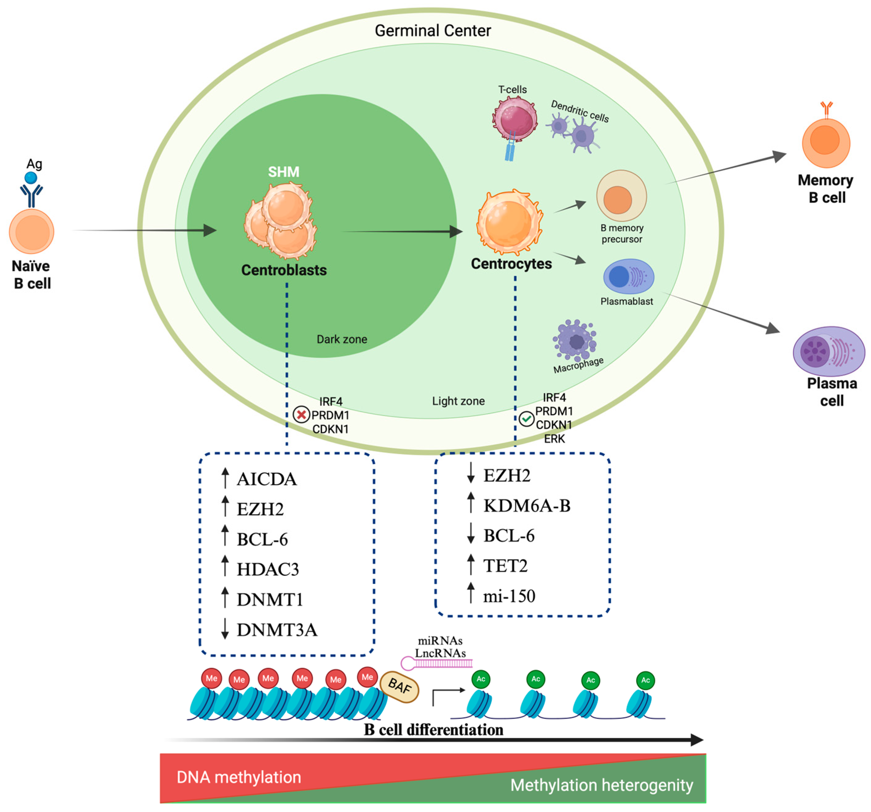
Figure 1. Epigenetic overview of B cell maturation. The main epigenetic regulators include DNA methylation, histone modifications, chromatin remodeling, and lnc-RNAs. The interplay among these four modifications regulates gene expression and shapes the molecular features that are specific to each cell subtype during B cell maturation. In centroblasts, DNA methylation mediated by DNMT1 is frequent while DNMT3 expression is downregulated. During this stage, AICDA is responsible for SHM introducing point mutations at cytosine residues. EZH2 is upregulated, inducing histone hypermethylation, silencing IRF4, PRMD1 (genes involved in plasma cells differentiation) and decreasing CDKN1levels. High levels of BCL-6 allow deacetylation modulated by HDAC3. In centrocytes, EZH2 levels decrease, resulting in transcription of genes involved in cell differentiation. DNA methylation levels are heterogenous among later stages of differentiation, reflecting heterogeneity of cell differentiation. Acetylation mediated by HAC promotes gene expression regulated by BAF complex and BET proteins. Increased mi-150 levels in late stage of maturation are necessary to complete B cell differentiation and GC exit. Created in BioRender.com. Gaidano G. (2025) https://BioRender.com/ddw3e9n (accessed on 19 August 2025). Lnc-RNA, long noncoding RNA; DNMT, DNA methyltransferase; AICDA, activation induced cytidine deaminase; SHM, Somatic Hypermutation; Ac, acetylation; Me: methylation.
Ijms 26 08132 g001
Figure 2. Mutations and main epigenetic regulators in B cell malignancies. The most common epigenetic regulators and their mutations are listed in colored boxes representing the most frequent mature B cell malignancies: (blue = DLBCL; orange = FL; red = CLL). Arrows indicate the functional consequences of mutations or deregulated non-coding RNAs on signaling pathways. Colored stars define the disease context in which each alteration occurs. Numbers identify the genes and the epigenetic regulators involved. Their impact on different cell pathways is reported by colored stars which define the disease; numbers identify the genes and the epigenetic regulators involved. CREBBP loss of function mutations result in overactivation of NOTCH and BCR pathways in DLBCL and FL. KMT2D mutations significantly impact on JAK-STAT and BCR signaling in FL. RAS-ERK and MYC pathways are activated in DLBCL. Increased mi-21 and mi-125 levels activate mTOR and NF-kB pathways in DLBCL, thus promoting proliferation. Mi-150 promotes FOXP1 expression, mediating B cell transformation in DLBCL, FL and CLL. ARID1A mutations result in increased tumor progression in FL and CLL. Hypermethylation of SFRP1 induces its own downregulation as well as the hyperactivation of WNT signaling in CLL. Increased mi-155 levels enhance BCR pathway, mi-101 downregulation leads to EZH2 overexpression and deletion of mi-15/16 increases BCL-2 expression promoting survival in CLL cells. Created in BioRender.com. Gaidano G. (2025) https://BioRender.com/nufxqjt (accessed on 19 August 2025). DLBCL, diffuse large B cell lymphoma; FL, follicular lymphoma; CLL, chronic lymphocytic leukemia.
Ijms 26 08132 g002
Figure 3. Therapeutic strategies for epigenetic alterations in B cell malignancies. The four main epigenetic modifications and their inhibitors are represented in the figure. Colored asterisks identify the diseases for which the drug is approved, or studies that are ongoing. (blue = DLBCL; orange = FL; red = CLL). Created in BioRender.com. Gaidano G. (2025) https://BioRender.com/nv3vzvq (accessed on 19 August 2025). HMTi, histone methyltransferase inhibitor; HDACi, histone deacetylase inhibitor; DNMTi, DNA methyltransferase inhibitor; BETi, bromodomain and extraterminal motif inhibitors.
Ijms 26 08132 g003
Table 1. Ongoing clinical trials of epigenetic drugs in DLBCL, FL, and CLL.
DrugEpigeneitc TargetPhaseSettingDiseasePrimary OutcomeIdentifier
Decitabine + anti PD-1DNMTPhase IIR/RDLBCLORRNCT05816746
ASTX727 + NivolumabDNMTPhase IR/RNHL and HLSafetyNCT05272384
Tazemetostat + MosunetuzumabEZH2Phase IIFirst-lineFLCRNCT05994235
Tulmimetostat DZR123
(CPI-0209)
EZH2Phase I/IIR/RB and T lymphomas, and solid tumorsSafety
ORR
NCT04104776
Tazemetostat + CARTEZH2Phase IR/RDLBCL, FL, and MCLSafetyNCT05934838
tazemetostat + lenalidomide + rituximabEZH2Phase I/IIR/RFLSafety
PFS
NCT04224493
Mevrometostat (PF-06821497)EZH2Phase IR/RFLSafetyNCT03460977
MevrometostatEZH2Phase IR/RFL, SCLC, and CRPCSafetyNCT03460977
Tazemetostat + BelinostatEZH2 + HDACsPhase IR/RB-NHL and T-NHLSafetyNCT05627245
HH2853EZH2Phase I/IIR/RDLBCL, and FLSafety
ORR
NCT04390737
SHR2554 + SHR1701EZH2Phase I/IIR/RB-cell lymphoma, and
Advanced/metastatic solid tumors
PFSNCT04407741
Chidamide + DecitabineHDACs + DNMT1Phase I/IIR/RNHL after CART cellsCR
AE
NCT04337606
Chidamide + LinperlisibHDACs + PI3KδPhase ⅡR/RFLCRNCT06158386
PurinostatHDACPhase IIR/RDLBCLORRNCT05563844
AbexinostatHDACPhase IIR/RDLBCLORRNCT03936153
Etinostat + ZEN003694HDAC + BETPhase I/IIR/RLymphoma, and advanced solid tumorsSafety
ORR
NCT05053971
Vorinostat + pembrolizumabHDACPhase IR/RDLBCL, FL, and HLSafetyNCT03150329
Tazemetostat + epcoritamabEZH2Phase IIR/RFLSafety
CR
NCT06575686
AbexinostatHDACPhase IIR/RFLCR or PRNCT03600441
Valemetostat + LenalidomideEZH1/EZH2Phase I/IIR/RFLSafetyNCT05683171
ValemetostatEZH1/EZH2Phase IIR/RB-cell lymphomaORRNCT04842877
Pevoneidstat + IbrutinibNEDD8-activating enzymePhase IR/RCLL + B-NHLSafetyNCT03479268
TulmimetostatEZH1/EZH2Phase I/IIR/RSolid tumors + LymphomaSafety
ORR
NCT04104776
Abbreviations: DNMT, DNA methyl transferase; R/R, relapsed/refractory; DLBCL, diffuse large B cell lymphoma; ORR, overall response rate; NHL, non-Hodgkin lymphoma; HL, Hodgkin lymphoma; EZH2, enhancer of zeste homolog 2; FL, follicular lymphoma; CR, complete remission; CART, chimeric antigene receptor T cells; MCL, mantle cell lymphoma; SCLC, small cell lung cancer; CRPR castration resistant prostate cancer; HDAC, histone deacetylase; BET, bromodomain and extraterminal motif.
Disclaimer/Publisher’s Note: The statements, opinions and data contained in all publications are solely those of the individual author(s) and contributor(s) and not of MDPI and/or the editor(s). MDPI and/or the editor(s) disclaim responsibility for any injury to people or property resulting from any ideas, methods, instructions or products referred to in the content.