Article

AI-Based Facial Phenotyping Supports a Shared Molecular Axis in PACS1-, PACS2-, and WDR37-Related Syndromes

1
Unit of Clinical Genetics and Functional Genomics, Department of Pharmacology and Physiology, School of Medicine, University of Zaragoza, CIBERER-GCV2 and IIS-Aragon-GIIS062, 50009 Zaragoza, Spain
2
Clinical and Molecular Genetics Area, Vall d’Hebron Hospital, Medicine Genetics Group, Vall d’Hebron Research Institute (VHIR), 08035 Barcelona, Spain
3
Unit of Neurophysiology, University Hospital Lozano Blesa, 50009 Zaragoza, Spain
4
Unit of Paediatric Cardiology, Service of Paediatrics, University Hospital Miguel Servet, 50009 Zaragoza, Spain
5
Unit of Clinical Genetics, Service of Paediatrics, Department of Paediatrics, University Hospital Lozano Blesa, School of Medicine, University of Zaragoza, CIBERER-GCV2 and IIS-Aragon-GIIS062, 50009 Zaragoza, Spain
*
Authors to whom correspondence should be addressed.
Int. J. Mol. Sci. 2025, 26(16), 7964; https://doi.org/10.3390/ijms26167964
Submission received: 17 June 2025 / Revised: 6 August 2025 / Accepted: 8 August 2025 / Published: 18 August 2025
(This article belongs to the Section Molecular Biology)

Abstract

Despite significant advances in gene discovery, the molecular basis of many rare genetic disorders remains poorly understood. The concept of disease modules, clusters of functionally related genes whose disruption leads to overlapping phenotypes, offers a valuable framework for interpreting these conditions. However, identifying such relationships remains particularly challenging in ultra-rare syndromes due to the limited number of documented cases. We hypothesized that AI-based facial phenotyping could aid in identifying shared molecular mechanisms by detecting phenotypic convergence among clinically related syndromes. To test this, we used Schuurs–Hoeijmakers syndrome (SHMS; OMIM #615009), caused by a recurrent de novo variant in PACS1, as a model to explore potential phenotypic and functional associations with PACS2-related disorder (DEE66; OMIM #618067) and WDR37-related disorder (NOCGUS; OMIM #618652). Facial photographs of individuals with SHMS were analyzed using the DeepGestalt and GestaltMatcher algorithms. In addition to consistently recognizing SHMS as a distinct clinical entity, the algorithms frequently matched DEE66 and NOCGUS, suggesting a shared facial gestalt. Binary comparisons further confirmed overlapping craniofacial features among the three disorders. These findings were supported by literature review, indicating clinical overlapping and potential functional associations. Overall, our results confirm the presence of consistent facial similarities among PACS1-, PACS2-, and WDR37-related syndromes and highlight the utility of AI-driven facial phenotyping as a complementary tool for uncovering clinically relevant relationships in ultra-rare genetic disorders.

1. Introduction

Rare genetic disorders (RGDs) affect an estimated 3.5–5.9% of the global population [1], with more than 7000 conditions currently described and nearly 5000 genes linked to phenotype-causing mutations [2]. The increasing use of high-throughput sequencing technologies, along with international research collaborations, has greatly accelerated the discovery of novel disease-associated genes over the past decade. Despite these advances, understanding the underlying biological mechanisms of RGDs remains a major challenge, particularly in the context of ultra-rare conditions [3,4,5].
More than 80% of RGDs manifest during childhood and frequently affect the central nervous system (CNS), with neurological symptoms reported in over 70% of cases [6,7,8]. Despite their marked genetic and clinical heterogeneity, growing evidence from network-based studies indicates that disease-associated genes often cluster within interconnected biological pathways, such as synaptic transmission, chromatin remodeling, or mTOR signaling, rather than acting in isolation [9,10]. These findings support the concept of disease modules: groups of functionally related genes whose disruption results in overlapping phenotypes [11].
Identifying such relationships in ultra-rare disorders is particularly difficult due to the small number of reported cases. However, phenotypic similarities, especially those involving craniofacial features, can provide important diagnostic and biological clues. Many RGDs, particularly NDDs, exhibit recurrent craniofacial patterns or a recognizable “gestalt” that can guide clinical and molecular hypothesis generation. The emergence of artificial intelligence (AI)-based facial phenotyping tools has transformed this process [12,13,14]. Algorithms such as DeepGestalt and GestaltMatcher, implemented in platforms like Face2Gene, use deep learning to detect and compare facial features, suggesting candidate syndromes or phenotypically similar conditions based on a patient’s facial image [15,16,17].
In addition to their diagnostic value, AI-based facial phenotyping tools have been proposed as a means to uncover biologically meaningful relationships among rare disorders [18]. In this study, we explore that potential by analyzing individuals with Schuurs–Hoeijmakers syndrome (SHMS, OMIM #615009), also known as PACS1 syndrome, a rare autosomal dominant NDD caused by a recurrent de novo variant in the PACS1 gene (c.607C>T, p.Arg203Trp) [19]. PACS1 encodes a multifunctional protein implicated in intracellular trafficking [20], chromatin regulation [21], and neural development [22]. However, the precise pathophysiological consequences of this variant remain poorly understood [23]. Notably, previous studies have identified clinical and molecular similarities between SHMS and disorders associated with variants in PACS2 (DEE66; OMIM #618067) and WDR37 (NOCGUS; OMIM #618652) [24,25,26], suggesting the existence of a PACS1–PACS2–WDR37 axis that may underlie shared phenotypic features across their respective syndromes.
Building on these observations, the aim of this study is to assess whether AI-driven facial analysis can detect phenotypic convergence among PACS1-, PACS2-, and WDR37-related syndromes, and whether such resemblance aligns with known or hypothesized functional relationships. While facial similarity patterns can be informative, they must be interpreted with caution and supported by additional biological evidence. To this end, we complemented our facial analysis with structured phenotypic annotation using the Human Phenotype Ontology (HPO) and a comprehensive review of the relevant literature. This integrative approach allows for a more robust interpretation of phenotypic convergence and supports the use of AI-driven phenotyping as a complementary strategy to identify biologically meaningful relationships among RGDs.

2. Results

2.1. Dysmorphic Facial Phenotype in Individuals with Schuurs–Hoeijmakers Syndrome

We report on 14 patients with the genetic variant c.607C>T, p.(Arg203Trp) in the PACS1 gene. The cohort comprised four females and ten males, aged between 2 and 35 years. A clinical summary of their facial features is presented in Figure 1. Overall, the most common craniofacial features observed included thin upper lip vermilion (HP:0000219), smooth philtrum (HP:0000319), wide mouth (HP:0000154), broad and prominent nasal tip (HP:0000455, HP:0005274), and bulbous nose (HP:0000414) (Figure 1).
Facial photographs of the 14 individuals were analyzed using the Face2Gene CLINIC application with the DeepGestalt algorithm (v.22.3.0). These photos had not been used in any prior training of the algorithm. SHMS was recommended among the top 30 syndromes and ranked as the first diagnosis for 42.86% (n = 6) of individuals, second for 14.26% (n = 2), and third for 21.43% (n = 3). Overall, 92.86% (n = 13) of the patient photos analyzed had SHMS ranked in their top ten potential diagnoses out of the 30 possible suggested syndromes, from among the more than 350 syndromes currently recognized by the DeepGestalt algorithm. Among these 13 with SHMS in the top ten rank, the median gestalt score was 0.30 ± 0.16 (Table S1).
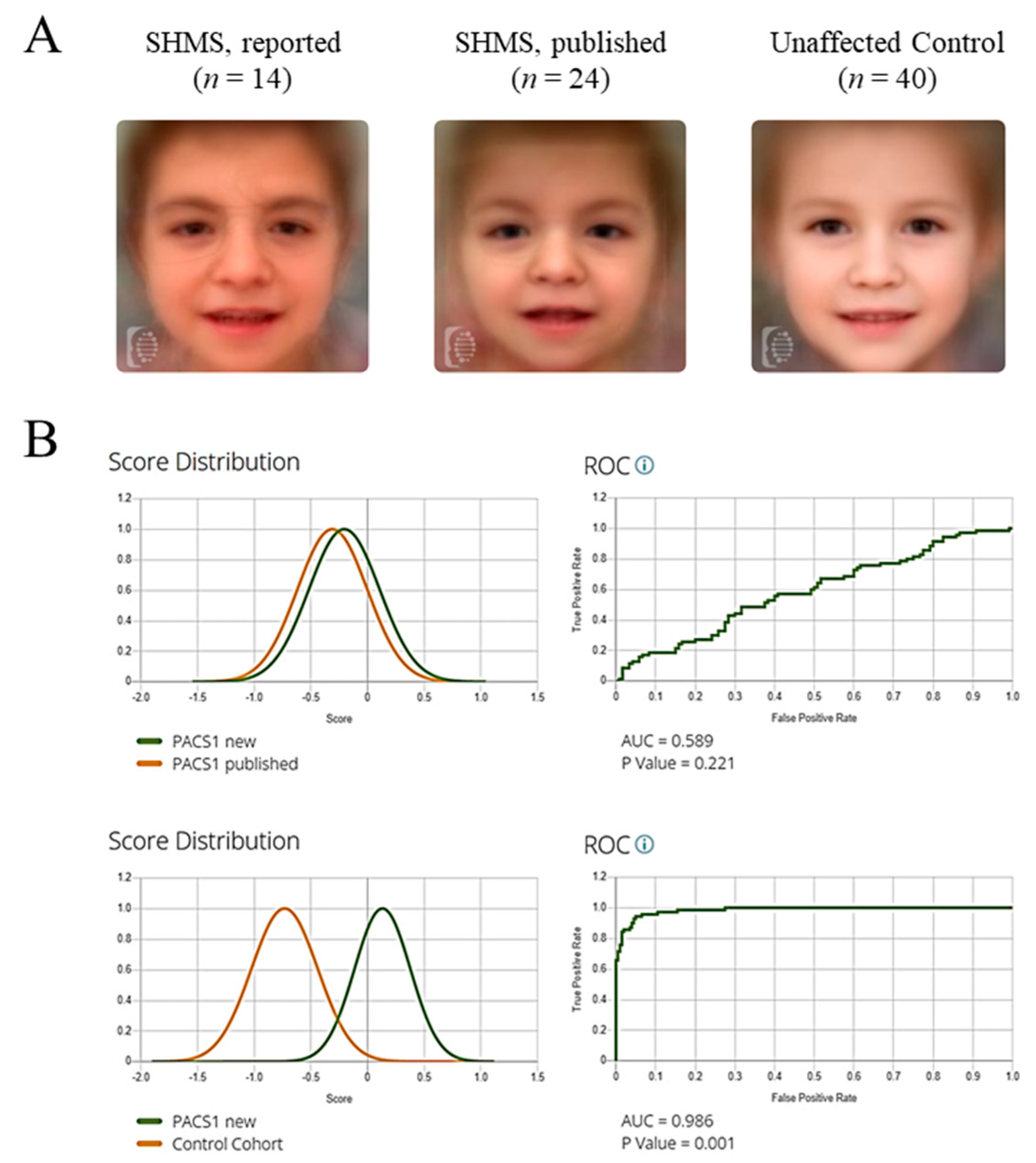
To evaluate facial similarity between our cohort (n = 14) and previously published cases of SHMS (n = 24) (Table S2) [19,27,28,29,30,31,32], we used binary comparison in the Face2Gene RESEARCH (v.22.3.0) application. Model performance was assessed using Receiver Operating Characteristic (ROC) curves, with the Area Under the Curve (AUC) reflecting discriminatory power. An AUC close to 1.0 indicates strong performance; values near 0.5 suggest random classification. The comparison yielded an AUC of 0.589 (p = 0.221), indicating no significant difference between the two SHMS groups. In contrast, a comparison between unaffected individuals (n = 40) and SHMS patients yielded an AUC of 0.986 with statistical significance (p < 0.001), confirming that SHMS presents a reasonably distinguishable facial gestalt (Figure 2). It is important to note that factors such as patient age, sex, and photo quality may influence the performance of the DeepGestalt algorithm. While this study did not systematically analyze these variables, the consistent identification of SHMS across varied demographics suggests a robust performance.

2.2. Common Differential Syndromes of SHMS Based on Gestalt Analysis

Based on the results above, photographs of the 14 individuals with SHMS from the present cohort and the 24 from previous publications were analyzed as a single group (n = 38) using DeepGestalt and GestaltMatcher (v.1.2.0) algorithms. We inquired about the common syndromes suggested in more than 50% of the photographs analyzed.
First, we applied the DeepGestalt algorithm. In addition to SHMS, another 10 syndromes were suggested among the 30 top syndrome matches (Table 1). Next, we examined ultra-rare syndromes using the GestaltMatcher algorithm. Curiously, developmental and epileptic encephalopathy-66 (DEE66; OMIM #618067), caused by alterations in PACS2 gene, was suggested in 33 of the 38 patients analyzed. The combination of both algorithms, DeepGestalt and GestaltMatcher, is graphically displayed as a tSNE plot in the Face2Gene CLINIC app. Using this application, the top syndromes matched by both algorithms were SHMS (37/38); DEE66 (30/38); Verheij syndrome (VRJS, OMIM #615583) (26/38), caused by a gene deletion involving the PUF60 gene; neuro-oculo-cardio-genitourinary syndrome (NOCGUS, OMIM #618652) (23/38), caused by heterozygous variants in the WDR37 gene; and Baraitser–Winter syndrome 1 (BRWS1, OMIM #243310) (20/38), caused by alterations in the ACTB gene (Table 1). Interestingly, the two syndromes previously proposed to share a molecular basis with PACS1, DEE66, and NOCGUS, emerged prominently in this analysis.

2.3. Quantification of Facial Gestalt Similarities Between Syndromes

Both DeepGestalt and GestaltMatcher quantify facial similarities using deep convolutional neural networks. We applied these tools to assess potential phenotypic overlaps that might indicate shared molecular pathways. In addition to individuals with SHMS (n = 38), we included publicly available facial images from academic publications of patients with DEE66 (n = 11) [25,33,34,35,36,37], VRJS (n = 23) [38,39,40,41], NOCGUS (n = 10) [42,43,44], and BRWS1 (n = 32) [45,46,47,48,49,50] (Table S3). These syndromes were prioritized for binary comparisons due to their consistent co-suggestion by the AI-based algorithms.
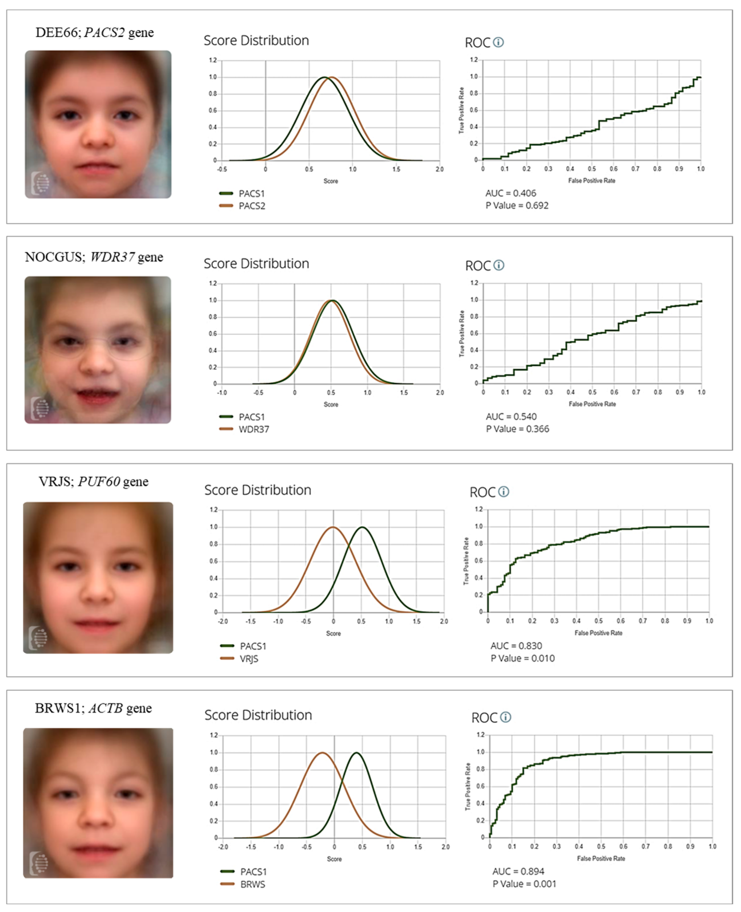
Binary comparisons revealed no statistically significant differences between SHMS and DEE66 (AUC = 0.406; p = 0.692), SHMS and NOCGUS (AUC = 0.540; p = 0.366), or DEE66 and NOCGUS (AUC = 0.493; p = 0.458), suggesting potential facial overlap among these PACS1-, PACS2-, and WDR37-related syndromes and potential molecular convergence. In contrast, the algorithm was able to differentiate SHMS from VRJS (AUC = 0.830; p = 0.010) and BRWS1 (AUC = 0.894; p = 0.001), supporting the distinctiveness of the SHMS facial phenotype in these comparisons (Figure 3).
Given the historical diagnostic overlap between SHMS and Cornelia de Lange syndrome (CdLS), and the fact that CdLS appeared among the top matches in over 50% of SHMS cases (Table 1), we also assessed this comparison. SHMS and CdLS were clearly differentiated (AUC = 0.996; p < 0.001), further reinforcing the specificity of the SHMS facial phenotype (Figure S1).
Taken together, these findings suggest that PACS1-, PACS2-, and WDR37-related syndromes share a recognizable facial signature that may reflect a common biological basis.

3. Discussion

Many rare genetic disorders (RGDs) are characterized by distinctive craniofacial features, making dysmorphological assessment a long-standing cornerstone of clinical genetics. In recent years, the integration of Human Phenotype Ontology (HPO) terminology and AI-based image analysis tools has significantly advanced the field, enabling the extraction of quantitative phenotypic descriptors from facial photographs. These technologies have enhanced diagnostic accuracy and supported clinical decision-making in genetics [51,52].
In this context, the application of AI-driven facial phenotyping in our study provides new perspectives on phenotypic and functional convergence among RGDs with overlapping facial features. Using Schuurs–Hoeijmakers syndrome (SHMS, or PACS1 syndrome) as a reference model, we demonstrate how computational analysis can refine syndrome-specific facial profiling and reveal phenotypic relationships across genetically unrelated conditions. The consistent identification of PACS1 syndrome by both DeepGestalt and GestaltMatcher supports the existence of a distinctive and well-characterized facial gestalt, reinforcing its classification as a distinct clinical entity [27,28,29,53,54]. In our cohort, DeepGestalt correctly ranked SHMS within the top 10 diagnoses in 92.86% of cases, an outcome consistent with its reported performance across other RGDs [16,17,55]. Beyond their diagnostic potential, the frequent co-identification of SHMS with Developmental and Epileptic Encephalopathy 66 (DEE66, linked to PACS2 variants) and Neuro-Oculo-Cardio-Genitourinary syndrome (NOCGUS, caused by WDR37 variants) suggests notable facial similarity despite distinct molecular etiologies. The low AUC values observed in binary comparisons among these syndromes indicate a high degree of morphological resemblance, demonstrating the sensitivity of AI-based approaches in detecting subtle phenotypic convergence. Importantly, these tools also preserved their discriminatory power, as evidenced by their ability to distinguish SHMS from Cornelia de Lange syndrome (CdLS), a historically difficult differential diagnosis. Taken together, these findings emphasizes the dual utility of AI-driven facial analysis, both as a diagnostic aid and as a hypothesis-generating tool for exploring shared developmental mechanisms in ultra-rare syndromes.
Nevertheless, our findings should be interpreted with caution. The limited ability of the model to distinguish SHMS, DEE66, and NOCGUS may reflect genuine phenotypic convergence, but could also result from technical constraints or small sample sizes, particularly for DEE66 and NOCGUS, which may affect generalizability and increase the risk of overfitting. These limitations must be considered when inferring shared molecular mechanisms from AI-derived phenotypic similarities. Looking ahead, emerging multimodal AI systems, including those incorporating large language models, may further improve diagnostic precision by integrating facial, clinical, genetic, and textual data [56,57].
From a clinical standpoint, syndromes associated with PACS1, PACS2, and WDR37 display marked phenotypic overlap. All three present with facial dysmorphism, significant neurodevelopmental impairment, and a wide range of neurological features [23]. Although PACS2-related syndrome primarily affects the nervous system, it also presents with clinical signs common to PACS1 and WDR37 syndromes, including feeding difficulties, postnatal growth retardation, and variable skeletal, cardiac, and genitourinary anomalies [56]. Ocular abnormalities like corneal opacity and coloboma are more frequent in WDR37 cases but have also been reported with PACS1 and PACS2 variants [24]. Seizures are a prominent hallmark across all three syndromes, particularly severe and often refractory in PACS2 and WDR37, with onset typically in early infancy [56]. Cerebellar hypoplasia and other brain abnormalities are frequently observed, especially in WDR37-related presentations, which also tend to show more profound neurocognitive impairment and multisystem involvement, including rare cases of early mortality [24,42]. In terms of craniofacial gestalt, common features include hypertelorism, broad nasal bridge, bulbous nose, short and smooth philtrum, thin upper lip, and wide mouth. While some traits such as arched eyebrows, sparse lateral eyebrows, and prominent pinnae appear more frequently in PACS2- and WDR37-related cases, these differences are subtle and not exclusive. Mild distinctions can be noted; triangular facial shape tends to be more frequently reported in PACS1, microcephaly, craniosynostosis, and auricular anomalies in WDR37, and synophrys or the absence of a smooth philtrum in a subset of PACS2 cases.
This significant phenotypic overlap supports the hypothesis that PACS1, PACS2, and WDR37 may participate in a shared molecular pathway involved in neurodevelopment and craniofacial morphogenesis. Transcriptomic data show that all three genes are expressed during early embryogenesis, particularly in neural crest-derived tissues relevant to facial development. PACS1, for instance, demonstrates high expression in the developing brain and facial mesenchyme, suggesting its role in early craniofacial patterning [58]. This aligns with current models of neurodevelopmental disorders, which emphasize the coordinated regulation of craniofacial and brain development by neural crest gene regulatory networks, and how their disruption can result in overlapping syndromic features [59].
At the molecular level, PACS1 and PACS2 are evolutionarily conserved paralogs that regulate intracellular trafficking by interacting with acidic cluster motifs via their furin-binding region (FBR). They are involved in the localization of key cytoplasmic proteins such as receptors, proteases, and ion channels [60,61,62,63], and also engage in nuclear processes related to chromatin regulation and DNA repair through interactions with class I and III histone deacetylases (HDACs) [21,64]. Beyond their well-established role in protein trafficking, PACS proteins have been identified as interaction partners of WDR37, a conserved WD40 repeat-containing protein, across multiple species ranging from invertebrates to mammals. The interaction between PACS1 and WDR37 has been shown to influence endoplasmic reticulum (ER) calcium signaling [25,65]. This interaction supports their functional interdependence and provides strong evidence that PACS1, PACS2, and WDR37 constitute a conserved molecular axis.
In summary, this study highlights the potential of combining AI-driven facial phenotyping with molecular and clinical data to uncover biologically meaningful relationships in RGDs. Our findings suggest that PACS1-, PACS2-, and WDR37-associated syndromes form a coherent phenotypic and molecular cluster. While further validation is necessary, this integrative approach offers a scalable framework to identify hidden connections among RGDs, with potential implications for diagnosis, disease classification, and the understanding of potential biological links among ultra-rare syndromes.

4. Materials and Methods

4.1. Patient Recruitment and Clinical Imaging

Clinical and genotyping information from 14 unrelated individuals diagnosed with PACS1 syndrome was obtained. Participants were recruited through a call for collaboration by the Spanish PACS1 Patient Association. Inclusion criteria comprised a confirmed clinical and molecular diagnosis of SHMS, availability of a detailed clinical evaluation, and the presence of a frontal facial photograph. Informed consent was obtained from parents or guardians of all individuals included in this study, in accordance with the local ethics committee (CEICA; PI15/00707, PI24/288). Written informed consent for the publication of photographs was also obtained. Facial images were uploaded to the Face2Gene CLINIC App for phenotypic analysis. Additional photographs used in this work were obtained from scientific literature, as appropriately referenced in each section. All selected images were standardly cropped, centered, and aligned. Control photographs, matched for age, sex, and ethnicity, were supplied by Face2Gene technical support. Photographs of individuals with Cornelia de Lange syndrome (CdLS) were obtained under the same ethics approval (CEICA; PI15/00707, PI24/288) and were collected through collaboration with the Spanish Cornelia de Lange Association.

4.2. Clinical Facial Deep Phenotyping

Phenotypic characterization of patients was performed by a US Board-certified clinical geneticist during clinical evaluation. Clinical data were collected using a standardized restricted-term questionnaire, and detailed phenotypic descriptions of the individuals were annotated using the Human Phenotype Ontology (HPO) nomenclature (https://hpo.jax.org/, accessed on 3 March 2025). A total of 42 HPO terms related to craniofacial features were included in the questionnaire.

4.3. Computational Facial Analysis

Computational facial analysis was performed by Face2Gene (F2G) applications (FDNA Inc., Boston, MA, USA) using three algorithms: DeepGestalt (v.22.3.0), GestalMatcher (v.1.2.0) and the newly described facial D-score (v.2.0). The DeepGestalt algorithm first preprocesses each photo to detect facial landmarks and align the face, which is then cropped into facial regions. Each region is fed into a Deep Convolutional Neural Network (DCNN) to generate a descriptor and a softmax vector indicating its correspondence to each syndrome included in the model. The output vectors of all regional DCNNs are then aggregated and sorted to produce the final ranked list of 30 suggested genetic syndromes displayed in Face2Gene, each assigned a gestalt score, where higher values reflect greater similarity in facial morphology to a particular disorder [16]. GestaltMatcher uses these descriptors to create a “Clinical Face Phenotype Space”, in which the distance between photos defines syndromic similarity, calculated via cosine distance in that space and expressed as a “similarity score” [19]. The facial D-score algorithm uses the same descriptors to distinguish between two classes of frontal facial photos: images of patients diagnosed with a rare genetic disease showing facial dysmorphic features, and an equivalently sampled control group of unaffected individuals. Scores above 0.75 indicate a likely presence of facial dysmorphia [35].

Supplementary Materials

The following supporting information can be downloaded at https://www.mdpi.com/article/10.3390/ijms26167964/s1.

Author Contributions

Conceptualization, J.d.R., F.J.R., A.L.-P., and J.P.; methodology, M.G.-S., A.L.-P., C.L.-C., M.A., and B.P.; software, J.d.R., and A.L.-P.; validation, J.d.R., L.T., L.A., M.G.-S., and C.L.-C.; formal analysis, J.d.R., L.T., L.A., P.P., and A.A.-C.; investigation, J.d.R., L.T., L.A., P.P., A.A.-C., M.G.-S., A.L.-P., C.L.-C., M.A., and B.P.; resources, P.P., A.A.-C., M.A., and B.P.; data curation, A.L.-P., and J.d.R.; writing—original draft preparation, A.L.-P.; writing—review and editing, A.L.-P., F.J.R., and J.P.; visualization, J.d.R., A.L.-P., and J.P.; supervision, F.J.R., and J.P.; project administration, F.J.R., and J.P.; funding acquisition, A.L.-P., F.J.R., and J.P. All authors have read and agreed to the published version of the manuscript.

Funding

This research was funded by Spanish Ministry of Health-ISCIII Fondo de Investigación Sanitaria (FIS) [Ref. PI23/01370, to F.J.R. and J.P.] and co-founded by European Union; Diputación General de Aragón-FEDER: European Social Fund [Reference Group B32_20R, to J.P.]; University of Zaragoza [JIUZ2023-SAL-06 to A.L.-P].

Institutional Review Board Statement

The study was conducted in accordance with the Declaration of Helsinki, and approved by the Ethics and Clinical Research Committee of the Government of Aragon (Spain) (CEICA; PI15/00707, PI24/288, date of approval 12 June 2024).

Informed Consent Statement

Informed consent was obtained from all subjects involved in the study. Written informed consent has been obtained from the patients or their legal guardians to publish this paper.

Data Availability Statement

The raw data supporting the conclusions of this article will be made available by the authors on request.

Acknowledgments

We are grateful to the patient and her parents for participating in this study, as well as to the Spanish Association of Families with PACS1. We also sincerely thank Rebeca Antoñanzas-Pérez for her valuable technical assistance and Nicole Fleischer of FDNA Inc. (Boston, MA, USA), provider of Face2Gene, for her kind cooperation during this study.

Conflicts of Interest

The authors declare no conflicts of interest.

Abbreviations

The following abbreviations are used in this manuscript:
AIArtificial intelligence
BRWS1Baraitser–Winter syndrome 1
CdLSCornelia de Lange syndrome
CEICAEthics and clinical research committee of the Government of Aragon
CNSCentral nervous system
DEE66Developmental and Epileptic Encephalopathy-66
F2GFace2Gene
HDACHistone deacetylase
HPOHuman phenotype ontology
NDDNeurodevelopmental disorder
NOCGUSNeuro-Oculo-Cardio-Genitourinary syndrome
OMIMOnline Mendelian Inheritance in Man
RGDRare genetic disorder
SHMSSchuurs–Hoeijmakers syndrome
PPIProtein–protein interaction
VRJSVerheij syndrome

References

  1. Wakap, S.N.; Lambert, D.M.; Olry, A.; Rodwell, C.; Gueydan, C.; Lanneau, V.; Murphy, D.; Le Cam, Y.; Rath, A. Estimating cumulative point prevalence of rare diseases: Analysis of the Orphanet database. Eur. J. Hum. Genet. 2020, 28, 165–173. [Google Scholar] [CrossRef]
  2. Online Mendelian Inheritance in Man, OMIM®. McKusick-Nathans Institute of Genetic Medicine, Johns Hopkins University (Baltimore, MD). Available online: https://omim.org (accessed on 22 July 2025).
  3. Monaco, L.; Zanello, G.; Baynam, G.; Jonker, A.H.; Julkowska, D.; Hartman, A.L.; O’cOnnor, D.; Wang, C.M.; Wong-Rieger, D.; Pearce, D.A. Research on rare diseases: Ten years of progress and challenges at IRDiRC. Nat. Rev. Drug. Discov. 2022, 21, 319–320. [Google Scholar] [CrossRef] [PubMed]
  4. Boycott, K.M.; Rath, A.; Chong, J.X.; Hartley, T.; Alkuraya, F.S.; Baynam, G.; Brookes, A.J.; Brudno, M.; Carracedo, A.; Dunnen, J.T.D.; et al. International Cooperation to Enable the Diagnosis of All Rare Genetic Diseases. Am. J. Hum. Genet. 2017, 100, 695–705. [Google Scholar] [CrossRef] [PubMed]
  5. Bamshad, M.J.; Nickerson, D.A.; Chong, J.X. Mendelian Gene Discovery: Fast and Furious with No End in Sight. Am. J. Hum. Genet. 2019, 105, 448–455. [Google Scholar] [CrossRef] [PubMed]
  6. McRae, J.F.; Clayton, S.; Fitzgerald, T.W.; Kaplanis, J.; Prigmore, E.; Rajan, D.; Sifrim, A.; Aitken, S.; Akawi, N.; Alvi, M.; et al. Prevalence and architecture of de novo mutations in developmental disorders. Nature 2017, 542, 433–438. [Google Scholar] [CrossRef]
  7. Sanders, S.J.; Sahin, M.; Hostyk, J.; Thurm, A.; Jacquemont, S.; Avillach, P.; Douard, E.; Martin, C.L.; Modi, M.E.; Moreno-De-Luca, A.; et al. A framework for the investigation of rare genetic disorders in neuropsychiatry. Nat. Med. 2019, 25, 1477–1487. [Google Scholar] [CrossRef]
  8. Lee, C.E.; Singleton, K.S.; Wallin, M.; Faundez, V. Rare Genetic Diseases: Nature’s Experiments on Human Development. iScience 2020, 23, 101123. [Google Scholar] [CrossRef]
  9. Barabási, A.L.; Gulbahce, N.; Loscalzo, J. Network medicine: A network-based approach to human disease. Nat. Rev. Genet. 2011, 12, 56–68. [Google Scholar] [CrossRef]
  10. Parenti, I.; Rabaneda, L.G.; Schoen, H.; Novarino, G. Neurodevelopmental Disorders: From Genetics to Functional Pathways. Trends Neurosci. 2020, 43, 608–621. [Google Scholar] [CrossRef]
  11. Buphamalai, P.; Kokotovic, T.; Nagy, V.; Menche, J. Network analysis reveals rare disease signatures across multiple levels of biological organization. Nat. Commun. 2021, 12, 6306. [Google Scholar] [CrossRef]
  12. Porras, A.R.; Rosenbaum, K.; Tor-Diez, C.; Summar, M.; Linguraru, M.G. Development and evaluation of a machine learning-based point-of-care screening tool for genetic syndromes in children: A multinational retrospective study. Lancet Digit. Health 2021, 3, e635–e643. [Google Scholar] [CrossRef]
  13. Hennocq, Q.; Willems, M.; Amiel, J.; Arpin, S.; Attie-Bitach, T.; Bongibault, T.; Bouygues, T.; Cormier-Daire, V.; Corre, P.; Dieterich, K.; et al. Next generation phenotyping for diagnosis and phenotype-genotype correlations in Kabuki syndrome. Sci. Rep. 2024, 14, 2330. [Google Scholar] [CrossRef]
  14. Ciancia, S.; Goedegebuure, W.J.; Grootjen, L.N.; Hokken-Koelega, A.C.S.; Kerkhof, G.F.; van der Kaay, D.C.M. Computer-aided facial analysis as a tool to identify patients with Silver-Russell syndrome and Prader-Willi syndrome. Eur. J. Pediatr. 2023, 182, 2607–2614. [Google Scholar] [CrossRef] [PubMed]
  15. Gurovich, Y.; Hanani, Y.; Bar, O.; Nadav, G.; Fleischer, N.; Gelbman, D.; Basel-Salmon, L.; Krawitz, P.M.; Kamphausen, S.B.; Zenker, M.; et al. Identifying facial phenotypes of genetic disorders using deep learning. Nat. Med. 2019, 25, 60–64. [Google Scholar] [CrossRef] [PubMed]
  16. Pantel, J.T.; Hajjir, N.; Danyel, M.; Elsner, J.; Abad-Perez, A.T.; Hansen, P.; Mundlos, S.; Spielmann, M.; Horn, D.; Ott, C.-E.; et al. Efficiency of Computer-Aided Facial Phenotyping (DeepGestalt) in Individuals with and Without a Genetic Syndrome: Diagnostic Accuracy Study. J. Med. Int. Res. 2020, 22, e19263. [Google Scholar] [CrossRef] [PubMed]
  17. Latorre-Pellicer, A.; Ascaso, Á.; Trujillano, L.; Gil-Salvador, M.; Arnedo, M.; Lucia-Campos, C.; Antoñanzas-Pérez, R.; Marcos-Alcalde, I.; Parenti, I.; Bueno-Lozano, G.; et al. Evaluating face2gene as a tool to identify cornelia de lange syndrome by facial phenotypes. Int. J. Mol. Sci. 2020, 21, 1042. [Google Scholar] [CrossRef]
  18. Hsieh, T.C.; Bar-Haim, A.; Moosa, S.; Ehmke, N.; Gripp, K.W.; Pantel, J.T.; Danyel, M.; Mensah, M.A.; Horn, D.; Rosnev, S.; et al. GestaltMatcher facilitates rare disease matching using facial phenotype descriptors. Nat. Genet. 2022, 54, 349–357. [Google Scholar] [CrossRef]
  19. Schuurs-Hoeijmakers, J.H.M.; Oh, E.C.; Vissers, L.E.L.M.; Swinkels, M.E.M.; Gilissen, C.; Willemsen, M.A.; Holvoet, M.; Steehouwer, M.; Veltman, J.A.; de Vries, B.B.; et al. Recurrent de novo mutations in PACS1 cause defective cranial-neural-crest migration and define a recognizable intellectual-disability syndrome. Am. J. Hum. Genet. 2012, 91, 1122–1127. [Google Scholar] [CrossRef]
  20. Crump, C.M.; Xiang, Y.; Thomas, L.; Gu, F.; Austin, C.; Tooze, S.A.; Thomas, G. PACS-1 binding to adaptors is required for acidic cluster motif-mediated protein traffic. EMBO J. 2001, 20, 2191–2201. [Google Scholar] [CrossRef]
  21. Mani, C.; Tripathi, K.; Luan, S.; Clark, D.W.; Andrews, J.F.; Vindigni, A.; Thomas, G.; Palle, K. The multifunctional protein PACS-1 is required for HDAC2- and HDAC3-dependent chromatin maturation and genomic stability. Oncogene 2020, 39, 2583–2596. [Google Scholar] [CrossRef]
  22. Villar-Pazos, S.; Thomas, L.; Yang, Y.; Chen, K.; Lyles, J.B.; Deitch, B.J.; Ochaba, J.; Ling, K.; Powers, B.; Gingras, S.; et al. Neural deficits in a mouse model of PACS1 syndrome are corrected with PACS1- or HDAC6-targeting therapy. Nat. Commun. 2023, 14, 6547. [Google Scholar] [CrossRef]
  23. Arnedo, M.; Ascaso, Á.; Latorre-Pellicer, A.; Lucia-Campos, C.; Gil-Salvador, M.; Ayerza-Casas, A.; Pablo, M.J.; Gómez-Puertas, P.; Ramos, F.J.; Bueno-Lozano, G.; et al. Molecular Basis of the Schuurs-Hoeijmakers Syndrome: What We Know about the Gene and the PACS-1 Protein and Novel Therapeutic Approaches. Int. J. Mol. Sci. 2022, 23, 9649. [Google Scholar] [CrossRef]
  24. Sakaguchi, Y.; Yoshihashi, H.; Uehara, T.; Miyama, S.; Kosaki, K.; Takenouchi, T. Coloboma may be a shared feature in a spectrum of disorders caused by mutations in the WDR37-PACS1-PACS2 axis. Am. J. Med. Genet. A 2021, 185, 884–888. [Google Scholar] [CrossRef]
  25. Sorokina, E.A.; Reis, L.M.; Thompson, S.; Agre, K.; Babovic-Vuksanovic, D.; Ellingson, M.S.; Hasadsri, L.; van Bever, Y.; Semina, E.V. WDR37 syndrome: Identification of a distinct new cluster of disease-associated variants and functional analyses of mutant proteins. Hum. Genet. 2021, 140, 1775–1789. [Google Scholar] [CrossRef] [PubMed]
  26. Byrd, D.T.; Han, Z.C.; Piggott, C.A.; Jin, Y. PACS-1 variant protein is aberrantly localized in Caenorhabditis elegans model of PACS1/PACS2 syndromes. Genetics 2024, 228, iyae118. [Google Scholar] [CrossRef] [PubMed]
  27. Schuurs-Hoeijmakers, J.H.M.; Landsverk, M.L.; Foulds, N.; Kukolich, M.K.; Gavrilova, R.H.; Greville-Heygate, S.; Hanson-Kahn, A.; Chitayat, D.; Glass, J.; Bernstein, J.A.; et al. Clinical delineation of the PACS1-related syndrome--Report on 19 patients. Am. J. Med. Genet. A 2016, 170, 670–675. [Google Scholar] [CrossRef]
  28. Gadzicki, D.; Döcker, D.; Schubach, M.; Menzel, M.; Schmorl, B.; Stellmer, F.; Biskup, S.; Bartholdi, D. Expanding the phenotype of a recurrent de novo variant in PACS1 causing intellectual disability. Clin. Genet. 2015, 88, 300–302. [Google Scholar] [CrossRef] [PubMed]
  29. Tenorio-Castaño, J.; Morte, B.; Nevado, J.; Martinez-Glez, V.; Santos-Simarro, F.; García-Miñaúr, S.; Palomares-Bralo, M.; Pacio-Míguez, M.; Gómez, B.; Arias, P.; et al. Schuurs-Hoeijmakers Syndrome (PACS1 Neurodevelopmental Disorder): Seven Novel Patients and a Review. Genes 2021, 12, 738. [Google Scholar] [CrossRef]
  30. Hoshino, Y.; Enokizono, T.; Imagawa, K.; Tanaka, R.; Suzuki, H.; Fukushima, H.; Arai, J.; Sumazaki, R.; Uehara, T.; Takenouchi, T.; et al. Schuurs-Hoeijmakers syndrome in two patients from Japan. Am. J. Med. Genet. A 2019, 179, 341–343. [Google Scholar] [CrossRef]
  31. Kurt Colak, F.; Eyerci, N.; Aytekin, C.; Eksioglu, A.S. Renpenning Syndrome in a Turkish Patient: de novo Variant c.607C>T in PACS1 and Hypogammaglobulinemia Phenotype. Mol. Syndromol. 2020, 11, 157–161. [Google Scholar] [CrossRef]
  32. Abdulqader, S.A.; Wli, W.A.; Qaryaqos, S.H. Schuurs-Hoeijmakers syndrome in a patient from Iraq-Kirkuk. Clin. Case Rep. 2021, 9, e04897. [Google Scholar] [CrossRef]
  33. Olson, H.E.; Jean-Marçais, N.; Yang, E.; Heron, D.; Tatton-Brown, K.; van der Zwaag, P.A.; Bijlsma, E.K.; Kamsteeg, E.J.; Backer, E.; Krock, B.L.; et al. A Recurrent De Novo PACS2 Heterozygous Missense Variant Causes Neonatal-Onset Developmental Epileptic Encephalopathy, Facial Dysmorphism, and Cerebellar Dysgenesis. Am. J. Hum. Genet. 2018, 102, 995–1007. [Google Scholar] [CrossRef]
  34. Cesaroni, E.; Matricardi, S.; Cappanera, S.; Marini, C. First reported case of an inherited PACS2 pathogenic variant with variable expression. Epileptic Disord. 2022, 24, 572–576. [Google Scholar] [CrossRef]
  35. Terrone, G.; Marchese, F.; Vari, M.S.; Severino, M.; Madia, F.; Amadori, E.; Del Giudice, E.; Romano, A.; Gennaro, E.; Zara, F.; et al. A further contribution to the delineation of epileptic phenotype in PACS2-related syndrome. Seizure 2020, 79, 53–55. [Google Scholar] [CrossRef]
  36. Sánchez-Soler, M.J.; Serrano-Antón, A.T.; López-González, V.; Guillén-Navarro, E. New case with the recurrent c.635G>A pathogenic variant in the PACS2 gene: Expanding the phenotype. Neurologia 2021, 36, 716–719. [Google Scholar] [CrossRef] [PubMed]
  37. Dentici, M.L.; Barresi, S.; Niceta, M.; Ciolfi, A.; Trivisano, M.; Bartuli, A.; Digilio, M.C.; Specchio, N.; Dallapiccola, B.; Tartaglia, M. Expanding the clinical spectrum associated with PACS2 mutations. Clin. Genet. 2019, 95, 525–531. [Google Scholar] [CrossRef] [PubMed]
  38. El Chehadeh, S.; Kerstjens-Frederikse, W.S.; Thevenon, J.; Kuentz, P.; Bruel, A.L.; Thauvin-Robinet, C.; Bensignor, C.; Dollfus, H.; Laugel, V.; Rivière, J.B.; et al. Dominant variants in the splicing factor PUF60 cause a recognizable syndrome with intellectual disability, heart defects and short stature. Eur. J. Hum. Genet. 2016, 25, 43–51. [Google Scholar] [CrossRef] [PubMed]
  39. Yamada, M.; Uehara, T.; Suzuki, H.; Takenouchi, T.; Kosaki, K. Protein elongation variant of PUF60: Milder phenotypic end of the Verheij syndrome. Am. J. Med. Genet. A 2020, 182, 2709–2714. [Google Scholar] [CrossRef]
  40. Low, K.J.; Ansari, M.; Abou Jamra, R.; Clarke, A.; El Chehadeh, S.; FitzPatrick, D.R.; Greenslade, M.; Henderson, A.; Hurst, J.; Keller, K.; et al. PUF60 variants cause a syndrome of ID, short stature, microcephaly, coloboma, craniofacial, cardiac, renal and spinal features. Eur. J. Hum. Genet. 2017, 25, 552–559. [Google Scholar] [CrossRef]
  41. Fennell, A.P.; Baxter, A.E.; Berkovic, S.F.; Ellaway, C.J.; Forwood, C.; Hildebrand, M.S.; Kumble, S.; McKeown, C.; Mowat, D.; Poke, G.; et al. The diverse pleiotropic effects of spliceosomal protein PUF60: A case series of Verheij syndrome. Am. J. Med. Genet. A 2022, 188, 3432–3447. [Google Scholar] [CrossRef]
  42. Hay, E.; Henderson, R.H.; Mansour, S.; Deshpande, C.; Jones, R.; Nutan, S.; Mankad, K.; Young, R.M.; Arno, G.; Moosajee, M.; et al. Expanding the phenotypic spectrum consequent upon de novo WDR37 missense variants. Clin. Genet. 2020, 98, 191–197. [Google Scholar] [CrossRef] [PubMed]
  43. Reis, L.M.; Sorokina, E.A.; Thompson, S.; Muheisen, S.; Velinov, M.; Zamora, C.; Aylsworth, A.S.; Semina, E.V. De Novo Missense Variants in WDR37 Cause a Severe Multisystemic Syndrome. Am. J. Hum. Genet. 2019, 105, 425–433. [Google Scholar] [CrossRef] [PubMed]
  44. Kanca, O.; Andrews, J.C.; Lee, P.T.; Patel, C.; Braddock, S.R.; Slavotinek, A.M.; Cohen, J.S.; Gubbels, C.S.; Aldinger, K.A.; Williams, J.; et al. De Novo Variants in WDR37 Are Associated with Epilepsy, Colobomas, Dysmorphism, Developmental Delay, Intellectual Disability, and Cerebellar Hypoplasia. Am. J. Hum. Genet. 2019, 105, 672–674, Erratum in Am. J. Hum. Genet. 2019, 105, 413–424. [Google Scholar] [CrossRef]
  45. Nie, K.; Huang, J.; Liu, L.; Lv, H.; Chen, D.; Fan, W. Identification of a De Novo Heterozygous Missense ACTB Variant in Baraitser-Winter Cerebrofrontofacial Syndrome. Front. Genet. 2022, 13, 828120. [Google Scholar] [CrossRef] [PubMed]
  46. Baumann, M.; Beaver, E.M.; Palomares-Bralo, M.; Santos-Simarro, F.; Holzer, P.; Povysil, G.; Müller, T.; Valovka, T.; Janecke, A.R. Further delineation of putative ACTB loss-of-function variants: A 4-patient series. Hum. Mutat. 2020, 41, 753–758. [Google Scholar] [CrossRef]
  47. Chacon-Camacho, O.F.; Barragán-Arévalo, T.; Villarroel, C.E.; Almanza-Monterrubio, M.; Zenteno, J.C. Previously undescribed phenotypic findings and novel ACTG1 gene pathogenic variants in Baraitser-Winter cerebrofrontofacial syndrome. Eur. J. Med. Genet. 2020, 63, 103877. [Google Scholar] [CrossRef]
  48. Hampshire, K.; Martin, P.M.; Carlston, C.; Slavotinek, A. Baraitser-Winter cerebrofrontofacial syndrome: Report of two adult siblings. Am. J. Med. Genet. A 2020, 182, 1923–1932. [Google Scholar] [CrossRef]
  49. Di Donato, N.; Kuechler, A.; Vergano, S.; Heinritz, W.; Bodurtha, J.; Merchant, S.R.; Breningstall, G.; Ladda, R.; Sell, S.; Altmüller, J.; et al. Update on the ACTG1-associated Baraitser-Winter cerebrofrontofacial syndrome. Am. J. Med. Genet. A 2016, 170, 2644–2651. [Google Scholar] [CrossRef]
  50. Verloes, A.; Di Donato, N.; Masliah-Planchon, J.; Jongmans, M.; Abdul-Raman, O.A.; Albrecht, B.; Allanson, J.; Brunner, H.; Bertola, D.; Chassaing, N.; et al. Baraitser-Winter cerebrofrontofacial syndrome: Delineation of the spectrum in 42 cases. Eur. J. Hum. Genet. 2015, 23, 292–301. [Google Scholar] [CrossRef]
  51. Gargano, M.A.; Matentzoglu, N.; Coleman, B.; Addo-Lartey, E.B.; Anagnostopoulos, A.V.; Anderton, J.; Avillach, P.; Bagley, A.M.; Bakštein, E.; Balhoff, J.P.; et al. The Human Phenotype Ontology in 2024: Phenotypes around the world. Nucleic Acids Res. 2024, 52, D1333–D1346. [Google Scholar] [CrossRef]
  52. Krawitz, P.M.; Lesmann, H.; Klinkhammer, H. The future role of facial image analysis in ACMG classification guidelines. Med. Genet. 2023, 35, 115–121. [Google Scholar] [CrossRef]
  53. Pagano, S.; Lopergolo, D.; De Falco, A.; Meossi, C.; Satolli, S.; Pasquariello, R.; Trovato, R.; Tessa, A.; Casalini, C.; Pfanner, L.; et al. Expanding the Clinical Spectrum Associated with the Recurrent Arg203Trp Variant in PACS1: An Italian Cohort Study. Genes 2025, 16, 227. [Google Scholar] [CrossRef]
  54. Latorre-Pellicer, A.; Trujillano, L.; del Rincón, J.; Peña-Marco, M.; Gil-Salvador, M.; Lucia-Campos, C.; Arnedo, M.; Puisac, B.; Ramos, F.J.; Ayerza-Casas, A.; et al. Heart Disease Characterization and Myocardial Strain Analysis in Patients with PACS1 Neurodevelopmental Disorder. J. Clin. Med. 2023, 12, 4052. [Google Scholar] [CrossRef]
  55. Reiter, A.M.V.; Pantel, J.T.; Danyel, M.; Horn, D.; Ott, C.E.; Mensah, M.A. Validation of 3 Computer-Aided Facial Phenotyping Tools (DeepGestalt, GestaltMatcher, and D-Score): Comparative Diagnostic Accuracy Study. J. Med. Internet Res. 2024, 26, e42904. [Google Scholar] [CrossRef] [PubMed]
  56. Thirunavukarasu, A.J.; Ting, D.S.J.; Elangovan, K.; Gutierrez, L.; Tan, T.F.; Ting, D.S.W. Large language models in medicine. Nat. Med. 2023, 29, 1930–1940. [Google Scholar] [CrossRef] [PubMed]
  57. Ao, G.; Chen, M.; Li, J.; Nie, H.; Zhang, L.; Chen, Z. Comparative analysis of large language models on rare disease identification. Orphanet J. Rare Dis. 2025, 20, 150. [Google Scholar] [CrossRef] [PubMed]
  58. BrainSpan: Atlas of the Developing Human Brain; Allen Institute for Brain Science. Available online: https://www.brainspan.org (accessed on 23 July 2025).
  59. Selleri, L.; Rijli, F.M. Shaping faces: Genetic and epigenetic control of craniofacial morphogenesis. Nat. Rev. Genet. 2023, 24, 610–626. [Google Scholar] [CrossRef]
  60. Thomas, G.; Aslan, J.E.; Thomas, L.; Shinde, P.; Shinde, U.; Simmen, T. Caught in the act-protein adaptation and the expanding roles of the PACS proteins in tissue homeostasis and disease. J. Cell Sci. 2017, 130, 1865–1876. [Google Scholar] [CrossRef]
  61. Blagoveshchenskaya, A.D.; Thomas, L.; Feliciangeli, S.F.; Hung, C.H.; Thomas, G. HIV-1 Nef downregulates MHC-I by a PACS-1- and PI3K-regulated ARF6 endocytic pathway. Cell 2002, 111, 853–866. [Google Scholar] [CrossRef]
  62. Simmen, T.; Aslan, J.E.; Blagoveshchenskaya, A.D.; Thomas, L.; Wan, L.; Xiang, Y.; Crump, C.M.; Hung, C.H.; Feliciangeli, S.F.; Thomas, G. PACS-2 controls endoplasmic reticulum-mitochondria communication and Bid-mediated apoptosis. EMBO J. 2005, 24, 717–729. [Google Scholar] [CrossRef]
  63. Köttgen, M.; Benzing, T.; Simmen, T.; Tauber, R.; Buchholz, B.; Feliciangeli, S.; Huber, T.B.; Schermer, B.; Kramer-Zucker, A.; Höpker, K.; et al. Trafficking of TRPP2 by PACS proteins represents a novel mechanism of ion channel regulation. EMBO J. 2005, 24, 705–716. [Google Scholar] [CrossRef]
  64. Rylaarsdam, L.; Rakotomamonjy, J.; Pope, E.; Guemez-Gamboa, A. iPSC-derived models of PACS1 syndrome reveal transcriptional and functional deficits in neuron activity. Nat. Commun. 2024, 15, 827. [Google Scholar] [CrossRef]
  65. Nair-Gill, E.; Bonora, M.; Zhong, X.; Liu, A.; Miranda, A.; Stewart, N.; Ludwig, S.; Russell, J.; Gallagher, T.; Pinton, P.; et al. Calcium flux control by Pacs1-Wdr37 promotes lymphocyte quiescence and lymphoproliferative diseases. EMBO J. 2021, 40, e104888. [Google Scholar] [CrossRef]
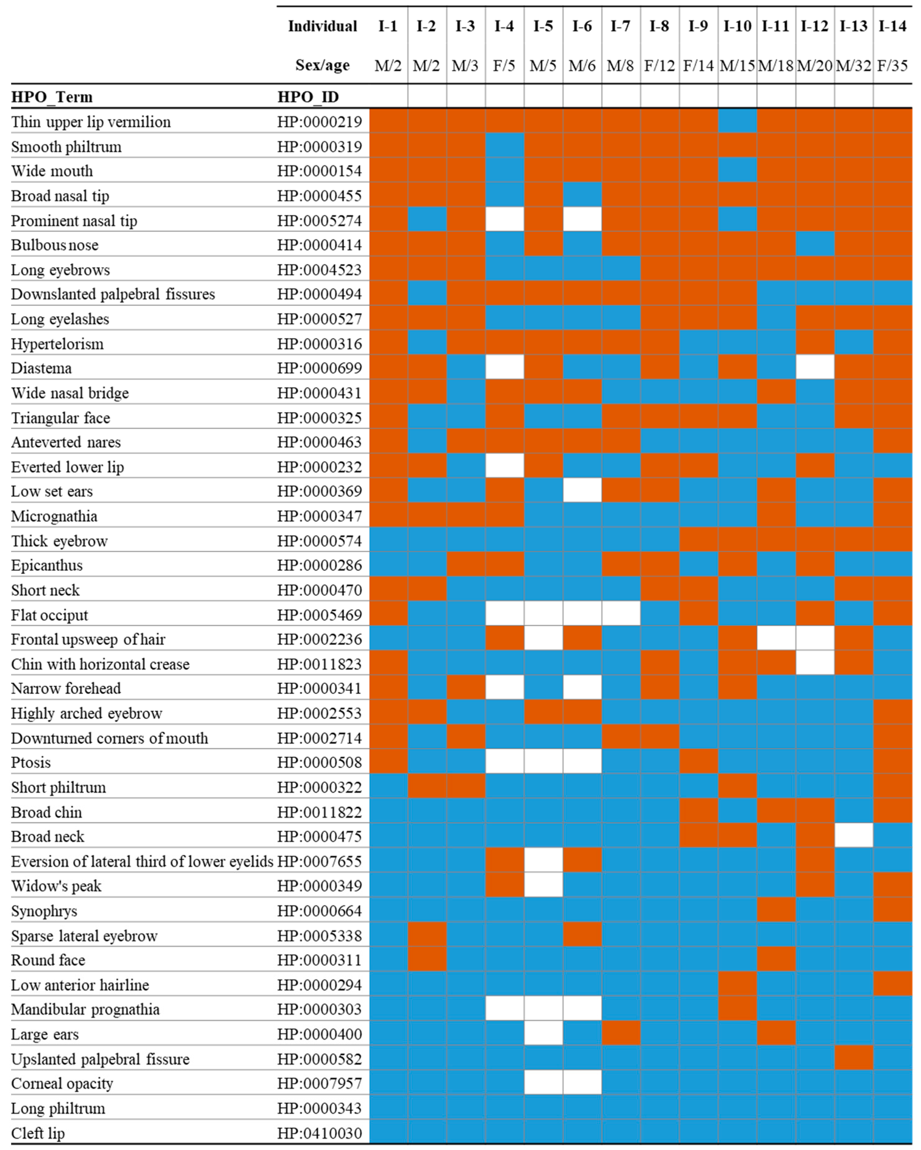
Figure 1. Craniofacial clinical evaluation of individuals with Schuurs–Hoeijmakers syndrome (SHMS). Feature presence is indicated in orange, absence in blue, and unknown or not applicable status in white, based on Human Phenotype Ontology (HPO) terms. M, male; F, female.
Ijms 26 07964 g001
Figure 2. Binary comparison of facial images of individuals with SHMS and unaffected controls. (A) Composite gestalt based upon 14 participants with SHMS, 24 previously published individuals with SHMS, and 40 unaffected individuals. (B) Score distribution and ROC curve obtained using DeepGestalt analysis. Upper panels: comparison between newly reported SHMS individuals and previously published cases. Lower panels: comparison between newly reported SHMS individuals and unaffected controls.
Ijms 26 07964 g002
Figure 3. Binary comparison of facial images of individuals with SHMS (n = 38) versus Epileptic Encephalopathy, Early Infantile, 66 (DEE66) (n = 11), Neuro-Oculo-Cardio-Genitourinary syndrome (NOCGUS) (n = 10), Verheij syndrome (VRJS) (n = 23), and Baraitser–Winter syndrome (BRWS1) (n = 32). Left: Composite gestalt image obtained based on the analyzed photographs. Right: Score distribution and ROC curve obtained by DeepGestalt analysis.
Ijms 26 07964 g003
Table 1. Syndromes identified by the DeepGestalt and GestaltMatcher algorithms in more than 50% of individuals with SHMS analyzed (n = 38).
Syndrome_idOMIM_idGene# PatientsGestalt_Score (Prom)
DeepGestalt
SHMS615009PACS1370.558
BRWS1243310ACTB330.244
NS1163950PTPN11330.297
KABUK1147920KMT2D280.223
RSTS1180849CREBBP250.213
AS105830UBE3A240.226
NF1162200NF1240.219
CdLS122470NIPBL230.215
CSS135900ARID1B220.211
HPMRS1239300PIGV210.250
KBGS148050ANKRD11200.195
GestaltMatcher
DEE66618067PACS2330.358
DeepGestalt and GestaltMatcher
SHMS615009PACS1370.467
DEE66618067PACS2300.366
VRJS615583PUF60260.357
NOCGUS618652WDR37230.363
BRWS1243310ACTB200.359
SHMS, Schuurs–Hoeijmakers syndrome; BRWS1, Baraitser–Winter syndrome 1; NS1, Noonan syndrome 1; KABUK1, Kabuki syndrome 1; RSTS1, Rubinstein–Taybi syndrome 1; AS, Angelman syndrome; NF1, Neurofibromatosis, type 1; CdLS, Cornelia de Lange syndrome; CSS, Coffin–Siris syndrome; HPMRS1, Hyperphosphatasia with impaired intellectual development syndrome 1; KBGS, KBG syndrome; DEE66, Developmental and Epileptic Encephalopathy 66; VRJS, Verheij syndrome; NOCGUS, Neuro-Oculo-Cardio-Genitourinary syndrome; #, number.
Disclaimer/Publisher’s Note: The statements, opinions and data contained in all publications are solely those of the individual author(s) and contributor(s) and not of MDPI and/or the editor(s). MDPI and/or the editor(s) disclaim responsibility for any injury to people or property resulting from any ideas, methods, instructions or products referred to in the content.