Article

A Single DNA Binding Site of DprA Dimer Is Required to Facilitate RecA Filament Nucleation

1
Petersburg Nuclear Physics Institute Named by B.P. Konstantinov of National Research Center «Kurchatov Institute», Orlova Roscha 1, 188300 Gatchina, Russia
2
Department of Microbial Biotechnology, Centro Nacional de Biotecnología, CNB-CSIC, 3 Darwin Str., 28049 Madrid, Spain
3
Research Center of Nanobiotechnologies, Peter the Great St. Petersburg Polytechnic University, Polytechnicheskaya, 29, 195251 St. Petersburg, Russia
4
National Research Center «Kurchatov Institute», Akademika Kurchatova pl. 1, 123182 Moscow, Russia
*
Authors to whom correspondence should be addressed.
Int. J. Mol. Sci. 2025, 26(16), 7873; https://doi.org/10.3390/ijms26167873
Submission received: 27 June 2025 / Revised: 11 August 2025 / Accepted: 13 August 2025 / Published: 15 August 2025
(This article belongs to the Section Molecular Biology)

Abstract

DprA (also known as Smf) is a conserved RecA mediator originally characterized by its role in natural chromosomal transformation, yet its widespread presence across bacteria hints at broader DNA metabolic functions. Here, we demonstrate that Bacillus subtilis DprA enhances the frequency of Escherichia coli Hfr conjugation in vivo. In vitro, RecA·ATP binds and cooperatively polymerizes in a 50-nucleotide (nt) polydeoxy T (dT)50 ssDNA to form dynamic filaments that SSB inhibits, an effect fully reversed by Bacillus subtilis DprA. Escherichia coli RecA bound to (dT)21 exhibits minimal dATPase activity, but the addition of B. subtilis DprA significantly stimulates RecA dATP hydrolysis. B. subtilis RecA·dATP readily assembles on (dT)20 complexes, and DprA allosterically activates RecA on even shorter (dT)15 substrates. Combining biochemical assays with a fully atomic model of the RecA–DprA–ssDNA complex, we proposed that only one DNA binding site of the DprA dimer engages the ssDNA during RecA loading, owing to steric constraints. This work refines the mechanism of DprA-mediated RecA nucleation and defines the minimal ssDNA footprint required for mediator activity.

1. Introduction

DprA is a ubiquitous, evolutionarily conserved protein that directly interacts with RecA and single-strand binding proteins (SsbA and SsbB) to facilitate natural chromosomal transformation, as supported by yeast two-hybrid, pull-down, and Förster resonance energy transfer data [1,2]. Upon entry of exogenous single-stranded DNA (ssDNA) into the cytosol, SsbA/B coat the incoming single-stranded DNA, blocking nonproductive binding. DprA dimers then partially displace the SsbA/B and load RecA onto ssDNA, acting as mediator proteins to initiate natural chromosomal transformation [3]. DprA’s role as a natural chromosomal transformation factor in numerous Firmicutes species has been well-established, with extensive studies on Bacillus subtilis (DrpABsu) and Streptococcus pneumoniae (DrpASpn) [2,4,5]. In Streptococcus pneumoniae, RecA (RecASpn) further destabilizes the DprASpn dimer interface to complete ssDNA hand-off, underscoring a finely tuned mediator mechanism [4]. Across Firmicutes and other phyla, high sequence conservation and the ability to bind both single- and double-stranded DNA support a critical, yet still underexplored, cellular function of the protein [2,5,6]. Biochemical studies have revealed at least three discrete activities of DprA proteins: (a) displacement of SsbA/B “roadblocks” and positive mediation of RecA filament assembly on ssDNA [5], (b) promotion of strand annealing between complementary Ssb-coated ssDNAs [7], and (c) counteracting function of negative RecA modulator RecX [8].
Moreover, in S. pneumoniae, DprASpn coordinates the shut-off of competence to prevent deleterious effects of prolonged DNA uptake [9]. In vitro reconstitution confirmed that pneumococcal DprASpn displaces SSB from ssDNA and stimulates RecAEco nucleation and growth on mixed DprASpn-ssDNA-SSB complexes [2], demonstrating heterospecific interaction across distant bacterial proteins.
Although first characterized in naturally competent species, DprA homologs are widespread—even in bacteria lacking known competence pathways—hinting at additional roles in DNA metabolism beyond transformation [10,11]. For example, overexpression of Escherichia coli DprA (DprAEc/SmfEc) restores chromosomal transformation in competent Haemophilus influenza mutants [12], indicating biologically significant cross-species RecA–DprA compatibility.
Structural analyses of DprA from Gram-positive and Gram-negative bacteria illuminate its multifunctionality. DprASpn comprises an N-terminal sterile alpha motif (SAM) and a C-terminal Rossmann fold that assemble into tail-to-tail dimers, with overlapping surfaces for DprA–DprA and DprA–RecA interactions. In contrast, Helicobacter pylori DprA (DprAHpy) features an N-terminal Rossmann fold and C-terminal winged-helix domain (DML1) yet retains conserved interfaces for ssDNA and RecA binding [13,14].
The DprAEco protein comprises 374-amino acid residues. While the crystal structure of full-length DprAEco, which contains SAM, RF, and DML1 domains, remains unresolved, the structure of its DprASpn homolog has been elucidated [13]. DprASpn forms tail-to-tail dimers via the association of a sterile alpha motif domain and a Rossmann fold. Structural mapping of key interacting residues within DprASpn indicates an overlap between the DprASpn–DprASpn and DprASpn–RecASpn interaction surfaces [13]. Overall, these features suggest a competitive interaction between DprASpn and a truncated variant of RecASpn lacking the first 27 residues [4]. Multiple pieces of in vitro evidence have demonstrated heterospecific interaction between DprASpn and non-cognate RecA proteins, raising the possibility of using DprA as a universal protein across species boundaries [2]. To assess whether a Firmicutes DprA is functional during Hfr conjugation, we examined the activity of plasmid-expressed DprA from B. subtilis (DprABsu). DprABsu was selected due to its well-characterized interaction with RecABsu, as confirmed by yeast two-hybrid assays and fluorescence resonance energy transfer (FRED) experiments [1]. In contrast, a binary interaction between full-length DprASpn and RecASpn was undetectable via yeast two-hybrid assays [13]. Interestingly, a truncated RecASpn variant lacking its N-terminal domain responsible for monomer-monomer interactions does bind full-length DprASpn [13].
However, the function of DprA in non-transformable bacteria remains unclear. Here, we investigated the ability of DprAEco to substitute for endogenous Smf/DprABsu during Escherichia coli Hfr conjugation and measured its impact on homologous recombination in vivo. In parallel, we employed defined oligodeoxythymidine substrates and ATP/dATP hydrolysis assays to dissect the molecular mechanism of DprA-mediated RecA loading. Finally, we built an atomic-level model of the RecA–DprA–ssDNA assembly to explain how steric constraints restrict active DNA engagement to a single protomer within the DprA dimer.

2. Results and Discussion

2.1. DprA Significantly Enhances Recombinational Exchange Frequency (FRE)

Conjugation continuously introduces new ssDNA that is transferred into recipients during Hfr mating, with markers transferred sequentially (leu+→ara+→thr+) according to the Haldane model of recombination [15]. FRE (frequency of recombinational exchanges) was defined as the average number of crossover events per E. coli genome equivalent per 100 min. Over the past 20 years, an assay capturing essentially all genetic exchanges during Hfr mating was developed to measure FRE. This approach aligns with earlier methods based on recombinant yield and marker–marker linkage [16].
At least two quantitative parameters have been utilized to characterize conjugational recombination—the yield of recombinants and the linkage of two transferred donor markers. For wild-type E. coli, FRE = 5.0. [17,18]. ΔFRE is calculated as FRE1/FRE2, where FRE1 is the FRE of the test and FRE2 that of the reference. To assess DprA’s role, we conducted Hfr conjugation experiments with IPTG-induced expression of either native DprAEco or heterologous DprABsu. Overexpression timing was optimized to avoid metabolic overload and approximate physiological levels. At 37 °C, DprAEco and DprABsu increased FRE by 2.6- and 6.4-fold relative to vector alone (Table 1a), indicating that DprABsu is a stronger recombination activator under these conditions. Lowering the induction temperature to 30 °C further amplified FRE 17- to 19-fold for DprAEco and DprABsu, respectively (Table 1b).
As previously described, FRE values in the wild-type background increased in a temperature-dependent manner, showing a 2-fold rise at 42 °C [16]. This unexpected trend underscores the robustness of the observed effect. This suggests that temperatures as high as 37 °C inhibit intracellular protein abundance or activity of proteins or both. Collectively, these data demonstrate that DprA acts as a potent positive regulator of HR in Gram-negative bacterial conjugation and that DprABsu efficiently substitutes for the endogenous mediator in E. coli cells.

2.2. DprABsu Promotes SSBEco Displacement from ssDNA

To date, there are no published reports of biochemical purification of DprAEco, likely due to its susceptibility to proteolytic degradation and poor solubility in aqueous media. The observation of heterospecific interactions between DprABsu and RecAEco, combined with the high degree of conservation between RecABsu and RecAEco, led us to use RecAEco to investigate the effect of DprABsu binding.
RecA filament assembly on SSB–ssDNA complexes can be monitored indirectly via ATP hydrolysis, and DprABsu has been validated as a mediator of this process.
To test whether DprABsu facilitates RecAEco nucleation on SSB-coated ssDNA, we preincubated circular M13mp8 ssDNA (12 μM) with SSBEco protein (0.5 μM) to achieve full coverage. Sub-stoichiometric RecAEco (1 μM) and increasing concentrations of DprABsu were then added, and ATP hydrolysis was recorded. In the initial minutes, in the absence of DprABsu, RecAEco exhibited a prolonged lag phase of ATP hydrolysis, indicating a temporary delay in filament nucleation due to the obstacle presented by SSB. Addition of DprABsu not only eliminated this lag but also increased the steady-state ATPase rate in proportion to DprABsu, indicating active SSBEco displacement and efficient RecAEco loading (Figure 1a). The maximal RecAEco ATPase rate was reached at 1 DprABsu monomer per 150-nt of DNA.
On circular ssDNA, RecAEco gradually displaces SSBEco, whereas on linear ssDNA with a free 5′ end, SSBEco more effectively dislodges RecAEco, reflecting the 5′→3′ polarity of RecAEco filament disassembly [19,20].
The dynamic equilibrium between SSBEco and RecAEco on ssDNA depends on their concentrations, the order of addition, and DNA topology. To focus strictly on direct protein–protein interactions, we used a linear, unstructured (dT)50 oligonucleotide. To directly assess DprABsu’s ability to overcome SSBEco-mediated inhibition, circular ssDNA was replaced by a linear 50-nt poly(dT). RecAEco (3 µM) was present in excess over dT50 (5 µM in nt), ensuring full substrate binding. When RecAEco was preincubated with (dT)50 and ATP, filaments formed rapidly and hydrolyzed ATP at a steady state rate near values ~25–30 min−1 (Figure 1b).
Addition of SSBEco (0.5 µM) to preformed RecAEco-DNA complexes rapidly displaced RecAEco, abolishing ATPase activity (Figure 1b). Strikingly, the addition of DprABsu (80 nM) completely restored ATP hydrolysis to its original rate, even when SSBEco was pre-bound. The kinetics returned to the same steady state, demonstrating complete and reversible SSBEco displacement. Because dT50 lacks secondary structure, these effects are attributable to specific protein–protein interactions. Two non–mutually exclusive mechanisms may account for DprABsu-mediated RecAEco loading: (i) DprA first binds ssDNA, displaces SSBEco via direct interaction, and thereby creates a platform for RecA nucleation [5,21]; (ii) DprABsu and RecAEco cooperatively interact to enhance RecAEco’s nucleation frequency on SSB-coated ssDNA, irrespective of which protein binds DNA first.

2.3. DprABsu Facilitates RecA Nucleation onto Short (dTn) Oligos

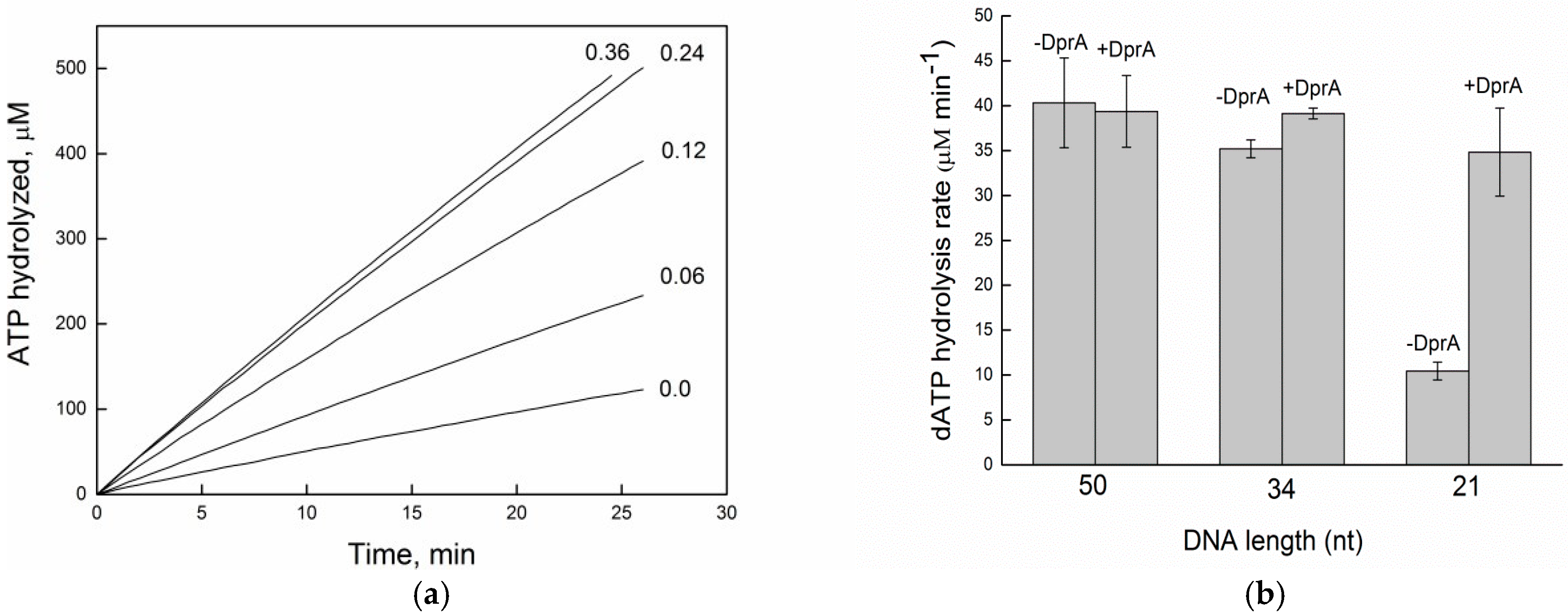
To know how DprABsu influences RecAEco filament formation on oligonucleotides of defined length, experiments were conducted in which RecAEco was added prior to or following DprABsu. RecAEco’s affinity for ssDNA increases with substrate length, yielding stable filaments on long oligos but failing on shorter ones [22,23]. Varying the substrate length helps isolate the nucleation phase from filament extension. Given the cooperative nature of monomer binding, an insufficient number of subunits in the filament compromises the complex stability. The other current view is that the nucleation occurs as a 5-subunit oligomer or even a dimer, followed by filament growth on ssDNA [24,25]. Short oligonucleotides, such as (dT)30 to (dT)40, proved to be poor substrates for RecA-mediated ATP hydrolysis and dynamic filament formation. Protein interaction with such limited-length DNA results in only modest ATP hydrolysis. However, increasing ssDNA length gradually enhances RecA-mediated hydrolysis, reaching peak levels. A (dT)34 oligo can accommodate 10–11 RecA monomers. Due to the dynamic between association and dissociation, the actual number of bound monomers may be lower at any given moment. At the concentration of RecA protein used in our experiment, ATP hydrolysis is activated by less than 20% of the potential maximum value (Figure 2a). We pre-incubated 3 µM RecAEco with (dT)n (5 µM in nt) in ATP and measured ATP hydrolysis. Short (dT)30–(dT)40 supported only modest activity, whereas (dT)34, capable of binding ~10–11 RecAEco monomers, activated less than 20% of the maximal ATPase rate (Figure 2a). At this concentration, RecAEco nucleation and filament growth are severely limited by substrate length. The addition of 0.06 μM DprABsu dramatically elevated RecAEco ATPase rates on (dT)34, regardless of protein addition order. Enhancement was linear with DprABsu concentration and reached saturation at a 1 DprABsu dimer/(dT)34 ratio (0.24 μM DprABsu), implying a single mediator dimer engages each DNA oligo [13].
To define the minimal substrate length required for DprABsu-mediated stimulation of RecAEco nucleation, ATP was replaced by dATP, which increases RecAEco·ssDNA stability and nucleation frequency [26,27].
Switching from ATP to dATP allowed us to define the minimum site size required for DprABsu-mediated RecA loading. Under dATP, RecAEco binds ssDNA with higher affinity and slower disassembly, enhancing nucleation events. RecAEco·dATP on (dT)34 hydrolyzed dATP efficiently (Figure 2b), confirming sufficient filament assembly. We then assayed dATP hydrolysis on oligos ranging from (dT)50 to (dT)21 in the presence or absence of DprABsu (0.3 µM DprA) (Figure 2b). On (dT)50, RecAEco·dATP activity was near-maximal, obscuring any mediator effect. As oligo length decreased, DprABsu’s stimulatory role became increasingly evident, with a 7- to 8-fold activation on (dT)21. RecAEco·dATP alone showed negligible hydrolysis on (dT)21, but DprABsu fully restored robust activity, indicating that, within the examined range, a 21-nt ssDNA (≈seven RecA protomers) is the minimal site for mediator-assisted nucleation. (Figure 2b).

2.4. DprABsu Promotes Dynamic RecABsu·dATP Filaments on Short ssDNA

RecABsu exhibits no detectable dATPase activity in the absence of ssDNA, and DprABsu lacks intrinsic dATPase function [7]. Thus, all observed dATP hydrolysis reflects RecABsu·dATP nucleation on the provided oligonucleotides. To determine the minimum substrate length required for DprA to stimulate RecA nucleation, experimental conditions were optimized using homologous proteins and dATP. The nucleotide length required for DprABsu to stimulate RecABsu·dATP nucleoprotein filament formation was measured.
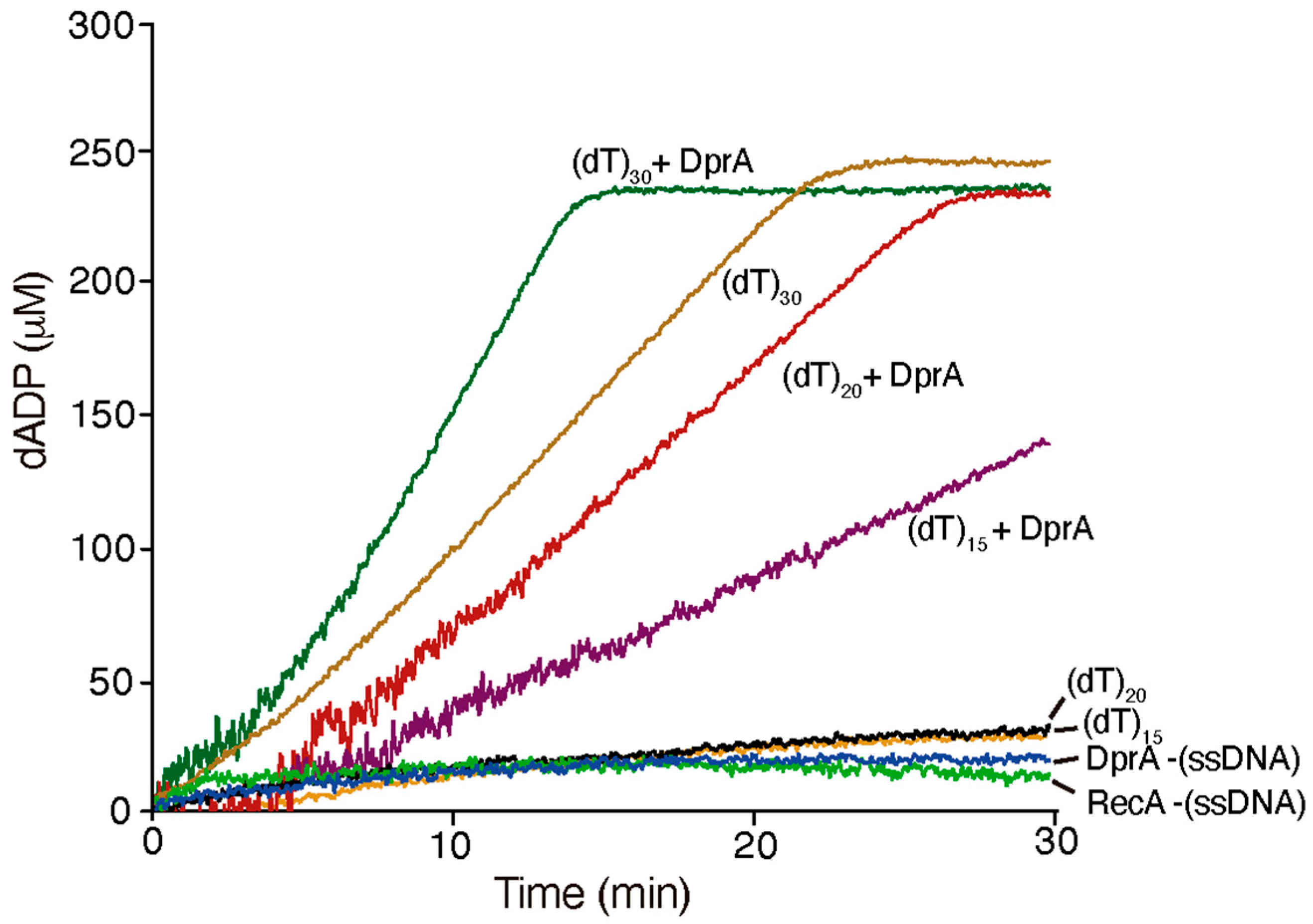
RecABsu·dATP (1 RecA monomer/2-nt (dT)n) binds to and cooperatively polymerises on (dT)30 ssDNA, forming a dynamic filament that hydrolyzes dATP with a kcat of 2.3 ± 0.3 min−1 (Figure 3). In contrast, with (dT)20 and (dT)15 ssDNA substrates, RecABsu·dATP showed significantly reduced hydrolysis rates (kcat = 0.2 ± 0.02 min−1 and 0.19 ± 0.02 min−1, respectively), only slightly above background levels observed with RecA or DprA in the absence of ssDNA (-ssDNA).
To test whether RecABsu·dATP undergoes a functional transition upon interaction with DprABsu, and whether short DNA substrates might confer structural stability for RecA loading, DprABsu was added to the reaction mixture. Addition of DprABsu not only restored but enhanced dATP hydrolysis across all lengths, yielding kcat values of 4.4 ± 0.4 min−1, 2.0 ± 0.4 min−1 on (dT)20, and (dT)15. The effect of DprABsu on maximizing RecABsu-mediated dATP hydrolysis with (dT)20 or 1.1 ± 0.2 min−1 on (dT)15 corresponded to an 8-fold and 10-fold activation on the shortest substrates, respectively. Thus, DprABsu-RecABsu interplay with a 20–21 long oligo will be further analyzed.
Co-crystal structures showed a binding footprint of 5-nt per DrpAHpy monomer and 3-nt per RecA monomer [14,28], and physical interaction between DprABsu and RecABsu has been demonstrated (see above). Based on this, we propose that DprABsu bound to (dT)20 ssDNA interacts with and recruits RecABsu·dATP, contributing to the loading of ~5 RecABsu·dATP protomers. The DprABsu-RecABsu interaction may elicit an allosteric effect on both proteins. Under these conditions, monomeric DprA binds 5-nt ssDNA and enhances RecABsu cooperativity, facilitating dynamic filaments. This hypothesis aligns with previous findings that a DprASpn monomer has only a 2.6-fold weaker binding affinity for (dT)20 compared to the DprASpn dimer [13]. DprASpn or DprABsu is sufficient to recruit homologous or heterologous RecA, in the ATP or dATP bound form, onto short ssDNA oligos [2,5,7].

2.5. Structural Model of the RecA-DprA-ssDNA Complex

Our dATP hydrolysis assays established that DprABsu caps and stabilizes RecA/RecABsu filaments on a (dT)21/20 substrate, implying that only one DprABsu DNA binding site engages each short ssDNA. When a DprABsu–RecABsu·dATP complex binds to a (dT)20 ssDNA, it moderately enhances dATP hydrolysis along the filament and yields RecABsu·dADP. This interaction appears sufficient to allosterically stabilize RecABsu, promoting cooperative ssDNA binding and the formation of helical nucleoprotein filaments. The N-terminal helix of RecABsu may destabilize the DprABsu dimer, favoring formation of a functional monomer, as suggested earlier [4,13]. This mechanism provides a molecular explanation for the transfer of short (dT) ssDNA from DprABsu to RecABsu. This hypothesis led us to refine an existing structural model of the complex to better align with the experimental data (Figure 4A,B).
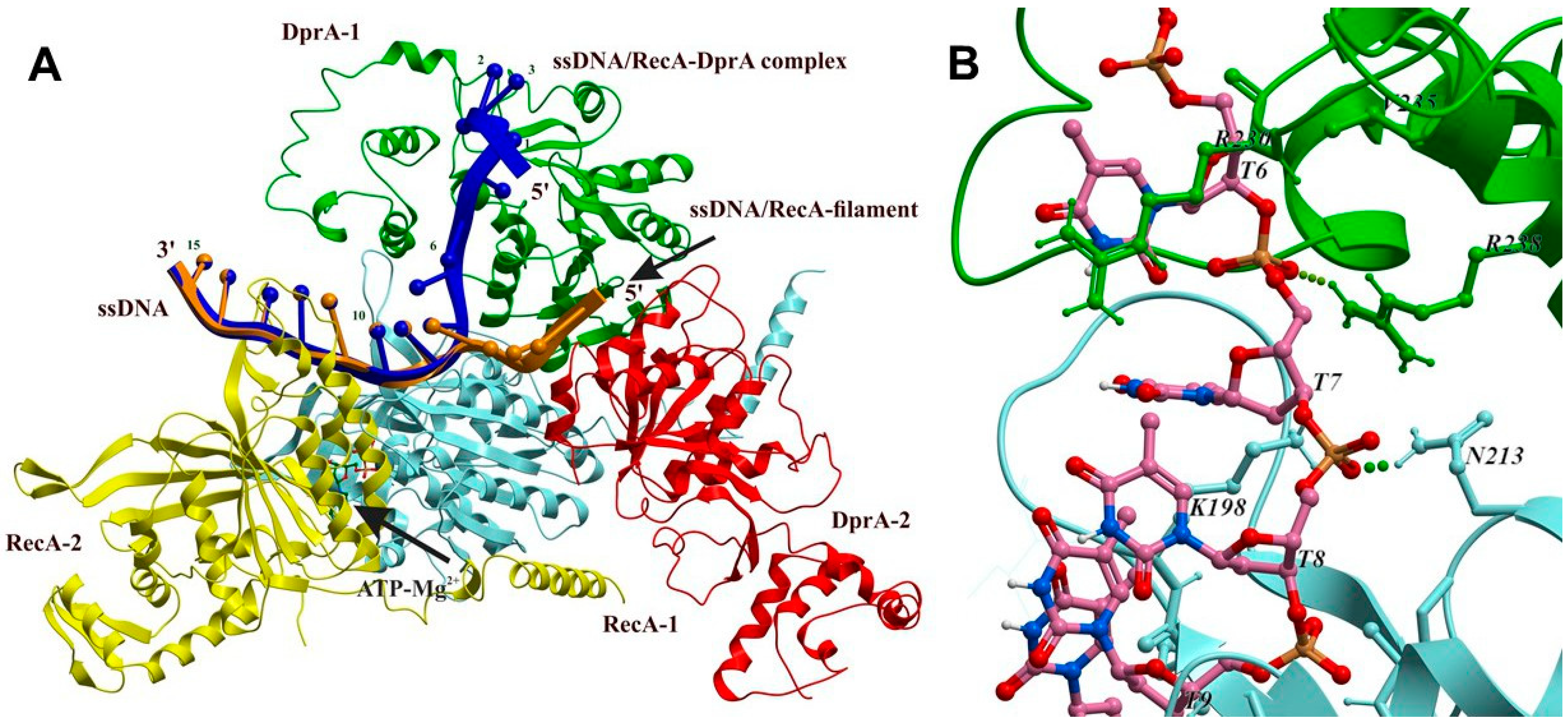
Using molecular modeling approaches based on three different incomplete complexes of highly homologous proteins, RecA, DprA, and ssDNA, we built the fully atomic structure of the DprA2-RecA2·ATP-(dT)15 complex (see Section 3). Specifically, we combined (1) the structure of the presynaptic nucleoprotein filament of Escherichia coli RecA protein bound to ssDNA (PDB ID: 3CMX), (2) the DprA–ssDNA complex from Helicobacter pylori (PDB ID: 4LJR), and (3) a fully atomic spatial model of the Streptococcus pneumoniae RecA-DprA dimer generated by molecular docking [13,14,28]. All specific interactions observed in the original structures were preserved in the reconstructed DprA2-RecA2·ATP-(dT)15 complex (Figure 4). To connect the DNA segments from the RecA-DNA (PDB ID: 3CMX) and DprA-DNA (PDB ID: 4LJR) structures, we inserted two additional nucleotides. The specific interactions of this new structural element with the DprA and RecA proteins are shown in Figure 4B.
Although this nucleoprotein complex has a multicomponent structure, obtained from the structural data of complexes of various compositions, the structure and conformation of its constituent proteins turned out to be compatible with each other and do not have any structural problems. However, the conformation of the 5′-end of the ssDNA presynaptic filament RecA (shown in brown) turned out to be incompatible with the presence of DprA proteins and directed towards the region of DprA protein–protein interface and not towards the known nucleotide binding site found in the crystal structure of the dimeric complex of DprA proteins with short fragments of ssDNA [14]. This obviously means that binding of DprA requires that the six nucleotide residues of the free 5′-end of ssDNA adopt a suitable conformation for binding to the nucleotide binding site of the DprA protein. Molecular modeling of the conformational transition of ssDNA showed that the nucleotide from the dimeric complex of the DprA protein [14] and ssDNA of the presynaptic RecA protein filament [28] can be connected without any steric strains by two mobile nucleotide residues, thus forming a continuous ssDNA chain running from the nucleotide binding site of the DprA protein to the RecA proteins helical filament, as shown in Figure 4. This fully atomic model reconciles our biochemical data with structural constraints, providing a molecular basis for DprA’s one-site mediation of RecA loading on short ssDNA stretches.
The ATP/dATP hydrolysis site of RecABsu is located at the protomer–protomer interface. Filament assembly on ssDNA requires direct contacts between loop L1 of one RecA protomer to contact L2 of its neighbor protomer every three nucleotide residues. Hence, each additional RecABsu subunit in the nucleoprotein filament engages three nucleotides of ssDNA [28]. From this requirement, one ATP-binding site (two RecABsuprotomers) demands at least 15 nt of ssDNA, whereas two sites (three protomers), which support higher ATP hydrolysis rates, require 18–20 nt. Our dATP hydrolysis assays with DprABsu-stimulated RecABsu filaments on (dT)21/(dT)20 substrates closely match these theoretical thresholds (Figure 2 and Figure 3).
Co-crystal structures of DNA complexes of DrpAHpy and RecA report footprints of 5 nt per DprAHpy monomer and 3 nt per RecAHpy protomer [14,28], and we have shown that DprABsu physically interacts with RecABsu.
One important question concerns the role of the second subunit of the DprA protein in the DprA2–RecA2·ATP–(dT)15 complex shown in Figure 4. It is evident that an identical symmetrical structure could theoretically be formed through the interaction between the second DprABsu subunit and a RecABsu protein. However, the simultaneous presence of both symmetrical structures within a single complex is sterically hindered, as the two symmetrically located RecABsu subunits compete for the same spatial location.
Recent structural data and molecular modeling of DprA complexes with ssDNA and RecA have led to the proposal of a molecular mechanism for DprA-mediated loading of RecABsu onto ssDNA [29]. This model suggests that DprA forms a dimeric nucleoprotein complex with ssDNA, providing a platform for RecA nucleation. Following this initial interaction, one DprA subunit dissociates, allowing RecA filament formation on ssDNA to proceed. According to the model, ssDNA binds simultaneously to both DprA subunits, whose DNA binding sites are approximately 50 Å apart, equivalent to a stretch of ssDNA about 10 nucleotides long in a fully extended conformation.
Experimental analysis of DprA–ssDNA binding indicates that stability depends on the DNA length [14]. Short ssDNA fragments (6–20 nucleotides) either do not bind or have very low affinity, whereas high-affinity binding was observed for ssDNA fragments around 35 nucleotides long. The structure of the dimeric DprA-DNA complex was resolved using X-ray crystallography. However, most of the (dT)35 ssDNA appeared disordered, and electron density was observed only for the 6-nt at the 5′ end. These 6-nt were used in the reconstruction of the DprA2-RecA2·ATP-(dT)15 complex shown in Figure 4. RecA interacts with and destabilizes the DprA dimer interface, facilitating the transfer of ssDNA from DprA to RecA.
Importantly, due to the symmetry of the DprA dimer, its two ssDNA binding sites are oriented in opposite directions, making it sterically impossible for a single ssDNA strand to occupy both sites of the DprA dimer simultaneously. Consequently, only a complex in which ssDNA binds to one DprA subunit (Figure 4) is sterically feasible. The second DprA subunit, however, can accommodate a separate ssDNA strand in the opposite orientation. In this scenario, the second subunit not only promotes initial loading of the first RecA monomer onto ssDNA but may also play a central role in the still poorly understood mechanism by which the RecA nucleoprotein filament efficiently searches for homologous DNA. Furthermore, the binding of ssDNA to the second subunit could modulate the ssDNA binding affinity of the first. Under these conditions, the minimum ssDNA length required is estimated at 6 + 9 = 15-nt nucleotides, a value that aligns with the experimental data obtained in this study.

3. Materials and Methods

3.1. Strains and Plasmids

The donor strain KL227 (HfrP4x metB) and recipient strains AB1157 (thr-1 leuB6 ara14 proA2 hisG4 argE3 thi-1 supE44 rpsL31) and recombination-deficient JC10289 (genetically identical to AB1157 but carrying ΔrecA-srlR306:Tn10 [=ΔrecA306]) were obtained from A.J. Clark’s collection. The strain JW5708-1 (ΔdprA-724::kan) was sourced from the Keio Collection (E. coli Genetic Resources at Yale CGSC). A null dprAdprA) strain was generated via P1 transduction, transferring the dprA-724 (del)::kan allele from JW5708-1 into AB1157, as described previously [30]. The pT7 plasmid (originally named pT7POL26) encodes T7 RNA polymerase under the control of a lacZ promoter. It was used to drive expression of DprA/Smf proteins via IPTG induction of the lac promoter. Plasmid pCB888, which contains the dprABsu gene under the control of a T7 RNA polymerase-driven promoter, was kindly provided by Prof. J.C. Alonso. Likewise, the pDprA plasmid, harboring the dprA gene with a similar promoter structure, was supplied by EUROGEN (Moscow, Russia).

3.2. Proteins

Wild-type RecAEco and RecABsu were purified as previously described [31,32]. Single-strand binding (SSB) protein was kindly provided by Prof. M. Cox (University of Wisconsin–Madison). Protein concentrations were determined using their native extinction coefficients: ϵ280 = 2.23 × 10⁴ M−1 cm−1 for RecA [33], 1.52 104 M−1 cm−1 for RecABsu, and 2.38 × 10⁴ M−1 cm−1 for SSB protein [34]. The concentration of DprABsu was determined using the native extinction coefficient ϵ280 = 4.5 × 10⁴ M−1 cm−1 [5].
DprABsu purification was performed following previously reported protocols [5], with some modifications. Briefly, E. coli BL21(DE3) pLysE cells were transformed with the pCB888. Cultures were grown at 25 °C, and protein expression was induced with 0.4 mM isopropyl-1-thio-β-D-galactopyranoside (IPTG) at an OD600 of 0.5. After induction, cells were incubated for 3 h, with rifampicin (200 µg/mL) added 30 min after IPTG induction.
DprAEco was purified from the clarified lysate via two-step chromatography using HisTrap HP 1 mL (GE Healthcare, Chicago, IL, USA) and HiTrap SP XL 1 mL columns (GE Healthcare, Chicago, USA). Protein-containing fractions were concentrated using Amicon Ultra-4 centrifugal filter (Merck, Darmstadt, Germany) with a 3 kDa cut-off, supplemented with 50% glycerol, and stored at −20 °C.

3.3. (d)ATP Hydrolysis Assays

A coupled enzyme spectrophotometric assay was employed to measure RecA-mediated ATP or dATP hydrolysis, as previously described [35,36]. In this system, the ADP or dADP generated by hydrolysis was converted back to ATP or dATP by a regeneration system involving pyruvate kinase and phosphoenolpyruvate (PEP). The resulting pyruvate was subsequently converted to lactate-by-lactate dehydrogenase, using NADH as a reducing agent. This reaction enabled indirect monitoring of nucleotide hydrolysis via the decrease in absorbance at 380 nm due to the oxidation of NADH to NAD+. ATP or dATP consumption over time was quantified using the NADH extinction coefficient ε380 = 1.21 mM−1 cm−1. All measurements were conducted using a Cary 5000 dual-beam spectrometer equipped with a temperature controller and a 12-position cell changer (Varian, Palo Alto, CA, USA). The path length was 1 cm, and the band pass was 2 nm. RecAEco ATPase and dATPase assays were performed in buffer A, composed of 25 mM Tris-HCl (pH 7.5, 88% cation), 10 mM MgCl2, 5% w/v glycerol, 1 mM dithiothreitol (DTT), and 2 mM ATP or dATP. The regeneration system included 3 mM PEP, 10 U/mL pyruvate kinase, 10 U/mL lactate dehydrogenase, 4.5 mM NADH, and 5 μM M13mp18 cssDNA or (dT) oligos. RecABsu dATPase assays were performed using buffer B, which consisted of 50 mM Tris-HCl [pH 7.5], 10 mM Mg(OAc)2, 5% w/v glycerol, 1 mM dithiothreitol (DTT), and 2.5 mM dATP. The regeneration components included 0.5 mM PEP, 10 U/mL pyruvate kinase, 10 U/mL lactate dehydrogenase, 4.5 mM NADH, and 10 μM (dT)n oligos. All reactions were repeated at least in triplicate, yielding consistent results.

3.4. Conjugation

Conjugation experiments were carried out essentially as previously described [16]. Expression was induced for no longer than 1 h prior to conjugation to limit cellular stress and maintain near-physiological protein levels. Both Hfr and F strains were cultured, mated, and recombinant progeny were selected at either 37 °C or 30 °C (as specified in the Table 1) for 1 h in mineral salts 56/2 medium supplied with all necessary growth factors at pH 7.5. The donor-to-recipient ratio in the mating mixture was 1:10, with 2–4 × 10⁷ donors and 2–4 × 10⁸ recipients per 1 mL. The yield of thr+ Strr and ara+ Strr recombinants from independent crosses (typically 5–7% relative to donor input) was normalized to account for the mating ability of each recipient strain. This was determined by measuring the yield of F’-lac+ transconjugants in control crosses between recipient and donor strains P200 F’-lac.
FRE (frequency of recombination exchange) values were calculated according to previously established procedures [16]. Changes in FRE (ΔFRE) resulting from recA mutation or accessory gene, relative to the FRE promoted by the wild-type E.coli recA, were computed using the following formula: ΔFRE = ln(2μ1 − 1)/(2 μ2 − 1), where μ1 denotes the linkage of selected thr+ or ara+ with unselected leu+ markers in crosses involving the wild-type E. coli strain AB1157, and μ2 corresponds to the linkage observed in the experimental cross. Uncertainty in relative FRE values was calculated as the deviations from the average value based on three independent experiments, using the formula [=2 × STDEV] in Excel-97 and entering replicate data points.

3.5. Reconstruction of the Structure of the RecA-ssDNA-DprABsu Complex

To construct a full-atom model of the RecA-ssDNA-DprABsu spatial structure, we utilized several crystal structures from the Protein Data Bank. These included the presynaptic nucleoprotein filament of RecAEco bound to RecA6–(ADP–AlF4–Mg)6–dT18 (PDB ID: 3CMX) [28], the Helicobacter pylori DprAhpy–ssDNA dimer complex (DprAHpy–dT6)2 (PDB ID: 4LJR) [14], and a molecular docking-derived spatial model of the RecA–DprA complex, kindly provided by the authors of this study [13]. Superimposition, energy minimization, and other molecular manipulations were performed using standard protocols of the ICM-Pro software package version 3.8-7c (Molsoft LLC, San Diego, CA, USA) [37], employing the ECEPP/3 force field as implemented in ICM-Pro [37,38].
The spatial structure of the DprA dimeric complex from E. coli was derived via homology modeling based on the DprAhpy crystal structure (PDB ID: 4LJR), using built-in modeling protocols in ICM-Pro. The RecA–sDNA–DprABsu complex was assembled through a stepwise superimposition of individual protein structures. Initially, the position of the DprABsu dimer was determined by aligning RecA monomers from the presynaptic filament (PDB ID: 3CMX) [28] and in the docking model of the RecA–DprABsu complex [13]. Next, the location of 6-nt at the 5′-end of ssDNA was defined by superposing the (DprAHpy-dT6)2 complex (PDB ID: 4LJR) [14] with the RecA–DprA model. The conformation of the 3′ ssDNA segment was adopted from its positioning within the RecA presynaptic filament (PDB ID: 3CMX) [28]. Finally, the two bridging nucleotides were modeled by minimizing the energy of the ssDNA strand, keeping its 5′ and 3′ends fixed.

4. Conclusions

In this study, we investigated the functionality of the DprAEco homolog in the context of Hfr conjugation. While most regulators only partially influence the genetic outcomes of recombination, thereby merely modulating the natural FRE values (used to assess in vivo recombinase activity) [16,18,39,40], our study identifies DprA/Smf as a potent activator of recombination, increasing FRE by approximately 18-fold, upon overexpression. This finding suggests that the DprA–RecA interaction in E. coli represents an additional layer of control over the primary recombination pathways and advances our understanding of homologous recombination regulation. Collectively, our data indicate that DprA proteins may play distinct roles across different bacterial species.
Through length-dependent experiments, we determined that a 20-nucleotide ssDNA substrate is sufficient to nucleate the RecAEco filament in conjunction with DprABsu. This led us to refine an existing structural model to better align with our biochemical data.
Building on previous mutagenesis-based analysis [4], which showed that DprASpn dimerization is essential for DNA binding and RecASpn loading, and consistent with data from H. pylori demonstrating that only the dimeric form is active [14], we propose a new mechanism.
Structural and biochemical characterization of the DprA proteins reveals that a DprA2–ssDNA complex forms with only one DprA subunit actively bound to ssDNA, while the second DprA subunit sterically stabilizes the initial RecA monomer in its DNA-bound conformation. Based on the known dimensions of the DprA–DNA binding site and the role of dimeric DrpA as the active species, we propose the formation of a ternary RecA–DprA–ssDNA complex during RecA filament nucleation. Within this configuration, the remaining 21 nt ssDNA stretch can accommodate, at most, a RecA trimer as the minimal nucleation unit, which then extends into a hexamer upon DprA dissociation.
This structural model of the DprA-ssDNA complex provides a compelling basis for further investigation into DprA and related proteins. Regulators of homologous recombination involved in chromosomal transformation should undoubtedly be considered promising targets for inhibition [41,42]. Further structural analysis of the DprA–RecA complex may ultimately support the rational design of compounds aimed at suppressing horizontal gene transfer.

Supplementary Materials

The following supporting information can be downloaded at: https://www.mdpi.com/article/10.3390/ijms26167873/s1.

Author Contributions

Conceptualization, D.B. and M.P.; Methodology, D.B. and I.B.; Software, M.P.; Protein Preparation, A.A., M.Y. and N.M.; Investigation, D.B., I.B. and B.C.; Resources, M.K.; Writing—Original Draft, D.B. and M.P.; Writing—Review and Editing, D.B., M.P. and B.C. All authors have read and agreed to the published version of the manuscript.

Funding

This work was supported in part by Ministerio de Ciencia e Innovación (MCIN)/Agencia Estatal de Investigación (AEI)/10.13039/501100011033/FEDER, EU, PID2021-122273NB-I00, as well as by the CSIC 2021AEP031 and 202520E100 to J.C.A. Funding for open access charges was provided by CSIC 202520E100. The research was partly supported by the Ministry of Science and Higher Education of the Russian Federation (grant No. 075-15-2024-630) to M.P. (molecular modeling) and (theme No. 1023031500033-1-1.6.7;1.6.4;1.6.8) I.B. (genetics). The research was supported by the Russian Science Foundation, project No. 24-44-02003, to M.Y. The work was carried out using the scientific equipment of the Center of Shared Usage, “The analytical center of nano- and biotechnologies of SPbPU”.

Data Availability Statement

The data that support the findings of this study are available on request from the corresponding authors.

Acknowledgments

We are grateful to Juan C. Alonso (Department of Microbial Biotechnology, Centro Nacional de Biotecnología, Madrid, Spain) for his generous gift of expression plasmids bearing the B. subtilis DprA for the E. coli strain. The authors would like to thank Michael M. Cox (University of Wisconsin–Madison) for the SSB protein. The authors thank Jessica Andreani and Sophie Quevillon-Cheruel (Institute of Integrative Biology of the Cell, Université Paris-Sud, Gif-sur-Yvette, France) for the structural model of the complex of the DprA dimer with the RecA protein. We acknowledge the help of Inna Kurdyumova, Daria Antonova, and Maryam Dmitrieva (Peter the Great St. Petersburg Polytechnic University) for the purification of B. subtilis DprA. We thank Alexander Yakimov for assistance throughout the project. B.C. acknowledges financial support from Ministerio de Ciencia e Innovación MCIN/Agencia Estatal de Investigación (AEI)/10.13039/501100011033/FEDER, EU, PID2021-122273NB-I00, as well as from CSIC 2021AEP031 and 202520E100.

Conflicts of Interest

The authors declare no conflicts of interest.

Abbreviations

The following abbreviations are used in this manuscript:
FREfrequency of recombination exchanges per DNA unit length
SSBsingle-stranded DNA binding protein
ssDNAsingle-strand DNA
poly(dT)extended polymers of deoxythymidine
ntnucleotide(s)
EMSAelectrophoretic mobility shift assay

References

  1. Kramer, N.; Hahn, J.; Dubnau, D. Multiple interactions among the competence proteins of Bacillus subtilis. Mol. Microbiol. 2007, 65, 454–464. [Google Scholar] [CrossRef] [PubMed]
  2. Mortier-Barriere, I.; Velten, M.; Dupaigne, P.; Mirouze, N.; Pietrement, O.; McGovern, S.; Fichant, G.; Martin, B.; Noirot, P.; Le Cam, E.; et al. A key presynaptic role in transformation for a widespread bacterial protein: DprA conveys incoming ssDNA to RecA. Cell 2007, 130, 824–836. [Google Scholar] [CrossRef] [PubMed]
  3. Kidane, D.; Ayora, S.; Sweasy, J.B.; Graumann, P.L.; Alonso, J.C. The cell pole: The site of cross talk between the DNA uptake and genetic recombination machinery. Crit. Rev. Biochem. Mol. Biol. 2012, 47, 531–555. [Google Scholar] [CrossRef]
  4. Quevillon-Cheruel, S.; Campo, N.; Mirouze, N.; Mortier-Barriere, I.; Brooks, M.A.; Boudes, M.; Durand, D.; Soulet, A.L.; Lisboa, J.; Noirot, P.; et al. Structure-function analysis of pneumococcal DprA protein reveals that dimerization is crucial for loading RecA recombinase onto DNA during transformation. Proc. Natl. Acad. Sci. USA 2012, 109, E2466–E2475. [Google Scholar] [CrossRef]
  5. Yadav, T.; Carrasco, B.; Hejna, J.; Suzuki, Y.; Takeyasu, K.; Alonso, J.C. Bacillus subtilis DprA recruits RecA onto single-stranded DNA and mediates annealing of complementary strands coated by SsbB and SsbA. J. Biol. Chem. 2013, 288, 22437–22450. [Google Scholar] [CrossRef]
  6. Sharma, D.K.; Misra, H.S.; Soni, I.; Rajpurohit, Y.S. Characterization of DNA Processing Protein A (DprA) of the Radiation-Resistant Bacterium Deinococcus radiodurans. Microbiol. Spectr. 2022, 10, e0347022. [Google Scholar] [CrossRef]
  7. Yadav, T.; Carrasco, B.; Serrano, E.; Alonso, J.C. Roles of Bacillus subtilis DprA and SsbA in RecA-mediated genetic recombination. J. Biol. Chem. 2014, 289, 27640–27652. [Google Scholar] [CrossRef]
  8. Le, S.; Serrano, E.; Kawamura, R.; Carrasco, B.; Yan, J.; Alonso, J.C. Bacillus subtilis RecA with DprA-SsbA antagonizes RecX function during natural transformation. Nucleic Acids Res. 2017, 45, 8873–8885. [Google Scholar] [CrossRef]
  9. Mirouze, N.; Berge, M.A.; Soulet, A.-L.; Mortier-Barriere, I.; Quentin, Y.; Fichant, G.; Granadel, C.; Noirot-Gros, M.-F.; Noirot, P.; Polard, P. Direct involvement of DprA, the transformation-dedicated RecA loader, in the shut-off of pneumococcal competence. Proc. Natl. Acad. Sci. USA 2013, 110, E1035–E1044. [Google Scholar] [CrossRef]
  10. Johnston, C.; Mortier-Barriere, I.; Khemici, V.; Polard, P. Fine-tuning cellular levels of DprA ensures transformant fitness in the human pathogen Streptococcus pneumoniae. Mol. Microbiol. 2018, 109, 663–675. [Google Scholar] [CrossRef]
  11. Brito, I.L. Examining horizontal gene transfer in microbial communities. Nat. Rev. Microbiol. 2021, 19, 442–453. [Google Scholar] [CrossRef]
  12. Smeets, L.C.; Becker, S.C.; Barcak, G.J.; Vandenbroucke-Grauls, C.M.; Bitter, W.; Goosen, N. Functional characterization of the competence protein DprA/Smf in Escherichia coli. FEMS Microbiol. Lett. 2006, 263, 223–228. [Google Scholar] [CrossRef]
  13. Lisboa, J.; Andreani, J.; Sanchez, D.; Boudes, M.; Collinet, B.; Liger, D.; van Tilbeurgh, H.; Guerois, R.; Quevillon-Cheruel, S. Molecular determinants of the DprA-RecA interaction for nucleation on ssDNA. Nucleic Acids Res. 2014, 42, 7395–7408. [Google Scholar] [CrossRef]
  14. Wang, W.; Ding, J.; Zhang, Y.; Hu, Y.; Wang, D.C. Structural insights into the unique single-stranded DNA-binding mode of Helicobacter pylori DprA. Nucleic Acids Res. 2014, 42, 3478–3491. [Google Scholar] [CrossRef] [PubMed]
  15. Lawley, T.D.; Gordon, G.S.; Wright, A.; Taylor, D.E. Bacterial conjugative transfer: Visualization of successful mating pairs and plasmid establishment in live Escherichia coli. Mol. Microbiol. 2002, 44, 947–956. [Google Scholar] [CrossRef] [PubMed]
  16. Lanzov, V.A.; Bakhlanova, I.V.; Clark, A.J. Conjugational hyperrecombination achieved by derepressing the LexA regulon, altering the properties of RecA protein and inactivating mismatch repair in Escherichia coli K-12. Genetics 2003, 163, 1243–1254. [Google Scholar] [CrossRef] [PubMed]
  17. Lloyd, R.G. Hyper-recombination in Escherichia coli K-12 mutants constitutive for protein X synthesis. J. Bacteriol. 1978, 134, 929–935. [Google Scholar] [CrossRef]
  18. Bakhlanova, I.V.; Dudkina, A.V.; Wood, E.A.; Lanzov, V.A.; Cox, M.M.; Baitin, D.M. DNA Metabolism in Balance: Rapid Loss of a RecA-Based Hyperrec Phenotype. PLoS ONE 2016, 11, e0154137. [Google Scholar] [CrossRef]
  19. Arenson, T.A.; Tsodikov, O.V.; Cox, M.M. Quantitative analysis of the kinetics of end-dependent disassembly of RecA filaments from ssDNA. J. Mol. Biol. 1999, 288, 391–401. [Google Scholar] [CrossRef]
  20. Bork, J.M.; Cox, M.M.; Inman, R.B. RecA protein filaments disassemble in the 5′ to 3′ direction on single-stranded DNA. J. Biol. Chem. 2001, 276, 45740–45743. [Google Scholar] [CrossRef]
  21. Hovland, E.; Beyene, G.T.; Frye, S.A.; Homberset, H.; Balasingham, S.V.; Gomez-Munoz, M.; Derrick, J.P.; Tonjum, T.; Ambur, O.H. DprA from Neisseria meningitidis: Properties and role in natural competence for transformation. Microbiology 2017, 163, 1016–1029. [Google Scholar] [CrossRef] [PubMed]
  22. Brenner, S.L.; Mitchell, R.S.; Morrical, S.W.; Neuendorf, S.K.; Schutte, B.C.; Cox, M.M. recA protein-promoted ATP hydrolysis occurs throughout recA nucleoprotein filaments. J. Biol. Chem. 1987, 262, 4011–4016. [Google Scholar] [CrossRef]
  23. Sussman, R.; Sharma, S.K.; Kuzirian, A. Catalytic activities of recA protein are dependent on the lattice length of the single-strand DNA ligand. Cell Cycle 2008, 7, 89–95. [Google Scholar] [CrossRef] [PubMed]
  24. Galletto, R.; Amitani, I.; Baskin, R.J.; Kowalczykowski, S.C. Direct observation of individual RecA filaments assembling on single DNA molecules. Nature 2006, 443, 875–878. [Google Scholar] [CrossRef] [PubMed]
  25. Bell, J.C.; Plank, J.L.; Dombrowski, C.C.; Kowalczykowski, S.C. Direct imaging of RecA nucleation and growth on single molecules of SSB-coated ssDNA. Nature 2012, 491, 274–278. [Google Scholar] [CrossRef]
  26. Menetski, J.P.; Kowalczykowski, S.C. Enhancement of Escherichia coli RecA protein enzymatic function by dATP. Biochemistry 1989, 28, 5871–5881. [Google Scholar] [CrossRef]
  27. Shan, Q.; Cox, M.M. RecA protein dynamics in the interior of RecA nucleoprotein filaments. J. Mol. Biol. 1996, 257, 756–774. [Google Scholar] [CrossRef]
  28. Chen, Z.; Yang, H.; Pavletich, N.P. Mechanism of homologous recombination from the RecA-ssDNA/dsDNA structures. Nature 2008, 453, 489–494. [Google Scholar] [CrossRef]
  29. Sharma, D.K.; Misra, H.S.; Bihani, S.C.; Rajpurohit, Y.S. Biochemical Properties and Roles of DprA Protein in Bacterial Natural Transformation, Virulence, and Pilin Variation. J. Bacteriol. 2023, 205, e0046522. [Google Scholar] [CrossRef]
  30. Csonka, L.N.; Clark, A.J. Construction of an Hfr strain useful for transferring recA mutations between Escherichia coli strains. J. Bacteriol. 1980, 143, 529–530. [Google Scholar] [CrossRef]
  31. Cox, M.M.; McEntee, K.; Lehman, I.R. A simple and rapid procedure for the large scale purification of the recA protein of Escherichia coli. J. Biol. Chem. 1981, 256, 4676–4678. [Google Scholar] [CrossRef] [PubMed]
  32. Manfredi, C.; Carrasco, B.; Ayora, S.; Alonso, J.C. Bacillus subtilis RecO nucleates RecA onto SsbA-coated single-stranded DNA. J. Biol. Chem. 2008, 283, 24837–24847. [Google Scholar] [CrossRef] [PubMed]
  33. Drees, J.C.; Lusetti, S.L.; Chitteni-Pattu, S.; Inman, R.B.; Cox, M.M. A RecA filament capping mechanism for RecX protein. Mol. Cell 2004, 15, 789–798. [Google Scholar] [CrossRef] [PubMed]
  34. Lohman, T.M.; Green, J.M.; Beyer, R.S. Large-scale overproduction and rapid purification of the Escherichia coli ssb gene product. Expression of the ssb gene under lambda PL control. Biochemistry 1986, 25, 21–25. [Google Scholar] [CrossRef]
  35. Morrical, S.W.; Lee, J.; Cox, M.M. Continuous association of Escherichia coli single-stranded DNA binding protein with stable complexes of recA protein and single-stranded DNA. Biochemistry 1986, 25, 1482–1494. [Google Scholar] [CrossRef]
  36. Lindsley, J.E.; Cox, M.M. Assembly and disassembly of RecA protein filaments occur at opposite filament ends. Relationship to DNA strand exchange. J. Biol. Chem. 1990, 265, 9043–9054. [Google Scholar] [CrossRef]
  37. Abagyan, R.; Totrov, M. Biased probability Monte Carlo conformational searches and electrostatic calculations for peptides and proteins. J. Mol. Biol. 1994, 235, 983–1002. [Google Scholar] [CrossRef]
  38. Arnautova, Y.A.; Jagielska, A.; Scheraga, H.A. A new force field (ECEPP-05) for peptides, proteins, and organic molecules. J. Phys. Chem. B 2006, 110, 5025–5044. [Google Scholar] [CrossRef]
  39. Baitin, D.M.; Bakhlanova, I.V.; Chervyakova, D.V.; Kil, Y.V.; Lanzov, V.A.; Cox, M.M. Two RecA protein types that mediate different modes of hyperrecombination. J. Bacteriol. 2008, 190, 3036–3045. [Google Scholar] [CrossRef]
  40. Bakhlanova, I.V.; Dudkina, A.V.; Baitin, D.M.; Knight, K.L.; Cox, M.M.; Lanzov, V.A. Modulating cellular recombination potential through alterations in RecA structure and regulation. Mol. Microbiol. 2010, 78, 1523–1538. [Google Scholar] [CrossRef]
  41. Yakimov, A.; Pobegalov, G.; Bakhlanova, I.; Khodorkovskii, M.; Petukhov, M.; Baitin, D. Blocking the RecA activity and SOS-response in bacteria with a short alpha-helical peptide. Nucleic Acids Res. 2017, 45, 9788–9796. [Google Scholar] [CrossRef]
  42. Yakimov, A.; Bakhlanova, I.; Baitin, D. Targeting evolution of antibiotic resistance by SOS response inhibition. Comput. Struct. Biotechnol. J. 2021, 19, 777–783. [Google Scholar] [CrossRef]
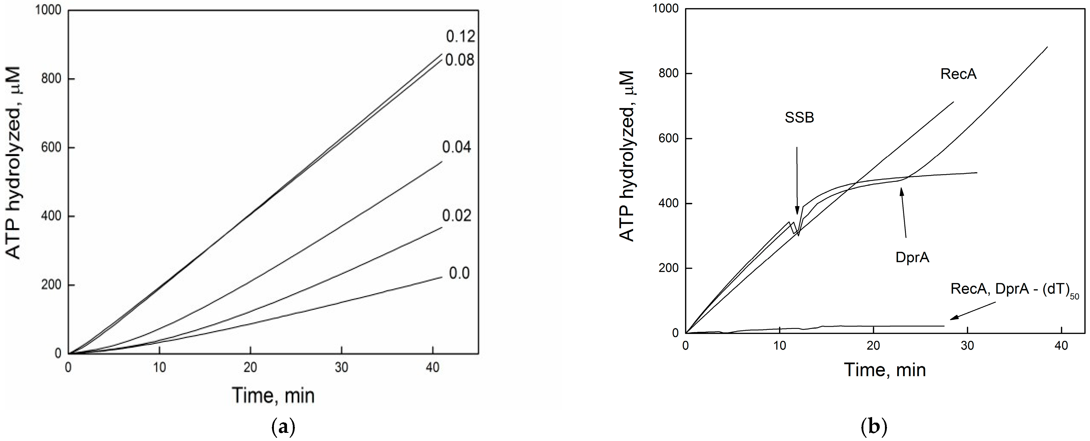
Figure 1. DprABsuantagonises SSBEco function. (a) DprABsu reverses SSBEco inhibition of RecAEco ATP hydrolysis. Reactions mixtures were carried out as described under “Experimental Procedures” and contained circular M13mp8 ssDNA (12 μM in nt), RecAEco(1 μM), SSBEco (0.5 μM), and the indicated concentrations of DprABsu. (b) RecA (3 μM) was preincubated with poly(dT)50 (5 μM in nt). When indicated, SSBEco (0.5 μM) or DprABsu (80 nM) was added. All assays were performed at least three times with consistent results.
Ijms 26 07873 g001
Figure 2. DprABsu enables RecAEco nucleation on short (dT)n oligonucleotides. (a) ATPase activity of RecAEco RecA (3 μM) on (dT)34 (5 μM in nt) in the presence of 2 mM ATP and increasing concentration of DprABsu. (b) dATPase activity of RecAEco (5 µM) on (dT)_n substrates (5 µM in nt), with or without DprABsu (0.3 µM) and 2 mM dATP. All experiments were carried out at least three times with consistent results. All reactions were performed as described under “Experimental Procedures. Data are mean ± SD from three independent experiments.
Ijms 26 07873 g002
Figure 3. Effect of DNA size on RecABsu-dependent dATP hydrolysis in the presence or absence of DprABsu protein. Reaction mixtures contained 3 μM RecABsu, 10 μM (dT)n (in nt), and 2.5 mM dATP in Buffer B. When indicated, 0.3 μM DprABsu was added. The dATP hydrolysis rate (Kcat) was determined as described in Section 3.3.
Ijms 26 07873 g003
Figure 4. Atomic model of the DprA2-RecA3·ATP-(dT)15 complex. Panel (A) shows DprABsu, with protomers shown in green and red, and the two RecABsu protomers are shown in cyan and yellow, respectively. The ssDNA (dT15) strand bridges both proteins; the first six 5′ terminal nucleotides (blue) adopt the DprA-bound conformation (PDB ID: 4LJR) [13], while the seven 3′ terminal nucleotides follow the RecA filament path (PDB ID: 3CMX) [28]. The conformations of the two bridging nucleotides (T7 and T8) were obtained using molecular modeling in this study. The original RecA nucleoprotein filament (brown) is overlaid for reference. ATP–Mg2+ at the RecA interface is shown in the ball-and-stick representation. Panel (B) provides a close-up of the DNA-bridging region, highlighting interactions with DprABsu (green) and RecABsu (cyan). PDB formatted file is presented in the Supplementary Materials.
Ijms 26 07873 g004
Table 1. (a) The dependence of FRE value on the elevated DprA expression in transconjugants of the AB1157 strain crossed with the KL227 donor at 37 °C. (b) The dependence of the FRE value on elevated expression of DprABsu in transconjugants of the AB1157 strain crossed with the KL227 donor at 30 °C.
Expression PlasmidsLinkage (μ) Between Selected thr+ and Unselected leu+ MarkersYield of thr+ Str R Recombinants (% to Donors)FREΔFRE
(a)
ABpT7, ΔdprApET210.93 ± 0.025 (600) *1.6%4.5 ± 1.01.0
ABpT7, ΔdprApDprA0.84 ± 0.032 (600)0.71%11.7 ± 1.52.6
ABpT7, ΔdprApDprABsu0.69 ± 0.021 (400)0.22%28.9 ± 5.36.4
(b)
ABpT7, ΔdprApET210.949 ± 0.024 (600) *1.7%3.2 ± 0.61.0
ABΔdprA, pET210.94 ± 0.018 (600)3.1%3.6 ± 0.51.1
ABpT7, ΔdprApDprA0.59 ± 0.025 (800)0.2%55.5 ± 6.317.3
ABpT7, ΔdprApDprABsu0.56 ± 0.036 (600)0.02%61.2 ± 8.519.1
* Numbers in parentheses indicate the number of clones analyzed.
Disclaimer/Publisher’s Note: The statements, opinions and data contained in all publications are solely those of the individual author(s) and contributor(s) and not of MDPI and/or the editor(s). MDPI and/or the editor(s) disclaim responsibility for any injury to people or property resulting from any ideas, methods, instructions or products referred to in the content.