Article

Mechanotransduction-Driven Modulation of L-Type Calcium Channels: Roles of Nitric Oxide, S-Nitrosylation, and cGMP in Rat Ventricular Cardiomyocytes

by
Olga V. Kamkina
,
Anastasia S. Rodina
,
Andre Kamkin
,
Andrei S. Bilichenko
,
Viktor E. Kazansky
,
Alexandra D. Zolotareva
,
Valentin I. Zolotarev
,
Stanislav A. Shileiko
,
Vadim M. Mitrokhin
and
Mitko I. Mladenov
*
Institute of Physiology, Pirogov Russian National Research Medical University, 117997 Moscow, Russia
*
Author to whom correspondence should be addressed.
Int. J. Mol. Sci. 2025, 26(15), 7539; https://doi.org/10.3390/ijms26157539 (registering DOI)
Submission received: 6 June 2025 / Revised: 18 July 2025 / Accepted: 31 July 2025 / Published: 4 August 2025
(This article belongs to the Special Issue New Insights into Cardiac Ion Channel Regulation 3.0)

Abstract

L-type Ca2+ channels, particularly CaV1.2, play a crucial role in cardiac excitation-contraction coupling and are known to exhibit mechanosensitivity. However, the mechanisms regulating their response to mechanical stress remain poorly understood. To investigate the mechanosensitivity and nitric oxide (NO)-dependent regulation of L-type Ca2+ channels in rat ventricular cardiomyocytes, we used RNA sequencing to assess isoform expression and whole-cell patch-clamp recordings to measure L-type Ca2+ current (ICa,L) under controlled mechanical and pharmacological conditions. RNA sequencing revealed predominant expression of CaV1.2 (TPM: 0.1170 ± 0.0075) compared to CaV1.3 (0.0021 ± 0.0002) and CaV1.1 (0.0002 ± 0.0002). Local axial stretch (6–10 μm) consistently reduced ICa,L in proportion to stretch magnitude. The NO donor SNAP (200 μM) had variable effects on basal ICa,L in unstretched cells (stimulatory, inhibitory, or biphasic) but consistently restored stretch-reduced ICa,L to control levels. Ascorbic acid (10 μM), which reduces S-nitrosylation, increased basal ICa,L and partially restored the reduction caused by stretch, implicating S-nitrosylation in channel regulation. The sGC inhibitor ODQ (5 μM) decreased ICa,L in both stretched and unstretched cells, indicating involvement of the NO–cGMP pathway. Mechanical stress modulates L-type Ca2+ channels through a complex interplay between S-nitrosylation and NO–cGMP signaling, with S-nitrosylation playing a predominant role in stretch-induced effects. This mechanism may represent a key component of cardiac mechanotransduction and could be relevant for therapeutic targeting in cardiac pathologies involving mechanically induced dysfunction.

1. Introduction

L-type Ca2+ channels are essential for cardiac function and mediate the predominant pathway for Ca2+ entry during the action potential of cardiomyocytes. Among these channels, CaV1.2 plays a particularly important role by allowing Ca2+ entry during the initial Ca2+ influx phase that initiates Ca2+ release from the sarcoplasmic/endoplasmic reticulum, which in turn initiates contraction of the cardiomyocyte [1]. CaV1.2 channels are predominantly localized within the T-tubule system associated with the sarcomeric Z-lines, where they are in close proximity to ryanodine receptors on the sarcoplasmic reticulum [1]. Such an arrangement allows high efficiency in excitation-contraction coupling because of rapid calcium-induced calcium release [1]. This Ca2+-induced Ca2+ release is crucial for the coherence of the excitation-contraction coupling and heart function [1].
Recent studies have demonstrated that L-type Ca2+ channels possess significant mechanosensitivity, and their gating properties are modulated by mechanical stress. Studies have shown that CaV1.2 channels exhibit both single-channel and whole-cell current mechanical sensitivity [2]. Similarly, CaV1.3 channels have been found to be mechanosensitive [3], but their expression in ventricular myocytes is markedly lower than in CaV1.2. This mechanosensitivity places these channels in a separate category from mechano-gated channels (MGCs), and they are called mechano-sensitive channels (MSCs). A mechano-sensitive channel (MSC) is often considered any ion channel that is mechanically gated by forces such as membrane tension, cytoskeletal deformation, or extracellular matrix interactions. It should to be noted that another subclass of MSCs—mechanically gated channels (MGCs)—are directly opened by membrane stretch or pressure. This difference enables us to distinguish channels regulated indirectly by mechanical forces from channels that are inherently gated by such forces.
The regulation of L-type Ca2+ channels by nitric oxide (NO) is a complex and controversial field. It has previously been demonstrated that NO regulates the channel through two separate pathways that include the classic NO-sGC-cGMP pathway and direct S-nitrosylation of the channel proteins. The action of NO on basal Ca2+ currents (ICa,L) has been described as activation, inhibition, absence of effect, or concentration dependence of the effect [4]. This wide variety of responses indicates complex regulatory mechanisms that may depend on specific cellular conditions and experimental parameters. Based on studies using NO-sensitive dyes, there is evidence that NO production is elevated in cardiomyocytes in response to mechanical stress like cell stretching [5,6]. However, it is still unclear how NO modulates L-type Ca2+ channels under mechanical stress. Although NO mediates its effects through the soluble guanylyl cyclase/cyclic guanosine monophosphate pathway (sGC/cGMP), others have demonstrated the importance of direct S-nitrosylation of channel proteins [7].
The present investigation aims to reveal the signaling pathways involved in the control of L-type Ca2+ channels (e.g., CaV1.2) by both mechanical stretch and NO in rat ventricular cardiomyocytes. We particularly focus on the effects of mechanical stretch on channel activity and examine the roles of S-nitrosylation and NO-cGMP pathways in these changes. Understanding these mechanisms is important since they may provide therapeutic targets relevant to cardiac pathologies associated with mechanical stress and changes in Ca2+ handling.
Through integrated RNA sequencing, patch-clamp recording, and pharmacological approaches, we show that mechanical stretch uniformly suppressed ICa,L via mechanisms mediated by both pathways. Our findings provide a new perspective on the intricate crosstalk between mechanical stress and NO signaling in the regulation of cardiac Ca2+ channels and may open new opportunities for designing targeted therapeutic regimens for heart disease. To gain further insights into the molecular profiles of stretch-induced changes in cardiomyocyte function, we performed genome-wide RNA-seq analysis. The objective was to identify systemic changes in gene expression with a special focus on mechanosensitive ion channels (especially transient receptor potential M7 (TRPM7), Piezo1, and the KCNK family), members of the NO-sGC-cGMP pathway (NOS1–3, GUCY1A3), and enzymes involved in cytoskeletal remodeling and redox homeostasis. Unlike classical qPCR, RNA-seq has the advantage of hypothesis-free scanning for novel transcripts and pathway-level changes that might guide subsequent functional investigations.

2. Results

2.1. Expression Profile of Voltage-Gated Calcium Channels and Their Auxiliary Subunits

2.1.1. L-Type Calcium Channel Predominance in Rat Ventricular Cardiomyocytes

RNA sequencing of rat ventricular myocytes identified a clear pattern in the expression of voltage-gated calcium channels (Figure 1A). Among the L-type channels, CaV1.2 (CACNA1C) was the predominant isoform (0.117 ± 0.0118 TPM), followed by CaV3.2 (CACNA1H) (0.1981 ± 0.0129 TPM) and CaV3.1 (CACNA1G) (0.0669 ± 0.0154 TPM). CaV1.3 (CACNA1D) had minimal expression with 0.0019 ± 0.0002 TPM compared to CaV1.1, CaV1.4, CaV2.2, and CaV3.3, which were undetectable or negligible. CaV2.1 (0.0088 ± 0.0023 TPM) and CaV2.3 (0.0004 ± 0.0002 TPM) were found at very low levels, as expected, as they are mainly expressed in neuronal tissues.
These data from three independent biological replicates unambiguously identify CaV1.2 as the predominant voltage-gated calcium channel in adult rat ventricular cardiomyocytes.

2.1.2. Auxiliary Subunit Expression Profile of L-Type Calcium Channels

To fully characterize the molecular composition of L-type calcium channel complexes in rat ventricular myocytes, we determined the expression profile of auxiliary subunits α2δ and β that are known to alter channel function (Figure 1B). Among β subunits, CACNB2 (0.2143 ± 0.0400 TPM) was most abundant, followed by CACNB3 (0.1852 ± 0.0239 TPM) and CACNB1 (0.0324 ± 0.0025 TPM), with very low levels of CACNB4 (0.0005 ± 0.0001 TPM). Regarding the α2δ subunits, CACNA2D2 (0.1986 ± 0.0219 TPM) and CACNA2D1 (0.0421 ± 0.0062 TPM) were predominant, whereas CACNA2D3 and CACNA2D4 were expressed below the detection limit. Among γ subunits, only CACNG6 showed substantial expression (0.7805 ± 0.1128 TPM) while CACNG1 was not detected. These auxiliary subunits likely contribute to the functional regulation of CaV1.2 channel complexes in the ventricular myocardium. Notably, the majority of γ subunits (CACNG2–8) were excluded from this analysis, as they have been functionally reclassified as transmembrane AMPA receptor regulatory proteins (TARPs) rather than true auxiliary calcium channel subunits [8,9]. No members of the γ family have been recognized as bona fide L-type calcium channel modulators except CACNG1 and CACNG6 [10,11].
Collectively, these data indicate that CACNB2, CACNB3, CACNA2D2, CACNA2D1, and CACNG6 are the predominant auxiliary subunits expressed in rat ventricular cardiomyocytes. The substantial expression of these regulators serves as a molecular blueprint for the regulation of CaV1.2 function in subsequent electrophysiological studies.

2.1.3. Implications for T-Type Channel Contribution to Recorded Currents

Although T-type calcium channels—particularly CaV3.1—were detectably expressed in the RNA sequencing dataset used in our experiment, their functional role in the recorded calcium currents was insignificant due to the specific design of our electrophysiological protocol. Patch-clamp recordings were performed using a Vh of −80 mV, a holding potential that effectively inactivates T-type Ca2+ channels—typically activated at more negative potentials (approximately −70 to −40 mV). Furthermore, our pulse protocol, which ranged from −40 mV to +40 mV, was focused on a voltage range that preferentially activates L-type Ca2+ channels, ensuring that the recorded currents predominantly reflected L-type activity. In addition, the absence of mechanosensitivity in T-type channels is supported by the literature. Foundational electrophysiology has shown that T-type channels do not open in response to mechanical stimuli (hydrostatic pressure, membrane stretch) that robustly open channels of other types, such as N-type channels [12,13]. This functional separation is consistent with structural studies that have shown that T-type channels do not have the important mechanosensitive motifs (including amphipathic helices and lipid-sensitive domains) present in many stretch-activated ion channels [14].
All of these points together support that the stretch-modulated Ca2+ currents we observed in our experiments mainly represent L-type CaV1.2 channels as the predominant functional subtype in rat ventricular cardiomyocytes, which are structurally suitable for mechanotransduction.

2.1.4. Impact of Mechanical Stretch on L-Type Ca2+ Currents

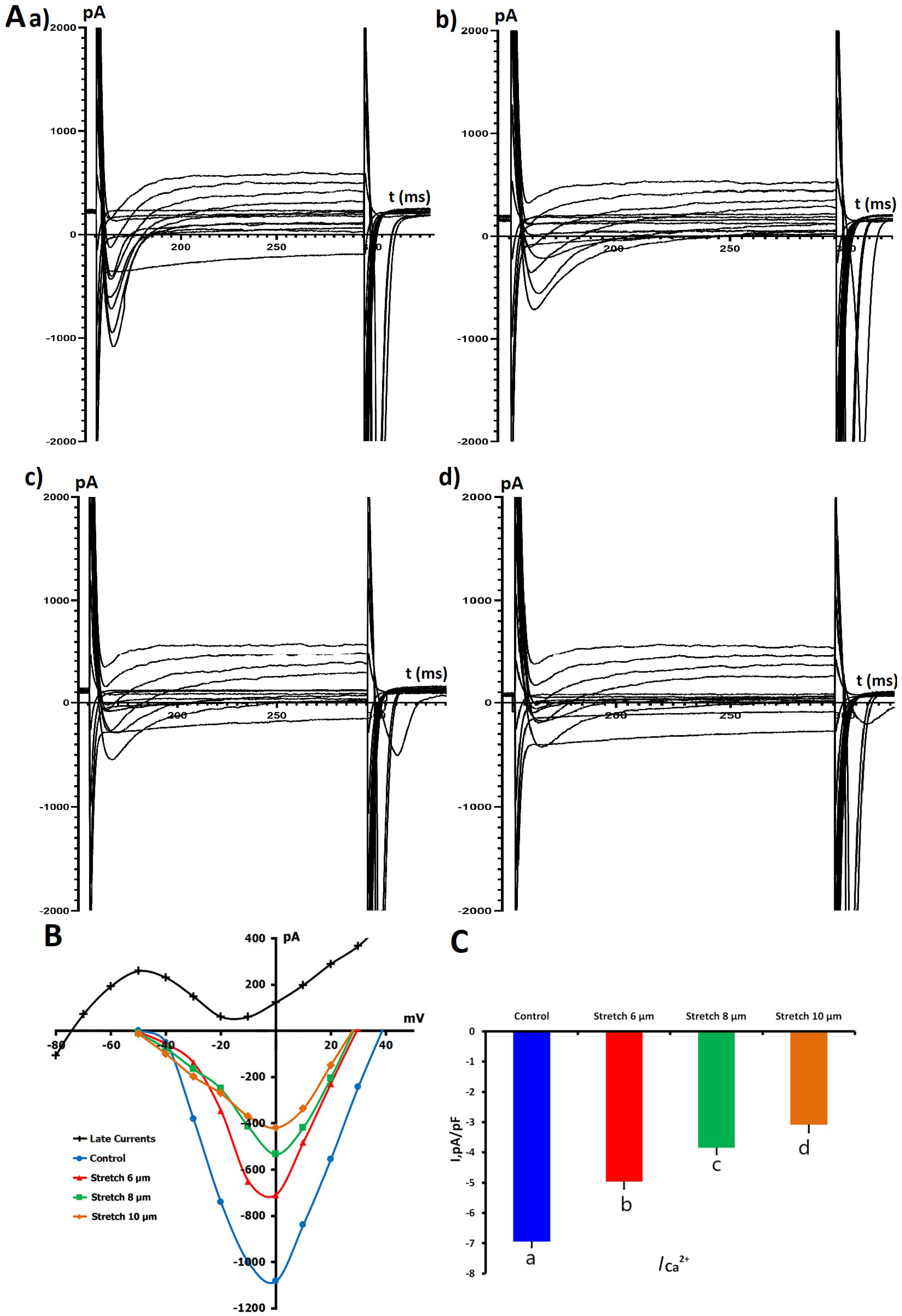
We investigated the mechanosensitivity of ICa,L through measurement of ICa,L before and during applied axial stretch. To maintain high-quality electrophysiological data, only cells with stable access resistance and <10% baseline current drift over the course of the protocol were included for analysis. In Figure 2, the voltage-dependent properties of ICa,L and the modulation of ICa,L by different amounts of mechanical stretch (6, 8, and 10 μm) are shown.
Under control conditions, in K+in/K+out solution, baseline ICa,L density was −6.95 ± 0.18 pA/pF (n = 7, m = 4), with sarcomere length 1.83 ± 0.01 μm. Local axial stretch produced a consistent and graded reduction in ICa,L amplitude, correlating with increases in sarcomere length. ICa,L reduction was observed with 6 μm stretch (sarcomere length 2.00 ± 0.01 μm), where ICa,L diminished to −4.96 ± 0.24 pA/pF. The current continued to decrease with 8 μm stretch (sarcomere length 2.10 ± 0.01 μm) to −3.84 ± 0.22 pA/pF, reaching −3.08 ± 0.19 pA/pF at 10 μm stretch (sarcomere length 2.17 ± 0.01 μm). All changes were statistically significant compared to the control (p < 0.01).

2.1.5. Effects of NO Donor SNAP on ICa,L

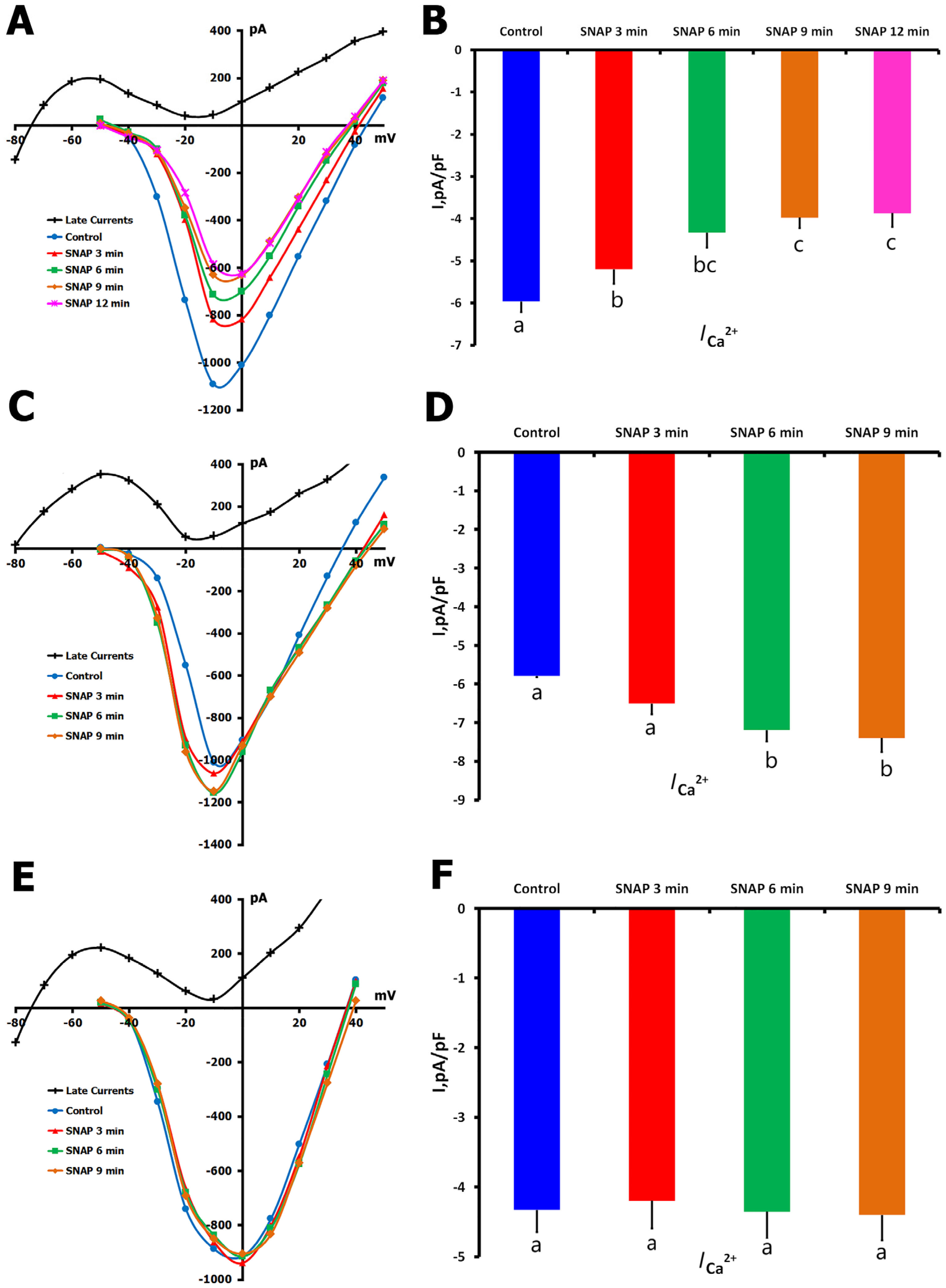
Heterogeneities in the modulation of ICa,L by application of S-nitroso-N-acetyl-D,L-penicillamine (SNAP) (200 µM) in unstretched cardiomyocytes were obtained, revealing three distinct response profiles. These are shown in Figure 3 as a reduction in ICa,L (Figure 3A,B), an enhancement of ICa,L (Figure 3C,D), and no significant current size change (Figure 3E,F).
The most common response (66.6% of cells; n = 22) was an ICa,L decrease from −5.96 ± 0.25 to −3.87 ± 0.33 pA/pF after 12 min of SNAP exposure. During the treatment phase, a statistically significant difference compared to the control was found, but no significant differences were confirmed between time points (3, 6, 9, and 12 min; p = ns).
In a smaller subset of cells (9%; n = 3), SNAP significantly enhanced ICa,L from −5.79 ± 0.31 to −7.40 ± 0.36 pA/pF after 9 min (p < 0.01 vs. control), with no significant differences between intermediate time points (p = ns).
The remaining cells (24.4%, n = 8) showed no change in ICa,L throughout the recording period, as compared with the baseline (control: −7.72 ± 0.23 pA/pF; p = ns at each time point).

2.1.6. Effects of SNAP on ICa,L During Mechanical Stretch

To address the interplay between NO signaling and mechanical stress in the modulation of L-type Ca2+ channels, we applied the following two complementary experimental protocols:
(1)
SNAP application during sustained mechanical stretch;
(2)
Mechanical stretch was applied to cells pretreated with SNAP.
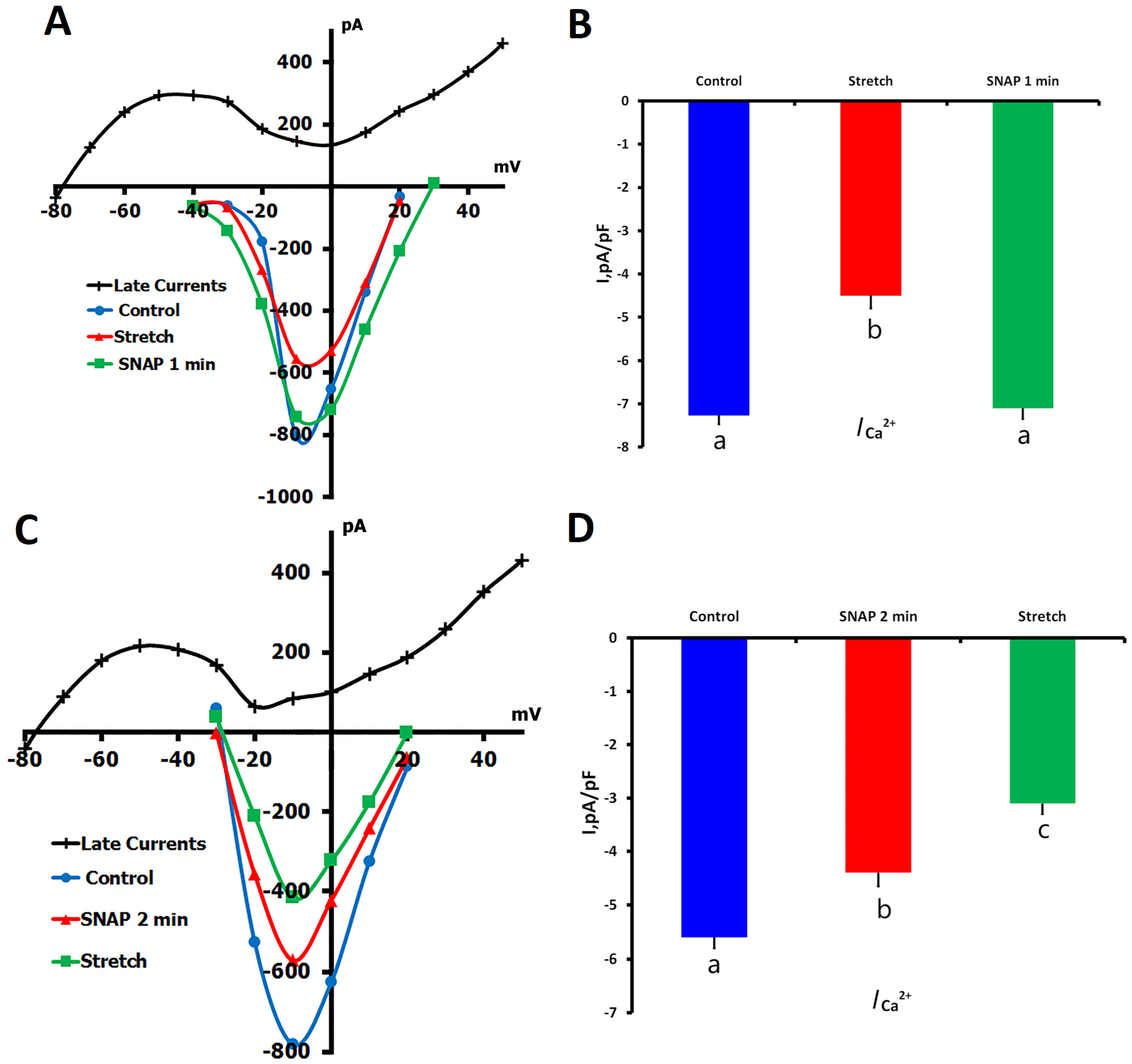
In protocol 1, application of local axial stretch (6 μm) significantly decreased ICa,L from −7.27 ± 0.20 pA/pF (control) to −4.50 ± 0.30 pA/pF (p < 0.01; n = 7, m = 5). When SNAP (200 μM) was perfused during the maintained stretch, ICa,L quickly returned to −7.10 ± 0.40 pA/pF within 1 min (p = ns vs. control), a value not significantly different from the baseline, but significantly higher than during stretching alone (p < 0.01). These results indicate that NO can rapidly nullify the effect of stretch on L-type Ca2+ channels (Figure 4A,B).
In the second protocol, SNAP was applied to unstretched cells, followed by a decrease in ICa,L from −5.60 ± 0.40 pA/pF to −4.50 ± 0.40 pA/pF after 2 min (p < 0.01). When these SNAP-treated cells were subsequently subjected to 6 μm stretch, ICa,L was further reduced to −3.30 ± 0.40 pA/pF (p < 0.01 vs. both control and SNAP alone), demonstrating that the mechanical stimulus was able to further reduce basal myocyte ICa,L when superimposed on that from SNAP (Figure 4C,D).
The divergent results observed in these two experimental settings—SNAP reversing stretch-induced inhibition when applied after stretch but augmenting inhibition when applied before stretch—serve to illustrate the temporal context of both the NO and the mechanical pathways. These findings are consistent with the idea that NO and stretch regulate ICa,L via partly independent but convergent mechanisms and that the particular sequence of activation is crucial for dictating channel behavior. The rapid reversibility of stretch effects by SNAP also indicates a dynamic regulatory mechanism that may be pertinent to potential therapy for diseases associated with altered mechanical load (cardiac hypertrophy or failure).

2.2. Effects of sGC Inhibition by ODQ on ICa,L Regulation

2.2.1. ODQ Inhibition of ICa,L in Unstretched Cells

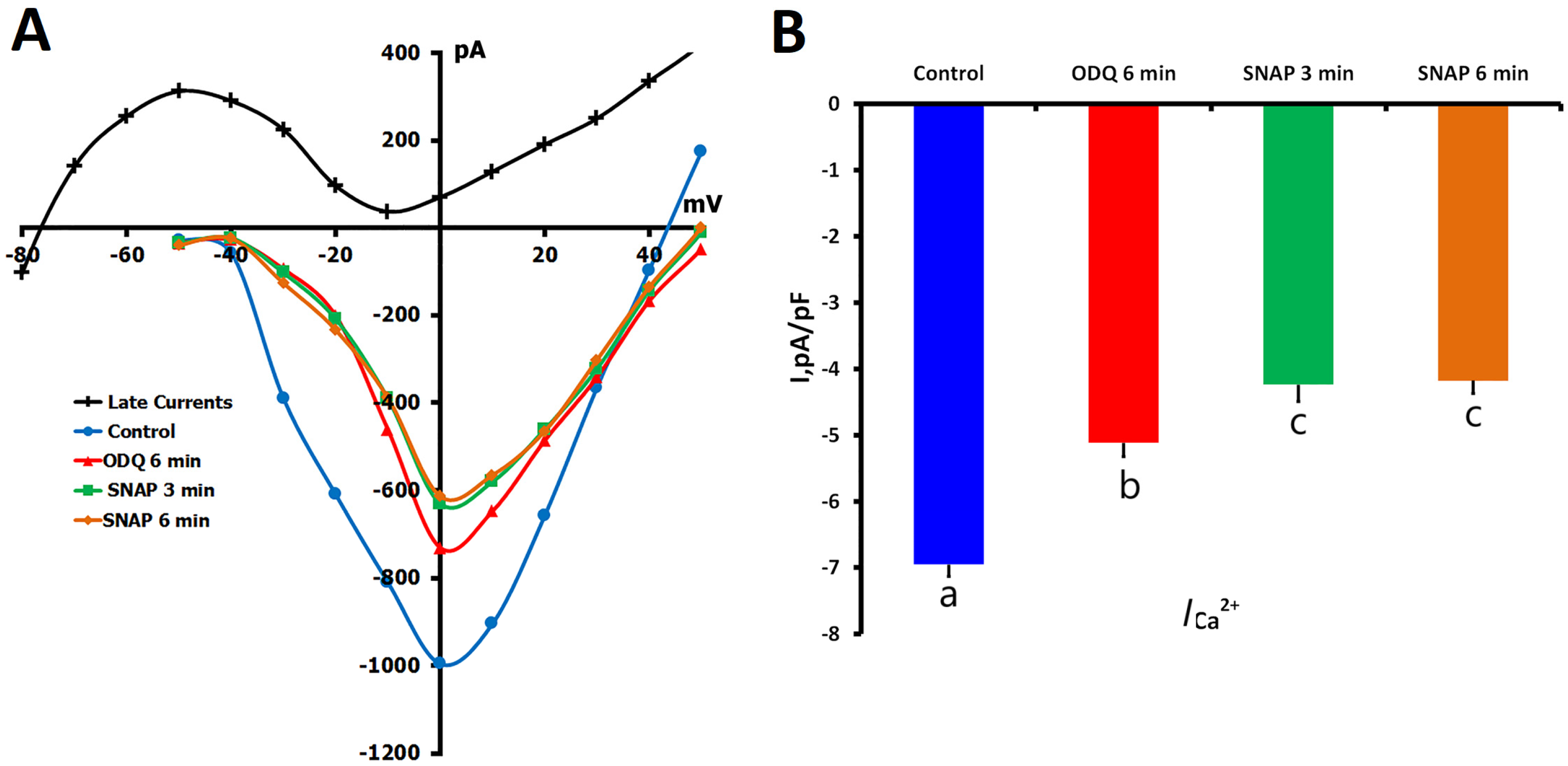
To determine the involvement of the NO-sGC-cGMP signaling pathway in the regulation of L-type Ca2+ channels, we initially investigated the impact of sGC inhibition by 1H-[1,2,4]-oxadiazolo[4,3-a]quinoxalin-1-one (ODQ) (5 μM) in unstretched ventricular myocytes. ICa,L was significantly attenuated by the application of ODQ for 6 min from −6.88 ± 0.18 pA/pF to −5.23 ± 0.21 pA/pF (p < 0.01; n = 13, m = 7), suggesting that tonic sGC activity is involved in determining basal Ca2+ current amplitude (Figure 5A,B).
Upon subsequent introduction of SNAP (200 μM) into the ODQ-containing perfusate ICa,L decreased further to −4.49 ± 0.25 pA/pF at 3 min (p < 0.01 vs. ODQ alone) with no further significant change at 6 min (−4.45 ± 0.19 pA/pF; p = ns vs. 3-min value). This additive inhibition indicates that SNAP affects ICa,L through sGC-independent pathways, probably through other NO signaling pathways such as S-nitrosylation.

2.2.2. ODQ Effects on ICa,L During Mechanical Stretch

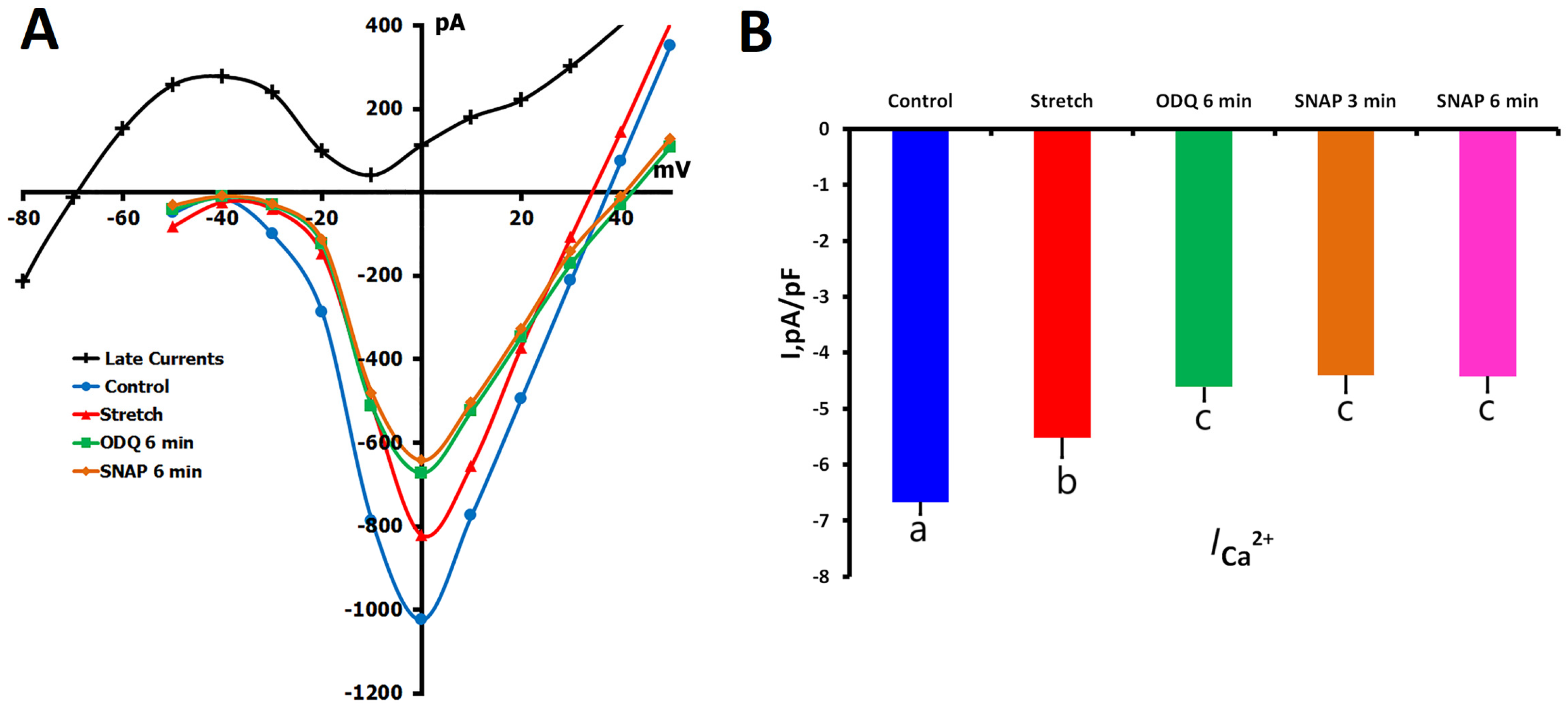
We further investigated the effect of sGC inhibition on the regulation of Ca2+ current under mechanical stress. In these experiments (n = 7, m = 4), 6 μm mechanical stretch decreased ICa,L from control levels of −6.67 ± 0.22 pA/pF to −5.52 ± 0.35 pA/pF (p < 0.01), as previously reported (Figure 6A,B). ODQ applied subsequently reduced ICa,L to −4.61 ± 0.26 pA/pF after 6 min (p < 0.01 vs. stretch alone).
In the case of stretched myocytes, the presence of SNAP did not produce notable modifications of ICa,L when the cells were subjected to ODQ: −4.42 ± 0.40 pA/pF at 3 min and −4.48 ± 0.42 pA/pF at 6 min (p = ns). This insensitivity toward SNAP is in contrast to the further inhibitory effect in unstretched cells, implying that mechanical stretch modulates the channel sensitivity to NO-mediated regulation.
Collectively, the findings under stretched and unstretched conditions suggest that the NO-sGC-cGMP pathway participates in the modulation of Ca2+ channels in a context-specific manner. Mechanical stress also seems to alter the balance of sGC-dependent and sGC-independent NO signaling, indicating the dynamic crosstalk between mechano-stimulated and biochemical control of L-type Ca2+ channels in cardiomyocytes.

2.3. Effects of Ascorbic Acid (AA) on ICa,L Regulation

2.3.1. AA Modulation of ICa,L in Unstretched Cells

To examine the role of S-nitrosylation in the modulation of L-type Ca2+ channels, we used ascorbic acid (AA, 10 μM), a reducing agent that has been shown to specifically remove S-nitrosylation from proteins. In our study, the effect of AA alone and its interaction with NO donor SNAP in unstretched ventricular cardiomyocytes were examined.
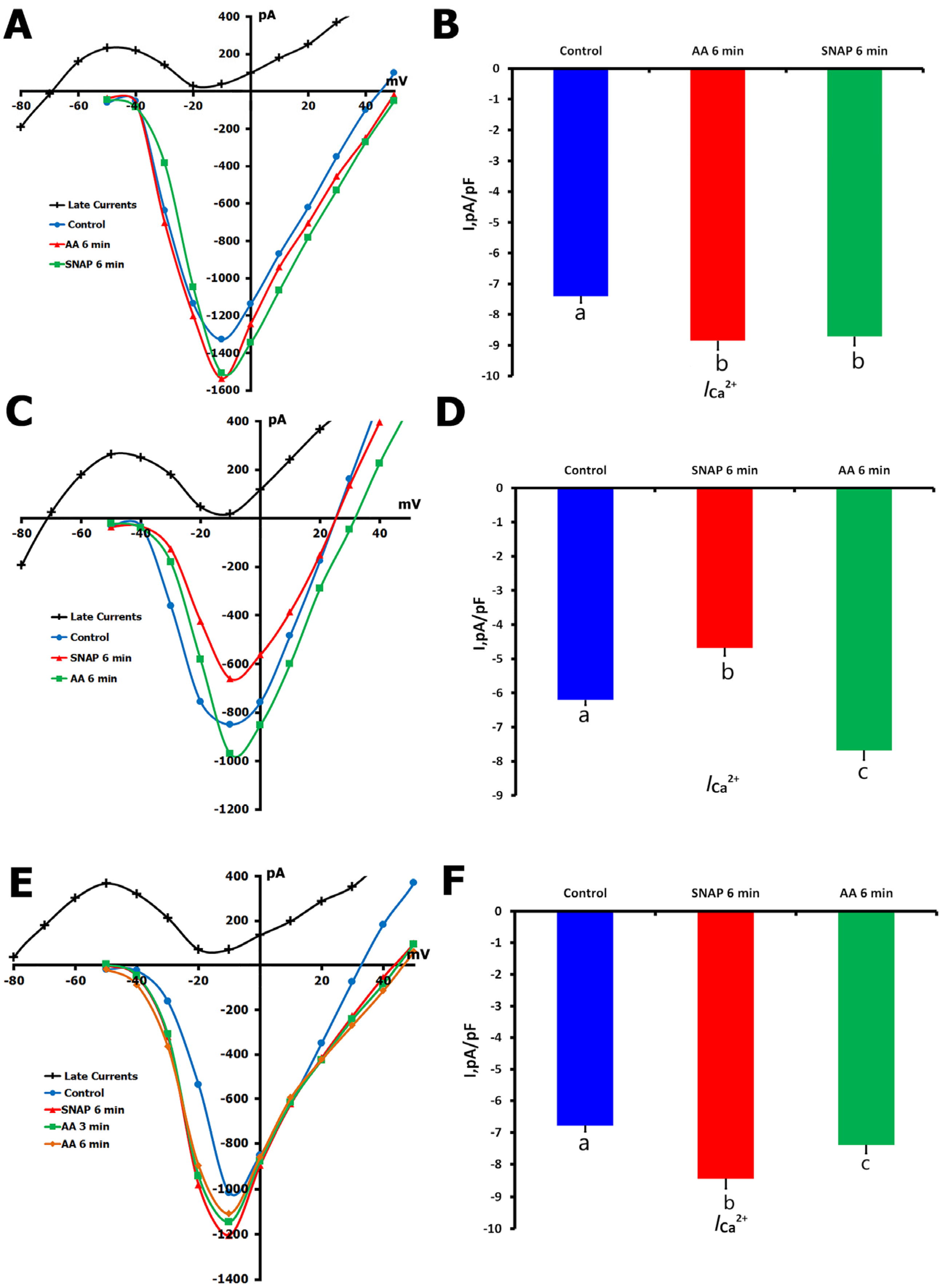
In the first series of experiments (n = 8, m = 3), we tested the impact of AA application alone. Baseline ICa,L was −7.57 ± 0.02 pA/pF and increased to −8.77 ± 0.03 pA/pF after 6 min of AA perfusion (p < 0.01; Figure 7A,B). When 200 μM SNAP was added in the continued presence of AA, there was no significant change in ICa,L (−8.54 ± 0.03 pA/pF at 6 min, p = ns compared to AA alone). This indicates that AA pretreatment prevented SNAP-induced channel modulation, possibly by scavenging S-nitrosylation targets before the addition of SNAP.
In a second series of experiments, we reversed the order of application to assess the effect of AA on SNAP-modulated ICa,L. We identified two distinct response patterns.
In the first pattern (n = 7, m = 4), SNAP alone decreased ICa,L from −6.44 ± 0.15 pA/pF to −5.02 ± 0.23 pA/pF over 6 min (p < 0.01). Subsequent AA application reversed ICa,L to −7.37 ± 0.28 pA/pF (p < 0.01 versus SNAP; Figure 7C,D), which demonstrated that AA successfully reversed the SNAP-induced suppression, supporting the major role of S-nitrosylation in the inhibitory effect.
In the second pattern (n = 6, m = 3), SNAP first increased ICa,L from −7.10 ± 0.02 pA/pF to −8.25 ± 0.31 pA/pF (p < 0.01). The enhancement of ICa,L by SNAP was partially reversed by AA treatment, −7.59 ± 0.03 pA/pF (p < 0.01 vs. SNAP; Figure 7E,F), but the resultant amplitude was still significantly greater than that of control (p < 0.05). This indicates that SNAP’s stimulatory effect is mediated by S-nitrosylation as well as by a non-S-nitrosylation mechanism.
Taken together, these findings underscore the key role of S-nitrosylation in the regulation of L-type Ca2+ channel activity and uncover the dual potential of NO signaling that is considered in light of the redox status of the channels.

2.3.2. AA Effects on Stretch-Reduced ICa,L

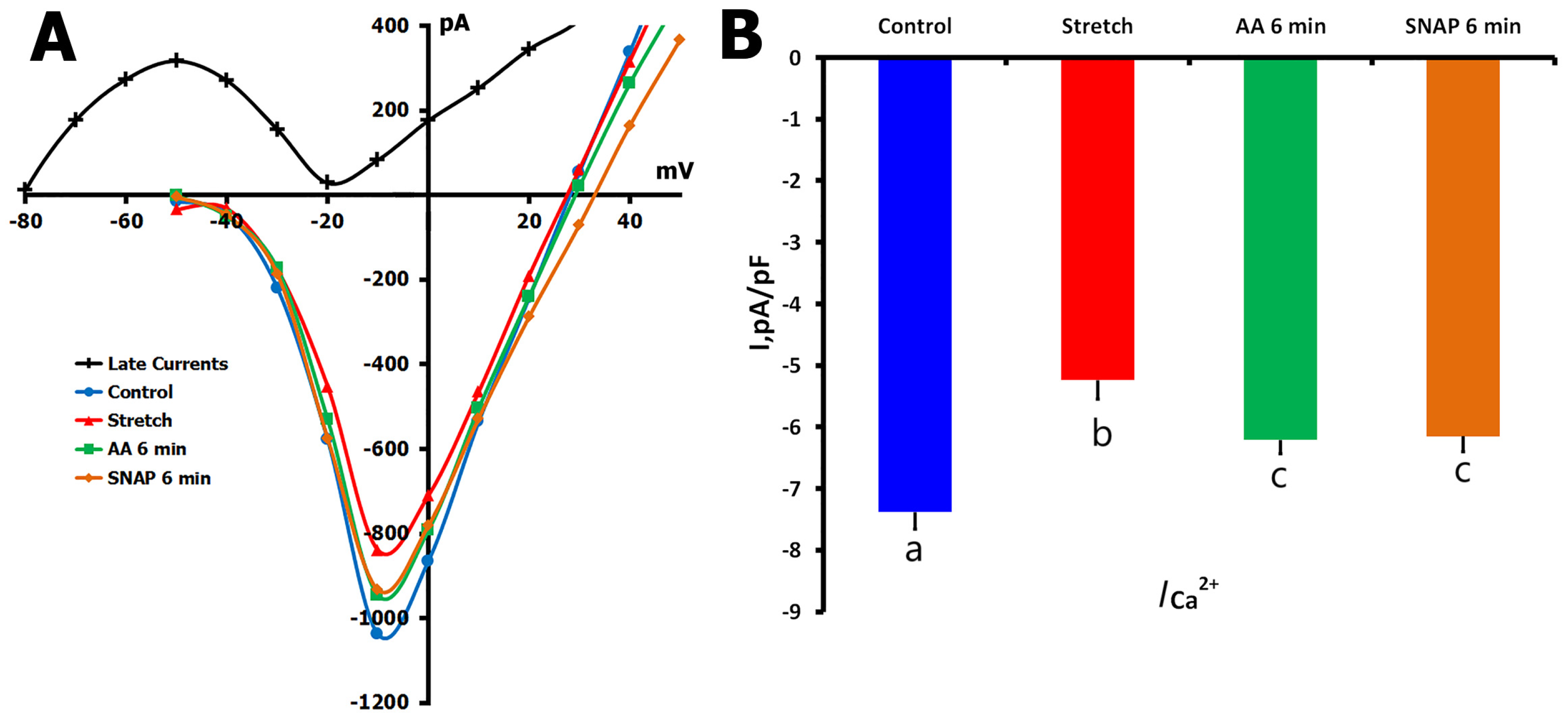
Following the characterization of AA’s effects on basal Ca2+ channel activity, we next investigated its ability to modulate ICa,L in cardiomyocytes subjected to mechanical stretch. In these experiments (n = 7, m = 3), mechanical stretching of the cell membrane by 6 μm significantly reduced ICa,L from a control value of −7.31 ± 0.28 pA/pF to −5.24 ± 0.31 pA/pF (p < 0.01; Figure 8A,B).
Perfusion with AA (10 μM) for 6 min partially restored the Ca2+ current, resulting in an ICa,L of −6.11 ± 0.22 pA/pF (p < 0.01 vs. stretch; p < 0.05 vs. control), indicating that at least part of the stretch-induced inhibition is mediated through S-nitrosylation of L-type Ca2+ channels. In contrast, in the continued presence of mechanical stretch, when SNAP (200 μM) was added after AA treatment, no further reduction of ICa,L was observed (−6.05 ± 0.25 pA/pF, p = ns vs. AA alone), suggesting no further regulation of ICa,L by NO at this time point.
These results suggest that mechanical stretch decreases ICa,L, at least in part, through pathways related to S-nitrosylation, and that AA can reverse this effect. However, the partial recovery and its resistance to additional SNAP modulation indicate the involvement of S-nitrosylation-independent mechanisms in stretch-induced downregulation of Ca2+ channel activity.

2.4. Effects of NEM on ICa,L Regulation

2.4.1. Biphasic NEM Effects on ICa,L in Unstretched Cells

To further investigate the role of protein thiol groups in Ca2+ channel regulation, we employed N-ethylmaleimide (NEM), which irreversibly alkylates thiol groups and prevents S-nitrosylation. Due to the compound’s cellular toxicity at physiological temperature, these experiments were conducted at 22 °C with careful monitoring of cell viability. Initial experiments revealed that even at reduced temperatures, NEM concentrations above 200 μM or extended exposure time led to rapid cell deterioration, consistent with previous reports in the literature.
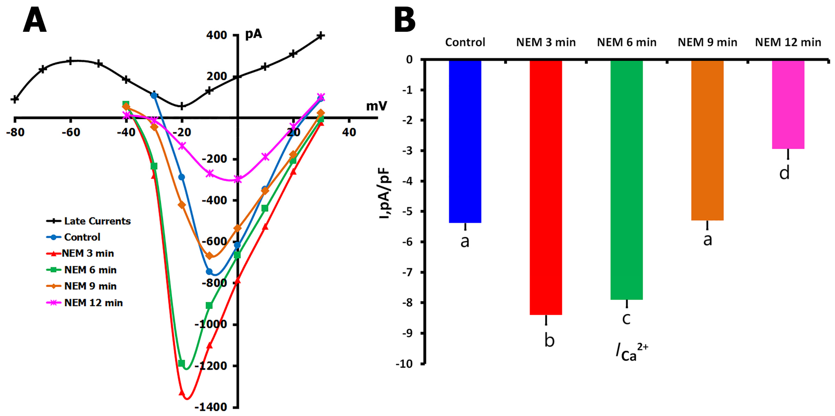
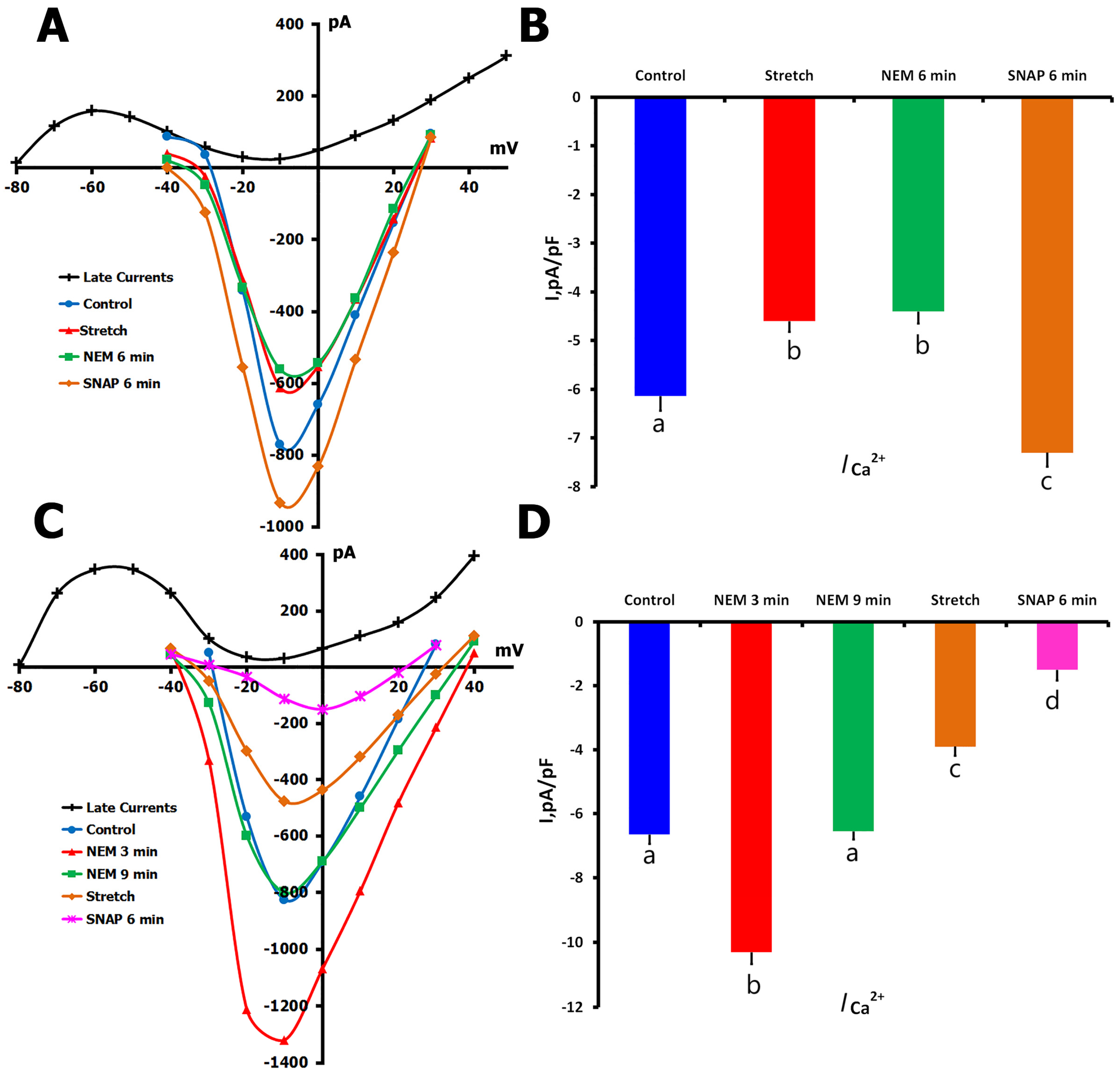
In unstretched cells (n = 10, m = 4), NEM (200 μM) produced a striking biphasic response in ICa,L (Figure 9A,B). From control values of −5.40 ± 0.3 pA/pF, the current increased dramatically to −8.40 ± 0.3 pA/pF within the first 3 min of NEM exposure. This initial increase was followed by a slight decline to −7.90 ± 0.4 pA/pF at 6 min, then a more substantial reduction to near-control levels (−5.30 ± 0.3 pA/pF) at 9 min. By 12 min, ICa,L had fallen significantly below control values to −2.92 ± 0.2 pA/pF. This complex temporal response pattern suggests that different populations of thiol groups, possibly with varying accessibility or reactivity, contribute to Ca2+ channel regulation.

2.4.2. NEM Effects on Stretch-Modified ICa,L

To understand how thiol modification affects stretch-dependent channel regulation, we employed two distinct experimental protocols (Figure 10). In the first approach (n = 7, m = 3), we initially stretched the cells, confirming the typical reduction in ICa,L from −6.14 ± 0.3 pA/pF to −4.60 ± 0.22 pA/pF (Figure 10A,B). Subsequent application of NEM to these stretched cells produced minimal change in current amplitude (−4.40 ± 0.29 pA/pF). However, when SNAP was added to the NEM-containing solution, we observed a surprising increase in ICa,L to −7.30 ± 0.33 pA/pF, exceeding control levels.
In the second protocol (n = 6, m = 3), we reversed the sequence by applying NEM before mechanical stretch (Figure 10C,D). The initial NEM application produced the characteristic biphasic response, with ICa,L increasing from −6.65 ± 0.29 pA/pF to −10.3 ± 0.37 pA/pF after 3 min, then returning to near-control levels (−6.55 ± 0.25 pA/pF) after 12 min. When these NEM-treated cells were subsequently stretched, ICa,L decreased to −3.91 ± 0.29 pA/pF, and the addition of SNAP caused a further reduction to −1.66 ± 0.33 pA/pF.
The contrasting responses to SNAP in these two protocols—enhancement of current when applied after stretch versus inhibition when applied before stretch—suggest that the temporal sequence of thiol modification and mechanical stress critically determines the final functional state of the channels. Furthermore, the ability of mechanical stretch to modify channel function even after NEM treatment indicates that some aspects of stretch-dependent regulation may occur independently of thiol group availability.

3. Discussion

3.1. Expression Pattern and Mechanosensitivity

Our RNA-seq data conclusively validate CaV1.2 (CACNA1C) as the dominant member of the group of voltage-gated calcium channels expressed in rat ventricular cardiomyocytes, both in overall transcript amount and distribution among other L- and T-type isoforms [15,16]. Notably, CaV1.3 and CaV1.1 had negligible expression, while T-type channels (particularly CaV3.1 and CaV3.2) were expressed at magnitudes far below those of CaV1.2. Functionally, this expression profile is highly supportive of our conclusion that ICa,L recorded in our studies is mostly due to CaV1.2 channel activity with minimal contribution from T-type calcium channels. In addition, their auxiliary subunit profile (with high levels of CACNB2, CACNA2D2, and CACNG6 expression) is indicative of a molecular composition more suitable for greater functional modulation of CaV1.2. Critically, these findings provide a molecular rationale for our decision to narrow our investigations of mechanosensitivity and NO modulation to CaV1.2 channel activity and exclude significant confounding effects of other Ca2+ channel subtypes.
We found a uniform suppression of ICa,L during mechanical stretch, which supported previous results in different preparations [17,18]. Similar Ca2+ current reductions in response to axial stretch have been found in guinea pig ventricular myocytes [17,18], indicating a common mechanosensitive mechanism among species. F-actin or similar cytoskeletal components, e.g., microtubules, have also been suggested to participate in the modulation of mechanosensitive channels and CaV1.2 gating, providing additional evidence for an integrated structural–functional response to mechanical stimulation [19].
Although some heterogeneity was observed in I/V relationships, it may be attributed to the intrinsic cell-to-cell variability in cells isolated from the tissue. To ensure data reliability, only recordings with stable series resistance and comparable membrane capacitance were included. Representative current traces also confirm the reproducibility of our electrophysiological findings (Figure 2A).
Collectively, the RNA-seq data confirm CaV1.2 as the major L-type Ca2+ channel isoform in this cellular environment, thus rationalizing the observed electrophysiological effects (Figure 1A,B). The stretch-dependent decrease in ICa,L and its modulation by NO donors and inhibitors reflect changes in the functional state of CaV1.2 activity. Interestingly, NO signaling seems able to suppress this mechanosensitive suppression and even promote it. This dual sensitivity is probably a consequence of separate yet overlapping mechanisms (i.e., S-nitrosylation and cGMP-mediated signaling) that work autonomously but interact with mechanical stimuli. These findings underscore the significance of temporal dynamics and subcellular compartmentalization in deciphering the manner by which mechanical- and redox-activated signals converge at the level of a single, biologically relevant ion channel.

3.2. NO Signaling Complexity and Mechanisms of Regulation

The differential modulation of Ca2+ channels by NO underscores the complex control of cardiac electrophysiological properties by redox signaling. Previous studies have shown conflicting results for the effect of NO donors: SNAP increased basal ICa,L in human atrial myocytes [20], while it decreased the current in neonatal rat ventricular strips [21]. These data are in accordance with our observation that biphasic changes induced by nitro-L-arginine methyl ester (L-NAME) or diethylenetriaminepentaacetic acid (DTPA) were seen, which probably corresponded to drug concentrations, as has been reported with 3-morpholinosydnonimine (SIN-1) in frog ventricular myocytes [22].
Such apparent inconsistencies can be explained by multiple levels of physiological complexity. The first is due to the fact that the three isoforms of NO synthase (NOS)—neuronal (nNOS), endothelial (eNOS), and inducible (iNOS)—have different subcellular localizations and activation pathways. For instance, eNOS is membrane-bound and can be stimulated mechanically, while nNOS is located near the sarcoplasmic reticulum and transversal tubules [23]. These spatial differences lead to an NO production that is locally regulated and may affect the NO environment of neighboring L-type Ca2+ channels.
Second, NO acts through two major types of signaling: rapid, reversible S-nitrosylation of protein thiols and the classical NO-sGC-cGMP pathway. The preferential choice of one pathway over the other is determined by redox status, availability of oxygen, and cofactors such as tetrahydrobiopterin (BH4) [24]. Third, the cellular context (species, developmental stage, experimental approach) is crucial for both channel expression and the machinery that orchestrates the upstream regulation. Together, these factors explain the conditional facilitation of, or decrease in, ICa,L by NO [25].
Our findings indicate that mechanical stimulation modulates L-type Ca2+ channel function by a fine-tuned interplay between S-nitrosylation and sGC-cGMP signaling. ODQ- and AA-treatment experiments lend further support to crosstalk processes in our system and indicate S-nitrosylation to be functionally relevant. Moreover, the differential effects of various NO donors suggest that the local activity of NO synthase (NOS)—particularly endothelial (eNOS) and/or neuronal NOS (nNOS)—is target-specific, probably depending on the nature of the protein substrates or the availability of reactive cysteine residues [26]. The phenomenon of transnitrostylation, in which intermediate proteins accept nitrosyl groups, complicates the issue even further [27]. Furthermore, S-nitrosylation itself may also suppress NOS activity, suggesting complex feedback regulation in the NO signaling system [28]. However, the specific spatial and molecular dynamics of these interactions are not fully elucidated.
NO has been demonstrated to be generated locally when the heart undergoes mechanical stretching, demonstrating the site specificity of NO release in ventricular cardiomyocytes [5]. This compartmentalization is essential for efficient ion channel regulation, particularly NOS3-mediated regulation. Moreover, NOS3−/− mice are unable to produce stretch-induced NO responses, unlike NOS1−/− and NOS2−/− animals [29,30]. Approximately 20% of total NOS3 expression in the heart is derived from ventricular myocytes [31], highlighting its important role in mechanotransduction.
These findings underscore the importance of accurate spatial and temporal measurement of NO. The development of new techniques, such as biotin-switch assays, S-nitrosoproteomics, and fluorescent NO biosensors [32,33], has provided the opportunity to study S-nitrosylation and localization during mechanical manipulations. Moreover, cell-type specificity of this transduction pathway is indicated by varying NO production in cardiomyocytes and fibroblasts [34].
An additional complexity is related to the interaction between NO and β-adrenergic signaling. NO-releasing compounds have been reported to suppress isoprenaline-induced ICa,L in atrioventricular node (AVN) cells [32], suggesting coordinated regulation during mechanical and neurohumoral stress. This interaction may be responsible for adaptation of cardiac excitability to varying physiological demands. Such differences between studies could be due to variation in NO donor, redox status, phosphorylation, and/or NOS isoform contributions [35]. The cell response to NO is also dependent on developmental stage, cell type (atrial vs. ventricular), and metabolic condition [35,36].
Therefore, the relative significance of NO and the extent to which NO produces distinct outcomes vary in response to specific signaling parameters, so effects of NO must be interpreted within this context to reconcile discrepancies between reported data and their translational relevance.

3.3. Integration with Cellular Ca2+ Handling

The interrelationship between stretch-induced ionic currents and Ca2+ handling appears well tuned. 1,2-Bis(o-aminophenoxy) ethane-N,N,N’,N’-tetraacetic acid (BAPTA) does not block stretch-activated currents but alters mechanical effects on ICa,L [19]. It has also been found that BAPTA-AM was capable of inhibiting the stretch-induced decrease in ICa,L in guinea pig ventricular myocytes, implying the mediation of Ca2+-dependent inactivation in the mechanical regulation of L-type Ca2+ channels [37].
The stretch-induced regulation of ICa,L occurs most likely in combination with Ca2+-handling. Mechanical distortion may induce an increase in the intracellular Ca2+ level by a direct influx through MSCs [38] or by release of the Ca2+ from the sarcoplasmic reticulum [6]. The decrease of ICa,L upon stretch (Figure 2) may therefore be a protective feedback mechanism that restrains Ca2+ overload and ensures intracellular homeostasis under mechanical load.

3.4. Regulation Through Multiple Pathways

The intricate interaction between mechanical tension and Ca2+ channel activity consists of several, although partly redundant, signaling steps. The NO-sGC-cGMP system is not the only mediator of the stretch-induced changes, although it may play a significant role. Mechanical forces may also influence other ion transport pathways such as the Na+/Ca2+ exchanger (NCX), particularly if stretch-activated channels are also the site of enhanced Na+ influx and a rise in intracellular Na+ [39]. Furthermore, some K+ channels are also mechanosensitive and can potentially sense changes in intracellular Ca2+ or Na+ concentration [40].
Gadolinium (Gd3+), which has been commonly used as a non-specific blocker, provides valuable information about mechanosensitivity in a larger context. It has been demonstrated to block not only stretch-activated channels but also L-type Ca2+ channels, BKCa channels [41], and delayed rectifier potassium currents [42]. These wide-range inhibitory effects indicate that mechanosensitivity may be a more prevalent feature of ion channels than has been generally recognized.
The diversity of responses to SNAP in unstretched cells probably reflects the heterogeneity of either Ca2+ channel subtypes or of their regulatory environment. A similar variety of responses to SIN-1 has been described with either stimulation or inhibition of the Ca2+ current (ICa,L) in guinea pig ventricular myocytes stimulated with isoprenaline [43] and in ferret ventricular myocytes [44]. This heterogeneity might thus allow fine-tuned contextual modulation of cardiac responses to mechanical input.
It should be noted that while this study concentrates on L-type Ca2+ channels, the cardiac action potential is maintained by the combined activity of a number of types of ion channels [45]. Voltage-sensitive sodium channels (NaV1.5) and numerous potassium channel isoforms, such as TREK-1, KATP, and delayed rectifier channels, are also modulated by mechanical stretch and shape action potential morphology and conduction [46]. For instance, mechanical stimulation might increase late Na+ current or indirectly modulate NCX through intracellular Na+ buildup [47]. Mechanosensitive K+ channels, for their part, may abbreviate action potential duration, and thus, could work with lower ICa,L to prevent Ca2+ overload [48].
Taken together, the interaction and crosstalk of these mechanosensitive ion channels—particularly under disease-stretch or neurohumoral activation—present a major challenge in understanding the mechano-electrical feedback in the heart. Subsequent investigations should focus on decoding such interactions at the molecular and functional levels to fully appreciate their roles in health and disease.

3.5. Limitations and Future Directions

Although this research has expanded our knowledge about the mechanosensitive properties of L-type Ca2+ channels, several limitations need to be considered. While isolated rat ventricular cardiomyocytes are valuable for manipulating experimental conditions, they do not fully replicate the complexity of the whole working heart, where mechanical signaling is integrated within the multicellular network and influenced by neurohumoral factors. For enhanced translational relevance, future studies could integrate whole-heart models or human induced pluripotent stem cell-derived cardiomyocytes (hiPSC-CMs).
Our pharmacological data with ODQ, SNAP, AA, and NEM strongly suggest that both S-nitrosylation and NO-sGC-cGMP signaling are involved in stretch-induced modulation of ICa,L but the conclusions remain inferential, as no direct biochemical evidence for post-translational modification of CaV1.2, such as S-nitrosylation, is present. The involvement of individual NOS isoforms is also unclear. Further studies utilizing direct detection techniques (e.g., biotin-switch assays) or isoform-specific knockout models are required to definitively determine the molecular basis for stretch-dependent regulation.
Furthermore, the time and location of NO signaling are still poorly defined, and there is a need to focus on compartmentalizing S-nitrosylation and cGMP pathways. Advanced real-time imaging techniques and biosensors will be indispensable in dissecting these interactions in subcellular compartments.
A more global perspective of cardiac mechanotransduction would also necessitate analyzing the crosstalk between the mechanosensitive Ca2+, Na+, and K+ channels. Mechanical modulation of late Na+ current and (or) NCX activity and (or) different K+ conductances could participate in synergy with L-type Ca2+ channel regulation to determine the final electrophysiological response to mechanical stress. These interactions should be viewed as integrated mechanisms rather than single components. Subsequent studies need to investigate these interactions together rather than individually.
Moreover, our patch-clamp protocol employed low EGTA (0.01 mmol/L) to maintain near physiological intracellular Ca2+ changes. Although this approach allows Ca2+-dependent inactivation and signaling to occur, it may confound the distinction between direct mechanical effects and Ca2+-mediated secondary responses. Complementary studies using higher concentrations of rapid Ca2+ buffers, such as BAPTA, could help isolate the contribution of direct mechanical gating.
Finally, it is intriguing to note that the targeting of NO-mediated mechanosensitive pathways may be a promising therapeutic strategy for cardiac diseases that requires further investigation. Such signaling circuits may be modulated by approaches that control them under pathologies associated with altered mechanical load and redox state, such as heart failure and hypertrophic cardiomyopathy.
In conclusion, this investigation provides novel insight into the NO-mediated mechano-electrical feedback in cardiomyocytes; however, future studies are required for a comprehensive understanding of the molecular basis, to confirm results in humans, and to determine therapeutic utility.

4. Materials and Methods

4.1. Animals and Cardiomyocyte Isolation

Male Wistar rats (8 weeks old, 180–200 g) were housed under standard conditions (12:12 h light:dark cycle) with ad libitum access to food. All experiments complied with the Guide for the Care and Use of Laboratory Animals (8th edition, 2011) and were approved by the institutional Ethics Committee.
Rats were anesthetized with ketamine (80 mg/kg) and xylazine (10 mg/kg) with heparin (1000 U/kg) added to prevent blood coagulation. Hearts were rapidly excised and mounted on a Langendorff apparatus for retrograde perfusion at 37 °C. Initial perfusion used Ca2+-free physiological salt solution (PSS) containing (in mmol/L): 118 NaCl, 4 KCl, 1 MgCl2, 1.6 NaH2PO4, 24 NaHCO3, 5 sodium pyruvate, 20 taurine, and 10 glucose (pH 7.4, carbogen-bubbled). After 5 min, hearts were perfused with enzyme medium (Ca2+-free PSS supplemented with 10 μmol/L CaCl2, 0.2 mg/mL type II collagenase, and 1 mg/mL BSA) for 18–20 min. Ventricles were then excised, cut into strips, and mechanically dissociated in modified Kraftbrühe (KB) medium.

4.2. Patch-Clamp Recordings

Whole-cell patch-clamp recordings were performed at 37 °C using borosilicate glass electrodes (1.8–2.2 MΩ). The external solution contained (mmol/L): 150 NaCl, 5.4 KCl, 1.8 CaCl2, 1.2 MgCl2, 20 glucose, and 5 HEPES (pH 7.4). The internal solution contained (mmol/L) 140 KCl, 5 Na2ATP, 5 MgCl2, 0.01 EGTA, and 10 HEPES/KOH (pH 7.3). Currents were recorded using an Axopatch 200B amplifier (Molecular Devices, Sunnyvale, CA, USA) and pClamp 10 software, filtered at 2 kHz and sampled at 5 kHz. In K+in/K+out solutions, the current through L-type Ca2+ channels (ICa,L) was estimated as the difference between the negative peak Ca2+ current and the late current (IL) in control conditions [49,50]. Whole-cell patch-clamp recordings were performed using an internal solution containing (in mmol/L): 130 KCl, 1 MgCl2, 5 Na2ATP, 0.01 EGTA, and 10 HEPES; pH adjusted to 7.2 with KOH. A low EGTA concentration was used to permit physiological Ca2+ dynamics and avoid artificial suppression of Ca2+-sensitive mechanisms.
In addition, cells with Rs values exceeding 4 MΩ at any point during the experiment were excluded from the final analysis to minimize potential voltage-clamp errors and to ensure the accuracy of current–voltage relationships, particularly under mechanical stretch conditions. Data were digitized at 10 kHz and filtered at 2 kHz.

4.3. Mechanical Stretch Protocol

Local axial stretch was applied using a fire-polished glass stylus (diameter 14 ± 0.8 µm) controlled by a motorized micromanipulator (MP 285, Sutter, accuracy 0.2 µm). The stylus and patch pipette were positioned 40 µm apart at 45° angles to the glass bottom. Sarcomere length was measured using an Olympus XM10 camera and CellSens (Olympus Corporation, Tokyo, Japan), version 1.18, (https://www.olympus-lifescience.com/en/software/cellsens/; accessed on 1 April 2025), before stretch (1.83 ± 0.01 µm) and during stretches of 4–10 µm.

4.4. Pharmacological Interventions

S-nitroso-N-acetylpenicillamine (SNAP) (200 µmol/L) was used as an NO donor. 1H-[1,2,4]Oxadiazolo[4,3-a]quinoxalin-1-one (ODQ) (5 µmol/L) was employed to inhibit sGC. Ascorbic acid (AA) (10 μmol/L) was used to inhibit S-nitrosylation. N-ethylmaleimide (NEM), (200 µmol/L) was applied as a thiol-alkylating agent at 22 °C due to temperature-dependent toxicity. All compounds were prepared fresh before experiments.

4.5. RNA Sequencing

RNA was isolated from cardiomyocytes using TRIzol followed by chloroform extraction and RNeasy mini kit purification. RNA quality was assessed using NanoDrop (Thermo Fisher Scientific, Waltham, MA, USA) and the Qi-RNA kit (Qiagen, Hilden, Germany). Libraries were prepared using the NEB Ultra II RNA kit (New England Biolabs, Ipswich, MA, USA) with NEBNext Poly(A) magnetic isolation and unique dual-indexing. Sequencing was performed on an Illumina NovaSeq 6000 (2 × 150 bp paired-end), (Illumina, San Diego, CA, USA). Raw reads were quality-checked using FastQC v0.11.5, trimmed with Trimmomatic v0.36, and aligned to the rat reference genome (mRatBN7.2) using HISAT2 (version 2.2.1), https://daehwankimlab.github.io/hisat2/ (accessed on 1 March 2025) [51,52,53,54]. Expression levels were calculated as transcripts per million (TPM).

4.6. Data Analysis

Data were analyzed using pClamp 10.2 software. For statistical comparisons, p-values are reported as exact values where possible. One-way repeated measures ANOVA with the Holm–Sidak post hoc test was used, and comparisons are described using group names (e.g., control, stretch, drug treatment) for clarity rather than arbitrary lettering. Normality was verified using the Shapiro–Wilk test. Data are presented as mean ± SEM, with n representing the number of cells (249) and m representing the number of rats (114).

5. Conclusions

We show that ICa,L is decreased by mechanical stretch in cardiomyocytes and that this effect is mediated by NO production through both S-nitrosylation and the NO-sGC-cGMP pathway. The opposing effects of NO donors and inhibitors indicate a more complex regulatory network that involves overlapping, context-sensitive mechanisms. Such findings provide further insights into cardiac mechano-electrical coupling and have potentially wide-ranging implications in settings where mechanical stress is a major driver of pathology, such as in infarction, heart failure, and hypertrophy.
From a translational perspective, selective intervention in NO signaling might represent new therapeutic options in diseases involving mechanically mediated stimuli. Pharmacological manipulation of both S-nitrosylation and cGMP-dependent signaling might be expected to enhance Ca2+ handling and performance of the heart during mechanical stress.
Future studies will also need to determine the molecular interactions between NO signaling components and CaV1.2 and their spatial and temporal properties in health and disease. In human cardiomyocytes, including those from induced pluripotent stem cells, studies will be required to validate these mechanisms and evaluate their clinical applicability.
Although the idea of targeting NO-mediated mechanosensitivity is at an exploratory stage, this research provides a mechanistic framework for further exploration. Additional disease models and in vivo studies are required to evaluate the therapeutic potential of modulating the stretch-sensitive Ca2+ channel. However, our results provide a platform for further translational research on the mechanical regulation of cardiac function.

Author Contributions

Conceptualization, A.K. and V.M.M., methodology, V.E.K. and V.I.Z.; software, A.S.B. and A.S.R.; validation, V.M.M., M.I.M., and O.V.K.; formal analysis, A.S.B. and V.E.K.; investigation, A.K.; resources, A.S.R.; data curation, A.D.Z.; writing-original draft preparation, A.K. and O.V.K.; writing—review and editing, M.I.M.; visualization, S.A.S.; supervision, A.K.; project administration, S.A.S. and A.D.Z.; funding acquisition, A.K. All authors have read and agreed to the published version of the manuscript.

Funding

This research received no external funding.

Institutional Review Board Statement

The animal study protocol was approved by the Institutional Review Board of Institute of Physiology, Pirogov, Russian National Research Medical University, Moscow, Russian Federation (protocol code # 27/2023 issued 25 December 2023).

Informed Consent Statement

Not applicable.

Data Availability Statement

The data that support the findings of this study are available in Gene Expression Omnibus (GEO) at https://www.ncbi.nlm.nih.gov/geo/query/acc.cgi?acc=GSE285899 (accessed on 1 March 2025). All further data gathered in this study (including manual patch-clamp measurements and their analysis procedures) are available from the corresponding author upon request.

Conflicts of Interest

The authors declare no conflicts of interest.

Abbreviations

AAAscorbic Acid
ATPAdenosine Triphosphate
BAPTA1,2-Bis(o-aminophenoxy)ethane-N,N,N′,N′-Tetraacetic Acid
BSABovine Serum Albumin
CMsCardiomyocytes
cGMPCyclic Guanosine Monophosphate
EGTAEthylene Glycol Tetraacetic Acid
GEOGene Expression Omnibus
hiPSC-CMsHuman-Induced Pluripotent Stem Cell–Derived Cardiomyocytes
ICa,LL-type Calcium Current
iPSCsInduced Pluripotent Stem Cells
IV curveCurrent–Voltage Curve
KB mediumKraftbrühe Medium
MGCsMechanically Gated Channels
mRNAMessenger Ribonucleic Acid
MSCsMechano-Sensitive Channels
NEBNew England Biolabs
NEMN-Ethylmaleimide
NONitric Oxide
NOSNitric Oxide Synthase
NOS1/2/3Nitric Oxide Synthase Isoforms (neuronal, inducible, endothelial)
ODQ1H-[1,2,4]Oxadiazolo[4,3-a]quinoxalin-1-one
PSSPhysiological Salt Solution
RNARibonucleic Acid
ROSReactive Oxygen Species
RT-qPCRReal-Time Quantitative Polymerase Chain Reaction (if mentioned, inferred from context)
sGCSoluble Guanylyl Cyclase
SEMStandard Error of the Mean
SIN-13-Morpholinosydnonimine
SNAPS-nitroso-N-acetylpenicillamine
TPMTranscripts Per Million
TRIzolCommercial Reagent for RNA Isolation

References

  1. Bers, D.M. Cardiac excitation–contraction coupling. Nature 2002, 415, 198–205. [Google Scholar] [CrossRef] [PubMed]
  2. Lyford, G.L.; Strege, P.R.; Shepard, A.; Ou, Y.; Ermilov, L.; Miller, S.M.; Gibbons, S.J.; Farrugia, G. α(1C) (Ca(v)1.2) L-type calcium channel mediates mechanosensitive calcium regulation. Am. J. Physiol.-Cell Physiol. 2002, 283, C1001–C1008. [Google Scholar] [CrossRef]
  3. Vincent, P.F.; Bouleau, Y.; Petit, C.; Dulon, D. A synaptic F-actin network controls otoferlin-dependent exocytosis in auditory inner hair cells. eLife 2015, 4, e10988. [Google Scholar] [CrossRef]
  4. Ziolo, M.T. The fork in the nitric oxide road: Cyclic GMP or nitrosylation? Nitric Oxide 2008, 18, 153–156. [Google Scholar] [CrossRef]
  5. Boycott, H.E.; Nguyen, M.N.; Vrellaku, B.; Gehmlich, K.; Robinson, P. Nitric oxide and mechano-electrical transduction in cardiomyocytes. Front. Physiol. 2020, 11, 1629. [Google Scholar] [CrossRef]
  6. Petroff, M.G.; Kim, S.H.; Pepe, S.; Dessy, C.; Marbán, E.; Balligand, J.L.; Sollott, S.J. Endogenous nitric oxide mechanisms mediate the stretch dependence of Ca2⁺ release in cardiomyocytes. Nat. Cell Biol. 2001, 3, 867–873. [Google Scholar] [CrossRef]
  7. Gonzalez, D.R.; Treuer, A.V.; Sun, Q.A.; Stamler, J.S.; Hare, J.M. S-nitrosylation of cardiac ion channels. J. Cardiovasc. Pharmacol. 2009, 54, 188–195. [Google Scholar] [CrossRef]
  8. Chen, L.; Chetkovich, D.M.; Petralia, R.S.; Swanson, G.T.; Wenthold, R.J.; Bredt, D.S. Stargazin regulates synaptic targeting of AMPA receptors by two distinct mechanisms. Nature 2000, 408, 936–943. [Google Scholar] [CrossRef] [PubMed]
  9. Greger, I.H.; Khatri, L.; Kong, X.; Ziff, E.B. AMPA receptor tetramerization is mediated by Q/R editing. Neuron 2003, 40, 763–774. [Google Scholar] [CrossRef]
  10. Klugbauer, N.; Marais, E.; Hofmann, F. Calcium channel α2δ subunits: Differential expression, function, and drug binding. J. Bioenerg. Biomembr. 2003, 35, 639–647. [Google Scholar] [CrossRef] [PubMed]
  11. Sandoz, G.; López-Gonzalez, I.; Grunwald, D.; Bichet, D.; Altafaj, X.; Weiss, N.; Mourre, C. Cavβ-subunit displacement is a key step in L-type calcium channel activation. J. Physiol. 2004, 555, 685–700. [Google Scholar]
  12. Calabrese, B.; Tabarean, I.V.; Juranka, P.; Morris, C.E. Mechanosensitivity of N-type calcium channel currents. Biophys. J. 2002, 83, 2560–2574. [Google Scholar] [CrossRef] [PubMed]
  13. Beyder, A.; Rae, J.L.; Bernard, C.E.; Strege, P.R.; Sachs, F.; Farrugia, G. Mechanosensitivity of Nav1.5, a voltage-sensitive sodium channel. J. Physiol. 2010, 588, 4969–4985. [Google Scholar] [CrossRef] [PubMed]
  14. Jin, P.; Jan, L.Y.; Jan, Y.N. Mechanosensitive ion channels: Structural features relevant to mechanotransduction mechanisms. Annu. Rev. Neurosci. 2020, 43, 207–229. [Google Scholar] [CrossRef]
  15. Youm, J.B.; Han, J.; Kim, N.; Zhang, Y.H.; Kim, E.; Leem, C.H. Role of stretch-activated channels in the heart: Action potential and Ca2⁺ transients. In Mechanosensitivity in Cells and Tissues; Kamkin, A., Kiseleva, I., Eds.; Academia: Moscow, Russia, 2005; pp. 103–125. [Google Scholar]
  16. Youm, J.B.; Han, J.; Kim, N.; Zhang, Y.H.; Kim, E.; Joo, H.; Leem, C.H. Role of stretch-activated channels on the stretch-induced changes of rat atrial myocytes. Prog. Biophys. Mol. Biol. 2006, 90, 186–206. [Google Scholar] [CrossRef]
  17. Sasaki, N.; Mitsuiye, T.; Noma, A. Effects of mechanical stretch on membrane currents of single ventricular myocytes of guinea-pig heart. Jpn. J. Physiol. 1992, 42, 957–970. [Google Scholar] [CrossRef]
  18. Kamkin, A.; Kiseleva, I.; Isenberg, G. Stretch-activated currents in ventricular myocytes: Amplitude and arrhythmogenic effects increase with hypertrophy. Cardiovasc. Res. 2000, 48, 409–420. [Google Scholar] [CrossRef]
  19. Kamkin, A.; Kiseleva, I.; Isenberg, G. Ion selectivity of stretch-activated cation currents in mouse ventricular myocytes. Pflügers Arch.-Eur. J. Physiol. 2003, 446, 220–231. [Google Scholar] [CrossRef] [PubMed]
  20. Vandecasteele, G.; Eschenhagen, T.; Fischmeister, R. Role of the NO-cGMP pathway in the muscarinic regulation of the L-type Ca2⁺ current in human atrial myocytes. J. Physiol. 1998, 506, 653–663. [Google Scholar] [CrossRef]
  21. Vulcu, S.D.; Wegener, J.W.; Nawrath, H. Differences in the nitric oxide-soluble guanylyl cyclase signaling pathway in the myocardium of neonatal and adult rats. Eur. J. Pharmacol. 2000, 406, 247–255. [Google Scholar] [CrossRef] [PubMed]
  22. Méry, P.; Pavoine, C.; Belhassen, L.; Pecker, F.; Fischmeister, R. Nitric oxide regulates cardiac Ca2⁺ current. J. Biol. Chem. 1993, 268, 26286–26295. [Google Scholar] [CrossRef]
  23. Barouch, L.A.; Harrison, R.W.; Skaf, M.W.; Rosas, G.O.; Cappola, T.P.; Kobeissi, Z.A.; Hobai, I.A.; Lemmon, C.A.; Burnett, A.L.; O’Rourke, B.; et al. Nitric oxide regulates the heart by spatial confinement of nitric oxide synthase isoforms. Nature 2002, 416, 337–339. [Google Scholar] [CrossRef] [PubMed]
  24. Förstermann, U.; Sessa, W.C. Nitric oxide synthases: Regulation and function. Eur. Heart J. 2012, 33, 829–837. [Google Scholar] [CrossRef]
  25. Cittadini, A.; Monti, M.G.; Iaccarino, G.; Castiello, M.C.; Baldi, A.; Bossone, E.; Longobardi, S.; Marra, A.M.; Petrillo, V.; Saldamarco, L.; et al. SOCS1 gene transfer accelerates the transition to heart failure through the inhibition of the gp130/JAK/STAT pathway. Cardiovasc. Res. 2012, 96, 381–390. [Google Scholar] [CrossRef]
  26. Burkard, N.; Rokita, A.G.; Kaufmann, S.G.; Hallhuber, M.; Wu, R.; Hu, K.; Hofmann, U.; Stilli, D.; Dirschinger, R.; Ruth, P.; et al. Conditional neuronal nitric oxide synthase overexpression impairs myocardial contractility. Circ. Res. 2007, 100, e32–e34. [Google Scholar] [CrossRef]
  27. Evangelista, A.M.; Kohr, M.J.; Murphy, E. S-nitrosylation: Specificity, occupancy, and interaction with other post-translational modifications. Antioxid. Redox Signal. 2013, 19, 1209–1219. [Google Scholar] [CrossRef]
  28. Murphy, E.; Kohr, M.; Sun, J.; Nguyen, T.; Steenbergen, C. S-nitrosylation: A radical way to protect the heart. J. Mol. Cell. Cardiol. 2012, 52, 568–577. [Google Scholar] [CrossRef] [PubMed]
  29. Kazanski, V.; Kamkin, A.; Makarenko, E.; Lysenko, N.; Sutiagin, P.V.; Kiseleva, I. Role of nitric oxide in the regulation of mechanosensitive ionic channels in cardiomyocytes: Contribution of NO-synthases. Bull. Exp. Biol. Med. 2010, 150, 263–267. [Google Scholar] [CrossRef]
  30. Kazanski, V.; Kamkin, A.; Makarenko, E.; Lysenko, N.; Lapina, N.; Kiseleva, I. The role of nitric oxide in the regulation of mechanically gated channels in the heart. In Mechanosensitivity and Mechanotransduction; Kamkin, A., Kiseleva, I., Eds.; Springer: Dordrecht, The Netherlands, 2011; pp. 109–140. [Google Scholar]
  31. Gödecke, A.; Heinicke, T.; Kamkin, A.; Kiseleva, I.; Strasser, R.H.; Decking, U.K.; Schrader, J. Inotropic response to β-adrenergic receptor stimulation and anti-adrenergic effect of ACh in endothelial NO synthase-deficient mouse hearts. J. Physiol. 2001, 532, 195–204. [Google Scholar] [CrossRef] [PubMed]
  32. Deng, Y.; Qi, H.; Ma, Y.; Liu, S.; Zhao, M.; Guo, Z.; Li, Y.; Zhang, Y.; Ren, T. A flexible and highly sensitive organic electrochemical transistor-based biosensor for continuous and wireless nitric oxide detection. Proc. Natl. Acad. Sci. USA 2022, 119, e2208060119. [Google Scholar] [CrossRef]
  33. Gomes, F.O.; Maia, L.B.; Loureiro, J.A.; Pereira, M.C.; Delerue-Matos, C.; Moura, I.; Moura, J.J.G. Biosensor for direct bioelectrocatalysis detection of nitric oxide using nitric oxide reductase incorporated in carboxylated single-walled carbon nanotubes/lipidic bilayer nanocomposite. Bioelectrochemistry 2019, 127, 76–86. [Google Scholar] [CrossRef] [PubMed]
  34. Liao, X.; Liu, J.M.; Du, L.; Tang, A.; Shang, Y.; Wang, S.Q.; Li, X.; Li, Y. Nitric oxide signaling in stretch-induced apoptosis of neonatal rat cardiomyocytes. FASEB J. 2006, 20, 1883–1885. [Google Scholar] [CrossRef]
  35. Han, X.; Kobzik, L.; Balligand, J.L.; Kelly, R.A.; Smith, T.W. Nitric oxide synthase (NOS3)-mediated cholinergic modulation of Ca2⁺ current in adult rabbit atrioventricular nodal cells. Circ. Res. 1996, 78, 998–1008. [Google Scholar] [CrossRef]
  36. Massion, P.B.; Pelat, M.; Belge, C.; Balligand, J.L. Modulation of cardiac contraction, relaxation and rate by the endothelial nitric oxide synthase (eNOS): Lessons from genetically modified mice. J. Physiol. 2003, 546, 63–75. [Google Scholar] [CrossRef]
  37. Belus, A.; White, E. Streptomycin and intracellular calcium modulate the response of single guinea-pig ventricular myocytes to axial stretch. J. Physiol. 2003, 546, 501–509. [Google Scholar] [CrossRef]
  38. Sigurdson, W.; Ruknudin, A.; Sachs, F. Calcium imaging of mechanically induced fluxes in tissue-cultured chick heart: Role of stretch-activated ion channels. Am. J. Physiol. 1992, 262, H1110–H1115. [Google Scholar] [CrossRef]
  39. Isenberg, G.; Kazanski, V.; Kondratev, D.; Wendt-Gallitelli, M.F.; Kiseleva, I.; Kamkin, A. Differential effects of stretch and compression on membrane currents and [Na⁺]c in ventricular myocytes. Prog. Biophys. Mol. Biol. 2003, 82, 43–56. [Google Scholar] [CrossRef]
  40. Wendt-Gallitelli, M.F.; Voigt, T.; Isenberg, G. Microheterogeneity of subsarcolemmal sodium gradients. Electron probe microanalysis in guinea-pig ventricular myocytes. J. Physiol. 1993, 472, 33–44. [Google Scholar] [CrossRef]
  41. Tang, Q.Y.; Qi, Z.; Naruse, K.; Sokabe, M. Characterization of a functionally expressed stretch-activated BKCa channel cloned from chick ventricular myocytes. J. Membr. Biol. 2003, 196, 185–200. [Google Scholar] [CrossRef] [PubMed]
  42. Hongo, K.; Pascarel, C.; Cazorla, O.; Gannier, F.; Le Guennec, J.Y.; White, E. Gadolinium blocks the delayed rectifier potassium current in isolated guinea-pig ventricular myocytes. Exp. Physiol. 1997, 82, 647–656. [Google Scholar] [CrossRef] [PubMed]
  43. Wahler, G.M.; Dollinger, S.J. Nitric oxide donor SIN-1 inhibits mammalian cardiac calcium current through cGMP-dependent protein kinase. Am. J. Physiol. 1995, 268, C45–C54. [Google Scholar] [CrossRef] [PubMed]
  44. Campbell, D.L.; Stamler, J.S.; Strauss, H.C. Redox modulation of L-type calcium channels in ferret ventricular myocytes. J. Gen. Physiol. 1996, 108, 277–293. [Google Scholar] [CrossRef] [PubMed]
  45. Song, T.; Hui, W.; Huang, M.; Guo, Y.; Yu, M.; Yang, X.; Liu, Y.; Chen, X. Dynamic Changes in Ion Channels during Myocardial Infarction and Therapeutic Challenges. Int. J. Mol. Sci. 2024, 25, 6467. [Google Scholar] [CrossRef]
  46. Bechard, E.; Bride, J.; Le Guennec, J.-Y.; Brette, F.; Demion, M. TREK-1 in the Heart: Potential Physiological and Pathophysiological Roles. Front. Physiol. 2022, 13, 1095102. [Google Scholar] [CrossRef]
  47. Del Canto, I.; Santamaría, L.; Genovés, P.; Such-Miquel, L.; Arias-Mutis, O.; Zarzoso, M.; Soler, C.; Parra, G.; Tormos, Á.; Alberola, A.; et al. Effects of the Inhibition of Late Sodium Current by GS967 on Stretch-Induced Changes in Cardiac Electrophysiology. Cardiovasc. Drugs Ther. 2018, 32, 413–425. [Google Scholar] [CrossRef]
  48. Ji, S.; John, S.A.; Lu, Y.; Weiss, J.N. Mechanosensitivity of the Cardiac Muscarinic Potassium Channel: A Novel Property Conferred by Kir3.4 Subunit. J. Biol. Chem. 1998, 273, 1324–1328. [Google Scholar] [CrossRef]
  49. Isenberg, G.; Klöckner, U. Calcium currents of isolated bovine ventricular myocytes are fast and of large amplitude. Pflügers Arch.-Eur. J. Physiol. 1982, 395, 30–41. [Google Scholar] [CrossRef] [PubMed]
  50. Kamkin, A.G.; Kamkina, O.V.; Kazansky, V.E.; Mitrokhin, V.M.; Bilichenko, A.; Nasedkina, E.A.; Shileiko, S.A.; Rodina, A.S.; Zolotareva, A.D.; Zolotarev, V.I.; et al. Identification of RNA reads encoding different channels in isolated rat ventricular myocytes and the effect of cell stretching on L-type Ca2⁺ current. Biol. Direct 2023, 18, 70. [Google Scholar] [CrossRef]
  51. Andrews, S. FASTQC: A Quality Control Tool for High Throughput Sequence Data [Internet]. 2010. Available online: http://www.bioinformatics.babraham.ac.uk/projects/fastqc/ (accessed on 1 November 2024).
  52. Bolger, M.A.; Lohse, M.; Usadel, B. Trimmomatic: A flexible trimmer for Illumina sequence data. Bioinformatics 2014, 30, 2114–2120. [Google Scholar] [CrossRef]
  53. Kim, D.; Langmead, B.; Salzberg, S.L. HISAT: A fast spliced aligner with low memory requirements. Nat. Methods 2015, 12, 357–360. [Google Scholar] [CrossRef] [PubMed]
  54. Chen, S.; Zhou, Y.; Chen, Y.; Gu, J. fastp: An ultra-fast all-in-one FASTQ preprocessor. Bioinformatics 2018, 34, i884–i890. [Google Scholar] [CrossRef] [PubMed]
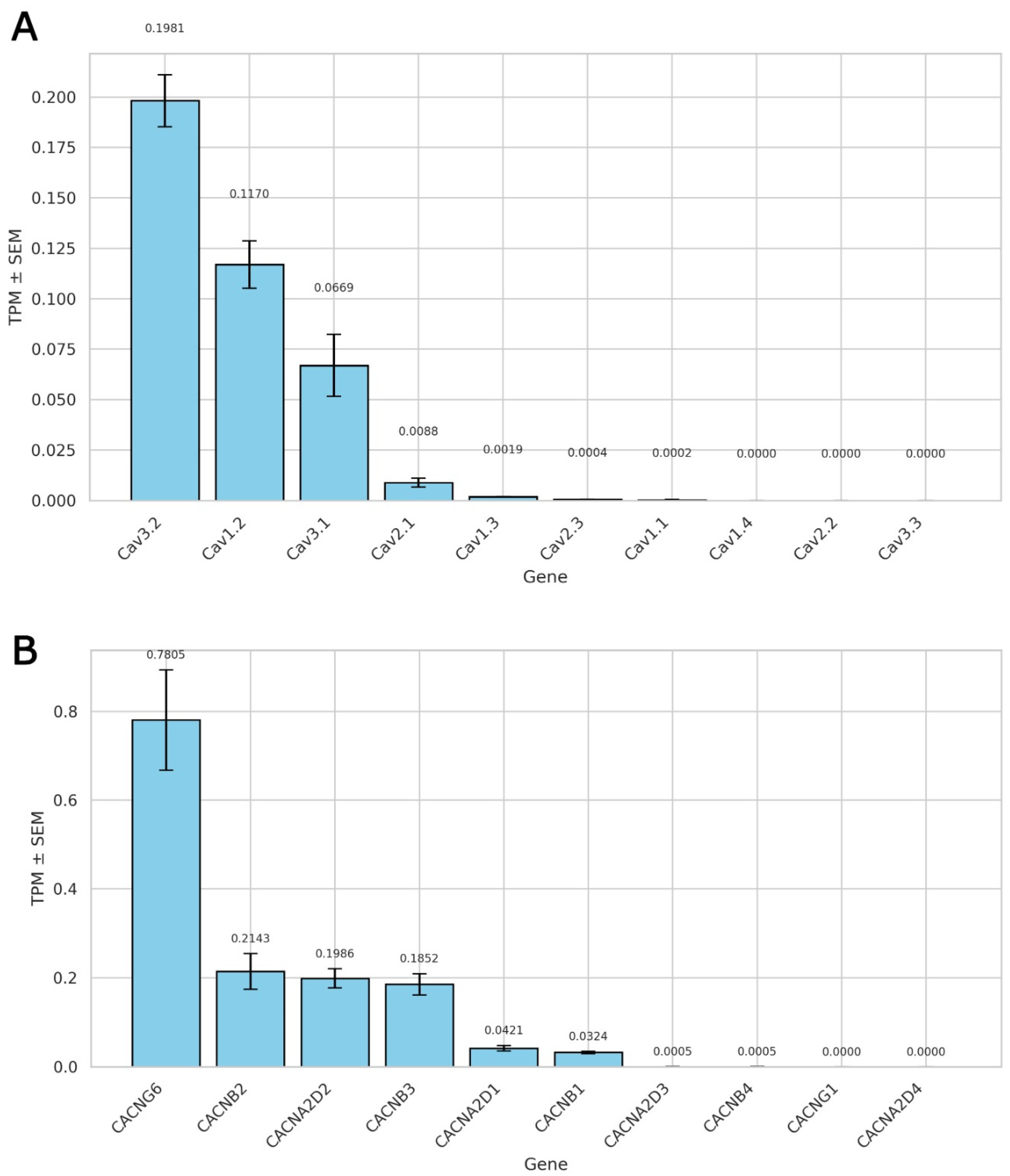
Figure 1. RNA sequencing-based expression profiles of voltage-gated calcium channels and their auxiliary subunits in rat ventricular cardiomyocytes. (A) Transcript abundance of pore-forming α1 subunits, expressed as transcripts per million (TPM) ± SEM (n = three biological replicates). (B) Expression profile of auxiliary subunits associated with L-type calcium channels. TPM values ± SEM are shown for three biological replicates.
Ijms 26 07539 g001
Figure 2. Effects of mechanical stretch on L-type Ca2+ current (ICa,L) in rat ventricular cardiomyocytes. (A) Representative whole-cell ICa,L traces recorded under control conditions (a) and during mechanical stretch of 6 μm (b), 8 μm (c), and 10 μm (d). Currents were elicited using a standard depolarizing pulse protocol (for details, please see the Materials and Methods section). (B) Current–voltage (I/V) relationships of ICa,L under control conditions and during mechanical stretch of 6, 8, and 10 μm. The net late current (IL,Net; for clarification, please see the Materials and Methods section) is shown in black, indicating time-dependent decay during sustained depolarization. (C) Mean peak ICa,L current density (pA/pF) under control conditions and at increasing levels of stretch (6, 8, and 10 μm). Lowercase letters (a, b, c, d) above the bars indicate statistically significant differences between the groups (p < 0.05). Statistical comparisons were made using repeated measures ANOVA with the Holm–Sidak post hoc test. The data are presented as mean ± SEM. Superimposed traces in panels (A(ad)) partially overlap at later time points due to the nature of multi-trace electrophysiological recordings. This overlap does not affect the interpretation of current amplitude, kinetics, or time scale.
Ijms 26 07539 g002
Figure 3. Effects of SNAP (200 µmol/L) on ICa,L in unstretched cells in K+in/K+out solutions: (reduction in ICa,L (A,B), enhancement of ICa,L (C,D), and no significant current size change (E,F). (A) Representative I/V curves under control conditions and following 3, 6, 9, and 12 min of SNAP application. Cell capacitance = 175 pF. (B) Mean ICa,L density at baseline and after 3, 6, 9, and 12 min of SNAP exposure. Lowercase letters (a, b, c) above bars indicate statistically significant differences between the time points (p < 0.05). (C) I/V curves showing ICa,L under control conditions and at 3, 6, and 9 min after SNAP application. Cell capacitance = 170 pF. (D) Mean ICa,L density at baseline and during SNAP exposure. Lowercase letters (a, b) denote significant differences between groups (p < 0.05). (E) I/V curves recorded under control conditions and following 3, 6, and 9 min of SNAP application. Cell capacitance = 120 pF. (F) Mean ICa,L density remained unchanged over time (p = ns across all time points). Lowercase letters (a) indicate no statistically significant differences between groups. For all I/V plots (A,C,E), the net late current (IL,Net) used for ICa,L calculation is depicted in black. Statistical comparisons were made using repeated measures ANOVA with the Holm–Sidak post hoc test. The data are presented as mean ± SEM.
Ijms 26 07539 g003
Figure 4. Effects of mechanical stretch and SNAP application on ICa,L in rat ventricular cardiomyocytes. Experiments were performed in K+in/K+out solutions to evaluate how SNAP (200 µM) modulates ICa,L during and after mechanical stretch. (A) Representative I/V curves show ICa,L under control conditions (blue circles), during 6 μm axial stretch (red triangles), and after 1 min of SNAP application while stretch was maintained (green squares). Cell capacitance = 135 pF. (B) Mean ICa,L densities corresponding to each condition. Lowercase letters (a, b) above the bars indicate statistically significant differences between the groups (p < 0.05). (C) I/V curves demonstrate ICa,L under control conditions (blue circles), after 2 min of SNAP application (red triangles), and during subsequent 6 μm stretch (green squares). Cell capacitance = 150 pF. (D) Mean ICa,L densities for each condition. Lowercase letters (a, b, c) above the bars indicate statistically significant differences between the groups (p < 0.05). For both (A,C), the net late current (IL,Net) used to calculate ICa,L is shown as a black curve. Statistical comparisons were made using repeated measures ANOVA with the Holm–Sidak post hoc test. Data are presented as mean ± SEM.
Ijms 26 07539 g004
Figure 5. Effects of ODQ (5 µM) and SNAP (200 µM) on ICa,L in unstretched rat ventricular cardiomyocytes. Experiments were conducted in K+in/K+out solutions to assess the influence of soluble guanylyl cyclase (sGC) inhibition and subsequent NO donor application on ICa,L. (A) Representative I/V curves show ICa,L under control conditions (blue circles), after 6 min of ODQ application (red triangles), and following SNAP addition at 3 min (green squares) and 6 min (orange diamonds). The late component of the current (IL,Net), used for current density calculation, is shown in black. Cell capacitance = 155 pF. (B) Mean ICa,L densities for each condition. Lowercase letters (a, b, c) above the bars indicate statistically significant differences between the groups (p < 0.05). Statistical comparisons were made using repeated measures ANOVA with the Holm–Sidak post hoc test. Data are presented as mean ± SEM.
Ijms 26 07539 g005
Figure 6. Combined effects of mechanical stretch, ODQ (5 µM), and SNAP (200 µM) on ICa,L in rat ventricular cardiomyocytes. Experiments were performed in K+in/K+out solutions to assess how sGC inhibition and NO signaling interact with mechanical stress to regulate ICa,L. (A) Representative I/V curves show ICa,L under control conditions (blue circles), during local axial stretch of 6 μm (red triangles), and after 6 min of ODQ application while stretch was maintained (green squares). The net late current (IL,Net), used for current density calculation, is shown in black. Cell capacitance = 170 pF. (B) Mean ICa,L densities under each condition. Lowercase letters (a, b, c) above the bars indicate statistically significant differences between the groups (p < 0.05). Statistical analysis was performed using repeated measures ANOVA with the Holm–Sidak post hoc test. Data are presented as mean ± SEM.
Ijms 26 07539 g006
Figure 7. Effects of ascorbic acid (AA, 10 µM) and SNAP (200 µM) on ICa,L in unstretched rat ventricular cardiomyocytes. Experiments were performed in K+in/K+out solutions to evaluate how AA modulates ICa,L under basal conditions and in the presence of the NO donor SNAP. (A) Representative I/V curves showing ICa,L under control conditions (blue circles), after 6 min of AA perfusion (red triangles), and following an additional 6 min of SNAP application in the presence of AA (green squares). The net late current (IL,Net) is shown in black; cell capacitance = 180 pF. (B) Mean ICa,L values under the same conditions. Lowercase letters (a, b) above the bars indicate statistically significant differences between the groups (p < 0.05). (C) I/V curves show ICa,L under control conditions (blue circles), after 6 min of SNAP application (red triangles), and following 6 min of AA perfusion (green squares); cell capacitance = 135 pF. (D) Mean ICa,L values under the same conditions. Lowercase letters (a, b, c) above the bars indicate statistically significant differences between the groups (p < 0.05). (E) I/V curves show ICa,L under control conditions (blue circles), after 6 min of SNAP application (red triangles), and following an additional 6 min of AA application (green squares); cell capacitance = 155 pF. (F) Mean ICa,L values under the same conditions. Lowercase letters (a, b, c) above the bars indicate statistically significant differences between the groups (p < 0.05). In all panels (A,C,E), IL,Net used for current density calculation is shown in black. Statistical analysis was performed using repeated measures ANOVA with the Holm–Sidak post hoc test. Data are presented as mean ± SEM.
Ijms 26 07539 g007
Figure 8. Effects of ascorbic acid (AA, 10 µM) and SNAP (200 µM) on ICa,L in mechanically stretched rat ventricular cardiomyocytes. Experiments were conducted in K+in/K+out solutions to assess whether AA and SNAP modulate stretch-induced suppression of ICa,L. (A) Representative I/V curves show ICa,L under control conditions (blue circles), during 6 μm axial stretch (red triangles), and after 6 min of AA perfusion while stretch was maintained (green squares). Subsequent SNAP application for 6 min produced no additional significant change (orange diamonds). The late current (IL,Net) used for ICa,L calculation is shown in black; cell capacitance = 170 pF. (B) Mean ICa,L densities across all conditions. Lowercase letters (a, b, c) above the bars indicate statistically significant differences between the groups (p < 0.05). Statistical analysis was performed using repeated measures ANOVA with the Holm–Sidak post hoc test. Data are presented as mean ± SEM.
Ijms 26 07539 g008
Figure 9. Biphasic effects of N-ethylmaleimide (NEM, 200 µM) on ICa,L in unstretched rat ventricular cardiomyocytes at 22 °C. Experiments were conducted in K+in/K+out solutions to evaluate the time-dependent effects of thiol alkylation by NEM on ICa,L under basal (unstretched) conditions. (A) Representative I/V curves show ICa,L at baseline (blue circles) and following 3 min (red triangles), 6 min (green squares), 9 min (orange diamonds), and 12 min (purple stars) of continuous NEM perfusion. The net late current (IL,Net) used for current calculation is shown in black. Cell capacitance = 160 pF. (B) Mean ICa,L densities corresponding to each time point. Lowercase letters (a, b, c, d) above the bars indicate statistically significant differences between the groups (p < 0.05). Statistical comparisons were made using repeated measures ANOVA with the Holm–Sidak post hoc test. Data are presented as mean ± SEM.
Ijms 26 07539 g009
Figure 10. Effects of N-ethylmaleimide (NEM, 200 µM) and SNAP (200 µM) on ICa,L during mechanical stretch in rat ventricular cardiomyocytes at 22 °C. Experiments were performed in K+in/K+out solutions to evaluate how thiol alkylation by NEM and NO signaling via SNAP affect ICa,L under conditions of mechanical stress. (A) Representative I/V curves show ICa,L under control conditions (blue circles), after 6 μm axial stretch (red triangles), and following 6 min of NEM perfusion during stretch (green squares). The net late current (IL,Net) used for current calculation is shown in black. Cell capacitance = 130 pF. (B) Mean ICa,L densities under the same conditions. Lowercase letters (a, b, c) above the bars indicate statistically significant differences between the groups (p < 0.05). (C) I/V curves show ICa,L under control conditions (blue circles), after 3 min of NEM perfusion (red triangles), and after an additional 6 min of NEM (green squares). IL,Net is shown as a black curve; cell capacitance = 130 pF. (D) Mean ICa,L densities for each condition. Lowercase letters (a, b, c, d) above the bars indicate statistically significant differences between the groups (p < 0.05). Statistical comparisons were made using repeated measures ANOVA with the Holm–Sidak post hoc test. Data are presented as mean ± SEM.
Ijms 26 07539 g010
Disclaimer/Publisher’s Note: The statements, opinions and data contained in all publications are solely those of the individual author(s) and contributor(s) and not of MDPI and/or the editor(s). MDPI and/or the editor(s) disclaim responsibility for any injury to people or property resulting from any ideas, methods, instructions or products referred to in the content.