Article

Membrane Progesterone Receptor Beta Regulates the Decidualization of Endometrial Stromal Cells in Women with Endometriosis

by
Dora Maria Velázquez-Hernández
1,
Edgar Ricardo Vázquez-Martínez
1,
Oliver Cruz-Orozco
2,
José Roberto Silvestri-Tomassoni
2,
Brenda Sánchez-Ramírez
2,
Andrea Olguín-Ortega
2,
Luis F. Escobar-Ponce
3,
Mauricio Rodríguez-Dorantes
4 and
Ignacio Camacho-Arroyo
1,*
1
Unidad de Investigación en Reproducción Humana, Instituto Nacional de Perinatología-Facultad de Química, Universidad Nacional Autónoma de México, Ciudad de México 11000, Mexico
2
Departamento de Ginecología Quirúrgica, Instituto Nacional de Perinatología, Ciudad de México 11000, Mexico
3
Coordinación de Cirugía Pévica Avanzada, Instituto Nacional de Perinatología, Ciudad de México 11000, Mexico
4
Instituto Nacional de Medicina Genómica, Periférico Sur 4809, Ciudad de México 14610, Mexico
*
Author to whom correspondence should be addressed.
Int. J. Mol. Sci. 2025, 26(15), 7297; https://doi.org/10.3390/ijms26157297
Submission received: 10 June 2025 / Revised: 8 July 2025 / Accepted: 9 July 2025 / Published: 28 July 2025
(This article belongs to the Section Molecular Endocrinology and Metabolism)

Abstract

Endometriosis is a disorder characterized by the presence of endometrial tissue outside the uterus, leading to dyspareunia, chronic pelvic pain, dysuria, and infertility. The latter has been related to implantation failure associated with alterations in decidualization, a process regulated by sex hormones such as progesterone. Membrane progesterone receptor β (mPRβ) exhibits a lower expression in endometriotic tissues than in normal endometrial ones. However, the role of mPRβ in decidualization is unknown. This work aimed to investigate whether mPRβ plays a role in the decidualization of endometrial stromal cells (ESCs) derived from women with and without endometriosis. The mPR agonist OrgOD-2 induced the gene expression of key decidualization markers (insulin-like growth factor binding protein 1, prolactin, transcription factor heart and neural crest derivatives-expressed transcript 2, and fork-head transcription factor) in healthy ESCs, eutopic (uterine cavity), and ectopic (outside of the uterine cavity) ESCs from women with endometriosis. Notably, the expression of the decidualization markers was lower in endometriotic cells than in healthy endometrial ones. An siRNA mediated knockdown of mPRβ reduced the expression of decidualization-associated genes in ESCs treated with a decidualization stimuli, regardless of whether cells were derived from healthy women or those with endometriosis. Our data suggest that progesterone, through mPRβ activation, regulates the decidualization process in endometrial stromal cells from women with and without endometriosis.

1. Introduction

Endometriosis is one of the most common benign gynecological disorders affecting women of reproductive age [1]. It presents stromal and epithelial endometrial cells outside the uterine cavity, and the main symptoms of this disease include dyspareunia, chronic pelvic pain, dysuria, painful defecation, bloating, and constipation [1,2]. Moreover, approximately 25 to 40% of women with endometriosis present infertility, often linked to ovarian insufficiency, pelvic adhesions, and implantation difficulties [3]. RNA sequencing analysis has revealed that cells derived from eutopic (lining of the uterine cavity) and ectopic (outside the uterine cavity) endometrium of women with endometriosis exhibit a transcriptional profile associated with infertility [4].
Elevated estradiol levels characterize endometriosis, driving the growth and persistence of endometriotic tissue and inducing inflammation and pain [5]. Additionally, progesterone (P4) resistance in endometriosis contributes to lesion growth, chronic inflammation, aberrant gene expression, and a non-receptive endometrium [6].
Successful establishment of pregnancy requires endometrial decidualization, a process involving the morphological and functional transformation of endometrial stromal cells (ESCs) into decidua-like epithelial cells. Decidual cells express a wide range of molecules, including Heart and Neural Crest Derivatives-Expressed Transcript 2 (HAND2), Fork head Box O1 (FOXO1), Bone Morphogenetic Protein 2 (BMP2), a member of the Transforming Growth Factor-Beta (TGF-β) superfamily, Homeobox A10 (HOXA10), Insulin-Like Growth Factor-Binding Protein 1 (IGFBP1), Prolactin (PRL), and Zinc Finger and BTB Domain-Containing 16 (ZBTB16). These molecules enable immune cell recruitment, vascular remodeling, and stimulation of the endometrial glandular system to facilitate embryo implantation [7,8,9,10,11,12,13,14,15]. Defects in decidualization and embryo implantation have been associated with recurrent pregnancy loss, preeclampsia, and infertility [3,16]. Decidualization is a hormone-dependent process, with P4 as a key regulator, acting through its receptors, which are categorized into intracellular (classical) and membrane (non-classical) receptors [17,18]. Chromatin immunoprecipitation sequencing studies and gene expression profiling have demonstrated the role of intracellular P4 receptors (PR-A and PR-B) in regulating the expression of distinct sets of genes during decidualization and pregnancy establishment [19]. Furthermore, PR-A and PR-B regulate the expression of various transcription factors, such as AP-1, FOSL2, and JUN, which are pivotal in gene expression regulation during decidualization [20].
Previous studies have suggested that P4 effects on decidualization-associated gene expression are not solely attributed to PR activation in human immortalized ESCs, indicating the involvement of other regulatory mechanisms [21]. In the case of non-classical P4 receptors, decreased expression of the P4 receptor membrane component (PGRMC1) in human ESCs has been linked to accelerated decidualization, whereas its overexpression inhibits decidualization [22,23].
Membrane P4 receptors (mPRs) belong to the progestin and adipoQ receptors PAQR family and present five subtypes in vertebrates: PAQR 7 (mPRα), PAQR 8 (mPRβ), PAQR 5 (mPRγ), PAQR 6 (mPRδ), and PAQR 9 (mPRε). mPRs can respond to P4 by activating G proteins or interacting with adapter proteins such as PGRMC and amyloid adapter protein containing pleckstrin homology domain, phosphotyrosine binding domain, and leucine zipper motif 1 (APPL), activating multiple signaling pathways [24,25]. mPRs exhibit varying expression patterns in the endometrium of rodents, bovines, rhesus monkeys, and humans across the reproductive cycle. In humans, mPRα overexpression occurs during the secretory phase of the menstrual cycle, while mPRγ and mPRε transcripts are elevated in the proliferative phase and reduced in the secretory one [26,27]. Notably, mPRβ expression appears stable throughout the menstrual cycle, although a reduced expression has been observed in pathologies such as recurrent spontaneous abortions [28]. In endometriosis, decreased mRNA and protein expression of mPRβ has been reported in eutopic and ectopic endometriotic tissue compared to endometrial tissue from women without the disease [29]. Nevertheless, the involvement of mPRβ in the decidualization of ESCs from women with and without endometriosis remains unknown.

2. Results

2.1. Demographical and Clinical Data

No differences in demographic data were observed between women with endometriosis and women without the disease, as shown in Table 1. Notably, 100% of women without the disease had full-term pregnancies, whereas more than 50% of women with endometriosis experienced abortions.

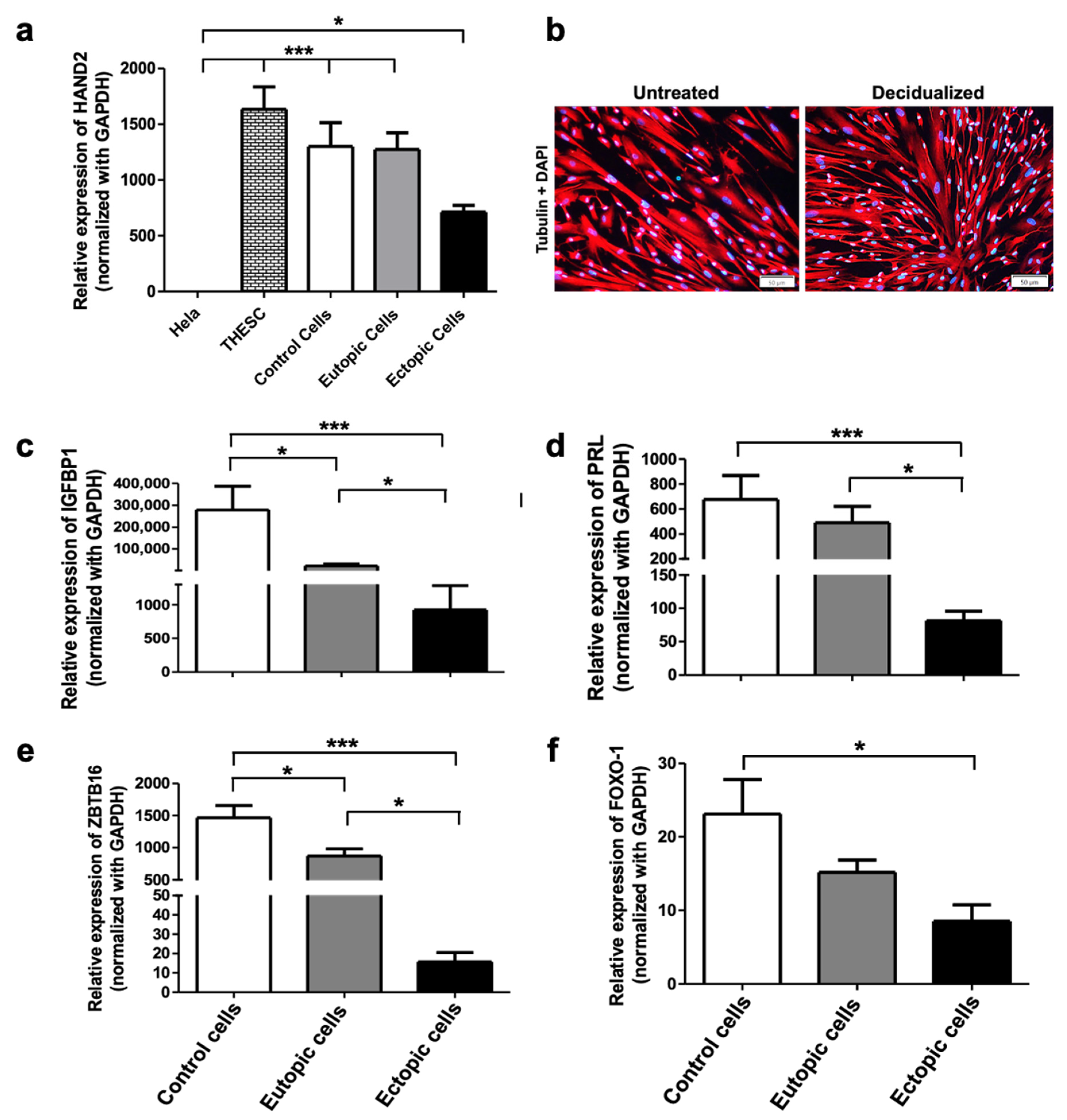
2.2. Decidualization of Human ESCs

ESCs obtained from biopsies of women with or without endometriosis were isolated, cultured, and subjected to RT-qPCR assay to firstly analyze HAND2 gene expression, which is highly expressed in endometrial stromal cells, to confirm that the primary cultures obtained from the biopsies were enriched in ESCs [8]. Additionally, stromal identity was further supported by immunofluorescence staining of vimentin, a mesenchymal marker, in representative cultures (Supplementary Figure S1). As depicted in Figure 1a, HAND2 expression was detected in THESC cells (positive control), ESCs from women without endometriosis and in eutopic and ectopic ESCs from endometriosis patients. We observed a lower HAND2 expression in ectopic cells than in the controls, consistent with findings previously reported [8]. Subsequently, stromal cells were treated with a decidualization cocktail (0.5 mM cAMP, 10 nM E2, and 1 µM P4). The transition from a fibroblast morphology to a rounded and cobblestone shape, characteristic of decidualized cells, is observed in Figure 1b. The expression levels of the decidualization markers IGFBP1, PRL, ZBTB16, and FOXO-1 were assessed by RT-qPCR (Figure 1c–f). Notably, a reduced expression of IGFBP1 and ZBTB16 was observed in the eutopic cells, which was even more pronounced in the ectopic cells compared to control ones (Figure 1c–f).

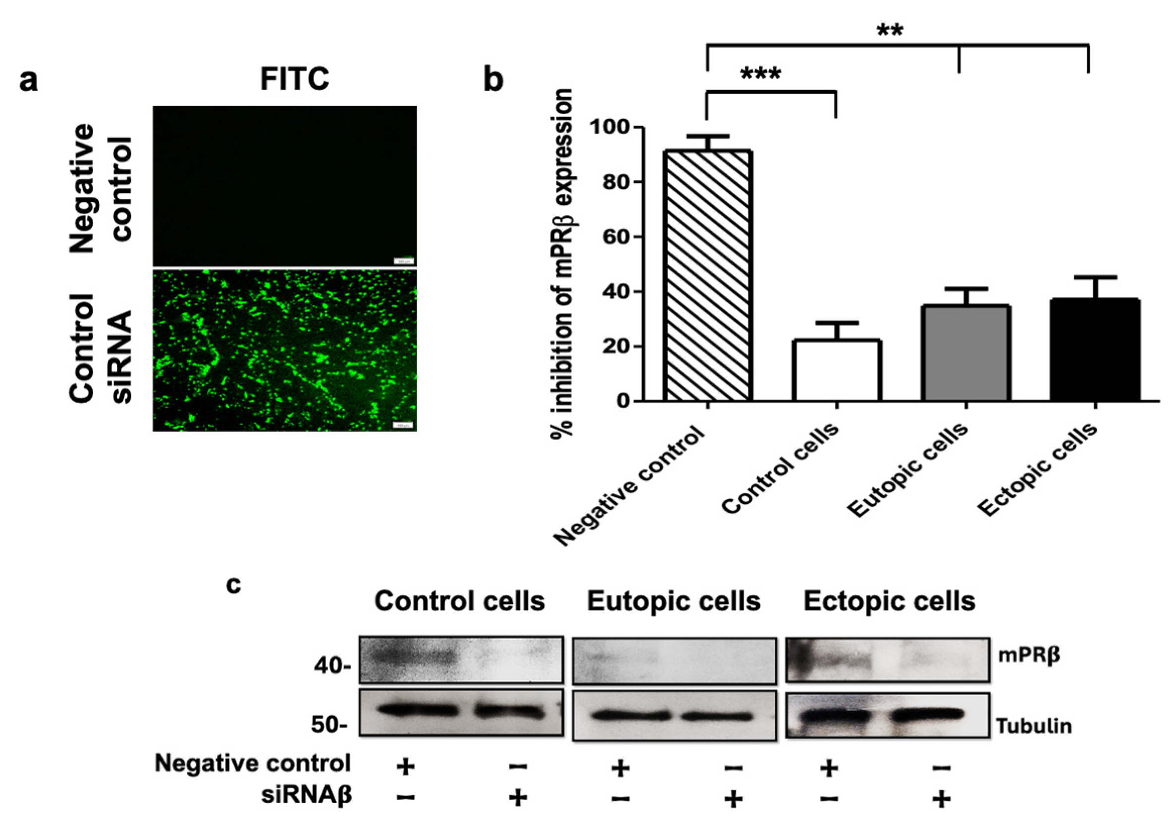
2.3. mPRs Participation in the Decidualization of Human Endometrial Stromal Cells

To assess the role of mPRs in the decidualization of human endometrial stromal cells, we administered 50 nM and 100 nM of the selective mPR agonist Org OD 02-0 + E2 + cAMP. Both Org OD 02-0 concentrations mimicked the effects of P4 by upregulating the expression of IGFBP1, PRL, HAND2, and ZBTB16 in control, eutopic, and ectopic cells from women with endometriosis (Figure 2a–d), suggesting the involvement of mPRs in the decidualization process of endometrial cells. Interestingly, in the case of the decidualization marker ZBTB16, none of the tested concentrations of Org OD 02-0 replicated the effect of P4 in control, eutopic or ectopic cells derived from women with or without endometriosis (Figure 2e). A 78% decrease in IGFBP1 expression in eutopic cells and a 99% decrease in ectopic cells compared to healthy endometrial stromal cells was observed (Figure 2f). Additionally, PRL expression was reduced by 77% in cells derived from ectopic tissue (Figure 2g).

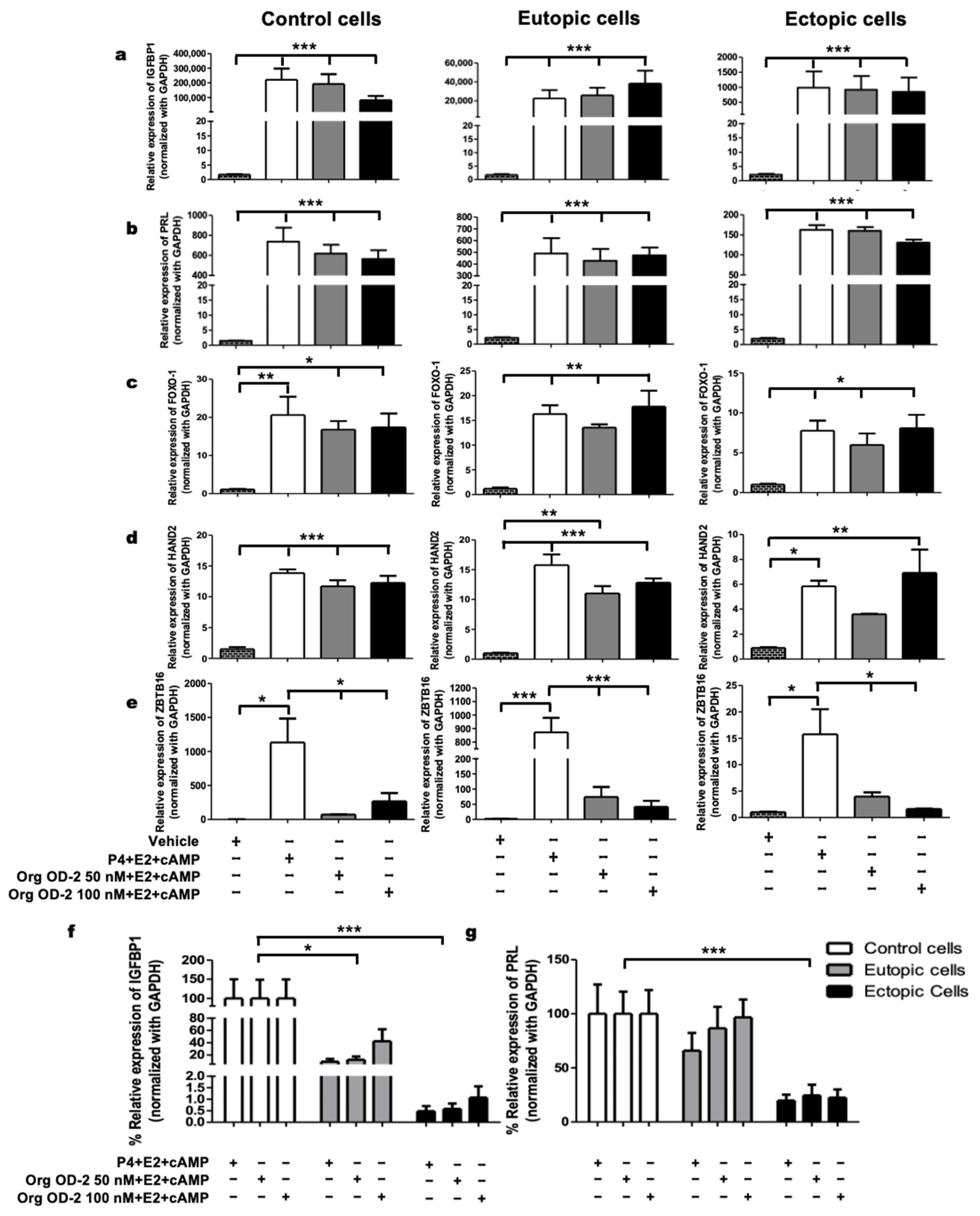
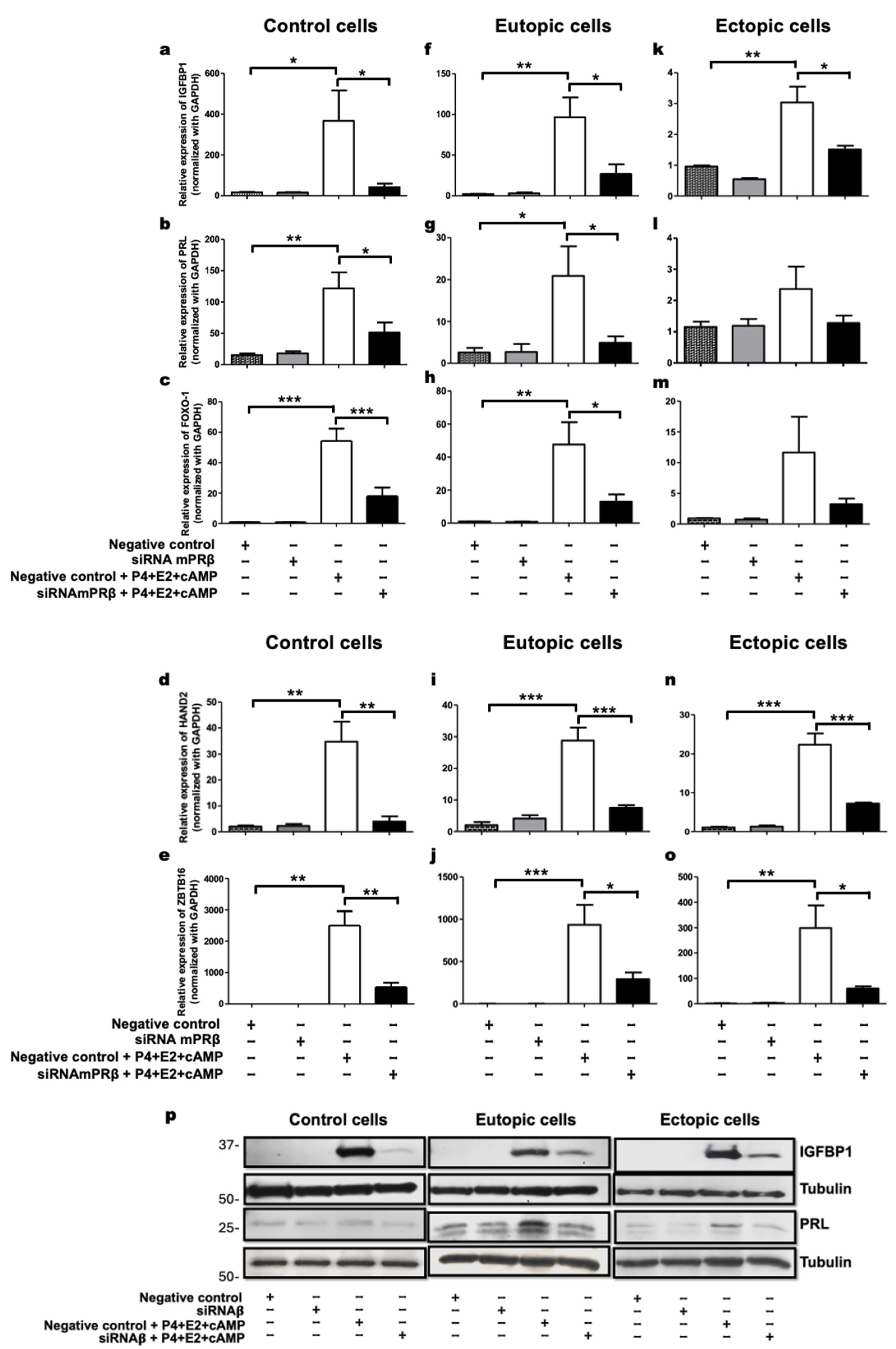
2.4. mPRβ Induces the Decidualization of Human Endometrial Stromal Cells

To demonstrate the involvement of a specific mPR in the decidualization of endometrial stromal cells derived from control and women with endometriosis, we employed an mPRβ siRNA. We selected mPRβ due to its putative role in embryo implantation and its downregulation in the endometrium of patients with endometriosis [29]. We transfected control, eutopic, and ectopic endometrial stromal cells with mPRβ siRNA, negative control (scramble), or transfection efficiency control siRNA (FITC conjugate)-A. As depicted in Figure A1, siRNA transfection efficiency reached 90%, with mPRβ expression levels decreasing by 77.73% in control cells, 65.08% in eutopic cells, and 62.87% in ectopic cells compared to the negative control (Figure A1b). Protein levels were also evaluated, showing a decrease in cells transfected with siRNAβ compared to those transfected with the negative control (Figure A1c).
Our findings demonstrate that the transfection with mPRβ siRNA resulted in a 60–80% reduction in the expression of the decidualization markers IGFBP1, PRL, HAND2, FOXO1, and ZBTB16 in control cells (Figure 3a–e) and a 60–70% reduction in eutopic cells (Figure 3f–j) compared to the negative control + E2 + P4 + cAMP. In ectopic cells, a 50–80% decrease was observed in the expression of IGFBP1, HAND2, and ZBTB16 compared to the same negative control (Figure 3k,n,o). For PRL and FOXO1, a decreasing trend in expression levels was noted in ectopic cells (Figure 3l,m). Additionally, the protein levels of IGFBP1 and PRL were reduced following transfection with mPRβ siRNA, confirming that mPRβ is essential for the decidualization process in cells from healthy women and women with endometriosis (Figure 3p).

3. Discussion

The human endometrium undergoes complex and dynamic changes to establish a suitable microenvironment for pregnancy. The decidualization of ESCs plays a crucial role in initiating and facilitating implantation [7]. While the involvement of PR in endometrial receptivity is well-established, recent interest has shifted towards non-classical receptors such as PGRMC1 and 2 and mPRs. Currently, limited information exists regarding the role of each receptor subtype in uterine function, prompting our investigation into whether mPRs contribute to the decidualization process of ESCs in women with and without endometriosis. Notably, P4 regulates the expression of decidualization markers such as IGFBP1, PRL, FOXO1, HAND2, and ZBTB16 [7,8,9,10,11,12,13,14,30].
Our results demonstrate a decrease in the expression of decidualization markers in cells derived from women with endometriosis compared to those without the disease when exposed to P4 + E2 + cAMP, consistent with previous findings [31,32,33]. It has been proposed that P4 resistance in endometriosis may result from a decreased PR-B/PR-A ratio [34]. Interestingly, when a specific mPR agonist (Org OD 02-0) was added to the decidualization cocktail, a similar decrease in decidualization markers was observed (Figure 2f,g), suggesting a potential contribution of mPRs to P4 resistance in endometriosis [29].
The Org OD 02-0 compound provided insights into the involvement of some mPRs (e.g., mPRα, mPRγ, mPRε, or mPRβ) expressed in the endometrium [26,27] in the decidualization of ESCs from women with and without endometriosis. Subsequently, we sought to identify the specific mPR subtype involved in this process. While mPRγ and mPRε are upregulated in the proliferative phase [26,27], their involvement in decidualization was deemed unlikely due to the timing of this process in the menstrual cycle. Conversely, mPRα’s association with PGRMC1, whose role in decidualization is known, made it less suitable for investigation [22,23]. Consequently, mPRβ was selected due to its consistent expression throughout the menstrual cycle, higher expression compared to mPRα in the endometrium, and observed downregulation in women with recurrent abortions and endometriosis [28,29].
Our findings suggest that mPRβ regulates the expression of IGFBP1, PRL, FOXO1, HAND2, and ZBTB16 during decidualization, as evidenced by decreased gene expression levels following mPRβ inhibition in control and eutopic cells. While the expression of PRL and FOXO1 showed a trend towards reduction in ectopic cells, the effect was less pronounced compared to other decidualization markers. This observation suggests that additional receptors contribute to gene regulation in these cells.
Interestingly, ZBTB16 does not exhibit increased expression levels in response to the agonist Org OD 02-0, a preferential agonist of mPRα. However, blocking mPRβ expression results in a more than 70% reduction in ZBTB16 expression, reinforcing the notion that mPRβ is essential for decidualization in cells from healthy women and those with endometriosis.

4. Materials and Methods

4.1. Tissue Collection

This study included 20 participants with endometriosis, specifically ovarian and deep infiltrating types, diagnosed through laparoscopy and histological analysis, and 20 control women without the disease from the Instituto Nacional de Perinatología, Mexico City. In both groups, women were between 18 and 45 years old, presented regular menstrual cycles, and had not undergone hormonal therapy in the three months before sample collection. All participants provided informed consent for their participation in this study. Most samples from women with endometriosis and those free of disease were collected during the proliferative phase of the menstrual cycle. Eutopic tissue biopsies were obtained using a Pipelle cannula (Laboratoire CCD, Paris, France), while ectopic tissue was obtained with surgical scissors during laparoscopic surgeries. This study received approval from the Research, Biosecurity, and Ethics Committees of the Instituto Nacional de Perinatología (reference number 2019-1-26).

4.2. Primary Cell Culture of ESCs

Primary endometrial stromal cells were isolated, as described in a previous study [21]. Briefly, the endometrial (eutopic) and endometriotic (ectopic) tissues were minced using scalpel blades and incubated in Hank’s Balanced Salt Solution (composition: NaCl 140 mM, KCl 5 mM, CaCl2 1 mM, MgSO4 0.4 mM, MgCl2 0.5 mM, Na2HPO4 0.3 mM, C6H12O6 6 mM) with 10 mg/mL type I collagenase (Gibco, Grand Island, NY, USA) and 1 mg/mL DNase I (Boehringer Mannheim, Mannheim, Germany) with agitation at 37 °C for 2 h. The digested tissue was passed through a 40 µm nylon cell strainer (FALCON, Chicago, IL, USA), which allowed the separation of stromal cells from epithelial and glandular fragments based on size. Stromal cells that passed through the strainer were collected and cultured in DMEM/F-12 supplemented with 10% FBS, antibiotic-antimycotic (Gibco, Life Technologies, Grand Island, NY, USA), and gentamicin (Gibco, Life Technologies, USA) at 37 °C and 5% CO2. All experiments were performed using primary cultures at passages 2 and 3.

4.3. Cell Lines

The THESC cell line (endometrial stromal cell line immortalized by reversible human telomerase transcriptase; ATCC CRL-4003, Manassas, VA, USA passage 22) was cultured in DMEM/F12 without phenol red (Gibco, USA), supplemented with 1.5 g/L sodium bicarbonate, 1.0 mM sodium pyruvate (Gibco, USA), 0.5% insulin-transferrin-selenium (ITS; Gibco, USA), and penicillin/streptomycin (Gibco, USA), along with 10% fetal bovine serum (FBS; Biowest, Heathfield, UK). The HeLa cells (passage 10) were cultured in DMEM (Gibco, USA), supplemented with penicillin/streptomycin (Gibco, USA), and 10% fetal bovine serum (FBS; Biowest, UK). Both cell lines were maintained in a controlled environment at 37 °C and 5% CO2. The cell line THESC was used as a positive control of ESCs, and the epithelioid cervix carcinoma cell line HeLa was used as a negative control.

4.4. Decidualization Treatments

Human endometrial cells were cultured in 6-well plates within phenol red-free DMEM/F-12 supplemented with 10% charcoal-stripped FBS (SH30068.03, CYTIVA, Marlborough, MA USA) and antibiotic-antimycotic (Gibco, Life Technologies, NY). A decidualization cocktail: 0.5 mM of cAMP (B7880 Sigma-Aldrich, St. Louis, MO, USA) + 10 nM of estradiol (E2) (E2758, Sigma-Aldrich, St. Louis, MO, USA) + 1 μM of P4 (P8783, Sigma-Aldrich, St. Louis, MO, USA) [21], or 50 or 100 nM of the mPR agonist 10-Ethenyl-19-norprogesterone (Org OD 02-0) (Axon 2085, Med Chem, Reston, VA, USA) was added to the culture media for 48 h to induce decidualization. Cells were washed twice with cold PBS, and the RT-qPCR technique was performed as described below in Section 4.5 to firstly observe the enrichment of ESCs in the culture with HAND2 expression, and then to evaluate the expression of decidualization markers PRL, IGFBP1, FOXO-1, and ZBTB16.

4.5. RNA Isolation and Reverse Transcription-Quantitative PCR (RT-qPCR)

Total RNA was isolated from human endometrial stromal cells using QIAzol reagent (Qiagen, Valencia, CA, USA) according to the manufacturer’s protocol. Briefly, tissue samples were homogenized in QIAzol, followed by the addition of chloroform to induce phase separation. After centrifugation, the aqueous phase containing RNA was carefully collected, precipitated with isopropanol, washed with ethanol, and finally resuspended in RNase-free water. RNA integrity and purity were evaluated by agarose gel electrophoresis and spectrophotometry, respectively. Complementary DNA (cDNA) was synthesized from 1 μg of total RNA in a 20 μL reaction volume containing M-MLV reverse transcriptase (Invitrogen, Waltham, MA, USA), 10 mM of each deoxynucleotide (dNTPs) (Invitrogen, USA), and the appropriate buffer, according to the manufacturer’s instructions. The synthesized cDNA was subsequently used for quantitative real-time PCR (RT-qPCR) to amplify gene fragments corresponding to decidualization markers: PRL, IGFBP1, FOXO1, and ZBTB16. Reactions were carried out using PowerUp SYBR Green PCR Master Mix (Applied Biosystems, Vilnius, Lithuania) and detected with the CFX96 Touch Real-Time PCR Detection System (Bio-Rad, Hercules, CA, USA). The cycling conditions comprised 40 cycles of 95 °C for 15 s and 60 °C for 1 min. Each sample was amplified in triplicate, and data were analyzed using the ΔΔCt method, with GAPDH as the internal control. Primer sequences are provided in Table 2.

4.6. Immunofluorescence

ESCs were cultured on coverslips to visualize the decidualization. Cells were washed with PBS, fixed with 4% paraformaldehyde for 15 min, permeabilized with 0.1% Triton X-100, and blocked with BSA 1% (Promega, Madison, WI, USA) (Research Organics, Cleveland, OH, USA) for 1 h. Cells were incubated with an anti-β-tubulin antibody (ab7291, Abcam, Cambridge, UK) or anti-vimentin (ab92597 EPR3776) in a blocking solution at 4 °C overnight. After three washes with PBS, cells were incubated in the dark at room temperature with Alexa Fluor 594-conjugated secondary antibody (ab150116) or Alexa Fluor 488-conjugated secondary antibody (ab150097) for 1 h. This was followed by incubation with DAPI for 6 min. After washing, coverslips were mounted using Aqua-Poly/Mount. Cell fluorescence was examined using an inverted microscope (Olympus IX73 Tokyo, Japan).

4.7. mPRβ siRNA Transfection

Human endometrial cells were cultured in 6-well plates within phenol red-free DMEM/F-12 containing 10% charcoal-stripped FBS (35-072-CV, CA) and antibiotic-antimycotic (Gibco, Life Technologies, NY). siRNA transfections were performed in serum-free Opti-MEM (Gibco, Life Technologies, NY) using 35 pmol of PAQR8 siRNA (ID85315 Origene Technologies, Rockville, MD, USA) and universal scrambled negative control siRNA or positive control siRNA (FITC Conjugated, sc-36869 Santa Cruz Biotechnology, Dallas, TX, USA) and mixed with lipofectamine when the cells reached 80% confluence. After 7 h, transfected cells with positive control siRNA were observed under a microscope (Olympus IX73 Tokyo, Japan). The decidualization cocktail of 0.5 mM cAMP + 10 nM E2 + 1 μM P4 was added to the culture media for 24 h to induce decidualization. Subsequently the RT-qPCR assays were performed, as described in Section 4.5 to evaluate the expression of decidualization markers IGFBP1, PRL, HAND2, FOXO-1, and ZBTB16.

4.8. Western Blot

Protein was isolated from cells using RIPA buffer (50 mM Tris-HCl, pH 8, 150 mM NaCl, 1% NP-40, 1 mM EDTA, 0.1% SDS, 0.5% sodium deoxycholate) supplemented with a protease inhibitor cocktail (cOmplete™, EDTA-free, 11873580001, Roche, Mannheim, Germany). Then, 60 μg of protein was separated on a 10% SDS-PAGE gel (Invitrogen, Life Technologies, Carlsbad, CA, USA) and electrophoretically transferred onto nitrocellulose membranes (Bio-Rad, cat. 1620097). Membranes were blocked with 1% BSA (Promega, Madison, WI, USA) and incubated overnight at 4 °C with the respective primary antibodies: mPRβ (sc-50109 (n15), Santa Cruz Biotechnology), IGFBP-1 (PA5-78020, Invitrogen), and PRL (PA5-95712, Invitrogen). Detection was performed using the Immobilon Forte kit (Millipore, Burlington, MA, USA) and horseradish peroxidase (HRP)-conjugated secondary antibodies, including rabbit anti-goat (sc-2768, Santa Cruz Biotechnology), anti-mouse HRP (ab205719, Abcam), and anti-rabbit HRP (ab6721, Abcam). β-Tubulin (ab7291, Abcam) was used as a control to ensure equal protein loading across samples.

4.9. Statistical Analysis

Data were analyzed using either One-way ANOVA followed by post hoc Tukey’s and Kruskal–Wallis’s test followed by Dunns, using PRISM software (version 8.0; GraphPad) to determine differences between experimental groups. Statistical significance was set at p < 0.05.

5. Conclusions

This study is pioneering in demonstrating that mPRs, particularly mPRβ, are involved in the decidualization of stromal cells in women with and without endometriosis. Notably, the induction of decidualization using the mPR agonist Org OD 02-0 was lower in endometriotic cells than in healthy ones. This type of research is essential to elucidate the role of mPRs in infertility associated with endometriosis and emphasize that the expression or activity of non-classical P4 receptors can answer many questions associated with P4 resistance and problems in embryo implantation.

Supplementary Materials

The supporting information can be downloaded at: https://www.mdpi.com/article/10.3390/ijms26157297/s1.

Author Contributions

Writing—original draft preparation and methodology: D.M.V.-H.; Writing—review and editing: E.R.V.-M. and M.R.-D.; Recruiting, attending women, and collecting the biopsy: O.C.-O., J.R.S.-T., L.F.E.-P., A.O.-O. and B.S.-R.; Writing—review and editing, project administration, investigation, and funding acquisition: I.C.-A. All authors have read and agreed to the published version of the manuscript.

Funding

This work was supported by Instituto Nacional de Perinatología (number. 2019-1-26) and a DGAPA postdoctoral Fellowship program of the Universidad Nacional Autónoma de México for Dora María Velázquez-Hernández.

Institutional Review Board Statement

This study was conducted in accordance with the Declaration of Helsinki and approved by the Research, Biosecurity, and Ethics Committees of the Instituto Nacional de Perinatología (reference number 2019-1-26).

Informed Consent Statement

Written informed consent has been obtained from the patients to publish this paper.

Data Availability Statement

The datasets generated and/or analyzed during the current study are available from the corresponding author upon reasonable request.

Acknowledgments

We thank Maria Cristina Castañeda Patlán (Facultad de Medicina, Departamento de Bioquímica, Universidad Nacional Autónoma de México) and Karina Hernández-Ortega (Facultad de Química, Departamento de Biología, Universidad Nacional Autónoma de México) for their technical support and Moisés León-Juárez for the donation of the THESC cell line.

Conflicts of Interest

The authors declare no conflicts of interest.

Abbreviations

The following abbreviations are used in this manuscript:
mPRβMembrane Progesterone Receptor β
ESCsEndometrial Stromal Cells
HAND2 Heart and Neural Crest Derivatives-Expressed Transcript 2
FOXO1Fork head Box O1
BMP2Bone Morphogenetic Protein 2 a member of the Transforming Growth Factor-Beta (TGF-β) superfamily
HOXA10Homeobox A10
IGFBP1Insulin-Like Growth Factor-Binding Protein 1
PRLProlactin
ZBTB16Zinc Finger and BTB Domain-Containing 16
PAQRProgestin and AdipoQ Receptors
Org OD 02-0 10-Ethenyl-19-norprogesterone
cAMPCyclic Adenosine Monophosphate

Appendix A

Figure A1. Transfection of control and mPRβ siRNA into human endometrial stromal cells. (a) Representative image of control cells transfected with control siRNA (FITC conjugate) for 7 h, scale bar: 100 μm. (b) Control, eutopic, and ectopic cells were transfected with mPRβ siRNA or negative control for 48 h. The percentage of transfection was evaluated by the expression of mPRβ using RT-qPCR. The data in the figures represent mean ± SEM obtained from 10 independent experiments. Statistical analysis was performed using a one-way analysis of variance (ANOVA) followed by the Tukey post hoc test. Statistical significance was considered as follows: ** p ≤ 0.01 and *** p ≤ 0.001. (c) Representative image of protein levels of mPRβ in control, eutopic, and ectopic transfected cells with mPRβ siRNA or negative control for 48 h.
Ijms 26 07297 g0a1

References

  1. Taylor, H.S.; Kotlyar, A.M.; Flores, V.A. Endometriosis is a chronic systemic disease: Clinical challenges and novel innovation. Lancet 2021, 397, 839–852. [Google Scholar] [CrossRef] [PubMed]
  2. Pasalic, E.; Tambuwala, M.M.; Jahjefendic-Hromic, A. Endometriosis: Classification, pathophysiology, and treatment options. Pathol. Res. Pract. 2023, 251, 154847. [Google Scholar] [CrossRef] [PubMed]
  3. Prescott, J.; Farland, L.V.; Tobias, D.K.; Gaskins, A.J.; Spiegelman, D.; Chavarro, J.E.; Rich-Edwards, J.W.; Barbieri, R.L.; Missmer, S.A. A prospective cohort study of endometriosis and subsequent risk of infertility. Hum. Reprod. 2016, 31, 1475–1482. [Google Scholar] [CrossRef]
  4. Feng, X.; Qi, L.; Xu, X.; Feng, Y.; Gong, X.; Aili, A.; Tong, X. Analysis of differences in the transcriptomic profiles of eutopic and ectopic endometriums in women with ovarian endometriosis. PeerJ 2021, 9, e11045. [Google Scholar] [CrossRef]
  5. Chantalat, E.; Valera, M.C.; Vaysse, C.; Noirrit, E.; Rusidze, M.; Weyl, A.; Vergriete, K.; Buscail, E.; Lluel, P.; Fontaine, C.; et al. Estrogen Receptors and Endometriosis. Int. J. Mol. Sci. 2020, 21, 2815. [Google Scholar] [CrossRef] [PubMed]
  6. Zhang, P.; Wang, G. Progesterone Resistance in Endometriosis: Current Evidence and Putative Mechanisms. Int. J. Mol. Sci. 2023, 24, 6992. [Google Scholar] [CrossRef]
  7. Ochoa-Bernal, M.A.; Fazleabas, A.T. Physiologic Events of Embryo Implantation and Decidualization in Human and Non-Human Primates. Int. J. Mol. Sci. 2020, 21, 1973. [Google Scholar] [CrossRef] [PubMed]
  8. Murata, H.; Tanaka, S.; Tsuzuki-Nakao, T.; Kido, T.; Kakita-Kobayashi, M.; Kida, N.; Hisamatsu, Y.; Tsubokura, H.; Hashimoto, Y.; Kitada, M.; et al. The transcription factor HAND2 up-regulates transcription of the IL15 gene in human endometrial stromal cells. J. Biol. Chem. 2020, 295, 9596–9605. [Google Scholar] [CrossRef]
  9. Grinius, L.; Kessler, C.; Schroeder, J.; Handwerger, S. Forkhead transcription factor FOXO1A is critical for induction of human decidualization. J. Endocrinol. 2006, 189, 179–187. [Google Scholar] [CrossRef]
  10. Huang, P.; Deng, W.; Bao, H.; Lin, Z.; Liu, M.; Wu, J.; Zhou, X.; Qiao, M.; Yang, Y.; Cai, H.; et al. SOX4 facilitates PGR protein stability and FOXO1 expression conducive for human endometrial decidualization. eLife 2022, 11, e72073. [Google Scholar] [CrossRef]
  11. Liao, Z.; Tang, S.; Jiang, P.; Geng, T.; Cope, D.I.; Dunn, T.N.; Guner, J.; Radilla, L.A.; Guan, X.; Monsivais, D. Impaired bone morphogenetic protein (BMP) signaling pathways disrupt decidulization in endometriosis. Commun. Biol. 2024, 7, 227. [Google Scholar] [CrossRef] [PubMed]
  12. Xu, Y.; Lu, J.; Wu, J.; Jiang, R.; Guo, C.; Tang, Y.; Wang, H.; Kong, S.; Wang, S. HOXA10 co-factor MEIS1 is required for the decidualization in human endometrial stromal cell. J. Mol. Endocrinol. 2020, 64, 249–258. [Google Scholar] [CrossRef]
  13. Ng, S.W.; Norwitz, G.A.; Pavlicev, M.; Tilburgs, T.; Simon, C.; Norwitz, E.R. Endometrial decidualization: The primary driver of pregnancy health. Int. J. Mol. Sci. 2020, 21, 4092. [Google Scholar] [CrossRef]
  14. Kommagani, R.; Szwarc, M.M.; Vasquez, Y.M.; Peavey, M.C.; Mazur, E.C.; Gibbons, W.E.; Lanz, R.B.; DeMayo, F.J.; Lydon, J.P. The Promyelocytic Leukemia Zinc Finger Transcription Factor Is Critical for Human Endometrial Stromal Cell Decidualization. PLoS Genet. 2016, 12, e1005937. [Google Scholar] [CrossRef] [PubMed]
  15. Arlier, S.; Kayisli, U.A.; Semerci, N.; Ozmen, A.; Larsen, K.; Schatz, F.; Lockwood, C.J.; Guzeloglu-Kayisli, O. Enhanced ZBTB16 Levels by Progestin-Only Contraceptives Induces Decidualization and Inflammation. Int. J. Mol. Sci. 2023, 24, 10532. [Google Scholar] [CrossRef]
  16. Garrido-Gomez, T.; Dominguez, F.; Quiñonero, A.; Diaz-Gimeno, P.; Kapidzic, M.; Gormley, M.; Ona, K.; Padilla-Iserte, P.; McMaster, M.; Genbacev, O.; et al. Defective decidualization during and after severe preeclampsia reveals a possible maternal contribution to the etiology. Proc. Natl. Acad. Sci. USA 2017, 114, E8468–E8477. [Google Scholar] [CrossRef]
  17. Cope, D.I.; Monsivais, D. Progesterone Receptors Signaling in Uterus Is Essential for Pregnancy Success. Cell 2022, 11, 1474. [Google Scholar] [CrossRef] [PubMed]
  18. Medina-Laver, Y.; Rodríguez-Varela, C.; Salsano, S.; Labarta, E.; Domínguez, F. What Do We Know about Classical and Non-Classical Progesterone Receptors in the Human Female Reproductive Tract? A Review. Int. J. Mol. Sci. 2021, 22, 11278. [Google Scholar] [CrossRef]
  19. Kaya, H.S.; Hantak, A.M.; Stubbs, L.J.; Taylor, R.N.; Bagchi, I.C.; Bagchi, M.K. Roles of Progesterone Receptor A and B Isoforms During Human Endometrial Decidualization. Mol. Endocrinol. 2015, 29, 882–895. [Google Scholar] [CrossRef]
  20. Chi, R.A.; Wang, T.; Adams, N.; Wu, S.P.; Young, S.L.; Spencer, T.E.; DeMayo, F. Human Endometrial Transcriptome and Progesterone Receptor Cistrome Reveal Important Pathways and Epithelial Regulators. J. Clin. Endocrinol. Metab. 2020, 105, e1419–e1439. [Google Scholar] [CrossRef]
  21. Retis-Resendiz, A.M.; Cid-Cruz, Y.; Velázquez-Hernández, D.M.; Romero-Reyes, J.; León-Juárez, M.; García-Gómez, E.; Camacho-Arroyo, I.; Vázquez-Martínez. cAMP regulates the progesterone receptor gene expression through the protein kinase A pathway during decidualization in human immortalized endometrial stromal cells. Steroids 2024, 203, 109363. [Google Scholar] [CrossRef]
  22. Salsano, S.; González-Martín, R.; Quiñonero, A.; Pérez-Debén, S.; Domínguez, F. Deciphering the Role of PGRMC1 During Human Decidualization Using an In Vitro Approach. J. Clin. Endocrinol. Metab. 2021, 106, 2313–2327. [Google Scholar] [CrossRef] [PubMed]
  23. Tsuru, A.; Yoshie, M.; Kojima, J.; Yonekawa, R.; Azumi, M.; Kusama, K.; Nishi, H.; Tamura, K. PGRMC1 Regulates Cellular Senescence via Modulating FOXO1 Expression in Decidualizing Endometrial Stromal Cells. Biomolecules 2022, 12, 1046. [Google Scholar] [CrossRef] [PubMed]
  24. Tomas, P. Membrane Progesterone Receptors (mPRs, PAQRs): Review of Structural and Signaling Characteristics. Cells 2022, 11, 1785. [Google Scholar] [CrossRef]
  25. Nader, N.; Dib, M.; Hodeify, R.; Courjaret, R.; Elmi, A.; Hammad, A.S.; Dey, R.; Huang, X.Y.; Machaca, K. Membrane progesterone receptor induces meiosis in Xenopus oocytes through endocytosis into signaling endosomes and interaction with APPL1 and Akt2. PLoS Biol. 2020, 18, e3000901. [Google Scholar] [CrossRef] [PubMed]
  26. Kowalik, K.; Dobrzyn, K.; Rekawiecki, R.; Kotwica, J. Expression of membrane progestin receptors (mPRs) α, β and γ in the bovine uterus during the oestrous cycle and pregnancy. Theriogenology 2019, 140, 171–179. [Google Scholar] [CrossRef]
  27. Fernandes, M.S.; Pierron, V.; Michalovich, D.; Astle, S.; Thornton, S.; Peltoketo, H.; Lam, E.W.; Gellersen, B.; Huhtaniemi, I.; Allen, J.; et al. Regulated expression of putative membrane progestin receptor homologues in human endometrium and gestational tissues. J. Endocrinol. 2005, 187, 89–101. [Google Scholar] [CrossRef]
  28. Rahnama, R.; Rafiee, M.; Fouladi, S.; Akbari-Fakhrabadi, M.; Mehrabian, F.; Rezaei, A. Gene expression analysis of membrane progesterone receptors in women with recurrent spontaneous abortion: A case control study. BMC Res. Notes 2019, 12, 790. [Google Scholar] [CrossRef]
  29. Vázquez-Martínez, E.R.; Bello-Alvarez, C.; Hermenegildo-Molina, A.L.; Solís-Paredes, M.; Parra-Hernández, S.; Cruz-Orozco, O.; Silvestri-Tomassoni, J.R.; Escobar-Ponce, L.F.; Hernández-López, L.A.; Reyes-Mayoral, C.; et al. Expression of Membrane Progesterone Receptors in Eutopic and Ectopic Endometrium of Women with Endometriosis. BioMed Res. Int. 2020, 2020, 2196024. [Google Scholar] [CrossRef]
  30. Tanaka, K.; Sakai, K.; Matsushima, M.; Matsuzawa, Y.; Izawa, T.; Nagashima, T.; Furukawa, S.; Kobayashi, Y.; Iwashita, M. Branched-chain amino acids regulate insulin-like growth factor-binding protein 1 (IGFBP1) production by decidua and influence trophoblast migration through IGFBP1. Mol. Hum. Reprod. 2016, 22, 890–899. [Google Scholar] [CrossRef]
  31. Bunch, K.; Tinnemore, D.; Huff, S.; Hoffer, Z.S.; Burney, R.O.; Stallings, J.D. Expression Patterns of Progesterone Receptor, Membrane Components 1 and 2 in Endometria From Women with and Without Endometriosis. Reprod. Sci. 2014, 21, 190–197. [Google Scholar] [CrossRef] [PubMed]
  32. Cai, X.; Xu, M.; Zhang, H.; Zhang, M.; Wang, J.; Mei, J.; Zhang, Y.; Zhou, J.; Zhen, X.; Kang, N.; et al. Endometrial stromal PRMT5 plays a crucial role in decidualization by regulating NF-κB signaling in endometriosis. Cell Death Discov. 2022, 8, 408. [Google Scholar] [CrossRef] [PubMed]
  33. Klemmt, P.A.; Carver, J.G.; Kennedy, S.H.; Koninckx, P.R.; Mardon, H.J. Stromal cells from endometriotic lesions and endometrium from women with endometriosis have reduced decidualization capacity. Fertil. Steril. 2006, 85, 564–572. [Google Scholar] [CrossRef] [PubMed]
  34. Chae, U.; Min, J.Y.; Kim, S.H.; Ihm, H.J.; Oh, Y.S.; Park, S.Y.; Chae, H.D.; Kim, C.H.; Kang, B.M. Decreased progesterone receptor B/A ratio in endometrial cells by tumor necrosis factor-alpha and peritoneal fluid from patients with endometriosis. Yonsei Med. J. 2016, 57, 1468–1474. [Google Scholar] [CrossRef]
Figure 1. Decidualization of endometrial stromal cells from women with endometriosis. (a) Expression of HAND2 in various cell types: Hela cells of epithelial origin, THESC cell line derived from the endometrium of healthy women, control cells (women without endometriosis), eutopic cells (obtained from endometriotic lesions in the uterine cavity), and ectopic cells (obtained from endometriotic lesions outside the uterine cavity). (b) Immunofluorescence: representative image of the decidualization of control cells treated with the decidualization cocktail (E2 + cAMP + P4) scale bar 50 μm. (cf) Expression of decidualization markers IGFBP1, PRL, ZBTB16, and FOXO1 in control, eutopic, and ectopic cells treated with the decidualization cocktail for 48 h. Data in the figures represent mean + SEM obtained from 10 independent experiments. (a) Statistical analysis was performed using one-way analysis of variance (ANOVA) followed by the Tukey post hoc test. (cf) Statistical analysis was performed using Kruskal–Wallis followed by Dunns’s Multiple Comparison test. Statistical significance was considered as follows: * p ≤ 0.05 and *** p ≤ 0.001.
Ijms 26 07297 g001
Figure 2. mPRs participate in the decidualization of endometrial stromal cells from women with and without endometriosis. (a) Expression of IGFBP1 and (b), PRL (c), FOXO1, (d) HAND2, and (e) ZBTB16 in control, eutopic, and ectopic cells treated with vehicle, 50 nM and 100 nM of the selective mPR agonist Org OD2 + 10 nM E2 + 0.5 mM cAMP. The graphs (f,g) depict the comparison in percentage of expression levels of decidualization markers IGFBP1 and PRL among control cells (women without endometriosis), eutopic, and ectopic cells derived from women with endometriosis. The data presented in the figures represent mean ± SEM obtained from 10 patients per group. Statistical analysis was conducted using the One-way ANOVA analysis followed by the Tukey post hoc test. Statistical significance was considered as follows: * p ≤ 0.05, ** p ≤ 0.01 and *** p ≤ 0.001.
Ijms 26 07297 g002
Figure 3. mPRβ induces the decidualization of human endometrial stromal cells. (ap) Control, eutopic, and ectopic cells were starved for 24 h. Subsequently, they were transfected with mPRβ siRNA or negative control for 24 h, followed by treatment with the decidualization cocktail (10 nM E2 + 0.5 mM cAMP + 1 μM P4) for 24 h. The expression levels of the decidualization markers IGFBP1, PRL, FOXO-1, HAND2, and ZBTB16 were evaluated by RT-qPCR and the protein levels of IGFBP1 and PRL were observed by Western blot. The data in the figures (ao) represent mean and SEM obtained from 10 independent experiments. Statistical analysis was performed using one-way analysis of variance (ANOVA) followed by the Tukey post hoc test. Statistical significance was considered as follows: * p ≤ 0.05, ** p ≤ 0.01 and *** p ≤ 0.001 (p) Representative image of protein levels of IGFBP1 and PRL in control, eutopic, and ectopic cells transfected with mPRβ siRNA or a negative control for 24 h. Following transfection, cells were treated with the decidualization cocktail (0.5 mM cAMP + 10 nM E2 + 1 µM P4) for an additional 24 h.
Ijms 26 07297 g003
Table 1. Clinical characteristic of the women included in this study.
Clinical CharacteristicsControl (n = 20)Women with Endometriosis (n = 20)
Age years (mean ± SD)38.4 (± 6.6)33.22 (± 9.7)
Number of women with full-term pregnancies204
Number of women with abortions211
Number of women in the proliferative phase during biopsy collection1311
Number of women in the secretory phase during biopsy collection 79
n: Number of women.
Table 2. Primers used in the present study.
GENEFORWARD 5′3′REVERSE 5′3′
HAND 2GAGGAAGAAGGAGCTGAACGAAGTCCGGCCTTTGGTTTTCTTG
IGFBP1TCCTTTGGGACGCCATCAGTACGATGTCTCCTGTGCCTTGGCTA
PRLCATATTGCGATCCTGGAATGAGCTCCTCAATCTCTACAGCTTTGGA
FOXO-1AGGDTTAGTGAGCAGGTTACACTGGACTGCTTCTCTCAGTTCC
ZBTB16TTGTCTGTGATCAGTGCGGTGCCATGTCAGTGCCAGTATG
PAQR8AGGACGCAGCAAACAGGACAGCC AACACAGGCAGGAATAA
GAPDHGACAGTCAGCCGCATCTTCTGCCCAATACGAC CAAATCCGT
Disclaimer/Publisher’s Note: The statements, opinions and data contained in all publications are solely those of the individual author(s) and contributor(s) and not of MDPI and/or the editor(s). MDPI and/or the editor(s) disclaim responsibility for any injury to people or property resulting from any ideas, methods, instructions or products referred to in the content.

