Article

Metabolic Culture Medium Enhances Maturation of Human iPSC-Derived Cardiomyocytes via Cardiac Troponin I Isoform Induction

by
Daria V. Goliusova
1,2,
Agnessa P. Bogomolova
3,
Alina V. Davidenko
1,
Kristina A. Lavrenteva
1,
Margarita Y. Sharikova
1,
Elena A. Zerkalenkova
4,5,
Ekaterina M. Vassina
1,
Alexandra N. Bogomazova
1,
Maria A. Lagarkova
1,
Ivan A. Katrukha
3,6 and
Olga S. Lebedeva
1,*
1
Lopukhin Federal Research and Clinical Center of Physical-Chemical Medicine, 119435 Moscow, Russia
2
Koltzov Institute of Developmental Biology of Russian Academy of Sciences, 119334 Moscow, Russia
3
Hytest Ltd., 20520 Turku, Finland
4
Dmitry Rogachev National Medical Research Center of Pediatric Hematology, Oncology and Immunology, 117198 Moscow, Russia
5
Department of Biochemistry and Molecular Biology, Faculty of General Medicine, Pirogov Russian National Research Medical University, 117997 Moscow, Russia
6
Department of Biochemistry, Biological Faculty, Lomonosov Moscow State University, 119991 Moscow, Russia
*
Author to whom correspondence should be addressed.
Int. J. Mol. Sci. 2025, 26(15), 7248; https://doi.org/10.3390/ijms26157248 (registering DOI)
Submission received: 3 July 2025 / Revised: 16 July 2025 / Accepted: 22 July 2025 / Published: 26 July 2025

Abstract

Human induced pluripotent stem cell-derived cardiomyocytes (iCMs) provide a powerful platform for investigating cardiac biology. However, structural, metabolic, and electrophysiological immaturity of iCMs limits their capacity to model adult cardiomyocytes. Currently, no universally accepted criteria or protocols for effective iCMs maturation exist. This study aimed to identify practical culture conditions that promote iCMs maturation, thereby generating more physiologically relevant in vitro cardiac models. We evaluated the effects of short- and long-term culture in media supplemented with various stimulatory compounds under 2D conditions, focusing on intracellular content and localization of slow skeletal troponin I (ssTnI) and cardiac troponin I (cTnI) isoforms. Our findings demonstrate that the multicomponent metabolic maturation medium (MM-1) effectively enhances the transition toward a more mature iCM phenotype, as evidenced by increased cTnI expression and formation of cross-striated myofibrils. iCMs cultured in MM-1 more closely resemble adult cardiomyocytes and are compatible with high-resolution single-cell techniques such as electron microscopy and patch-clamp electrophysiology. This work provides a practical and scalable approach for advancing the maturation of iPSC-derived cardiac models, with applications in disease modeling and drug screening.

1. Introduction

Since their discovery in 2006 [1], human induced pluripotent stem cells (iPSCs) have become one of the most widely used platforms in both fundamental and translational research [2,3]. A major limitation, however, lies in the immaturity of iPSC-derived cell types compared to their somatic counterparts in vivo. In the absence of external stimuli, iPSC derivatives—including chondrocytes [4], neurons [5], fibroblasts [6], pancreatic β-cells [7], and cardiomyocytes [8]—retain immature characteristics that limit their utility for disease modeling and complicate mechanistic studies in vitro. Over the past decade, extensive efforts have been made to develop effective protocols for the maturation of human iPSC-derived cardiomyocytes (iCMs). Cardiovascular diseases still represent the foremost cause of mortality, contributing to approximately one-third of all deaths worldwide [9]. As the global burden of cardiovascular disease continues to rise—with projections indicating a 90% increase in prevalence between 2025 and 2050 [10]—iCMs have emerged as a valuable model system for cardiac research and development. These cells not only facilitate the study of fundamental heart biology [11] but also offer a promising human-relevant alternative to animal models, particularly for investigating genetic cardiomyopathies [12].
Cardiomyocyte maturation in vivo is a prolonged, multistage process beginning in the perinatal period and continuing into early childhood, encompassing morphological, metabolic, and electrophysiological changes that culminate by 6–10 years of age [13,14,15]. During this process, cardiomyocytes transition from small, round cells into elongated, rod-shaped cells with highly organized myofibrils, widened Z-discs, and developed T-tubules and sarcoplasmic reticulum [16]. Around birth and throughout the neonatal period, cytokinesis becomes decoupled from karyokinesis, causing cells to arrest in the G1 phase of the cell cycle, resulting in binucleation and/or polyploidy. This shift enables hypertrophic heart growth not through proliferation, but through cellular enlargement and ultimately fusion of neighbouring cells, resulting in syncytium formation [17,18,19]. Simultaneously, the metabolic profile switches from glycolysis to fatty acid β-oxidation, providing a more efficient ATP source for postnatal physiology [20,21]. By approximately six years of age, cardiomyocytes exhibit distinct structural localization of gap junction proteins, ensuring continuous ion and molecular transport across intercalated discs and enabling synchronized electrical conduction [13].
Despite advances in differentiation protocols, iCMs in vitro retain a fetal-like phenotype characterized by small, round or irregular morphology, mononuclearity, absence of a branched T-tubule network, and failure to form syncytia. They remain proliferative, rely predominantly on glycolysis, exhibit gene expression profiles typical of fetal cardiomyocytes, and display immature electrophysiological properties, including low resting membrane potential and reduced action potential amplitude [15,22,23,24,25]. The first major challenge in iCM’s maturation lies in the limited understanding of the genetic and regulatory mechanisms governing perinatal cardiomyocyte development. Current maturation strategies aim to recapitulate key structural, metabolic, and electrophysiological transitions by applying physical and chemical stimuli. These include mechanical and/or electrical stimulation, modification of substrate stiffness and geometry, biochemical cues, long-term culture, co-culture, organoid formation, tissue engineering constructs, and in vivo implantation in animal models [8,11,26]. The progress in bioelectrode materials design and microfabrication provides a novel platform for the development of heart-on-a-chip technologies with controlled microenvironments for iCM culture and maturation in vitro [27,28]. In some promising cases, well-designed and implemented maturation strategies have enabled the identification of phenotype-genotype correlations in personalized iCM-based models of inherited cardiomyopathies [29,30,31,32,33,34,35]. However, such successes remain rare, largely due to the high costs and the need for specialized equipment. The second critical bottleneck is the lack of standardized, consensus-based markers for defining iCMs maturity, making it difficult to compare maturation strategies across studies [36,37,38,39]. While comprehensive multiparametric analyses provide the most detailed insights into iCMs maturation, they require interdisciplinary expertise and substantial resources.
This study aimed to identify accessible and effective culture conditions for enhancing the maturation of human iPSC-derived cardiomyocytes, thereby creating a more physiologically relevant in vitro model of myocardium. We focused on accessible approaches to promote iCMs maturation, adopting a widely used strategy involving culture in media supplemented with maturation-promoting factors. As a first-tier maturation marker, we selected the ratio of slow skeletal troponin I (ssTnI) to cardiac troponin I (cTnI)—a robust, in vivo-validated indicator proposed by Bedada et al. in 2014—as a quantifiable measure of structural iCMs maturation across laboratories [14]. We supplemented this with analysis of the intracellular localization of cTnI and ssTnI at the single-cell level to further assess structural maturation.

2. Results

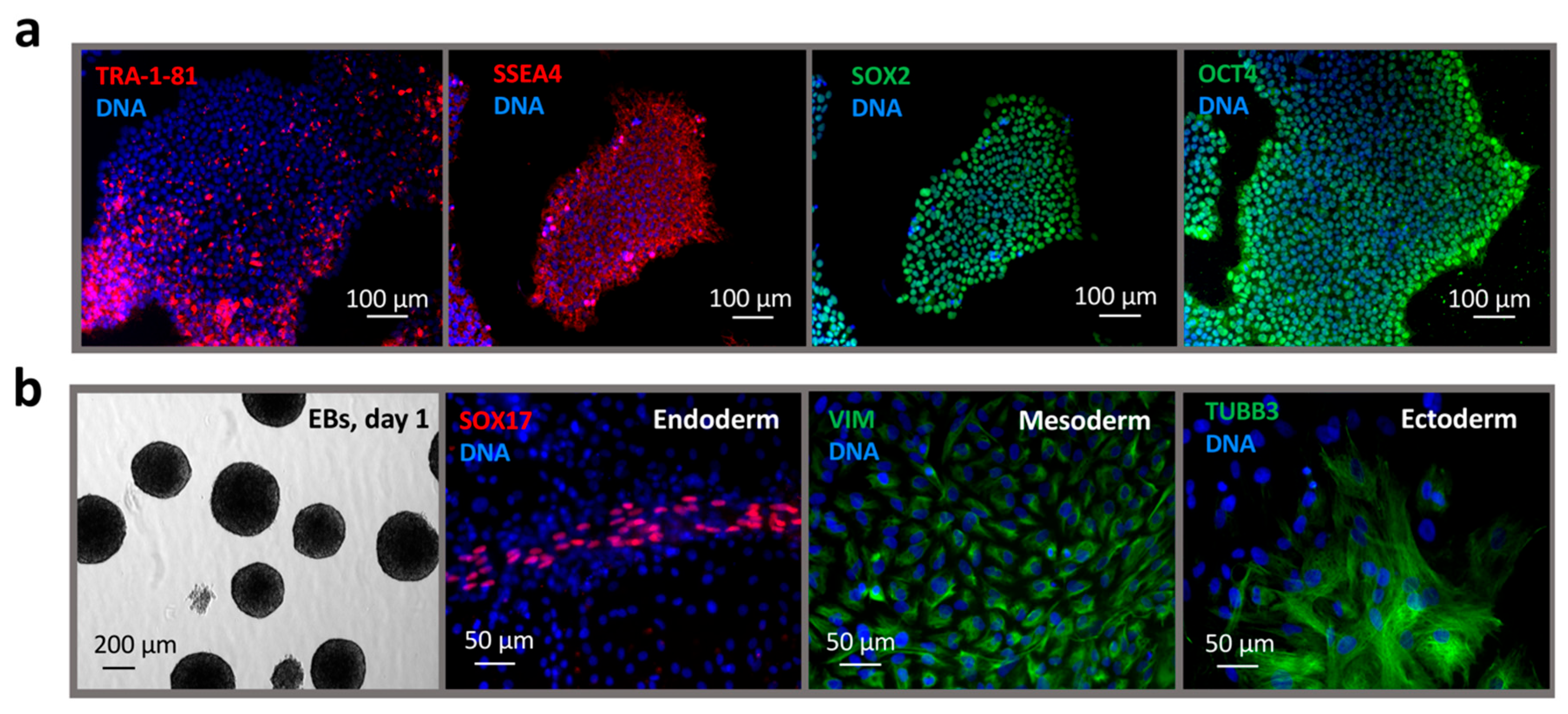
2.1. Reprogramming of Dermal Fibroblasts to a Pluripotent State

A primary dermal fibroblast culture (Figure 1a) was established from a skin biopsy of a healthy donor. A total of 1.5 × 10⁵ passage 1 dermal fibroblasts were transduced with the four Yamanaka reprogramming factors. Approximately three weeks after transduction, colonies exhibiting the typical morphology of human pluripotent stem cells (hPSCs) began to emerge. In total, 12 colonies were selected. The reprogramming efficiency, calculated as the ratio of colonies formed to the number of transduced cells, was 0.008%. Molecular and functional characterization of the resulting iPSC line, RCPCMi013-A (IPSAVE2S), confirmed its pluripotent status. The cells exhibited typical hPSC morphology (Figure 1b), a normal 46, XY karyotype (Figure 1c), and genetic identity to the parental fibroblasts across 19 STR markers and the amelogenin locus (data available upon request). They were negative for Mycoplasma spp. contamination (Figure 1d) and expressed the core pluripotency genes OCT4, SOX2, SALL4, and FOXD3 (Figure 1e), as well as the surface markers SSEA4 and TRA-1-81 (Figure 1f). The resulting RCPCMi013-A (IPSAVE2S) cell line passport is provided in Supplementary Table S1.
Immunostaining confirmed expression of nuclear (OCT4, SOX2) and surface (SSEA4, TRA-1-81) pluripotency markers (Figure 2a). The IPSAVE2S cells were capable of forming embryoid bodies and differentiating spontaneously into derivatives of all three germ layers. Immunostaining showed positive expression of SOX17 (endoderm), vimentin (VIM, mesoderm), and β-III-tubulin (TUBB3, ectoderm) (Figure 2b).

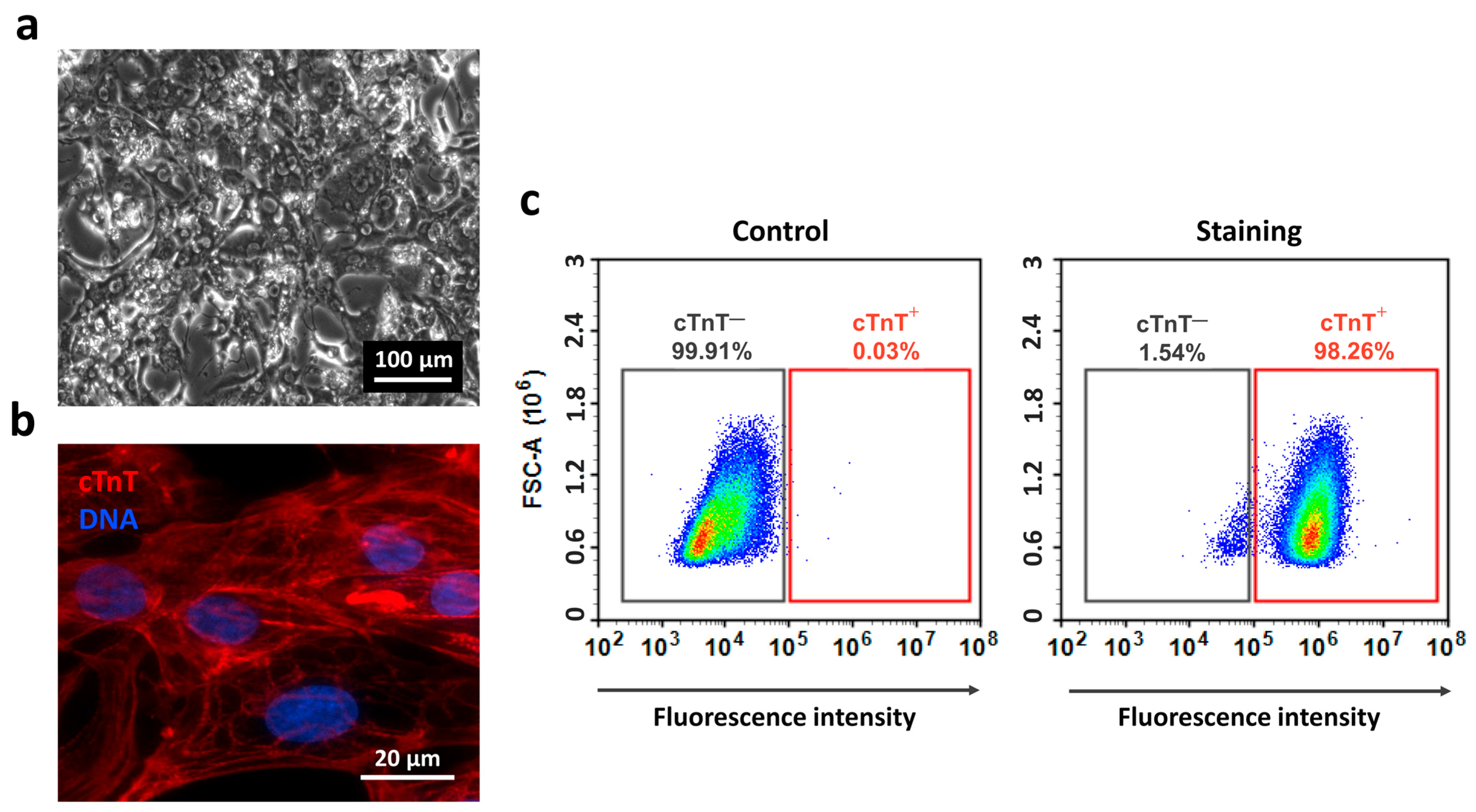
2.2. Differentiation of iPSCs into Cardiomyocytes

To assess the cardiogenic differentiation potential of the IPSAVE2S line, we conducted a pilot differentiation experiment aimed at generating and characterizing cardiomyocytes. For four passages prior to the start of differentiation, IPSAVE2S cells were passaged at a consistent density of 0.35 × 10⁵ cells/cm2 every three days. At passage 18, the cells were seeded at a density of 1.25 × 10⁵ cells/cm2 and subjected to differentiation using the commercial STEMdiff Ventricular Cardiomyocyte Differentiation Kit according to the manufacturer’s protocol. By day 8, the first clusters of spontaneously contracting iCMs were observed. Between days 10 and 14, non-myocyte populations present in the culture were selectively eliminated through metabolic stress selection in a glucose-free medium supplemented with lactate. This selection step led to the death of cells unable to utilize lactate as a primary energy source, while the iCMs monolayer exhibited spontaneous contractile activity (Supplementary Video S1). Subsequently, the cells were cultured in STEMdiff Cardiomyocyte Maintenance Medium. By day 22, cultures consisted predominantly of spontaneously contracting iCMs displaying light nuclear and dark cytoplasmic morphology (Figure 3a). Immunostaining revealed strong sarcomeric cross-striation for cardiac troponin T (cTnT) (Figure 3b), and flow cytometry indicated 98% positivity compared to isotype controls (Figure 3c). These results demonstrate that the IPSAVE2S line differentiates efficiently into cardiomyocytes.

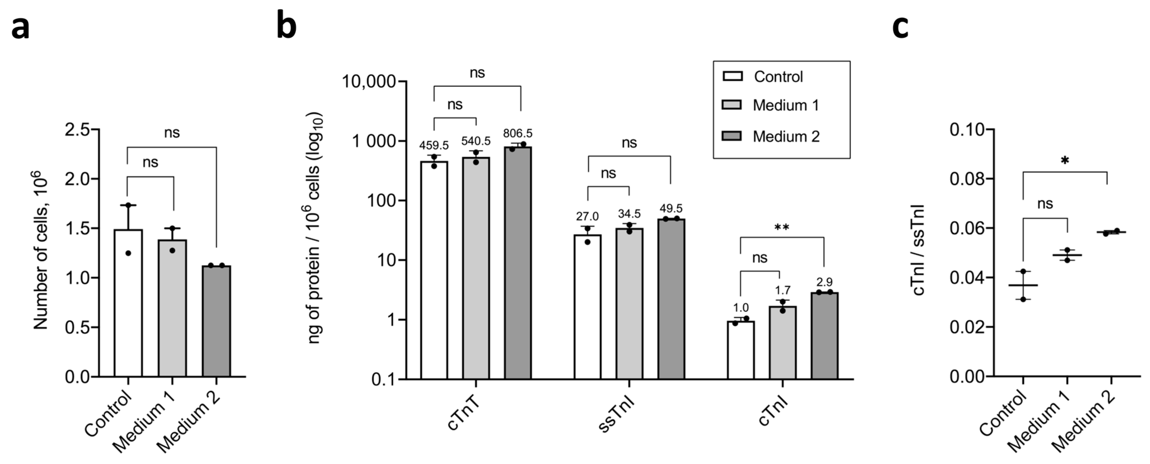
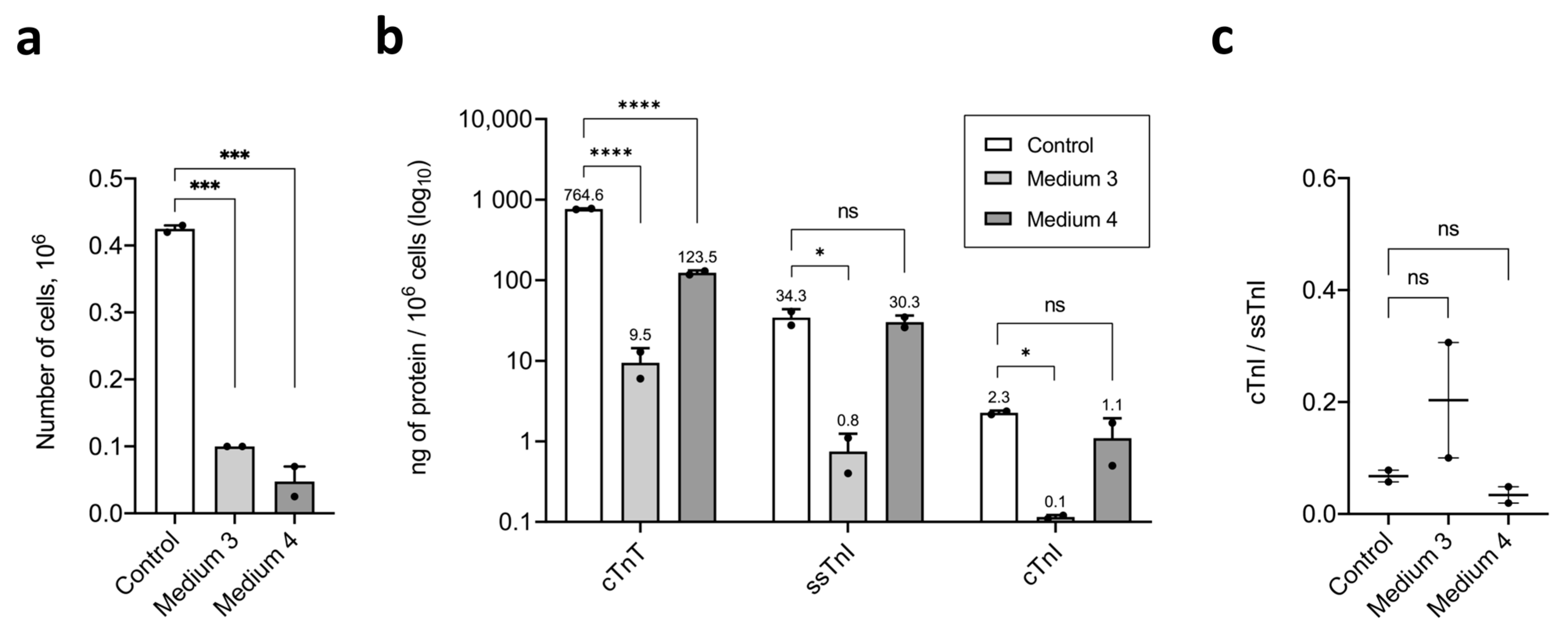
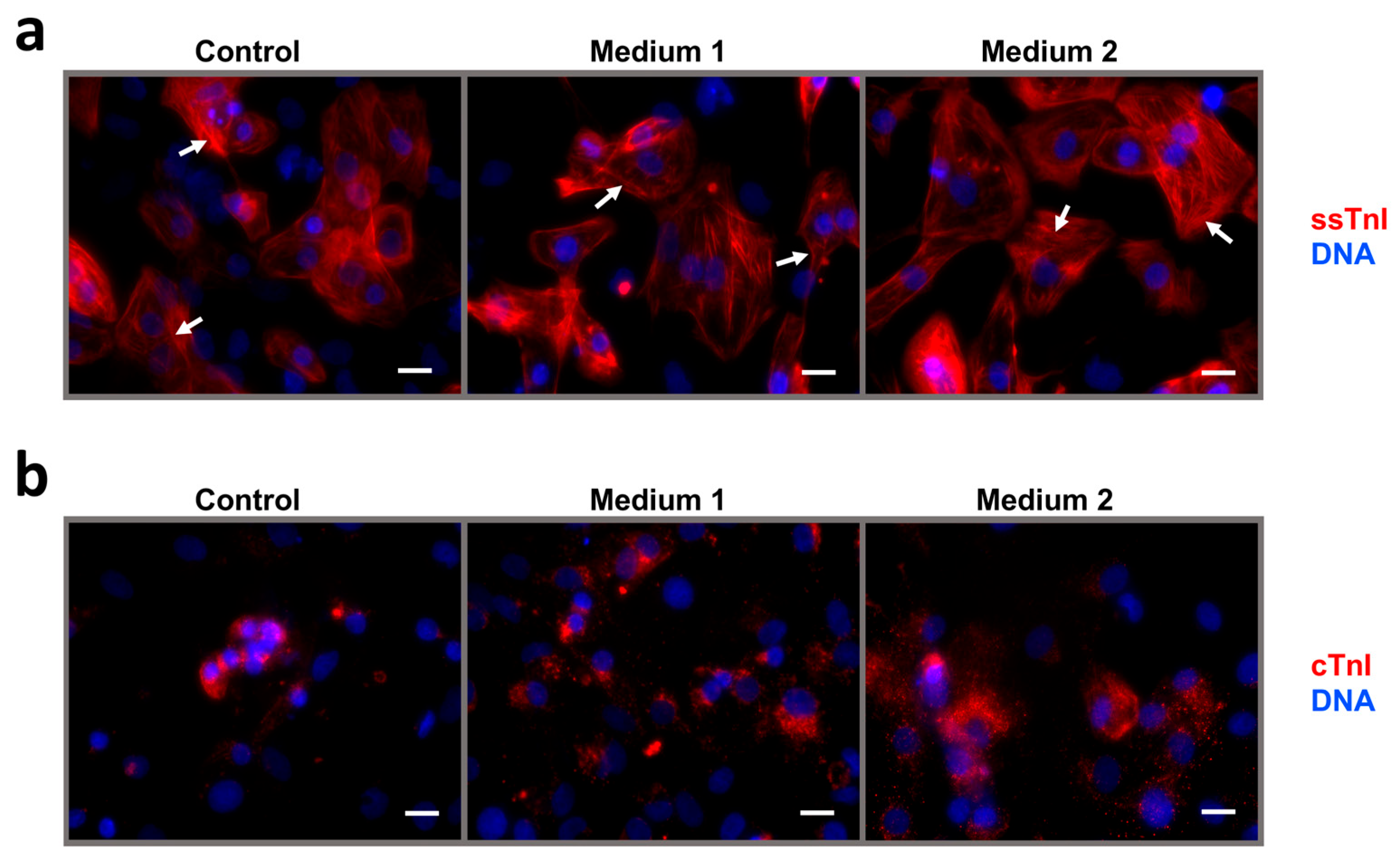
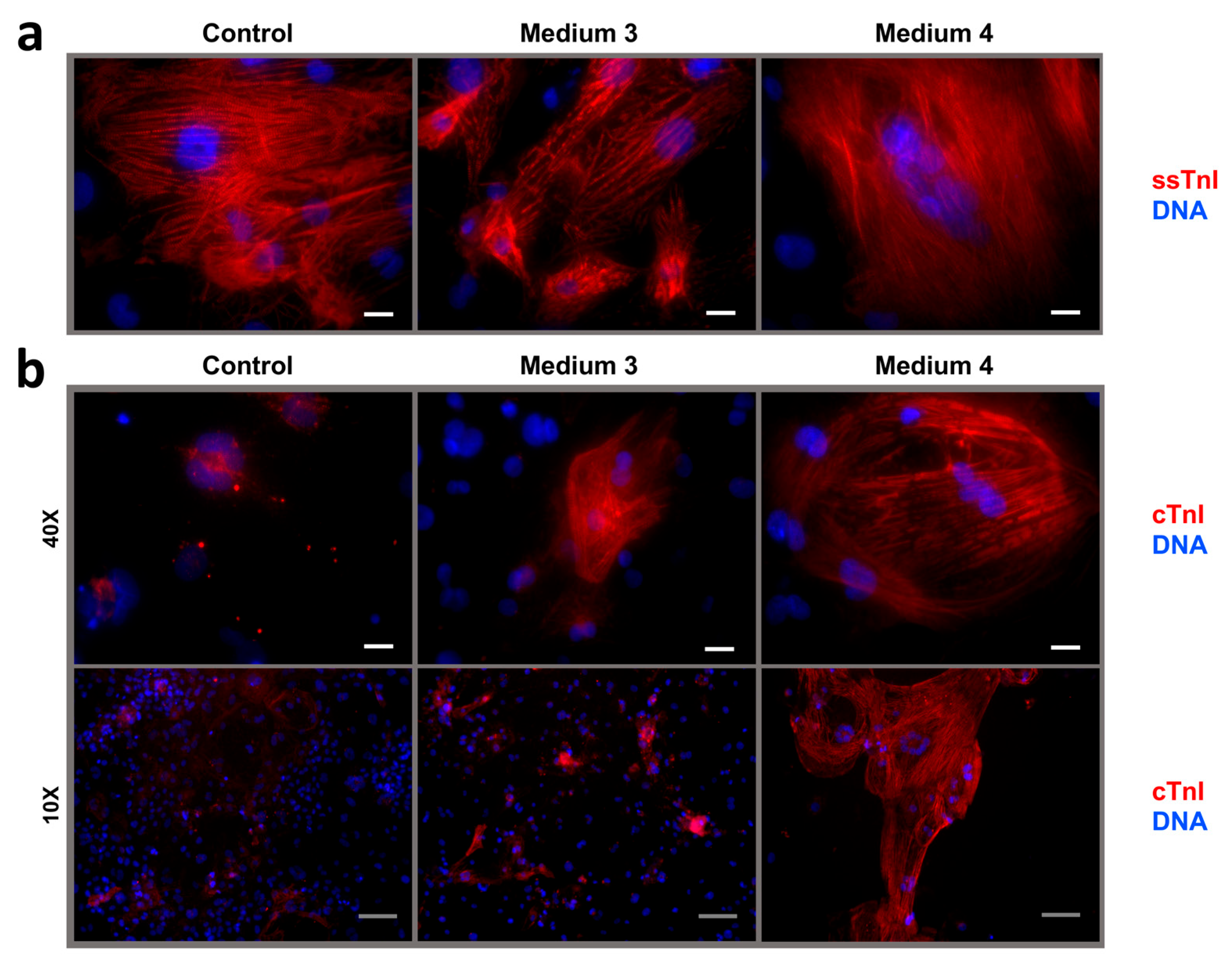
2.3. Maturation and Comparative Analysis of iCMs Maturity

To analyze cTnI, ssTnI, and cTnT quantitative content, cTnI/ssTnI ratios, and intracellular localization pattern of cTnI and ssTnI in human iCMs, we differentiated wild-type hiPSCs (IPSAVE2S) using the STEMdiff Ventricular Cardiomyocyte Differentiation Kit and performed a short-term (4-day) or long-term (22-day) maturation of the resulting iCM cultures following metabolic selection (Figure 4). We used four types of maturation media containing maturation-promoting factors (Table 1). The unmodified commercial STEMdiff Cardiomyocyte Maintenance Medium was used as the control condition in all experiments.
In the first experiment, AVE2S-iCMs were derived from passage 31 of IPSAVE2S. Short-term maturation was performed over 4 days using the STEMdiff Cardiomyocyte Maintenance Medium supplemented either with T3, IGF-1, dexamethasone, and lipid concentrate (Medium 1) or with etoposide and lipid concentrate (Medium 2) (Figure 4a). In the second experiment, AVE2S-iCMs were derived from passage 38 of IPSAVE2S. Maturation was carried out over 22 days in either RPMI-1640 with standard glucose levels supplemented with T3, IGF-1, dexamethasone, and NeuroMax (Medium 3), or in low-glucose DMEM supplemented with L-glutamine, non-essential amino acids, sodium lactate, vitamin B12, biotin, creatine, L-taurine, L-carnitine, ascorbic acid, human albumin, lipid concentrate, NeuroMax, and KnockOut SR (Medium 4) (Figure 4b). At the end of the experiments, iCMs were fixed for immunostaining and collected for fluoroimmunoassay. Quantitative data on cell counts, concentrations of cTnI, ssTnI, and cTnT, as well as cTnI/ssTnI ratios in the resulting AVE2S-iCMs samples are summarized in Supplementary Table S2.
Compared with control conditions, culture in Medium 1 did not result in statistically significant differences in live cell count (1.4915 × 10⁶ cells in control vs. 1.3875 × 10⁶ cells in Medium 1) (Figure 5a), cTnT content (459.825 ng/106 cells in control vs. 540.645 ng/106 cells in Medium 1), ssTnI content (27.065 ng/106 cells in control vs. 34.64 ng/106 cells in Medium 1), or cTnI content (0.96 ng/106 cells in control vs. 1.71 ng/106cells in Medium 1) (Figure 5b) in the iCMs cultures at the end of the experiment. After maturation in Medium 1, the ssTnI level remained 20 times higher than that of cTnI (34.64 ng/106 cells for ssTnI vs. 1.71 ng/106 cells for cTnI), while in the control, this difference was 28.2-fold (27.065 ng/106 cells for ssTnI vs. 0.96 ng/106 cells for cTnI) (Figure 5b). The cTnI/ssTnI ratio remained <1:0.049 in Medium 1 and 0.037 in control, with no significant difference (Figure 5c).
Similarly, Medium 2 did not affect live cell count (1.4915 × 10⁶ cells in control vs. 1.125 × 10⁶ cells in Medium 2) (Figure 5a), cTnT content (459.825 ng/106 cells in control vs. 806.575 ng/106 cells in Medium 2), or ssTnI content (27.065 ng/106 cells in control vs. 49.745 ng/106 cells in Medium 2), but did result in a 3-fold increase in cTnI content (0.96 ng/106 cells in control vs. 2.9 ng/106 cells in Medium 2) (Figure 5b). After maturation in Medium 2, the ssTnI level remained 17 times higher than that of cTnI (49.745 ng/106 cells for ssTnI vs. 2.9 ng/106 cells for cTnI), while in the control, this difference was 28.2-fold (27.065 ng/106 cells for ssTnI vs. 0.96 ng/106 cells for cTnI) (Figure 5b). The cTnI/ssTnI ratio remained below 1 (0.059), however, increased slightly versus control (0.037) (Figure 5c).
Compared with the control, culture in Medium 3 led to a 4.25-fold decrease in cell number (0.425 × 10⁶ cells in control vs. 0.1 × 10⁶ cells in Medium 3) (Figure 6a), 80.9-fold reduction in cTnT content (764.59 ng/106 cells in control vs. 9.45 ng/106 cells in Medium 3), 44.8-fold reduction in ssTnI content (34.305 ng/106 cells in control vs. 0.765 ng/106 cells in Medium 3), and 19.7-fold reduction in cTnI content (2.26 ng/106 cells in control vs. 0.115 ng/106 cells in Medium 3) (Figure 6b). Following maturation, ssTnI levels remained 6.7 times higher than cTnI (0.765 ng/106 cells for ssTnI vs. 0.115 ng/106 cells for cTnI), while in the control, this ratio was 15.2-fold (34.305 ng/106 cells for ssTnI vs. 2.26 ng/106 cells for cTnI) (Figure 6b). The cTnI/ssTnI ratio was 0.203, not significantly different from control (0.068) (Figure 6c).
The culture in Medium 4 (MM-1) resulted in an 8.9-fold reduction in cell count (0.425 × 10⁶ cells in control vs. 0.0475 × 10⁶ cells in Medium 4) (Figure 6a) and a 6-fold reduction in cTnT content (764.59 ng/106 cells in control vs. 123.42 ng/106 cells in Medium 4), while ssTnI (34.305 ng/106 cells in control vs. 30.315 ng/106 cells in Medium 4) and cTnI (2.26 ng/106 cells in control vs. 1.095 ng/106 cells in Medium 4) levels were not significantly changed (Figure 6b). After maturation in MM-1, ssTnI remained 27.7 times higher than cTnI (30.315 ng/106 cells for ssTnI vs. 1.095 ng/106 cells for cTnI), while in the control, this ratio was 15.2-fold (34.305 ng/106 cells for ssTnI vs. 2.26 ng/106 cells for cTnI) (Figure 6b). The cTnI/ssTnI ratio was 0.034, again not significantly different from the control 0.068 value (Figure 6c).
Immunostaining for ssTnI revealed well-organized sarcomeric striations in all iCMs in the short-term maturation experiment (Figure 7a). In contrast, cTnI staining remained diffuse across all samples, though signal intensity trended upward from control to Medium 2 (Figure 7b). In the long-term maturation experiment, immunostaining for ssTnI revealed the same well-organized sarcomeric striations in all iCMs (Figure 8a). Unexpectedly, immunostaining for cTnI revealed isolated cTnI+-iCMs with cross-striation following maturation in Medium 3, and clusters of large cTnI+-iCMs with clear striation in Medium MM-1 (Figure 8b).
Thus, in the short-term maturation experiment, no significant differences in the number of live cells collected for fluoroimmune analysis were observed across all iCM cultures. Levels of cTnT, ssTnI, and cTnI were similar between treated and control cultures, except for a modest increase in cTnI in cells exposed to etoposide and lipid concentrate. The cTnI/ssTnI ratio remained below 1 in all conditions: 0.037 (control), 0.049 (Medium 1), and 0.059 (Medium 2). Treatment with Medium 2 resulted in a slight but statistically significant increase in the cTnI/ssTnI ratio. Immunostaining for ssTnI revealed a cross-striated pattern of myofibrils in all iCM cultures. In contrast, cTnI staining showed diffuse cytoplasmic localization, with a trend toward signal enhancement from control to Medium 1 to Medium 2, but without detectable cross-striation as a marker of iCM’s structural maturity.
In summary, the long-term maturation experiment revealed a significant reduction in live cell count in both matured iCM cultures compared to the control. In Medium 3, all troponins—cTnT, ssTnI, and cTnI—were significantly reduced, whereas in MM-1, only the cTnT level decreased. The cTnI/ssTnI ratio remained below 1 in all conditions: 0.068 (control), 0.203 (Medium 3), and 0.034 (MM-1). Immunostaining for ssTnI demonstrated a cross-striated pattern of iCM myofibrils in the analyzed cell cultures. Unexpectedly, we observed isolated mature cTnI+-iCMs with evident cross-striations of sarcomeres in Media 3 and clustered mature cTnI+-iCMs with evident cross-striations of sarcomeres in MM-1.

3. Discussion

Globally, considerable effort is being focused on developing strategies for the maturation of cardiomyocytes derived from human induced pluripotent stem cells (iCMs), aiming to improve the physiological relevance of cardiac models. Despite progress in identifying transcriptional regulators involved in cardiomyocyte maturation both in vivo [25] and in vitro [40,41], our understanding of the key upstream triggers and master regulators remains incomplete. Consequently, no universal strategy currently exists to activate the cardiomyocyte maturation program and reproducibly induce the formation of structurally and functionally mature iCMs in vitro. Most current maturation protocols focus not on gene expression modulation, but on biophysical or engineering interventions—such as 3D tissue constructs, mechanical or electrical stimulation—which often require specialized equipment and interdisciplinary resources. Among more accessible approaches, supplementation of 2D cultures with hormonal, metabolic, or signaling factors remains one of the most practical strategies [11,26].
In this study, we systematically evaluated four media formulations designed to promote cardiomyocyte maturation in 2D culture by short-term or long-term exposure to stimulatory compounds. Our objective was to identify a robust and scalable approach that can be integrated into standard protocols for producing patient-specific or healthy iCMs suitable for rare cardiomyopathy modeling. As a foundation, we derived and characterized a new wild-type iPSC line (IPSAVE2S) from dermal fibroblasts using a non-integrating Sendai virus system delivering hOCT3/4, hSOX2, hKLF4, and hc-MYC [42], which is known for its high reprogramming efficiency and low incidence of chromosomal abnormalities [43]. The iPSC line met established pluripotency criteria [44].
For differentiation, we selected a reproducible protocol optimized for ventricular cardiomyocyte generation with a stated efficiency > 80%, based on cTnT expression. To enhance culture homogeneity, we incorporated a 4-day metabolic selection phase exploiting glucose deprivation, which selectively eliminates non-cardiomyocyte lineages [45]. This approach yielded a highly pure cTnT+-iCMs population.
As a first-line marker of structural maturation, we employed the ratio of cTnI to ssTnI, a well-established developmental metric. In mammalian heart development, including humans, only ssTnI is expressed during early stages. Expression of cTnI begins between 20 and 33 weeks of gestation, with an approximate 1:1 ratio at birth. A permanent isoform switch favoring cTnI occurs postnatally, and in the adult heart, only cTnI is expressed [46,47,48,49]. Thus, the cTnI/ssTnI ratio serves as a quantitative indicator of iCMs maturity: <1 in fetal-like iCMs, ~1 in neonatal, and >1 in mature postnatal cardiomyocytes [14]. Unlike many other markers, this ratio can be measured at both the population and single-cell levels across all stages of culture.
As an internal control, we also measured cTnT, a cardiomyocyte-specific component of the sarcomeric troponin complex [50]. cTnT is the only troponin T isoform expressed in human cardiomyocytes throughout ontogeny [51]. Since we used antibodies targeting constitutively expressed cTnT epitopes [52], we did not distinguish between embryonic and adult splice isoforms of cTnT [53,54]. All troponin levels were quantified using a highly sensitive and specific sandwich fluoroimmunoassay [55,56,57].
Among stimulatory maturation factors, we used fatty acids, T3, IGF-1, dexamethasone, etoposide, and multicomponent metabolic maturation medium 1 (MM-1). Medium 1 (T3, IGF-1, dexamethasone, and fatty acids) and Medium 2 (etoposide and fatty acids) were prepared using the commercial STEMdiff Cardiomyocyte Maintenance Medium as the basal formulation. Medium 3 (T3, IGF-1, dexamethasone, and NeuroMax) was based on standard-glucose RPMI-1640 [33]. The 12-component Medium 4 (MM-1) was based on glucose-free DMEM, following the published metabolic maturation medium protocol [31].

3.1. Fatty Acids

Fatty acids are the primary energy source for adult human cardiomyocytes [20,21]. In contrast, standard culture media typically rely on glucose (10–25 mM) as the dominant metabolic substrate and contain only minimal lipid content. For example, RPMI medium supplemented with B27 and insulin—a commonly used formulation for iCM culture—contains less than 10 μM total lipids [58], whereas the concentration of circulating fatty acids in neonatal human serum is approximately 300 μM [59]. To mimic the metabolic environment of the postnatal heart and promote oxidative metabolism, one established strategy for iCMs maturation involves supplementing culture media with exogenous fatty acids—such as palmitate, oleate, or linoleic acid—and reducing glucose levels or replacing glucose with galactose to suppress glycolysis [33,60]. In this study, we applied a chemically defined lipid concentrate containing both saturated and unsaturated fatty acids, in combination with additional stimulatory compounds, to support metabolic maturation of iCMs.

3.2. T3, IGF-1, and Dexamethasone

Culturing iCMs in media supplemented with hormones and fatty acids is a widely used strategy that mimics key aspects of in vivo cardiac development [61,62,63]. The thyroid hormone T3 and the glucocorticoid dexamethasone are critical for neonatal adaptation and exhibit a surge in both concentration and biological activity during late gestation [64,65,66]. IGF-1 is also a key developmental regulator of cardiomyocyte growth and structural remodeling [67,68]. Prior studies have shown that combined treatment with T3, IGF-1, and dexamethasone synergistically enhances the functional maturation of iCMs [29].

3.3. Etoposide

Etoposide is a semisynthetic derivative of podophyllotoxin, a compound extracted from Podophyllum peltatum roots, and has not previously been applied to promote iCMs maturation. It acts as a chemotherapeutic agent through inhibition of topoisomerase II, leading to DNA strand breaks and impaired DNA synthesis [69,70]. In a study investigating etoposide-induced cardiotoxicity, Nemade et al. showed that treatment with 10–30 μM etoposide activated transcriptional and miRNA programs associated with mitochondrial remodeling, contractile protein regulation, and myocardial hypertrophy in human iCMs. However, 48 h treatment at concentrations of 10, 15, or 30 μM did not affect expression of TNNI3, which encodes cTnI [71]. In this study, we addressed the question of whether a prolonged (96 h) 25 μM etoposide exposure can stimulate TnI isoforms switch and cTnI expression at the protein level in iCMs without significant toxic side-effects.

3.4. Metabolic Maturation Medium

In 2020, Feyen et al. introduced a metabolic maturation medium (MM) designed to promote oxidative metabolism and enhance the physiological function of iCMs [31]. This formulation is based on DMEM with reduced glucose (3 mM) and elevated lactate (10 mM), supplemented with vitamin B12, biotin, creatine, taurine, carnitine, ascorbic acid, fatty acids bound to bovine serum albumin (AlbuMAX), B27, KnockOut SR, and essential amino acids. Culturing iCMs in MM for 3–5 weeks was shown to enhance metabolic activity, electrophysiology, and contractile function. Cells exhibited increased expression of genes related to ion channel function and mitochondrial metabolism, including genes involved in cristae formation, alongside enhanced contractile force and increased reliance on Na+ and Ca2+ currents [31]. More recently, Fetterman et al. validated the effectiveness of similar multicomponent maturation media in promoting various aspects of iCMs development [72].

3.5. Comparative Effects of Experimental Conditions

We found that 96 h exposure to 25 μM etoposide (Medium 2) resulted in a detectable increase in cTnI protein expression without significant change in the number of viable cells in culture. However, this increase was insufficient to drive complete isoform switching from slow skeletal TnI to cardiac TnI; the cTnI/ssTnI ratio remained below 1, consistent with a fetal-like phenotype. Given this partial effect, it may be of interest to evaluate whether a more prolonged etoposide exposure could further promote structural maturation of iCMs. Nevertheless, the use of etoposide, e.g., in patient-specific iCMs is questionable, as its cytotoxic effects may distort disease-relevant molecular profiles and compromise cell viability.
Neither short-term (4-day) combined treatment with T3, IGF-1, dexamethasone, and fatty acids (Medium 1), nor prolonged (22-day) exposure to T3, IGF-1, dexamethasone, and NeuroMax (Medium 3) promoted isoform switching from ssTnI to cTnI across the iCMs culture. In both conditions, the cTnI/ssTnI ratio remained below 1, indicating that the cells retained a fetal-like phenotype. Immunocytochemical staining for ssTnI and cTnI suggested early signs of maturation and modest upregulation of cTnI expression in response to Medium 1; however, four days of treatment were insufficient to drive isoform switching. Interestingly, extending the duration of T3, IGF-1, and dexamethasone exposure (Medium 3) enhanced maturation at the single-cell level: for the first time, we detected individual cTnI+-iCMs with clear sarcomeric cross-striations. At the same time, however, this prolonged treatment resulted in a marked loss of cell viability; therefore, further use of Medium 3 was discontinued. It may still be valuable to assess this hormonal combination under alternative basal conditions, such as STEMdiff Cardiomyocyte Maintenance Medium, to mitigate cytotoxic effects.
Medium 4 was prepared according to the published formulation for metabolic maturation of iCMs [31], with functional substitution of two components: AlbuMAX (fatty acids bound to bovine serum albumin) and B27 supplement were replaced with a chemically defined lipid concentrate (containing both saturated and unsaturated fatty acids), recombinant human albumin, and the NeuroMax supplement (B27 analogue). Our results show that culturing iCMs in MM-1 for 22 days did not induce global isoform switching from ssTnI to cTnI; the cTnI/ssTnI ratio remained below one across the culture, consistent with a fetal-like phenotype. However, compared to other tested conditions, this approach most effectively promoted cTnI expression at the single-cell level, indicating localized isoform transition. Only in MM-1–treated cultures did we observe clusters of cTnI+-iCMs exhibiting well-defined sarcomeric cross-striations.
These findings suggest that MM-1 promotes structural maturation in a subpopulation of cells, even in the absence of a culture-wide shift in TnI isoform expression. Future studies should include a quantitative assessment of cTnI+-iCMs in MM-1 cultures under extended treatment durations and/or refined medium composition, such as the addition of galactose, insulin-transferrin-selenium supplement, T3, dexamethasone, or other supportive factors.

4. Materials and Methods

4.1. Cell Material and Troponin-Specific Antibodies

Biological material was obtained from a healthy donor who provided informed consent prior to study inclusion. Monoclonal antibodies (mAbs) specific to troponins—skTnI58, skTnI27, skTnI30 [55], Y306, Y603, MF4, 329, 406—along with recombinant human cardiac troponin ITC-complex (cITC) and recombinant human slow skeletal troponin IC-complex (ssIC) were kindly provided by HyTest (Turku, Finland).

4.2. Isolation and Culture of Fibroblasts

Dermal fibroblasts were isolated from a skin biopsy (~1 cm2) of a healthy 45-year-old male donor. The biopsy was cut into 2–3 mm2 fragments, placed on a 60-mm Petri dish lid (SPL Life Science, Pocheon, Republic of Korea) in a drop of DMEM (PanEco, Moscow, Russia) containing 20 μg/mL gentamicin (PanEco, Russia). Fragments were transferred onto 35-mm Petri dishes (SPL Life Science, Pocheon, Republic of Korea) and covered with a 24 × 24 mm coverslip (Menzel-Gläser, Braunschweig, Germany). Fibroblast culture medium composed of DMEM (PanEco, Russia), 20% fetal bovine serum (FBS) (HyClone, Hyde Park, UT, USA), 1% MEM non-essential amino acids (PanEco, Russia), 2 mM alanyl-glutamine (PanEco, Russia), 10 μg/mL gentamicin, 50 U/mL penicillin, and 50 μg/mL streptomycin (PanEco, Russia) was added (3 mL per dish). Cultures were maintained at 37 °C, 5% CO2. Primary keratinocytes emerged from explants within 3–5 days, followed by fibroblasts. From day 5 onward, the medium was changed every 3 days, reducing FBS concentration to 10%. Upon reaching 100% confluency, fibroblasts were passaged using 0.25% trypsin-EDTA solution (Gibco, New York, NY, USA): wells were washed twice with 1 mL of Hank’s solution (PanEco, Russia), then 1 mL of trypsin was added. Cells were incubated in a CO2 incubator for 5–8 min under microscopic control. Cells were then resuspended, and trypsin was inactivated by adding an equal volume of DMEM (PanEco, Russia) supplemented with 10% FBS (HyClone, USA). The cell suspension was centrifuged for 5 min at 200× g (Eppendorf Centrifuge 5804R, Eppendorf, Hamburg, Germany). Cell counting was performed using an improved Neubauer chamber C-Chip (INCYTO, Cheonan, Republic of Korea) with 10 µL of a single-cell suspension stained 1:1 with trypan blue (PanEco, Russia). Fibroblasts were seeded at a density of 0.05 × 10⁵ cells/cm2 onto six-well culture plates (SPL Life Science, Republic of Korea) or 35-mm Petri dishes (SPL Life Science, Republic of Korea) in fibroblast culture medium. For cryopreservation cell pellets were resuspended in 1 mL of DMEM (PanEco, Russia) containing 10% FBS (HyClone, USA) and 10% DMSO (PanEco, Russia), transferred into cryovials (SPL Life Science, Republic of Korea), cooled at −70 °C for 24 h in a controlled-rate freezer (Sanyo, Osaka, Japan) and subsequently stored in liquid nitrogen.

4.3. Generation and Culture of iPSCs

Passage 1 dermal fibroblasts cultured on 35-mm Petri dish (SPL Life Science, Republic of Korea) were reprogrammed into induced pluripotent stem cells (iPSCs) by transduction with pluripotency factors hKOS (vector carrying hKLF4, hOCT3/4, and hSOX2), hc-MYC, and hKLF4 using the CytoTune iPS 2.0 Sendai Reprogramming Kit (ThermoFisher Scientific, Waltham, MA, USA) according to the manufacturer’s protocol. Colonies were manually picked and transferred to Matrigel-coated wells (Matrigel hES-qualified matrix, Corning, Corning, NY, USA) of 24-well culture plates (SPL Life Science, Republic of Korea) in mTeSR1 medium (Stemcell Technologies, Vancouver, BC, Canada) supplemented with 5 μM ROCK inhibitor Y-27632 (Stemcell Technologies, Canada). After 24 h, the culture medium was changed to mTeSR1. iPSC clones were cultured until expansion in mTeSR1 with daily medium change. Upon 70–80% confluence, iPSCs were passaged using 0.05% trypsin-EDTA solution (Gibco, USA). The cell suspension was centrifuged for 5 min at 200× g (Eppendorf Centrifuge 5804R). Viable cells were counted using an improved Neubauer chamber C-Chip (INCYTO, Republic of Korea) with 10 µL of single-cell suspension stained 1:1 with trypan blue (PanEco, Russia). iPSCs were plated at a 1:4 split ratio at 0.3–0.5 × 10⁵ cells/cm2 on Matrigel-coated culture plastic in culture medium containing a 4:1 mixture of GibriS-8 (PanEco, Russia) and mTeSR1 (GibriS-8/mTeSR1) supplemented with 5 μM ROCK inhibitor Y-27632 (Stemcell Technologies, Canada). After 24 h culture medium was changed to GibriS-8/mTeSR1. iPSCs were cultured in the presence of 50 U/mL penicillin and 50 μg/mL streptomycin (PanEco, Russia) with daily medium change. For cryopreservation, 5–8 × 10⁵ cells were resuspended in 0.5 mL fetal bovine serum (FBS) (Himedia, Mumbai, India), transferred into cryovials, mixed with 0.5 mL FBS containing 20% dimethyl sulfoxide (DMSO) (PanEco, Russia), gently mixed once, and stored at –70 °C for 24 h before transfer to liquid nitrogen for long-term storage.

4.4. Validation of iPSC Line

Genetic, molecular, and functional characterization of iPSCs was performed using primary antibodies against TRA-1-81 (Santa Cruz Biotechnology, Dallas, TX, USA, Cat.#sc-21706, 1:15 dilution) (Flow cytometry), Vimentin (Invitrogen, ThermoFisher Scientific, USA, Cat.#MA5-11883, 1:250 dilution), and SOX17 (Abcam, Cambridge, U.K., Cat.#ab224637, 1:100 dilution) (Immunocytochemical staining) as described previously [73].

4.5. Differentiation and Culture of iPSC-Derived Cardiomyocytes

iPSCs were differentiated into ventricular iCMs on 12-well culture plates (SPL Life Science, Republic of Korea) under standard conditions (37 °C, 5% CO2) in the presence of 50 U/mL and 50 µg/mL penicillin-streptomycin (PanEco, Russia) using the STEMdiff Ventricular Cardiomyocyte Differentiation Kit (Stemcell Technologies, Canada) following the manufacturer’s protocol. At the end of differentiation (days 10–14), metabolic selection of iCMs was performed for 4 days in CDM3L medium composed of glucose- and glutamine-free RPMI-1640 (PanEco, Russia), 0.5 mg/mL recombinant human albumin (eEnzyme, Gaithersburg, MD, USA), 1.2 mM ascorbic acid (Sigma-Aldrich, St. Louis, MO, USA), 4 mM sodium lactate (PanReac, Barcelona, Spain), supplemented with 1× Glutamax (Gibco, USA) or 1.84 mM alanyl-glutamine (PanEco, Russia). iCMs were passaged using 0.25% trypsin-EDTA solution (PanEco, Russia): wells were washed twice with Hank’s solution (PanEco, Russia), trypsin was added and incubated for 6–10 min under microscopic control, cells were resuspended into a single-cell suspension, trypsin was inactivated by twice the volume of DMEM (PanEco, Russia) with 10% FBS (Himedia, India), and centrifuged for 5 min at 200× g (Eppendorf Centrifuge 5804R, Eppendorf, Germany). The pellet was resuspended in STEMdiff Cardiomyocyte Maintenance Medium (Stemcell Technologies, Canada) supplemented with 10% FBS and 5 µM ROCK inhibitor Y-27632, and plated at a 1:2 ratio on 24-well culture plates (SPL Life Science, Republic of Korea) precoated with Matrigel (Matrigel hES-qualified matrix, Corning, USA). For plating iCMs on glass, wells of eight-well culture slide flasks (SPL Life Science, Republic of Korea) were pretreated with 1 mg/mL polyethyleneimine (PEI, branched, Sigma-Aldrich, USA) for 1 h at room temperature, rinsed three times with distilled water, and coated with Matrigel. iCMs were cultured in STEMdiff Cardiomyocyte Maintenance Medium according to the manufacturer’s instructions.

4.6. Maturation of iCMs

Maturation of metabolically selected iCMs was carried out via a short protocol over 4 days (days 14–18 of the experiment) or a long protocol over 22 days (days 18–40 of the experiment) by culturing under standard conditions (37 °C, 5% CO2) in media containing maturation-promoting supplements as follows.
Short protocol:
  • Medium 1—STEMdiff Cardiomyocyte Maintenance Medium (Stemcell Technologies, Canada), 100 nM T3 (Sigma-Aldrich, Cat.#T6397, USA), 1 µM Dexamethasone (injectable solution, KRKA, Novo Mesto, Slovenia), 100 ng/mL rhIGF-1 (Stemcell Technologies, Cat.#78022, Canada), 1000× Chemically Defined Lipid Concentrate (Gibco, Cat.#11905-031, USA).
  • Medium 2—STEMdiff Cardiomyocyte Maintenance Medium (Stemcell Technologies, Canada), 25 µM Etoposide (Selleckchem, Houston, TX, Cat.#S1225, USA), 1000× Chemically Defined Lipid Concentrate (Gibco, Cat.#11905-031, USA).
Long protocol:
  • Medium 3—RPMI-1640 with 11 mM Glucose (Gibco, Cat.#21870-076, USA), 100 nM Triiodothyronine (Sigma-Aldrich, Cat.#T6397, USA), 1 µM Dexamethasone (injectable solution, KRKA, Slovenia), 100 ng/mL rgIGF-1 (Stemcell Technologies, Cat.#78022, Canada), and 50× NeuroMax supplement (PanEco, Cat.#FR-0305, Russia)
  • Medium 4—Glucose-free DMEM (Gibco, Cat.#A14430-01, USA) supplemented with 3 mM D-Glucose (injectable solution, Armavir Biological Factory, Armavir, Russia), 4 mM L-Glutamine (Corning, Cat.#25-005-CI, USA), 100× Non-essential amino acids for MEM (PanEco, Cat.#F115/100p, Russia), 10 mM Sodium lactate (PanReac, Cat.#143397.1211, Spain), 5 µg/mL Vitamin B12 (injectable solution, Dalhimfarm, Khabarovsk, Russia), 0.82 µM Biotin (PanReac, Cat.#A0969.0001, Spain), 5 mM Creatine monohydrate (dietary supplement, Evalar, Biysk, Russia), 2 mM L-Taurine (intravitreal solution, Dalhimfarm, Russia), 2 mM L-Carnitine (injectable solution, Ellara, Pokrov, Russia), 0.5 mM Ascorbic acid (Sigma-Aldrich, Cat.#A4544-256, USA), 0.5 mg/mL rhAlbumin (eEnzyme, Cat.#HSA-1g, USA), 1000× Chemically defined lipid concentrate (Gibco, Cat.#11905-031, USA), 50× NeuroMax supplement (PanEco, Cat.#FR-0305, Russia), and 1% KnockOut SR (Gibco, Cat.#10828-028, USA).
All media were supplemented with 50 U/mL, 50 μg/mL penicillin-streptomycin (PanEco, Russia). Stock solutions of maturation factors were prepared according to the manufacturers’ instructions. A stock solution of creatine monohydrate was prepared in distilled water and sterilized by filtration through a 0.22 µm filter. Medium changes were performed every 2 days (short protocol) or every 3 days (long protocol) with 0.5 mL or 1 mL of culture media per cm2 of cultureware, respectively.
The unmodified STEMdiff Cardiomyocyte Maintenance Medium (Stemcell Technologies, Canada) was used as a control condition for all experiments in accordance with the manufacturer’s protocol for long-term maintenance of iPSC-derived cardiomyocytes.

4.7. Preparation of iCMs Lysates

Cells were detached from the substrate using 0.25% trypsin on day 18 or day 40 from the start of differentiation, resuspended to a single-cell suspension, and counted using an improved Neubauer counting chamber C-Chip (INCYTO, Republic of Korea). The cell suspension was centrifuged for 4 min at 200× g (Eppendorf Centrifuge 5804R, Germany), the supernatant was completely removed, and the cell pellets were frozen at −20 °C in two technical replicates. After thawing, the pellets were resuspended in lysis buffer (10 mM KH2PO4, 150 mM NaCl, pH 7.4, 0.1% Triton X-100, phenylmethylsulfonyl fluoride, aprotinin) at a ratio of ≥5 × 10³ cells/µL buffer and processed by sonication using a Misonix X-400 Ultrasonic Liquid Processor (Misonix, Farmingdale, NY, USA). For further analysis by sandwich-type fluoroimmunoassay (FIA), cell lysates were diluted in antigen buffer (20 mM Tris-HCl, 150 mM KCl, 5 mM CaCl2, 7.5% bovine serum albumin (BSA), and pH 7.5, 0.15% NaN3). Samples containing ≥0.05 × 10⁶ cells were prepared in two dilutions: 1:50 and 1:200. Samples with <0.05 × 10⁶ cells were diluted once at a 1:4 ratio. Each dilution was analyzed in two technical replicates.

4.8. Sandwich Fluoroimmunoassay (FIA)

The concentrations of ssTnI, cTnI, and cTnT in lysates were measured by FIA using the antibody pairs skTnI58-skTnI27 (capture and detection antibodies, respectively) for ssTnI, Y306-Y603 for cTnI, and 329-406 for cTnT. Calibrators included ssIC for the skTnI58-skTnI27 system and cITC for the Y306-Y603 and 329-406 systems. The list of antibodies used is provided in Table 2. For analysis, monoclonal antibodies (mAbs) at 2 µg/mL were immobilized onto the surface of polystyrene 96-well plates (Greiner Bio-One, Kremsmünster, Austria) in buffer containing 10 mM KH2PO4, 150 mM NaCl, pH 7.4, in a volume of 50 µL per well. After incubation for 40 min at 25 °C with constant shaking, the wells were washed with wash buffer (10 mM Tris-HCl, pH 7.8, 0.9% NaCl, 0.025% Tween 40, 0.05% NaN3) using a PlateWasher instrument (Perkin Elmer, Springfield, IL, USA). Next, europium–chelate–conjugated mAbs were added in 25 µL volume in buffer containing 50 mM Tris-HCl, pH 7.8, 0.9% NaCl, 0.5% BSA, 0.01% Tween 40, 0.5% NaN3, followed by 25 µL of test samples diluted in antigen buffer (20 mM Tris-HCl, 150 mM KCl, 5 mM CaCl2, 7.5% BSA, pH 7.5, 0.15% NaN3). The mixture was incubated at 25 °C with constant shaking for 40 min and then washed again with wash buffer. Subsequently, 50 µL of enhancement solution (0.1 M CH3COOH, pH 3.2, 50 µM trioctylphosphine oxide, 50 µM 4,4,4-trifluoro-1-(2-naphthyl)-1,3-butanedione, 0.1% Triton X-100) was added to each well, incubated for 10 min at 25 °C with constant shaking, and fluorescence intensity was measured in the wells using a Victor 1420 Multilabel Counter (Perkin-Elmer, Waltham, MA, USA). To construct calibration curves, serial dilutions of ssIC or cITC were prepared in the concentration range of 0.08–80 ng/mL (troponin I concentration in complex).

4.9. Calculation of the cTnI/ssTnI Ratio in iCMs

The quantitative content of ssTnI, cTnI, and cTnT in cell lysates was determined using the concentration values (ng/mL) of ssTnI, cTnI, and cTnT in the tested samples. The amount of protein (ng) was normalized to one million cells (million cells) collected for lysis. The resulting values were used to calculate the cTnI/ssTnI ratio.

4.10. Immunocytochemical Staining (ICC)

Fixation of iCM cultures with approximately more than 50% confluence was performed on days 20, 22, and 40 from the start of differentiation: cells were washed once with Hank’s solution, incubated for 15 min in 4% paraformaldehyde solution (PanReac, Spain), washed with Hank’s solution, permeabilized for 10 min with 0.2% Triton X-100 solution (Sigma-Aldrich, USA), incubated for 30 min in blocking solution containing 0.1% Tween20 (PanReac, Spain) in PBS (PanEco, Russia) (PBS-T), 5% goat serum (Gibco, USA), and 5% fetal bovine serum (FBS) (Himedia, India), then stained overnight at 4 °C with primary monoclonal antibodies (see Table 2) diluted in blocking solution. Primary antibodies were washed three times for 5 min each with 0.1% PBS-T solution and then stained with secondary antibodies (see Table 2) diluted in 0.1% PBS-T for 30 min in the dark at room temperature. Secondary antibodies were washed twice for 5 min with 0.1% PBS-T, cells were incubated for 10 min in 100 ng/mL DAPI solution (Sigma-Aldrich, USA) in the dark at room temperature, washed once with PBS (PanEco, Russia), and mounted with PBS (PanEco, Russia) containing 0.02% NaN3 (Sigma-Aldrich, USA). Preparations on slides were mounted using Mowiol mounting medium (Sigma-Aldrich, USA). Microphotographs of cells were obtained using an inverted fluorescence microscope Olympus IX53F (Olympus, Tokyo, Japan) with cellSens Standard Version 1.11 software (Olympus, Japan), and a direct fluorescence microscope Nikon Eclipse Ni (Nikon, Tokyo, Japan) with NIS-Elements BR Version 5.30.06 software (Nikon, Japan) at the same excitation light intensity and exposure settings. Image processing was performed using open-source ImageJ2 Version 2.9.0 and GIMP Version2.10 software.

4.11. Flow Cytometry (FC)

On day 22 from the start of differentiation, iCMs were detached from the substrate using 0.25% trypsin. The resulting suspension was filtered through a 100 µm pore-size cell strainer (Nest, Shenzhen, China), and 0.25 × 10⁶ cells were taken for staining. Sample preparation was conducted at room temperature with cell pelleting by centrifugation using a bucket rotor (Eppendorf Centrifuge 5804R, Germany) at 300 g for 3 min. Cells were fixed for 10 min with 2% paraformaldehyde (PanReac, Spain), washed once with Hank’s solution (PanEco, Russia), permeabilized for 10 min with 0.2% Triton X-100 (Sigma-Aldrich, USA), incubated for 30 min in blocking solution containing 0.1% Tween20 (PanReac, Spain) in PBS (PanEco, Russia) (PBS-T), 5% goat serum (Gibco, USA), and 5% fetal bovine serum (FBS) (Himedia, India), then primary monoclonal antibodies against cTnT (see Table 2) were added and incubated for 1 h at room temperature. Primary antibodies were washed twice for 5 min with 0.1% PBS-T, and stained with secondary antibodies (see Table 2) diluted in 0.1% PBS-T for 30 min in the dark at room temperature. Secondary antibodies were washed twice for 5 min in PBS (PanEco, Russia), and the pellet was resuspended in 100 µL PBS (PanEco, Russia). Sample analysis was performed on a NovoCyte 3000 Flow Cytometer (ACEA Biosciences, San Diego, CA, USA) using ACEA NovoExpress Version 1.5.0 software (ACEA Biosciences, USA). The absence of nonspecific binding of secondary antibody was preliminarily confirmed on iCMs (Supplementary Figure S1). cTnT+-iCMs were gated on density plots by fluorescence intensity of the used marker (Alexa Fluor 488, Molecular Probes Inc., Eugene, OR, USA) and forward scatter (FSC) relative to autofluorescence of unstained iCMs.

4.12. Statistical Data Analysis

Data are presented as mean ± standard deviation (SD). Statistical analysis was performed using GraphPad Prism software (version 8.4.0). Significance for pairwise comparisons was calculated using ordinary one-way ANOVA with Dunnett’s post hoc test for normally distributed samples. A p-value < 0.05 was considered statistically significant.

5. Conclusions

We established and characterized a new human iPSC line, IPSAVE2S, demonstrated its high efficiency in differentiating into ventricular iCMs, and tested four distinct 2D culture media aimed at enhancing cardiomyocyte maturation. Our measurements of ssTnI and cTnI content are consistent with previous reports indicating that, in the absence of external stimuli, iCMs retain a fetal-like phenotype.
We show for the first time that the multicomponent metabolic maturation medium MM-1—formulated with reduced glucose, elevated lactate, essential amino acids, vitamin B12, biotin, creatine, taurine, carnitine, ascorbic acid, serum replacement, recombinant human albumin, defined fatty acids, and NeuroMax—promotes cTnI expression at the protein level and facilitates structural maturation in localized iCMs clusters. MM-1–based maturation may be readily integrated into routine protocols for generating human iPSC-derived cardiac models suitable for single-cell applications such as electron microscopy, confocal imaging, or patch-clamp electrophysiology. In addition to biochemical stimulation, the future integration of various in vitro maturation strategies—including iCMs electrostimulation and heart-on-a-chip technology—could significantly accelerate the elucidation of the fundamental principles governing cardiomyocyte maturation.
Among future strategies, targeting gene regulatory pathways that drive the transition to the postnatal cardiomyocyte state appears particularly promising. However, the identification of key in vivo maturation triggers remains a significant and interdisciplinary challenge in developmental cardiobiology.

Supplementary Materials

The following supporting information can be downloaded at: https://www.mdpi.com/article/10.3390/ijms26157248/s1.

Author Contributions

Conceptualization, O.S.L., I.A.K. and D.V.G.; methodology, O.S.L., I.A.K., D.V.G. and A.P.B.; validation, D.V.G. and A.P.B.; formal analysis, D.V.G. and A.P.B.; investigation, D.V.G., A.P.B., A.V.D., K.A.L., M.Y.S. and E.A.Z.; resources, A.N.B., M.A.L. and I.A.K.; writing—original draft preparation, D.V.G. and A.P.B.; writing—review and editing, E.M.V., M.A.L., O.S.L. and I.A.K.; visualization, D.V.G.; supervision, O.S.L. and I.A.K.; project administration, O.S.L. and D.V.G.; funding acquisition, A.N.B. and M.A.L. All authors have read and agreed to the published version of the manuscript.

Funding

This research was funded by the state-supported project “Heart organoid 25-27” (R&D registration No. 125030703325-7). Antibodies were provided by HyTest (Finland), which had no involvement in study design, conduct, or publication decisions.

Institutional Review Board Statement

The study was conducted in accordance with the Declaration of Helsinki and approved by the Ethics Committee of the Lopukhin Federal Research and Clinical Center of Physical-Chemical Medicine (protocol No. 1, dated 1 June 2021).

Informed Consent Statement

Informed consent was obtained from the subject involved in the study.

Data Availability Statement

The original contributions presented in this study are included in the article/Supplementary Material. Further inquiries can be directed to the corresponding author.

Acknowledgments

We thank our colleagues from Lomonosov Moscow State University, Irina B. Alieva for providing Mowiol, Daria V. Serebryanaya for advice and etoposide, and Alfiia R. Mustafina for input on figure design.

Conflicts of Interest

Authors Agnessa P. Bogomolova and Ivan A. Katrukha were employed by the company Hytest Ltd. The remaining authors declare that the research was conducted in the absence of any commercial or financial relationships that could be construed as a potential conflict of interest. The authors declare that this study received funding from HyTest (Finland). The funder was not involved in the study design, collection, analysis, interpretation of data, the writing of this article or the decision to submit it for publication.

Abbreviations

The following abbreviations are used in this manuscript:
ATPadenosine triphosphate
BSAbovine serum albumin
CDM3Lchemically defined Medium 3 without glucose with lactate
cITCternary human cardiac troponin complex (I-T-C)
cTnIcardiac troponin I
cTnTcardiac troponin T
DAPI4′,6-diamidino-2-phenylindole
DMEMDulbecco’s modified Eagle medium
DMSOdimethyl sulfoxide
DNAdeoxyribonucleic acid
EBsembryoid bodies
EDTAethylenediaminetetraacetic acid
FBSfetal bovine serum
FCflow cytometry
FIAfluoroimmunoassay
FSCforward scatter
hc-MYChuman cellular myelocytomatosis oncogene
hKLF4human Krüppel-like factor-4
hOCT3/4human octamer-binding transcription tactor-3/4
hPSChuman pluripotent stem cells
hSOX2human SRY-box transcription factor 2
ICCimmunocytochemical staining
iCMsinduced pluripotent stem cell-derived cardiomyocytes
IGF-1insulin-like growth factor 1
iPSCsinduced pluripotent stem cells
MEMminimum essential medium
MM-1metabolic maturation medium 1
PBSphosphate-buffered saline
PEIpolyethyleneimine
ROCKrho-associated protein kinase
RPMIRoswell Park Memorial Institute
SEMstandard error of the mean
skTnIskeletal troponin I
SOX17SRY-box transcription factor 17
SRserum replacement
ssICbinary human slow skeletal troponin complex (I-C)
ssTnIslow skeletal troponin I
STRshort tandem repeat
T3triiodothyronine
TNNI3troponin I3, cardiac type
TRA-1-81podocalyxin-like protein 1

References

  1. Takahashi, K.; Yamanaka, S. Induction of Pluripotent Stem Cells from Mouse Embryonic and Adult Fibroblast Cultures by Defined Factors. Cell 2006, 126, 663–676. [Google Scholar] [CrossRef]
  2. Liu, G.; David, B.T.; Trawczynski, M.; Fessler, R.G. Advances in Pluripotent Stem Cells: History, Mechanisms, Technologies, and Applications. Stem Cell Rev. Reports 2020, 16, 3–32. [Google Scholar] [CrossRef]
  3. Hui, K.K.; Yamanaka, S. IPS Cell Therapy 2.0: Preparing for next-Generation Regenerative Medicine. BioEssays 2024, 1–23. [Google Scholar] [CrossRef]
  4. Eremeev, A.V.; Pikina, A.S.; Ruchko, E.S.; Sidorov, V.S.; Ragozin, A.O. Fabrication of Cartilage Tissue Substitutes From Cells With Induced Pluripotency. Extrem. Med. 2022, 30–41. [Google Scholar] [CrossRef]
  5. Lebedeva, O.S.; Sharova, E.I.; Grekhnev, D.A.; Skorodumova, L.O.; Kopylova, I.V.; Vassina, E.M.; Oshkolova, A.; Novikova, I.V.; Krisanova, A.V.; Olekhnovich, E.I.; et al. An Efficient 2D Protocol for Differentiation of IPSCs into Mature Postmitotic Dopaminergic Neurons: Application for Modeling Parkinson’s Disease. Int. J. Mol. Sci. 2023, 24, 7297. [Google Scholar] [CrossRef]
  6. Bogomiakova, M.E.; Sekretova, E.K.; Anufrieva, K.S.; Khabarova, P.O.; Kazakova, A.N.; Bobrovsky, P.A.; Grigoryeva, T.V.; Eremeev, A.V.; Lebedeva, O.S.; Bogomazova, A.N.; et al. IPSC-Derived Cells Lack Immune Tolerance to Autologous NK-Cells Due to Imbalance in Ligands for Activating and Inhibitory NK-Cell Receptors. Stem Cell Res. Ther. 2023, 14, 1–18. [Google Scholar] [CrossRef]
  7. Barsby, T.; Otonkoski, T. Maturation of Beta Cells: Lessons from in Vivo and in Vitro Models. Diabetologia 2022, 65, 917–930. [Google Scholar] [CrossRef]
  8. Hong, Y.; Zhao, Y.; Li, H.; Yang, Y.; Chen, M.; Wang, X.; Luo, M.; Wang, K. Engineering the Maturation of Stem Cell-Derived Cardiomyocytes. Front. Bioeng. Biotechnol. 2023, 11, 1–19. [Google Scholar] [CrossRef]
  9. Lindstrom, M.; Decleene, N.; Dorsey, H.; Fuster, V.; Johnson, C.O.; Legrand, K.E.; Mensah, G.A.; Razo, C.; Stark, B.; Turco, V.; et al. Summary of Global Burden of Disease Study Methods. J. Am. Coll. Cardiol. 2022, 80, 2372–2425. [Google Scholar] [CrossRef]
  10. Chong, B.; Jayabaskaran, J.; Jauhari, S.M.; Chan, S.P.; Goh, R.; Kueh, M.T.W.; Li, H.; Chin, Y.H.; Kong, G.; Anand, V.V.; et al. Global Burden of Cardiovascular Diseases: Projections from 2025 to 2050. Eur. J. Prev. Cardiol. 2024, zwae281. [Google Scholar] [CrossRef]
  11. Lu, Y.; Liu, Y.; Yan, Y.; Fooladi, S.; Qyang, Y. Advancements in Techniques for Human IPSC-Derived Cardiomyocytes Maturation: Mechanical and Electrical Stimulation Approaches. Biophys. Rev. 2025, 169–183. [Google Scholar] [CrossRef]
  12. Brodehl, A.; Ebbinghaus, H.; Deutsch, M.A.; Gummert, J.; Gärtner, A.; Ratnavadivel, S.; Milting, H. Human Induced Pluripotent Stem-Cell-Derived Cardiomyocytes as Models for Genetic Cardiomyopathies. Int. J. Mol. Sci. 2019, 20, 4381. [Google Scholar] [CrossRef]
  13. Peters, N.S.; Severs, N.J.; Rothery, S.M.; Lincoln, C.; Yacoub, M.H.; Green, C.R. Spatiotemporal Relation between Gap Junctions and Fascia Adherens Junctions during Postnatal Development of Human Ventricular Myocardium. Circulation 1994, 90, 713–725. [Google Scholar] [CrossRef]
  14. Bedada, F.B.; Chan, S.S.K.; Metzger, S.K.; Zhang, L.; Zhang, J.; Garry, D.J.; Kamp, T.J.; Kyba, M.; Metzger, J.M. Acquisition of a Quantitative, Stoichiometrically Conserved Ratiometric Marker of Maturation Status in Stem Cell-Derived Cardiac Myocytes. Stem Cell Reports 2014, 3, 594–605. [Google Scholar] [CrossRef]
  15. Maroli, G.; Braun, T. The Long and Winding Road of Cardiomyocyte Maturation. Cardiovasc. Res. 2021, 117, 712–726. [Google Scholar] [CrossRef]
  16. Uscategui Calderon, M.; Gonzalez, B.A.; Yutzey, K.E. Cardiomyocyte-Fibroblast Crosstalk in the Postnatal Heart. Front. Cell Dev. Biol. 2023, 11, 1–12. [Google Scholar] [CrossRef]
  17. Li, F.; Wang, X.; Capasso, J.M.; Gerdes, A.M. Rapid Transition of Cardiac Myocytes from Hyperplasia to Hypertrophy during Postnatal Development. J. Mol. Cell. Cardiol. 1996, 28, 1737–1746. [Google Scholar] [CrossRef]
  18. Mollova, M.; Bersell, K.; Walsh, S.; Savla, J.; Das, L.T.; Park, S.Y.; Silberstein, L.E.; Dos Remedios, C.G.; Graham, D.; Colan, S.; et al. Cardiomyocyte Proliferation Contributes to Heart Growth in Young Humans. Proc. Natl. Acad. Sci. USA 2013, 110, 1446–1451. [Google Scholar] [CrossRef]
  19. Patterson, M.; Barske, L.; Van Handel, B.; Rau, C.D.; Gan, P.; Sharma, A.; Parikh, S.; Denholtz, M.; Huang, Y.; Yamaguchi, Y.; et al. Frequency of Mononuclear Diploid Cardiomyocytes Underlies Natural Variation in Heart Regeneration. Nat. Genet. 2017, 49, 1346–1353. [Google Scholar] [CrossRef]
  20. Lopaschuk, G.D.; Jaswal, J.S. Energy Metabolic Phenotype of the Cardiomyocyte during Development, Differentiation, and Postnatal Maturation. J. Cardiovasc. Pharmacol. 2010, 56, 130–140. [Google Scholar] [CrossRef]
  21. Paredes, A.; Justo-Méndez, R.; Jiménez-Blasco, D.; Núñez, V.; Calero, I.; Villalba-Orero, M.; Alegre-Martí, A.; Fischer, T.; Gradillas, A.; Sant’Anna, V.A.R.; et al. γ-Linolenic Acid in Maternal Milk Drives Cardiac Metabolic Maturation. Nature 2023, 618, 365–373. [Google Scholar] [CrossRef]
  22. Schwinger, R.H.G.; Böhm, M.; Erdmann, E. Different Negative Inotropic Activity of Ca2+-Antagonists in Human Myocardial Tissue. Klin. Wochenschr. 1990, 68, 797–805. [Google Scholar] [CrossRef]
  23. Chen, H.S.V.; Kim, C.; Mercola, M. Electrophysiological Challenges of Cell-Based Myocardial Repair. Circulation 2009, 120, 2496–2508. [Google Scholar] [CrossRef]
  24. Ulmer, B.M.; Eschenhagen, T. Human Pluripotent Stem Cell-Derived Cardiomyocytes for Studying Energy Metabolism. Biochim. Biophys. Acta-Mol. Cell Res. 2020, 1867, 118471. [Google Scholar] [CrossRef]
  25. Kannan, S.; Miyamoto, M.; Zhu, R.; Lynott, M.; Guo, J.; Chen, E.Z.; Colas, A.R.; Lin, B.L.; Kwon, C. Trajectory Reconstruction Identifies Dysregulation of Perinatal Maturation Programs in Pluripotent Stem Cell-Derived Cardiomyocytes. Cell Rep. 2023, 42, 112330. [Google Scholar] [CrossRef]
  26. Sugiura, T.; Shahannaz, D.C.; Ferrell, B.E. Global Translational Medicine Advancements in Cardiac Regenerative Therapy: Scalable Human IPSC-Derived Cardiomyocyte Differentiation and Maturation. Glob. Transl. Med. 2025, 4, 1–15. [Google Scholar] [CrossRef]
  27. Dou, W.; Daoud, A.; Chen, X.; Wang, T.; Malhi, M.; Gong, Z.; Mirshafiei, F.; Zhu, M.; Shan, G.; Huang, X.; et al. Ultrathin and Flexible Bioelectronic Arrays for Functional Measurement of IPSC-Cardiomyocytes under Cardiotropic Drug Administration and Controlled Microenvironments. Nano Lett. 2023, 23, 2321–2331. [Google Scholar] [CrossRef]
  28. Lopez-Buenafe, G.d.R.; Alonso-Cabrera, J.A.; Marcuello, C.; Ortiz-Perez, M.; Benito-Lopez, F.; Colom, A.; Basabe-Desmonts, L.; Saez, J. Fabrication and Characterization of PEDOT:PSS-Based Microstructured Electrodes for In Vitro Cell Culture. Adv. Mater. Interfaces 2025, 2500097. [Google Scholar] [CrossRef]
  29. Birket, M.J.; Ribeiro, M.C.; Kosmidis, G.; Ward, D.; Leitoguinho, A.R.; van de Pol, V.; Dambrot, C.; Devalla, H.D.; Davis, R.P.; Mastroberardino, P.G.; et al. Contractile Defect Caused by Mutation in MYBPC3 Revealed under Conditions Optimized for Human PSC-Cardiomyocyte Function. Cell Rep. 2015, 13, 733–745. [Google Scholar] [CrossRef]
  30. Veerman, C.C.; Mengarelli, I.; Lodder, E.M.; Kosmidis, G.; Bellin, M.; Zhang, M.; Dittmann, S.; Guan, K.; Wilde, A.A.M.; Schulze-Bahr, E.; et al. Switch from Fetal to Adult SCN5A Isoform in Human Induced Pluripotent Stem Cell-Derived Cardiomyocytes Unmasks the Cellular Phenotype of a Conduction Disease-Causing Mutation. J. Am. Heart Assoc. 2017, 6. [Google Scholar] [CrossRef]
  31. Feyen, D.A.M.; McKeithan, W.L.; Bruyneel, A.A.N.; Spiering, S.; Hörmann, L.; Ulmer, B.; Zhang, H.; Briganti, F.; Schweizer, M.; Hegyi, B.; et al. Metabolic Maturation Media Improve Physiological Function of Human IPSC-Derived Cardiomyocytes. Cell Rep. 2020, 32. [Google Scholar] [CrossRef]
  32. Agarwal, R.; Paulo, J.A.; Toepfer, C.N.; Ewoldt, J.K.; Sundaram, S.; Chopra, A.; Zhang, Q.; Gorham, J.; Depalma, S.R.; Chen, C.S.; et al. Filamin C Cardiomyopathy Variants Cause Protein and Lysosome Accumulation. Circ. Res. 2021, 129, 751–766. [Google Scholar] [CrossRef]
  33. Peters, M.C.; Maas, R.G.C.; Van Adrichem, I.; Doevendans, P.A.M.; Mercola, M.; Šarić, T.; Buikema, J.W.; Van Mil, A.; Chamuleau, S.A.J.; Sluijter, J.P.G.; et al. Metabolic Maturation Increases Susceptibility to Hypoxia-Induced Damage in Human IPSC-Derived Cardiomyocytes. Stem Cells Transl. Med. 2022, 11, 1040–1051. [Google Scholar] [CrossRef]
  34. Wang, B.Z.; Nash, T.R.; Zhang, X.; Rao, J.; Abriola, L.; Kim, Y.; Zakharov, S.; Kim, M.; Luo, L.J.; Morsink, M.; et al. Engineered Cardiac Tissue Model of Restrictive Cardiomyopathy for Drug Discovery. Cell Reports Med. 2023, 4, 100976. [Google Scholar] [CrossRef]
  35. Ohiri, J.C.; Dellefave-Castillo, L.; Tomar, G.; Wilsbacher, L.; Choudhury, L.; Barefield, D.Y.; Fullenkamp, D.; Gacita, A.M.; Monroe, T.O.; Pesce, L.; et al. Reduction of Filamin C Results in Altered Proteostasis, Cardiomyopathy, and Arrhythmias. J. Am. Heart Assoc. 2024, 13, 1–15. [Google Scholar] [CrossRef]
  36. Bedada, F.B.; Wheelwright, M.; Metzger, J.M. Maturation Status of Sarcomere Structure and Function in Human IPSC-Derived Cardiac Myocytes. Biochim. Biophys. Acta-Mol. Cell Res. 2016, 1863, 1829–1838. [Google Scholar] [CrossRef]
  37. Li, J.; Feng, X.; Wei, X. Modeling Hypertrophic Cardiomyopathy with Human Cardiomyocytes Derived from Induced Pluripotent Stem Cells. Stem Cell Res. Ther. 2022, 13, 1–20. [Google Scholar] [CrossRef]
  38. Kermani, F.; Mosqueira, M.; Peters, K.; Lemma, E.D.; Rapti, K.; Grimm, D.; Bastmeyer, M.; Laugsch, M.; Hecker, M.; Ullrich, N.D. Membrane Remodelling Triggers Maturation of Excitation–Contraction Coupling in 3D-Shaped Human-Induced Pluripotent Stem Cell-Derived Cardiomyocytes. Basic Res. Cardiol. 2023, 118, 1–16. [Google Scholar] [CrossRef]
  39. Deogharia, M.; Venegas-Zamora, L.; Agrawal, A.; Shi, M.; Jain, A.K.; McHugh, K.J.; Altamirano, F.; Marian, A.J.; Gurha, P. Histone Demethylase KDM5 Regulates Cardiomyocyte Maturation by Promoting Fatty Acid Oxidation, Oxidative Phosphorylation, and Myofibrillar Organization. Cardiovasc. Res. 2024, 120, 630–643. [Google Scholar] [CrossRef]
  40. Kumar, A.; He, S.; Mali, P. Systematic Discovery of Transcription Factors That Improve HPSC-Derived Cardiomyocyte Maturation via Temporal Analysis of Bioengineered Cardiac Tissues. APL Bioeng. 2023, 7, 1–41. [Google Scholar] [CrossRef]
  41. Kojima, H.; Sadahiro, T.; Muraoka, N.; Yamakawa, H.; Hashimoto, H.; Ishii, R.; Gosho, M.; Abe, Y.; Yamada, Y.; Nakano, K.; et al. MEF2C/P300-Mediated Epigenetic Remodeling Promotes the Maturation of Induced Cardiomyocytes. Stem Cell Reports 2023, 18, 1274–1283. [Google Scholar] [CrossRef]
  42. Takahashi, K.; Tanabe, K.; Ohnuki, M.; Narita, M.; Ichisaka, T.; Tomoda, K.; Yamanaka, S. Induction of Pluripotent Stem Cells from Adult Human Fibroblasts by Defined Factors. Cell 2007, 131, 861–872. [Google Scholar] [CrossRef]
  43. Schlaeger, T.M.; Daheron, L.; Brickler, T.R.; Entwisle, S.; Chan, K.; Cianci, A.; DeVine, A.; Ettenger, A.; Fitzgerald, K.; Godfrey, M.; et al. A Comparison of Non-Integrating Reprogramming Methods. Nat. Biotechnol. 2015, 33, 58–63. [Google Scholar] [CrossRef]
  44. Smith, L.; Quelch-Cliffe, R.; Liu, F.; Aguilar, A.H.; Przyborski, S. Evaluating Strategies to Assess the Differentiation Potential of Human Pluripotent Stem Cells: A Review, Analysis and Call for Innovation. Stem Cell Rev. Reports 2024, 107–125. [Google Scholar] [CrossRef]
  45. Burridge, P.W.; Matsa, E.; Shukla, P.; Lin, Z.C.; Churko, J.M.; Ebert, A.D.; Lan, F.; Diecke, S.; Huber, B.; Mordwinkin, N.M.; et al. Chemically Defined and Small Molecule-Based Generation of Human Cardiomyocytes. Nat Methods 2014, 11, 855–860. [Google Scholar] [CrossRef]
  46. Bhavsar, P.K.; Dhoot, G.K.; Cumming, D.V.E.; Butler-Browne, G.S.; Yacoub, M.H.; Barton, P.J.R. Developmental Expression of Troponin I Isoforms in Fetal Human Heart. FEBS Lett. 1991, 292, 5–8. [Google Scholar] [CrossRef]
  47. Hunkeler, N.M.; Kullman, J.; Murphy, A.M. Troponin I Isoform Expression in Human Heart. Circ. Res. 1991, 69, 1409–1415. [Google Scholar] [CrossRef]
  48. Sasse, S.; Brand, N.J.; Kyprianou, P.; Dhoot, G.K.; Wade, R.; Arai, M.; Periasamy, M.; Yacoub, M.H.; Barton, P.J.R. Troponin I Gene Expression during Human Cardiac Development and in End-Stage Heart Failure. Circ. Res. 1993, 72, 932–938. [Google Scholar] [CrossRef]
  49. Siedner, S.; Krüger, M.; Schroeter, M.; Metzler, D.; Roell, W.; Fleischmann, B.K.; Hescheler, J.; Pfitzer, G.; Stehle, R. Developmental Changes in Contractility and Sarcomeric Proteins from the Early Embryonic to the Adult Stage in the Mouse Heart. J. Physiol. 2003, 548, 493–505. [Google Scholar] [CrossRef]
  50. Katrukha, I.A. Human Cardiac Troponin Complex. Structure and Functions. Biochem. 2013, 78, 1447–1465. [Google Scholar] [CrossRef]
  51. Wei, B.; Jin, J.P. TNNT1, TNNT2, and TNNT3: Isoform Genes, Regulation, and Structure-Function Relationships. Gene 2016, 582, 1–13. [Google Scholar] [CrossRef]
  52. Anderson, P.A.W.; Malouf, N.N.; Oakeley, A.E.; Pagani, E.D.; Allen, P.D. Troponin T Isoform Expression in Humans. A Comparison among Normal and Failing Adult Heart, Fetal Heart, and Adult and Fetal Skeletal Muscle. Circ. Res. 1991, 69, 1226–1233. [Google Scholar] [CrossRef]
  53. Jin, J.P.; Lin, J.J. Rapid Purification of Mammalian Cardiac Troponin T and Its Isoform Switching in Rat Hearts during Development. J. Biol. Chem. 1988, 263, 7309–7315. [Google Scholar] [CrossRef]
  54. Jin, J.P. Alternative RNA Splicing-Generated Cardiac Troponin T Isoform Switching: A Non-Heart-Restricted Genetic Programming Synchronized in Developing Cardiac and Skeletal Muscles. Biochem. Biophys. Res. Commun. 1996, 225, 883–889. [Google Scholar] [CrossRef]
  55. Bogomolova, A.P.; Katrukha, I.A.; Emelin, A.M.; Zabolotsky, A.I.; Bereznikova, A.V.; Lebedeva, O.S.; Deev, R.V.; Katrukha, A.G. Development of Immunochemical Systems for Detection of Human Skeletal Troponin I Isoforms. Biochem. 2025, 90, 349–363. [Google Scholar] [CrossRef]
  56. Paterson, N.; Biggart, E.M.; Chapman, R.S.; Beastall, G.H. Evaluation of a Time-Resolved Immunofluorometric Assay for Serum Thyroid Stimulating Hormone. Ann. Clin. Biochem. 1985, 22, 606–611. [Google Scholar] [CrossRef]
  57. Vylegzhanina, A.V.; Kogan, A.E.; Katrukha, I.A.; Koshkina, E.V.; Bereznikova, A.V.; Filatov, V.L.; Bloshchitsyna, M.N.; Bogomolova, A.P.; Katrukha, A.G. Full-Size and Partially Truncated Cardiac Troponin Complexes in the Blood of Patients with Acute Myocardial Infarction. Clin. Chem. 2019, 65, 882–892. [Google Scholar] [CrossRef]
  58. Brewer, G.J.; Cotman, C.W. Survival and Growth of Hippocampal Neurons in Defined Medium at Low Density: Advantages of a Sandwich Culture Technique or Low Oxygen. Brain Res. 1989, 494, 65–74. [Google Scholar] [CrossRef]
  59. Makinde, A.O.; Kantor, P.F.; Lopaschuk, G.D. Maturation of Fatty Acid and Carbohydrate Metabolism in the Newborn Heart. Mol. Cell. Biochem. 1998, 188, 49–56. [Google Scholar] [CrossRef]
  60. Knight, W.E.; Cao, Y.; Dillon, P.; Song, K. A Simple Protocol to Produce Mature Human-Induced Pluripotent Stem Cell-Derived Cardiomyocytes. STAR Protoc. 2021, 2, 100912. [Google Scholar] [CrossRef]
  61. Yang, X.; Rodriguez, M.; Pabon, L.; Fischer, K.A.; Reinecke, H.; Regnier, M.; Sniadecki, N.J.; Ruohola-Baker, H.; Murry, C.E. Tri-Iodo-l-Thyronine Promotes the Maturation of Human Cardiomyocytes-Derived from Induced Pluripotent Stem Cells. J. Mol. Cell. Cardiol. 2014, 72, 296–304. [Google Scholar] [CrossRef]
  62. Parikh, S.S.; Blackwell, D.J.; Gomez-Hurtado, N.; Frisk, M.; Wang, L.; Kim, K.; Dahl, C.P.; Fiane, A.; Tønnessen, T.; Kryshtal, D.O.; et al. Thyroid and Glucocorticoid Hormones Promote Functional T-Tubule Development in Human-Induced Pluripotent Stem Cell-Derived Cardiomyocytes. Circ. Res. 2017, 121, 1323–1330. [Google Scholar] [CrossRef]
  63. Horikoshi, Y.; Yan, Y.; Terashvili, M.; Wells, C.; Horikoshi, H.; Fujita, S.; Bosnjak, Z.J.; Bai, X. Fatty Acid-Treated Induced Pluripotent Stem Cell-Derived Human Cardiomyocytes Exhibit Adult Cardiomyocyte-Like Energy Metabolism Phenotypes. Cells 2019, 8, 1095. [Google Scholar] [CrossRef]
  64. Forhead, A.J.; Fowden, A.L. Thyroid Hormones in Fetal Growth and Prepartum Maturation. J. Endocrinol. 2014, 221. [Google Scholar] [CrossRef]
  65. Gomez-Garcia, M.J.; Quesnel, E.; Al-attar, R.; Laskary, A.R.; Laflamme, M.A. Maturation of Human Pluripotent Stem Cell Derived Cardiomyocytes in Vitro and in Vivo. Semin. Cell Dev. Biol. 2021, 118, 163–171. [Google Scholar] [CrossRef]
  66. Ivy, J.R.; Carter, R.N.; Zhao, J.F.; Buckley, C.; Urquijo, H.; Rog-Zielinska, E.A.; Panting, E.; Hrabalkova, L.; Nicholson, C.; Agnew, E.J.; et al. Glucocorticoids Regulate Mitochondrial Fatty Acid Oxidation in Fetal Cardiomyocytes. J. Physiol. 2021, 599, 4901–4924. [Google Scholar] [CrossRef]
  67. McMullen, J.R.; Shioi, T.; Huang, W.Y.; Zhang, L.; Tarnavski, O.; Bisping, E.; Schinke, M.; Kong, S.; Sherwood, M.C.; Brown, J.; et al. The Insulin-like Growth Factor 1 Receptor Induces Physiological Heart Growth via the Phosphoinositide 3-Kinase(P110α) Pathway. J. Biol. Chem. 2004, 279, 4782–4793. [Google Scholar] [CrossRef]
  68. Laustsen, P.G.; Russell, S.J.; Cui, L.; Entingh-Pearsall, A.; Holzenberger, M.; Liao, R.; Kahn, C.R. Essential Role of Insulin and Insulin-Like Growth Factor 1 Receptor Signaling in Cardiac Development and Function. Mol. Cell. Biol. 2007, 27, 1649–1664. [Google Scholar] [CrossRef]
  69. Pommier, Y.; Leo, E.; Zhang, H.; Marchand, C. DNA Topoisomerases and Their Poisoning by Anticancer and Antibacterial Drugs. Chem. Biol. 2010, 17, 421–433. [Google Scholar] [CrossRef]
  70. Choudhari, A.S.; Mandave, P.C.; Deshpande, M.; Ranjekar, P.; Prakash, O. Phytochemicals in Cancer Treatment: From Preclinical Studies to Clinical Practice. Front. Pharmacol. 2020, 10, 1–17. [Google Scholar] [CrossRef]
  71. Nemade, H.; Chaudhari, U.; Acharya, A.; Hescheler, J.; Hengstler, J.G.; Papadopoulos, S.; Sachinidis, A. Cell Death Mechanisms of the Anti-Cancer Drug Etoposide on Human Cardiomyocytes Isolated from Pluripotent Stem Cells. Arch. Toxicol. 2018, 92, 1507–1524. [Google Scholar] [CrossRef]
  72. Fetterman, K.A.; Blancard, M.; Lyra-Leite, D.M.; Vanoye, C.G.; Fonoudi, H.; Jouni, M.; DeKeyser, J.M.L.; Lenny, B.; Sapkota, Y.; George, A.L.; et al. Independent Compartmentalization of Functional, Metabolic, and Transcriptional Maturation of HiPSC-Derived Cardiomyocytes. Cell Rep. 2024, 43, 114160. [Google Scholar] [CrossRef]
  73. Goliusova, D.V.; Lebedeva, O.S.; Sharikova, M.Y.; Kopylova, I.V.; Teryakova, M.V. Derivation of RCPCMi011-A Induced Pluripotent Stem Cell Line from Fibroblasts of a Patient with Restrictive Cardiomyopathy Caused by c. 7416 _ 7418delGAA Mutation in the FLNC Gene. Russ. J. Dev. Biol. 2024, 55, 347–355. [Google Scholar] [CrossRef]
Figure 1. Molecular and genetic characterization of the RCPCMi013-A (IPSAVE2S) iPSC line. (a) Morphology of primary dermal fibroblasts isolated from a skin biopsy (indicated by red arrows); (b) morphology of an iPSC colony; (c) karyogram of iPSCs; (d) electropherogram of amplification products for the Mycoplasma spp. 16S rRNA gene in iPSCs (M—DNA length marker, Fibro—original dermal fibroblasts, −K—negative control, +K—positive control); (e) electrophoregram of amplification products for pluripotency genes OCT4, SOX2, SALL4, FOXD3, and the housekeeping gene GAPDH in iPSCs and the original fibroblasts; (f) flow cytometry data showing expression of surface pluripotency markers SSEA4 (left) and TRA-1-81 (right) in iPSCs (unstained cells are shown in green, isotype control in blue, stained cells in red). p.—cell passage; bp—base pairs.
Ijms 26 07248 g001
Figure 2. Molecular and functional characterization of the RCPCMi013-A (IPSAVE2S) iPSC line. (a) Immunostaining for nuclear (OCT4, SOX2) and surface (SSEA4, TRA-1-81) pluripotency markers; (b) morphology of embryoid bodies (EBs) derived from iPSCs and immunostaining of EBs-derived cells for markers of the three germ layers: SOX17 (endoderm), VIM (mesoderm), and TUBB3 (ectoderm). Cell nuclei are counterstained with DAPI.
Ijms 26 07248 g002
Figure 3. AVE2S-iCMs morphology and cardiac troponin T expression. (a) Cell morphology under phase contrast (passage 1, day 22); (b) immunocytochemical staining for cTnT (passage 2, day 30); cell nuclei are counterstained with DAPI; (c) flow cytometry fluorescence density plots illustrating the proportion of cTnT-negative cells (cTnT, grey rectangle) and cTnT-positive cells (cTnT+, red rectangle) in iCMs culture (passage 1, day 22). FSC-A—forward scatter area.
Ijms 26 07248 g003
Figure 4. Schematic representation of human iCMs derivation and maturation protocols. (a) Short-term (4-day) maturation experiment; (b) long-term (22-day) maturation experiment. FIA—fluoroimmunoassay; ICC—immunocytochemical staining.
Ijms 26 07248 g004
Figure 5. Troponin content in human iCMs after short-term maturation in Media 1 and 2. (a) Number of live cells in samples collected for fluoroimmunoassay; (b) content of troponins (cTnT, ssTnI, and cTnI) per 106 cells in the analyzed samples; (c) ratio of TnI isoforms (cTnI/ssTnI), expressed in ng of protein per 106 cells. Data are presented as mean ± SD. ns—not significant; * p < 0.05; ** p < 0.005.
Ijms 26 07248 g005
Figure 6. Troponin content in human iCMs after long-term maturation in Media 3 and 4. (a) Number of live cells in samples collected for fluoroimmunoassay; (b) content of troponins (cTnT, ssTnI, cTnI) per 106 cells in the analyzed samples; (c) ratio of TnI isoforms (cTnI/ssTnI), expressed in ng of protein per 106 cells. Data are presented as mean ± SD. ns—not significant; * p < 0.05; *** p < 0.0005; **** p < 0.0001.
Ijms 26 07248 g006
Figure 7. Intracellular localization of troponin I isoforms in human iCMs after short-term maturation in Media 1 and 2. (a) ssTnI; (b) cTnI. Cell nuclei are counterstained with DAPI. White arrows indicate iCMs myofibrils exhibiting a cross-striated pattern. Scale bar: 20 µm.
Ijms 26 07248 g007
Figure 8. Intracellular localization of troponin I isoforms in human iCMs after long-term maturation in Media 3 and 4 (MM-1). (a) ssTnI; (b) cTnI. Cell nuclei are counterstained with DAPI. White scale bar: 20 µm; grey scale bar: 100 µm. Magnification: 40× and 10× objectives.
Ijms 26 07248 g008
Table 1. Culture media and supplements used for human iCMs maturation.
NameBasal MediumMaturation FactorsMaturation Protocol
Medium 1STEMdiff Cardiomyocyte
Maintenance Medium
Triiodothyronine (T3), dexamethasone,
insulin-like growth factor 1 (IGF-1),
chemically defined lipid concentrate
Short (4-day)
Medium 2STEMdiff Cardiomyocyte Maintenance MediumEtoposide, chemically defined lipid concentrateShort (4-day)
Medium 3RPMI-1640 with GlucoseT3, dexamethasone, IGF-1,
NeuroMax supplement (NeuroMax)
Long (22-day)
Medium 4
(MM-1)
DMEM without GlucoseD-glucose, L-glutamine, non-essential amino acids,
sodium lactate, vitamin B12, biotin, creatine
monohydrate, L-taurine, L-carnitine, ascorbic acid,
albumin, chemically defined lipid concentrate,
NeuroMax, KnockOut serum replacement (KnockOut SR)
Long (22-day)
Table 2. Antibodies used for FIA, ICC staining, and flow cytometry of iCMs.
Target ProteinAntibodyManufacturer, Cat.#, or ReferenceDilution
FIA
ssTnIskTnI58[55]-
skTnI27[55]-
cTnIY306HyTest #RC4T21-
Y603HyTest #RC4T21-
cTnT329HyTest #4T19cc-
406HyTest #4T19cc-
ICC
ssTnIskTnI30[55]1:1000
cTnIMF4HyTest #4T211:1000
Goat anti-Mouse IgG (H + L)Alexa Fluor 555-conjugated secondary antibodyInvitrogen #A-214221:1000
FC
cTnT406HyTest #4T19CC1:1000
Goat anti-Mouse IgG (H + L)Alexa Fluor 488-conjugated secondary antibodyInvitrogen #A-110011:1000
Disclaimer/Publisher’s Note: The statements, opinions and data contained in all publications are solely those of the individual author(s) and contributor(s) and not of MDPI and/or the editor(s). MDPI and/or the editor(s) disclaim responsibility for any injury to people or property resulting from any ideas, methods, instructions or products referred to in the content.

