Article

Fever in Children with Cancer: Pathophysiological Insights Using Blood Transcriptomics

1
Department of Pediatrics & Adolescent Medicine, Copenhagen University Hospital, Rigshospitalet, 2100 Copenhagen, Denmark
2
Center for Genomic Medicine, Copenhagen University Hospital, Rigshospitalet, 2100 Copenhagen, Denmark
3
Department of Pediatrics & Adolescent Medicine, Hans Christian Andersen Children’s Hospital, 5000 Odense, Denmark
4
Department of Pediatrics & Adolescent Medicine, Aarhus University Hospital, 8200 Aarhus, Denmark
5
Department of Pediatrics & Adolescent Medicine, Aalborg University Hospital, 9000 Aalborg, Denmark
6
Department of Clinical Medicine, Faculty of Health & Medical Sciences, Copenhagen University, 2200 Copenhagen, Denmark
*
Author to whom correspondence should be addressed.
Int. J. Mol. Sci. 2025, 26(15), 7126; https://doi.org/10.3390/ijms26157126
Submission received: 26 May 2025 / Revised: 1 July 2025 / Accepted: 20 July 2025 / Published: 24 July 2025
(This article belongs to the Section Molecular Pathology, Diagnostics, and Therapeutics)

Abstract

Fever is a frequent complication in children receiving chemotherapy, primarily caused by bloodstream infections and non-infectious inflammation. Yet, the pathophysiological mechanisms remain unclear, and diagnostics are insufficient, which often results in continued antibiotic treatment despite negative blood cultures. In a nationwide study, we collected whole blood in PAXgene tubes from 168 febrile episodes in children with hematological malignancies, including 37 episodes with bacteremia, and performed single-cell RNA sequencing. We compared transcriptomic profiles between febrile children with and without bacteremia. In children with bacteremia, differentially expressed genes were related to immunoregulation and cardiac and vascular function. Children without bacteremia had distinct gene expression patterns, suggesting a viral or other inflammatory cause of fever. Several differentially expressed genes overlapped with previously published transcriptomics-based diagnostic signatures developed in immunocompetent children. In conclusion, blood transcriptomics provided novel insights into the pathophysiological mechanisms of febrile children with hematological malignancies. We found differentially expressed genes suggesting viral infections or non-bacterial inflammation as causes of fever in children with negative blood cultures, supporting early antibiotic discontinuation in children with cancer.

1. Introduction

Fever is a frequent and clinically important complication in children with cancer, most often observed during chemotherapy-induced neutropenia [1,2,3,4]. Bloodstream infections are the most common cause, accounting for 10–30% of fever episodes, and are associated with significant morbidity and mortality [5,6,7,8,9]. Other infections include viral infections, localized infections, such as skin infections related to central venous catheters, and, less commonly, fungal infections [10,11]. Non-infectious causes include cytokine release from chemotherapy-induced cell death, mucositis, transfusion reactions, and drug-induced fever, e.g., from cytarabine [12,13].
Distinguishing bloodstream infections from other causes of fever in children with cancer is essential to guiding targeted antibiotics and avoiding unnecessary treatment and hospitalization. Robust and generalizable tools for risk stratification are lacking, and current diagnostic methods remain slow or imprecise despite advances in pathogen-specific molecular techniques [14,15,16,17,18]. Early cessation of antibiotic therapy is now recommended in low-risk patients, but the duration in high-risk children without bacteremia but prolonged neutropenia remains a major research gap [19,20]. During the last decade, molecular technologies have enabled insights into the pathogenesis of the different causes of fever in immunocompetent children [21,22]. One method is transcriptomic profiling of whole blood, which detects activated genes in white blood cells and provides an unbiased analysis of biological processes, including immunological responses to infection [23,24]. Transcriptomics also holds promise for host-based diagnostics [22,25,26,27,28,29,30,31,32,33,34,35,36]. Although transcriptomics has posed challenges in children with cancer due to their low white blood cell counts [37,38], one study indicated that children with bacteremia have distinct immune responses from children with negative blood cultures [39].
Using transcriptomics, we aimed to investigate molecular changes in febrile children with hematological malignancies. We also aimed to assess the performance of a diagnostic two-gene signature, previously shown to yield promising results in immunocompetent children [26,28].

2. Results

The study included 168 episodes of fever from 93 children with hematological malignancies, comprising 56 boys (60%) and 37 girls (40%) with a median age of 5 years (IQR 3–10). Seventy-seven (83%) had acute lymphoblastic leukemia, eight (9%) had non-Hodgkin lymphoma, six had acute myeloid or bilineal leukemia (6%), and two (2%) had Hodgkin lymphoma (Figure 1).
Among the 168 fever episodes, 37 (22%) involved bacteremia, including 12 caused by Enterobacterales (Escherichia coli, Klebsiella pneumoniae, and Enterobacter cloacae), 8 by Pseudomonas aeruginosa, 7 by viridians group streptococci, 6 by Staphylococcus aureus, 2 by enterococci, 1 by Stenotrophomonas maltophilia, and 1 by Acinetobacter pittii (Tables S1 and S2). Of these, 12 had more than one pathogen identified. In six febrile episodes, coagulase-negative staphylococci were identified in one (n = 4) or two (n = 2) blood cultures. The levels of C-reactive protein (CRP), procalcitonin (PCT), white blood cell counts (WBC), and absolute neutrophil counts (ANC) are shown in Table 1.

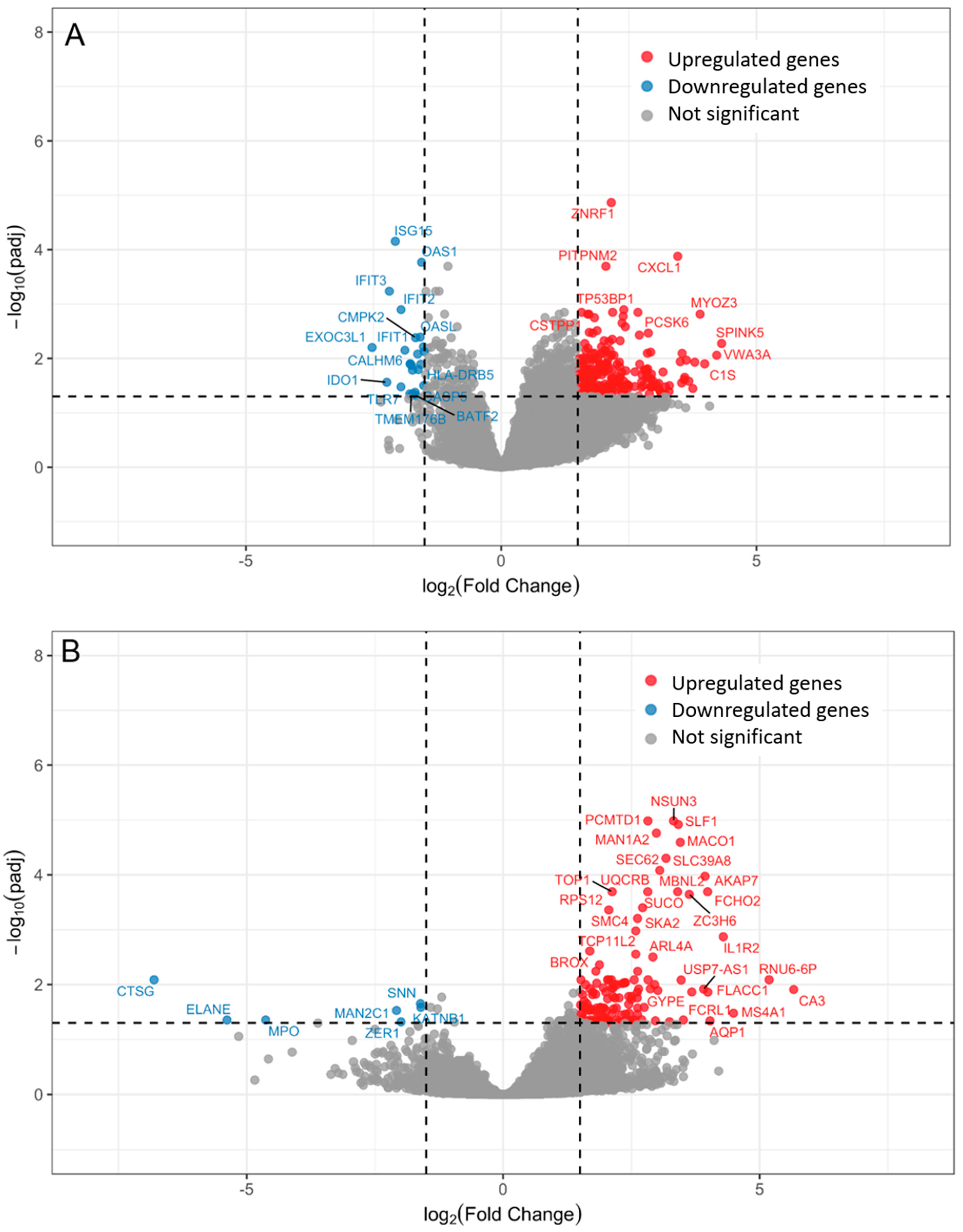
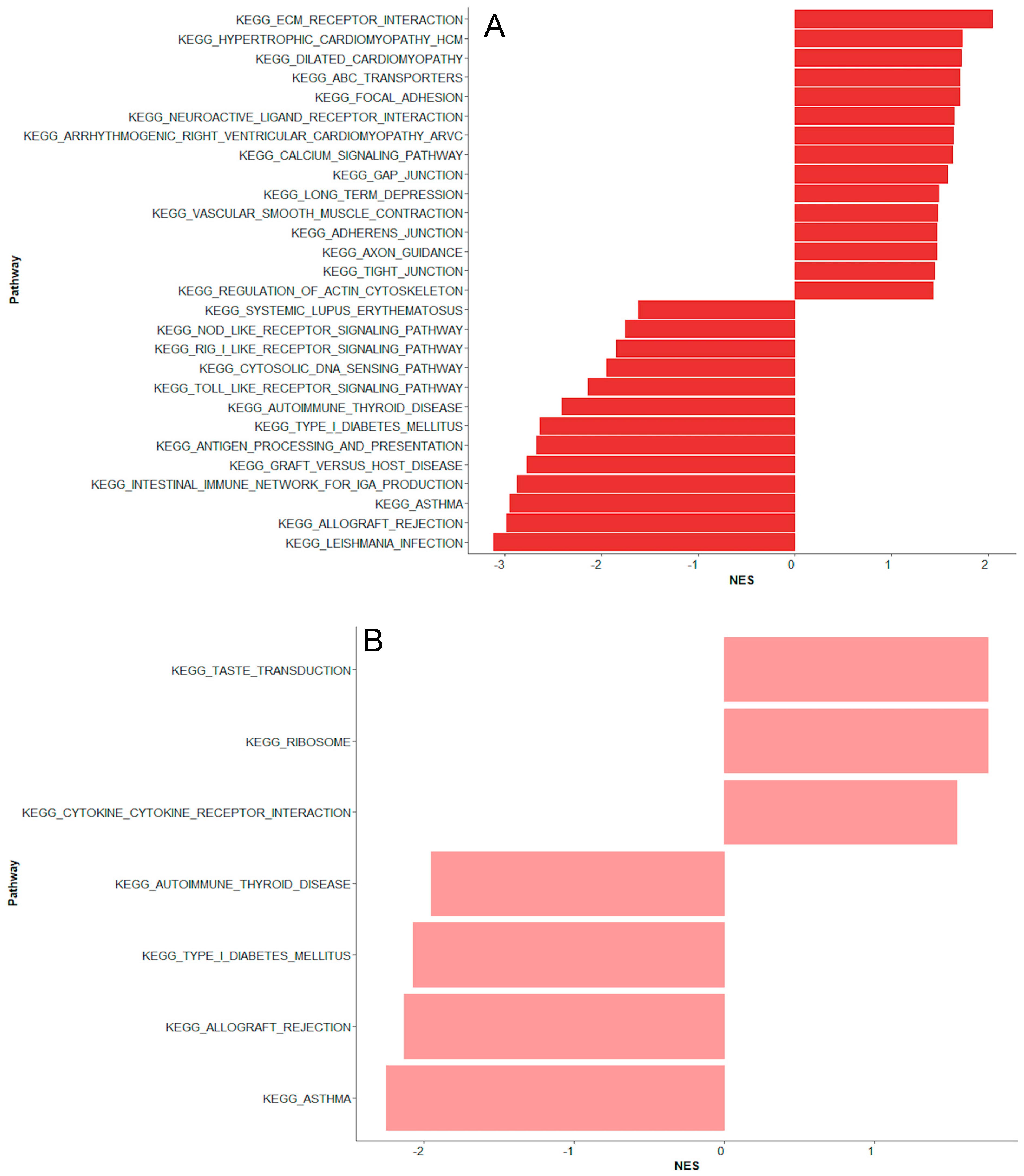
2.1. Bacteremia Versus Unexplained Fever with Lov C-Reactive Protein

Transcriptomics revealed 266 differentially expressed genes in children with bacteremia compared to unexplained fever with low CRP levels (Figure 2A). Eleven of the top 20 differentially expressed genes were associated with immune response (Table 2).
Four genes were upregulated, including C-X-C Motif Chemokine Ligand 1 (CXCL1), Zinc and Ring Finger 1 (ZNRF1), BTB Domain and CNC Homolog 2 (BACH2), and Tumor Protein 53 Binding Protein 1 (TP53BP1). Several interferon-induced genes related to antiviral responses were downregulated, including ISG15 Ubiquitin Like Modifier (ISG15), 2′-5′-Oligoadenylate Synthetase 1 (OAS1), 2′-5′-Oligoadenylate Synthetase Like (OASL), Interferon Induced Protein With Tetratricopeptide Repeats 1, 2 and 3 (IFIT1, IFIT2, IFIT3), and Cytidine/Uridine Monophosphate Kinase 2 (CMPK2) (Table 2). In line, the gene set enrichment analysis identified two downregulated pathways related to antiviral responses, “Antigen processing and presentation” and “Toll-like receptor signaling pathway” (Figure 3A).
In addition, three of the top 20 differentially expressed genes were associated with cardiac and vascular function, including Proprotein Convertase Subtilisin/Kexin Type 6 (PCSK6), C-X-C Motif Chemokine Ligand 1 (CXCL1), and Myozenin 3 (MYOZ3) (Table 2). In line, the gene set enrichment analysis identified upregulated pathways linked to cardiac and vascular function, such as “Dilated cardiomyopathy”, “Hypertrophic cardiomyopathy”, “Vascular smooth muscle contraction”, “Regulation of actin cytoskeleton”, and “Calcium signaling pathway” (Figure 3A). Several top differentially expressed genes were associated with cytoskeletal organization and cellular homeostasis (Table 2).
On day 2 of fever onset, 134 genes distinguished children with bacteremia from unexplained fever with low CRP levels. Only one gene, SS18L1 Subunit of BAF Chromatin Remodeling Complex (SS18L1), overlapped from day 1 to day 2. Among the top 20 differentially expressed genes, three related to immune regulation and neutrophil effector function, including Cathepsin G (CTSG), myeloperoxidase (MPO), and Elastase, Neutrophil Expressed (ELANE), were downregulated. The remaining were mainly involved in DNA and RNA processing and repair as well as protein modification and transport (Table S3). Three distinct pathways were upregulated: “Cytokine cytokine receptor interaction”, “Ribosome”, and “Taste transduction” (Figure 3B). Three downregulated pathways overlapped with those from day 1, including “Type I diabetes mellitus”, “Autoimmune thyroid disease”, and “Asthma”.

2.2. Unexplained Fever with High C-Reactive Protein Versus Bacteremia

Comparing unexplained fever episodes with high CRP to those with bacteremia, 192 genes were differentially expressed (Table S4). Four of the top 20 differentially expressed upregulated genes were immune-related, including Interferon Alpha Inducible Protein 27 (IFI27), Fatty Acid Binding Protein 5 (FABP5), and Chloride Channel 3 (CLC). CXCL Motif Chemokine Ligand 1 (CXCL1) was downregulated together with differentially expressed genes linked to vascular dysfunction, including Perlecan (HSPG2) and Myozenin 3 (MYOZ3). The remaining top differentially expressed genes were primarily related to mitochondrial energy production and function, RNA processing, and gene regulation (Table S4). The gene set enrichment analysis identified downregulated pathways related to cardiac function, including “Dilated cardiomyopathy”, “Hypertrophic cardiomyopathy”, and “Calcium signaling pathway” (Figure 3C).

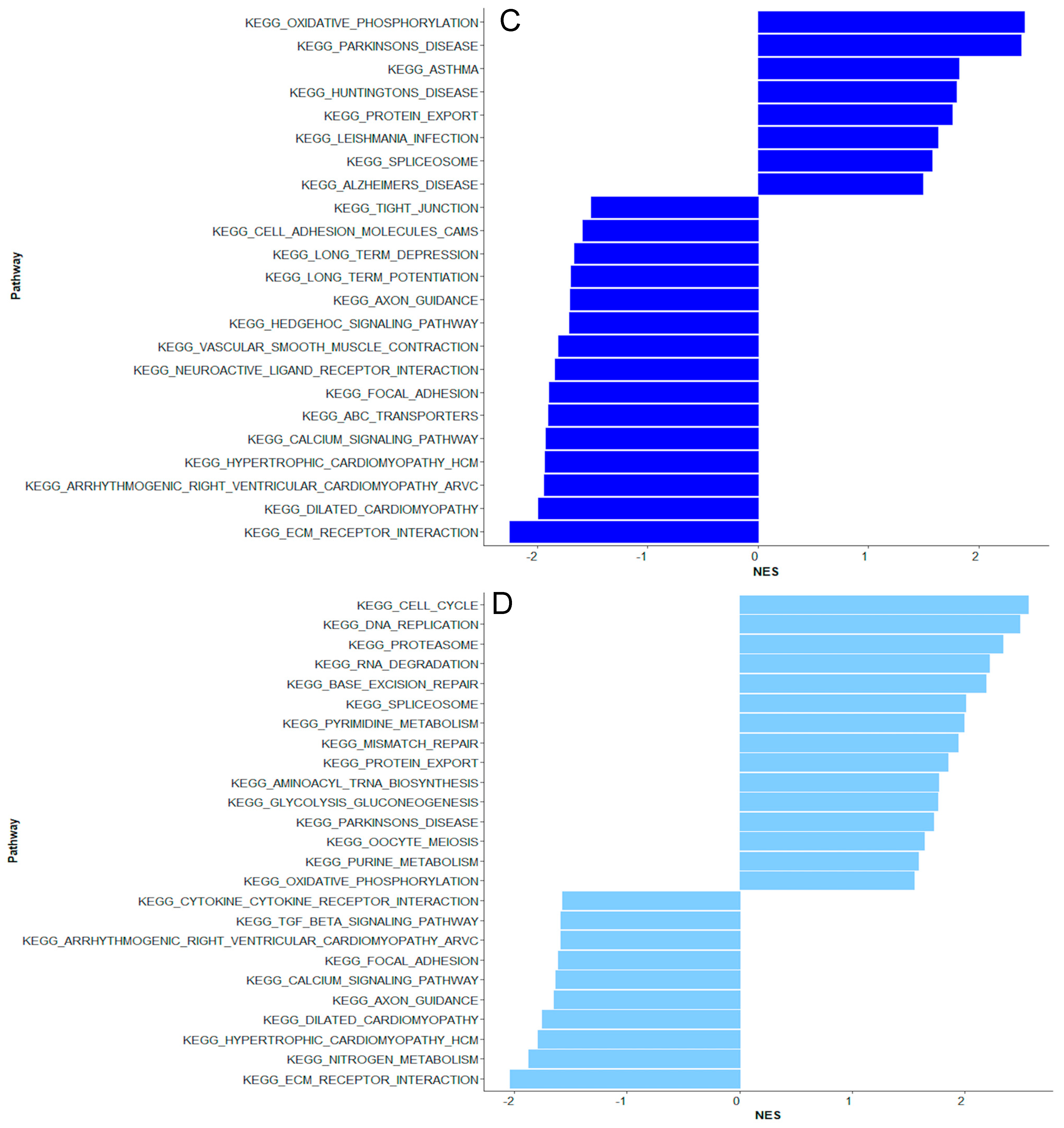
2.3. Coagulase-Negative Staphylococci vs. Bacteremia Caused by Gram-Negative and Other Gram-Positive Bacteria

No genes were differentially expressed in children with coagulase-negative staphylococci compared to bacteremia caused by Gram-negative and other Gram-positive bacteria. The gene set enrichment analysis revealed similar up- and downregulated pathways to those observed in unexplained fever with high CRP (Figure 3D).

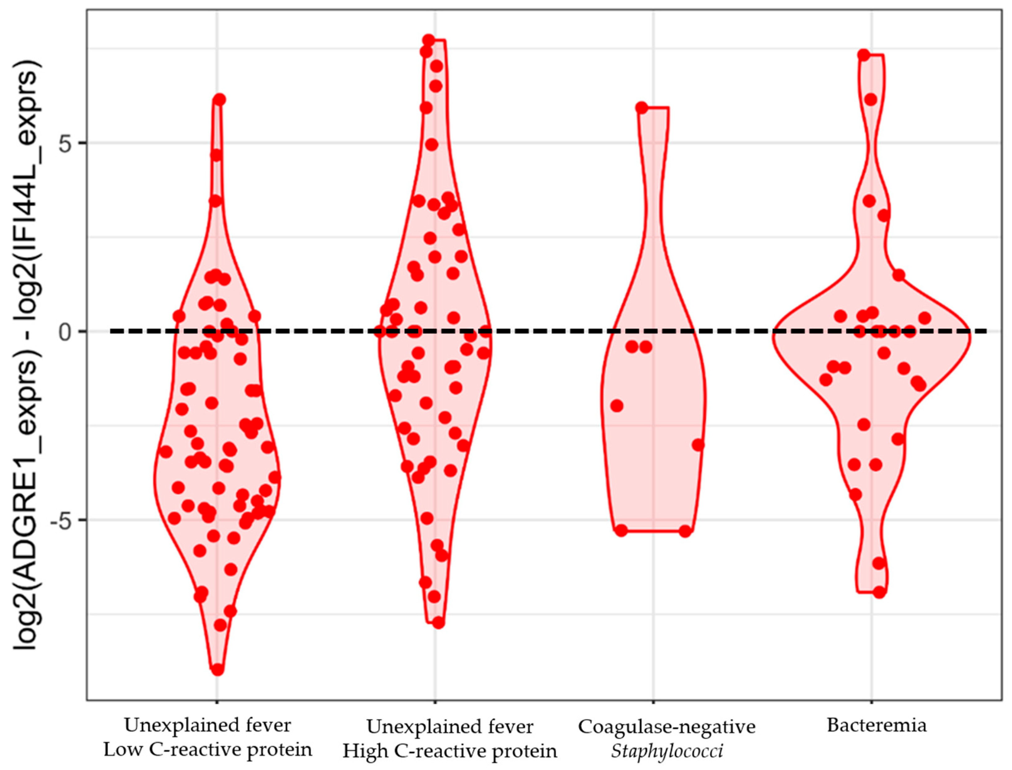
2.4. Testing the Performance of a 2-Gene Signature

The ADGRE1 and IFI44L 2-gene signature identified 14 (52%) of 27 episodes with bacteremia on day 1 as a bacterial infection (sensitivity 0.52, 95% CI 0.32–0.71) and 39 (60%) of 65 episodes without bacterial infection and low CRP as not having bacterial infections (specificity 0.60, 95% CI 0.47–0.72), using the proposed threshold of 0 (Figure 4). We could not identify an alternative risk score threshold as the groups had considerable overlaps in the disease risk score values (Figure 4).

3. Discussion

In this prospective cohort study, we demonstrated the diagnostic potential of transcriptomic profiling in children with hematological malignancies by identifying distinct gene expression patterns distinguishing children with bacteremia from those without. Children with bacteremia had increased expression of genes related to antibacterial immune responses, along with upregulated genes and pathways associated with cardiac and vascular function, potentially indicating sepsis. In contrast, children with unexplained fever had transcriptomic patterns dominated by interferon-related pathways, suggesting viral or other non-bacterial causes of fever. These results argue against false-negative blood cultures and support the potential for early discontinuation of antibiotics in these children despite persistent neutropenia.
This study is the largest to date investigating transcriptomic profiling in febrile children with cancer. Transcriptomic investigations of immunocompromised patients have posed challenges due to their low white blood cell counts, limiting the extraction of sufficient mRNA for gene expression analysis. Only two studies have investigated transcriptomic profiling in febrile children with cancer [37,39]. One study by Haeusler et al. found sufficient RNA for sequencing in 91% of peripheral blood mononuclear cells collected in EDTA tubes. However, this method requires immediate processing to preserve RNA, limiting its utility in clinical and research settings [39]. The other study by Wahlund et al. used RNA-stabilizing tubes, similar to our study, which preserve RNA at collection and allow storage at room temperature for up to 72 h before freezing. However, using a standard RNA-sequencing protocol, Wahlund et al. concluded that immunocompromised children had too few white blood cells for reliable gene expression analysis [37]. In our study, no samples were excluded due to low amounts of RNA, and 91% were successfully sequenced using a single-cell mRNA-sequencing protocol, which enhances mRNA capture and quantification [38].
In children with bacteremia, 266 genes were differentially expressed compared to children with unexplained fever and low CRP. This finding aligns with those of Haeusler et al., who found significant changes in gene expression were observed between children with bloodstream infection and unexplained fever [39]. In our study, several key immunoregulatory genes related to bacterial infection were upregulated, including ZNFR1, involved in TLR4 signaling induced by Gram-negative bacteria; BACH, involved in the adaptive immune response; and CXCL1, a chemokine that promotes neutrophil recruitment and has recently been shown to increase earlier than CRP in children with cancer and bacteremia. Genes and pathways related to cardiac and vascular function were upregulated, such as MYOZ3 and HSPG2, which may reflect hyperdynamic cardiac function and vascular dysregulation during sepsis. CXCL1 may also contribute to vascular dysfunction during sepsis [23,40,41,42]. Pathways linked to calcium signaling were upregulated, as also found by Haeusler et al., consistent with its role in inflammatory activation, vascular permeability, cardiac muscle contraction, and coagulation [39]. Collectively, transcriptomic profiling at fever onset in children with bacteremia was characterized by innate and adaptive immune responses, cardiac and vascular function, and cytoskeletal organization.
A rapid change in gene expression was observed during the first 24 h of fever onset in children with bacteremia. Only one gene overlapped from day 1 to day 2, and no pathways were associated with cardiac or vascular function as seen on day 1. Several upregulated genes were related to cellular homeostasis, indicating a shift towards recovery [43,44]. Neutrophil-expressed genes were downregulated on day 2, reflecting a reduced innate immune response, which may suggest recovery or inappropriate immune suppression [43,45,46]. Thus, transcriptomic changes may vary during the infectious course. This dynamic RNA host response within 24 h may reflect physiological and immunological changes following treatment, such as antibiotics and fluid therapy [47]. Supporting this, Haeusler et al. [39] identified pathways involved in immune recovery and metabolic restoration on day 2, and Cazalis et al. [44] reported that over 70% of the leukocyte transcriptome was altered within 24 h in adults with septic shock, with substantial changes within the first hours. This highlights the importance of investigating transcriptomic changes when developing diagnostic signatures.
Bacteremia with coagulase-negative staphylococci is a common clinical dilemma, as it is often difficult to determine whether the organism is the cause of fever or merely a contaminant. We, therefore, analyzed these episodes separately. We found that coagulase-negative staphylococci had distinct pathways compared to Gram-negative and other Gram-positive infections, while most pathways overlapped with unexplained fever and high CRP. This suggests that coagulase-negative staphylococci are pathophysiologically different from other causes of bloodstream infection. Moreover, these findings may indicate that the coagulase-negative staphylococci were contaminants and that fever in these children was caused by a non-bacterial infection. However, the small sample size limits the ability to draw definitive conclusions.
Children with unexplained fever and low CRP had differentially expressed genes linked to interferon-induced immune responses, including IFIT1, IFIT2, and IFIT3, commonly upregulated during viral infections [48]. This suggests that children with unexplained fever and low CRP may have had fever due to (1) respiratory viral infections without focal signs, as respiratory viruses have been reported to be frequent in children with non-focal febrile neutropenic patients, (2) other non-focal infections, such as enterovirus, and/or (3) self-limiting reactivation of systemic viruses such as cytomegalovirus (CMV) and Epstein-Barr virus (EBV), although such reactivation has recently been reported to be rare in children with febrile neutropenia [49,50,51,52,53,54]. This remains speculative as we did not test for viral infections in children with non-focal febrile neutropenia. Alternatively, the observed interferon-response may reflect sterile inflammation due to non-infectious conditions, as described by Sweeney et al. [43], and thus potentially related to chemotherapy-induced tissue damage. If the observed downregulation of interferon genes in children with bacteremia is reflected at the protein level, the clinical implications could include a higher susceptibility to reactivation of latent viruses. This aligns with Cabler et al., who have reported that viremia, e.g., CMV and EBV, was frequent in pediatric sepsis and linked to a higher mortality [55].
Children with unexplained fever and high CRP had a gene expression profile distinct from those with bacteremia, including downregulated CXCL1, MYOZ3, and HSPG2. They had upregulated IFI27, a type-I interferon-induced gene commonly upregulated during viral infection and other inflammatory responses, and recently suggested as a target for immunoregulatory therapies [56,57,58,59,60]. The distinct profile argues against false negative blood cultures and supports the potential for shorter antibiotic courses despite persistent neutropenia. Shorter antibiotic courses in high-risk children with culture-negative febrile episodes remain a research gap and are currently being investigated in two randomized controlled trials [20,61,62].
Diagnostics based on transcriptomics have shown promise in immunocompetent children, whereas their diagnostic value in children with cancer remains uncertain. Neither the study by Haeusler et al. [39] nor ours included enough febrile episodes to develop and validate a diagnostic signature. The previously published 2-gene signature, based on IFI44L and ADGRE1, performed poorly in our cohort. Still, several differentially expressed genes identified in our study overlapped with those frequently appearing in other published signatures differentiating bacterial from non-bacterial infections developed in immunocompetent children, including IFI27, OASL, and ISG15 [63,64]. This suggests that children with hematological malignancies may have select differentially expressed genes comparable to those of immunocompetent children, despite differences in immune cell composition. Thus, the finding supports the potential applicability of diagnostic signatures developed in immunocompetent populations to immunocompromised children. Perspectives involve investigations of other specific signatures developed in children [27], including innovative multi-class diagnostic signatures, designed to concurrently account for specific pathogens [32].
The main limitation of this study was the small sample size, which limited the investigation of the pathophysiology in specific bloodstream infections and hindered diagnostic signature development and validation. A second limitation was the lack of systematic testing for viral infections, potentially leading to undetected cases, as suggested by the gene expression analysis in children with unexplained fever. Third, as the day 1 and 2 samples were from different patients, sample variability limits direct comparison, unlike in Haeusler et al., where paired samples were used [39]. Still, both studies observed changes in gene expression over time. The main strengths included high RNA preservation, enabling successful sequencing with a single-cell mRNA-sequencing protocol, identification of immune genes overlapping with published signatures, and a study population encompassing the largest pediatric cancer cohort to date.

4. Materials and Methods

This nationwide observational study prospectively included children and adolescents aged 0–17 years with hematological malignancies, admitted to one of the four Pediatric Oncology Departments in Denmark from 1 June 2019 to 31 July 2021. Patients were eligible for inclusion if admitted with fever and re-inclusion if a new fever episode occurred. A new fever episode was defined as fever developing after termination of antibiotics or if the fever re-occurred during antibiotic therapy after at least 72 h of being afebrile (‘breakthrough fever’). Fever was defined as a temperature exceeding 38.5 °C or between 38 °C and 38.5 °C measured twice within one hour. Febrile episodes excluded localized infections, such as skin infections, possible blood culture contaminants, e.g., Micrococcus and Corynebacterium species, and confirmed viral infections. Patients without focal signs were not tested for respiratory viruses.
The patients were managed according to the Danish guidelines for fever in children with cancer, which included the administration of antipseudomonal antibiotics after blood cultures were obtained. They received anti-leukemic treatment according to the Nordic Society of Paediatric Hematology and Oncology (NOPHO) protocols, which did not include antibacterial prophylactic treatment except for weekly co-trimoxazole.
Whole blood (2.5 mL) for RNA analysis was collected at admission for fever, or within 24 h, in PAX gene® Blood RNA Tubes (cat. no. 762165, Qiagen, Hilden, Germany) together with blood cultures and routine blood biochemistry. The samples were incubated at room temperature for at least two hours and frozen to −20 °C within 72 h. Demographic and clinical data were collected in real time from the electronic medical records into a REDCap database.

4.1. Definitions

RNA samples were defined as "day 1" if collected at admission due to fever, before initiating antibiotics, or at the time of fever onset in a patient receiving antibiotics. RNA samples collected 8–24 h after fever onset (on the following day) were defined as “day 2” samples.
We grouped febrile episodes by blood culture status and CRP levels, as those with high CRP and negative blood cultures remain clinically ambiguous, potentially reflecting culture-negative bacteremia, viral infection, or hyperinflammation. Similarly, febrile episodes with coagulase-negative staphylococci bacteremia were grouped separately. Thus, the cohort was divided into four groups:
  • Bacteremia: Positive blood cultures, excluding coagulase-negative staphylococci
  • Unexplained fever with low CRP: Negative blood culture and a maximum CRP below 35 mg/L
  • Unexplained fever with high CRP: Negative blood culture and a maximum CRP of 35 mg/L or above
  • Coagulase-negative staphylococci

4.2. RNA Sequencing and Quality Control

A total of 185 samples were analyzed, with 19 samples processed according to a previously published protocol [38], and 166 using the same protocol with the following minor modifications: Total RNA was isolated from stabilized blood using the QIAsymphony PAXgene Blood RNA Kit (Qiagen, Cat. Nr. 762635), according to the manufacturer’s instructions, and eluted in 100 µL. RNA concentration was quantified by NanoDrop. As RNA yields were within the recommended working range of the SMART-Seq HT Kit (Takara Bio, Shiga, Japan, Cat. Nr. 634436), the Eppendorf Concentrator Plus step used in the previously published protocol was omitted [38]. Samples with an RNA concentration above 12 ng/µL (group 1) and 5–12 ng/µL (group 2) were diluted to 1 ng/µL and 1:2, respectively, and samples with an RNA concentration below 5 ng/µL (group 3) were not further diluted. High-quality, full-length cDNA was generated by priming mRNA with oligo(dT) using the SMART-Seq HT Kit, as per the manufacturer’s instructions. The amount of mRNA used as input was standardized as follows: 1 µL of diluted mRNA for samples of group 1; 5 µL of diluted mRNA for samples of group 2; 10.5 µL of undiluted mRNA for samples of group 3. cDNA was amplified for 13 total PCR cycles and purified with AMPure XP beads (Beckman Coulter Life Sciences, Brea, CA, USA, A63881). cDNA concentration was measured with a Qubit Fluorometer (Thermo Fisher Scientific, Waltham, MA, USA, Q32854), and cDNA was diluted to 0.2 ng/µL, tagmented and barcoded using the Nextera XT DNA Library Preparation Kit (Illumina, San Diego, CA, USA, Cat. Nr. FC-131-1096), as per manufacturer’s instructions. Final libraries were purified with AMPure XP beads. DNA concentration was measured with a Qubit Fluorometer, and fragment size was determined with a Tape Station using High Sensitivity D5000 reagents (Agilent, Santa Clara, CA, USA, Cat. Nr. 5067-5593). Samples were paired-end sequenced on a NextSeq 500 Sequencer (Illumina) to reach a sequencing depth of 1 million raw reads per sample.
The Illumina raw reads were exported as fastq files and trimmed using TrimGalore! v.0.4.0 [65]. The trimmed sequences were aligned to the Genome Reference Consortium Human Build 38 (GRCh38) by STAR (Spliced Transcripts Alignment to a Reference) [66]. For quality control, principal component analysis was performed to check for batch effect, mitochondrial content, age, and gender. Low-quality samples were defined as those with expressed genes less than or equal to 5000, a number of unmapped reads greater than 218 (262,144), and mitochondrial genes exceeding 30%. Seventeen (9%) of 185 samples were excluded, including 6 lost during the processing steps and eleven excluded after quality control, resulting in a final study population of 168 febrile episodes.

4.3. Differential Expression and Pathway Analysis

The primary analysis compared differentially expressed genes between febrile episodes with bacteremia and unexplained fever with low CRP on day 1, followed by a comparison of the same groups on day 2. Additionally, separate analyses compared unexplained fever with high CRP to febrile episodes with bacteremia and episodes with coagulase-negative staphylococci to those with bacteremia. Raw RNA-seq count data were imported into RStudio (v4.3.3). Differential gene expression analysis was performed using DESeq2 (v1.37.0), with neutropenia status (yes/no), febrile infection episodes, and RNA sample collection time considered in the design formula. Genes with fewer than 10 total counts across all samples were filtered out before performing differential expression analysis. Differentially expressed genes (DEGs) were identified with a threshold of absolute value of log2FoldChange(log2FC) more than 1.5, and adjusted p-value (padj) less than 0.05. Volcano plots were used to visualize differentially expressed genes. The function of differentially expressed genes was obtained from GeneCards [67].
Gene set enrichment analysis (GSEA) was performed using fgsea (v1.30.0) package, with log2FC as the ranking metric. Significantly enriched pathways were determined based on padj through Kyoto Encyclopedia of Genes and Genomes (KEGG) pathway analysis, uncovering biological processes associated with differentially expressed genes.
To investigate the performance of a previously published 2-gene signature [26,28], a disease risk score was calculated as the log2-transformed expression difference between ADGRE1 and IFI44L, using a threshold for bacterial assignment of above 0, as proposed by Pennisi et al. [26]. Expression values were extracted from DESeq2-normalized counts, and log2-transformed values were computed with a small offset (+0.1) to prevent log transformation errors. The scores were visualized using violin plots and sina plots [68,69].

4.4. Study Approvals

Patients were recruited under the approval of the research ethics committees of the Ethics Committee of the Capital Region of Denmark (H-2-2010-002) and the Danish Data Protection Agency (P-2019-29). Informed oral and written parental consent was provided before participation.

5. Conclusions

In conclusion, this study provided novel insights into the pathophysiological mechanisms in febrile children with hematological malignancies, revealing immunoregulatory genes associated with bacterial infection and genes and pathways related to cardiac and vascular function, aligning with sepsis. Children with unexplained fever had gene expression profiles different from those with bacteremia, including several interferon-induced genes, suggesting a viral or non-bacterial cause of inflammation, and challenging the assumption of false-negative blood cultures. This finding supports early discontinuation of antibiotics in children with unexplained fever. Furthermore, the study identified several genes overlapping with previously published signatures, supporting the potential for developing transcriptomics-based diagnostics in children with cancer.

Supplementary Materials

The following supporting information can be downloaded at: https://www.mdpi.com/article/10.3390/ijms26157126/s1.

Author Contributions

Conceptualization, L.M.S., K.H.S.D., Y.G., V.P., K.S., F.O.B., N.H.V., and U.N.; methodology, L.M.S., K.H.S.D., Y.G., V.P., L.M., F.O.B., N.H.V., and U.N.; formal analysis, L.M.S., K.H.S.D., Y.G., F.O.B., N.H.V., and U.N.; writing—original draft preparation, L.M.S., K.H.S.D., Y.G., L.M., F.O.B., N.H.V., and U.N.; writing—review and editing, L.M.S., K.H.S.D., Y.G., L.L.H., V.P., L.M., D.G., I.K., R.T., K.S., F.O.B., N.H.V., and U.N.; visualization, L.M.S., K.H.S.D., Y.G., and F.O.B.; project administration, L.M.S., K.H.S.D., Y.G., L.L.H., V.P., L.M., D.G., I.K., R.T., K.S., F.O.B., N.H.V., and U.N.; funding acquisition, L.M.S., N.H.V., and U.N. All authors have read and agreed to the published version of the manuscript.

Funding

The study was funded by The Lundbeck Foundation (R249-2017-1300), The Danish Childhood Cancer Foundation (2017-2076), The Danish Cancer Society (R167-A11072-17-S2), and Innovation Fund Denmark (0176-00020B). The funders had no role in the design and conduct of the study.

Institutional Review Board Statement

The study was approved by the research ethics committees of the Ethics Committee of the Capital Region of Denmark (H-2-2010-002) and the Danish Data Protection Agency (P-2019-29). Clinical trials ID: NCT04669418.

Informed Consent Statement

Informed consent was obtained from all subjects included in the study.

Data Availability Statement

The datasets presented in this article are not readily available in adherence to guidelines established by the Danish Data Protection Agency, which classifies RNA sequences as personally identifiable information. Requests to access the datasets should be directed to the corresponding author.

Acknowledgments

The authors wish to thank the staff at the pediatric oncology departments of Rigshospitalet (Copenhagen University Hospital), Aarhus University Hospital, H.C. Andersen Children’s Hospital (Odense), and Aalborg University Hospital for their invaluable support and assistance during the study period.

Conflicts of Interest

The authors declare no conflicts of interest.

References

  1. Castagnola, E.; Fontana, V.; Caviglia, I.; Caruso, S.; Faraci, M.; Fioredda, F.; Garrè, M.L.; Moroni, C.; Conte, M.; Losurdo, G.; et al. A Prospective Study on the Epidemiology of Febrile Episodes during Chemotherapy-Induced Neutropenia in Children with Cancer or after Hemopoietic Stem Cell Transplantation. Clin. Infect. Dis. 2007, 45, 1296–1304. [Google Scholar] [CrossRef]
  2. van der Velden, F.J.S.; de Vries, G.; Martin, A.; Lim, E.; von Both, U.; Kolberg, L.; Carrol, E.D.; Khanijau, A.; Herberg, J.A.; De, T.; et al. Febrile illness in high-risk children: A prospective, international observational study. Eur. J. Pediatr. 2023, 182, 543–554. [Google Scholar] [CrossRef]
  3. Sørensen, M.C.L.; Andersen, M.M.; Rostgaard, K.; Schmiegelow, K.; Mikkelsen, T.S.; Wehner, P.S.; Olsen, M.; Søegaard, S.H.; Hjalgrim, L.L. Treatment-related mortality among children with cancer in Denmark during 2001–2021. Acta Oncol. 2024, 63, 294–302. [Google Scholar] [CrossRef]
  4. Inaba, H.; Pei, D.; Wolf, J.; Howard, S.C.; Hayden, R.T.; Go, M.; Varechtchouk, O.; Hahn, T.; Buaboonnam, J.; Metzger, M.L.; et al. Infection-related complications during treatment for childhood acute lymphoblastic leukemia. Ann. Oncol. 2017, 28, 386–392. [Google Scholar] [CrossRef]
  5. Alp, S.; Akova, M. Management of febrile neutropenia in the era of bacterial resistance. Ther. Adv. Infect. Dis. 2013, 1, 37–43. [Google Scholar] [CrossRef] [PubMed]
  6. Alali, M.; Mayampurath, A.; Dai, Y.; Bartlett, A.H. A prediction model for bacteremia and transfer to intensive care in pediatric and adolescent cancer patients with febrile neutropenia. Sci. Rep. 2022, 12, 7429. [Google Scholar] [CrossRef] [PubMed]
  7. Lima, M.A.F.; de Sá Rodrigues, K.E.; Vanucci, M.F.; da Silva, P.L.L.; Baeta, T.; Oliveira, I.P.; Romanelli, R.M.d.C. Bloodstream infection in pediatric patients with febrile neutropenia induced by chemotherapy. Hematol. Transfus. Cell Ther. 2023, 45, 170–175. [Google Scholar] [CrossRef] [PubMed]
  8. Baskaran, N.D.; Gan, G.G.; Adeeba, K.; Sam, I.-C. Bacteremia in patients with febrile neutropenia after chemotherapy at a university medical center in Malaysia. Int. J. Infect. Dis. 2007, 11, 513–517. [Google Scholar] [CrossRef]
  9. Klein, K.; van Litsenburg, R.R.L.; de Haas, V.; Dors, N.; van den Heuvel-Eibrink, M.M.; Knops, R.R.G.; Tissing, W.J.E.; Versluys, B.A.; Zwaan, C.M.; Kaspers, G.J.L. Causes of early death and treatment-related death in newly diagnosed pediatric acute myeloid leukemia: Recent experiences of the Dutch Childhood Oncology Group. Pediatr. Blood Cancer 2020, 67, e28099. [Google Scholar] [CrossRef]
  10. Torres, J.P.; De la Maza, V.; Kors, L.; Villarroel, M.; Piemonte, P.; Izquierdo, G.; Salgado, C.; Tordecilla, J.; Contardo, V.; Farfán, M.J.; et al. Respiratory Viral Infections and Coinfections in Children with Cancer, Fever and Neutropenia: Clinical Outcome of Infections Caused by Different Respiratory Viruses. Pediatr. Infect. Dis. J. 2016, 35, 949–954. [Google Scholar] [CrossRef]
  11. Vissing, N.H.; Lausen, B.; Hutchings Hoffmann, M.; Als-Nielsen, B.; Schmiegelow, K.; Helweg-Larsen, J.; Arendrup, M.C.; Nygaard, U. Aspergillus flavus Infections in Children with Leukemia Despite Liposomal Amphotericin-B Prophylaxis. Pediatr. Infect. Dis. J. 2021, 40, 749–752. [Google Scholar] [CrossRef]
  12. Ishii, E.; Hara, T.; Mizuno, Y.; Ueda, K. Vincristine-induced fever in children with leukemia and lymphoma. Cancer 1988, 61, 660–662. [Google Scholar] [CrossRef]
  13. Chappell, T.L.; Pflaster, E.G.; Namata, R.; Bell, J.; Miller, L.H.; Pomputius, W.F.; Boutilier, J.J.; Messinger, Y.H. Bloodstream Infections in Childhood Acute Myeloid Leukemia and Machine Learning Models: A Single-institutional Analysis. J. Pediatr. Hematol. Oncol. 2025, 47, e26–e33. [Google Scholar] [CrossRef]
  14. Avilés-Robles, M.; Schnur, J.J.; Dorantes-Acosta, E.; Márquez-González, H.; Ocampo-Ramírez, L.A.; Chawla, N.V. Predictors of Septic Shock or Bacteremia in Children Experiencing Febrile Neutropenia Post-Chemotherapy. J. Pediatr. Infect. Dis. Soc. 2022, 11, 498–503. [Google Scholar] [CrossRef]
  15. van der Velden, F.J.S.; Gennery, A.R.; Emonts, M. Biomarkers for Diagnosing Febrile Illness in Immunocompromised Children: A Systematic Review of the Literature. Front. Pediatr. 2022, 10, 828569. [Google Scholar] [CrossRef]
  16. Haeusler, G.M.; Phillips, R.; Slavin, M.A.; Babl, F.E.; De Abreu Lourenco, R.; Mechinaud, F.; Thursky, K.A.; Australian PICNICC study group and the PREDICT network. Re-evaluating and recalibrating predictors of bacterial infection in children with cancer and febrile neutropenia. EClinicalMedicine 2020, 23, 100394. [Google Scholar] [CrossRef] [PubMed]
  17. Zhu, Y.; Gan, M.; Ge, M.; Dong, X.; Yan, G.; Zhou, Q.; Yu, H.; Wang, X.; Cao, Y.; Lu, G.; et al. Diagnostic Performance and Clinical Impact of Metagenomic Next-Generation Sequencing for Pediatric Infectious Diseases. J. Clin. Microbiol. 2023, 61, e00115-23. [Google Scholar] [CrossRef] [PubMed]
  18. Arbefeville, S.S.; Timbrook, T.T.; Garner, C.D. Evolving strategies in microbe identification—A comprehensive review of biochemical, MALDI-TOF MS and molecular testing methods. J. Antimicrob. Chemother. 2024, 79, i2–i8. [Google Scholar] [CrossRef]
  19. Lehrnbecher, T.; Robinson, P.D.; Ammann, R.A.; Fisher, B.; Patel, P.; Phillips, R.; Beauchemin, M.P.; Carlesse, F.; Castagnola, E.; Davis, B.L.; et al. Guideline for the Management of Fever and Neutropenia in Pediatric Patients with Cancer and Hematopoietic Cell Transplantation Recipients: 2023 Update. J. Clin. Oncol. 2023, 41, 1774–1785. [Google Scholar] [CrossRef]
  20. Lehrnbecher, T.; Averbuch, D.; Castagnola, E.; Cesaro, S.; Ammann, R.A.; Garcia-Vidal, C.; Kanerva, J.; Lanternier, F.; Mesini, A.; Mikulska, M.; et al. 8th European Conference on Infections in Leukaemia: 2020 guidelines for the use of antibiotics in paediatric patients with cancer or post-haematopoietic cell transplantation. Lancet Oncol. 2021, 22, e270–e280. [Google Scholar] [CrossRef]
  21. Nygaard, U.; Nielsen, A.B.; Dungu, K.H.S.; Drici, L.; Holm, M.; Ottenheijm, M.E.; Nielsen, A.B.; Glenthøj, J.P.; Schmidt, L.S.; Cortes, D.; et al. Proteomic profiling reveals diagnostic signatures and pathogenic insights in multisystem inflammatory syndrome in children. Commun. Biol. 2024, 7, 688. [Google Scholar] [CrossRef]
  22. Buonsenso, D.; Sodero, G.; Valentini, P. Transcript host-RNA signatures to discriminate bacterial and viral infections in febrile children. Pediatr. Res. 2022, 91, 454–463. [Google Scholar] [CrossRef]
  23. Jenner, R.G.; Young, R.A. Insights into host responses against pathogens from transcriptional profiling. Nat. Rev. Microbiol. 2005, 3, 281–294. [Google Scholar] [CrossRef] [PubMed]
  24. Mejias, A.; Cohen, S.; Glowinski, R.; Ramilo, O. Host transcriptional signatures as predictive markers of infection in children. Curr. Opin. Infect. Dis. 2021, 34, 552–558. [Google Scholar] [CrossRef] [PubMed]
  25. Kaforou, M.; Herberg, J.A.; Wright, V.J.; Coin, L.J.M.; Levin, M. Diagnosis of Bacterial Infection Using a 2-Transcript Host RNA Signature in Febrile Infants 60 Days or Younger. JAMA 2017, 317, 1577–1578. [Google Scholar] [CrossRef] [PubMed]
  26. Pennisi, I.; Rodriguez-Manzano, J.; Moniri, A.; Kaforou, M.; Herberg, J.A.; Levin, M.; Georgiou, P. Translation of a Host Blood RNA Signature Distinguishing Bacterial From Viral Infection Into a Platform Suitable for Development as a Point-of-Care Test. JAMA Pediatr. 2021, 175, 417–419. [Google Scholar] [CrossRef]
  27. Mahajan, P.; Kuppermann, N.; Mejias, A.; Suarez, N.; Chaussabel, D.; Casper, T.C.; Smith, B.; Alpern, E.R.; Anders, J.; Atabaki, S.M.; et al. Association of RNA Biosignatures with Bacterial Infections in Febrile Infants Aged 60 Days or Younger. JAMA 2016, 316, 846–857. [Google Scholar] [CrossRef]
  28. Herberg, J.A.; Kaforou, M.; Wright, V.J.; Shailes, H.; Eleftherohorinou, H.; Hoggart, C.J.; Cebey-López, M.; Carter, M.J.; Janes, V.A.; Gormley, S.; et al. Diagnostic Test Accuracy of a 2-Transcript Host RNA Signature for Discriminating Bacterial vs Viral Infection in Febrile Children. JAMA 2016, 316, 835–845. [Google Scholar] [CrossRef]
  29. Anderson, S.T.; Kaforou, M.; Brent, A.J.; Wright, V.J.; Banwell, C.M.; Chagaluka, G.; Crampin, A.C.; Dockrell, H.M.; French, N.; Hamilton, M.S.; et al. Diagnosis of childhood tuberculosis and host RNA expression in Africa. N. Engl. J. Med. 2014, 370, 1712–1723. [Google Scholar] [CrossRef]
  30. Schlapbach, L.J.; Ganesamoorthy, D.; Wilson, C.; Raman, S.; George, S.; Snelling, P.J.; Phillips, N.; Irwin, A.; Sharp, N.; Marsney, R.L.; et al. Host gene expression signatures to identify infection type and organ dysfunction in children evaluated for sepsis: A multicentre cohort study. Lancet Child Adolesc. Health 2024, 8, 325–338. [Google Scholar] [CrossRef]
  31. Wright, V.J.; Herberg, J.A.; Kaforou, M.; Shimizu, C.; Eleftherohorinou, H.; Shailes, H.; Barendregt, A.M.; Menikou, S.; Gormley, S.; Berk, M.; et al. Diagnosis of Kawasaki Disease Using a Minimal Whole-Blood Gene Expression Signature. JAMA Pediatr. 2018, 172, e182293. [Google Scholar] [CrossRef] [PubMed]
  32. Habgood-Coote, D.; Wilson, C.; Shimizu, C.; Barendregt, A.M.; Philipsen, R.; Galassini, R.; Calle, I.R.; Workman, L.; Agyeman, P.K.A.; Ferwerda, G.; et al. Diagnosis of childhood febrile illness using a multi-class blood RNA molecular signature. Med 2023, 4, 635–654.e5. [Google Scholar] [CrossRef] [PubMed]
  33. Viz-Lasheras, S.; Gómez-Carballa, A.; Bello, X.; Rivero-Calle, I.; Dacosta, A.I.; Kaforou, M.; Habgood-Coote, D.; Cunnington, A.J.; Emonts, M.; Herberg, J.A.; et al. A diagnostic host-specific transcriptome response for Mycoplasma pneumoniae pneumonia to guide pediatric patient treatment. Nat. Commun. 2025, 16, 673. [Google Scholar] [CrossRef]
  34. Viz-Lasheras, S.; Gómez-Carballa, A.; Pardo-Seco, J.; Bello, X.; Rivero-Calle, I.; Dacosta, A.I.; Kaforou, M.; Habgood-Coote, D.; Cunnington, A.J.; Emonts, M.; et al. A 5-transcript signature for discriminating viral and bacterial etiology in pediatric pneumonia. iScience 2025, 28, 111747. [Google Scholar] [CrossRef]
  35. Channon-Wells, S.; Habgood-Coote, D.; Vito, O.; Galassini, R.; Wright, V.J.; Brent, A.J.; Heyderman, R.S.; Anderson, S.T.; Eley, B.; Martinón-Torres, F.; et al. Integration and validation of host transcript signatures, including a novel 3-transcript tuberculosis signature, to enable one-step multiclass diagnosis of childhood febrile disease. J. Transl. Med. 2024, 22, 802. [Google Scholar] [CrossRef]
  36. Tsalik, E.L.; Henao, R.; Montgomery, J.L.; Nawrocki, J.W.; Aydin, M.; Lydon, E.C.; Ko, E.R.; Petzold, E.; Nicholson, B.P.; Cairns, C.B.; et al. Discriminating Bacterial and Viral Infection Using a Rapid Host Gene Expression Test. Crit. Care Med. 2021, 49, 1651–1663. [Google Scholar] [CrossRef]
  37. Wahlund, M.; Sinha, I.; Broliden, K.; Saghafian-Hedengren, S.; Nilsson, A.; Berggren, A. The Feasibility of Host Transcriptome Profiling as a Diagnostic Tool for Microbial Etiology in Childhood Cancer Patients with Febrile Neutropenia. Int. J. Mol. Sci. 2020, 21, 5305. [Google Scholar] [CrossRef]
  38. Probst, V.; Smedegaard, L.M.; Simonyan, A.; Guo, Y.; Østrup, O.; Dungu, K.H.S.; Vissing, N.H.; Nygaard, U.; Bagger, F.O. A Protocol for Low-Input RNA-Sequencing of Patients with Febrile Neutropenia Captures Relevant Immunological Information. Int. J. Mol. Sci. 2023, 24, 10251. [Google Scholar] [CrossRef]
  39. Haeusler, G.M.; Garnham, A.L.; Li-Wai-Suen, C.S.; Clark, J.E.; Babl, F.E.; Allaway, Z.; Slavin, M.A.; Mechinaud, F.; Smyth, G.K.; Phillips, B.; et al. Blood transcriptomics identifies immune signatures indicative of infectious complications in childhood cancer patients with febrile neutropenia. Clin. Transl. Immunol. 2022, 11, e1383. [Google Scholar] [CrossRef]
  40. Weischendorff, S.; De Pietri, S.; Rathe, M.; Frandsen, T.L.; Hasle, H.; Nielsen, C.H.; Moser, C.; Müller, K. Markers of neutrophil chemotaxis for identification of blood stream infections in children with acute lymphoblastic leukemia undergoing induction treatment. Eur. J. Haematol. 2023, 110, 762–771. [Google Scholar] [CrossRef]
  41. Lee, C.-Y.; Lai, T.-Y.; Tsai, M.-K.; Chang, Y.-C.; Ho, Y.-H.; Yu, I.-S.; Yeh, T.-W.; Chou, C.-C.; Lin, Y.-S.; Lawrence, T.; et al. The ubiquitin ligase ZNRF1 promotes caveolin-1 ubiquitination and degradation to modulate inflammation. Nat. Commun. 2017, 8, 15502. [Google Scholar] [CrossRef]
  42. Korbecki, J.; Maruszewska, A.; Bosiacki, M.; Chlubek, D.; Baranowska-Bosiacka, I. The Potential Importance of CXCL1 in the Physiological State and in Noncancer Diseases of the Cardiovascular System, Respiratory System and Skin. Int. J. Mol. Sci. 2022, 24, 205. [Google Scholar] [CrossRef]
  43. Sweeney, T.E.; Shidham, A.; Wong, H.R.; Khatri, P. A comprehensive time-course-based multicohort analysis of sepsis and sterile inflammation reveals a robust diagnostic gene set. Sci. Transl. Med. 2015, 7, 287ra71. [Google Scholar] [CrossRef]
  44. Cazalis, M.-A.; Lepape, A.; Venet, F.; Frager, F.; Mougin, B.; Vallin, H.; Paye, M.; Pachot, A.; Monneret, G. Early and dynamic changes in gene expression in septic shock patients: A genome-wide approach. Intensive Care Med. Exp. 2014, 2, 20. [Google Scholar] [CrossRef]
  45. Tang, B.M.; Huang, S.J.; McLean, A.S. Genome-wide transcription profiling of human sepsis: A systematic review. Crit. Care 2010, 14, R237. [Google Scholar] [CrossRef]
  46. Wong, H.R. Genome-wide expression profiling in pediatric septic shock. Pediatr. Res. 2013, 73, 564–569. [Google Scholar] [CrossRef]
  47. Tosi, M.; Coloretti, I.; Meschiari, M.; De Biasi, S.; Girardis, M.; Busani, S. The Interplay between Antibiotics and the Host Immune Response in Sepsis: From Basic Mechanisms to Clinical Considerations: A Comprehensive Narrative Review. Antibiotics 2024, 13, 406. [Google Scholar] [CrossRef] [PubMed]
  48. Dalskov, L.; Gad, H.H.; Hartmann, R. Viral recognition and the antiviral interferon response. EMBO J. 2023, 42, e112907. [Google Scholar] [CrossRef] [PubMed]
  49. Suryadevara, M.; Tabarani, C.M.; Bartholoma, N.; Rosenberg, H.F.; Domachowske, J.B. Nasopharyngeal detection of respiratory viruses in febrile neutropenic children. Clin. Pediatr. 2012, 51, 1164–1167. [Google Scholar] [CrossRef] [PubMed]
  50. Torres, J.P.; Santolaya, M.E. Respiratory viral infections in children with cancer and febrile neutropenia and children undergoing hematopoietic stem cell transplantation. Curr. Opin. Infect. Dis. 2024, 37, 407–412. [Google Scholar] [CrossRef]
  51. Barnbrock, A.; Berger, A.; Lauten, M.; Demmert, M.; Klusmann, J.-H.; Ciesek, S.; Bochennek, K.; Lehrnbecher, T. Frequency and clinical impact of viraemia in paediatric patients undergoing therapy for cancer. Sci. Rep. 2024, 14, 14867. [Google Scholar] [CrossRef]
  52. Handous, I.; Achour, B.; Marzouk, M.; Rouis, S.; Hazgui, O.; Brini, I.; Khelif, A.; Hannachi, N.; Boukadida, J. Co-infections of human herpesviruses (CMV, HHV-6, HHV-7 and EBV) in non-transplant acute leukemia patients undergoing chemotherapy. Virol. J. 2020, 17, 37. [Google Scholar] [CrossRef]
  53. Obrová, K.; Grumaz, S.; Remely, M.; Czurda, S.; Krickl, I.; Herndlhofer, S.; Gleixner, K.V.; Sperr, W.R.; Größlinger, L.; Frank, T.; et al. Presence of viremia during febrile neutropenic episodes in patients undergoing chemotherapy for malignant neoplasms. Am. J. Hematol. 2021, 96, 719–726. [Google Scholar] [CrossRef] [PubMed]
  54. Phasuk, N.; Keatkla, J.; Rattanasiri, S.; Techasaensiri, C.; Anurathapan, U.; Apiwattanakul, N. Monitoring of cytomegalovirus infection in non-transplant pediatric acute lymphoblastic leukemia patients during chemotherapy. Medicine 2019, 98, e14256. [Google Scholar] [CrossRef] [PubMed]
  55. Cabler, S.S.; Storch, G.A.; Weinberg, J.B.; Walton, A.H.; Brengel-Pesce, K.; Aldewereld, Z.; Banks, R.K.; Cheynet, V.; Reeder, R.; Holubkov, R.; et al. Viral DNAemia and DNA Virus Seropositivity and Mortality in Pediatric Sepsis. JAMA Netw. Open 2024, 7, e240383. [Google Scholar] [CrossRef] [PubMed]
  56. Gao, J.; Zhu, X.; Wu, M.; Jiang, L.; Wang, F.; He, S. IFI27 may predict and evaluate the severity of respiratory syncytial virus infection in preterm infants. Hereditas 2021, 158, 3. [Google Scholar] [CrossRef]
  57. Min, Z.; Ye, Z.; Gang, L.; Boyu, D.; Xueyan, X. IFI27 as a potential indicator for severe Enterovirus 71-infected hand foot and mouth disease. Virus Res. 2020, 289, 198149. [Google Scholar] [CrossRef]
  58. Tang, B.M.; Shojaei, M.; Parnell, G.P.; Huang, S.; Nalos, M.; Teoh, S.; O’Connor, K.; Schibeci, S.; Phu, A.L.; Kumar, A.; et al. A novel immune biomarker IFI27 discriminates between influenza and bacteria in patients with suspected respiratory infection. Eur. Respir. J. 2017, 49, 1602098. [Google Scholar] [CrossRef]
  59. Shojaei, M.; McLean, A.S. Interferon-stimulated gene IFI27 as a multifaceted candidate target in precision medicine. Trends Immunol. 2025, 46, 219–228. [Google Scholar] [CrossRef]
  60. Villamayor, L.; López-García, D.; Rivero, V.; Martínez-Sobrido, L.; Nogales, A.; DeDiego, M.L. The IFN-stimulated gene IFI27 counteracts innate immune responses after viral infections by interfering with RIG-I signaling. Front. Microbiol. 2023, 14, 1176177. [Google Scholar] [CrossRef]
  61. Schmiegelow, K. Early Termination of Empirical Antibiotics in High-Risk Febrile Neutropenia in Children with Cancer: An Open-Label, Randomised, Controlled Trial. Available online: https://clinicaltrials.gov/study/NCT04637464 (accessed on 11 April 2025).
  62. Murdoch Childrens Research Institute. Early Versus Late Stopping of Antibiotics in Children with Cancer and High-Risk Febrile Neutropenia. Available online: https://clinicaltrials.gov/study/NCT04948463 (accessed on 11 April 2025).
  63. Bodkin, N.; Ross, M.; McClain, M.T.; Ko, E.R.; Woods, C.W.; Ginsburg, G.S.; Henao, R.; Tsalik, E.L. Systematic comparison of published host gene expression signatures for bacterial/viral discrimination. Genome Med. 2022, 14, 18. [Google Scholar] [CrossRef] [PubMed]
  64. Chawla, D.G.; Cappuccio, A.; Tamminga, A.; Sealfon, S.C.; Zaslavsky, E.; Kleinstein, S.H. Benchmarking transcriptional host response signatures for infection diagnosis. Cell Syst. 2022, 13, 974–988.e7. [Google Scholar] [CrossRef]
  65. Martin, M. Cutadapt removes adapter sequences from high-throughput sequencing reads. EMBnet. J. 2011, 17, 10–12. [Google Scholar] [CrossRef]
  66. Dobin, A.; Davis, C.A.; Schlesinger, F.; Drenkow, J.; Zaleski, C.; Jha, S.; Batut, P.; Chaisson, M.; Gingeras, T.R. STAR: Ultrafast universal RNA-seq aligner. Bioinformatics 2013, 29, 15–21. [Google Scholar] [CrossRef]
  67. GeneCards. Available online: www.genecards.com (accessed on 1 March 2025).
  68. Hintze, J.L.; Nelson, R.D. Violin Plots: A Box Plot-Density Trace Synergism. Am. Stat. 1998, 52, 181–184. [Google Scholar] [CrossRef]
  69. Sidiropoulos, N.; Sohi, S.H.; Pedersen, T.L.; Porse, B.T.; Winther, O.; Rapin, N.; Bagger, F.O. SinaPlot: An Enhanced Chart for Simple and Truthful Representation of Single Observations Over Multiple Classes. J. Comput. Graph. Stat. 2018, 27, 673–676. [Google Scholar] [CrossRef]
Figure 1. Classification of febrile episodes. Febrile children were classified according to blood cultures and CRP. The primary comparison was between febrile episodes with bacteremia and unexplained fever with low CRP. Seventeen (9%) samples from 185 febrile episodes were excluded (6 during processing steps and 11 after quality control), resulting in a final study population of 168 episodes. CoNS = Coagulase-negative staphylococci; CRP = C-reactive protein.
Ijms 26 07126 g001
Figure 2. Volcano plot illustrating differentially expressed genes between febrile episodes with bacteremia and unexplained fever with low CRP on day 1 (A) and day 2 (B). Each point represents a gene. The x-axis represents the log2 fold change, and the y-axis represents the log10 (p-value). The dashed lines represent the statistical significance thresholds after adjusting for multiple testing. Upregulated genes in children with bacteremia compared to unexplained fever with low CRP-reactive protein are shown in red, such as C-X-C Motif Chemokine Ligand 1 (CXCL1), and downregulated in blue, such as ISG15 Ubiquitin Like Modifier (ISG15). Genes that did not differ significantly between groups are gray. One gene overlapped between the day 1 and day 2 samples.
Ijms 26 07126 g002
Figure 3. Gene enrichment pathways. Biological process enrichment analysis of differentially expressed genes across four comparisons: (A) Bacteremia day 1 versus unexplained fever with low CRP, (B) bacteremia day 2 versus unexplained fever with low CRP day 2, (C) unexplained fever with high CRP day 1 versus bacteremia day 1, and (D) coagulase-negative staphylococci day 1 versus bacteremia day 1. Pathways were identified through Kyoto Encyclopedia of Genes and Genomes (KEGG) analysis and displayed by Normalized Enrichment Score (NES), which reflects the direction and strength of enrichment. NES > 0 indicates upregulated pathways and NES < 0 indicates downregulated pathways in the first group of each comparison. NES are shown on the x-axis, and gene set names are on the y-axis. NES = Normalized Enrichment Score.
Ijms 26 07126 g003aIjms 26 07126 g003b
Figure 4. The performance of the previously published 2-gene signature, including ADGRE1 and IFI44L, for unexplained fever with low and high CRP protein, coagulase-negative staphylococci, and bacteremia. To investigate the performance of the 2-gene signature, a disease risk score for each febrile episode was calculated by subtracting log2 IFI44L expression from log2 ADGRE1 expression. The dotted line represents the proposed threshold of 0 or above for assignment to bacterial infections [25,26]. All calculations used febrile episodes on day 1.
Ijms 26 07126 g004
Table 1. Hematological and inflammatory parameters in patients with febrile episodes.
Febrile EpisodesWBC
×109 cells/L
ANC
×109 cells/L
CRP
mg/L
PCT
ng/mL
Positive blood culture
Bacteremia
   -
Day 1
0.5 (0.1–2.4)0.0 (0.0–1.4)84 (40–130)5.9 (0.7–15.9)
   -
Day 2
0.3 (0.1–0.5)0.0 (0.0–0.0)200 (150–287)4.3 (0.4–95.0)
Coagulase-negative staphylococci1.5 (0.5–2.3)0.8 (0.0–1.6)58 (17–115)0.5 (0.1–1.3)
Negative blood culture
C-reactive protein < 35 mg/L
   -
Day 1
3.6 (1.6–5.9)2.5 (0.8–4.6)7 (4–20)0.2 (0.1–0.3)
   -
Day 2
1.6 (1.1–3.2)0.3 (0.1–1.1)19 (8–25)0.2 (0.1–0.3)
C-reactive protein ≥ 35 mg/L0.7 (0.3–0.6)0.0 (0.0–0.3)88 (54–137)0.7 (0.3–1.5)
ANC = Absolute Neutrophil Count; CRP = Absolute Neutrophil Count; PCT = Procalcitonin; WBC = White Blood Cell Count.
Table 2. Differentially expressed genes in febrile episodes with bacteremia vs. unexplained fever with low C-reactive protein on day 1.
Gene SymbolGene Namelog2 FCpadjGene Function
Immune response and regulation
CXCL1C-X-C Motif Chemokine Ligand 13.460.0001Chemotactic activity for neutrophils
ZNRF1Zinc and Ring Finger 12.16<0.0001Regulation of inflammation, TLR4-activated immune response, controlling TLR3 trafficking
BACH2BTB Domain and CNC Homolog 21.720.0015Regulates T-cell function, B-cell maturation, and inflammatory response
TP53BP1Tumor Protein 53 Binding Protein 12.400.0013DNA repair in lymphocytes; class switch recombination in B cells
ISG15ISG15 Ubiquitin-Like Modifier−2.07<0.0001Interferon-induced; essential in innate antiviral immune response
OAS12′-5′-Oligoadenylate Synthetase 1−1.560.0002Interferon-induced; essential in innate antiviral immune response
IFIT3Interferon-Induced Protein with Tetratricopeptide Repeats 3−2.190.0006Interferon-induced antiviral protein; inhibitor of cellular and viral processes, cell migration, proliferation, signaling, and viral replication
IFIT2Interferon-Induced Protein with Tetratricopeptide Repeats 2−1.960.0013Interferon-induced antiviral protein; inhibits expression of viral messenger RNAs
OASL2′-5′-Oligoadenylate Synthetase Like−1.590.0041Negative regulation of viral genome replication, IL-27-mediated signaling pathway
CMPK2Cytidine/Uridine Monophosphate Kinase 2−1.680.0042Immunomodulatory and antiviral activities
IFIT1Interferon-Induced Protein with Tetratricopeptide Repeats 1−1.890.0071Interferon-induced antiviral protein; inhibits viral replication
Other
SS18L1SS18L1 Subunit of BAF Chromatin Remodeling Complex1.700.0015Calcium-responsive transactivator; an essential subunit of a neuron-specific chromatin-remodeling complex
CSTPP1Centriolar Satellite-Associated Tubulin Polyglutamylase Complex Regulator 11.580.0014Cytoskeletal organization, nuclear shape, and cilium disassembly
PITPNM2Phosphatidylinositol Transfer Protein Membrane Associated 22.050.0002Calcium ion binding and lipid binding
MYOZ3Myozenin 33.900.0015Skeletal and cardiac muscle; calcineurin-interacting, helps tether calcineurin to the sarcomere
ARHGEF9Cdc42 Guanine Nucleotide
Exchange Factor 9
2.180.0014Brain-specific; receptor recruitment in GABAergic and glycinergic synapses
DUSP6Dual Specificity Phosphatase 6−1.510.0075Inactivates MAP kinases associated with cellular proliferation and differentiation
PCSK6Proprotein Convertase
Subtilisin/Kexin Type 6
2.680.0014Vascular remodeling; processes protein and peptide precursors in the liver, gut, and brain
ODF3BOuter Dense Fiber of Sperm Tails 3B−1.520.0061Functional element
EXOC3L1Exocyst Complex Component 3 Like 1−2.530.0063Part of the exocyst; possible role in regulating exocytosis of insulin granules
Gene functions were retrieved from https://www.genecards.org/.
Disclaimer/Publisher’s Note: The statements, opinions and data contained in all publications are solely those of the individual author(s) and contributor(s) and not of MDPI and/or the editor(s). MDPI and/or the editor(s) disclaim responsibility for any injury to people or property resulting from any ideas, methods, instructions or products referred to in the content.

