Article

Identification of Genetic Variants Using Next-Generation Sequencing in Pediatric Myelodysplastic Syndrome: From Disease Biology to Clinical Applications

by
Viviane Lamim Lovatel
1,
Gerson Moura Ferreira
2,
Beatriz Ferreira da Silva
1,
Rayane de Souza Torres
1,
Rita de Cássia Barbosa da Silva Tavares
3,
Ana Paula Silva Bueno
4,
Eliana Abdelhay
2 and
Teresa de Souza Fernandez
1,*
1
Cytogenetic Laboratory, Cell and Gene Therapy Program, Instituto Nacional de Câncer (INCA), Rio de Janeiro 20230-130, RJ, Brazil
2
Stem Cell Laboratory, Instituto Nacional de Câncer, Rio de Janeiro 20230-130, RJ, Brazil
3
Bone Marrow Transplantation Center, Instituto Nacional de Câncer (INCA), Rio de Janeiro 20230-130, RJ, Brazil
4
Faculdade de Medicina, Instituto de Pediatria e Puericultura Martagão Gesteira, Universidade Federal do Rio de Janeiro, Rio de Janeiro 21941-912, RJ, Brazil
*
Author to whom correspondence should be addressed.
Int. J. Mol. Sci. 2025, 26(14), 6907; https://doi.org/10.3390/ijms26146907
Submission received: 8 May 2025 / Revised: 14 July 2025 / Accepted: 16 July 2025 / Published: 18 July 2025

Abstract

This study aimed to identify genetic variants using a customized next-generation sequencing (NGS) panel for pediatric myelodysplastic syndrome (pMDS) and to explore their associations with cytogenetic and clinical characteristics. Cytogenetic analyses were conducted using G-banding and fluorescence in situ hybridization. NGS was performed with the Ion Torrent Personal Genome Machine for the following genes: GATA2, RUNX1, CEBPA, ANKRD26, ETV6, SAMD9, SAMD9L, PTPN11, NRAS, SETBP1, DDX41, TP53, FLT3, SRP72, and JAK3. Analyses were performed with Ion Reporter 5.20.8.0 software. Genetic variants were classified using the dbSNP, 1000 Genomes, COSMIC, and Varsome databases. We analyzed 25 cases of pMDS; 15 presented abnormal karyotypes, and 19 showed genetic variants. Among the 29 variants identified across 12/15 genes, 27% were pathogenic and 14% were likely pathogenic, with NRAS and GATA2 most frequently associated with disease progression. A new somatic variant of uncertain significance in SETBP1 was detected in seven patients showing heterogeneous clinical outcomes. Genetic variants were found in 7/10 patients with normal karyotypes, indicating that submicroscopic alterations can shed light on disease biology. Our results highlight the critical role of a targeted NGS panel in identifying molecular alterations associated with pMDS pathogenesis, thereby enhancing diagnostic precision, prognosis, and aiding in treatment selection.

1. Introduction

Pediatric myelodysplastic syndrome (pMDS) is a rare clonal hematopoietic disease that accounts for 5–10% of pediatric hematological malignancies. This neoplasm is characterized by ineffective hematopoiesis and bone marrow dysplasia of one or more cell lines. It also carries a high risk of progression to acute myeloid leukemia (AML) [1,2]. Furthermore, diagnosing pMDS is particularly challenging due to the significant heterogeneity in clinical presentation. Its overlapping symptoms with other conditions, such as inherited bone marrow failure syndromes, also make the diagnosis more difficult [3].
In this context, cytogenetics has been an important tool for the diagnosis and risk stratification of MDS. Specific cytogenetic abnormalities, such as chromosome 7 alterations, are especially relevant in pediatric cases, holding significant prognostic value and guiding clinical management [4]. However, approximately half of the pMDS patients have normal karyotypes, underscoring the importance of identifying and integrating molecular data to advance our understanding of pMDS biology [1,4,5].
Recent advances in the genomic understanding of MDS have enhanced our knowledge of its molecular landscape. These insights have led to updates in the classification systems and the development of more refined prognostic models, thereby improving both diagnostic accuracy and risk stratification [2,3]. However, most of the knowledge about molecular alterations in MDS has been acquired from studies in adult patients [4,5]. This highlights a particular challenge, as pediatric cases of MDS often display significant differences, including variations in genetic profiles and treatment approaches, compared to adults [6].
The largest study to date that performed comprehensive whole-exome sequencing in a pMDS cohort analyzed 46 primary cases to better understand the somatic and germline variants associated with this neoplasm [5]. This study, along with previous research, revealed that in pMDS, unlike in adults, mutations in genes vital for epigenetic regulation, such as TET2, ASXL1, and DNMT3A, as well as RNA-splicing genes like SF3B1 and U2AF1, are rare. In contrast, Ras/MAPK signaling pathway mutations are notably more prevalent in pediatric cases [5,6,7,8].
Pediatric MDS also shows a higher frequency of germline variants. Specifically, genetic variants in GATA2 and SAMD9/SAMD9L are found in 7% and 8%, respectively, of pMDS patients with chromosome 7 alterations. Overall, GATA2 is the most common genetic alteration associated with an increased risk of developing MDS and AML [8]. Moreover, germline mutations in other important genes, such as RUNX1, ETV6, CEBPA, ANKRD26, and SRP72 have been detected in a small subset of pMDS patients [8,9,10]. Additionally, the precise mechanisms through which germline predisposition contributes to pMDS remain poorly elucidated. However, some studies have shown that the acquisition of somatic variants, depending on the underlying germline alteration, may contribute to either increased disease aggressiveness with progression to leukemia or hematopoietic stem cell adaptation, resulting in improved hematopoiesis [8,9]. Therefore, the evaluation and identification of these variants have implications for treatment strategies and prognostic results [11,12,13].
Due to the rarity of pMDS, studies evaluating the genomic architecture and clinical implications of these alterations in the pediatric age group are still limited. Moreover, the existing myeloid panels used in next-generation sequencing (NGS) were primarily developed based on the biology of MDS in adult patients [3]. Consequently, a thorough evaluation of genetic variants specific to pediatric cases is essential, as it may significantly influence risk stratification, therapeutic decisions, and prognostic assessment for this age group [11,12,13]. Therefore, the present study aimed to analyze genetic variants in pMDS using a customized NGS panel and investigate their association with clinical and cytogenetic characteristics, with a particular focus on the progression from MDS to acute myeloid leukemia (AML).

2. Results

2.1. Clinical and Cytogenetic Characteristics

This study was carried out on BM samples of twenty-five pediatric patients diagnosed with MDS, fifteen males and ten females. The median age at diagnosis was eight years (ranging from three months to seventeen years). Of the 25 patients, 84% (21/25) exhibited hypocellular bone marrow (BM). Pancytopenia, characterized by the involvement of all three cellular lineages, was present in 48% (12/25) of the cases. Among the patients, thirteen were diagnosed with refractory cytopenia of childhood (RCC) and twelve were classified into advanced subtypes, including eight cases of myelodysplastic syndrome with excess of blasts (MDS-EB) and three cases of MDS/AML. During the follow-up period, disease progression was observed in 56% (14/25) of patients (Table 1).
Abnormal karyotypes were observed in 60% (15/25) of the patients at diagnosis. Chromosome 7 alterations were present in five patients; among them, one had biclonal alteration (patient n.3), one had clonal evolution with the acquisition of del(X)(q23) (patient n.5), and three had monosomy 7 as the sole chromosomal abnormality. Besides this, during subsequent follow-ups with these patients, a patient with RCC and transitory monosomy 7 was observed (patient n.1). This patient was diagnosed at three months old and had a progressive loss of monosomy 7. At two years old, monosomy 7 could only be detected by fluorescence in situ hybridization (FISH) analysis in 7% of the BM cells, and one year later, this genetic alteration was no longer detectable. Complex karyotypes were detected in three patients with advanced pMDS subtypes and were associated with evolution from MDS to AML. Chromosomal translocations were present in three patients. These chromosomal alterations were associated with the evolution to AML. Trisomy 8, as the sole chromosomal abnormality, was observed in one patient (patient n.11), who showed evolution to AML. Interestingly, trisomy 8 was present in cases with chromosomal translocations (patients n.10 and n.25), complex karyotypes (patients n.7 and n.8), associated with del(11)(q23) (patient n.13), involved in clonal evolution associated with add(12)(p12) (patient n.12), and also present in a biclonal chromosomal abnormality (patient n.24). All these patients showed evolution from MDS to AML. Concerning the ten patients with normal karyotypes, only two were diagnosed with advanced subtypes, and only one showed evolution to AML (Table 1).

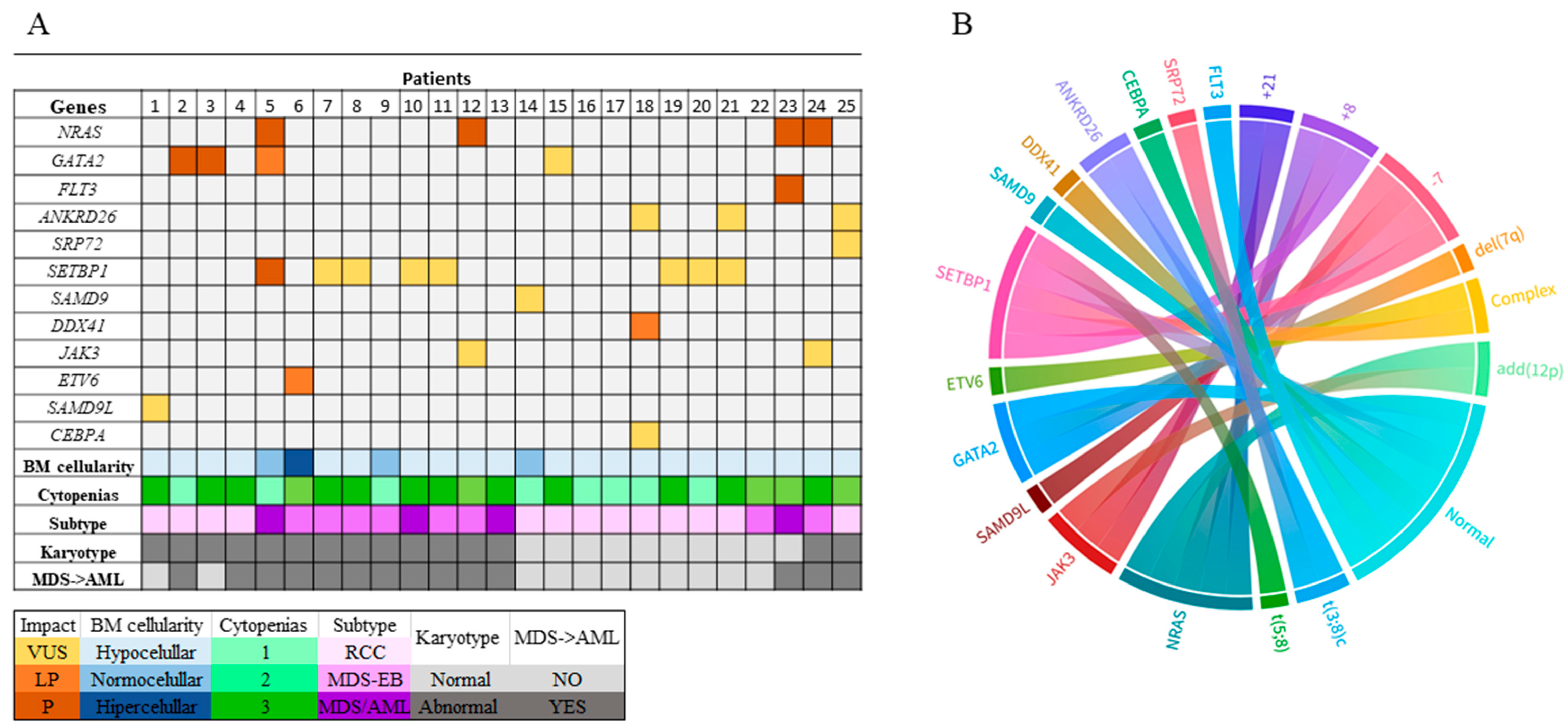
2.2. Genomic Alterations

Genetic variants were identified in 76% of cases (19/25), encompassing 12 different genes: NRAS, GATA2, FLT3, ANKRD26, SRP72, SETBP1, SAMD9, SAMD9L, DDX41, JAK3, ETV6, and CEBPA. In total, 29 variants were identified, with 27.6% (8/29) classified as pathogenic, 10.3% (3/29) as likely pathogenic, and 62.1% (18/29) as variants of uncertain significance (VUS). Concerning the consequences of the variants, missense variants represented 68.97% (20/29), followed by frameshift insertion at 13.8% (4/29) and frameshift deletion at 13.8% (4/29). Based on the variant allele frequency (VAF) of ≥ 40%, most pathogenic (5/8) and likely-pathogenic (2/3) variants had a suggestive germline origin (Table 2). The controls did not show genetic variants among the studied genes.
Genetic variants in SETBP1 were the most frequent, corresponding to 27.5% (8/29) of the total variants identified in this study, followed by NRAS and GATA2 with 13.79% (4/29) each. The genes with the highest number of pathogenic variants were NRAS and GATA2, though FLT3 and SETBP1 also had variants classified as pathogenic. Likely-pathogenic variants were also detected in GATA2, as well as in ETV6 and DDX41.
We identified genetic variants that were not previously reported; they were detected in four genes: SAMD9L (c.2663C>T/p.Ser888Phe), ETV6 (c.1359A>G/p.Ter453Trp), GATA2 (c.-3CCGG>G), and SETBP1 (c.3461A>C/p.His1154Pro). Notably, the novel variant in SETBP1 was observed in seven patients. Among them, six cases involved the variant as an isolated finding. The allele frequency (VAF) of this novel variant ranged from 2.86% to 6.14%, with robust sequencing coverage exceeding 2000 reads.
Isolated genetic variants were identified in eleven patients, predominantly affecting the SETBP1 (6/11) and GATA2 (3/11) genes. However, only two cases with the SETBP1 variant (patients n.19, 20) and one case with the GATA2 variant (patient n.15) had no associated cytogenetic alteration. The other patients had abnormal karyotypes, including two complex karyotypes (patients n. 7, 8), one translocation (patient n.10), and one trisomy 8 (patient n.11) associated with the SETBP1 variant. Meanwhile, the GATA2 variant was associated with monosomy 7 in both cases (patients n. 2, 3).
The association of genetic variants with clinical features showed that the patients with hypocellularity and pancytopenia also had chromosomal alterations and presented disease progression to AML. Concerning the clinical impact of genetic variants, the VUS were commonly observed in patients with RCC who lacked cytogenetic abnormalities and showed no disease progression. On the other hand, patients with EB-MDS and MDS/AML had a higher number of pathogenic and pathogenic-like variants. These patients also presented cytogenetic alterations, mainly involving chromosomes 7 and 8 (Figure 1A,B).

3. Discussion

In this study, a customized NGS panel for 15 genes was designed to analyze genetic variants in pMDS. These genes were selected based on a literature review focusing on their role in the pathogenesis of pMDS and genome sequencing in pMDS [5,8,10]. Based on previous studies, we used a VAF > 40% to indicate a germline origin of genetic variants [3,5]. In our study, 44.8% (13/29) of the variants were found to have a possible germline origin (Table 2). Our results reinforce the important role of germline susceptibility in pMDS [5,8].
Our results showed that higher frequencies of pathogenic variants were identified in NRAS and GATA2. Around 10–30% of myeloid malignancies have NRAS variants associated with an aggressive disease, presenting increased proliferation and resistance to therapies [15]. In MDS, there are three prevalent mutational hotspots: G12, G13, and Q61+ [15,16]. Here, we observed four patients with NRAS variants located within these hotspots. Two patients had the variant at G12 that is particularly prevalent in MDS, accounting for 50–70% of the cases with variants in this gene [5,15].
NRAS encodes a GTPase that, when mutated, constitutively binds to GTP, promoting oncogenesis. However, NRAS variants do not always function as independent prognostic factors. Their interaction with other genetic alterations is common and can lead to varying outcomes. For instance, NRAS mutations in combination with TP53 have been associated with the development of lethal AML [15,16]. In our study, all patients with NRAS variants presented additional alterations. Patient n. 5 had GATA2 and SETBP1 gene variants; three exhibited chromosomal 7 alterations (patients n. 2, 3, and 5). These patients had disease evolution in accordance with the synergic adverse impact suggested for NRAS variants with additional alterations [15].
Regarding GATA2, three patients with monosomy 7 had pathogenetic or likely-pathogenic variants with a suggestive germline origin [5]. GATA2 encodes a major hematopoietic transcription factor that plays an important role in early hematopoiesis. In pMDS, GATA2 alterations are common genetic variants, representing 7% of all patients and 66.67% of those with chromosome 7 alterations, mainly in advanced subtypes [9,17]. In the present study, GATA2 variants were observed in two patients with RCC and one with MDS/AML.
In pediatric patients with MDS, an association between monosomy 7 or del(7q) and genetic variants in SAMD9/SAMD9L has also been reported. However, unlike GATA2, it is predominantly observed in the initial stage of pMDS, the subtype RCC [9]. In the present study, SAMD9L and SAMD9 variants were also observed only in patients with RCC. Interestingly, one of these patients had transient monosomy 7 (patient no. 1), and after approximately three years, the cytogenetic alteration was no longer detectable. This patient harbored two variants: a somatic one (p.Phe1307Ser) and a previously unreported germline variant in SAMD9L (p.Ser888Phe), located in the region that encodes a P-loop/NTPase protein, which plays a crucial role in several cellular processes, including apoptosis. Transient monosomy 7 has also been described in other studies, attributing this phenomenon to an adaptive response resulting from the somatic genetic rescue of variants in SAMD9/SAMD9L [5,9,18,19].
Likely-pathogenic variants were also identified in the ETV6 and DDX41 genes, both of which are key to hematopoiesis in myeloid neoplasms [2,3,20,21]. Here, patient no. 6, with the ETV6 variant, exhibited a complex karyotype, a very poor prognosis, and disease relapse after HSCT. ETV6 functions as a transcriptional repressor, playing a crucial role in maintaining hematopoiesis while also acting as a tumor suppressor [20,22]. Alterations in ETV6 may interact with other genetic alterations to form a network that drives the self-renewal of leukemic stem cells and facilitates disease progression [20,22]. DDX41 is vital for innate immunity, genomic stability, mRNA splicing, and ribosome biogenesis. Dysfunctional DDX41 predisposes individuals to neoplasia, mainly of myeloid origin, although the pathogenic mechanisms are not fully understood [21]. In our study, patient no.18, who had a normal karyotype alongside DDX41 variants, as well as variants in ANKRD26 and CEBPA, responded well to HSCT.
In the present study, most of the variants detected were classified as VUS. This finding has become increasingly common in cancer research with the widespread adoption of massively parallel sequencing technologies. However, in hematologic malignancy, the limited available literature on VUS in genomic databases makes their interpretation challenging [23]. Recently, VUS in genes associated with myeloid neoplasms have been identified as markers of clonal hematopoiesis and potential indicators of prognosis in leukemia and myelodysplasia [24]. Therefore, reporting VUS and thoroughly assessing their potential impact is crucial for advancing the understanding of their role, especially in rare diseases where the genetic landscape is less well-characterized [22].
Among the genes in this panel, variants in SETBP1 were observed in eight patients, being the most frequent VUS. It is important to note that our study identified a novel somatic variant, p.His1154Pro SETBP1, which was present in seven patients. Regarding these seven patients, six had only this genetic variant among the genes studied. This genetic variant is located at the AT-hook domain of transcription factor SETBP1, which is essential for DNA binding to promoter regions. Alterations in this domain lead to the upregulation of several genes involved in cellular processes important for hematopoiesis, such as MECOM, HOXA9/10, and RUNX1 [25]. Beyond that, a previous study showed that a single SETBP1 variant could be associated with a worse prognosis in myeloid neoplasms, regardless of the variant classification as VUS [22,25]. In our cases, the outcome of patients with this alteration was variable. Three patients with RCC showed normal karyotype and no disease progression. Conversely, in advanced subtypes, all patients had cytogenetic alterations mainly involving chromosomal gains and presented with disease evolution. These results showed a probable pathway involving the variant in SETBP1 and an abnormal karyotype, which might involve trisomy 8. However, these results underscore the need for further research on the genomics of pMDS to understand the impact of this novel genetic variant in the biology of pMDS.
In this study, over half of the patients with normal karyotypes exhibited genetic variants. For example, one patient had a normal karyotype and two pathogenic variants, one in NRAS and one in FLT3. This patient presented an advanced subtype at diagnosis and an unfavorable outcome, with evolution to AML. Altogether, these results demonstrated that investigating submicroscopic alterations in patients with normal karyotypes can shed light on the molecular mechanisms involved in disease development and progression, as well as identify potential novel treatment targets. Nowadays, allogeneic hematopoietic stem cell transplantation remains the only potentially curative treatment for pMDS, and the rapid indication of this treatment is crucial for optimizing patient outcomes [14,26]. Therefore, identifying molecular biomarkers that may indicate rapid disease progression is essential to aid patient clinical management.
The genomic landscape of adult MDS has been extensively explored, allowing the development of commercial NGS panels. However, some essential genes in the pathology of pMDS are not included [2,3]. Despite some limitations of this study, such as the small number of patients analyzed, retrospective data collection, and the absence of skin fibroblast genomic analyses, this study showed the importance of using an NGS panel focused on pMDS molecular alterations. The use of this targeted panel with only 15 genes was able to bring new information about the biology of pMDS, allowing these results to be applied in the clinic. Such an approach enhances diagnostic and prognostic accuracy.
Currently, investigating molecular alterations through high-coverage sequencing platforms allows for the identification of new alterations that may have clinical relevance. Studies utilizing this approach open a new avenue for future research in pMDS, guiding treatment toward precision medicine.

4. Material and Methods

4.1. Patients and Controls

This study was conducted on retrospective BM samples sent to the cytogenetic laboratory from 2010 to 2023 as part of standard care for cytogenetic analyses and also as a research study, for which the remaining aliquots were used for molecular analyses. The patients were diagnosed at Instituto Nacional de Câncer (Rio de Janeiro, Brazil) and Instituto de Puericultura e Pediatria Martagão Gesteira (Rio de Janeiro, Brazil). The diagnosis was based on morphologic, cytogenetic, immunophenotypic, and clinical characteristics. None of the patients had previously been treated for malignancy, nor did they have a genetic syndrome. The subtype definition followed the International Consensus Classification of Myeloid Neoplasms [2]. We also used three bone marrow donor samples as controls for NGS analyses. This study was approved by the Ethics and Research Committee of the National Cancer Institute (reference number # 3401739) in accordance with the Declaration of Helsinki.

4.2. Cytogenetic Analyses

Classical cytogenetic analysis was performed on BM cells using G-banding [10]. Samples were initially collected in heparinized tubes. The cells were counted and cultured at a concentration of 5 × 10⁶ to 10 × 106 cells/mL in a medium composed of 80% RPMI and 20% fetal bovine serum. The cultures were incubated at 37 °C in a 5% CO2 atmosphere for 24 h. To arrest cells in metaphase, colcemid was added to the cultures at a final concentration of 0.05 µg/mL during the final hour of incubation. After incubation, cells were subjected to hypotonic shock treatment using 0.075 M KCl for 20 min, and then fixed in a methanol/acetic acid solution (3:1 ratio). For the G-banding procedure, approximately 10 slides were prepared per patient. Fixed cells were dropped onto clean slides from a height and flame-fixed using a Bunsen burner. These slides were incubated at room temperature for about 24h to age before banding. The slides were incubated in a 0.1% trypsin solution in Dulbecco’s solution (8 g NaCl, 0.2 g KCl, 0.2 g KH2PO4, 1.5 g NaH2PO4 per 1 L distilled water, pH 7.8) for 1 s to 1 min. After incubation, they were washed in 0.9% NaCl and stained with 2% Giemsa in phosphate buffer (14.075 g NaH2PO4 per 1 L of distilled water, pH 6.8) for 15 min. A minimum of 20 metaphases were analyzed per BM sample from a patient. Chromosomes were identified and arranged according to the International System for Human Cytogenomic Nomenclature (ISCN), 2020 [27]. Karyotype images were captured using the Ikaros Karyotyping System (MetaSystems, Zeiss, Altlussheim, Germany).
Fluorescence in situ hybridization (FISH) analyses were performed to confirm chromosomal alterations, particularly in cases where clone sizes detected by G-banding were small. For these analyses, cytogenetic culture samples were dropped onto clean slides using a water bath method, allowing them to dry on a heated plate at 42 °C. The slides were then incubated in 2× SSC buffer (prepared from 20× SSC: 3.0 M NaCl and 0.3 M sodium citrate, pH 7.0) for 20 min at room temperature. Following incubation, the slides were incubated in an ethanol series—70%, 90%, and 100%—for 2 min each at room temperature. The probes were prepared and applied according to the manufacturer’s instructions. FISH was conducted using the following probes: del(7q)/-7 (D7S486 “spectrum orange”/CEP7 “spectrum green”) and +8 (LSI c-MYC 8q24, “spectrum orange”). The hybridization area was covered with a coverslip and sealed with rubber cement (Marabu, Tamm, Germany). Slides were incubated for 16 h in a hybridization chamber (Thermobrite, Leica, Richmond, VA, USA). After hybridization, the slides were washed in 0.4× SSC + 0.3% Tween at 73 °C for 2 min, followed by washing in 0.2× SSC + 0.1% Tween at room temperature for 1 min. The slides were then counterstained with DAPI. Analysis was performed using a fluorescence microscope (Olympus BX51, Miami, FL, USA), and images were captured using the ISIS imaging system (MetaSystems, Zeiss, Altlussheim, Germany).

4.3. Next-Generation Sequencing

Genetic variants were analyzed using next-generation sequencing (NGS) on the Ion Torrent platform. A customized panel was employed, targeting complete coding regions with 98.84% coverage, which included 25 bp intronic padding around each exon. The panel generated 259 amplicon sets, divided into two pools: Pool 1, containing 132 amplicons, and Pool 2, containing 127 amplicons. Sequencing was performed using the human reference genome (hg19, GRCh37). The standard DNA amplicon size was 375 base pairs, yielding a total of 47,355 base pairs sequenced. Additionally, a total of 15 genes associated with pMDS were selected based on the published scientific literature [4,5,6]. The genes investigated in this panel were GATA2, RUNX1, CEBPA, ANKRD26, ETV6, SAMD9, SAMD9L, PTPN11, NRAS, SETBP1, DDX41, TP53, FLT3, SRP72, and JAK3.
Genomic DNA (20 ng) quantified by the Qubit dsDNA HS Assay Kit (Thermo Fisher Scientific, Waltham, MA, USA) was used to prepare libraries for NGS using the Ion AmpliSeq Library Kit 2.0 (2 pools by sample). Barcoded adapters (Ion Xpress Barcode Adapters, Thermo Fisher Scientific, Waltham, MA, USA) were added to each sample. The purification was performed with AMPure XP purification (Beckman Coulter, Brea, CA, USA), and the quantification of the libraries was performed using the Ion Library TaqMan Quantification Kit (Thermo Fisher Scientific, Waltham, MA, USA) via real-time PCR. The Ion One Touch 2 instrument, using the Ion PGM Hi-Q-View OT2 kit, was used to conduct the emulsion PCR. Library enrichment was performed with ionic sphere particles (ISPs) on the Ion One Touch ES instrument (Thermo Fisher Scientific, Waltham, MA, USA). Libraries were loaded onto the Ion 316 v2 chip and sequenced on the Ion Torrent PGM sequencer (Thermo Fisher Scientific, Waltham, MA, USA). All steps were followed according to the manufacturer’s guidelines. Sequence alignment and analyses were performed using Ion Reporter software 5.20.8.0 [available online: https://ionreporter.thermofisher.com (accessed on 18 June 2025)]. The average base coverage depth exceeded 1000×, with over 95% of bases sequenced on target. Variant identification and visual confirmation were performed using the Integrative Genomics Viewer (IGV) tool.
Further analyses were performed using Ensembl Variant Effect Predictor (VEP) [https://www.ensembl.org/info/docs/tools/vep/index.html (accessed on 15 February 2025)], and genomic variants with a variant allelic frequency (VAF) of ≥2% were evaluated through the following databases: dbSNP [https://www.ncbi.nlm.nih.gov/snp/ (accessed on 20 February 2025)], 1000 Genomes [https://www.internationalgenome.org/ (accessed on 20 February 2025)], COSMIC [https://cancer.sanger.ac.uk/cosmic/ (accessed on 20 February 2025)], ClinVar [https://www.ncbi.nlm.nih.gov/clinvar/ (accessed on 20 February 2025)], and Varsome [https://varsome.com/ (accessed on 20 February 2025)]. Benign and likely-benign variants were excluded from the results. Pathogenic and likely-pathogenic variants were annotated with ClinVar and COSMIC, while unreported variants were based on in silico analysis obtained from tools in Varsome, as well as the variants classified as VUS.

Author Contributions

V.L.L., B.F.d.S., R.d.S.T. and T.d.S.F. wrote the manuscript. G.M.F. conducted the NGS experiment. V.L.L., G.M.F. and T.d.S.F. analyzed the NGS data. V.L.L., B.F.d.S. and T.d.S.F. analyzed the cytogenetic data. R.d.C.B.d.S.T. and A.P.S.B. were responsible for patient care and reviewed the clinical data. E.A. and T.d.S.F. reviewed the manuscript critically. T.d.S.F. conceived the study, its design, and its coordination, and provided financial support. All authors have read and agreed to the published version of the manuscript.

Funding

This study was funded by Fundação Carlos Chagas Filho de Amparo à Pesquisa do Estado do Rio de Janeiro—FAPERJ, under grant number FAPERJ/E-26/201.218/2022, and Conselho Nacional de Desenvolvimento Científico e Tecnológico (CNPq).

Institutional Review Board Statement

The study was conducted following the Declaration of Helsinki and approved by the Ethics and Research Committee of the National Cancer Institute (reference number # 3401739, approval in 2019).

Informed Consent Statement

Informed consent was obtained from the legal guardians of all subjects involved in the study.

Data Availability Statement

The raw data supporting the conclusions of this article will be made available by the authors on request.

Conflicts of Interest

The authors declare no conflicts of interest.

References

  1. Chisholm, K.M.; Bohling, S.D. Childhood Myelodysplastic Syndrome. Clin. Lab. Med. 2023, 43, 639–655. [Google Scholar] [CrossRef] [PubMed]
  2. Arber, D.A.; Orazi, A.; Hasserjian, R.P.; Borowitz, M.J.; Calvo, K.R.; Kvasnicka, H.-M.; Wang, S.A.; Bagg, A.; Barbui, T.; Branford, S.; et al. International Consensus Classification of Myeloid Neoplasms and Acute Leukemias: Integrating morphologic, clinical, and genomic data. Blood 2022, 140, 1200–1228. [Google Scholar] [CrossRef] [PubMed]
  3. Bernard, E.; Tuechler, H.; Greenberg, P.L.; Hasserjian, R.P.; Ossa, J.E.A.; Nannya, Y.; Devlin, S.M.; Creignou, M.; Pinel, P.; Monnier, L.; et al. Molecular International Prognostic Scoring System for Myelodysplastic Syndromes. NEJM Evid. 2022, 1, EVIDoa2200008. [Google Scholar] [CrossRef] [PubMed]
  4. Babcock, S.; Calvo, K.R.; Hasserjian, R.P. Pediatric myelodysplastic syndrome. Semin. Diagn. Pathol. 2023, 40, 152–171. [Google Scholar] [CrossRef] [PubMed]
  5. Schwartz, J.R.; Ma, J.; Lamprecht, T.; Walsh, M.; Wang, S.; Bryant, V.; Song, G.; Wu, G.; Easton, J.; Kesserwan, C.; et al. The genomic landscape of pediatric myelodysplastic syndromes. Nat Commun. 2017, 8, 1557. [Google Scholar] [CrossRef] [PubMed]
  6. McCall, D.; Abuasab, T.; Rodriguez-Sevilla, J.J.; Mohamed, S.F.; Patnaik, A.; Devireddy, K.; Arani, N.; Sheikh, I.; Jamshidi, R.; Gibson, A.; et al. Characteristics and outcomes of children, adolescent, and young adult patients with myelodysplastic neoplasms: A single-center retrospective analysis. Leuk Res. 2024, 144, 107563. [Google Scholar] [CrossRef] [PubMed]
  7. Pastor, V.; Hirabayashi, S.; Karow, A.; Wehrle, J.; Kozyra, E.J.; Nienhold, R.; Ruzaike, G.; Lebrecht, D.; Yoshimi, A.; Niewisch, M.; et al. Mutational landscape in children with myelodysplastic syndromes is distinct from adults: Specific somatic drivers and novel germline variants. Leukemia 2017, 31, 759–762. [Google Scholar] [CrossRef] [PubMed]
  8. Li, Y.; Cheng, L.; Peng, Y.; Wang, L.; Zhang, W.; Yin, Y.; Zhang, J.; Wu, X. The role of genetic factors in pediatric myelodysplastic syndromes with different outcomes. BMC Pediatr. 2024, 24, 28. [Google Scholar] [CrossRef] [PubMed]
  9. Wlodarski, M.W.; Hirabayashi, S.; Pastor, V.; Starý, J.; Hasle, H.; Masetti, R.; Dworzak, M.; Schmugge, M.; Van Den Heuvel-Eibrink, M.; Ussowicz, M.; et al. Prevalence, clinical characteristics, and prognosis of GATA2-related myelodysplastic syndromes in children and adolescents. Blood 2016, 127, 1387–1397. [Google Scholar] [CrossRef] [PubMed]
  10. Inam, Z.; Pan, M.; Diab, Y.; Schore, R.; Vatsayan, A.; Cheng, J. Pediatric Myelodysplastic Syndrome. Arch. Pathol. Lab. Med. 2024, 149, 679–687. [Google Scholar] [CrossRef] [PubMed]
  11. Avagyan, S.; Shimamura, A. Lessons from pediatric MDS: Approaches to germline predisposition to hematologic malignancies. Front. Oncol. 2022, 12, 813149. [Google Scholar] [CrossRef] [PubMed]
  12. Lovatel, V.L.; Bueno, A.P.; de Kós, E.A.A.; Meyer, L.G.C.; Ferreira, G.M.; Kalonji, M.d.F.; de Mello, F.V.; Milito, C.B.; da Costa, E.S.; Abdelhay, E.; et al. A Novel Constitutional t(3;8)(p26;q21) and ANKRD26 and SRP72 Variants in a Child with Myelodysplastic Neoplasm: Clinical Implications. J. Clin. Med. 2023, 12, 3171. [Google Scholar] [CrossRef] [PubMed]
  13. Kristan, A.; Debeljak, N. Targeted Next-Generation Sequencing in Rare Diseases. Methods Mol. Biol. 2025, 2866, 45–57. [Google Scholar] [PubMed]
  14. Alawieh, D.; Cysique-Foinlan, L.; Willekens, C.; Renneville, A. RAS mutations in myeloid malignancies: Revisiting old questions with novel insights and therapeutic perspectives. Blood Cancer J. 2024, 14, 72. [Google Scholar] [CrossRef] [PubMed]
  15. Pikman, Y.; Stieglitz, E. Targeting the Ras pathway in pediatric hematologic malignancies. Curr. Opin. Pediatr. 2021, 33, 49–58. [Google Scholar] [CrossRef] [PubMed]
  16. Calvo, K.R.; Hickstein, D.D. The spectrum of GATA2 deficiency syndrome. Blood 2023, 141, 1524–1532. [Google Scholar] [CrossRef] [PubMed]
  17. Sahoo, S.S.; Erlacher, M.; Wlodarski, M.W. Genetic and Clinical Spectrum of SAMD9 and SAMD9L Syndromes: From Variant Interpretation to Patient Management. Blood 2025, 145, 475–485. [Google Scholar] [CrossRef] [PubMed]
  18. Cammenga, J. Of gains and losses: SAMD9/SAMD9L and monosomy 7 in myelodysplastic syndrome. Exp. Hematol. 2024, 134, 104217. [Google Scholar] [CrossRef] [PubMed]
  19. Zhou, F.; Chen, B. Acute myeloid leukemia carrying ETV6 mutations: Biologic and clinical features. Hematology 2018, 9, 608–612. [Google Scholar] [CrossRef] [PubMed]
  20. Makishima, H.; Saiki, R.; Nannya, Y.; Korotev, S.; Gurnari, C.; Takeda, J.; Momozawa, Y.; Best, S.; Krishnamurthy, P.; Yoshizato, T.; et al. Germ line DDX41 mutations define a unique subtype of myeloid neoplasms. Blood 2023, 141, 534–549. [Google Scholar] [CrossRef] [PubMed]
  21. Enjeti, A.K.; Walker, N.; Fahey, O.; Johnston, E.; Legge-Wilkinson, H.; Ramsurrun, N.; Sillar, J.; Lincz, L.F.; Ziolkowski, A.; Mossman, D. Certainty in uncertainty: Determining the rate and reasons for reclassification of variants of uncertain significance in haematological malignancies. EJHaem 2024, 5, 957–963. [Google Scholar] [CrossRef]
  22. Makishima, H. Somatic SETBP1 mutations in myeloid neoplasms. Int. J. Hematol. 2017, 105, 732–742. [Google Scholar] [CrossRef] [PubMed]
  23. Giudice, V.; Serio, B.; Errichiello, S.; Ferrara, I.; Galdiero, A.; Bertolini, A.; Visconti, R.; De Novellis, D.; Guariglia, R.; Luponio, S.; et al. Subclones with variants of uncertain clinical significance might contribute to ineffective hemopoiesis and leukemia predisposition. Eur. J. Haematol. 2023, 111, 729–741. [Google Scholar] [CrossRef] [PubMed]
  24. Heredia-Torrejón, M.; Montañez, R.; González-Meneses, A.; Carcavilla, A.; Medina, M.A.; Lechuga-Sancho, A.M. VUS next in rare diseases? Deciphering genetic determinants of biomolecular condensation. Orphanet J. Rare Dis. 2024, 19, 327. [Google Scholar] [CrossRef] [PubMed]
  25. de Kós, E.A.A.; Lovatel, V.L.; Tavares, R.D.C.B.; Ferreira, G.M.; Gomes, B.; Bueno, A.P.S.; da Costa, E.S.; de Souza Fernandez, T. Outcome of Allogeneic Hematopoietic Stem Cell Transplantation in a Child with Myelodysplastic Neoplasm with Complex Karyotype and ETV6 Variant. Mediterr. J. Hematol. Infect. Dis. 2024, 16, e2024040. [Google Scholar] [CrossRef] [PubMed]
  26. Niemeyer, C.M.; Strahm, B. Allogeneic Hematopoietic Cell Transplantation in Pediatric MDS Including Refractory Cytopenia of Childhood and in Juvenile Myelomonocytic Leukemia. In The EBMT Handbook: Hematopoietic Cell Transplantation and Cellular Therapies, 8th ed.; Sureda, A., Corbacioglu, S., Greco, R., Kröger, N., Carreras, E., Eds.; Springer: Cham, Switzerland, 2024; Chapter 75. [Google Scholar]
  27. McGowan-Jordan, J.; Hastings, R.J.; Moore, S. (Eds.) ISCN 2020: An International System for Human Cytogenomic Nomenclature; Karger: Basel, Switzerland, 2020. [Google Scholar]
Figure 1. Overall clinical, cytogenetic, and genetic spectrum of pMDS patients. (A) The corresponding color coding is defined in the legend. P: pathogenic; LP: likely pathogenic; VUS: variant of uncertain significance; BM: bone marrow; RCC: refractory cytopenia of childhood; AML: acute myeloid leukemia; MDS-EB: myelodysplastic syndrome with excess of blasts. (B) The ribbon plot was created using Flourish [https://flourish.studio/ (accessed on 31 March 2025)], which shows the associations between cytogenetic profiles and genetic variants observed in the present study.
Ijms 26 06907 g001
Table 1. Clinical–pathological characteristics of pediatric patients with MDS.
Patient (n.)Age/
Gender
KaryotypeSubtypeBM
Cellularity
CytopeniaBlast
Counts (%)
Evolution from
MDS to AML
HSCTAlive
13 months/F45,XX,-7[24]/46,XX[2]RCChypocellularN, T, A0%NONOYES
210/M45,XY,-7[26]RCChypocellularT4.5%YESYESNO
31/M46,XY,del(7)(q22q32)[5]/45,XY,-7[15]RCChypocellularN, T, A2%NOYESNO
415/M45,XY,-7[24]RCChypocellularN, T, A3.8%YESNONO
511/F45,XX,-7[25]/45,X,del(X)(q23),-7[5]MDS/AMLnormocellularN20–30%YESNONO
6 *3/M49,XY,del(3)(q21),del(6)(q21),+der(6)del(6)(q21)MDS-EBhypercellularN, T16%YESYESNO
+8,+der(12)del(12)(p11)[21]
76/F58,XX,+X,+3,+5,+6,+8,+10,+11,+12,+13,+18,+20,+21[5]/MDS-EBhypocellularN, T, A16%YESNONO
58,XX,idem,dup(1)(q21q31)[14]/46,XX[21]
85/M52,XY,+6,+8,+14,+16,+19,+22[3]/46,XY[19]MDS-EBhypocellularN, T, A5.8%YESNONO
912/F46,XX,der(2)t(2;15)(q37;q21)[25]MDS-EBnormocellularT16%YESNONO
105/M46,XY,t(5;8)(q32;q22)[23]/46,XY[8]MDS/AMLhypocellularN, T, A28%YESNONO
1116/M47,XY,+8[30]/46,XY[25]MDS-EBhypocellularN, T, A10%YESNONO
1213/M46,XY,add(12)(p12)[19]/47,XY,+8,add(12)(p12)[11]MDS-EBhypocellularN, T10%YESYESYES
131/M47,XY,+8,del(11)(q23)[9]/46,XY[13]MDS/AMLhypocellularN, T, A22%YESNONO
1417/M46,XY[30]RCCnormocellularT0.3%NONOYES
152/F46,XX[30]RCChypocellularN, T, A0.5%NONOYES
1610/F46,XX[21]RCChypocellularT0.6%NONOYES
1710/F46,XX[20]RCChypocellularN0%NONOYES
185/F46,XX[30]RCChypocellularN1%NOYESYES
191/M46,XY[25]RCChypocellularN, T, A2%NONOYES
2016/M46,XY[30]RCChypocellularT1.5%NONOYES
217/M46,XY[35]RCChypocellularN, T, A1%NONOYES
221/M46,XY[20]MDS-EBhypocellularN, T18%NONONO
2316/M46,XY[24]MDS/AMLhypocellularN, T20%YESNONO
244/F47,XX,+21[5]/47,XX,+8[3]/46,XX[20]MDS-EBhypocellularN, T, A15%YESNONO
25 *4/F46,XX,t(3;8)(q29;q11)c[26]RCChypocellularN, T5.4%YESNOYES
RCC: refractory cytopenia of childhood; MDS-EB: myelodysplastic syndrome with excess of blasts; AML: acute myeloid leukemia; N: neutropenia; T: thrombocytopenia; A: anemia. * Patients previously reported [12,14].
Table 2. Genetic variants identified in patients with pMDS.
Patient (n.) GeneLocusdbsnpImpactConsequenceGenotypeVAF (%)ProteinCoding
1SAMD9Lchr7: 92761365rs1199597457VUSMA/G10.76p.Phe1307Serc.3920T>C
SAMD9Lchr7: 92762622-VUSMG/A47.80p.Ser888Phec.2663C>T
2GATA2chr3: 128200723rs387906634pMC/T45.73p.Arg361Hisc.1082G>A
3GATA2chr3: 128205858rs1291114301PFITC/TC100.00p.Glu6GlyfsTer179c.16_17insG
5NRASchr1: 115258747rs121913237PMC/T50.52p.Gly12Asp, p.?c.35G>A, c.*2043G>A
GATA2chr3: 128200776rs1313081073LPFDTCTGGCGGCCGA/T64.71p.Ser340LysfsTer40c.1018_1028del TCGGCCGCCAG
SETBP1chr18: 42531907rs267607042PMG/A48.95p.Asp868Asnc.2602G>A
6 *ETV6chr12: 12043980-LPSPA/G3.32p.Ter453Trpc.1359A>G
7SETBP1chr18: 42532766 VUSMA/C4.42p.His1154Proc.3461A>C
8SETBP1chr18: 42532766-VUSMA/C3.37p.His1154Proc.3461A>C
10SETBP1chr18: 42532766-VUSMA/C2.86p.His1154Proc.3461A>C
11SETBP1chr18: 42532766 VUSMA/C6.14p.His1154Proc.3461A>C
12NRASchr1: 115256530rs1057519834, rs121913254PMG/T47.29p.Gln61Lysc.181C>A
JAK3chr19: 17953950rs55778349VUSMG/C49.75p.Pro151Argc.452C>G
14SAMD9chr7: 92733609rs375515095VUSFDATT/A52.69p.Glu600AspfsTer12c.1800_1801 delAA
15GATA2chr3: 128205877-VUSFICCGG/C4.05 c.-3CCGG>G
18DDX41chr5: 176943921rs1554111842LPFIT/TACCT48.16p.Arg10ValfsTer20c.25_26 insAGGT
ANKRD26chr10: 27337805rs1216270855, rs561705414VUSFDCCAT/C2.67p.Asp579delc.1736_173 delATG
CEBPAchr19: 33793023rs1289919155VUSFDCGCC/C2.94p.Gly104delc.295_297delGGC
19SETBP1chr18: 42532766-VUSMA/C3.62p.His1154Proc.3461A>C
20SETBP1chr18: 42532766-VUSMA/C3.21p.His1154Proc.3461A>C
21ANKRD26chr10: 27389160rs779596861VUSMC/G47.18p.Glu32Aspc.96G>C
SETBP1chr18: 42532766-VUSMA/C4.51p.His1154Proc.3461A>C
23NRASchr1: 115258747rs121913237pMC/A15.35p.Gly12Val, p.?c.35G>T, c.*2043G>T
FLT3chr13: 28609669rs1568403015PFIA/AG8.79p.Gly521TrpfsTer5c.1559_1560insC
24NRASchr1: 115258744rs121434596PMC/T5.10p.Gly13Asp, p.?c.38G>A, c.*2046G>A
JAK3chr19: 17945970rs1568403015VUSMG/A38.60p.Arg657Trpc.1969C>T
25 *SRP72chr4: 57361553rs34419325VUSSA/G50.61p.Lys557=c.1671A>G
ANKRD26chr10: 27353007rs12359281VUSMATC/GTC50.40p.Ile425Valc.1273A>G
* Patients previously reported [12,14]; n.: patient number; P: pathogenic; LP: likely pathogenic; VUS: variant of uncertain significance; M: missense; FI: frameshift insertion; S: synonymous; FD: frameshift deletion; SP: stop loss; VAF: variant allelic frequency.
Disclaimer/Publisher’s Note: The statements, opinions and data contained in all publications are solely those of the individual author(s) and contributor(s) and not of MDPI and/or the editor(s). MDPI and/or the editor(s) disclaim responsibility for any injury to people or property resulting from any ideas, methods, instructions or products referred to in the content.

