Article

Alternative Lengthening of Telomeres: The Need for ATRX Mutations Is Lineage-Dependent

Department of Science, Roma Tre University, Viale G. Marconi 446, 00146 Rome, Italy
*
Author to whom correspondence should be addressed.
Int. J. Mol. Sci. 2025, 26(14), 6765; https://doi.org/10.3390/ijms26146765
Submission received: 28 May 2025 / Revised: 26 June 2025 / Accepted: 10 July 2025 / Published: 15 July 2025
(This article belongs to the Section Molecular Genetics and Genomics)

Abstract

During carcinogenesis, cells must acquire a telomere maintenance mechanism in order to avoid telomere shortening-induced replicative senescence. While most tumors activate telomerase, a minority of them employ a recombinational mechanism called Alternative Lengthening of Telomeres (ALT). One of the most investigated features is the association between ALT and ATRX mutations, since this has been shown to be the gene with the highest rate of mutations among ALT tumors. However, most of these studies, and in particular, mechanistic studies in vitro, have been carried out on mesenchymal tumors (sarcomas). In the present study, using genomic and expression data from the DepMap portal, we identified several non-mesenchymal ALT cell lines, and we compared the incidence of ATRX and other gene mutations between ALT cell lines of different origins (mesenchymal, neural, epithelial, hematopoietic). We confirmed that ATRX is frequently mutated in mesenchymal and neural ALT cell lines but not in epithelial ones. Our results showed that mutations of ATRX or other proteins involved in the maintenance of telomere integrity are needed for ALT activation in all cell types, and ATRX is preferentially mutated in mesenchymal ALT cells. Besides a more precise interpretation of the role of ATRX loss in ALT establishment, we proposed a model in which mutation of this gene impairs differentiation in mesenchymal and neural cells (but not in epithelial ones). Therefore, we explained the high incidence of ATRX mutations in mesenchymal and neural tumors with the fact that they both trigger ALT and impair differentiation, thus promoting two steps at once in the process of carcinogenesis.

1. Introduction

Cells with unlimited proliferation are characterized by the ability to avoid telomere shortening. This is achieved either by activation of telomerase or by a homologous recombination (HR)-based mechanism called ALT (Alternative Lengthening of Telomeres). These telomere maintenance mechanisms (TMMs) have been identified by many researchers as tumor-specific targets for possible therapies.
Many phenomena that lead to telomerase activation have been discovered, i.e., TERT (the gene encoding for the catalytic subunit of telomerase) amplification, mutations of the TERT promoter, and mutations/amplifications of TERT-activating oncogenes [1].
Concerning ALT, knowledge of its mechanisms has made progress in the last two decades, whereas the understanding of its activation remains poor [2]. One of the most investigated features is the association between ALT and ATRX mutations [3,4]. ATRX is a helicase and chromatin remodeler, a member of the SWI/SNF family, which, forming a complex with DAXX, deposits the histone variant H3.3 on telomeric, pericentromeric, and other heterochromatic regions [5]. It also plays a role in the establishment of Senescence-Associated Heterochromatic Foci [6] and is essential for replication stress tolerance and restart at stalled replication forks [7]. A hypothesis shared by many researchers is that in the absence of ATRX, H3 histone is not deposited at telomeres, which thus turn from heterochromatic to euchromatic, creating an environment permissive for ALT [8].
In vitro studies aimed at activating ALT by ATRX silencing gave conflicting results [9]. Nonetheless, ATRX remains the gene with the highest rate of mutations among ALT tumors. Moreover, Lovejoy et al. [10] studied many ALT cell lines, investigating both the presence of ALT markers and ATRX alterations. The results showed that almost all ALT cell lines, i.e., 16/22 (73%), were ATRX deficient. However, the composition of this cell line panel was heavily biased by its mesenchymal origin, as three cell lines were osteosarcomas and 17 were transformed fibroblasts (many of which were subclones from the same individual). Presenting the data in another way, it can be seen that 67% of osteosarcoma cell lines (2/3) and 82% of fibroblast cell lines (14/17) were ATRX deficient, whereas a lung cancer cell line (SKLU-1) and a mesothelial cell line (MeT-4A) were both ATRX proficient. From these data (although few), it could be argued that the incidence of ATRX loss is high among ALT cells of mesenchymal origin but low among those of epithelial origin. Moreover, another difference related to the histological types of tumors is that the incidence of ALT has been reported as far higher among (mesenchymal) sarcomas than in (epithelial) carcinomas [11,12].
The aim of this paper was to compare the incidence of ATRX and other gene mutations between ALT cell lines of different origins (mesenchymal, neural, epithelial, hematopoietic).

2. Results

2.1. Identification of ALT Cell Lines

Among the 1854 CCLE cell lines in the DepMap portal [13], using literature data, 16 cell lines were characterized as ALT, 206 as telomerase-positive and three as tumor cells without TMM (Table S1 in Supplementary Material). The unequivocal criterion is the presence of telomerase activity for telomerase-positive cell lines, while for ALT cells it is the absence of telomerase activity and the presence of ALT markers (long and heterogeneous telomeres, C-circles, ALT-associated PML Bodies). The other cell lines for which TMM characterization is absent in the literature were classified as “unknown”.
TERT expression data from the DepMap portal were used to identify ALT cell lines among those from the CCLE panel for which TMM is unknown (Table S2 in Supplementary Material). The lowest value of TERT expression shown by telomerase-positive cell lines was chosen as the first cut-off (Figure 1A,B). In this way, 65 cell lines showing TERT expression values lower than the cut-off were classified as telomerase-negative and 1105 as telomerase putative (those with values above the cut-off). Subsequently, telomerase-negative cell lines with short telomeres (low telomere content, Figure 1C) were discarded (Table S2 in Supplementary Materials). In this way, 60 cell lines were classified as ALT putative, which, added to 16 cell lines known to be ALT, gives a total of 76.
The use of this stringent method may have led to the exclusion of some cell lines that are ALT-positive and telomerase-negative because they have TERT but no TERC expression. However, the aim of this research was to study the incidence of mutations among ALT cells, and we are quite sure that we have not included telomerase-positive lines among the ones classified as ALT (indeed, while ALT cell lines with TERT expression can exist, no telomerase-positive cell can exist without TERT expression). Moreover, our results show an incidence of ALT of 5.6% (Figure 1D) among CCLE lines, which is similar to previous estimates in tumors [11,12,14]. In particular, the incidence is 23.9% in lines of mesenchymal origin (sarcomas), 6.8% in those of neural origin (which includes also melanomas and neuroendocrine tumors), 3.9% in epithelial lines and 0.9% in blood cell lines (Figure 1D). The fact that mesenchymal cells represent just 8% of all cell lines analyzed but represent 34% of the lines classified as ALT (Figure 1E) confirms the fact that this TMM is more common in sarcomas [12].
In particular, the incidence of ALT is 63% in osteosarcomas (10/16), 25% in chondrosarcomas (1/4), 36% in liposarcomas (4/11), 25% in leiomyosarcomas (1/4), 75% in alveolar rhabdomyosarcoma (6/8), 29% in embryonal rhabdomyosarcomas (2/7), and 0% in Ewing’s sarcomas (0/22). Moreover, we found a 6% incidence in astrocytomas (1/17), 6% in glioblastomas (3/51), 18% in neuroblastomas (3/17), 75% in malignant peripheral nerve sheath tumors (MPNST, 3/4), and 7% in melanomas (6/86).

2.2. Incidence of ATRX Alterations and ATRX Expression in ALT Cell Lines

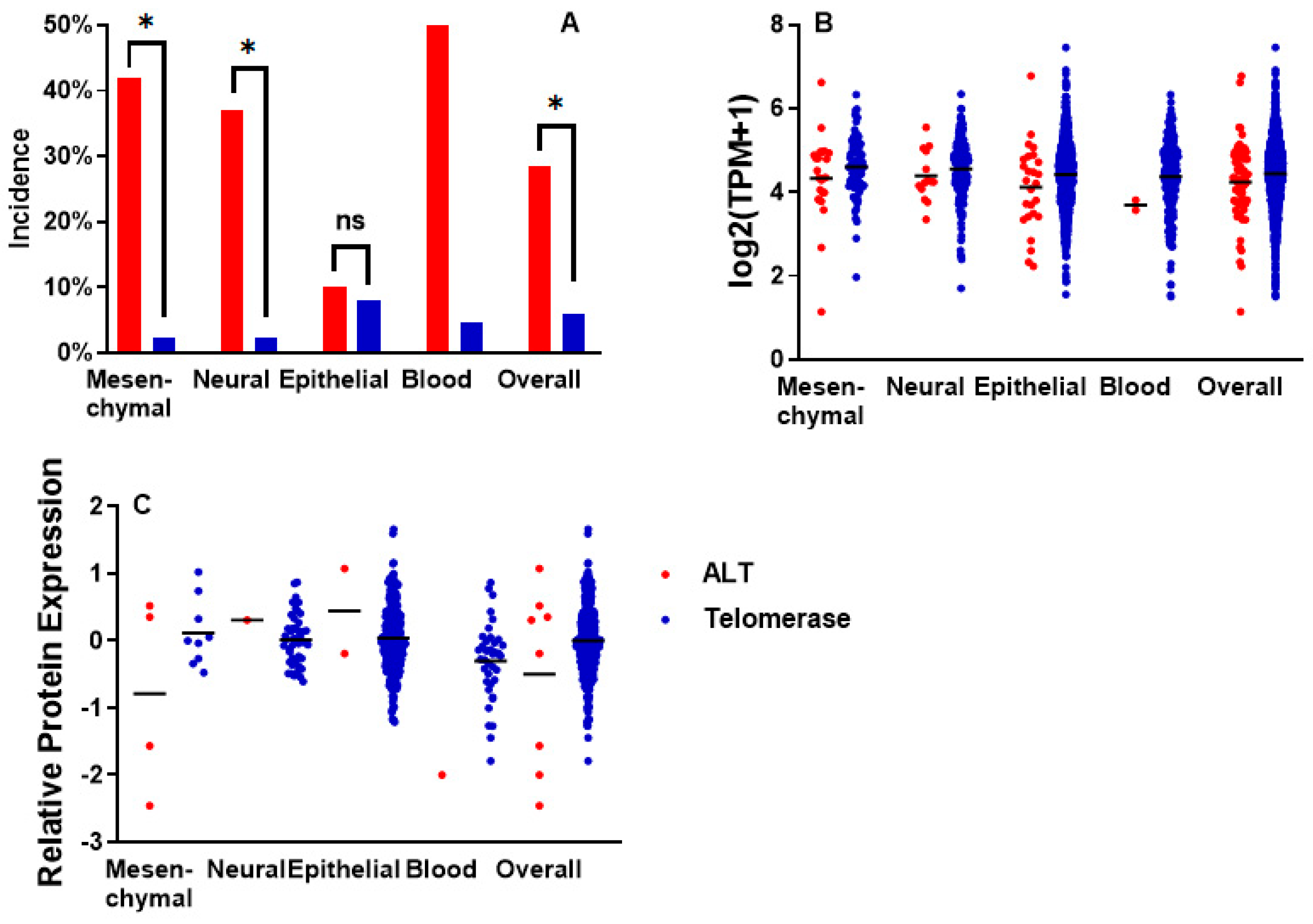
The incidence of ATRX alterations (which include damaging mutations, copy number loss, and structural variations, Table S3 in Supplementary Materials) was significantly higher (p < 0.0001) in ALT cells compared with telomerase-positive ones (Figure 2A). In particular, this was true for those lines of mesenchymal and neural origin (p < 0.0001, both), but not for the epithelial ones (p = 0.67). Also, for blood cell lines, the difference was significant (p = 0.004), but only 2 of them were ALT lines in this group.
In every group, ATRX RNA expression was lower in ALT cell lines compared with telomerase-positive ones (Figure 2B). The difference was not significant in mesenchymal (p = 0.14), neural (p = 0.39), epithelial (p = 0.06) and blood cell lines (p = 0.28). It was significant only in merging all cell lines, independently of their origin (p = 0.01).
Data for ATRX protein expression were much scarcer (Figure 2C). Among mesenchymal cell lines, the mean of the 4 ALT cell lines was lower, but not significantly (p = 0.11), than the telomerase ones. Among neural cell lines, the only ALT cell line available showed a value higher than the mean of the telomerase ones. Considering all cell lines, independently of their origin, the value for ALT cell lines was lower, but not significantly (p = 0.17), than the telomerase ones.

2.3. Incidence of Mutations in ALT Cell Lines

Besides ATRX, the most common altered genes are STAG2, WRN, SMARCAL1, POT1, BRCA2, SLF2, and SP100 (Figure 3, Table 1 and Table S3 in Supplementary Material), which all belong to pathways of telomeric integrity maintenance and are known repressors of ALT [15,16,17,18,19,20,21]. Also, HNRNPA1 showed damaging alterations in some cell lines: this protein allows RPA-to-POT1 switch on telomeric single-stranded DNA at the end of telomere replication [22], and its absence could prolong the presence of RPA (a key enzyme for the first steps of homologous recombination) and thus trigger telomeric recombination [23,24]. Mutations in genes implied in H3 histone deposition (DAXX, ASF1A, ASF1B, H3-3A) are much less common (and ASF1B showed no mutation at all).
Interestingly, RAD21, a cohesion involved in ALT-associated promyelocytic leukemia body [25], and RECQL4, a helicase involved in telomeric integrity maintenance [26], are often amplified in ALT cell lines (Figure 3). However, this may merely be due to the fact that they are neighbors of MYC (on the chromosome band 8q24), an oncogene often amplified both in ALT and telomerase-positive tumors. We also observed amplifications of TOP3A, a gene involved in fork stall resolution [27] and whose amplification has been proposed to be associated with ALT and mutually exclusive with ATRX mutations [28] (see below).
Other frequently altered genes are RB1 and MYC (a well-known oncosuppressor and oncogene, respectively), NF1 (known for being often mutated in neural tumors), and PAXX (often fused with FOXO1 in alveolar rhabdomyosarcomas).
Finally, the p53 pathway is altered in almost all ALT lines (89%), either by TP53 mutation or MDM2, MDM4, PPM1D amplification, confirming previous observations [29,30]. U2OS cell line (which is TP53 wild type) has been previously reported as p53 proficient or deficient, depending on the results of MDM2 copy number evaluation [31,32]. However, Kleiblova et al. [33] finally identified a gain-of-function mutation of PPM1D in U2OS that impairs the p53 pathway. We also discovered that the HUO9 cell line harbors this same mutation.
Although it is not one of the main aims of this research, we noted a difference (which may interest the reader) in the types of TP53 alterations between ALT and telomerase-positive cell lines: whereas the first ones show both point mutations and gene deletions, the latter ones show only point mutations. We explain this with the fact that the TP53 gene overlaps (partially and antisense) with the WRAP53 gene (better known as TCAB1): being the protein encoded by the latter essential for the assembly of the telomerase enzyme, a loss of this genomic region would be detrimental for telomerase-positive cells but not for ALT ones.
Analysis of mutual exclusivity and co-occurrence of gene alterations gave no significant results. We would mention that unadjusted p values were significant for mutual exclusivity between ATRX and POT1 (p = 0.048), ATRX and BRCA2 (p = 0.048), WRN and STAG2 (p = 0.049); however, also in these cases, False Discovery Rate analysis gave no significant results (q = 0.75 each). Since de Nonneville et al. [28] observed mutual exclusivity between ATRX mutations and TOP3A amplification in osteosarcoma, we tested this hypothesis in our sample. Among the 10 ALT osteosarcoma cell lines, 5 harbored ATRX alterations and no TOP3A amplification, 2 harbored no ATRX alteration and TOP3A amplification, and 3 neither of the 2; statistical analysis, however, gave a non-significant result (p = 0.44). Looking at TOP3A expression (Figure S1 in Supplementary Material), ATRX-wild type, TOP3A-amplified cells showed the highest values, and the difference with ATRX-deficient, TOP3A-wild type ones was significant (p = 0.001). However, the difference between ATRX-wild type cells (both TOP3A-amplified and wild-type) and ATRX-deficient, TOP3A-wild type ones was not significant (p = 0.81). This seems to contradict the hypothesis that in osteosarcoma, either ATRX mutations or TOP3A overexpression are needed for ALT.
We previously confirmed that ATRX is the most frequently altered gene in ALT cells (Figure 3). Its alteration can be viewed as a defect in the H3-3 histone deposition pathway, leading to telomeric euchromatinization and thus ALT establishment, as proposed in the past [8]. However, other members of this pathway (DAXX, ASF1A, ASF1B, H3-3A) are rarely altered in our ALT cell line panel (Figure 3). In particular, the percentage of cell lines with one of these genes being altered is significantly higher in ALT compared with telomerase-positive cells (Figure 4A), if we consider mesenchymal and neural lines (p < 0.0001, both), but not in epithelial ones (p = 0.46).
ATRX absence, on the other hand, can be viewed as the cause of defects in telomeric fork resolution, cohesion, and unwinding (which, also in this case, can lead to ALT). The percentage of cell lines with alterations in genes belonging to this pathway (Figure 4B) is significantly higher in ALT cells compared with telomerase-positive ones; this is true for mesenchymal, neural, and also epithelial lines (p < 0.0001, for all).

3. Discussion

Since ALT is much more common in mesenchymal and neural tumors [11,12], research on the role of ATRX has been conducted almost solely in these tumor types. In this study, we identified a wider panel of ALT-positive cell lines, including carcinomas, and we estimated the incidence of ATRX-damaging mutations among them. Our method may have given some false negatives (i.e., we have excluded some true ALT-positive cell lines), but we are sure to have not included false negatives (i.e., ALT-negative identified as ALT-positive). When we began this study, the TMMs of RH4, HS729, and TM31 were unknown, and we, using the protocol presented above, predicted that they were ALT-positive (Figure 3). Before we finished writing this article, we experimentally demonstrated that RH4 was ALT-positive [34]. Moreover, HS729 and TM31 were confirmed as ALT positive [35] (M. Zimmermann personal communication), giving us confidence in the robustness of our method. Also, the similarity between our results and the incidence of ALT in tumor samples [11,12,14] is encouraging.
Data on ATRX mRNA expression seemed to show a trend for lower values in ALT cell lines, but this is not statistically significant. Moreover, many ATRX-deficient ALT cell lines showed high values. This is because these cell lines may transcribe mRNA from this gene, although this mRNA will not be translated into a functional protein. Therefore, protein expression data would be much more informative for the investigation of ATRX presence in the different cell lines. Unfortunately, protein expression data are very scarce (Figure 2). Therefore, the best way to study the ATRX status of the different cell lines is to investigate the presence of gene mutations and alterations. We have shown that ATRX is the most altered gene in ALT cell lines with a mesenchymal or neural origin but not among those with an epithelial one (Figure 2A and Figure 3).
The first (and still popular) hypothesis linking ATRX loss and ALT is that without ATRX-mediated deposition of H3.3 histone, telomeres become euchromatic and prone to recombination [8]. It should be added, however, that the actual euchromatic/heterochromatic status of telomeres is still not clear [36]. Following this hypothesis, replication stress at telomeres would be a consequence, or by-product, of the absence of ATRX and its helicase activity. Other authors [37], instead, consider the telomeric replication stress (caused by ATRX inactivation) as the cause of ALT: this stress, in fact, consists of the stall and collapse of the replication fork, which activates HR at telomeres. If ALT is triggered by loss of telomeric H3.3 histone, we should find mutations not only in ATRX but also in other genes involved in this pathway. Our results, however, show that these (including mutations of H3-3A) are very rare among ALT-positive cell lines (Figure 4A). It could be objected that this may be due to the lethality of this kind of mutation. However, they are present in other types of tumors and, in particular, H3-3A mutations have a high incidence among giant cell tumors of bone [38], and so far, all the tumors of this type analyzed have active telomerase [39]. On the other hand, the incidence of loss of proteins involved in fork stall resolving (SMARCAL1, WRN, SLF2, BRCA2) and repression of recombination (STAG2, POT1, SP100, Figure 4B) makes us believe that ALT is triggered by replication stress at telomeres rather than the absence of H3.3 deposition. For clarity, we must add that knocking down of SMARCAL1 [21], WRN [40], SLF2 [21], BRCA2 [19], STAG2 [18], POT1 [16], and SP100 [15] have been shown to induce activation of ALT or, at least, of some of its markers (C-Circles, telomeric recombination, etc.).
However, even if one disagrees with us (and thus believes that ATRX loss triggers ALT because H3.3 deposition is hampered), it should be noted that ATRX loss is peculiar to mesenchymal and neural ALT cell lines and almost absent in epithelial ones; thus, loss of H3.3 deposition would be a mechanism for ALT establishment only in mesenchymal and neural cells and not a universal feature.
Also, in vitro, ATRX loss increases the proportion of cell lines activating ALT in p53-defective fibroblasts but not in p53-defective epithelial cells [41].
From our results, it seems that for ALT establishment, a defect in the telomeric fork repair pathway is needed and that ATRX alterations are specifically enriched in mesenchymal and neural ALT cell lines. This could be due to other functions exerted by ATRX specifically in mesenchymal and neural (but not in epithelial) cells that need to be hampered. In addition to its telomeric role, ATRX is also involved in regulating the expression of imprinted genes [42]. ATRX protein is highly enriched at all known imprinting control regions [43,44] and in the absence of Atrx, H19 and Igf2 expression are increased in mouse brain [43,44,45]. Atrx-null mice also show the downregulation of Neuroligin 4 and other enzymes important for neuronal differentiation [45], and, in general, ATRX loss impairs neurodifferentiation [46]. Although the nervous system has attracted most of the attention, some studies investigated the effects of ATRX inactivation on muscle cells and osteoblasts, showing an inability to differentiate [47,48]. Thus, ATRX loss seems to induce replication stress and impaired differentiation in neural and mesenchymal cells, giving at once two steps in the process of ALT tumorigenesis. Is ATRX inactivation dispensable for epithelial differentiation? Answering this question would help elucidate differences in ATRX loss incidence between ALT epithelial and mesenchymal/neural cells. Similar hypotheses can be also drawn for SMARCAL1 (which is often lost in ALT glioblastomas and is mutually exclusive with ATRX). In fact, besides its well-known role as a helicase responsible for telomeric integrity maintenance, it has recently been shown that its loss impairs differentiation [49].
In addition to altering the expression of genes driving differentiation, ATRX loss (as said before) impairs the expression of imprinted genes and, for example, increases the levels of H19 and IGF2 [45]. IGF2 is a growth factor [50], and H19 is a non-coding RNA essential for tumorigenesis [51] that is reactivated in most tumors [52]. Moreover, H19 is upregulated by oncogenes, such as c-Myc [52]. One study showed that H19 and IGF2 are downregulated during fibroblast senescence and that reactivation of H19 upregulates IGF2, increasing proliferation and avoiding senescence [53]. On the other hand, in prostate epithelial and urothelial cells, aging is associated with loss of imprinting of IGF2, leading to increased expression [54]. Thus, it is tempting to put forward this hypothesis: (a) ALT cells (which usually lack alterations in genes such as Myc) need reactivation of factors like IGF2 or H19 in order to bypass senescence and/or differentiation; (b) ATRX loss is needed in mesenchymal and neural cells in order to reactivate these imprinted genes; (c) ATRX loss is not needed in epithelial cells because they reactivate IGF2 during senescence. Of course, there are few studies to support this speculation, but we think that this issue (i.e., loss of imprinting due to ATRX mutations in different tissues) deserves further research.
In conclusion, viewing ALT establishment as a “multi-hit” process, we hypothesized the need for inactivation of the p53 pathway (in order to cope with replication and telomeric stress [55,56]), of the “telomeric integrity maintenance” pathway (causing de-repression of telomeric recombination) and of other ones (yet to be discovered) that may be lineage specific. As in mesenchymal and neural cells, one of these unknown pathways may rely on ATRX, the inactivation of this gene “kills two birds with one stone” (de-repression of telomeric recombination and lineage-specific pathway) in these lineages but gives only one “hit” in epithelial cells (de-repression of telomeric recombination), explaining the higher incidence of ATRX inactivation in the former (Figure 5). As we have helped to identify more ALT epithelial cell lines, our last recommendation is to perform more studies on these in order to have a clearer understanding of the ALT phenomenon.

4. Material and Methods

Cell lines present in the DepMap portal (https://depmap.org/portal, accessed on 9 July 2025), version 2023Q2, were classified, using literature data, according to their TMM, i.e., as ALT, or telomerase-positive, or without TMM. Those with no data in the literature were classified as unknown. The criterion for classifying a cell line as telomerase-positive was the presence of measured telomerase activity in the literature. The criterion for classifying a cell line as ALT was the absence of telomerase activity and the presence of ALT markers (long and heterogeneous telomeres, C-circles, ALT-associated PML Bodies). Three cell lines are reported in the literature as cancerous but with no TMM (i.e., telomerase- and ALT-negative). Thirty-eight cell lines are reported as fibroblasts in the DepMap portal; these were used just for comparison of TERT expression and telomere content values. Cell lines classified as unknown were subsequently subdivided on the basis of their TERT expression values (downloaded from the DepMap portal): the lowest value of TERT expression shown by known telomerase-positive cell lines was chosen as a threshold, and those cell lines with values above it were classified as “telomerase putative”. Vice versa, cell lines with values below this threshold were classified as “telomerase-negative”. The latter were further scrutinized for telomere length using data of “telomere content” obtained with TelSeq [57] and present in the DepMap portal [58]. Those cell lines with telomere content value below −1 (corresponding to 5 kb) were interpreted as having short telomeres and thus discarded. “Telomerase-negative” cell lines with telomere content value above −1 were classified as “ALT putative”.
ATRX RNA and protein expression and mutation data were downloaded from the DepMap portal. A gene was considered to have a damaging alteration if it had one or more of the following: nonsense mutation, out-of-frame insertion or deletion, splice site mutation, fusion, or copy number loss. Missense mutations were not considered as damaging unless they are reported as oncogenic in the literature or databanks.
Comparison of (log-transformed) expression data was performed using Student’s t test. Comparison of incidence data was performed using the Chi-squared test. In order to test mutual exclusivity or co-occurrence of gene alterations, we used a one-sided Fisher’s Exact Test. We also used the Log2 Odds Ratio to assess the direction of association, where negative values indicate trends towards mutual exclusivity and positive values indicate trends toward co-occurrence. To adjust for multiple hypotheses across multiple gene pairs, we used the Benjamini–Hochberg False Discovery Rate correction procedure.

Supplementary Materials

The following supporting information can be downloaded at: https://www.mdpi.com/article/10.3390/ijms26146765/s1. References [59,60,61,62,63,64,65,66,67,68,69,70,71,72,73,74,75,76,77,78,79,80,81,82,83,84,85,86,87,88,89,90,91,92,93,94,95,96,97,98] are cited in the supplementary materials.

Author Contributions

I.U.: writing—original draft, methodology, investigation, formal analysis, data curation, writing—review and editing. A.S.: writing—review and editing. All authors have read and agreed to the published version of the manuscript.

Funding

This research received no external funding.

Institutional Review Board Statement

Not applicable.

Informed Consent Statement

Not applicable.

Data Availability Statement

Original data were retrieved from the DepMap portal (https://depmap.org/portal, accessed on 9 July 2025). All data used in this article are available in the Supplementary Material. This paper does not report original code.

Conflicts of Interest

The authors declare no conflicts of interest.

References

  1. Yuan, X.; Larsson, C.; Xu, D. Mechanisms underlying the activation of TERT transcription and telomerase activity in human cancer: Old actors and new players. Oncogene 2019, 38, 6172–6183. [Google Scholar] [CrossRef] [PubMed]
  2. O’Sullivan, R.J.; Greenberg, R.A. Mechanisms of Alternative Lengthening of Telomeres. Cold Spring Harb. Perspect. Biol. 2025, 17, a041690. [Google Scholar] [CrossRef] [PubMed]
  3. Clatterbuck Soper, S.F.; Meltzer, P.S. ATRX/DAXX: Guarding the Genome against the Hazards of ALT. Genes 2023, 14, 790. [Google Scholar] [CrossRef] [PubMed]
  4. Xu, F.; Yu, D.; Guo, J.; Hu, J.; Zhao, Y.; Jiang, C.; Meng, X.; Cai, J.; Zhao, Y. From pathology to therapy: A comprehensive review of ATRX mutation related molecular functions and disorders. Mutat. Res. Rev. Mutat. Res. 2025, 795, 108537. [Google Scholar] [CrossRef] [PubMed]
  5. Lewis, P.W.; Elsaesser, S.J.; Noh, K.M.; Stadler, S.C.; Allis, C.D. Daxx is an H3.3-specific histone chaperone and cooperates with ATRX in replication-independent chromatin assembly at telomeres. Proc. Natl. Acad. Sci. USA 2010, 107, 14075–14080. [Google Scholar] [CrossRef] [PubMed]
  6. Kovatcheva, M.; Liao, W.; Klein, M.E.; Robine, N.; Geiger, H.; Crago, A.M.; Dickson, M.A.; Tap, W.D.; Singer, S.; Koff, A. ATRX is a regulator of therapy induced senescence in human cells. Nat. Commun. 2017, 8, 386. [Google Scholar] [CrossRef] [PubMed]
  7. Leung, J.W.C.; Ghosal, G.; Wang, W.; Shen, X.; Wang, J.; Li, L.; Chen, J. Alpha thalassemia/mental retardation syndrome X-linked gene product ATRX is required for proper replication restart and cellular resistance to replication stress. J. Biol. Chem. 2013, 288, 6342–6350. [Google Scholar] [CrossRef] [PubMed]
  8. Blasco, M.A. The epigenetic regulation of mammalian telomeres. Nat. Rev. Genet. 2007, 8, 299–309. [Google Scholar] [CrossRef] [PubMed]
  9. Udroiu, I.; Marinaccio, J.; Sgura, A. Effects of p53 and ATRX inhibition on telomeric recombination in aging fibroblasts. Front. Oncol. 2024, 14, 1322438. [Google Scholar] [CrossRef] [PubMed]
  10. Lovejoy, C.A.; Li, W.; Reisenweber, S.; Thongthip, S.; Bruno, J.; de Lange, T.; De, S.; Petrini, J.H.J.; Sung, P.A.; Jasin, M.; et al. Loss of ATRX, genome instability, and an altered DNA damage response are hallmarks of the alternative lengthening of telomeres pathway. PLoS Genet. 2012, 8, e1002772. [Google Scholar] [CrossRef] [PubMed]
  11. Bryan, T.M.; Englezou, A.; Dalla-Pozza, L.; Dunham, M.A.; Reddel, R.R. Evidence for an alternative mechanism for maintaining telomere length in human tumors and tumor-derived cell lines. Nat. Med. 1997, 3, 1271–1274. [Google Scholar] [CrossRef] [PubMed]
  12. Heaphy, C.M.; Subhawong, A.P.; Hong, S.M.; Goggins, M.G.; Montgomery, E.A.; Gabrielson, E.; Netto, G.J.; Epstein, J.I.; Lotan, T.L.; Westra, W.H.; et al. Prevalence of the alternative lengthening of telomeres telomere maintenance mechanism in human cancer subtypes. Am. J. Pathol. 2011, 179, 1608–1615. [Google Scholar] [CrossRef] [PubMed]
  13. Tsherniak, A.; Vazquez, F.; Montgomery, P.G.; Weir, B.A.; Kryukov, G.; Cowley, G.S.; Gill, S.; Harrington, W.F.; Pantel, S.; Krill-Burger, J.M.; et al. Defining a Cancer Dependency Map. Cell 2017, 170, 564–576. [Google Scholar] [CrossRef] [PubMed]
  14. MacKenzie, D., Jr.; Watters, A.K.; To, J.T.; Young, M.W.; Muratori, J.; Wilkoff, M.H.; Abraham, R.G.; Plummer, M.M.; Zhang, D. ALT Positivity in Human Cancers: Prevalence and Clinical Insights. Cancers 2021, 13, 2384. [Google Scholar] [CrossRef] [PubMed]
  15. Jiang, W.Q.; Zhong, Z.H.; Henson, J.D.; Neumann, A.A.; Chang, A.C.M.; Reddel, R.R. Suppression of alternative lengthening of telomeres by Sp100-mediated sequestration of the MRE11/RAD50/NBS1 complex. Mol. Cell. Biol. 2005, 25, 2708–2721. [Google Scholar] [CrossRef] [PubMed]
  16. Wu, L.; Multani, A.S.; He, H.; Cosme-Blanco, W.; Deng, Y.; Deng, J.M.; Bachilo, O.; Pathak, S.; Tahara, H.; Bailey, S.M.; et al. Pot1 deficiency initiates DNA damage checkpoint activation and aberrant homologous recombination at telomeres. Cell 2006, 126, 49–62. [Google Scholar] [CrossRef] [PubMed]
  17. Sugimoto, M.; Furuichi, Y.; Ide, T.; Goto, M. Involvement of WRN helicase in immortalization and tumorigenesis by the telomeric crisis pathway. Oncol. Lett. 2011, 2, 609–611. [Google Scholar] [CrossRef] [PubMed]
  18. Daniloski, Z.; Smith, S. Loss of tumor suppressor STAG2 promotes telomere recombination and extends the replicative lifespan of normal human cells. Cancer Res. 2017, 77, 5530–5542. [Google Scholar] [CrossRef] [PubMed]
  19. Kwon, M.; Lee, J.J.; Min, J.; Hwang, K.; Park, S.G.; Kim, E.H.; Kim, B.C.; Bhak, J.; Lee, H. Brca2 abrogation engages with the alternative lengthening of telomeres via break-induced replication. FEBS J. 2019, 286, 1841–1858. [Google Scholar] [CrossRef] [PubMed]
  20. Scott, W.A.; Dhanji, E.Z.; Dyakov, B.J.A.; Dreseris, E.S.; Asa, J.S.; Grange, L.J.; Mirceta, M.; Pearson, C.E.; Stewart, G.S.; Gingras, A.-C.; et al. ATRX proximal protein associations boast roles beyond histone deposition. PLoS Genet. 2021, 17, e1009909. [Google Scholar] [CrossRef] [PubMed]
  21. Liu, H.; Xu, C.; Diplas, B.H.; Brown, A.; Strickland, L.M.; Yao, H.; Ling, J.; E McLendon, R.; Keir, S.T.; Ashley, D.M.; et al. Cancer-associated SMARCAL1 loss-of-function mutations promote alternative lengthening of telomeres and tumorigenesis in telomerase-negative glioblastoma cells. Neuro-Oncol. 2023, 25, 1563–1575. [Google Scholar] [CrossRef] [PubMed]
  22. Flynn, R.L.; Centore, R.C.; O’sUllivan, R.J.; Rai, R.; Tse, A.; Songyang, Z.; Chang, S.; Karlseder, J.; Zou, L. TERRA and hnRNPA1 orchestrate an RPA-to-POT1 switch on telomeric single-stranded DNA. Nature 2011, 471, 532–536. [Google Scholar] [CrossRef] [PubMed]
  23. Raghunandan, M.; Geelen, D.; Majerova, E.; Decottignies, A. NHP2 downregulation counteracts hTR-mediated activation of the DNA damage response at ALT telomeres. EMBO J. 2021, 40, e106336. [Google Scholar] [CrossRef] [PubMed]
  24. Udroiu, I.; Marinaccio, J.; Sgura, A. Many functions of telomerase components: Certainties, doubts, and inconsistencies. Int. J. Mol. Sci. 2022, 23, 15189. [Google Scholar] [CrossRef] [PubMed]
  25. Zhao, B.; Wang, Z.J.; Yi, B.Q.; Ma, H.C.; Xu, H.M. hRad21 overexpresses and localizes to the ALT-associated promyelocytic leukemia body in ALT cells. Cancer Biol. Ther. 2010, 9, 978–983. [Google Scholar] [CrossRef] [PubMed]
  26. Ghosh, A.K.; Rossi, M.L.; Singh, D.K.; Dunn, C.; Ramamoorthy, M.; Croteau, D.L.; Liu, Y.; Bohr, V.A. RECQL4, the protein mutated in Rothmund-Thomson syndrome, functions in telomere maintenance. J. Biol. Chem. 2012, 287, 196–209. [Google Scholar] [CrossRef] [PubMed]
  27. Lu, R.; O’Rourke, J.J.; Sobinoff, A.P.; Allen, J.A.M.; Nelson, C.B.; Tomlinson, C.G.; Lee, M.; Reddel, R.R.; Deans, A.J.; Pickett, H.A. The FANCM-BLM-TOP3A-RMI complex suppresses alternative lengthening of telomeres (ALT). Nat. Commun. 2019, 10, 2252. [Google Scholar] [CrossRef] [PubMed]
  28. de Nonneville, A.; Salas, S.; Bertucci, F.; Sobinoff, A.P.; Adélaïde, J.; Guille, A.; Finetti, P.; Noble, J.R.; Churikov, D.; Chaffanet, M.; et al. TOP3A amplification and ATRX inactivation are mutually exclusive events in pediatric osteosarcomas using ALT. EMBO Mol. Med. 2022, 14, e15859. [Google Scholar] [CrossRef] [PubMed]
  29. Henson, J.D.; Reddel, R.R. Assaying and investigating Alternative Lengthening of Telomeres activity in human cells and cancers. FEBS Lett. 2010, 584, 3800–3811. [Google Scholar] [CrossRef] [PubMed]
  30. Farooqi, A.S.; Dagg, R.A.; Choi, L.M.R.; Shay, J.W.; Reynolds, C.P.; Lau, L.M. Alternative lengthening of telomeres in neuroblastoma cell lines is associated with a lack of MYCN genomic amplification and with p53 pathway aberrations. J. Neuro-Oncol. 2014, 119, 17–26. [Google Scholar] [CrossRef] [PubMed]
  31. Flørenes, V.A.; Mælandsmo, G.M.; Forus, A.; Andreassen, Å.; Myklebost, O.; Fodstad, Ø. MDM2 gene amplification and transcript levels in human sarcomas: Relationship to TP53 gene status. JNCI J. Natl. Cancer Inst. 1994, 86, 1297–1302. [Google Scholar] [CrossRef] [PubMed]
  32. Landers, J.E.; Cassel, S.L.; George, D.L. Translational enhancement of mdm2 oncogene expression in human tumor cells containing a stabilized wild-type p53 protein. Cancer Res. 1997, 57, 3562–3568. [Google Scholar] [PubMed]
  33. Kleiblova, P.; Shaltiel, I.A.; Benada, J.; Sevcik, J.; Pecháčková, S.; Pohlreich, P.; Voest, E.E.; Dundr, P.; Bartek, J.; Kleibl, Z.; et al. Gain-of-function mutations of PPM1D/Wip1 impair the p53-dependent G1 checkpoint. J. Cell Biol. 2013, 201, 511–521. [Google Scholar] [CrossRef] [PubMed]
  34. Udroiu, I.; Marinaccio, J.; Goffi, R.S.; Micheli, E.; Sgura, A. Specificity and sensitivity of ALT-associated markers in cancer cells. FEBS Lett. 2025, 599, 989–1005. [Google Scholar] [CrossRef] [PubMed]
  35. Zimmermann, M.; Bernier, C.; Kaiser, B.; Fournier, S.; Li, L.; Desjardins, J.; Skeldon, A.; Rimkunas, V.; Veloso, A.; Young, J.T.; et al. Guiding ATR and PARP inhibitor combinations with chemogenomic screens. Cell Rep. 2022, 40, 111081. [Google Scholar] [CrossRef] [PubMed]
  36. Udroiu, I.; Sgura, A. Alternative lengthening of telomeres and chromatin status. Genes 2019, 11, 45. [Google Scholar] [CrossRef] [PubMed]
  37. Hoang, S.M.; O’Sullivan, R.J. Alternative Lengthening of Telomeres: Building bridges to connect chromosome ends. Trends Cancer 2020, 6, 247–260. [Google Scholar] [CrossRef] [PubMed]
  38. Behjati, S.; Tarpey, P.S.; Presneau, N.; Scheipl, S.; Pillay, N.; Van Loo, P.; Wedge, D.C.; Cooke, S.L.; Gundem, G.; Davies, H.; et al. Distinct H3F3A and H3F3B driver mutations define chondroblastoma and giant cell tumor of bone. Nat. Genet. 2013, 45, 1479–1482. [Google Scholar] [CrossRef] [PubMed]
  39. Forsyth, R.G.; De Boeck, G.; Bekaert, S.; De Meyer, T.; Taminiau, A.H.; Uyttendaele, D.; Roels, H.; Praet, M.M.; Hogendoorn, P.C. Telomere biology in giant cell tumour of bone. J. Pathol. 2008, 214, 555–563. [Google Scholar] [CrossRef] [PubMed]
  40. Laud, P.R.; Multani, A.S.; Bailey, S.M.; Wu, L.; Ma, J.; Kingsley, C.; Lebel, M.; Pathak, S.; DePinho, R.A.; Chang, S. Elevated telomere-telomere recombination in WRN-deficient, telomere dysfunctional cells promotes escape from senescence and engagement of the ALT pathway. Genes Dev. 2005, 19, 2560–2570. [Google Scholar] [CrossRef] [PubMed]
  41. Napier, C.E.; Huschtscha, L.I.; Harvey, A.; Bower, K.; Noble, J.R.; Hendrickson, E.A.; Reddel, R.R. ATRX represses alternative lengthening of telomeres. Oncotarget 2015, 6, 16543. [Google Scholar] [CrossRef] [PubMed]
  42. Bérubé, N.G. ATRX in chromatin assembly and genome architecture during development and disease. Biochem. Cell Biol. 2011, 89, 435–444. [Google Scholar] [CrossRef] [PubMed]
  43. Kernohan, K.D.; Jiang, Y.; Tremblay, D.C.; Bonvissuto, A.C.; Eubanks, J.H.; Mann, M.R.; Bérubé, N.G. ATRX partners with cohesin and MeCP2 and contributes to developmental silencing of imprinted genes in the brain. Dev. Cell 2010, 18, 191–202. [Google Scholar] [CrossRef] [PubMed]
  44. Voon, H.P.; Hughes, J.R.; Rode, C.; De La Rosa-Velazquez, I.A.; Jenuwein, T.; Feil, R.; Higgs, D.R.; Gibbons, R.J. ATRX plays a key role in maintaining silencing at interstitial heterochromatic loci and imprinted genes. Cell Rep. 2015, 11, 405–418. [Google Scholar] [CrossRef] [PubMed]
  45. Kernohan, K.D.; Vernimmen, D.; Gloor, G.B.; Bérubé, N.G. Analysis of neonatal brain lacking ATRX or MeCP2 reveals changes in nucleosome density, CTCF binding and chromatin looping. Nucleic Acids Res. 2014, 42, 8356–8368. [Google Scholar] [CrossRef] [PubMed]
  46. Bieluszewska, A.; Wulfridge, P.; Doherty, J.; Ren, W.; Sarma, K. ATRX histone binding and helicase activities have distinct roles in neuronal differentiation. Nucleic Acids Res. 2022, 50, 9162–9174. [Google Scholar] [CrossRef] [PubMed]
  47. Huh, M.S.; O’dEa, T.P.; Ouazia, D.; McKay, B.C.; Parise, G.; Parks, R.J.; Rudnicki, M.A.; Picketts, D.J. Compromised genomic integrity impedes muscle growth after Atrx inactivation. J. Clin. Investig. 2012, 122, 4412–4423. [Google Scholar] [CrossRef] [PubMed]
  48. Chen, Y.-T.; Jiang, M.-M.; Leynes, C.; Adeyeye, M.; Majano, C.F.; Ibrahim, B.; Polak, U.; Hung, G.; Jin, Z.; Lanza, D.G.; et al. ATRX silences Cartpt expression in osteoblastic cells during skeletal development. J. Clin. Investig. 2025, 135, e163587. [Google Scholar] [CrossRef] [PubMed]
  49. Deguchi, K.; Clewing, J.M.; Elizondo, L.I.; Hirano, R.; Huang, C.; Choi, K.; Sloan, E.A.; Lücke, T.; Marwedel, K.M.; Powell, R.D.; et al. Neurologic phenotype of Schimke immuno-osseous dysplasia and neurodevelopmental expression of SMARCAL1. J. Neuropathol. Exp. Neurol. 2008, 67, 565–577. [Google Scholar] [CrossRef] [PubMed]
  50. Beatty, A.; Rubin, A.M.; Wada, H.; Heidinger, B.; Hood, W.R.; Schwartz, T.S. Postnatal expression of IGF2 is the norm in amniote vertebrates. Proc. R. Soc. B 2022, 289, 20212278. [Google Scholar] [CrossRef] [PubMed]
  51. Matouk, I.J.; DeGroot, N.; Mezan, S.; Ayesh, S.; Abu-lail, R.; Hochberg, A.; Galun, E. The H19 non-coding RNA is essential for human tumor growth. PLoS ONE 2007, 2, e845. [Google Scholar] [CrossRef] [PubMed]
  52. Barsyte-Lovejoy, D.; Lau, S.K.; Boutros, P.C.; Khosravi, F.; Jurisica, I.; Andrulis, I.L.; Tsao, M.S.; Penn, L.Z. The c-Myc oncogene directly induces the H19 noncoding RNA by allele-specific binding to potentiate tumorigenesis. Cancer Res. 2006, 66, 5330–5337. [Google Scholar] [CrossRef] [PubMed]
  53. Tang, H.; Yao, F.; Yin, M.; Liao, Y.; Li, K.; Li, L.; Xiao, X.; Guo, J.; Hu, F.; Feng, H. Anti-senescent effects of long non-coding RNA H19 on human dermal fibroblast cells through impairing microRNA-296-5p-dependent inhibition of IGF2. Cell. Signal. 2022, 94, 110327. [Google Scholar] [CrossRef] [PubMed]
  54. Fu, V.X.; Schwarze, S.R.; Kenowski, M.L.; LeBlanc, S.; Svaren, J.; Jarrard, D.F. A loss of insulin-like growth factor-2 imprinting is modulated by CCCTC-binding factor down-regulation at senescence in human epithelial cells. J. Biol. Chem. 2004, 279, 52218–52226. [Google Scholar] [CrossRef] [PubMed]
  55. Akter, J.; Katai, Y.; Sultana, P.; Takenobu, H.; Haruta, M.; Sugino, R.P.; Mukae, K.; Satoh, S.; Wada, T.; Ohira, M.; et al. Loss of p53 suppresses replication stress-induced DNA damage in ATRX-deficient neuroblastoma. Oncogenesis 2021, 10, 73. [Google Scholar] [CrossRef] [PubMed]
  56. Udroiu, I.; Marinaccio, J.; Sgura, A. Inhibition of p53 and ATRX increases telomeric recombination in primary fibroblasts. FEBS Open Bio 2023, 13, 1683–1698. [Google Scholar] [CrossRef] [PubMed]
  57. Ding, Z.; Mangino, M.; Aviv, A.; UK10KConsortium Spector, T.; Durbin, R. Estimating telomere length from whole genome sequence data. Nucleic Acids Res. 2014, 42, e75. [Google Scholar] [CrossRef] [PubMed]
  58. Hu, K.; Ghandi, M.; Huang, F.W. Integrated evaluation of telomerase activation and telomere maintenance across cancer cell lines. eLife 2021, 10, e66198. [Google Scholar] [CrossRef] [PubMed]
  59. Abid, H.Z.; McCaffrey, J.; Raseley, K.; Young, E.; Lassahn, K.; Varapula, D.; Riethman, H.; Xiao, M. Single-molecule analysis of subtelomeres and telomeres in Alternative Lengthening of Telomeres (ALT) cells. BMC Genom. 2020, 21, 485. [Google Scholar] [CrossRef] [PubMed]
  60. Alawi, F.; Lin, P.; Ziober, B.; Patel, R. Dyskerin expression correlates with active proliferation independently of telomerase. Head Neck 2010, 33, 1041. [Google Scholar] [CrossRef] [PubMed]
  61. Atri, S.; Nasoohi, N.; Hodjat, M. Azacitidine, as a DNMT inhibitor decreases hTERT gene expression and telomerase activity more effective compared with HDAC inhibitor in human head and neck squamous cell carcinoma cell lines. Curr. Mol. Pharmacol. 2021, 14, 60–67. [Google Scholar] [CrossRef] [PubMed]
  62. Binz, N.; Shalaby, T.; Rivera, P.; Shin-Ya, K.; Grotzer, M.A. Telomerase inhibition, telomere shortening, cell growth suppression and induction of apoptosis by telomestatin in childhood neuroblastoma cells. Eur. J. Cancer 2005, 41, 2873–2881. [Google Scholar] [CrossRef] [PubMed]
  63. Bojovic, B.; Crowe, D.L. Resistance to telomerase inhibition by human squamous cell carcinoma cell lines. Int. J. Oncol. 2011, 38, 1175–1181. [Google Scholar] [CrossRef] [PubMed]
  64. Bovée, J.V.; van den Broek, L.J.; Cleton-Jansen, A.M.; Hogendoorn, P.C. Chondrosarcoma is not characterized by detectable telomerase activity. J. Pathol. 2001, 193, 354–360. [Google Scholar] [CrossRef] [PubMed]
  65. Brosnan-Cashman, J.A.; Yuan, M.; Graham, M.K.; Rizzo, A.J.; Myers, K.M.; Davis, C.; Zhang, R.; Esopi, D.M.; Raabe, E.H.; Eberhart, C.G.; et al. ATRX loss induces multiple hallmarks of the alternative lengthening of telomeres (ALT) phenotype in human glioma cell lines in a cell line-specific manner. PLoS ONE 2018, 13, e0204159. [Google Scholar] [CrossRef] [PubMed]
  66. Counter, C.M.; Hahn, W.C.; Wei, W.; Caddle, S.D.; Beijersbergen, R.L.; Lansdorp, P.M.; Sedivy, J.M.; Weinberg, R.A. Dissociation among in vitro telomerase activity, telomere maintenance, and cellular immortalization. Proc. Natl. Acad. Sci. USA 1998, 95, 14723–14728. [Google Scholar] [CrossRef] [PubMed]
  67. Diplas, B.H.; He, X.; Brosnan-Cashman, J.A.; Liu, H.; Chen, L.H.; Wang, Z.; Moure, C.J.; Killela, P.J.; Loriaux, D.B.; Lipp, E.S.; et al. The genomic landscape of TERT promoter wildtype-IDH wildtype glioblastoma. Nat. Commun. 2018, 9, 2087. [Google Scholar] [CrossRef] [PubMed]
  68. Flynn, R.L.; Cox, K.E.; Jeitany, M.; Wakimoto, H.; Bryll, A.R.; Ganem, N.J.; Bersani, F.; Pineda, J.R.; Suvà, M.L.; Benes, C.H.; et al. Alternative lengthening of telomeres renders cancer cells hypersensitive to ATR inhibitors. Science 2015, 347, 273–277. [Google Scholar] [CrossRef] [PubMed]
  69. Fujiwara, M.; Kamma, H.; Wu, W.; Yano, Y.; Homma, S.; Satoh, H. Alternative lengthening of telomeres in the human adrenocortical carcinoma cell line H295R. Int. J. Oncol. 2006, 29, 445–451. [Google Scholar] [CrossRef] [PubMed]
  70. Glaessl, A.; Bosserhoff, A.K.; Buettner, R.; Hohenleutner, U.; Landthaler, M.; Stolz, W. Increase in telomerase activity during progression of melanocytic cells from melanocytic naevi to malignant melanomas. Arch. Dermatol. Res. 1999, 291, 81–87. [Google Scholar] [CrossRef] [PubMed]
  71. Gordon, K.E.; Ireland, H.; Roberts, M.; Steeghs, K.; McCaul, J.A.; MacDonald, D.G.; Parkinson, E.K. High levels of telomere dysfunction bestow a selective disadvantage during the progression of human oral squamous cell carcinoma. Cancer Res. 2003, 63, 458–467. [Google Scholar] [PubMed]
  72. Hou, M.; Xu, D.; Bjorkholm, M.; Gruber, A. Real-time quantitative telomeric repeat amplification protocol assay for the detection of telomerase activity. Clin. Chem. 2001, 47, 519–524. [Google Scholar] [CrossRef] [PubMed]
  73. Hu, Y.; Bobb, D.; Lu, Y.; He, J.; Dome, J.S. Effect of telomerase inhibition on preclinical models of malignant rhabdoid tumor. Cancer Genet. 2014, 207, 403–411. [Google Scholar] [CrossRef] [PubMed]
  74. Isaka, K.; Nishi, H.; Sagawa, Y.; Nakada, T.; Osakabe, Y.; Serizawa, H.; Ebihara, Y.; Takayama, M. Establishment of a new human cell line (EN) with TP53 mutation derived from endometrial carcinoma. Cancer Genet. Cytogenet. 2003, 141, 20–25. [Google Scholar] [CrossRef] [PubMed]
  75. Khaw, A.K.; Silasudjana, M.; Banerjee, B.; Suzuki, M.; Baskar, R.; Hande, M.P. Inhibition of telomerase activity and human telomerase reverse transcriptase gene expression by histone deacetylase inhibitor in human brain cancer cells. Mutat. Res./Fundam. Mol. Mech. Mutagen. 2007, 625, 134–144. [Google Scholar] [CrossRef] [PubMed]
  76. Kim, N.W.; Piatyszek, M.A.; Prowse, K.R.; Harley, C.B.; West, M.D.; Ho, P.L.C.; Coviello, G.M.; Wright, W.E.; Weinrich, S.L.; Shay, J.W. Specific association of human telomerase activity with immortal cells and cancer. Science 1994, 266, 2011–2015. [Google Scholar] [CrossRef] [PubMed]
  77. Kleideiter, E.; Schwab, M.; Friedrich, U.; Koscielniak, E.; Schäfer, B.W.; Klotz, U. Telomerase activity in cell lines of pediatric soft tissue sarcomas. Pediatr. Res. 2003, 54, 718–723. [Google Scholar] [CrossRef] [PubMed]
  78. Kyo, S.; Kanaya, T.; Ishikawa, H.; Ueno, H.; Inoue, M. Telomerase activity in gynecological tumors. Clin. Cancer Res. 1996, 2, 2023–2028. [Google Scholar] [PubMed]
  79. Lee, J.C.; Jong, H.S.; Yoo, C.G.; Han, S.K.; Shim, Y.S.; Kim, Y.W. Telomerase activity in lung cancer cell lines and tissues. Lung Cancer 1998, 21, 99–103. [Google Scholar] [CrossRef] [PubMed]
  80. Lin, Z.; Lim, S.; Viani, M.A.; Sapp, M.; Lim, M.S. Down-regulation of telomerase activity in malignant lymphomas by radiation and chemotherapeutic agents. Am. J. Pathol. 2001, 159, 711–719. [Google Scholar] [CrossRef] [PubMed]
  81. Ma, H.; Urquidi, V.; Wong, J.; Kleeman, J.; Goodison, S. Telomerase reverse transcriptase promoter regulation during myogenic differentiation of human RD rhabdomyosarcoma cells. Mol. Cancer Res. 2003, 1, 739–746. [Google Scholar] [PubMed]
  82. Maellaro, E.; Pacenti, L.; Del Bello, B.; Valentini, M.A.; Mangiavacchi, P.; De Felice, C.; Rubegni, P.; Luzi, P.; Miracco, C. Different effects of interferon-α on melanoma cell lines: A study on telomerase reverse transcriptase, telomerase activity and apoptosis. Br. J. Dermatol. 2003, 148, 1115–1124. [Google Scholar] [CrossRef] [PubMed]
  83. Mason-Osann, E.; Dai, A.; Floro, J.; Lock, Y.J.; Reiss, M.; Gali, H.; Matschulat, A.; Labadorf, A.; Flynn, R.L. Identification of a novel gene fusion in ALT positive osteosarcoma. Oncotarget 2018, 9, 32868–32880. [Google Scholar] [CrossRef] [PubMed]
  84. Milas, M.; Yu, D.; Sun, D.; Pollock, R.E. Telomerase activity of sarcoma cell lines and fibroblasts is independent of p53 status. Clin. Cancer Res. 1998, 4, 1573–1579. [Google Scholar] [PubMed]
  85. Mochida, A.; Gotoh, E.; Senpuku, H.; Harada, S.; Kitamura, R.; Takahashi, T.; Yanagi, K. Telomere size and telomerase activity in Epstein-Barr virus (EBV)-positive and EBV-negative Burkitt’s lymphoma cell lines. Arch. Virol. 2005, 150, 2139–2150. [Google Scholar] [CrossRef] [PubMed]
  86. Nishi, H.; Ohyashiki, K.; Fujito, A.; Yahata, N.; Ohyashiki, J.H.; Isaka, K.; Takayama, M. Expression of telomerase subunits and localization of telomerase activation in hydatidiform mole. Placenta 1999, 20, 317–323. [Google Scholar] [CrossRef] [PubMed]
  87. Panier, S.; Maric, M.; Hewitt, G.; Mason-Osann, E.; Gali, H.; Dai, A.; Labadorf, A.; Guervilly, J.-H.; Ruis, P.; Segura-Bayona, S.; et al. SLX4IP antagonizes promiscuous BLM activity during ALT maintenance. Mol. Cell 2019, 76, 27–43. [Google Scholar] [CrossRef] [PubMed]
  88. Parsch, D.; Brassat, U.; Brümmendorf, T.H.; Fellenberg, J. Consequences of telomerase inhibition by BIBR1532 on proliferation and chemosensitivity of chondrosarcoma cell lines. Cancer Investig. 2008, 26, 590–596. [Google Scholar] [CrossRef] [PubMed]
  89. Rha, S.Y.; Jeung, H.C.; Yang, W.I.; Kim, J.J.; Oh, T.J.; An, S.W.; Chung, H.C. Alteration of hTERT full-length variant expression level showed different gene expression profiles and genomic copy number changes in breast cancer. Oncol. Rep. 2006, 15, 749–755. [Google Scholar] [CrossRef] [PubMed]
  90. Saretzki, G.; Petersen, S.; Petersen, I.; Kölble, K.; von Zglinicki, T. hTERT gene dosage correlates with telomerase activity in human lung cancer cell lines. Cancer Lett. 2002, 176, 81–91. [Google Scholar] [CrossRef] [PubMed]
  91. Sato, N.; Maehara, N.; Mizumoto, K.; Nagai, E.; Yasoshima, T.; Hirata, K.; Tanaka, M. Telomerase activity of cultured human pancreatic carcinoma cell lines correlates with their potential for migration and invasion. Cancer 2001, 91, 496–504. [Google Scholar] [CrossRef] [PubMed]
  92. Shervington, A.; Patel, R.; Lu, C.; Cruickshanks, N.; Lea, R.; Roberts, G.; Dawson, T.; Shervington, L. Telomerase subunits expression variation between biopsy samples and cell lines derived from malignant glioma. Brain Res. 2007, 1134, 45–52. [Google Scholar] [CrossRef] [PubMed]
  93. Tahara, H.; Kuniyasu, H.; Yokozaki, H.; Yasui, W.; Shay, J.W.; Ide, T.; Tahara, E. Telomerase activity in preneoplastic and neoplastic gastric and colorectal lesions. Clin. Cancer Res. 1995, 1, 1245–1251. [Google Scholar] [PubMed]
  94. Terasaki, T.; Kyo, S.; Takakura, M.; Maida, Y.; Tsuchiya, H.; Tomita, K.; Inoue, M. Analysis of telomerase activity and telomere length in bone and soft tissue tumors. Oncol. Rep. 2004, 11, 1307–1311. [Google Scholar] [CrossRef] [PubMed]
  95. Ulaner, G.A.; Hoffman, A.R.; Otero, J.; Huang, H.Y.; Zhao, Z.; Mazumdar, M.; Gorlick, R.; Meyers, P.; Healey, J.H.; Ladanyi, M. Divergent patterns of telomere maintenance mechanisms among human sarcomas: Sharply contrasting prevalence of the alternative lengthening of telomeres mechanism in Ewing’s sarcomas and osteosarcomas. Genes Chromosomes Cancer 2004, 41, 155–162. [Google Scholar] [CrossRef] [PubMed]
  96. Villa, R.; Folini, M.; Perego, P.; Supino, R.; Setti, E.; Daidone, M.G.; Zunino, F.; Zaffaroni, N. Telomerase activity and telomere length in human ovarian cancer and melanoma cell lines: Correlation with sensitivity to DNA damaging agents. Int. J. Oncol. 2000, 16, 995–1002. [Google Scholar] [CrossRef] [PubMed]
  97. Wege, H.; Chui, M.S.; Le, H.T.; Tran, J.M.; Zern, M.A. SYBR Green real-time telomeric repeat amplification protocol for the rapid quantification of telomerase activity. Nucleic Acids Res. 2002, 31, e3. [Google Scholar] [CrossRef] [PubMed]
  98. Yu, E.Y.; Cheung, I.Y.; Feng, Y.; Rabie, M.O.; Roboz, G.J.; Guzman, M.L.; Cheung, N.-K.V.; Lue, N.F. Telomere trimming and DNA damage as signatures of high risk neuroblastoma. Neoplasia 2019, 21, 689–701. [Google Scholar] [CrossRef] [PubMed]
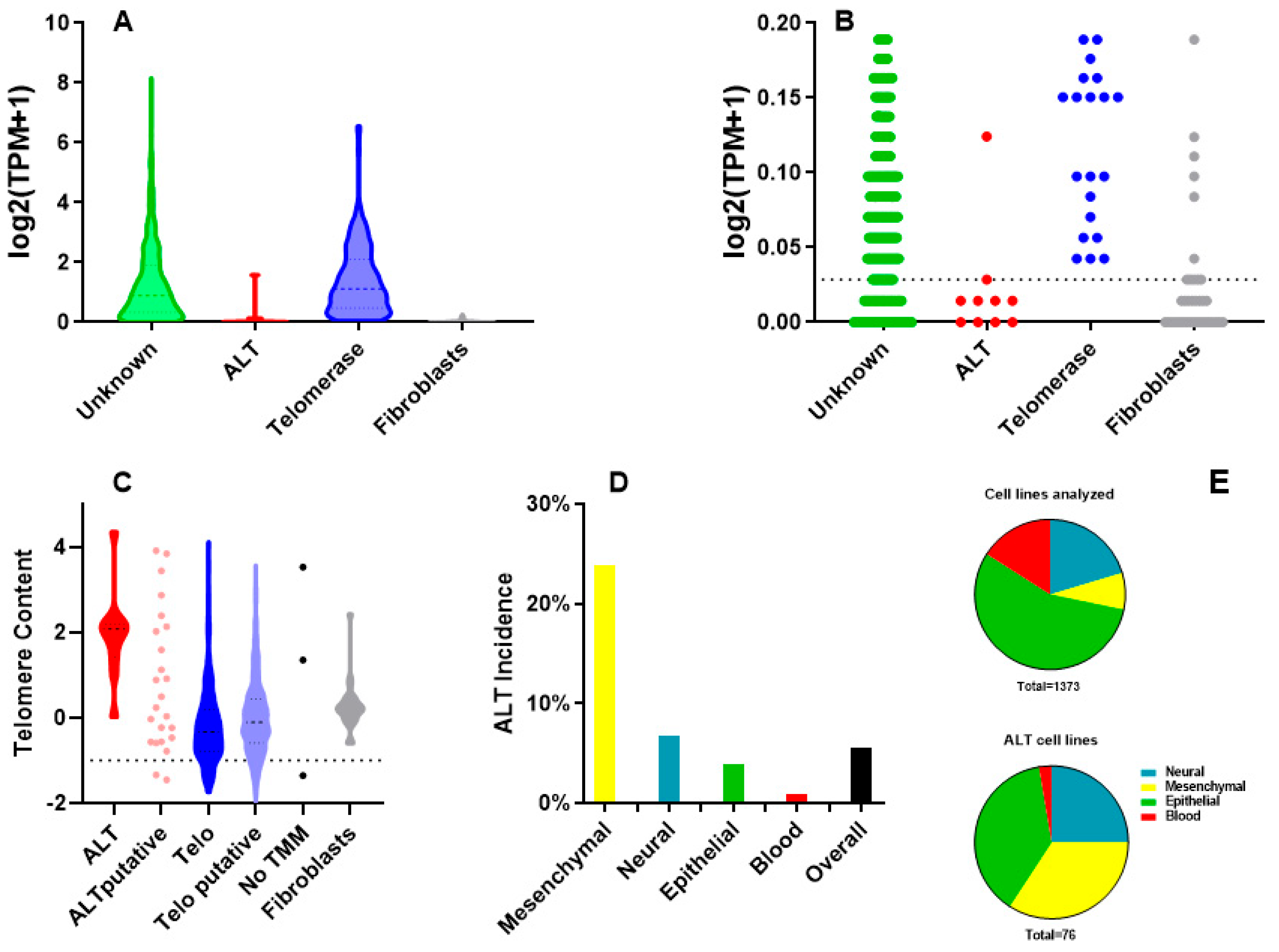
Figure 1. Identification of ALT cell lines. (A): TERT expression in cell lines with known and unknown telomere maintenance mechanisms (TMM). (B): Magnification of data in A; dotted line represents cut-off value for TERT expression: unknown cell lines with values above this threshold are classified as putative telomerase-positive, those below as putative ALT. (C): Telomere length evaluated by TelSeq; putative ALT cell lines with telomere content values below the threshold (shown as a dotted line) are discarded. (D): ALT incidence in different types of tumors. (E): percentage of types of tumors among all the cell lines analyzed (above) and among the ALT cell lines (below).
Ijms 26 06765 g001
Figure 2. ATRX alterations and expression. (A): incidence of ATRX damaging alterations (damaging mutations, copy loss and structural variations; data from 76 ALT-positive and 1299 telomerase-positive cell lines (statistical significance is not shown for blood cell lines, since only 2 are ALT-positive). (B): ATRX mRNA expression values (data from 66 ALT-positive and 1235 telomerase-positive cell lines). (C): ATRX protein expression values (data from 8 ALT-positive and 342 telomerase-positive cell lines). Asterisks represent significant differences, ns: not significant.
Ijms 26 06765 g002
Figure 3. Mutations in ALT cell lines.
Ijms 26 06765 g003
Figure 4. Incidence of damaging alterations of ALT repressor genes. (A): genes belonging to the H3.3 histone deposition pathway. (B): genes belonging to the telomeric integrity maintenance pathway. Asterisks represent significant differences, ns: not significant (statistical test was not performed on blood cell lines, since only 2 are ALT-positive). Incidence values are calculated from 76 ALT-positive and 1299 telomerase-positive cell lines.
Ijms 26 06765 g004
Figure 5. Hypothesis on the different roles of ATRX in mesenchymal and epithelial cells. Small arrows (←) indicate stimulation; blunt arrows (⊢) indicate inhibition.
Ijms 26 06765 g005
Table 1. Most frequent alterations in ALT cell lines. Note that ATRX belongs both to the telomeric integrity and the H3-3 deposition pathway.
PathwayGenesAlterations
ATRXFrequent damaging alterations in mesenchymal (42%) and neural cells (37%), much rarer in epithelial ones (10%)
Telomeric
integrity
WRNDamaging alterations in epithelial (24%), mesenchymal (12%) and neural cells (11%)
STAG2Damaging alterations in mesenchymal (15%) and neural cells (16%), much rarer in epithelial ones (3%)
SMARCAL1Damaging alterations in neural cells (13%), much rarer in mesenchymal cells (4%), absent in epithelial ones
H3-3
deposition
DAXX, ASF1A,
H3-3A
Quite rare damage mutations in mesenchymal and neural cells (<7%), none in epithelial ones
TOP3AAmplified in 11% of cell lines
Cell
proliferation
RB1Damaging alterations in 19% of cell lines
MYCAmplified in 31% of cell lines
P53TP53Frequent damaging alterations (68%)
MDM2Amplified in 13% of cell lines
Disclaimer/Publisher’s Note: The statements, opinions and data contained in all publications are solely those of the individual author(s) and contributor(s) and not of MDPI and/or the editor(s). MDPI and/or the editor(s) disclaim responsibility for any injury to people or property resulting from any ideas, methods, instructions or products referred to in the content.