Review

Oxidative Stress in HIV-Associated Neurodegeneration: Mechanisms of Pathogenesis and Therapeutic Targets

by
Sophia Gagliardi
1,†,
Tristan Hotchkin
1,†,
Grace Hillmer
1,‡,
Maeve Engelbride
1,‡,
Alexander Diggs
1,
Hasset Tibebe
1,
Coco Izumi
1,
Cailyn Sullivan
1,
Cecelia Cropp
1,
Olive Lantz
1,
Dacia Marquez
1,
Jason Chang
1,
Jiro Ezaki
1,
Alexander George Zestos
2,
Anthony L. Riley
3 and
Taisuke Izumi
1,4,*
1
Department of Biology, College of Arts & Sciences, American University, Washington, DC 20016, USA
2
Department of Chemistry, College of Arts & Sciences, American University, Washington, DC 20016, USA
3
Department of Neuroscience, College of Arts & Sciences, American University, Washington, DC 20016, USA
4
District of Columbia Center for AIDS Research, Washington, DC 20052, USA
*
Author to whom correspondence should be addressed.
These authors contributed equally to this work.
These authors also contributed equally to this work.
Int. J. Mol. Sci. 2025, 26(14), 6724; https://doi.org/10.3390/ijms26146724
Submission received: 11 June 2025 / Revised: 8 July 2025 / Accepted: 11 July 2025 / Published: 13 July 2025

Abstract

Treatment for HIV infection has become more manageable due to advances in combination antiretroviral therapy (cART). However, HIV still significantly affects the central nervous system (CNS) in infected individuals, even with effective plasma viral suppression, due to persistent viral reservoirs and chronic neuroinflammation. This ongoing inflammation contributes to the development of HIV-associated neurocognitive disorders (HANDs), including dementia and Alzheimer’s disease-like pathology. These complications are particularly prevalent among the aging population with HIV. This review aims to provide a comprehensive overview of HAND, with a focus on the contribution of oxidative stress induced by HIV-mediated reactive oxygen species (ROS) production through viral proteins such as gp120, Tat, Nef, Vpr, and reverse transcriptase. In addition, we discuss current and emerging therapeutic interventions targeting HAND, including antioxidant strategies and poly (ADP-ribose) polymerase (PARP) inhibitors. These are potential adjunctive approaches to mitigate neuroinflammation and oxidative damage in the CNS.

1. Introduction

Human Immunodeficiency Virus type 1 (HIV-1) is still one of the most significant global public health challenges [1]. As of 2023, the World Health Organization (WHO) reported that there are 39.9 million people living with HIV (PLWH) worldwide, with approximately 630,000 deaths attributed to HIV-related illnesses [2,3]. If left untreated, HIV can progress to acquired immunodeficiency syndrome (AIDS), resulting in death due to the inability of the immune system to fight infection [4,5]. Combination antiretroviral therapy (cART), which employs multiple antiviral agents to inhibit various stages of the HIV replication cycle, has revolutionized the treatment of HIV [6]. cART effectively suppresses plasma viral load to undetectable levels (<50 copies/mL), significantly improving long-term survival and overall health outcomes in individuals infected with HIV. Additionally, the development of long-acting antivirals, such as cabotegravir and lenacapavir, has further improved the quality of life for PLWH by reducing dosing frequency and enhancing adherence [7,8]. Despite these advancements in controlling viremia, a complete cure for HIV remains elusive, as the virus persists in latent reservoirs within host cells despite cART. Notably, even in individuals with well-controlled plasma viral loads on cART, HIV can significantly affect the central nervous system (CNS), leading to HIV-associated neurocognitive disorders (HANDs), a spectrum of cognitive impairments ranging from asymptomatic or mild deficits to severe HIV-associated dementia (HAD) [9,10]. While the incidence of HAD has declined because of widespread cART use, milder forms of cognitive impairment remain prevalent, affecting up to 50% of treated individuals [11,12]. As the HIV-positive population continues to age, there is increasing concern about the intersection of chronic HIV infection with age-related neurodegenerative diseases, including Alzheimer’s disease (AD), AD-related dementias (ADRD), Lewy body disease (LBD), vascular contributions to cognitive impairment and dementia (VCID), and frontotemporal dementia (FTD) [13]. Neurodegenerative diseases such as AD and FTD are prevalent in the general population, independent of HIV infection. These conditions arise from a complex interplay of genetic predispositions, environmental exposures, and lifestyle factors, many of which contribute to chronic inflammation within the CNS [14,15,16,17,18,19,20]. A key driver of this inflammation is the activation of microglia cells, myeloid-derived immune cells that act as the primary resident macrophages of the CNS [21,22,23,24]. Microglia cells play essential roles in immune surveillance, regulation of inflammation, and the maintenance of neural homeostasis. When activated, they release proinflammatory cytokines, such as interleukin-1β (IL-1β) and tumor necrosis factor-alpha (TNF-α), which induce the neuroinflammatory milieu [15,22]. Neuroinflammation in the brain leads to the release of harmful molecules, including reactive oxygen species (ROS), which can damage neurons and impair synaptic communication, ultimately disrupting overall brain function. This damage can contribute to the development of neurodegenerative diseases [22,25,26]. As aging occurs, these microglial cells tend to be activated more frequently, thus resulting in an increased release of proinflammatory cytokines that damage the brain. In aging populations, one factor contributing to microglial activation is the accumulation of misfolded proteins, such as amyloid-β (Aβ) plaques and neurofibrillary tangles, hallmark features of neurodegenerative diseases that appear early in the course of AD [26,27,28].
Microglial cells are primary targets of HIV infection and serve as the predominant viral reservoir within brain tissue [29,30,31,32,33]. Even in individuals with suppressed plasma viral loads on cART, HIV can promote a chronic inflammatory environment in the CNS by activating microglial cells, contributing to neuronal dysfunction and degeneration [34,35,36]. This microglia-mediated neuroinflammation is considered a primary driver of HAND pathogenesis [34,35,36,37]. A primary mechanism underlying HIV-induced neuroinflammation is the production of ROS by microglia triggered by viral infection or exposure to secreted viral proteins. In this review, we explore the molecular mechanisms of ROS generation and their role in driving neuroinflammation and neurodegeneration in the context of HAND, while also discussing emerging therapeutic strategies aimed at improving health outcomes for individuals living with chronic HIV infection.

2. Neurocognitive Disorders Caused by HIV Infection

HIV-related neurodegeneration is mediated via multiple mechanisms. The first of these mechanisms involves the virus and subsequent viral proteins that are secreted into the plasma and infiltrate the CNS by crossing the blood–brain barrier (BBB) (Figure 1[I]) [38,39]. This often occurs via infected monocytes that act as ‘Trojan horses’, which can establish a persistent viral reservoir in the brain (Figure 1[I]) [34]. HIV-infected monocytes and secreted viral proteins, such as gp120 and Tat, have been shown to disrupt BBB integrity and increase its permeability (Figure 1[I]) [34,40]. As a result of a compromised BBB, immune cells, viral particles, and neurotoxic HIV proteins can further infiltrate the CNS, leading to sustained inflammation and oxidative damage (Figure 1[II]) [38,41]. This breakdown of the barrier contributes to a self-reinforcing cycle of immune cell infiltration, sustained neuroinflammation, and progressive neurodegenerative changes. Even with effective antiretroviral therapy, a substantial proportion of individuals with HIV exhibit ongoing BBB dysfunction [42,43,44].
HIV infection of the brain leads to ongoing activation of resident immune cells, such as microglia and infiltrating macrophages [32,45]. Consistently, elevated levels of cytokines and chemokines can progressively damage neurons and synapses, leading to neuronal and synaptic dysfunction. As a result of this chronic immune activation, the production of ROS by infected immune cells is increased, establishing a self-perpetuating cycle of inflammation and oxidative stress [46,47]. This neuroinflammation is a central feature of HAND and can synergize with other neurodegenerative processes, such as mitochondrial dysfunction and the accumulation of misfolded proteins like Aβ and hyperphosphorylated tau (Figure 2) [48,49,50,51,52,53,54,55]. Oxidative stress has emerged as a pivotal mechanism in HIV-related neurocognitive impairment. HIV infection and its inducible inflammation disrupt the balance of reduction and oxidation reactions within the cell, leading to excessive ROS production in the CNS [56,57]. Viral proteins, particularly gp120, Tat, Nef, and Vpr, also initiate a cascade of harmful processes by exerting direct neurotoxic effects on neurons and glial cells [46]. Additionally, these proteins stimulate the production of ROS and proinflammatory cytokines within the brain [56]. The detailed mechanisms of viral protein-mediated ROS production are discussed in Section 3.

3. Mechanisms of Reactive Oxygen Species (ROS) Production by Human Immunodeficiency Virus

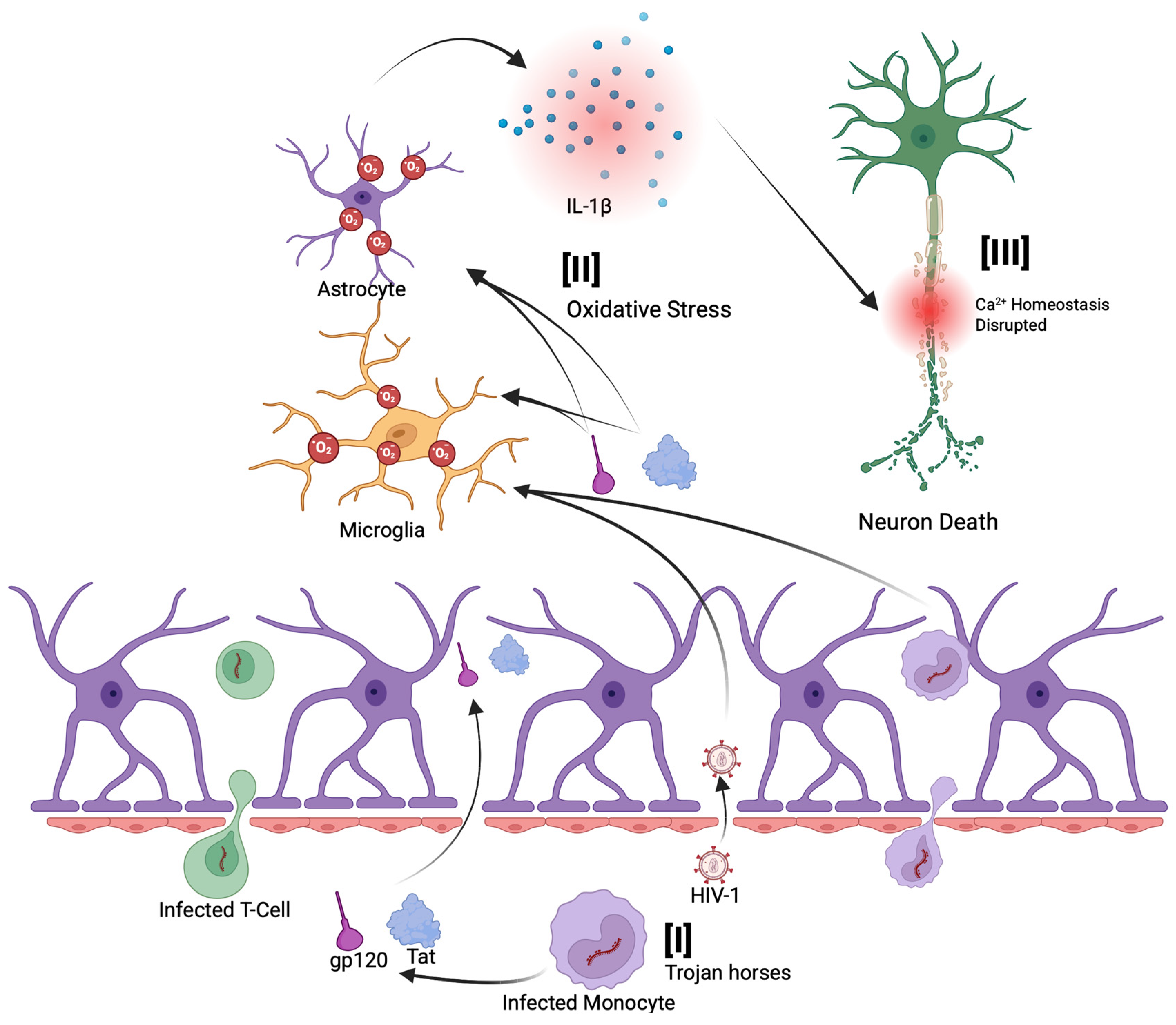
3.1. HIV Pathogenesis and ROS Production

HIV-1 is specifically known to induce the production of ROS in host cells, contributing to the pathogenesis of the virus [47,58]. As a result, HIV infection is associated with elevated oxidative stress in host cells. In both infected cells and bystander cells, HIV-1 triggers the overproduction of ROS compared to normal, healthy conditions [47,59]. Multiple HIV proteins, including the envelope glycoprotein (gp120), the regulatory protein (Tat), some accessory proteins (Nef and Vpr), and even the viral enzyme, RT, have been shown to stimulate ROS generation in host cells (Figure 3A) [58,60,61,62,63,64]. In addition to the viral proteins, there are other factors of HIV infection that can lead to the increased production of ROS. HIV infection involves the production of viral cDNA, including linear and circular forms such as 2-LTR circles [65]. HIV-derived cDNAs, particularly 2-LTR unintegrated forms, can activate the cGAS-STING pathway, which is known to drive innate immune responses and chronic inflammation [66]. This activation leads to increased production of ROS and oxidative stress, which can trigger ferroptosis, a distinct, iron-dependent form of regulated cell death characterized by the accumulation of lipid peroxides and membrane damage [67,68,69]. This pathway may also contribute to neurotoxicity in the context of HAND. Furthermore, prolonged use of cART, particularly regimens containing nucleoside RT inhibitors, has been associated with increased oxidative stress, which may contribute to neuronal injury and cognitive dysfunction [70,71]. This virus infection-induced oxidative stress plays a significant role in HIV pathogenesis, contributing to both direct and indirect effects, such as cellular damage and chronic inflammation. Notably, a persistent imbalance in the host redox state has been recognized as a key factor in various HIV-associated conditions, such as neurocognitive disorders and immune dysfunction, underlining the importance of a more comprehensive understanding of the role of ROS in HIV disease progression [47]. In the following subparagraph, we will discuss in more detail how each viral protein contributes to ROS production.

3.2. Envelope Glycoprotein (gp120) and Tat Mediated ROS Production

Several studies have shown that exposure to the HIV-1 viral envelope glycoprotein gp120 leads to increased ROS production and triggers intracellular oxidative stress in neural and immune cells, even in the absence of full viral infection. The glycoprotein gp120 stimulates oxidative stress in microglia and astrocytes, leading to the release of proinflammatory cytokines, such as IL-1β (Figure 1[II]), that disrupt neuronal calcium homeostasis and cause neuron death (Figure 1[III]) [64,72,73]. Specifically, Viviani et al. demonstrated that exposing glial cells to HIV gp120 leads to a marked increase in ROS production within those cells [60]. Additionally, their subsequent research indicated that gp120 induces IL-1β release from glial cells, which further contributes to neurotoxicity, highlighting a cellular-level mechanism for gp120-mediated production of ROS (Figure 3B) [74]. IL-1β, released by gp120-activated glial cells, subsequently disrupts calcium homeostasis in nearby neurons by enhancing N-methyl-D-aspartate receptor (NMDAR) activity, ultimately leading to neuronal injury and death [58,75]. Importantly, antioxidant-mediated neutralization of ROS prevented neuronal damage in a sandwich co-culture of primary hippocampal neurons and glial cells, confirming that the neurotoxicity was driven by ROS released from gp120-activated glia [60]. This study revealed an oxidative cascade initiated by gp120 in glial cells, characterized by ROS generation and IL-1β release, that subsequently disrupts the calcium homeostasis while also inducing neuronal death in adjacent cells (Figure 3B) [60]. Such a mechanism implicates oxidative stress as a driver of HIV-related neurotoxicity.
Further investigations found that certain neuron populations are especially vulnerable to gp120-induced oxidative stress. Agrawal et al. reported that dopamine-producing neurons (dopaminergic neurons), such as those in the substantia nigra of the brain, are particularly sensitive to oxidative stress caused by gp120 [76]. Dopaminergic neurons cultured in vitro underwent apoptosis at much lower peroxide concentrations than other neurons, indicating a heightened sensitivity to ROS. Correspondingly, when these neurons were exposed to HIV-1 gp120, they showed significant oxidative damage and apoptosis. This effect was mediated by gp120-induced calcium influx in neurons, with a selective vulnerability observed in dopaminergic cells, indicating that these neurons were more susceptible to gp120-induced toxicity than other neuronal subtypes. Dopaminergic neurons play a central role in the neurocognitive impairments associated with chronic HIV infection, as evidenced by reduced peak dopamine levels and slower reuptake rates that are linked to motivational and cognitive deficits such as apathy and depression, which are core features of HANDs [77]. Crucially, antioxidant interventions protect the cells [73,75,78]. The pretreatment with the antioxidant N-acetylcysteine (NAC) or the overexpression of antioxidant enzymes, such as superoxide dismutase (SOD) and glutathione peroxidase, blocked the gp120-induced cell death [76]. In vivo experiments reinforced these findings. When low doses of gp120 were injected into the brains of rats using a stereotaxic frame, it caused a rapid loss of dopaminergic neurons—an effect that was prevented by antioxidant enzyme treatment [79]. These findings provide compelling evidence that in vitro exposure to gp120 in primary human astrocytes induces ROS and contributes to selective neurotoxicity. While the role of gp120 in promoting ROS production and neurotoxicity is well established, more recent studies have elucidated the upstream molecular pathways involved. Ivanov et al. demonstrated that gp120 enhances ROS generation in astrocytes through multiple parallel mechanisms [47]. These include the upregulation of cytochrome P450 2E1 (CYP2E1), activation of NADPH oxidases NOX2 and NOX4, and promotion of Fenton and Weiss-Haber reactions—all of which contribute to oxidative stress [47,80,81,82]. Additionally, Pandhare et al. provided mechanistic evidence linking gp120 exposure to ROS generation in neuroblastoma cells [83]. They demonstrated that gp120 activates the tumor suppressor protein p53 via the CXCR4 co-receptor, which subsequently upregulates proline oxidase (POX). This cascade leads to increased ROS production and autophagic activity. Inhibition of POX reduced both ROS levels and autophagy, whereas POX overexpression amplified these responses.
The Tat protein can persist in the CNS despite cART and has been shown to promote AD-related pathology by binding to Aβ, disrupting its metabolism, and inducing tau hyperphosphorylation (Figure 2). These effects accelerate the formation of amyloid plaques and neurofibrillary tangles [84,85]. Such neurotoxic viral proteins contribute to synaptic damage, neuronal loss, and oxidative stress, driving the development of HAND and potentially increasing the risk of other dementias in individuals chronically living with HIV.

3.3. Nef-Mediated ROS Production

In addition to gp120 and Tat, the HIV accessory proteins Nef and Vpr significantly contribute to ROS induction within host cells, which further amplifies HIV-associated pathogenesis [86,87,88]. It has been established that Nef promotes oxidative stress by dysregulating the host cell’s NADPH oxidase complexes and mitochondrial function. One of the primary mechanisms is the interaction of Nef with components of the NADPH oxidase complex in macrophages and microglia [86,89]. Nef has been found to be associated with p22-phox, a subunit of the NADPH oxidase complex, leading to enhanced production of superoxide anions (Figure 3C[I]) [90]. This interaction effectively primes oxidase. In addition, Nef triggers intracellular kinases, including Src family kinases and PI3K, that lead to phosphorylation of p47-phox, which is a key cytosolic subunit of NADPH oxidase in the U937 human monoblastic cell line [91].
While expression of Nef did not activate the NADPH oxidase by itself in the microglia cell model, it is able to indirectly activate the NADPH oxidase by triggering the Vav/Rac1/p21-activated kinase (PAK) signaling pathway (Figure 3C[I]) [89]. This pathway is critical, as it plays a key role in initiating NADPH oxidase activity. This signaling cascade primes the cells for an amplified oxidative response upon subsequent stimulation, such as calcium ionophores, formyl peptide, or endotoxins [89]. Nef interacts with the guanine nucleotide exchange factor Vav, which in turn facilitates the exchange of GDP for GTP on Rac1, converting it to its active GTP-bound form. This activation of Rac1 leads to the stimulation of PAK signaling, culminating in the priming of the NADPH oxidase complex (gp91) for enhanced ROS production (Figure 3C[I]) [89,92]. Mutations in Nef that disrupt its interaction with this pathway abolish its ability to enhance ROS production, confirming that Nef-mediated activation of the PAK pathway is responsible for the increased superoxide release. Interestingly, Nef modulates ROS production in a biphasic manner, initiating an early increase in superoxide levels that is subsequently followed by a decline in NADPH oxidase activity [86]. This reduction has been attributed to Nef-induced soluble factors, such as IL-10, which suppress oxidase function. Overall, this highlights Nef’s complex regulatory influence on the cellular redox balance (Figure 3C[II]) [86]. The oxidative environment induced by Nef contributes to chronic inflammation by promoting inflammasome activation, which triggers pyroptosis in uninfected bystander CD4 T cells. This mechanism underlies the characteristic T-cell depletion observed in HIV infection (Figure 3C[III]), while also leading to the secretion of proinflammatory cytokines, such as IL-1β and IL-18, which sustain the aforementioned inflammatory milieu (Figure 3C[IV]) [93].

3.4. Vpr-Mediated ROS Production

Similarly, another accessory protein, Vpr, exerts potent oxidative effects by targeting mitochondria (Figure 3D) [88]. It localizes to mitochondrial membranes and interacts with components of the permeability transition pore complex, including the adenine nucleotide translocator (ANT) in the inner membrane and the voltage-dependent anion channel (VDAC) in the outer membrane. Vpr enters the mitochondria via the VDAC and binds to ANT, triggering the depolarization of the mitochondrial membrane. This results in an increase in the leakage of reactive species from the mitochondria (Figure 3D) [94]. Accordingly, Vpr can induce mitochondrial membrane permeabilization and uncouple the respiratory chain, leading to electron leakage and the subsequent elevation of ROS production. Vpr-induced mitochondrial membrane permeabilization also contributes to the release of pro-apoptotic factors, such as cytochrome c and apoptosis-inducing factor (AIF) from mitochondria into the cytosol, which activates the caspase cascade and apoptosis [88,95,96,97,98]. The brain and kidneys are particularly vulnerable to Vpr-induced toxicity, as Vpr has been implicated in HIV-associated neurodegeneration and nephropathy, largely through oxidative stress in neurons and renal tubular cells [99,100,101,102]. Importantly, Vpr-induced oxidative stress not only causes cell death but also can paradoxically stimulate HIV-1 replication. The excess ROS generated by Vpr activates redox-sensitive transcription factors, such as NF-κB and AP-1, which then enhance HIV gene expression (Figure 3D) [103,104]. Sandoval et al. recently demonstrated that Vpr-inducible DNA damage activates NF-κB through the DNA damage sensor ATM and the signaling adaptor NEMO, independently of the effects of Vpr on the cell cycle [105]. This NF-κB activation leads to increased HIV-1 transcription and promotes the production of proinflammatory cytokines, contributing to chronic immune activation in HIV infection [106]. Conversely, an antioxidant-rich environment can suppress HIV transcription. Israël et al. investigated whether shifting the cellular redox balance toward a more reduced state using butylated hydroxyanisole (BHA) could downregulate HIV promoter activity in lymphoblastoid and monocytic cell lines [107]. Their findings showed that antioxidant or free radical scavenger treatment in HIV-infected T cells and monocytes inhibited NF-κB nuclear translocation and reduced its activation, thereby suppressing HIV transcription. This led to a significant decrease in HIV long terminal repeat (LTR) promoter activity and consequently lower viral production. The drop in viral replication was accompanied by reduced secretion of NF-κB–dependent proinflammatory cytokines. These findings reveal a feed-forward loop in which HIV-induced ROS boosts NF-κB, which in turn enhances HIV replication and inflammatory gene expression.

3.5. Reverse Transcriptase (RT) Mediated ROS Production

While the HIV proteins, gp120, Tat, Nef, and Vpr, have well-documented roles in promoting oxidative stress, recent studies have also highlighted the role of HIV-1 RT in ROS generation [108,109]. RT is the viral enzyme responsible for converting RNA to DNA, but when expressed in host cells, it can have additional unexpected effects on cellular metabolism. Notably, expression of HIV-1 RT in mammalian cells has been shown to lead to significant ROS production (Figure 3E). Zakirova et al. demonstrated that introducing HIV-1 RT into a murine mammary carcinoma cell line (4T1-luc2) caused a marked increase in ROS levels, along with elevated lipid peroxidation and enhanced cell motility [62]. These effects were associated with the upregulation of epithelial–mesenchymal transition (EMT) markers, such as Twist, suggesting a link between RT-induced oxidative stress and pro-tumorigenic cellular changes (Figure 3E) [109]. Strikingly, RT variants with drug-resistance mutations that impair polymerase activity produced significantly less ROS, consequently losing their ability to promote tumor growth and metastasis [109]. In immunodeficient mice, tumors derived from cells expressing wild-type RT grew faster and formed more metastases than those expressing mutant RT, correlating with higher Twist expression and oxidative stress in wild-type RT tumors. These findings underscore that the enzymatic activity or structural integrity of HIV-1 RT is essential for driving ROS production and EMT-associated tumor progression. Additional evidence for RT mediated oxidative impact comes from studies in human cell lines. Isaguliants et al. reported that transient expression of HIV-1 RT in the human embryonic kidney cell line, HEK293 cells, triggers robust ROS generation and a cellular antioxidant response [108]. They observed a 10- to 15-fold increase in the transcription of detoxification enzymes, such as heme oxygenase-1 (HO-1), indicating significant oxidative stress in RT-expressing cells (Figure 3E). Notably, different RT gene variants produced varying levels of ROS, which in turn influenced both the magnitude and quality of the immune response elicited by a DNA vaccine targeting HIV-1 RT in their experimental model [108]. It was concluded that RT can modulate the host cellular environment through oxidative mechanisms, potentially contributing to viral pathogenesis and even oncogenic processes in cells where it is expressed.

4. Reactive Oxygen Species (ROS) and Chronic HIV-Associated Neurodegeneration

HIV-induced ROS production is not limited to acute neuronal injuries, but it also plays a role in the chronic neurological complications of HIV infection through downstream effects contributing to neural damage. While acute ROS production by gp120 causes immediate cellular damage, chronic oxidative stress produces long-term neurodegeneration in PLWH. In individuals with HAND, there is evidence of ongoing oxidative damage and disrupted lipid metabolism in the brain. Haughey et al. examined postmortem brain tissues and cerebrospinal fluid from individuals with HIV dementia and found clear signs of oxidative stress [110]. In these individuals, the levels of 4-hydroxynonenal (4-HNE), a reactive byproduct of lipid peroxidation, were significantly elevated in brain tissue and cerebrospinal fluid. 4-HNE has been shown to contribute to premature apoptosis via adduct formation, inhibition of survival pathways, and activation of apoptotic proteins, ultimately promoting neurodegenerative diseases such as Parkinson’s and Alzheimer’s [111,112]. Alongside 4-HNE, levels of ceramide, a sphingolipid known to promote apoptosis, were also increased [110]. Intracellular levels of ceramide increase when the activity of neutral sphingomyelinase (nSMase) or acid sphingomyelinase (aSMase) is elevated, as these enzymes convert sphingomyelin to ceramide (Figure 4) [113,114]. To explore causation, they exposed cultured neurons to the HIV-1 gp120 and Tat and observed that both provoked an accumulation of 4-HNE and ceramide within neurons (Figure 4) [110]. These findings suggest that oxidative stress and inflammatory signals triggered by HIV proteins can promote sphingomyelin breakdown and ceramide accumulation in neural cells. Given that ceramide is a potent mediator of programmed cell death, this pathway provides a mechanistic link between HIV-induced ROS and neuronal degeneration. Supporting this connection, pharmacological inhibition of ceramide synthesis protected neurons from gp120/Tat-induced toxicity [110]. These findings indicate that viral proteins induce oxidative stress in chronic HIV-associated brain disease that initiates a cascade of damaging lipid signaling, leading to excessive ceramide accumulation and subsequent apoptosis of neural cells (Figure 4). Ceramide generation mediated by nSMase has been reported to play a critical role in the late stages of HIV-1 maturation and replication, a key process required for the virus to acquire infectivity [7,115,116,117,118]. These findings suggest that ceramide generation broadly supports HIV infection at both cellular and molecular levels. This mechanism also provides a plausible explanation for how chronic HIV infection contributes to different neuropathological conditions, including dementia, through oxidative stress-driven pathways [7,115,116,117]. Additionally, the findings of Bachis et al. relating to the rapid loss of dopaminergic neurons in rat brains after low doses of gp120 were injected suggest a potential mechanism underlying the Parkinson’s disease-like symptoms occasionally observed in PLWH, wherein the gp120 protein may contribute to dopaminergic neuron loss through oxidative stress [79]. This ultimately links HIV infection to neurodegenerative processes.
There is substantial overlap between the mechanisms of HAND and those of AD. Chronic oxidative stress and inflammation in HIV can engage the same pathways that drive classic AD pathology. Epidemiological analyses further suggest that older people living with HIV may have a higher incidence of AD-like cognitive impairment compared to uninfected populations [13,56]. However, it remains under investigation whether HIV accelerates actual AD pathology or if observed AD-like symptoms in HIV are a byproduct of prolonged inflammation and vascular injury [13].
It is interesting to note how HIV itself might directly contribute to AD pathology. As mentioned above, the HIV Tat protein can bind to Aβ, impairing its clearance, and can trigger tau phosphorylation cascades (Figure 1[III]) [84]. On the other hand, some antiretroviral drugs may influence amyloid metabolism and could either mitigate or, in some cases, contribute to amyloid/tau accumulation (Figure 2) [119,120]. This complex interplay is the subject of active research, as studies attempt to discern whether long-term HIV-1 infection serves as a precipitating factor for the earlier onset of AD or other dementias. Neurocognitive issues also persist in HIV-infected individuals who are well-treated, highlighting the need for adjunctive strategies targeting these pathogenic mechanisms [121,122]. Approaches to reduce chronic inflammation and oxidative stress in PLWH are currently being explored. Although cART effectively suppresses plasma viral load over the long term, it does not fully protect the brain from the detrimental effects of chronic HIV infection [121,122]. One of the primary challenges is the limited penetration of cART into the CNS, largely due to the BBB, a tightly regulated structure composed of endothelial cells joined by tight junctions that restrict the passage of many substances, including several antiretroviral drugs [123]. This selective permeability can result in subtherapeutic drug concentrations in the brain, allowing HIV to persist and potentially develop resistance within CNS reservoirs [124,125]. In addition, some antiretroviral compounds have been associated with neurotoxic effects [126], which may contribute to neurocognitive impairments in PLWH, even when systemic viral suppression is achieved. Consequently, antioxidant therapies, such as glutathione precursors or NAC, have shown promise in preclinical models of HIV neurotoxicity through the attenuation of ROS levels and preventing neuron death caused by HIV proteins [75,78]. However, several challenges have limited their clinical efficacy, such as the same limited BBB penetration as cART, inconsistent clinical outcomes, potential pro-oxidant effects at high doses, and the complexity of HAND pathogenesis [127,128,129]. These challenges underscore the need for a comprehensive approach to treating HAND, potentially involving combination therapies that align with broader HIV cure strategies. In addition to targeting oxidative stress, anti-inflammatory treatments that reduce microglial activation have the potential to help preserve cognitive function. Ultimately, a more holistic strategy that aims to suppress viral replication, in addition to protecting long-term brain health, is essential. Details of current and future approaches for therapeutic intervention in HAND are discussed in Section 5. By addressing the overlapping mechanisms of HIV neuropathogenesis and AD, such as chronic inflammation and neuronal injury, it may be possible to reduce the risk of dementia among PLWH. Ongoing research continues to translate these mechanistic insights into therapeutic interventions, with the goal of ensuring that PLWH not only live longer but also maintain their cognitive vitality as they age.

5. Therapeutic Interventions for HIV-Associated Neurocognitive Disorder

5.1. Antioxidant

Given the detrimental effects of excess ROS production induced by various HIV-1 proteins, as discussed in Section 3 and Section 4, researchers have investigated antioxidant strategies as potential adjunctive therapies for HAND. Various preclinical studies have explored whether bolstering antioxidant defenses can mitigate HIV-induced oxidative stress (Figure 5 and Table 1). Agrawal et al. demonstrated that delivering exogenous antioxidant enzymes can protect cells from HIV protein-induced toxicity [130]. Using SV40 viral vectors, they introduced the genes for superoxide dismutase 1 (SOD1) and glutathione peroxidase 1 (GPx1) into primary human neurons, which were subsequently exposed to HIV-1 Tat. Co-expression of SOD1 and GPx1 effectively blocked Tat-induced calcium influx and neuronal apoptosis, whereas either enzyme alone provided insufficient protection. These results suggest that both superoxide and hydrogen peroxide, ROS intermediates detoxified by SOD and GPx, respectively, are involved in Tat-mediated neurotoxic signaling. Manda et al. provided clear experimental evidence supporting the protective effects of antioxidants in the context of HAND, particularly against cART-induced oxidative stress [131]. They demonstrated that cART, including azidothymidine (AZT) and indinavir (IDV), significantly induced oxidative stress and mitochondrial dysfunction in BBB endothelial cells (hCMEC/D3), leading to reduced intracellular glutathione, increased lipid peroxidation, mitochondrial membrane depolarization, ATP depletion, and apoptosis (Figure 5). Pretreatment with NACA, a cell-permeable thiol antioxidant, significantly reduced ROS levels, restored glutathione, preserved mitochondrial membrane potential, and prevented apoptosis of endothelial cells (Table 1). These findings suggest that antioxidants like NACA may protect the BBB from drug-induced oxidative stress, potentially helping to mitigate HAND in individuals treated with cART. In addition, Teodorof-Diedrich et al. provided strong experimental evidence supporting the therapeutic potential of antioxidants in mitigating neurocognitive damage associated with HIV and methamphetamine exposure [132]. In their study, NAC significantly reduced ROS levels in SK-N-MC cells, a human neuroepithelioma cell line, following exposure to HIV proteins, including gp120 and Tat, and methamphetamine. This antioxidant effect helped prevent mitochondrial fragmentation, supported autophagic processes, preserved mitochondrial integrity and function, and ultimately protected neuronal architecture. These in vitro mechanistic findings provide a strong rationale for translating antioxidant-based interventions into clinical research for HAND. Allard et al. conducted a double-blind, placebo-controlled trial to evaluate whether enhancing the antioxidant defenses of HIV-positive individuals could influence the disease progression (Figure 5 and Table 1) [133]. In this study, 49 PLWH were randomized to receive either a placebo or a daily oral supplementation of vitamin E and vitamin C, two well-known dietary antioxidants, for three months. The antioxidant-supplemented group showed a significant decrease in lipid peroxidation markers compared to controls, indicating that the vitamin regimen successfully lowered systemic oxidative stress. There was also a trend toward lower HIV viral load in the antioxidant-treated group after the supplementation period. Although this reduction in viral load was modest and did not reach statistical significance, it suggested a potentially beneficial effect. Another randomized, double-blind, placebo-controlled trial evaluated the safety and tolerability of OPC-14117, a lipophilic antioxidant, in individuals with HIV-associated cognitive impairment (Table 1) [134]. This study found that OPC-14117 was well tolerated with no significant adverse effects compared to placebo. While there were trends toward cognitive improvement in the treatment group, these changes were not statistically significant, suggesting the need for larger efficacy trials. In a multicenter, randomized, placebo-controlled phase II trial, Schifitto et al. evaluated the safety, tolerability, and efficacy of the selegiline transdermal system (STS), a skin patch formulation of the monoamine oxidase B (MAO-B) inhibitor selegiline, in individuals with HIV-associated cognitive impairment [135]. The primary outcome was the change in neuropsychological performance, measured by the NPZ-6 composite score. The study found that while STS was safe and well tolerated, there were no significant improvements in cognitive performance compared to placebo. These findings suggest that, although STS has a favorable safety profile, its efficacy in improving cognitive function in this population was not demonstrated within the study period. Overall, the findings of these clinical trials are consistent with mechanistic studies suggesting that reducing oxidative stress in vivo may help alleviate neurocognitive symptoms associated with HIV infection. However, antioxidant therapy remains an unproven strategy for treating HAND, and further empirical studies are needed to bridge the gap between in vitro mechanistic findings and in vivo clinical outcomes. Addressing this research question could help establish effective treatments for HAND and deepen our understanding of it. While early antioxidant agents have shown limited clinical efficacy, newer compounds, such as dimethyl fumarate (DMF) that activate endogenous antioxidant defenses via the Nrf2/ARE pathway, demonstrate promising potential [136,137]. However, direct evidence supporting the use of DMF specifically for the treatment of HAND remains lacking. Further research is needed to develop comprehensive, integrative strategies that combine pharmacological therapies with lifestyle interventions to effectively mitigate PLWH.

5.2. Poly (ADP-Ribose) Polymerase Inhibitors

Equally important to treating HAND is the urgent need to develop an effective HIV cure strategy. One of the major barriers to achieving a cure is the persistence of latent HIV infection [138,139,140,141,142,143,144]. Notably, microglial cells have been identified as a primary reservoir of HIV within the brain [29,30,31,32,145,146,147]. Although HDAC inhibitors have been extensively studied as latency-reversing agents (LRAs) in the ‘kick-and-kill’ strategy to eliminate HIV reservoir cells, clinical trials have not demonstrated a significant reduction in the size of latent reservoirs [148,149,150]. We recently reported that a combination of FDA-approved cancer drugs targeting poly (ADP-ribose) polymerase (PARP) enzymes with HDAC inhibitors effectively reactivates latent HIV infected cells (Figure 5) [151]. This reactivation enables subsequent elimination of infected cells through the stimulation of immune responses, particularly by natural killer (NK) cells, via a PARP inhibitor. In addition to their role in reducing the latent HIV reservoir, PARP inhibitors that target the PARP pathway have also been reported to suppress neuroinflammation [152]. The PARP protein family consists of 17 members that regulate a wide range of cellular processes, among which PARP-1 is one of the most well-studied and characterized [153,154,155,156]. PARP-1 is a nuclear enzyme that plays a central role in DNA repair and regulates transcriptional responses to genotoxic and oxidative stress through its poly (ADP-ribosyl)ation activity [157,158]. HIV-1 proteins such as gp120 and Tat have been shown to induce oxidative DNA damage indirectly through the generation of ROS, whereas Vpr contributes to DNA damage both by directly causing double-strand breaks and by promoting ROS production [63,105,159,160]. This DNA damage can lead to the activation of PARP-1, which depletes intracellular NAD+ and ATP, ultimately leading to neuronal death through a programmed cell death pathway known as parthanatos [161,162,163,164]. Concurrently, PARP-1 activation promotes the expression of proinflammatory genes through the NF-κB pathway, further intensifying neuroinflammation [162,165,166,167]. PARP inhibitors have been shown in vitro to reduce neurotoxicity and inflammatory injury by attenuating ROS production and microglial activation (Figure 5) [168,169,170]. This suggests a potential therapeutic strategy for limiting both direct neuronal damage and secondary inflammation in neurodegenerative and neuroinflammatory conditions. Recent preclinical studies using the SIV-infected macaque model have shown that while PARP-1 expression in the frontal cortex remained unchanged, transcripts of four other PARP family members, PARP9, PARP10, PARP12, and PARP14, were significantly upregulated [171]. In addition, neuronal mitochondria-specific PARP inhibitors could result in a greater neuroprotective effect in the traumatic brain injury (TBI) model mouse [172]. These findings suggest that PARP inhibition may offer therapeutic benefits for neuropathic conditions, supporting interest in their potential application for treating HAND. However, clinical evidence remains limited, and further investigation is required [173]. In broader models of neurodegenerative disease, PARP inhibitors have demonstrated potential in reducing glial activation, mitochondrial dysfunction, ROS production, neuroinflammation, and cognitive decline, suggesting their therapeutic utility may extend beyond HAND. Despite these promising findings, several challenges must be addressed before clinical translation. While several PARP inhibitors have been optimized for high CNS penetration, a critical feature for the effective treatment of brain disorders [174], most clinically approved PARP inhibitors, such as Talazoparib, Niraparib, and Rucaparib, show poor BBB penetration, limiting their use in treating CNS disorders [175]. Additionally, prolonged PARP inhibition raises concerns about genomic stability, particularly in aging individuals living with HIV who may require such treatment. However, we demonstrated that, when combined with HDAC inhibitors, PARP inhibitors can contribute to the reduction in the latent HIV reservoir, suggesting their dual potential in HIV cure strategies as well as in the prevention or mitigation of HAND [151]. Notably, PARP inhibitors are administered only briefly during the reactivation phase of latent reservoir cells to facilitate their elimination, which may minimize the risks associated with prolonged PARP inhibition in PLWH. Although four PARP inhibitors have been approved by the FDA for cancer treatment, further research, particularly pharmacokinetic optimization and clinical trials focused on HIV, is essential to assess the safety and therapeutic efficacy of PARP inhibition.

6. Conclusions and Future Directions

Despite the success of cART in controlling systemic HIV replication, HAND persists as a significant burden for PLWH, particularly as the population ages [6,7,8]. From the aforementioned population, approximately 50% of individuals infected with HIV develop some form of neurocognitive disorder [176]. Multiple studies have provided evidence that highlights oxidative stress as a central mechanism in HIV neuropathogenesis. Viral proteins, including gp120, Tat, Nef, and Vpr, in addition to viral RT, induce excessive production of ROS, triggering chronic neuroinflammation, neuronal damage, and ultimately, neurodegeneration. While numerous in vitro studies have provided valuable mechanistic insights into how specific HIV-1 proteins contribute to oxidative stress and neurotoxicity, the physiological relevance of these findings requires careful interpretation. The concentrations of viral proteins used in vitro often exceed those typically present in vivo, particularly in the brains of individuals on suppressive cART, where viral protein levels are expected to be very low [177]. Additionally, many of these studies rely on immortalized cell lines, which may not accurately represent the complex behavior and responses of primary CNS cells, thus limiting the translational relevance of their findings. Nevertheless, in vitro models remain essential tools for dissecting the molecular pathways underlying ROS-mediated damage, offering important context for understanding HAND pathogenesis.
Recent studies have highlighted a growing interest in exploring the link between neuroinflammation and intravenous drug use, particularly given the high prevalence of substance abuse among PLWH. Chronic use of opioids, cannabinoids, and methamphetamines has broadly been shown to induce neurotoxic effects similar to those observed in HIV infection, leading to increased oxidative stress and neuroinflammation [64,178,179,180,181,182]. For instance, research indicates that the inhibition of microglial activation can reduce both neuroinflammatory responses and drug-seeking behaviors [183], suggesting a potential therapeutic target for mitigating substance abuse-related neurotoxicity. Moreover, the co-occurrence of HIV and intravenous drug use is well-documented, with studies reporting significant comorbidity rates, underscoring the need for integrated treatment approaches [184,185]. Substance abuse of the aforementioned drugs may exacerbate the overproduction of ROS, further triggering neuroinflammatory pathways [186,187]. Additionally, certain treatment regimens can necessitate opioid prescriptions for pain management, such as those leading to neuropathic pain, which bears the risk of exacerbating neuroinflammation [188]. Previous research has shown opioid use to be associated with the activation of glial cells and subsequent neuroinflammation, potentially contributing to the development of HAND [188]. Furthermore, recreational drug use is known to compromise BBB integrity by downregulating tight junction proteins, thereby facilitating increased ROS production and neuroinflammation [189,190]. While it is challenging to generalize the effects of opioids, cannabinoids, and methamphetamines on neuroinflammation in PLWH due to their distinct mechanisms of action and metabolites, which can result in variable and sometimes counterintuitive outcomes, substance use remains an important clinical factor. Notably, an estimated 30% of PLWH with HAND have a history of substance use, highlighting the need for substance-specific investigations rather than broad categorizations when evaluating their contribution to neuroinflammation and oxidative stress [187].
Although early antioxidant strategies showed limited clinical efficacy [130,133,134,135], recent advances point to the therapeutic promise of newer compounds that activate endogenous defense pathways, such as dimethyl fumarate via Nrf2/ARE signaling [136,137]. In parallel, PARP inhibitors have emerged as a dual-function strategy, not only enhancing the latency-reversing efficacy of HDAC inhibitors in targeting latent HIV reservoirs as part of potential cure strategies [151], while also showing potential to mitigate neuroinflammation and oxidative damage associated with HAND [152,168,169,170,172,174]. Ultimately, managing HAND requires a multifaceted approach that goes beyond viral suppression. Future therapies will likely involve a combination of antiretroviral treatments, targeted antioxidants, inflammation modulators, and agents addressing HIV latency. Continued research, through well-designed in vivo studies and clinical trials, is essential to translate these mechanistic insights into effective interventions that preserve cognitive function and improve the quality of life for PLWH.
We are currently conducting research to address several of the outstanding questions described above. Ongoing studies are examining ROS expression in microglial cell lines treated with PARP inhibitors, which are also being explored for their ability to activate latent HIV reservoirs. Additionally, there is growing interest in utilizing human microglial cell lines and primary microglial models derived from monocytes, which more accurately reflect the characteristics of CNS-resident microglia in vitro. These models are being used to examine the effects of PARP inhibitors on both latent reservoir reduction and neuroprotection. Further investigation is planned using in vivo models such as EcoHIV-infected mice, a genetically modified HIV construct capable of establishing infection in murine hosts, to study the latency reservoir and HAND [191,192,193,194,195,196,197].

Author Contributions

All authors have written, reviewed, and agreed to submit this manuscript. All authors have read and agreed to the published version of the manuscript.

Funding

This research was partially supported by a research start-up fund from American University to T.I. Additional funding was provided by the National Institutes of Health (NIH), specifically through a grant from the National Institute of Allergy and Infectious Diseases (grant number: 7R15AI172610-02 to T.I.), National Institute of Mental Health (grant number: 5R41MH139088-02 to A.G.Z), by the Research Corporation for Scientific Advancement (RCSA) (grant number: SA-NCE-2025-004 to A.G.Z), by the Trone Family Eminent Scholar Chair in Neuroscience and Behavior (to A.L.R.), and in part by a 2024 award from the District of Columbia Center for AIDS Research (DC-CFAR), an NIH-funded program (P30AI117970). The DC-CFAR is supported by the following NIH Co-Funding and Participating Institutes and Centers: NIAID, NCI, NICHD, NHLBI, NIDA, NIMH, NIA, NIDDK, NIMHD, NIDCR, NINR, FIC, and OAR.

Institutional Review Board Statement

There is no Institutional Review Board Statement for this review article.

Informed Consent Statement

Not applicable.

Data Availability Statement

All data should be referred to in each reference.

Acknowledgments

We express our gratitude to the administrators and leadership of the authors’ institutions at American University for their continued support of this publication. The content is solely the responsibility of the authors and does not necessarily represent the official views of the NIH. All authors have read and approved the final version of the manuscript for publication.

Conflicts of Interest

The authors declare no conflicts of interest.

References

  1. Giovanetti, M.; Ciccozzi, M.; Parolin, C.; Borsetti, A. Molecular Epidemiology of HIV-1 in African Countries: A Comprehensive Overview. Pathogens 2020, 9, 1072. [Google Scholar] [CrossRef]
  2. Chen, Y.; Li, A.D.; Yang, Y.; Lu, J.; Xu, Y.; Ji, X.; Wu, L.; Han, L.; Zhu, B.; Xu, M. Global, regional and national burden of HIV/AIDS among individuals aged 15–79 from 1990 to 2021. AIDS Res. Ther. 2025, 22, 51. [Google Scholar] [CrossRef]
  3. Ayieko, J.; Thorp, M.; Ghebremichael, M. Renewing Our Focus on Vulnerable Populations Among People Living with HIV. Trop. Med. Infect. Dis. 2024, 9, 278. [Google Scholar] [CrossRef]
  4. Sudharshan, S.; Biswas, J. Introduction and immunopathogenesis of acquired immune deficiency syndrome. Indian J. Ophthalmol. 2008, 56, 357–362. [Google Scholar] [CrossRef]
  5. Balasubramaniam, M.; Pandhare, J.; Dash, C. Immune Control of HIV. J. Life Sci. 2019, 1, 4–37. [Google Scholar] [CrossRef]
  6. Weichseldorfer, M.; Reitz, M.; Latinovic, O.S. Past HIV-1 Medications and the Current Status of Combined Antiretroviral Therapy Options for HIV-1 Patients. Pharmaceutics 2021, 13, 1798. [Google Scholar] [CrossRef]
  7. McGraw, A.; Hillmer, G.; Medehincu, S.M.; Hikichi, Y.; Gagliardi, S.; Narayan, K.; Tibebe, H.; Marquez, D.; Mei Bose, L.; Keating, A.; et al. Exploring HIV-1 Maturation: A New Frontier in Antiviral Development. Viruses 2024, 16, 1423. [Google Scholar] [CrossRef]
  8. Nachega, J.B.; Scarsi, K.K.; Gandhi, M.; Scott, R.K.; Mofenson, L.M.; Archary, M.; Nachman, S.; Decloedt, E.; Geng, E.H.; Wilson, L.; et al. Long-acting antiretrovirals and HIV treatment adherence. Lancet HIV 2023, 10, e332–e342. [Google Scholar] [CrossRef]
  9. Smith, L.K.; Kuhn, T.B.; Chen, J.; Bamburg, J.R. HIV Associated Neurodegenerative Disorders: A New Perspective on the Role of Lipid Rafts in Gp120-Mediated Neurotoxicity. Curr. HIV Res. 2018, 16, 258–269. [Google Scholar] [CrossRef]
  10. Clifford, D.B.; Ances, B.M. HIV-associated neurocognitive disorder. Lancet Infect. Dis. 2013, 13, 976–986. [Google Scholar] [CrossRef]
  11. Heaton, R.K.; Clifford, D.B.; Franklin, D.R., Jr.; Woods, S.P.; Ake, C.; Vaida, F.; Ellis, R.J.; Letendre, S.L.; Marcotte, T.D.; Atkinson, J.H.; et al. HIV-associated neurocognitive disorders persist in the era of potent antiretroviral therapy: CHARTER Study. Neurology 2010, 75, 2087–2096. [Google Scholar] [CrossRef]
  12. Simioni, S.; Cavassini, M.; Annoni, J.M.; Rimbault Abraham, A.; Bourquin, I.; Schiffer, V.; Calmy, A.; Chave, J.P.; Giacobini, E.; Hirschel, B.; et al. Cognitive dysfunction in HIV patients despite long-standing suppression of viremia. AIDS 2010, 24, 1243–1250. [Google Scholar] [CrossRef]
  13. Chemparthy, D.T.; Kannan, M.; Gordon, L.; Buch, S.; Sil, S. Alzheimer’s-Like Pathology at the Crossroads of HIV-Associated Neurological Disorders. Vaccines 2021, 9, 930. [Google Scholar] [CrossRef]
  14. Breijyeh, Z.; Karaman, R. Comprehensive Review on Alzheimer’s Disease: Causes and Treatment. Molecules 2020, 25, 5789. [Google Scholar] [CrossRef]
  15. Stephenson, J.; Nutma, E.; van der Valk, P.; Amor, S. Inflammation in CNS neurodegenerative diseases. Immunology 2018, 154, 204–219. [Google Scholar] [CrossRef]
  16. Richards, R.I.; Robertson, S.A.; Kastner, D.L. Neurodegenerative diseases have genetic hallmarks of autoinflammatory disease. Hum. Mol. Genet. 2018, 27, R108–R118. [Google Scholar] [CrossRef]
  17. Madore, C.; Yin, Z.; Leibowitz, J.; Butovsky, O. Microglia, Lifestyle Stress, and Neurodegeneration. Immunity 2020, 52, 222–240. [Google Scholar] [CrossRef]
  18. Campbell, A. Inflammation, neurodegenerative diseases, and environmental exposures. Ann. N.Y. Acad. Sci. 2004, 1035, 117–132. [Google Scholar] [CrossRef]
  19. Soraci, L.; Corsonello, A.; Paparazzo, E.; Montesanto, A.; Piacenza, F.; Olivieri, F.; Gambuzza, M.E.; Savedra, E.V.; Marino, S.; Lattanzio, F.; et al. Neuroinflammaging: A Tight Line Between Normal Aging and Age-Related Neurodegenerative Disorders. Aging Dis. 2024, 15, 1726–1747. [Google Scholar] [CrossRef]
  20. Stoccoro, A.; Coppede, F. Exposure to Metals, Pesticides, and Air Pollutants: Focus on Resulting DNA Methylation Changes in Neurodegenerative Diseases. Biomolecules 2024, 14, 1366. [Google Scholar] [CrossRef]
  21. Xu, L.; He, D.; Bai, Y. Microglia-Mediated Inflammation and Neurodegenerative Disease. Mol. Neurobiol. 2016, 53, 6709–6715. [Google Scholar] [CrossRef]
  22. Subhramanyam, C.S.; Wang, C.; Hu, Q.; Dheen, S.T. Microglia-mediated neuroinflammation in neurodegenerative diseases. Semin. Cell Dev. Biol. 2019, 94, 112–120. [Google Scholar] [CrossRef]
  23. Bachiller, S.; Jimenez-Ferrer, I.; Paulus, A.; Yang, Y.; Swanberg, M.; Deierborg, T.; Boza-Serrano, A. Microglia in Neurological Diseases: A Road Map to Brain-Disease Dependent-Inflammatory Response. Front. Cell. Neurosci. 2018, 12, 488. [Google Scholar] [CrossRef]
  24. Dheen, S.T.; Kaur, C.; Ling, E.A. Microglial activation and its implications in the brain diseases. Curr. Med. Chem. 2007, 14, 1189–1197. [Google Scholar] [CrossRef]
  25. Isik, S.; Yeman Kiyak, B.; Akbayir, R.; Seyhali, R.; Arpaci, T. Microglia Mediated Neuroinflammation in Parkinson’s Disease. Cells 2023, 12, 1012. [Google Scholar] [CrossRef]
  26. Rodriguez-Gomez, J.A.; Kavanagh, E.; Engskog-Vlachos, P.; Engskog, M.K.R.; Herrera, A.J.; Espinosa-Oliva, A.M.; Joseph, B.; Hajji, N.; Venero, J.L.; Burguillos, M.A. Microglia: Agents of the CNS Pro-Inflammatory Response. Cells 2020, 9, 1717. [Google Scholar] [CrossRef]
  27. Nayak, D.; Roth, T.L.; McGavern, D.B. Microglia development and function. Annu. Rev. Immunol. 2014, 32, 367–402. [Google Scholar] [CrossRef]
  28. Oddo, S.; Caccamo, A.; Shepherd, J.D.; Murphy, M.P.; Golde, T.E.; Kayed, R.; Metherate, R.; Mattson, M.P.; Akbari, Y.; LaFerla, F.M. Triple-transgenic model of Alzheimer’s disease with plaques and tangles: Intracellular Abeta and synaptic dysfunction. Neuron 2003, 39, 409–421. [Google Scholar] [CrossRef]
  29. Wallet, C.; De Rovere, M.; Van Assche, J.; Daouad, F.; De Wit, S.; Gautier, V.; Mallon, P.W.G.; Marcello, A.; Van Lint, C.; Rohr, O.; et al. Microglial Cells: The Main HIV-1 Reservoir in the Brain. Front. Cell. Infect. Microbiol. 2019, 9, 362. [Google Scholar] [CrossRef]
  30. Tang, Y.; Chaillon, A.; Gianella, S.; Wong, L.M.; Li, D.; Simermeyer, T.L.; Porrachia, M.; Ignacio, C.; Woodworth, B.; Zhong, D.; et al. Brain microglia serve as a persistent HIV reservoir despite durable antiretroviral therapy. J. Clin. Investig. 2023, 133, e167417. [Google Scholar] [CrossRef]
  31. Gumbs, S.B.H.; Berdenis van Berlekom, A.; Kubler, R.; Schipper, P.J.; Gharu, L.; Boks, M.P.; Ormel, P.R.; Wensing, A.M.J.; de Witte, L.D.; Nijhuis, M. Characterization of HIV-1 Infection in Microglia-Containing Human Cerebral Organoids. Viruses 2022, 14, 829. [Google Scholar] [CrossRef] [PubMed]
  32. Schlachetzki, J.C.M.; Zhou, Y.; Glass, C.K. Human microglia phenotypes in the brain associated with HIV infection. Curr. Opin. Neurobiol. 2022, 77, 102637. [Google Scholar] [CrossRef] [PubMed]
  33. Plaza-Jennings, A.; Akbarian, S. Genomic Exploration of the Brain in People Infected with HIV-Recent Progress and the Road Ahead. Curr. HIV/AIDS Rep. 2023, 20, 357–367. [Google Scholar] [CrossRef]
  34. Strazza, M.; Pirrone, V.; Wigdahl, B.; Nonnemacher, M.R. Breaking down the barrier: The effects of HIV-1 on the blood-brain barrier. Brain Res. 2011, 1399, 96–115. [Google Scholar] [CrossRef]
  35. Woodburn, B.M.; Kanchi, K.; Zhou, S.; Colaianni, N.; Joseph, S.B.; Swanstrom, R. Characterization of Macrophage-Tropic HIV-1 Infection of Central Nervous System Cells and the Influence of Inflammation. J. Virol. 2022, 96, e0095722. [Google Scholar] [CrossRef]
  36. Gisslen, M.; Keating, S.M.; Spudich, S.; Arechiga, V.; Stephenson, S.; Zetterberg, H.; Di Germanio, C.; Blennow, K.; Fuchs, D.; Hagberg, L.; et al. Compartmentalization of cerebrospinal fluid inflammation across the spectrum of untreated HIV-1 infection, central nervous system injury and viral suppression. PLoS ONE 2021, 16, e0250987. [Google Scholar] [CrossRef] [PubMed]
  37. Uwishema, O.; Ayoub, G.; Badri, R.; Onyeaka, H.; Berjaoui, C.; Karabulut, E.; Anis, H.; Sammour, C.; Mohammed Yagoub, F.E.A.; Chalhoub, E. Neurological disorders in HIV: Hope despite challenges. Immun. Inflamm. Dis. 2022, 10, e591. [Google Scholar] [CrossRef]
  38. McRae, M. HIV and viral protein effects on the blood brain barrier. Tissue Barriers 2016, 4, e1143543. [Google Scholar] [CrossRef]
  39. Zhang, Y.L.; Ouyang, Y.B.; Liu, L.G.; Chen, D.X. Blood-brain barrier and neuro-AIDS. Eur. Rev. Med. Pharmacol. Sci. 2015, 19, 4927–4939. [Google Scholar]
  40. Atluri, V.S.; Hidalgo, M.; Samikkannu, T.; Kurapati, K.R.; Jayant, R.D.; Sagar, V.; Nair, M.P. Effect of human immunodeficiency virus on blood-brain barrier integrity and function: An update. Front. Cell. Neurosci. 2015, 9, 212. [Google Scholar] [CrossRef]
  41. Al-Obaidi, M.M.J.; Bahadoran, A.; Wang, S.M.; Manikam, R.; Raju, C.S.; Sekaran, S.D. Disruption of the blood brain barrier is vital property of neurotropic viral infection of the central nervous system. Acta Virol. 2018, 62, 16–27. [Google Scholar] [CrossRef] [PubMed]
  42. Bertrand, L.; Cho, H.J.; Toborek, M. Blood-brain barrier pericytes as a target for HIV-1 infection. Brain 2019, 142, 502–511. [Google Scholar] [CrossRef] [PubMed]
  43. Sun, Y.; Cai, M.; Liang, Y.; Zhang, Y. Disruption of blood-brain barrier: Effects of HIV Tat on brain microvascular endothelial cells and tight junction proteins. J. Neurovirol. 2023, 29, 658–668. [Google Scholar] [CrossRef] [PubMed]
  44. Lorin, V.; Danckaert, A.; Porrot, F.; Schwartz, O.; Afonso, P.V.; Mouquet, H. Antibody Neutralization of HIV-1 Crossing the Blood-Brain Barrier. mBio 2020, 11, e02424-20. [Google Scholar] [CrossRef]
  45. Barker, C.T.; Vaidya, N.K. Modeling HIV-1 infection in the brain. PLoS Comput. Biol. 2020, 16, e1008305. [Google Scholar] [CrossRef]
  46. Couret, J.; Chang, T.L. Reactive Oxygen Species in HIV Infection. EC Microbiol. 2016, 3, 597–604. [Google Scholar]
  47. Ivanov, A.V.; Valuev-Elliston, V.T.; Ivanova, O.N.; Kochetkov, S.N.; Starodubova, E.S.; Bartosch, B.; Isaguliants, M.G. Oxidative Stress during HIV Infection: Mechanisms and Consequences. Oxid. Med. Cell. Longev. 2016, 2016, 8910396. [Google Scholar] [CrossRef]
  48. Didonna, A. Tau at the interface between neurodegeneration and neuroinflammation. Genes Immun. 2020, 21, 288–300. [Google Scholar] [CrossRef]
  49. Cotto, B.; Natarajanseenivasan, K.; Langford, D. HIV-1 infection alters energy metabolism in the brain: Contributions to HIV-associated neurocognitive disorders. Prog. Neurobiol. 2019, 181, 101616. [Google Scholar] [CrossRef]
  50. Jha, N.K.; Sharma, A.; Jha, S.K.; Ojha, S.; Chellappan, D.K.; Gupta, G.; Kesari, K.K.; Bhardwaj, S.; Shukla, S.D.; Tambuwala, M.M.; et al. Alzheimer’s disease-like perturbations in HIV-mediated neuronal dysfunctions: Understanding mechanisms and developing therapeutic strategies. Open Biol. 2020, 10, 200286. [Google Scholar] [CrossRef]
  51. Williams, M.E.; Naude, P.J.W. The relationship between HIV-1 neuroinflammation, neurocognitive impairment and encephalitis pathology: A systematic review of studies investigating post-mortem brain tissue. Rev. Med. Virol. 2024, 34, e2519. [Google Scholar] [CrossRef] [PubMed]
  52. Kodidela, S.; Gerth, K.; Haque, S.; Gong, Y.; Ismael, S.; Singh, A.; Tauheed, I.; Kumar, S. Extracellular Vesicles: A Possible Link between HIV and Alzheimer’s Disease-Like Pathology in HIV Subjects? Cells 2019, 8, 968. [Google Scholar] [CrossRef]
  53. Gonzalez, J.; Wilson, A.; Byrd, D.; Cortes, E.P.; Crary, J.F.; Morgello, S. Neuronal accumulation of hyperphosphorylated tau protein predicts stable memory impairment in people living with HIV. AIDS 2023, 37, 1247–1256. [Google Scholar] [CrossRef]
  54. Fields, J.A.; Swinton, M.K.; Soontornniyomkij, B.; Carson, A.; Achim, C.L. Beta amyloid levels in cerebrospinal fluid of HIV-infected people vary by exposure to antiretroviral therapy. AIDS 2020, 34, 1001–1007. [Google Scholar] [CrossRef] [PubMed]
  55. Ozturk, T.; Kollhoff, A.; Anderson, A.M.; Christina Howell, J.; Loring, D.W.; Waldrop-Valverde, D.; Franklin, D.; Letendre, S.; Tyor, W.R.; Hu, W.T. Linked CSF reduction of phosphorylated tau and IL-8 in HIV associated neurocognitive disorder. Sci. Rep. 2019, 9, 8733. [Google Scholar] [CrossRef]
  56. Buckley, S.; Byrnes, S.; Cochrane, C.; Roche, M.; Estes, J.D.; Selemidis, S.; Angelovich, T.A.; Churchill, M.J. The role of oxidative stress in HIV-associated neurocognitive disorders. Brain Behav. Immun. Health 2021, 13, 100235. [Google Scholar] [CrossRef]
  57. Pulliam, L.; Calosing, C.; Sun, B.; Grunfeld, C.; Rempel, H. Monocyte activation from interferon-alpha in HIV infection increases acetylated LDL uptake and ROS production. J. Interferon Cytokine Res. 2014, 34, 822–828. [Google Scholar] [CrossRef] [PubMed]
  58. Harshithkumar, R.; Shah, P.; Jadaun, P.; Mukherjee, A. ROS Chronicles in HIV Infection: Genesis of Oxidative Stress, Associated Pathologies, and Therapeutic Strategies. Curr. Issues Mol. Biol. 2024, 46, 8852–8873. [Google Scholar] [CrossRef]
  59. Cevallos, C.; Ojeda, D.S.; Sanchez, L.; Urquiza, J.; Delpino, M.V.; Quarleri, J. HIV-induced bystander cell death in astrocytes requires cell-to-cell viral transmission. J. Neurochem. 2022, 163, 338–356. [Google Scholar] [CrossRef]
  60. Viviani, B.; Corsini, E.; Binaglia, M.; Galli, C.L.; Marinovich, M. Reactive oxygen species generated by glia are responsible for neuron death induced by human immunodeficiency virus-glycoprotein 120 in vitro. Neuroscience 2001, 107, 51–58. [Google Scholar] [CrossRef]
  61. Ronaldson, P.T.; Bendayan, R. HIV-1 viral envelope glycoprotein gp120 produces oxidative stress and regulates the functional expression of multidrug resistance protein-1 (Mrp1) in glial cells. J. Neurochem. 2008, 106, 1298–1313. [Google Scholar] [CrossRef]
  62. Zakirova, N.F.; Kondrashova, A.S.; Golikov, M.V.; Ivanova, O.N.; Ivanov, A.V.; Isaguliants, M.G.; Bayurova, E.O. Expression of HIV-1 Reverse Transcriptase in Murine Cancer Cells Increases Mitochondrial Respiration. Mol. Biol. 2022, 56, 795–807. [Google Scholar] [CrossRef]
  63. El-Amine, R.; Germini, D.; Zakharova, V.V.; Tsfasman, T.; Sheval, E.V.; Louzada, R.A.N.; Dupuy, C.; Bilhou-Nabera, C.; Hamade, A.; Najjar, F.; et al. HIV-1 Tat protein induces DNA damage in human peripheral blood B-lymphocytes via mitochondrial ROS production. Redox Biol. 2018, 15, 97–108. [Google Scholar] [CrossRef]
  64. Shah, A.; Kumar, S.; Simon, S.D.; Singh, D.P.; Kumar, A. HIV gp120- and methamphetamine-mediated oxidative stress induces astrocyte apoptosis via cytochrome P450 2E1. Cell Death Dis. 2013, 4, e850. [Google Scholar] [CrossRef]
  65. Butler, S.L.; Johnson, E.P.; Bushman, F.D. Human immunodeficiency virus cDNA metabolism: Notable stability of two-long terminal repeat circles. J. Virol. 2002, 76, 3739–3747. [Google Scholar] [CrossRef]
  66. Zhou, R.; Xie, X.; Li, X.; Qin, Z.; Wei, C.; Liu, J.; Luo, Y. The triggers of the cGAS-STING pathway and the connection with inflammatory and autoimmune diseases. Infect. Genet. Evol. 2020, 77, 104094. [Google Scholar] [CrossRef]
  67. Zhang, Z.; Yang, J.; Zhou, Q.; Zhong, S.; Liu, J.; Zhang, X.; Chang, X.; Wang, H. The cGAS-STING-mediated ROS and ferroptosis are involved in manganese neurotoxicity. J. Environ. Sci. 2025, 152, 71–86. [Google Scholar] [CrossRef]
  68. Sui, H.; Sun, Z.; Liu, C.; Xi, H. Ferritinophagy promotes microglia ferroptosis to aggravate neuroinflammation induced by cerebral ischemia-reperfusion injury via activation of the cGAS-STING signaling pathway. Neurochem. Int. 2025, 183, 105920. [Google Scholar] [CrossRef]
  69. Ding, L.; Zhang, R.; Du, W.; Wang, Q.; Pei, D. The role of cGAS-STING signaling pathway in ferroptosis. J. Adv. Res. 2024. [Google Scholar] [CrossRef]
  70. Akang, E.N. Combination antiretroviral therapy (cART)-induced hippocampal disorders: Highlights on therapeutic potential of Naringenin and Quercetin. IBRO Rep. 2019, 6, 137–146. [Google Scholar] [CrossRef]
  71. Akay, C.; Cooper, M.; Odeleye, A.; Jensen, B.K.; White, M.G.; Vassoler, F.; Gannon, P.J.; Mankowski, J.; Dorsey, J.L.; Buch, A.M.; et al. Antiretroviral drugs induce oxidative stress and neuronal damage in the central nervous system. J. Neurovirol. 2014, 20, 39–53. [Google Scholar] [CrossRef]
  72. Haughey, N.J.; Mattson, M.P. Calcium dysregulation and neuronal apoptosis by the HIV-1 proteins Tat and gp120. J. Acquir. Immune. Defic. Syndr. 2002, 31 (Suppl. 2), S55–S61. [Google Scholar] [CrossRef]
  73. Louboutin, J.P.; Reyes, B.A.; Agrawal, L.; Van Bockstaele, E.J.; Strayer, D.S. HIV-1 gp120-induced neuroinflammation: Relationship to neuron loss and protection by rSV40-delivered antioxidant enzymes. Exp. Neurol. 2010, 221, 231–245. [Google Scholar] [CrossRef]
  74. Viviani, B.; Gardoni, F.; Bartesaghi, S.; Corsini, E.; Facchi, A.; Galli, C.L.; Di Luca, M.; Marinovich, M. Interleukin-1 beta released by gp120 drives neural death through tyrosine phosphorylation and trafficking of NMDA receptors. J. Biol. Chem. 2006, 281, 30212–30222. [Google Scholar] [CrossRef]
  75. Steiner, J.; Haughey, N.; Li, W.; Venkatesan, A.; Anderson, C.; Reid, R.; Malpica, T.; Pocernich, C.; Butterfield, D.A.; Nath, A. Oxidative stress and therapeutic approaches in HIV dementia. Antioxid. Redox Signal. 2006, 8, 2089–2100. [Google Scholar] [CrossRef]
  76. Agrawal, L.; Louboutin, J.P.; Marusich, E.; Reyes, B.A.; Van Bockstaele, E.J.; Strayer, D.S. Dopaminergic neurotoxicity of HIV-1 gp120: Reactive oxygen species as signaling intermediates. Brain Res. 2010, 1306, 116–130. [Google Scholar] [CrossRef]
  77. Denton, A.R.; Samaranayake, S.A.; Kirchner, K.N.; Roscoe, R.F., Jr.; Berger, S.N.; Harrod, S.B.; Mactutus, C.F.; Hashemi, P.; Booze, R.M. Selective monoaminergic and histaminergic circuit dysregulation following long-term HIV-1 protein exposure. J. Neurovirol. 2019, 25, 540–550. [Google Scholar] [CrossRef]
  78. Cross, S.A.; Cook, D.R.; Chi, A.W.; Vance, P.J.; Kolson, L.L.; Wong, B.J.; Jordan-Sciutto, K.L.; Kolson, D.L. Dimethyl fumarate, an immune modulator and inducer of the antioxidant response, suppresses HIV replication and macrophage-mediated neurotoxicity: A novel candidate for HIV neuroprotection. J. Immunol. 2011, 187, 5015–5025. [Google Scholar] [CrossRef]
  79. Bachis, A.; Aden, S.A.; Nosheny, R.L.; Andrews, P.M.; Mocchetti, I. Axonal transport of human immunodeficiency virus type 1 envelope protein glycoprotein 120 is found in association with neuronal apoptosis. J. Neurosci. 2006, 26, 6771–6780. [Google Scholar] [CrossRef]
  80. Guengerich, F.P. Cytochrome P450 2E1 and its roles in disease. Chem. Biol. Interact. 2020, 322, 109056. [Google Scholar] [CrossRef]
  81. Biedrzycki, G.; Wolszczak-Biedrzycka, B.; Dorf, J.; Maciejczyk, M. The antioxidant barrier, oxidative/nitrosative stress, and protein glycation in allergy: From basic research to clinical practice. Front. Immunol. 2024, 15, 1440313. [Google Scholar] [CrossRef]
  82. Skonieczna, M.; Hejmo, T.; Poterala-Hejmo, A.; Cieslar-Pobuda, A.; Buldak, R.J. NADPH Oxidases: Insights into Selected Functions and Mechanisms of Action in Cancer and Stem Cells. Oxid. Med. Cell. Longev. 2017, 2017, 9420539. [Google Scholar] [CrossRef]
  83. Pandhare, J.; Dash, S.; Jones, B.; Villalta, F.; Dash, C. A Novel Role of Proline Oxidase in HIV-1 Envelope Glycoprotein-induced Neuronal Autophagy. J. Biol. Chem. 2015, 290, 25439–25451. [Google Scholar] [CrossRef] [PubMed]
  84. Hategan, A.; Masliah, E.; Nath, A. HIV and Alzheimer’s disease: Complex interactions of HIV-Tat with amyloid beta peptide and Tau protein. J. Neurovirol. 2019, 25, 648–660. [Google Scholar] [CrossRef] [PubMed]
  85. Jiang, H.; Niu, F.; Zheng, Y.; Xu, Y. CART mitigates oxidative stress and DNA damage in memory deficits of APP/PS1 mice via upregulating beta-amyloid metabolism-associated enzymes. Mol. Med. Rep. 2021, 23, 280. [Google Scholar] [CrossRef]
  86. Olivetta, E.; Pietraforte, D.; Schiavoni, I.; Minetti, M.; Federico, M.; Sanchez, M. HIV-1 Nef regulates the release of superoxide anions from human macrophages. Biochem. J. 2005, 390 Pt 2, 591–602. [Google Scholar] [CrossRef] [PubMed]
  87. Wang, T.; Green, L.A.; Gupta, S.K.; Kim, C.; Wang, L.; Almodovar, S.; Flores, S.C.; Prudovsky, I.A.; Jolicoeur, P.; Liu, Z.; et al. Transfer of intracellular HIV Nef to endothelium causes endothelial dysfunction. PLoS ONE 2014, 9, e91063. [Google Scholar] [CrossRef]
  88. Jacotot, E.; Ferri, K.F.; El Hamel, C.; Brenner, C.; Druillennec, S.; Hoebeke, J.; Rustin, P.; Metivier, D.; Lenoir, C.; Geuskens, M.; et al. Control of mitochondrial membrane permeabilization by adenine nucleotide translocator interacting with HIV-1 viral protein rR and Bcl-2. J. Exp. Med. 2001, 193, 509–519. [Google Scholar] [CrossRef]
  89. Vilhardt, F.; Plastre, O.; Sawada, M.; Suzuki, K.; Wiznerowicz, M.; Kiyokawa, E.; Trono, D.; Krause, K.H. The HIV-1 Nef protein and phagocyte NADPH oxidase activation. J. Biol. Chem. 2002, 277, 42136–42143. [Google Scholar] [CrossRef]
  90. Salmen, S.; Colmenares, M.; Peterson, D.L.; Reyes, E.; Rosales, J.D.; Berrueta, L. HIV-1 Nef associates with p22-phox, a component of the NADPH oxidase protein complex. Cell. Immunol. 2010, 263, 166–171. [Google Scholar] [CrossRef]
  91. Olivetta, E.; Mallozzi, C.; Ruggieri, V.; Pietraforte, D.; Federico, M.; Sanchez, M. HIV-1 Nef induces p47(phox) phosphorylation leading to a rapid superoxide anion release from the U937 human monoblastic cell line. J. Cell. Biochem. 2009, 106, 812–822. [Google Scholar] [CrossRef]
  92. Fackler, O.T.; Luo, W.; Geyer, M.; Alberts, A.S.; Peterlin, B.M. Activation of Vav by Nef induces cytoskeletal rearrangements and downstream effector functions. Mol. Cell. 1999, 3, 729–739. [Google Scholar] [CrossRef]
  93. Xia, C.; Zhang, X.; Harypursat, V.; Ouyang, J.; Chen, Y. The role of pyroptosis in incomplete immune reconstitution among people living with HIV: Potential therapeutic targets. Pharmacol. Res. 2023, 197, 106969. [Google Scholar] [CrossRef]
  94. Le Rouzic, E.; Benichou, S. The Vpr protein from HIV-1: Distinct roles along the viral life cycle. Retrovirology 2005, 2, 11. [Google Scholar] [CrossRef]
  95. Deniaud, A.; Brenner, C.; Kroemer, G. Mitochondrial membrane permeabilization by HIV-1 Vpr. Mitochondrion 2004, 4, 223–233. [Google Scholar] [CrossRef] [PubMed]
  96. Jacotot, E.; Ravagnan, L.; Loeffler, M.; Ferri, K.F.; Vieira, H.L.; Zamzami, N.; Costantini, P.; Druillennec, S.; Hoebeke, J.; Briand, J.P.; et al. The HIV-1 viral protein R induces apoptosis via a direct effect on the mitochondrial permeability transition pore. J. Exp. Med. 2000, 191, 33–46. [Google Scholar] [CrossRef] [PubMed]
  97. Muthumani, K.; Hwang, D.S.; Desai, B.M.; Zhang, D.; Dayes, N.; Green, D.R.; Weiner, D.B. HIV-1 Vpr induces apoptosis through caspase 9 in T cells and peripheral blood mononuclear cells. J. Biol. Chem. 2002, 277, 37820–37831. [Google Scholar] [CrossRef] [PubMed]
  98. Roumier, T.; Vieira, H.L.; Castedo, M.; Ferri, K.F.; Boya, P.; Andreau, K.; Druillennec, S.; Joza, N.; Penninger, J.M.; Roques, B.; et al. The C-terminal moiety of HIV-1 Vpr induces cell death via a caspase-independent mitochondrial pathway. Cell Death Differ. 2002, 9, 1212–1219. [Google Scholar] [CrossRef]
  99. Chen, Y.; Chen, Y.; Fu, J.; Sun, Z.; Li, H.; Xiao, W.; E, J.; Lo, B.Y.; Wang, N.; Zhang, W.; et al. Tubular-specific expression of HIV protein Vpr leads to severe tubulointerstitial damage accompanied by progressive fibrosis and cystic development. Kidney Int. 2023, 103, 529–543. [Google Scholar] [CrossRef]
  100. Na, H.; Acharjee, S.; Jones, G.; Vivithanaporn, P.; Noorbakhsh, F.; McFarlane, N.; Maingat, F.; Ballanyi, K.; Pardo, C.A.; Cohen, E.A.; et al. Interactions between human immunodeficiency virus (HIV)-1 Vpr expression and innate immunity influence neurovirulence. Retrovirology 2011, 8, 44. [Google Scholar] [CrossRef]
  101. Ferrucci, A.; Nonnemacher, M.R.; Wigdahl, B. Human immunodeficiency virus viral protein R as an extracellular protein in neuropathogenesis. Adv. Virus Res. 2011, 81, 165–199. [Google Scholar] [CrossRef] [PubMed]
  102. Snyder, A.; Alsauskas, Z.C.; Leventhal, J.S.; Rosenstiel, P.E.; Gong, P.; Chan, J.J.; Barley, K.; He, J.C.; Klotman, M.E.; Ross, M.J.; et al. HIV-1 viral protein r induces ERK and caspase-8-dependent apoptosis in renal tubular epithelial cells. AIDS 2010, 24, 1107–1119. [Google Scholar] [CrossRef]
  103. Averill-Bates, D.A. The antioxidant glutathione. Vitam. Horm. 2023, 121, 109–141. [Google Scholar] [CrossRef]
  104. Priya Dharshini, L.C.; Vishnupriya, S.; Sakthivel, K.M.; Rasmi, R.R. Oxidative stress responsive transcription factors in cellular signalling transduction mechanisms. Cell. Signal. 2020, 72, 109670. [Google Scholar] [CrossRef]
  105. Sandoval, C.; Nisson, K.; Fregoso, O.I. HIV-1 Vpr-induced DNA damage activates NF-kappaB through ATM-NEMO independent of cell cycle arrest. mBio 2024, 15, e0024024. [Google Scholar] [CrossRef] [PubMed]
  106. Tolomeo, M.; Tolomeo, F.; Cascio, A. The Complex Interactions Between HIV-1 and Human Host Cell Genome: From Molecular Mechanisms to Clinical Practice. Int. J. Mol. Sci. 2025, 26, 3184. [Google Scholar] [CrossRef]
  107. Israel, N.; Gougerot-Pocidalo, M.A.; Aillet, F.; Virelizier, J.L. Redox status of cells influences constitutive or induced NF-kappa B translocation and HIV long terminal repeat activity in human T and monocytic cell lines. J. Immunol. 1992, 149, 3386–3393. [Google Scholar] [CrossRef]
  108. Isaguliants, M.; Smirnova, O.; Ivanov, A.V.; Kilpelainen, A.; Kuzmenko, Y.; Petkov, S.; Latanova, A.; Krotova, O.; Engstrom, G.; Karpov, V.; et al. Oxidative stress induced by HIV-1 reverse transcriptase modulates the enzyme’s performance in gene immunization. Hum. Vaccin. Immunother. 2013, 9, 2111–2119. [Google Scholar] [CrossRef] [PubMed]
  109. Bayurova, E.; Jansons, J.; Skrastina, D.; Smirnova, O.; Mezale, D.; Kostyusheva, A.; Kostyushev, D.; Petkov, S.; Podschwadt, P.; Valuev-Elliston, V.; et al. HIV-1 Reverse Transcriptase Promotes Tumor Growth and Metastasis Formation via ROS-Dependent Upregulation of Twist. Oxid. Med. Cell. Longev. 2019, 2019, 6016278. [Google Scholar] [CrossRef]
  110. Haughey, N.J.; Cutler, R.G.; Tamara, A.; McArthur, J.C.; Vargas, D.L.; Pardo, C.A.; Turchan, J.; Nath, A.; Mattson, M.P. Perturbation of sphingolipid metabolism and ceramide production in HIV-dementia. Ann. Neurol. 2004, 55, 257–267. [Google Scholar] [CrossRef]
  111. Breitzig, M.; Bhimineni, C.; Lockey, R.; Kolliputi, N. 4-Hydroxy-2-nonenal: A critical target in oxidative stress? Am. J. Physiol. Cell Physiol. 2016, 311, C537–C543. [Google Scholar] [CrossRef] [PubMed]
  112. Li, Y.; Zhao, T.; Li, J.; Xia, M.; Li, Y.; Wang, X.; Liu, C.; Zheng, T.; Chen, R.; Kan, D.; et al. Oxidative Stress and 4-hydroxy-2-nonenal (4-HNE): Implications in the Pathogenesis and Treatment of Aging-related Diseases. J. Immunol. Res. 2022, 2022, 2233906. [Google Scholar] [CrossRef]
  113. Sindhu, S.; Leung, Y.H.; Arefanian, H.; Madiraju, S.R.M.; Al-Mulla, F.; Ahmad, R.; Prentki, M. Neutral sphingomyelinase-2 and cardiometabolic diseases. Obes. Rev. 2021, 22, e13248. [Google Scholar] [CrossRef] [PubMed]
  114. Truman, J.P.; Al Gadban, M.M.; Smith, K.J.; Hammad, S.M. Acid sphingomyelinase in macrophage biology. Cell. Mol. Life Sci. 2011, 68, 3293–3305. [Google Scholar] [CrossRef]
  115. Shamseddine, A.A.; Airola, M.V.; Hannun, Y.A. Roles and regulation of neutral sphingomyelinase-2 in cellular and pathological processes. Adv. Biol. Regul. 2015, 57, 24–41. [Google Scholar] [CrossRef]
  116. Waheed, A.A.; Zhu, Y.; Agostino, E.; Naing, L.; Hikichi, Y.; Soheilian, F.; Yoo, S.W.; Song, Y.; Zhang, P.; Slusher, B.S.; et al. Neutral sphingomyelinase 2 is required for HIV-1 maturation. Proc. Natl. Acad. Sci. USA 2023, 120, e2219475120. [Google Scholar] [CrossRef] [PubMed]
  117. Yoo, S.W.; Waheed, A.A.; Deme, P.; Tohumeken, S.; Rais, R.; Smith, M.D.; DeMarino, C.; Calabresi, P.A.; Kashanchi, F.; Freed, E.O.; et al. Inhibition of neutral sphingomyelinase 2 impairs HIV-1 envelope formation and substantially delays or eliminates viral rebound. Proc. Natl. Acad. Sci. USA 2023, 120, e2219543120. [Google Scholar] [CrossRef]
  118. McGraw, A.; Hillmer, G.; Choi, J.; Narayan, K.; Mehedincu, S.M.; Marquez, D.; Tibebe, H.; DeCicco-Skinner, K.L.; Izumi, T. Evaluating HIV-1 Infectivity and Virion Maturation across Varied Producer Cells with a Novel FRET-Based Detection and Quantification Assay. Int. J. Mol. Sci. 2024, 25, 6396. [Google Scholar] [CrossRef]
  119. Hui, L.; Ye, Y.; Soliman, M.L.; Lakpa, K.L.; Miller, N.M.; Afghah, Z.; Geiger, J.D.; Chen, X. Antiretroviral Drugs Promote Amyloidogenesis by De-Acidifying Endolysosomes. J. Neuroimmune Pharmacol. 2021, 16, 159–168. [Google Scholar] [CrossRef]
  120. Zulu, S.S.; Abboussi, O.; Simola, N.; Mabandla, M.V.; Daniels, W.M.U. Anti-HIV drugs promote beta-amyloid deposition and impair learning and memory in BALB/c mice. Acta Neuropsychiatr. 2020, 32, 257–264. [Google Scholar] [CrossRef]
  121. Sharma, I. Interrogating the impact of combination antiretroviral therapies on HIV-associated neurocognitive disorders. HIV Med. 2021, 22, 783–790. [Google Scholar] [CrossRef]
  122. Spooner, R.; Ranasinghe, S.; Urasa, S.; Yoseph, M.; Koipapi, S.; Mukaetova-Ladinska, E.B.; Lewis, T.; Howlett, W.; Dekker, M.; Kisoli, A.; et al. HIV-Associated Neurocognitive Disorders: The First Longitudinal Follow-Up of a cART-Treated Cohort of Older People in Sub-Saharan Africa. J. Acquir. Immune. Defic. Syndr. 2022, 90, 214–222. [Google Scholar] [CrossRef]
  123. Osborne, O.; Peyravian, N.; Nair, M.; Daunert, S.; Toborek, M. The Paradox of HIV Blood-Brain Barrier Penetrance and Antiretroviral Drug Delivery Deficiencies. Trends Neurosci. 2020, 43, 695–708. [Google Scholar] [CrossRef] [PubMed]
  124. Bertrand, L.; Velichkovska, M.; Toborek, M. Cerebral Vascular Toxicity of Antiretroviral Therapy. J. Neuroimmune Pharmacol. 2021, 16, 74–89. [Google Scholar] [CrossRef]
  125. Ingersoll, K.S.; Cohen, J. The impact of medication regimen factors on adherence to chronic treatment: A review of literature. J. Behav. Med. 2008, 31, 213–224. [Google Scholar] [CrossRef] [PubMed]
  126. Yuan, N.Y.; Kaul, M. Beneficial and Adverse Effects of cART Affect Neurocognitive Function in HIV-1 Infection: Balancing Viral Suppression against Neuronal Stress and Injury. J. Neuroimmune Pharmacol. 2021, 16, 90–112. [Google Scholar] [CrossRef]
  127. Adrian, M.D.; Alan, G.H. Why antioxidant therapies have failed in clinical trials. J. Theor. Biol. 2018, 457, 1–5. [Google Scholar] [CrossRef]
  128. Kolson, D.L. Developments in Neuroprotection for HIV-Associated Neurocognitive Disorders (HAND). Curr. HIV/AIDS Rep. 2022, 19, 344–357. [Google Scholar] [CrossRef]
  129. Louboutin, J.P.; Strayer, D. Role of Oxidative Stress in HIV-1-Associated Neurocognitive Disorder and Protection by Gene Delivery of Antioxidant Enzymes. Antioxidants 2014, 3, 770–797. [Google Scholar] [CrossRef]
  130. Agrawal, L.; Louboutin, J.P.; Strayer, D.S. Preventing HIV-1 Tat-induced neuronal apoptosis using antioxidant enzymes: Mechanistic and therapeutic implications. Virology 2007, 363, 462–472. [Google Scholar] [CrossRef]
  131. Manda, K.R.; Banerjee, A.; Banks, W.A.; Ercal, N. Highly active antiretroviral therapy drug combination induces oxidative stress and mitochondrial dysfunction in immortalized human blood-brain barrier endothelial cells. Free Radic. Biol. Med. 2011, 50, 801–810. [Google Scholar] [CrossRef]
  132. Teodorof-Diedrich, C.; Spector, S.A. Human Immunodeficiency Virus Type 1 and Methamphetamine-Mediated Mitochondrial Damage and Neuronal Degeneration in Human Neurons. J. Virol. 2020, 94, e00924-20. [Google Scholar] [CrossRef]
  133. Allard, J.P.; Aghdassi, E.; Chau, J.; Tam, C.; Kovacs, C.M.; Salit, I.E.; Walmsley, S.L. Effects of vitamin E and C supplementation on oxidative stress and viral load in HIV-infected subjects. AIDS 1998, 12, 1653–1659. [Google Scholar] [CrossRef]
  134. The Dana Consortium on the Therapy of HIV Dementia and Related Cognitive Disorders. Safety and tolerability of the antioxidant OPC-14117 in HIV-associated cognitive impairment. Neurology 1997, 49, 142–146. [Google Scholar] [CrossRef]
  135. Schifitto, G.; Zhang, J.; Evans, S.R.; Sacktor, N.; Simpson, D.; Millar, L.L.; Hung, V.L.; Miller, E.N.; Smith, E.; Ellis, R.J.; et al. A multicenter trial of selegiline transdermal system for HIV-associated cognitive impairment. Neurology 2007, 69, 1314–1321. [Google Scholar] [CrossRef]
  136. Scuderi, S.A.; Ardizzone, A.; Paterniti, I.; Esposito, E.; Campolo, M. Antioxidant and Anti-inflammatory Effect of Nrf2 Inducer Dimethyl Fumarate in Neurodegenerative Diseases. Antioxidants 2020, 9, 630. [Google Scholar] [CrossRef]
  137. Sandouka, S.; Singh, P.K.; Saadi, A.; Taiwo, R.O.; Sheeni, Y.; Zhang, T.; Deeb, L.; Guignet, M.; White, S.H.; Shekh-Ahmad, T. Repurposing dimethyl fumarate as an antiepileptogenic and disease-modifying treatment for drug-resistant epilepsy. J. Transl. Med. 2023, 21, 796. [Google Scholar] [CrossRef]
  138. Siliciano, J.D.; Siliciano, R.F. In Vivo Dynamics of the Latent Reservoir for HIV-1: New Insights and Implications for Cure. Annu. Rev. Pathol. 2022, 17, 271–294. [Google Scholar] [CrossRef]
  139. Sengupta, S.; Siliciano, R.F. Targeting the Latent Reservoir for HIV-1. Immunity 2018, 48, 872–895. [Google Scholar] [CrossRef]
  140. Wang, Z.; Gurule, E.E.; Brennan, T.P.; Gerold, J.M.; Kwon, K.J.; Hosmane, N.N.; Kumar, M.R.; Beg, S.A.; Capoferri, A.A.; Ray, S.C.; et al. Expanded cellular clones carrying replication-competent HIV-1 persist, wax, and wane. Proc. Natl. Acad. Sci. USA 2018, 115, E2575–E2584. [Google Scholar] [CrossRef]
  141. Rausch, J.W.; Parvez, S.; Pathak, S.; Capoferri, A.A.; Kearney, M.F. HIV Expression in Infected T Cell Clones. Viruses 2024, 16, 108. [Google Scholar] [CrossRef]
  142. Shan, L.; Siliciano, R.F. From reactivation of latent HIV-1 to elimination of the latent reservoir: The presence of multiple barriers to viral eradication. Bioessays 2013, 35, 544–552. [Google Scholar] [CrossRef]
  143. Ta, T.M.; Malik, S.; Anderson, E.M.; Jones, A.D.; Perchik, J.; Freylikh, M.; Sardo, L.; Klase, Z.A.; Izumi, T. Insights Into Persistent HIV-1 Infection and Functional Cure: Novel Capabilities and Strategies. Front. Microbiol. 2022, 13, 862270. [Google Scholar] [CrossRef]
  144. Sardo, L.; Parolin, C.; Yoshida, T.; Garzino-Demo, A.; Izumi, T. Editorial: Novel Insights Into a Functional HIV Cure. Front. Microbiol. 2021, 12, 797570. [Google Scholar] [CrossRef]
  145. Li, H.; McLaurin, K.A.; Illenberger, J.M.; Mactutus, C.F.; Booze, R.M. Microglial HIV-1 Expression: Role in HIV-1 Associated Neurocognitive Disorders. Viruses 2021, 13, 924. [Google Scholar] [CrossRef]
  146. Borrajo Lopez, A.; Penedo, M.A.; Rivera-Baltanas, T.; Perez-Rodriguez, D.; Alonso-Crespo, D.; Fernandez-Pereira, C.; Olivares, J.M.; Agis-Balboa, R.C. Microglia: The Real Foe in HIV-1-Associated Neurocognitive Disorders? Biomedicines 2021, 9, 925. [Google Scholar] [CrossRef]
  147. Watkins, B.A.; Dorn, H.H.; Kelly, W.B.; Armstrong, R.C.; Potts, B.J.; Michaels, F.; Kufta, C.V.; Dubois-Dalcq, M. Specific tropism of HIV-1 for microglial cells in primary human brain cultures. Science 1990, 249, 549–553. [Google Scholar] [CrossRef]
  148. Ling, L.; Kim, M.; Soper, A.; Kovarova, M.; Spagnuolo, R.A.; Begum, N.; Kirchherr, J.; Archin, N.; Battaglia, D.; Cleveland, D.; et al. Analysis of the effect of HDAC inhibitors on the formation of the HIV reservoir. mBio 2024, 15, e0163224. [Google Scholar] [CrossRef]
  149. Huang, L.; Lai, W.H.; Zhu, L.; Li, W.; Wei, L.; Lee, K.H.; Xie, L.; Chen, C.H. Elimination of HIV-1 Latently Infected Cells by Gnidimacrin and a Selective HDAC Inhibitor. ACS Med. Chem. Lett. 2018, 9, 268–273. [Google Scholar] [CrossRef]
  150. Zaikos, T.D.; Painter, M.M.; Sebastian Kettinger, N.T.; Terry, V.H.; Collins, K.L. Class 1-Selective Histone Deacetylase (HDAC) Inhibitors Enhance HIV Latency Reversal while Preserving the Activity of HDAC Isoforms Necessary for Maximal HIV Gene Expression. J. Virol. 2018, 92, e02110-17. [Google Scholar] [CrossRef]
  151. Tibebe, H.; Marquez, D.; McGraw, A.; Gagliardi, S.; Sullivan, C.; Hillmer, G.; Narayan, K.; Izumi, C.; Keating, A.; Izumi, T. Targeting Latent HIV Reservoirs: Effectiveness of Combination Therapy with HDAC and PARP Inhibitors. Viruses 2025, 17, 400. [Google Scholar] [CrossRef]
  152. Komirishetty, P.; Areti, A.; Yerra, V.G.; Ruby, P.K.; Sharma, S.S.; Gogoi, R.; Sistla, R.; Kumar, A. PARP inhibition attenuates neuroinflammation and oxidative stress in chronic constriction injury induced peripheral neuropathy. Life Sci. 2016, 150, 50–60. [Google Scholar] [CrossRef]
  153. Hottiger, M.O.; Hassa, P.O.; Luscher, B.; Schuler, H.; Koch-Nolte, F. Toward a unified nomenclature for mammalian ADP-ribosyltransferases. Trends Biochem. Sci. 2010, 35, 208–219. [Google Scholar] [CrossRef] [PubMed]
  154. Padovani, R.; Tognetti, F.; Proietti, D.; Pozzati, E.; Servadei, F. Extrathecal cavernous hemangioma. Surg. Neurol. 1982, 18, 463–465. [Google Scholar] [CrossRef]
  155. Wang, W.; Li, N.; Li, X.; Tran, M.K.; Han, X.; Chen, J. Tankyrase Inhibitors Target YAP by Stabilizing Angiomotin Family Proteins. Cell Rep. 2015, 13, 524–532. [Google Scholar] [CrossRef]
  156. Huang, S.M.; Mishina, Y.M.; Liu, S.; Cheung, A.; Stegmeier, F.; Michaud, G.A.; Charlat, O.; Wiellette, E.; Zhang, Y.; Wiessner, S.; et al. Tankyrase inhibition stabilizes axin and antagonizes Wnt signalling. Nature 2009, 461, 614–620. [Google Scholar] [CrossRef]
  157. Hong, B.K.; You, S.; Yoo, S.A.; Park, D.; Hwang, D.; Cho, C.S.; Kim, W.U. MicroRNA-143 and -145 modulate the phenotype of synovial fibroblasts in rheumatoid arthritis. Exp. Mol. Med. 2017, 49, e363. [Google Scholar] [CrossRef]
  158. Gibson, B.A.; Kraus, W.L. New insights into the molecular and cellular functions of poly(ADP-ribose) and PARPs. Nat. Rev. Mol. Cell Biol. 2012, 13, 411–424. [Google Scholar] [CrossRef]
  159. Samikkannu, T.; Ranjith, D.; Rao, K.V.; Atluri, V.S.; Pimentel, E.; El-Hage, N.; Nair, M.P. HIV-1 gp120 and morphine induced oxidative stress: Role in cell cycle regulation. Front. Microbiol. 2015, 6, 614. [Google Scholar] [CrossRef]
  160. Tachiwana, H.; Shimura, M.; Nakai-Murakami, C.; Tokunaga, K.; Takizawa, Y.; Sata, T.; Kurumizaka, H.; Ishizaka, Y. HIV-1 Vpr induces DNA double-strand breaks. Cancer Res. 2006, 66, 627–631. [Google Scholar] [CrossRef]
  161. Murata, M.M.; Kong, X.; Moncada, E.; Chen, Y.; Imamura, H.; Wang, P.; Berns, M.W.; Yokomori, K.; Digman, M.A. NAD+ consumption by PARP1 in response to DNA damage triggers metabolic shift critical for damaged cell survival. Mol. Biol. Cell 2019, 30, 2584–2597. [Google Scholar] [CrossRef]
  162. Huang, P.; Chen, G.; Jin, W.; Mao, K.; Wan, H.; He, Y. Molecular Mechanisms of Parthanatos and Its Role in Diverse Diseases. Int. J. Mol. Sci. 2022, 23, 7292. [Google Scholar] [CrossRef]
  163. Wang, Y.; Luo, W.; Wang, Y. PARP-1 and its associated nucleases in DNA damage response. DNA Repair 2019, 81, 102651. [Google Scholar] [CrossRef]
  164. Xu, X.; Sun, B.; Zhao, C. Poly (ADP-Ribose) polymerase 1 and parthanatos in neurological diseases: From pathogenesis to therapeutic opportunities. Neurobiol. Dis. 2023, 187, 106314. [Google Scholar] [CrossRef]
  165. Chiarugi, A.; Moskowitz, M.A. Poly(ADP-ribose) polymerase-1 activity promotes NF-kappaB-driven transcription and microglial activation: Implication for neurodegenerative disorders. J. Neurochem. 2003, 85, 306–317. [Google Scholar] [CrossRef]
  166. Choudhuri, S.; Garg, N.J. PARP1-cGAS-NF-kappaB pathway of proinflammatory macrophage activation by extracellular vesicles released during Trypanosoma cruzi infection and Chagas disease. PLoS Pathog. 2020, 16, e1008474. [Google Scholar] [CrossRef]
  167. Alano, C.C.; Garnier, P.; Ying, W.; Higashi, Y.; Kauppinen, T.M.; Swanson, R.A. NAD+ depletion is necessary and sufficient for poly(ADP-ribose) polymerase-1-mediated neuronal death. J. Neurosci. 2010, 30, 2967–2978. [Google Scholar] [CrossRef]
  168. d’Avila, J.C.; Lam, T.I.; Bingham, D.; Shi, J.; Won, S.J.; Kauppinen, T.M.; Massa, S.; Liu, J.; Swanson, R.A. Microglial activation induced by brain trauma is suppressed by post-injury treatment with a PARP inhibitor. J. Neuroinflamm. 2012, 9, 31. [Google Scholar] [CrossRef]
  169. Raghunatha, P.; Vosoughi, A.; Kauppinen, T.M.; Jackson, M.F. Microglial NMDA receptors drive pro-inflammatory responses via PARP-1/TRMP2 signaling. Glia 2020, 68, 1421–1434. [Google Scholar] [CrossRef]
  170. Kauppinen, T.M.; Suh, S.W.; Higashi, Y.; Berman, A.E.; Escartin, C.; Won, S.J.; Wang, C.; Cho, S.H.; Gan, L.; Swanson, R.A. Poly(ADP-ribose)polymerase-1 modulates microglial responses to amyloid beta. J. Neuroinflamm. 2011, 8, 152. [Google Scholar] [CrossRef]
  171. Mavian, C.; Ramirez-Mata, A.S.; Dollar, J.J.; Nolan, D.J.; Cash, M.; White, K.; Rich, S.N.; Magalis, B.R.; Marini, S.; Prosperi, M.C.F.; et al. Brain tissue transcriptomic analysis of SIV-infected macaques identifies several altered metabolic pathways linked to neuropathogenesis and poly (ADP-ribose) polymerases (PARPs) as potential therapeutic targets. J. Neurovirol. 2021, 27, 101–115. [Google Scholar] [CrossRef]
  172. Sun, J.; Liu, J.; Gao, C.; Zheng, J.; Zhang, J.; Ding, Y.; Gong, W.; Yang, M.; Li, Z.; Wang, Y.; et al. Corrigendum to ‘Targeted delivery of PARP inhibitors to neuronal mitochondria via biomimetic engineered nanosystems in a mouse model of traumatic brain injury’ [Acta Biomaterialia 140 (2022) 573–585]. Acta Biomater. 2023, 157, 734–736. [Google Scholar] [CrossRef]
  173. Balko, R.; Hurley, R.; Jatoi, A. Poly (ADP-Ribose) Polymerase Inhibition for Chemotherapy-Induced Peripheral Neuropathy: A Meta-Analysis of Placebo-Controlled Trials. J. Palliat. Med. 2019, 22, 977–980. [Google Scholar] [CrossRef]
  174. Nakajima, H.; Kakui, N.; Ohkuma, K.; Ishikawa, M.; Hasegawa, T. A newly synthesized poly(ADP-ribose) polymerase inhibitor, DR2313 [2-methyl-3,5,7,8-tetrahydrothiopyrano[4,3-d]-pyrimidine-4-one]: Pharmacological profiles, neuroprotective effects, and therapeutic time window in cerebral ischemia in rats. J. Pharmacol. Exp. Ther. 2005, 312, 472–481. [Google Scholar] [CrossRef]
  175. Tharamelveliyil Rajendran, A.; Dheeraj Rajesh, G.; Kumar, P.; Shivam Raju Dwivedi, P.; Shashidhara Shastry, C.; Narayanan Vadakkepushpakath, A. Selection of potential natural compounds for poly-ADP-ribose polymerase (PARP) inhibition in glioblastoma therapy by in silico screening methods. Saudi J. Biol. Sci. 2023, 30, 103698. [Google Scholar] [CrossRef]
  176. Wing, E.J. HIV and aging. Int. J. Infect. Dis. 2016, 53, 61–68. [Google Scholar] [CrossRef]
  177. Donoso, M.; D’Amico, D.; Valdebenito, S.; Hernandez, C.A.; Prideaux, B.; Eugenin, E.A. Identification, Quantification, and Characterization of HIV-1 Reservoirs in the Human Brain. Cells 2022, 11, 2379. [Google Scholar] [CrossRef]
  178. Sajja, R.K.; Rahman, S.; Cucullo, L. Drugs of abuse and blood-brain barrier endothelial dysfunction: A focus on the role of oxidative stress. J. Cereb. Blood Flow Metab. 2016, 36, 539–554. [Google Scholar] [CrossRef]
  179. Yamamoto, B.K.; Moszczynska, A.; Gudelsky, G.A. Amphetamine toxicities: Classical and emerging mechanisms. Ann. NY Acad. Sci. 2010, 1187, 101–121. [Google Scholar] [CrossRef]
  180. Ferris, M.J.; Mactutus, C.F.; Booze, R.M. Neurotoxic profiles of HIV, psychostimulant drugs of abuse, and their concerted effect on the brain: Current status of dopamine system vulnerability in NeuroAIDS. Neurosci. Biobehav. Rev. 2008, 32, 883–909. [Google Scholar] [CrossRef]
  181. Sanchez, A.B.; Kaul, M. Neuronal Stress and Injury Caused by HIV-1, cART and Drug Abuse: Converging Contributions to HAND. Brain Sci. 2017, 7, 25. [Google Scholar] [CrossRef]
  182. Borgmann, K.; Ghorpade, A. Methamphetamine Augments Concurrent Astrocyte Mitochondrial Stress, Oxidative Burden, and Antioxidant Capacity: Tipping the Balance in HIV-Associated Neurodegeneration. Neurotox. Res. 2018, 33, 433–447. [Google Scholar] [CrossRef]
  183. Jones, J.D. Potential of Glial Cell Modulators in the Management of Substance Use Disorders. CNS Drugs 2020, 34, 697–722. [Google Scholar] [CrossRef]
  184. Sil, S.; Thangaraj, A.; Chivero, E.T.; Niu, F.; Kannan, M.; Liao, K.; Silverstein, P.S.; Periyasamy, P.; Buch, S. HIV-1 and drug abuse comorbidity: Lessons learned from the animal models of NeuroHIV. Neurosci. Lett. 2021, 754, 135863. [Google Scholar] [CrossRef]
  185. Mahajan, S.D.; Aalinkeel, R.; Parikh, N.U.; Jacob, A.; Cwiklinski, K.; Sandhu, P.; Le, K.; Loftus, A.W.; Schwartz, S.A.; Quigg, R.J.; et al. Immunomodulatory Role of Complement Proteins in the Neuropathology Associated with Opiate Abuse and HIV-1 Co-Morbidity. Immunol. Investig. 2017, 46, 816–832. [Google Scholar] [CrossRef]
  186. Chambers, D.W. The Argument From Perfection. J. Calif. Dent. Assoc. 2017, 45, 65. [Google Scholar]
  187. Sil, S.; Niu, F.; Chivero, E.T.; Singh, S.; Periyasamy, P.; Buch, S. Role of Inflammasomes in HIV-1 and Drug Abuse Mediated Neuroinflammaging. Cells 2020, 9, 1857. [Google Scholar] [CrossRef]
  188. Fiellin, D.A. Substance use of disorders in HIV-infected patients: Impact and new treatment strategies. Top. HIV Med. 2004, 12, 77–82. [Google Scholar]
  189. Kadry, H.; Noorani, B.; Bickel, U.; Abbruscato, T.J.; Cucullo, L. Comparative assessment of in vitro BBB tight junction integrity following exposure to cigarette smoke and e-cigarette vapor: A quantitative evaluation of the protective effects of metformin using small-molecular-weight paracellular markers. Fluids Barriers CNS 2021, 18, 28. [Google Scholar] [CrossRef]
  190. Hashimoto, Y.; Tachibana, K.; Kondoh, M. Tight junction modulators for drug delivery to the central nervous system. Drug Discov. Today 2020, 25, 1477–1486. [Google Scholar] [CrossRef]
  191. Li, H.; Walker, M.; Ji, H.; Sikirzhytskaya, A.; Aksenova, M.; Shtutman, M.; Sikirzhytski, V.; Mactutus, C.F.; Booze, R.M. Identification of EcoHIV-Infected Cells in Microglia-Manipulated Transgenic Mice. J. Vis. Exp. 2024, e67150. [Google Scholar] [CrossRef]
  192. Gu, C.J.; Borjabad, A.; Hadas, E.; Kelschenbach, J.; Kim, B.H.; Chao, W.; Arancio, O.; Suh, J.; Polsky, B.; McMillan, J.; et al. EcoHIV infection of mice establishes latent viral reservoirs in T cells and active viral reservoirs in macrophages that are sufficient for induction of neurocognitive impairment. PLoS Pathog. 2018, 14, e1007061. [Google Scholar] [CrossRef]
  193. Kim, B.H.; Chao, W.; Hadas, E.; Borjabad, A.; Potash, M.J.; Volsky, D.J. EcoHIV Infection of Primary Murine Brain Cell Cultures to Model HIV Replication and Neuropathogenesis. Viruses 2024, 16, 693. [Google Scholar] [CrossRef]
  194. Li, H.; McLaurin, K.A.; Mactutus, C.F.; Booze, R.M. A Rat Model of EcoHIV Brain Infection. J. Vis. Exp. 2021, e62137. [Google Scholar] [CrossRef]
  195. Nedelcovych, M.T.; Kim, B.H.; Zhu, X.; Lovell, L.E.; Manning, A.A.; Kelschenbach, J.; Hadas, E.; Chao, W.; Prchalova, E.; Dash, R.P.; et al. Correction to: Glutamine Antagonist JHU083 Normalizes Aberrant Glutamate Production and Cognitive Deficits in the EcoHIV Murine Model of HIV-Associated Neurocognitive Disorders. J. Neuroimmune Pharmacol. 2021, 16, 693. [Google Scholar] [CrossRef]
  196. Jones, L.D.; Jackson, J.W.; Maggirwar, S.B. Modeling HIV-1 Induced Neuroinflammation in Mice: Role of Platelets in Mediating Blood-Brain Barrier Dysfunction. PLoS ONE 2016, 11, e0151702. [Google Scholar] [CrossRef]
  197. Surnar, B.; Shah, A.S.; Park, M.; Kalathil, A.A.; Kamran, M.Z.; Ramirez Jaime, R.; Toborek, M.; Nair, M.; Kolishetti, N.; Dhar, S. Correction to “Brain-Accumulating Nanoparticles for Assisting Astrocytes to Reduce Human Immunodeficiency Virus and Drug Abuse-Induced Neuroinflammation and Oxidative Stress”. ACS Nano 2024, 18, 22608. [Google Scholar] [CrossRef]
Figure 1. Basic mechanisms of HIV-related neurodegeneration. HIV-associated neurodegeneration begins with viral entry into the central nervous system (CNS) across the blood–brain barrier (BBB). [I] Secreted viral proteins increase BBB permeability, allowing HIV-infected monocytes and T cells to infiltrate the CNS, a mechanism often referred to as the “Trojan horse” model. [II] Once inside, the infiltrating virus, viral proteins, and infected cells induce oxidative stress in glial cells, leading to the production of proinflammatory cytokines such as interleukin-1β (IL-1β). [III] These inflammatory signals disrupt calcium homeostasis in neurons, ultimately contributing to neuronal injury and death.
Ijms 26 06724 g001
Figure 2. CNS disruption by Tat protein and amyloid-β interaction. Within the CNS, HIV-1 Tat protein binds to amyloid-β (Aβ), disrupting its clearance and promoting its accumulation. Tat also induces hyperphosphorylation of tau proteins, accelerating the formation of neurofibrillary tangles. Together, these effects enhance the development of both intracellular tangles and extracellular Aβ plaques, ultimately leading to synaptic loss and neuronal dysfunction in the CNS.
Ijms 26 06724 g002
Figure 3. HIV-induced production of reactive oxygen species (ROS). (A) Schematic representation of the HIV-1 genome, highlighting viral proteins implicated in ROS generation: gp120, Tat, Nef, Vpr, and reverse transcriptase (RT). (B) The envelope glycoprotein gp120 induces intracellular oxidative stress in the glial cells. Elevated ROS in glial cells promotes the release of the proinflammatory cytokine IL-1β, which disrupts calcium homeostasis in adjacent neurons, leading to neuronal injury and death. (C-I) The HIV accessory protein Nef interacts with the NADPH oxidase complex via the p22-phox subunit, increasing superoxide anion production and priming the complex for a respiratory burst. Nef also activates the Vav/Rac1/p21-activated kinase (PAK) signaling pathway, further enhancing NADPH oxidase activity. (C-II) Nef modulates ROS production in a biphasic manner, ultimately suppressing oxidase activity via IL-10 secretion. (C-III) The oxidative environment driven by Nef contributes to inflammasome activation, leading to pyroptosis of uninfected bystander CD4+ T cells. (C-IV) Elevated superoxide production increases ROS levels, promoting chronic inflammation through cytokines such as IL-1β and IL-18. (D) The HIV accessory protein Vpr localizes to the mitochondrial inner membrane, reaching the adenine nucleotide translocator (ANT) through the voltage-dependent anion channel (VDAC). This interaction induces mitochondrial membrane depolarization and permeabilization, resulting in elevated ROS production. The increased ROS promotes the release of pro-apoptotic factors such as cytochrome c and apoptosis-inducing factor (AIF), ultimately leading to apoptosis. Vpr-induced ROS also activates redox-sensitive transcription factors, including NF-κB and AP-1, which enhance HIV gene expression. NF-κB further drives the production of proinflammatory cytokines, contributing to chronic immune activation in PLWH. (E) HIV reverse transcriptase (RT), which converts viral RNA into DNA, also contributes to oxidative stress. Its expression in host cells induces ROS production, which is associated with upregulation of the epithelial–mesenchymal transition (EMT) marker Twist. This may promote tumorigenic processes, increased cell motility, and lipid peroxidation. In response to oxidative stress, RT-induced ROS also triggers a cellular antioxidant defense mechanism, including upregulation of heme oxygenase-1 (HO-1), a key detoxification enzyme.
Ijms 26 06724 g003
Figure 4. ROS-mediated chronic HIV-associated neurodegeneration. In neurons, ROS triggers increased levels of 4-hydroxynonenal (4-HNE), a marker of lipid peroxidation. ROS also activates acid sphingomyelinase (aSMase) and neutral sphingomyelinase (nSMase), catalyzing the breakdown of sphingomyelin into ceramide. Elevated 4-HNE and ceramide levels promote apoptosis.
Ijms 26 06724 g004
Figure 5. Antioxidants and PARP inhibitors suppress neuroinflammation. Antioxidants and PARP inhibitors mitigate oxidative stress by reducing the production of reactive oxygen species (ROS) in microglia and other CNS cells. This ROS suppression helps alleviate neuroinflammation and prevents subsequent neuronal death. PARP inhibitors also enhance the efficacy of HDAC inhibitor–mediated latency reversal and promote immune activation, particularly of natural killer (NK) cells, to eliminate reactivated HIV-infected cells. Combination antiretroviral therapy (cART) prevents further rounds of viral replication from reactivated cells. However, certain cART components, such as azidothymidine (AZT) and indinavir (IDV), have been shown to induce oxidative stress and mitochondrial dysfunction, underscoring the need for adjunctive strategies to protect CNS integrity.
Ijms 26 06724 g005
Table 1. Therapeutic Intervention for HIV-Associated Neurocognitive Disorder.
DrugMechanismStatusReference
NACAReduced neuronal death, preserved mitochondrial membrane potential, inhibited oxidative damagePreclinical in vitro study[130,132]
Vitamin E and Vitamin CReduced oxidative stress via decreased lipid peroxidation (measured by breath pentane, plasma lipid peroxides, and malondialdehyde)Randomized, double-blind, placebo-controlled trial[133]
OPC-14117Scavenges superoxide anion radicals; hypothesized to reduce oxidative neurotoxicity from HIV-infected macrophage-neuron interactionsPhase II—completed and discontinued[134]
STSInhibits monoamine oxidase B (MAO-B); reduces oxidative stress and may promote neurotrophic activityFDA-approved for major depressive disorder, but not for HAND[135]
DMFActivates Nrf2 pathway, reduces oxidative stress, and suppresses microglial-mediated neuroinflammationNo clinical trials in HAND yet; preclinical evidence only[136,137]
Disclaimer/Publisher’s Note: The statements, opinions and data contained in all publications are solely those of the individual author(s) and contributor(s) and not of MDPI and/or the editor(s). MDPI and/or the editor(s) disclaim responsibility for any injury to people or property resulting from any ideas, methods, instructions or products referred to in the content.

