Article

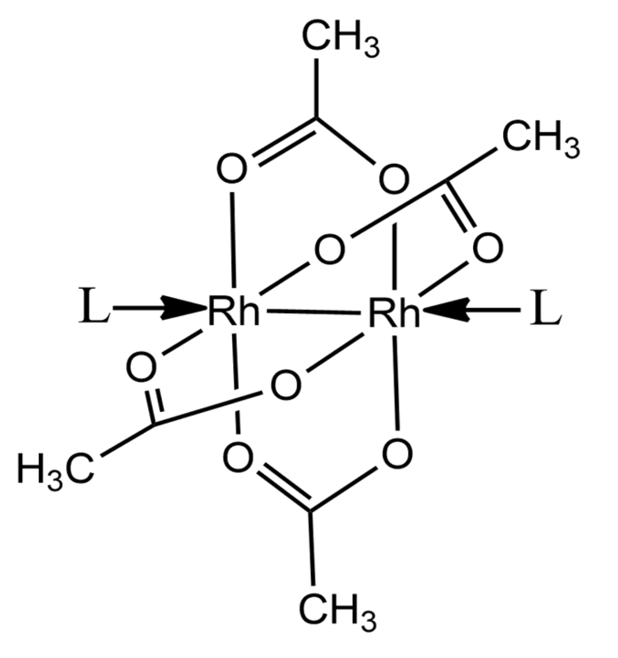
Dirhodium Tetraacetate Binding to Lysozyme at Body Temperature

Department of Chemical Sciences, University of Naples Federico II, 80126 Naples, Italy
*
Author to whom correspondence should be addressed.
Int. J. Mol. Sci. 2025, 26(14), 6582; https://doi.org/10.3390/ijms26146582
Submission received: 24 June 2025 / Revised: 5 July 2025 / Accepted: 8 July 2025 / Published: 9 July 2025
(This article belongs to the Special Issue Peptide and Protein Metalation)

Abstract

Paddlewheel dirhodium complexes are cytotoxic compounds that are also used as catalysts and in the formation of Rh-based artificial metalloenzymes. Low-temperature structures of adducts formed by the model protein hen egg white lysozyme (HEWL) with dirhodium tetraacetate ([Rh2(μ-O2CCH3)4]) when crystals of the protein were treated with the metal compound at 20 °C demonstrated that [Rh2(μ-O2CCH3)4] in part breaks down upon reaction with HEWL; dimeric Rh-Rh units bind the side chains of Asp18 and the C-terminal carboxylate, and monometallic fragments coordinate the side chains of Arg14 and His15 in 20% ethylene glycol, 0.100 M sodium acetate at pH 4.5 and 0.600 M sodium nitrate, while dimeric Rh-Rh units bind the side chains of Asn93 and Lys96, the C-terminal carboxylate and Asp101, with monometallic fragments that bind the side chains of Lys33 and His15 in 0.010 M HEPES pH 7.5 and 2.00 M sodium formate. To verify whether the binding of this metallodrug to proteins also occurs at body temperature, crystals of HEWL were grown in 0.010 M HEPES pH 7.5 and 2.00 M sodium formate at 37 °C and soaked with [Rh2(μ-O2CCH3)4] at the same temperature. X-ray diffraction data collected on these crystals at 37 °C demonstrate that [Rh2(μ-O2CCH3)4] reacts with proteins at body temperature. The structures of the Rh/HEWL adduct formed at 20 °C (obtained from data collected at 100 K) and at 37 °C under the same experimental conditions are very similar, with metal binding sites that are conserved. However, metal-containing fragment occupancy is higher in the structure obtained at 37 °C, suggesting a role of temperature in defining the protein metalation process.

1. Introduction

X-ray crystallography is routinely used for protein structure determination. The large majority of structures reported in the Protein Data Bank (PDB) have been obtained at 100 K [1], since data collection at this temperature minimizes the formation and diffusion of deleterious reactive species that are formed upon the collision of X-rays with protein atoms and reduces the crystal X-ray diffraction power [2]. Crystallographic experiments at cryogenic temperatures also have other advantages: the low temperature mitigates specific radiation damage [3], allows easy storage and transport of crystals and can be used to trap catalytic intermediates. However, the use of a low temperature is also associated with some disadvantages: functionally relevant enzyme conformations can be overlooked at low temperature; on the other hand, cryogenic temperature structures could show details that are not necessarily significant from a physiological point of view [4]. Recently, there has been growing interest in the search for functionally relevant information that may be missing in low-temperature structures [5]. It has been shown that temperature variations can drive the rearrangement of water molecules bound to a protein structure [6]. Furthermore, physiological temperature can induce conformational changes that hamper the formation of interactions between proteins and metal compounds [7,8]. The body temperature structure of the Nsp3 macrodomain of SARS-CoV-2 revealed a more expanded catalytic site when compared to the structure at low temperature [9], while some local conformational variations were observed in comparing the 310 K structure of SARS-CoV-2 Mpro with that solved at cryogenic and room temperatures [10]. These results have renewed interest in data collection at temperatures higher than 100 K [11].
Within this framework, very recently, Jacobs, Helliwell and Brink determined the crystal structure of a metal/protein adduct by collecting data at body temperature (37 °C/310 K) [12]. Along the same line, we solved the first structure of a polyoxidovanadate/protein adduct formed at 310 K [13]. A search in the Protein Data Bank revealed that these two X-ray structures are the only ones with reports of detailed atomic models of the interaction between a protein and a metal compound at 310 K. Since structural studies on adducts formed by proteins with metallodrugs at body temperature are important [14,15,16,17], there is a substantial information gap in this field. To obtain further data on this topic, here we studied the interaction of the paddlewheel compound dirhodium tetraacetate ([Rh2(μ-O2CCH3)4]) (Figure 1) with the model protein hen egg white lysozyme (HEWL) at body temperature, solving the X-ray structure of the Rh/HEWL adduct formed upon the reaction of the compound with the protein at 310 K. We chose this system since it had already been the subject of structural studies at 100 K [18]. In particular, it has been shown that [Rh2(μ-O2CCH3)4], a cytotoxic compound [19,20,21] that is also used as a catalyst [22,23,24,25], in part breaks down upon reaction with HEWL at 20 °C into several metal-containing fragments that are able to bind the protein. HEWL metalation by [Rh2(μ-O2CCH3)4] has been studied by solving the crystal structures of Rh/HEWL adducts formed in 20% ethylene glycol, 0.100 M sodium acetate at pH 4.5 and 0.600 M sodium nitrate, and in 0.010 M HEPES pH 7.5 and 2.00 M sodium formate [18]. In the former structure, Rh-Rh units have been found close to the Asp18 side chain and to the C-terminal carboxylate, while monometallic fragments have been found close to the side chains of Arg14 and His15. In the latter structure, Rh-Rh units bind the side chains of Asn93 and Lys96, the C-terminal carboxylate and Asp101, while monometallic fragments are found close to the side chains of Lys33 and His15.

2. Results and Discussion

2.1. Crystals of the Rh/HEWL Adduct at 310 K

To study HEWL metalation by [Rh2(μ-O2CCH3)4], crystals of Rh/HEWL adducts were obtained at 37 °C, treating crystals of the metal-free protein grown at 37 °C in 0.010 M HEPES pH 7.5 and 2.00 M sodium formate with a solution of the reservoir containing a large excess of [Rh2(μ-O2CCH3)4] for 1 day. This condition was chosen since it was used to obtain one of the already-solved structures of the Rh/HEWL adduct [18] and allows HEWL crystallization at 37 °C to occur in a short time (1 day). To collect data at 37 °C, it is important to reduce solvent evaporation from the crystal and successive dehydration. This is possible by adding paratone-N oil [26] or closing the protein crystals within a quartz capillary containing a few crystallization drops [12]. The Rh/HEWL adduct crystals were positioned within a borosilicate glass capillary (glass 0500, linear absorption coefficient 71.0 µ cm−1) closed with wax, maintained at 37 °C and containing droplets of the crystallization solution stored at 37 °C. X-ray diffraction data at 37 °C were collected at a resolution (2.12 Å, Table 1) that is lower than that of the 100 K structure (1.65 Å). In agreement with previous observations [5], data collected at 37 °C on the crystal of the Rh/HEWL adduct indicate a slight expansion of the cell volume when compared to that of the crystal at 100 K (233789 Å3 at 310 K vs. 231255 Å3 at 100 K).

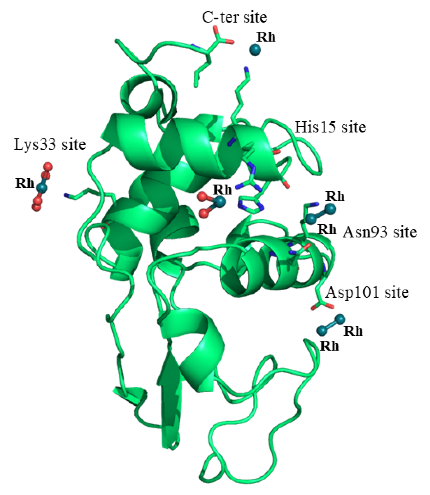
2.2. Structure of the Rh/HEWL Adduct at 310 K

The structure of the Rh/HEWL adduct at 37 °C contains 129 residues, 24 water molecules interacting with N or O atoms of the protein or water molecules, one sodium ion and seven Rh atoms with six water molecules as metal ligands, and refines with R-factor/R-free values of 0.198/0.261 (Table 1). The structure closely resembles that of the metal-free protein deposited in the PDB under the accession code 193L [27], with a root mean square deviation between the carbon alpha atoms (r.m.s.d.) of 0.30 Å (Figure 2). Inspection of the electron density map revealed the presence of five Rh binding sites (three monometallic centers and two dimetallic centers) on the protein surface. Rh centers are identified close to the C-terminal carboxylate, to the side chain of Lys13 and to the side chains of Lys33, Asn93 and Lys96, Asp101, and Arg14 and His15 (Figure 3).
Close to the side chain of Lys13 and to the C-terminal carboxylate, one Rh atom is observed (occupancy = 0.50). The metal is at a distance of 2.22 Å from the oxygen of the C-terminal carboxylate and 2.50 Å from the Lys nitrogen atom (Figure 3A). Another Rh atom (occupancy = 1.00), which coordinates four water molecules, is found close to the side chain of Lys33 (distance of the metal from the NZ atom of the Lys is 2.39 Å) (Figure 3B). The last monometallic fragment is found close to the side chains of Arg14 and His15 (occupancy = 1.00). At this site, the Rh center coordinates two water molecules, the NE2 atom of the His and the NH2 atom of the Arg, with distances of 2.81 and 2.39 Å, respectively (Figure 3C). Finally, two dirhodium binding sites were identified: the first Rh-Rh center is close to the Asp101 side chain with a distance between the metal and an oxygen of the Asp of 2.90 Å (occupancy = 0.60/0.60). In this region, the Rh–Rh distance is 2.21 Å (Figure 3D). The second Rh-Rh binding site is located close to the side chains of Asn93 and Lys96 (occupancy = 0.50/0.50). Here, one of the Rh atoms of the dirhodium center binds the OD1 atom of Asn93 and NZ atom of Lys96, with distances of 2.44 and 2.65 Å, respectively (Figure 3E). At this dinuclear site, the distance between the two Rh atoms is 2.22 Å.

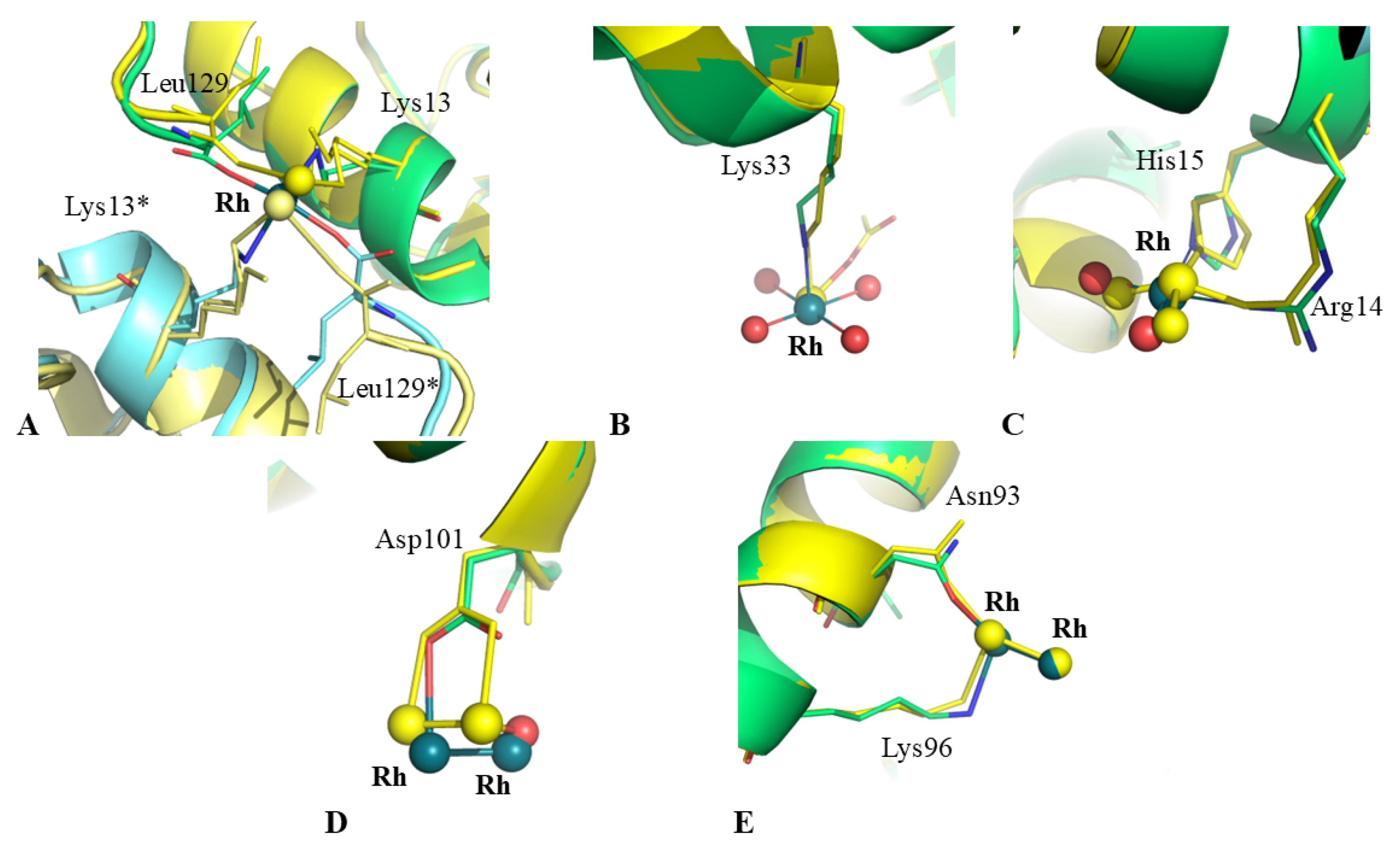
2.3. Structural Comparison with Data from Literature

X-ray structures of adducts formed upon reaction of [Rh2(μ-O2CCH3)4] with HEWL from literature have been obtained by both soaking and cocrystallization procedures [18]. Rh binding sites and metal ligands identified in these structures are reported in Table 2. The overall structure of the Rh/HEWL adduct reported here, deposited in the PDB under the accession code 9RUV, is almost identical to that of the Rh/HEWL adduct formed by soaking procedure under the same experimental conditions at 20 °C and derived from data collected at 100 K (r.m.s.d. = 0.27 Å, PDB code: 7BE2). The two structures show almost the same percentage of residues with hydrogen bonds (86% in the 100 K structure vs. 85% in the structure at 37 °C) and very similar values for the total accessible surface area (6528.8 vs. 6552.0 Å2) and for the exposed polar and non-polar accessible surface areas (1754.3 vs. 1695.4 Å2 and 3525.5 vs. 3598.2 Å2, respectively). Almost all the water molecules constituting the hydration shell of HEWL at 37 °C are conserved in the structure at 100 K. Detailed r.m.s.d. per residue calculations show that the temperature change does not lead to substantial changes in the position of protein atoms. R.m.s. deviations in carbon alpha atom positions in the 37 °C structure from those in the PDB structure with code 7BE2 show that only residues Gly4, 47–49, Pro70, Gly102, Gly117, 121–122 and 126–129 have r.m.s.d. values exceeding 0.40 Å. Notably, residues 47–49, 70, 102, 121–122 and 126–129 are close to Rh binding sites or to Rh binding sites of symmetry-related molecules. The Rh binding sites are also conserved (Table 2), although some modifications in the position of residue side chains involved in Rh binding are observed (Figure 4). This finding indicates that the reactivity of dirhodium tetraacetate with HEWL does not depend on the temperature used for the reaction of the metal complex with this biological macromolecule. This is in agreement with what has been observed in comparing the structure of the HEWL adduct with fac-[Et4N]2[Re(CO)3(Br)3] and imidazole collected at 100 K [28] and 37 °C [12], while different results have been obtained in comparing the structures of HEWL treated with [VIVO(acac)2] at 100 and 310 K [13]. A more detailed comparison between the structures of the Rh/HEWL adducts formed at low and body temperatures reveals some differences. In particular, it is interesting to note that the occupancy factors of metal centers in the structure at body temperature are, on average, higher than those found in the structure at 100 K (Table 2), although in the 100 K structure, a longer soaking time was used to obtain the adduct (7 days at 20 °C vs. 1 day at 37 °C). Although caution should be taken in interpreting this result, because of differences in the crystal dimensions and quality, in the diffusion of the metal complex within the crystal and in the soaking time, and possible radiation damage, data suggest that high temperature could enhance the HEWL metalation process by the rhodium compound, in agreement with what was found in the reaction of the same protein with cisplatin [29]. These findings could be significant since they indicate that the number of dirhodium-containing fragments that are bound to proteins at body temperature should be higher than previously believed. This datum also suggests that body temperature should be used to prepare artificial metalloenzymes based on dirhodium compounds since at this temperature, a higher number of Rh-Rh fragments bind proteins.
To verify whether the different temperatures used to prepare the crystals of the adduct and to collect X-ray diffraction data and the different occupancies of metal-containing fragments bound to HEWL could influence protein flexibility, a B-factor analysis was carried out. This allowed us to obtain information on the differences in the mobility of protein residues, a feature that is important for protein function. Obviously, B-factors in the 37 °C structure are higher than those found in the 100 K structure. Thus, to carry out a proper comparison, B-relatives (see Materials and Methods for details) were calculated (Figure 5). Inspection of Figure 5 reveals that the B-relative profiles of the two structures are very similar, with 98% of the residues that present differences in B-relatives lower than 0.3.

3. Materials and Methods

3.1. Crystallization of HEWL and Formation of Rh/HEWL Adduct at 37 °C

[Rh2(μ-O2CCH3)4] and HEWL were purchased from Merk Life Science S.r.l. (Milan, Italy) at the highest grade of purity and used without further purification. Crystals of Rh/HEWL adducts reported here were obtained using the soaking strategy at 37 °C. Metal-free HEWL crystals were grown at 37 °C using the sitting drop vapor diffusion method [30]. To obtain these crystals, the protein concentration was increased when compared to the concentration used at 20 °C. In particular, 1 µL of the protein, concentrated up to 100 mg/mL, was mixed with 1 µL of the reservoir solution containing 0.010 M HEPES pH 7.5 and 2.00 M sodium formate. Under these conditions, crystals of the protein grow in less than one day. The protein crystals were then soaked in a solution of the reservoir containing a large excess of [Rh2(μ-O2CCH3)4] for 1 day at 37 °C.

3.2. Data Collection at 37 °C, Structure Solution and Refinement

To collect X-ray diffraction data at 37 °C, crystals of the Rh/HEWL adduct formed at body temperature using the soaking experiment were fished with a nylon loop and positioned within a glass capillary containing a small drop of the reservoir solution. The capillary was then closed with wax. Data collection was carried out at the XRD2 beamline of ELETTRA synchrotron, Trieste, Italy. A complete data set can be obtained using a crystal orientation of 180° and an oscillation of 1° with an exposure time of 0.05 s per image, allowing a complete data set to be collected within 36 s. Data were scaled using Autoproc [31]. Data collection and refinement statistics are reported in Table 1. The resolution was chosen based on the CC1/2 value (cutoff > 0.3). This allowed us to include data at lower signal levels that have been demonstrated to improve protein structure models [32].
Molecular replacement was carried out using Phaser [33] from the CCP4i 7.0 software suite, using PDB code 193L [27] as the search model. Model building was carried out using Coot [34]; refinements were performed with Refmac5 [35].
Geometry and protein statistics were evaluated using the PDB validation server (www.rcsb.org), and structural analysis was carried out using the server VADAR (http://vadar.wishartlab.com/) [36] and Coot routines [34]. To carry out a detailed comparison between the structure of the adduct reported here and that of the same adduct obtained at 100 K, we verified that the radiation damage of the crystal at 37 °C was comparable to that of the low-temperature structure, analyzing the Bdamage parameter according to Garman and coworkers [37,38].
For a proper comparison of the flexibility of the protein residues, B-relatives were calculated by dividing the B-factor of the carbon alpha atoms of each residue by the average B-factor of the same atoms of the whole protein, as described in reference [39].

4. Conclusions

It is known that dirhodium tetracarboxylates bind proteins [20,40,41,42,43,44,45]. Crystals of [Rh2(μ-O2CCH3)4] with the model proteins HEWL [18] and bovine pancreatic RNase [46] have been reported; computational studies have been carried out to unveil the molecular bases of the interaction between dirhodium compounds and proteins [47,48]. However, at present, little is known about the effect of temperature on the binding of dirhodium complexes to proteins. Thus, in this work, we have studied the reactivity of dirhodium tetraacetate with HEWL at 37 °C. Our data demonstrate that Rh compounds react with the protein at body temperature, producing results that are very similar to those obtained when soaking was carried out at 20 °C, and by solving the structure of the adduct at 100 K, we found that dirhodium centers and monometallic fragments bind the enzyme at the level of the C-terminal carboxylate and close to the side chains of Lys13, Lys33, Asn93 and Lys96, Asp101, and Arg14 and His15, without altering the proteins overall conformation. These findings suggest that dirhodium compounds could also bind proteins in vivo in the cellular milieu. A strict comparison of the structures of the Rh/HEWL adducts obtained under the same experimental conditions using different temperatures for crystal growth and the soaking procedure also suggests that temperature can affect the protein metalation process: the degree of metalation increases at higher temperatures. This result, together with previous data [29], suggests that caution should be taken in administering metallodrugs to patients with fever.

Author Contributions

A.M. designed the concept and supervised the experiments. G.T. and G.F. performed the experimental work. A.M. wrote the manuscript. All authors have read and agreed to the published version of the manuscript.

Funding

This study was funded by MIUR PRIN2022 (project code: 2022JMFC3X, “Protein Metalation by Anticancer Metal-based Drugs”).

Institutional Review Board Statement

Not applicable.

Informed Consent Statement

Not applicable.

Data Availability Statement

Data is contained within the article.

Acknowledgments

The authors thank the XRD2 beamline Elettra synchrotron staff for their technical assistance during data collection.

Conflicts of Interest

The authors declare no conflicts of interest.

Abbreviations

CDCircular dichroism
HEWLHen egg white lysozyme
PDBProtein Data Bank
R.m.s.d.Root mean square deviation
RNase ABovine pancreatic ribonuclease

References

  1. Thompson, M.C. Combining Temperature Perturbations with X-Ray Crystallography to Study Dynamic Macromolecules: A Thorough Discussion of Experimental Methods. In Methods in Enzymology; Elsevier: Amsterdam, The Netherlands, 2023; Volume 688, pp. 255–305. ISBN 978-0-443-15926-8. [Google Scholar]
  2. Garman, E. “Cool” Crystals: Macromolecular Cryocrystallography and Radiation Damage. Curr. Opin. Struct. Biol. 2003, 13, 545–551. [Google Scholar] [CrossRef] [PubMed]
  3. Garman, E.F.; Weik, M. Radiation Damage in Macromolecular Crystallography. In Protein Crystallography; Wlodawer, A., Dauter, Z., Jaskolski, M., Eds.; Methods in Molecular Biology; Springer: New York, NY, USA, 2017; Volume 1607, pp. 467–489. ISBN 978-1-4939-6998-2. [Google Scholar]
  4. Juers, D.H.; Matthews, B.W. Cryo-Cooling in Macromolecular Crystallography: Advantages, Disadvantages and Optimization. Quart. Rev. Biophys. 2004, 37, 105–119. [Google Scholar] [CrossRef]
  5. Doukov, T.; Herschlag, D.; Yabukarski, F. Instrumentation and Experimental Procedures for Robust Collection of X-Ray Diffraction Data from Protein Crystals across Physiological Temperatures. J. Appl. Crystallogr. 2020, 53, 1493–1501. [Google Scholar] [CrossRef] [PubMed]
  6. Aponte-Santamaría, C.; Fischer, G.; Båth, P.; Neutze, R.; De Groot, B.L. Temperature Dependence of Protein-Water Interactions in a Gated Yeast Aquaporin. Sci. Rep. 2017, 7, 4016. [Google Scholar] [CrossRef]
  7. Ferraro, G.; Garribba, E.; Merlino, A. Exploring Polyoxidovanadate–Protein Interaction. Trends Chem. 2025, 7, 3–6. [Google Scholar] [CrossRef]
  8. Hu, J.; Park, S.J.; Walter, T.; Orozco, I.J.; O.‘Dea, G.; Ye, X.; Du, J.; Lü, W. Physiological Temperature Drives TRPM4 Ligand Recognition and Gating. Nature 2024, 630, 509–515. [Google Scholar] [CrossRef]
  9. Schuller, M.; Correy, G.J.; Gahbauer, S.; Fearon, D.; Wu, T.; Díaz, R.E.; Young, I.D.; Carvalho Martins, L.; Smith, D.H.; Schulze-Gahmen, U.; et al. Fragment Binding to the Nsp3 Macrodomain of SARS-CoV-2 Identified through Crystallographic Screening and Computational Docking. Sci. Adv. 2021, 7, eabf8711. [Google Scholar] [CrossRef]
  10. Ebrahim, A.; Riley, B.T.; Kumaran, D.; Andi, B.; Fuchs, M.R.; McSweeney, S.; Keedy, D.A. The Temperature-Dependent Conformational Ensemble of SARS-CoV-2 Main Protease (Mpro). IUCrJ 2022, 9, 682–694. [Google Scholar] [CrossRef]
  11. Fischer, M. Macromolecular Room Temperature Crystallography. Quart. Rev. Biophys. 2021, 54, e1. [Google Scholar] [CrossRef]
  12. Jacobs, F.J.F.; Helliwell, J.R.; Brink, A. Body Temperature Protein X-Ray Crystallography at 37 °C: A Rhenium Protein Complex Seeking ac Physiological Condition Structure. Chem. Commun. 2024, 60, 14030–14033. [Google Scholar] [CrossRef]
  13. Tito, G.; Ferraro, G.; Garribba, E.; Merlino, A. Formation of Mixed-Valence Cage-Like Polyoxidovanadates at 37 °C Upon Reaction of VIV O(Acetylacetonato)2 with Lysozyme. Chem. –A Eur. J. 2025, 31, e202500488. [Google Scholar] [CrossRef]
  14. Merlino, A. Recent Advances in Protein Metalation: Structural Studies. Chem. Commun. 2021, 57, 1295–1307. [Google Scholar] [CrossRef]
  15. Marzo, T.; Ferraro, G.; Merlino, A.; Messori, L. Protein Metalation by Inorganic Anticancer Drugs. In Encyclopedia of Inorganic and Bioinorganic Chemistry; Scott, R.A., Ed.; Wiley: Hoboken, NJ, USA, 2020; pp. 1–17. ISBN 978-1-119-95143-8. [Google Scholar]
  16. Merlino, A.; Marzo, T.; Messori, L. Protein Metalation by Anticancer Metallodrugs: A Joint ESI MS and XRD Investigative Strategy. Chem. –A Eur. J. 2017, 23, 6942–6947. [Google Scholar] [CrossRef]
  17. Messori, L.; Merlino, A. Protein Metalation by Metal-Based Drugs: X-Ray Crystallography and Mass Spectrometry Studies. Chem. Commun. 2017, 53, 11622–11633. [Google Scholar] [CrossRef]
  18. Loreto, D.; Ferraro, G.; Merlino, A. Unusual Structural Features in the Adduct of Dirhodium Tetraacetate with Lysozyme. Int. J. Mol. Sci. 2021, 22, 1496. [Google Scholar] [CrossRef]
  19. Erck, A.; Rainen, L.; Whileyman, J.; Chang, I.-M.; Kimball, A.P.; Bear, J. Studies of Rhodium(II) Carboxylates as Potential Antitumor Agents. Exp. Biol. Med. 1974, 145, 1278–1283. [Google Scholar] [CrossRef]
  20. Bear, J.L.; Gray, H.B., Jr.; Rainen, L.; Chang, I.M.; Howard, R. Interaction of Rhodium (II) Carboxylates With Molecules of Biologic Importance. Cancer Chemother. Rep. 1975, 59, 611–620. [Google Scholar]
  21. Chang, I.; Woo, W.S. Effects of Rh2 (O2CC2H5) 4L2 on the Replication of Ehrlich Tumor Cells in Vivo. Korean Biochem. J. 1976, 9, 175–180. [Google Scholar]
  22. Loreto, D.; Maity, B.; Morita, T.; Nakamura, H.; Merlino, A.; Ueno, T. Cross-Linked Crystals of Dirhodium Tetraacetate/RNase A Adduct Can Be Used as Heterogeneous Catalysts. Inorg. Chem. 2023, 62, 7515–7524. [Google Scholar] [CrossRef]
  23. Hrdina, R. Dirhodium(II, II) Paddlewheel Complexes. Eur. J. Inorg. Chem. 2021, 2021, 501–528. [Google Scholar] [CrossRef]
  24. Chifotides, H.T.; Dunbar, K.R. Rhodium Compounds. In Multiple Bonds Between Metal Atoms; Cotton, F.A., Murillo, C.A., Walton, R.A., Eds.; Springer: New York, NY, USA, 2005; pp. 465–589. ISBN 978-0-387-25084-7. [Google Scholar]
  25. Cotton, F.A.; Murillo, C.A.; Walton, R.A. Multiple Bonds Between Metal Atoms; Springer Science & Business Media: Berlin/Heidelberg, Germany, 2006. [Google Scholar]
  26. Hope, H. Cryocrystallography of Biological Macromolecules: A Generally Applicable Method. Acta Crystallogr. B Struct. Sci. 1988, 44, 22–26. [Google Scholar] [CrossRef]
  27. Vaney, M.C.; Maignan, S.; Riès-Kautt, M.; Ducruix, A. High-Resolution Structure (1.33 Å) of a HEW Lysozyme Tetragonal Crystal Grown in the APCF Apparatus. Data and Structural Comparison with a Crystal Grown under Microgravity from SpaceHab-01 Mission. Acta Crystallogr. D Biol. Crystallogr. 1996, 52, 505–517. [Google Scholar] [CrossRef] [PubMed]
  28. Jacobs, F.J.F.; Helliwell, J.R.; Brink, A. Time-Series Analysis of Rhenium(I) Organometallic Covalent Binding to a Model Protein for Drug Development. IUCrJ 2024, 11, 359–373. [Google Scholar] [CrossRef] [PubMed]
  29. Ferraro, G.; Pica, A.; Russo Krauss, I.; Pane, F.; Amoresano, A.; Merlino, A. Effect of Temperature on the Interaction of Cisplatin with the Model Protein Hen Egg White Lysozyme. J. Biol. Inorg. Chem. 2016, 21, 433–442. [Google Scholar] [CrossRef]
  30. Russo Krauss, I.; Ferraro, G.; Pica, A.; Márquez, J.A.; Helliwell, J.R.; Merlino, A. Principles and Methods Used to Grow and Optimize Crystals of Protein–Metallodrug Adducts, to Determine Metal Binding Sites and to Assign Metal Ligands. Metallomics 2017, 9, 1534–1547. [Google Scholar] [CrossRef]
  31. Vonrhein, C.; Flensburg, C.; Keller, P.; Sharff, A.; Smart, O.; Paciorek, W.; Womack, T.; Bricogne, G. Data Processing and Analysis with the autoPROC Toolbox. Acta Crystallogr. D Biol. Crystallogr. 2011, 67, 293–302. [Google Scholar] [CrossRef]
  32. Karplus, P.A.; Diederichs, K. Linking Crystallographic Model and Data Quality. Science 2012, 336, 1030–1033. [Google Scholar] [CrossRef]
  33. McCoy, A.J.; Grosse-Kunstleve, R.W.; Adams, P.D.; Winn, M.D.; Storoni, L.C.; Read, R.J. Phaser Crystallographic Software. J. Appl. Crystallogr. 2007, 40, 658–674. [Google Scholar] [CrossRef]
  34. Emsley, P.; Cowtan, K. Coot: Model-Building Tools for Molecular Graphics. Acta Crystallogr. D Biol. Crystallogr. 2004, 60, 2126–2132. [Google Scholar] [CrossRef]
  35. Murshudov, G.N.; Skubák, P.; Lebedev, A.A.; Pannu, N.S.; Steiner, R.A.; Nicholls, R.A.; Winn, M.D.; Long, F.; Vagin, A.A. REFMAC 5 for the Refinement of Macromolecular Crystal Structures. Acta Crystallogr. D Biol. Crystallogr. 2011, 67, 355–367. [Google Scholar] [CrossRef]
  36. Willard, L. VADAR: A Web Server for Quantitative Evaluation of Protein Structure Quality. Nucleic. Acids. Res. 2003, 31, 3316–3319. [Google Scholar] [CrossRef] [PubMed]
  37. Shelley, K.L.; Dixon, T.P.E.; Brooks-Bartlett, J.C.; Garman, E.F. RABDAM: Quantifying Specific Radiation Damage in Individual Protein Crystal Structures. J. Appl. Crystallogr. 2018, 51, 552–559. [Google Scholar] [CrossRef] [PubMed]
  38. Shelley, K.L.; Garman, E.F. Quantifying and Comparing Radiation Damage in the Protein Data Bank. Nat. Commun. 2022, 13, 1314. [Google Scholar] [CrossRef] [PubMed]
  39. Merlino, A.; Russo Krauss, I.; Castellano, I.; Vendittis, E.D.; Rossi, B.; Conte, M.; Vergara, A.; Sica, F. Structure and Flexibility in Cold-Adapted Iron Superoxide Dismutases: The Case of the Enzyme Isolated from Pseudoalteromonas Haloplanktis. J. Struct. Biol. 2010, 172, 343–352. [Google Scholar] [CrossRef]
  40. Loreto, D.; Merlino, A. The Interaction of Rhodium Compounds with Proteins: A Structural Overview. Coord. Chem. Rev. 2021, 442, 213999. [Google Scholar] [CrossRef]
  41. Enriquez Garcia, A.; Jalilehvand, F.; Niksirat, P.; Gelfand, B.S. Methionine Binding to Dirhodium(II) Tetraacetate. Inorg. Chem. 2018, 57, 12787–12799. [Google Scholar] [CrossRef]
  42. Howard, R.A.; Spring, T.G.; Bear, J. L The Interaction of Rhodium (II) Carboxylates with Enzymes. Cancer Res. 1976, 36, 4402–4405. [Google Scholar]
  43. Loreto, D.; Esposito, A.; Demitri, N.; Guaragna, A.; Merlino, A. Reactivity of a Fluorine-Containing Dirhodium Tetracarboxylate Compound with Proteins. Dalton Trans. 2022, 51, 3695–3705. [Google Scholar] [CrossRef]
  44. Loreto, D.; Esposito, A.; Demitri, N.; Guaragna, A.; Merlino, A. Digging into Protein Metalation Differences Triggered by Fluorine Containing-Dirhodium Tetracarboxylate Analogues. Dalton Trans. 2022, 51, 7294–7304. [Google Scholar] [CrossRef]
  45. Loreto, D.; Fasulo, F.; Muñoz-García, A.B.; Pavone, M.; Merlino, A. Unexpected Imidazole Coordination to the Dirhodium Center in a Protein Environment: Insights from X-Ray Crystallography and Quantum Chemistry. Inorg. Chem. 2022, 61, 8402–8405. [Google Scholar] [CrossRef]
  46. Ferraro, G.; Pratesi, A.; Messori, L.; Merlino, A. Protein Interactions of Dirhodium Tetraacetate: A Structural Study. Dalton Trans. 2020, 49, 2412–2416. [Google Scholar] [CrossRef] [PubMed]
  47. Tolbatov, I.; Marrone, A. Reaction of Dirhodium and Diruthenium Paddlewheel Tetraacetate Complexes with Nucleophilic Protein Sites: A Computational Study. Inorganica Chim. Acta 2022, 530, 120684. [Google Scholar] [CrossRef]
  48. Tolbatov, I.; Umari, P.; Marrone, A. Diruthenium Paddlewheel Complexes Attacking Proteins: Axial versus Equatorial Coordination. Biomolecules 2024, 14, 530. [Google Scholar] [CrossRef] [PubMed]
Figure 1. Paddlewheel structure of [Rh2(µ-O2CCH3)4]. L indicates possible ligands in the axial position.
Ijms 26 06582 g001
Figure 2. Overall structure of Rh/HEWL adduct at 37 °C. Rh binding sites are evidenced (Rh atoms are colored in dark green, water molecules in red). PDB code: 9RUV.
Ijms 26 06582 g002
Figure 3. Details of the Rh binding sites in the structure of the Rh/HEWL adduct formed at 37 °C: Lys13 and C-terminal carboxylate (A), Lys33 (B), Arg14-His15 (C), Asp101 (D) and Asn93-Lys96 (E). 2Fo-Fc electron density maps are contoured at 1.0 σ (light blue). Rh atoms are colored in dark green, water molecules in red. Asterisk (*) indicates residues from symmetry-related molecules.
Ijms 26 06582 g003
Figure 4. Overlay of the Rh binding sites in the structure at 37 °C vs. that from data collected at 100 K (PDB code 7BE2, yellow). Lys13 and C-terminal carboxylate (A), Lys33 (B), Arg14-His15 (C), Asp101 (D) and Asn93-Lys96 (E). Rh atoms are colored in dark green, water molecules in red. Asterisk (*) indicates residues from symmetry-related molecules.
Ijms 26 06582 g004
Figure 5. B-relative (Brelative) versus residue number trend in Rh/HEWL structures obtained at 37 °C and 100 K. B-relative is defined as the ratio between the B-factor of the carbon α atom of each residue and the average B-factor of the carbon α atoms of the whole molecule.
Ijms 26 06582 g005
Table 1. Data collection and refinement statistics.
Crystal
Temperature37 °C
Crystallization condition0.01 M HEPES pH 7.5 and 2.00 M sodium formate
Data collection
Space groupP43212
a = b (Å)80.43
c (Å)36.14
Resolution range (Å)56.87–2.12 (2.15–2.12)
Unique reflections6717 (342)
Completeness (%)94.0 (100.0)
Redundancy11.4 (11.8)
Rmerge (%)0.092 (2.788)
Average I/σ (I)13.2 (0.9)
CC1/20.998 (0.405)
Anom. completeness (%)93.7 (100.0)
Anom. redundancy6.4 (6.3)
Refinement
Resolution range (Å)56.87–2.12
N. of reflections6183
N. of reflections (working set)382
R-factor/R-free (%)19.8/26.1
N. of atoms1043
Average B-factors (Å2)
All atoms61.2
Rh occupancy0.50/1.00/1.00/0.60/0.50
Rh atoms100.5/125.1/159.9/103.2/115.7/134.7/139.1
R.m.s.d. from ideality
Bond lengths (Å)0.007
Bond angles (°)1.57
Ramachandran values (%)
Most favored/Additionally allowed91.9/7.3
Outliers0.8
Table 2. Comparison of Rh binding sites between the structure of the Rh/HEWL adduct obtained at 37 °C and the structure obtained at 20 °C. Table describes the ligands at each Rh binding site. Values in parentheses refer to the occupancy of metal and ligands. Asterisk (*) indicates residues from symmetry-related molecules.
Rh/HEWL AdductCrystal at 310 KData from LiteratureData from LiteratureData from LiteratureData from LiteratureData from LiteratureData from Literature
Crystallization condition0.01 M HEPES pH 7.5 and 2.0 M sodium formate0.1 M sodium acetate, 20% ethylene glicol, 0.1 M sodium nitrate
PDB code9RUV7BE27BDZ7BE07BE17BEB7BEC
Binding sitesMetal fragments at binding sites
C-ter siteRh (0.50)
Rh * (0.50)
Leu129 (1)
Leu129 * (1)
Lys13 (1)
Lys13 * (1)
Rh (0.45)
Rh * (0.45)
Leu129 (1)
Leu129 * (1)
Lys13 (1)
Lys13 * (1)
H2O (0.5)
H2O * (0.5)
Rh (0.65)
Rh * (0.65)
Leu129 (1)
Leu129 * (1)
Lys13 (1)
Lys13 * (1)
Act (0.60)
Act (0.60)
H2O (0.65)
H2O * (0.65)
Rh (0.65)
Rh * (0.65)
Leu129 (1)
Leu129 * (1)
Lys13 (1)
Lys13 * (1)
Act (0.65)
Act (0.65)
H2O (0.65)
H2O* (0.65)
Rh (0.65)
Rh* (0.65)
Leu129 (1)
Leu129* (1)
Lys13 (1)
Lys13* (1)
Act (0.65)
Act (0.65)
H2O (0.65)
H2O * (0.65)
Rh (0.7)
Rh * (0.7)
Leu129 (1)
Leu129 * (1)
Lys13 (1)
Lys13 * (1)
Act (0.7)
Act (0.7)
H2O (0.7)
H2O * (0.7)
Rh (0.4)
Rh * (0.4)
Leu129 (1)
Leu129 * (1)
Lys13 (1)
Lys13 * (1)
Lys33 siteRh (1)
Lys33 (1)
H2O (1)
H2O (1)
H2O (1)
H2O (1)
Rh (0.20)
Lys33 (1)
Act (0.33)
-----
His15 siteRh (1)
His15 (1)
Arg14 (1)
H2O (1)
H2O (1)
Rh (0.35)
His15 (1)
Arg14 (1)
H2O (0.35)
H2O (0.35)
Rh (0.35)
His15 (1)
Arg14 (1)
Act (1)
H2O (0.35)
H2O (0.65)
Rh (0.5)
His15 (1)
Arg14 (1)
Act (1)
H2O (0.5)
Rh (0.3)
His15 (1)
Arg14 (0.5)
Act (1)
Rh (0.3)
His15 (1)
Arg14 (0.5)
Act (1)
H2O (0.3)
H2O (0.3)
Rh (0.25)
His15 (1)
Arg14 (0.5)
Act (1)
H2O (1)
Asp101 siteRh (0.60)
Rh (0.60)
Asp101 (1)
Rh (0.40)
Rh (0.40)
Asp101 (1)
H2O (0.40)
-----
Asn93 siteRh (0.50)
Rh (0.50)
Asn93 (1)
Lys96 (1)
Rh (0.30)
Rh (0.30)
Asn93 (1)
Lys96 (1)
H2O (1)
-----
Asp18 site--Rh (0.40)
Rh (0.40)
Asp18 (1)
Rh (0.20)
Rh (0.20)
Asp18 (1)
H2O (0.20)
---
Disclaimer/Publisher’s Note: The statements, opinions and data contained in all publications are solely those of the individual author(s) and contributor(s) and not of MDPI and/or the editor(s). MDPI and/or the editor(s) disclaim responsibility for any injury to people or property resulting from any ideas, methods, instructions or products referred to in the content.

