
Dietary Gluten-Free Regimen Does Not Affect the Suppression of the Inflammatory Response in the Arachidonic Acid Cascade in Hashimoto’s Disease

 ,
, 
Abstract
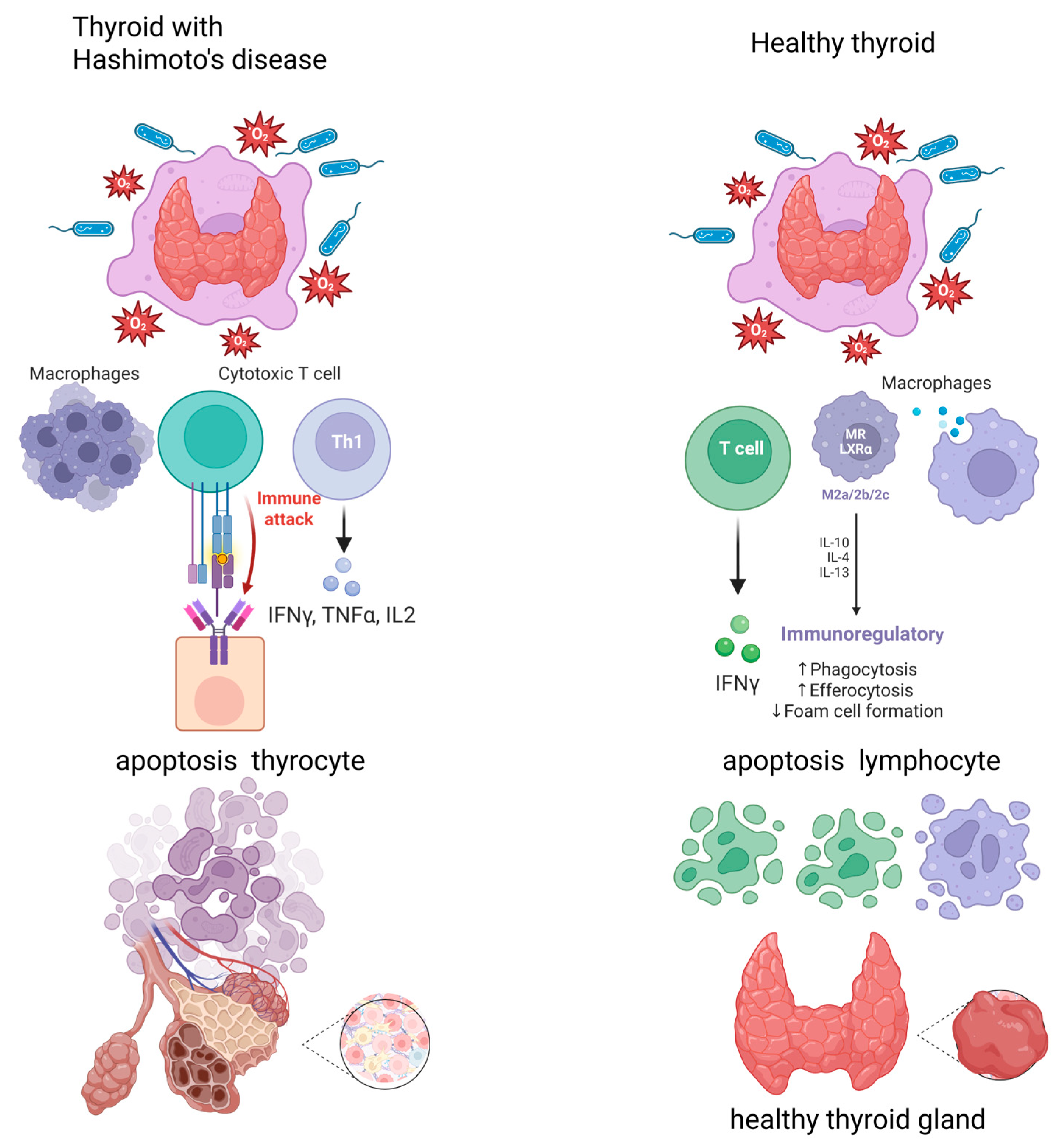
1. Introduction
2. Results
2.1. Characteristics of Study Group
2.2. Pro-Inflammatory Derivatives of Arachidonic Acid
2.3. Summary of Results
- Using a gluten-free diet for three months did not lead to a statistically significant change in the anthropometric and biochemical parameters, except for TSH (which resulted from concomitant levothyroxine treatment in the patients).
- The levels of mediators from the lipoxygenase pathway (LTB4) increased statistically significantly after gluten elimination, and the monooxygenase pathway (16RS-HETE) decreased after gluten elimination from the diet (trend).
- There was a positive correlation among TXB2, LTB4, and body weight before and after three months of following a gluten-free diet.
- There was a positive correlation among 16RS-HETE, body weight, and body fat mass before implementing a gluten-free diet.
- LTB4 was significantly correlated with CRP before and after following a gluten-free diet.
3. Discussion
4. Materials and Methods
4.1. Study Group
4.2. Dietary Intervention
4.3. Eicosanoid Extraction
4.4. High-Performance Liquid Chromatography (HPLC)
4.5. Statistical Analysis
5. Conclusions
Author Contributions
Funding
Institutional Review Board Statement
Informed Consent Statement
Data Availability Statement
Conflicts of Interest
References
- Klubo-Gwiezdzinska, J.; Wartofsky, L. Hashimoto thyroiditis: An evidence-based guide to etiology, diagnosis and treatment. Pol. Arch. Intern. Med. 2022, 132, 16222. [Google Scholar] [CrossRef]
- Hiromatsu, Y.; Satoh, H.; Amino, N. Hashimoto’s thyroiditis: History and future outlook. Hormones 2013, 12, 12–18. [Google Scholar] [CrossRef]
- Wu, Y.; Cai, T.; Xu, W.; Yang, X.; Gu, P.; Zhang, J. Polymorphisms of B-lymphocyte-associated genes CD20 and FCRL5 are associated with susceptibility to autoimmune thyroid diseases. Hum. Immunol. 2024, 85, 111165. [Google Scholar] [CrossRef]
- Zaletel, K.; Gaberšček, S. Hashimoto’s Thyroiditis: From Genes to the Disease. Curr. Genom. 2011, 12, 576–588. [Google Scholar] [CrossRef]
- Liontiris, M.I.; Mazokopakis, E.E. A concise review of Hashimoto thyroiditis (HT) and the importance of iodine, selenium, vitamin D and gluten on the autoimmunity and dietary management of HT patients. Points that need more investigation. Hell. J. Nucl. Med. 2017, 20, 51–56. [Google Scholar]
- Pyzik, A.; Grywalska, E.; Matyjaszek-Matuszek, B.; Roliński, J. Immune disorders in Hashimoto’s thyroiditis: What do we know so far? J. Immunol. Res. 2015, 2015, 979167. [Google Scholar] [CrossRef]
- Chistiakov, D.A. Immunogenetics of Hashimoto’s thyroiditis. J. Autoimmune Dis. 2005, 2, 1. [Google Scholar] [CrossRef]
- Ai, J.; Leonhardt, J.M.; Heymann, W.R. Autoimmune thyroid diseases: Etiology, pathogenesis, and dermatologic manifestations. J. Am. Acad. Dermatol. 2003, 48, 641–659. [Google Scholar] [CrossRef]
- Martin, S.A.; Brash, A.R.; Murphy, R.C. The discovery and early structural studies of arachidonic acid. J. Lipid Res. 2016, 57, 1126–1132. [Google Scholar] [CrossRef]
- Wang, T.; Fu, X.; Chen, Q.; Patra, J.K.; Wang, D.; Wang, Z.; Gai, Z. Arachidonic Acid Metabolism and Kidney Inflammation. Int. J. Mol. Sci. 2019, 20, 3683. [Google Scholar] [CrossRef]
- Meirer, K.; Steinhilber, D.; Proschak, E. Inhibitors of the arachidonic acid cascade: Interfering with multiple pathways. Basic Clin. Pharmacol. Toxicol. 2014, 114, 83–91. [Google Scholar] [CrossRef] [PubMed]
- Korbecki, J.; Baranowska-Bosiacka, I.; Gutowska, I.; Chlubek, D. Cyclooxygenase pathways. Acta Biochim. Pol. 2014, 61, 639–649. [Google Scholar] [CrossRef]
- Wang, B.; Wu, L.; Chen, J.; Dong, L.; Chen, C.; Wen, Z.; Hu, J.; Fleming, I.; Wang, D.W. Metabolism pathways of arachidonic acids: Mechanisms and potential therapeutic targets. Signal Transduct. Target Ther. 2021, 6, 94. [Google Scholar] [CrossRef]
- Christie, W.W.; Harwood, J.L. Oxidation of polyunsaturated fatty acids to produce lipid mediators. Essays Biochem. 2020, 64, 401–421. [Google Scholar]
- Pobłocki, J.; Pańka, T.; Szczuko, M.; Telesiński, A.; Syrenicz, A. Whether a Gluten-Free Diet Should Be Recommended in Chronic Autoimmune Thyroiditis or Not?—A 12-Month Follow-Up. J. Clin. Med. 2021, 10, 3240. [Google Scholar] [CrossRef] [PubMed]
- Melini, V.; Melini, F. Gluten-Free Diet: Gaps and Needs for a Healthier Diet. Nutrients 2019, 11, 170. [Google Scholar] [CrossRef] [PubMed]
- Passali, M.; Josefsen, K.; Frederiksen, J.L.; Antvorskov, J.C. Current Evidence on the Efficacy of Gluten-Free Diets in Multiple Sclerosis, Psoriasis, Type 1 Diabetes and Autoimmune Thyroid Diseases. Nutrients 2020, 12, 2316. [Google Scholar] [CrossRef]
- Ostrowska, L.; Gier, D.; Zyśk, B. The Influence of Reducing Diets on Changes in Thyroid Parameters in Women Suffering from Obesity and Hashimoto’s Disease. Nutrients 2021, 13, 862. [Google Scholar] [CrossRef]
- Araújo, E.M.Q.; Ramos, H.E.; Trevisani, V.F.M. Commentary: Effect of gluten-free diet on autoimmune thyroiditis progression in patients with no symptoms or histology of celiac disease: A meta-analysis. Front. Endocrinol. 2024, 15, 1459941. [Google Scholar] [CrossRef]
- Ashok, T.; Patni, N.; Fatima, M.; Lamis, A.; Siddiqui, S.W. Celiac Disease and Autoimmune Thyroid Disease: The Two Peas in a Pod. Cureus 2022, 14, e26243. [Google Scholar] [CrossRef]
- Ihnatowicz, P.; Wątor, P.; Drywień, M.E. The importance of gluten exclusion in the management of Hashimoto’s thyroiditis. Ann. Agric. Environ. Med. 2021, 28, 558–568. [Google Scholar] [CrossRef] [PubMed]
- Mikulska, A.A.; Karaźniewicz-Łada, M.; Filipowicz, D.; Ruchała, M.; Główka, F.K. Metabolic Characteristics of Hashimoto’s Thyroiditis Patients and the Role of Microelements and Diet in the Disease Management—An Overview. Int. J. Mol. Sci. 2022, 23, 6580. [Google Scholar] [CrossRef] [PubMed]
- Krysiak, R.; Szkróbka, W.; Okopień, B. The Effect of Gluten-Free Diet on Thyroid Autoimmunity in Drug-Naïve Women with Hashimoto’s Thyroiditis: A Pilot Study. Exp. Clin. Endocrinol. Diabetes 2019, 127, 417–422. [Google Scholar] [CrossRef] [PubMed]
- Sategna-Guidetti, C.; Volta, U.; Ciacci, C.; Usai, P.; Carlino, A.; De Franceschi, L.; Camera, A.; Pelli, A.; Brossa, C. Prevalence of thyroid disorders in untreated adult celiac disease patients and effect of gluten withdrawal: An Italian multicenter study. Am. J. Gastroenterol. 2001, 96, 751–757. [Google Scholar] [CrossRef]
- Ventura, A.; Neri, E.; Ughi, C.; Leopaldi, A.; Città, A.; Not, T. Gluten-dependent diabetes-related and thyroid-related autoantibodies in patients with celiac disease. J. Pediatr. 2000, 137, 263–265. [Google Scholar] [CrossRef]
- Malandrini, S.; Trimboli, P.; Guzzaloni, G.; Virili, C.; Lucchini, B. What about TSH and Anti-Thyroid Antibodies in Patients with Autoimmune Thyroiditis and Celiac Disease Using a Gluten-Free Diet? A Systematic Review. Nutrients 2022, 14, 1681. [Google Scholar] [CrossRef]
- Carroccio, A.; D’Alcamo, A.; Cavataio, F.; Soresi, M.; Seidita, A.; Sciumè, C.; Geraci, G.; Iacono, G.; Mansueto, P. High Proportions of People with Nonceliac Wheat Sensitivity Have Autoimmune Disease or Antinuclear Antibodies. Comp. Study Gastroenterol. 2015, 149, 596–603.e1. [Google Scholar] [CrossRef]
- Vojdani, A.; Lambert, J.; Vojdani, E. The Gut-Brain Axis: Autoimmune and Neuroimmune Disorders. Altern. Ther. Health Med. 2016, 22 (Suppl. S3), 31–46. [Google Scholar]
- Riseh, S.H.; Farhang, M.A.; Mobasseri, M.; Jafarabadi, M.A. The Relationship between Thyroid Hormones, Antithyroid Antibodies, Anti-Tissue Transglutaminase and Anti-Gliadin Antibodies in Patients with Hashimoto’s Thyroiditis. Acta Endocrinol. 2017, 13, 174–179. [Google Scholar] [CrossRef]
- Kulkarni, A.; Pineros, A.R.; Walsh, M.A.; Casimiro, I.; Ibrahim, S.; Hernandez-Perez, M.; Orr, K.S.; Glenn, L.; Nadler, J.L.; Morris, M.A.; et al. 12-Lipoxygenase governs the innate immune pathogenesis of islet inflammation and autoimmune diabetes. JCI Insight 2021, 6, e147812. [Google Scholar] [CrossRef]
- Wang, N.; Zhao, X.; Wang, W.; Peng, Y.; Bi, K.; Dai, R. Targeted profiling of arachidonic acid and eicosanoids in rat tissue by UFLC-MS/MS: Application to identify potential markers for rheumatoid arthritis. Talanta 2017, 162, 479–487. [Google Scholar] [CrossRef] [PubMed]
- Marchiori, R.C.; Pereira, L.A.F.; Naujorks, A.A.; Rovaris, D.L.; Meinerz, D.F.; Duarte, M.M.F.; Rocha, J.B.T. Improvement of blood inflammatory marker levels in patients with hypothyroidism under levothyroxine treatment. BMC Endocr. Disord. 2015, 15, 32. [Google Scholar] [CrossRef] [PubMed]
- Lai, R.; Yin, B.; Feng, Z.; Deng, X.; Lv, X.; Zhong, Y.; Peng, D. The causal relationship between 41 inflammatory cytokines and hypothyroidism: Bidirectional two-sample Mendelian randomization study. Front. Endocrinol. 2024, 14, 1332383. [Google Scholar] [CrossRef] [PubMed]
- Leffler, D.A.; Dennis, M.; Edwards George, J.B.; Jamma, S.; Magge, S.; Cook, E.F. Proste, sprawdzone badanie przestrzegania diety bezglutenowej u dorosłych chorych na celiakię. Clin. Gastroenterol. Hepatol. 2009, 7, 530–536.e2. [Google Scholar] [CrossRef]
- Szczuko, M.; Kotlęga, D.; Palma, J.; Zembroń-Łacny, A.; Tylutka, A.; Gołąb-Janowska, M.; Drozd, A. Lipoxins, RevD1 and 9, 13 HODE as the most important derivatives after an early incident of ischemic stroke. Sci. Rep. 2020, 10, 12849. [Google Scholar] [CrossRef]
- Drozd, A.; Szczuko, M.; Bohatyrewicz, A.; Jurewicz, A.; Kotlęga, D. High Levels of Thromboxane (TX) Are Associated with the Sex-Dependent Non-Dipping Phenomenon in Ischemic Stroke Patients. J. Clin. Med. 2022, 11, 2652. [Google Scholar] [CrossRef]
- Szczuko, M.; Syrenicz, A.; Szymkowiak, K.; Przybylska, A.; Szczuko, U.; Pobłocki, J.; Kulpa, D. Doubtful Justification of the Gluten-Free Diet in the Course of Hashimoto’s Disease. Nutrients 2022, 14, 1727. [Google Scholar] [CrossRef]

| Parameters | Before the Diet | After the Diet | p-Value | ||
|---|---|---|---|---|---|
| Mean | SD | Mean | SD | ||
| Age (years) | 37.472 | 7.966 | 37.911 | 8.966 | 0.804 |
| Height (cm) | 167.085 | 4.749 | 166.842 | 5.989 | 0.814 |
| Body weight (kg) | 69.734 | 12.083 | 71.036 | 12.285 | 0.456 |
| BMI (kg/m2) | 25.418 | 3.858 | 25.472 | 4.286 | 0.923 |
| Body fat mass (kg) | 25.131 | 7.574 | 26.015 | 8.793 | 0.543 |
| % body fat | 34.550 | 5.836 | 35.677 | 7.397 | 0.390 |
| ATPO (IU/mL) | 187.879 | 137.993 | 197.512 | 233.735 | 0.803 |
| ATG (IU/mL) | 318.946 | 562.052 | 283.350 | 478.578 | 0.243 |
| TSH (µIU/mL) | 3.222 | 2.425 | 1.793 | 1.211 | 0.005 * |
| fT3 (pg/mL) | 2.921 | 0.508 | 2.884 | 0.426 | 0.690 |
| fT4 (ng/dL) | 2.902 | 144.954 | 1.352 | 0.208 | 0.426 |
| CRP (mg/L) | 1.570 | 1.450 | 1.430 | 1.060 | 0.568 |
| Mediators (µg/mL) | Before the Diet | After the Diet | p-Value | ||
|---|---|---|---|---|---|
| Mean ± SD | Median | Mean ± SD | Median | ||
| TXB2 | 1.417 ± 2.193 | 0.610 | 1.553 ± 1.773 | 1.223 | 0.764 |
| PGE2 | 8.400 ± 9.901 | 5.612 | 8.661 ± 9.216 | 5.552 | 0.904 |
| LTB4 | 0.202 ± 0.112 | 0.176 | 0.421 ± 0.273 | 0.343 | 0.0000 * |
| 16RS-HETE | 5.361 ± 3.349 | 4.245 | 4.019 ± 2.657 | 3.643 | 0.054 ** |
| Before the Diet | ||||
|---|---|---|---|---|
| TXB2 | PGE2 | LTB4 | 16RS HETE | |
| Body weight (kg) | 0.338596 | −0.202877 | −0.011094 | 0.343364 |
| BMI (kg/m2) | 0.260730 | −0.150895 | 0.016461 | 0.248190 |
| Body fat mass (g) | 0.340965 | −0.224413 | 0.052039 | 0.335862 |
| % body fat | 0.249897 | −0.230690 | 0.089829 | 0.281437 |
| ATPO (IU/mL) | 0.012256 | 0.034259 | −0.136327 | −0.196726 |
| ATG (IU/mL) | 0.218033 | 0.149694 | −0.035316 | 0.168946 |
| TSH (uIU/mL) | −0.201970 | 0.071220 | −0.107853 | −0.225742 |
| fT3 (pg/mL) | −0.048557 | −0.138112 | −0.099590 | −0.186665 |
| fT4 (ng/dL) | 0.253603 | −0.059071 | 0.053083 | 0.075085 |
| CRP (mg/L) | 0.042549 | 0.1701 | 0.395769 | 0.111715 |
| After the Diet | ||||
| TXB2 | PGE2 | LTB4 | 16RS HETE | |
| Body weight (kg) | 0.044887 | −0.192943 | −0.043332 | −0.067551 |
| BMI (kg/m2) | −0.060214 | −0.125306 | 0.089704 | −0.037661 |
| Body fat mass (g) | −0.007947 | −0.144111 | 0.032910 | −0.064960 |
| % body fat | −0.048196 | −0.073610 | 0.104520 | −0.031625 |
| ATPO (IU/mL) | 0.248300 | −0.050716 | −0.038358 | 0.021351 |
| ATG (IU/mL) | −0.090165 | −0.070293 | −0.173284 | −0.160617 |
| TSH (uIU/mL) | −0.142625 | −0.155101 | −0.248174 | −0.119689 |
| fT3 (pg/mL) | 0.124574 | 0.040813 | 0.091432 | 0.044906 |
| fT4 (ng/dL) | −0.039495 | −0.088208 | 0.011553 | −0.218691 |
| CRP (mg/L) | −0.04035 | 0.278355 | 0.641592 | −0.09011 |
Disclaimer/Publisher’s Note: The statements, opinions and data contained in all publications are solely those of the individual author(s) and contributor(s) and not of MDPI and/or the editor(s). MDPI and/or the editor(s) disclaim responsibility for any injury to people or property resulting from any ideas, methods, instructions or products referred to in the content. |
© 2025 by the authors. Licensee MDPI, Basel, Switzerland. This article is an open access article distributed under the terms and conditions of the Creative Commons Attribution (CC BY) license (https://creativecommons.org/licenses/by/4.0/).
Share and Cite
Szczuko, M.; Kwiatkowska, L.; Szczuko, U.; Rudak, L.; Ryterska, K.; Syrenicz, A.; Pobłocki, J.; Drozd, A. Dietary Gluten-Free Regimen Does Not Affect the Suppression of the Inflammatory Response in the Arachidonic Acid Cascade in Hashimoto’s Disease. Int. J. Mol. Sci. 2025, 26, 6507. https://doi.org/10.3390/ijms26136507
Szczuko M, Kwiatkowska L, Szczuko U, Rudak L, Ryterska K, Syrenicz A, Pobłocki J, Drozd A. Dietary Gluten-Free Regimen Does Not Affect the Suppression of the Inflammatory Response in the Arachidonic Acid Cascade in Hashimoto’s Disease. International Journal of Molecular Sciences. 2025; 26(13):6507. https://doi.org/10.3390/ijms26136507
Chicago/Turabian StyleSzczuko, Małgorzata, Lidia Kwiatkowska, Urszula Szczuko, Leon Rudak, Karina Ryterska, Anhelli Syrenicz, Jakub Pobłocki, and Arleta Drozd. 2025. "Dietary Gluten-Free Regimen Does Not Affect the Suppression of the Inflammatory Response in the Arachidonic Acid Cascade in Hashimoto’s Disease" International Journal of Molecular Sciences 26, no. 13: 6507. https://doi.org/10.3390/ijms26136507
APA StyleSzczuko, M., Kwiatkowska, L., Szczuko, U., Rudak, L., Ryterska, K., Syrenicz, A., Pobłocki, J., & Drozd, A. (2025). Dietary Gluten-Free Regimen Does Not Affect the Suppression of the Inflammatory Response in the Arachidonic Acid Cascade in Hashimoto’s Disease. International Journal of Molecular Sciences, 26(13), 6507. https://doi.org/10.3390/ijms26136507