
Inflammation and Cognition in Bipolar Disorder: Diverging Paths of Interleukin-6 and Outcomes


Abstract
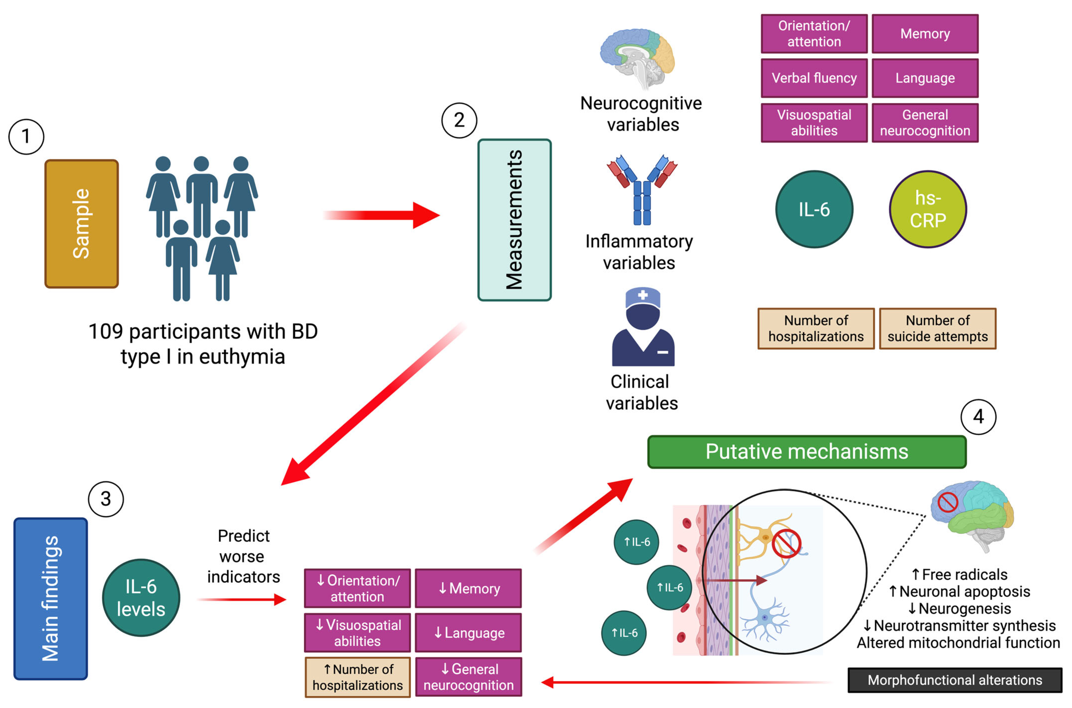
1. Introduction
2. Results
2.1. Descriptive Analysis
2.2. Bivariate Analysis
2.3. Linear Regression Models
3. Discussion
4. Materials and Methods
4.1. Design
4.2. Participants
4.3. Instruments
4.4. Clinical Variables
4.5. Inflammatory Variables
4.5.1. hs-CRP
4.5.2. IL-6
4.6. Statistical Analysis
5. Conclusions
Author Contributions
Funding
Institutional Review Board Statement
Informed Consent Statement
Data Availability Statement
Conflicts of Interest
Abbreviations
| BD | Bipolar disorder |
| hs-CRP | High-sensitivity C-reactive protein |
| IL-6 | Interleukin-6 |
| ACER-T | Addenbrooke’s Cognitive Examination Revised—Total |
| ACER-L | Addenbrooke’s Cognitive Examination Revised—Language |
| ACER-VE | Addenbrooke’s Cognitive Examination Revised—Visuospatial abilities |
| ACER-OA | Addenbrooke’s Cognitive Examination Revised—Orientation/attention |
| ACER-M | Addenbrooke’s Cognitive Examination Revised—Memory |
| ACER-VF | Addenbrooke’s Cognitive Examination Revised—Verbal fluency |
| TE | Time of evolution |
| NH | Number of hospitalizations |
| SA | Number of suicide attempts |
| GWAS | Genome-wide association study |
| SMD | Standardized mean difference |
| CI | Confidence interval |
References
- McIntyre, R.S.; Berk, M.; Brietzke, E.; Goldstein, B.I.; López-Jaramillo, C.; Kessing, L.V.; Malhi, G.S.; Nierenberg, A.A.; Rosenblat, J.D.; Majeed, A.; et al. Bipolar Disorders. Lancet 2020, 396, 1841–1856. [Google Scholar] [CrossRef]
- Gordovez, F.J.A.; McMahon, F.J. The Genetics of Bipolar Disorder. Mol. Psychiatry 2020, 25, 544–559. [Google Scholar] [CrossRef] [PubMed]
- Guglielmo, R.; Miskowiak, K.W.; Hasler, G. Evaluating Endophenotypes for Bipolar Disorder. Int. J. Bipolar. Disord. 2021, 9, 17. [Google Scholar] [CrossRef]
- Baena-Oquendo, S.; Valencia, J.G.; Vargas, C.; López-Jaramillo, C. Neuropsychological Aspects of Bipolar Disorder. Rev. Colomb. Psiquiatr. 2022, 51, 218–226. [Google Scholar] [CrossRef]
- Bourne, C.; Aydemir, O.; Balanzá-Martínez, V.; Bora, E.; Brissos, S.; Cavanagh, J.T.O.; Clark, L.; Cubukcuoglu, Z.; Dias, V.V.; Dittmann, S.; et al. Neuropsychological Testing of Cognitive Impairment in Euthymic Bipolar Disorder: An Individual Patient Data Meta-Analysis. Acta. Psychiatr. Scand. 2013, 128, 149–162. [Google Scholar] [CrossRef] [PubMed]
- Bora, E.; Özerdem, A. Meta-Analysis of Longitudinal Studies of Cognition in Bipolar Disorder: Comparison with Healthy Controls and Schizophrenia. Psychol. Med. 2017, 47, 2753–2766. [Google Scholar] [CrossRef]
- Ehrlich, T.J.; Ryan, K.A.; Burdick, K.E.; Langenecker, S.A.; McInnis, M.G.; Marshall, D.F. Cognitive Subgroups and Their Longitudinal Trajectories in Bipolar Disorder. Acta. Psychiatr. Scand. 2022, 146, 240–250. [Google Scholar] [CrossRef] [PubMed]
- Simjanoski, M.; McIntyre, A.; Kapczinski, F.; de Azevedo Cardoso, T. Cognitive Impairment in Bipolar Disorder in Comparison to Mild Cognitive Impairment and Dementia: A Systematic Review. Trends Psychiatry Psychother. 2022, 44, e20210300. [Google Scholar] [CrossRef]
- Douglas, K.M.; Gallagher, P.; Robinson, L.J.; Carter, J.D.; McIntosh, V.V.W.; Frampton, C.M.A.; Watson, S.; Young, A.H.; Ferrier, I.N.; Porter, R.J. Prevalence of Cognitive Impairment in Major Depression and Bipolar Disorder. Bipolar Disord. 2018, 20, 260–274. [Google Scholar] [CrossRef]
- Martino, D.J. Neurodevelopment as an Alternative to Neuroprogression to Explain Cognitive Functioning in Bipolar Disorder. Psychol. Med. 2024, 54, 4469–4474. [Google Scholar] [CrossRef]
- Strejilevich, S.A.; Samamé, C.; Martino, D.J. The Trajectory of Neuropsychological Dysfunctions in Bipolar Disorders: A Critical Examination of a Hypothesis. J. Affect. Disord. 2015, 175, 396–402. [Google Scholar] [CrossRef] [PubMed]
- Martino, D.J.; Strejilevich, S.A.; Manes, F. Neurocognitive Functioning in Early-Onset and Late-Onset Older Patients with Euthymic Bipolar Disorder. Int. J. Geriatr. Psychiatry 2013, 28, 142–148. [Google Scholar] [CrossRef] [PubMed]
- Konarski, J.Z.; McIntyre, R.S.; Kennedy, S.H.; Rafi-Tari, S.; Soczynska, J.K.; A Ketter, T. Volumetric Neuroimaging Investigations in Mood Disorders: Bipolar Disorder versus Major Depressive Disorder. Bipolar Disord. 2008, 10, 1–37. [Google Scholar] [CrossRef]
- Sæther, L.S.; Ueland, T.; Haatveit, B.; Maglanoc, L.A.; Szabo, A.; Djurovic, S.; Aukrust, P.; Roelfs, D.; Mohn, C.; Ormerod, M.B.E.G.; et al. Inflammation and Cognition in Severe Mental Illness: Patterns of Covariation and Subgroups. Mol. Psychiatry 2022, 28, 1284–1292. [Google Scholar] [CrossRef] [PubMed]
- Rosenblat, J.D.; Brietzke, E.; Mansur, R.B.; Maruschak, N.A.; Lee, Y.; McIntyre, R.S. Inflammation as a Neurobiological Substrate of Cognitive Impairment in Bipolar Disorder: Evidence, Pathophysiology and Treatment Implications. J. Affect. Disord. 2015, 188, 149–159. [Google Scholar] [CrossRef]
- Mullins, N.; Forstner, A.J.; O’Connell, K.S.; Coombes, B.; Coleman, J.R.I.; Qiao, Z.; Als, T.D.; Bigdeli, T.B.; Børte, S.; Bryois, J.; et al. Genome-Wide Association Study of More than 40,000 Bipolar Disorder Cases Provides New Insights into the Underlying Biology. Nat. Genet. 2021, 53, 817–829. [Google Scholar] [CrossRef]
- Leboyer, M.; Soreca, I.; Scott, J.; Frye, M.; Henry, C.; Tamouza, R.; Kupfer, D.J. Can Bipolar Disorder Be Viewed as a Multi-System Inflammatory Disease? J. Affect. Disord. 2012, 141, 1–10. [Google Scholar] [CrossRef]
- Altamura, M.; Leccisotti, I.; Mollica, A.; Maddalena, S.; Altamura, C.; Moretti, M.; Bellomo, A. Inflammatory biomarkers, cognitive functioning, and brain imaging abnormalities in bipolar disorder: A systematic review. Clin. Neuropsychiatry 2024, 21, 32–62. [Google Scholar]
- Cunha, Â.B.; Andreazza, A.C.; Gomes, F.A.; Frey, B.N.; Da Silveira, L.E.; Gonçalves, C.A.; Kapczinski, F. Investigation of Serum High-Sensitive C-Reactive Protein Levels across All Mood States in Bipolar Disorder. Eur. Arch. Psychiatry Clin. Neurosci. 2008, 258, 300–304. [Google Scholar] [CrossRef]
- Chang, H.H.; Wang, T.Y.; Lee, I.H.; Lee, S.Y.; Chen, K.C.; Huang, S.Y.; Yang, Y.K.; Lu, R.B.; Chen, P.S. C-Reactive Protein: A Differential Biomarker for Major Depressive Disorder and Bipolar II Disorder. World J. Biol. Psychiatry 2017, 18, 63–70. [Google Scholar] [CrossRef]
- Modabbernia, A.; Taslimi, S.; Brietzke, E.; Ashrafi, M. Cytokine Alterations in Bipolar Disorder: A Meta-Analysis of 30 Studies. Biol. Psychiatry 2013, 74, 15–25. [Google Scholar] [CrossRef] [PubMed]
- Dargél, A.A.; Godin, O.; Kapczinski, F.; Kupfer, D.J.; Leboyer, M. C-Reactive Protein Alterations in Bipolar Disorder: A Meta-Analysis. J. Clin. Psychiatry 2015, 76, 142–150. [Google Scholar] [CrossRef]
- Rowland, T.; Perry, B.I.; Upthegrove, R.; Barnes, N.; Chatterjee, J.; Gallacher, D.; Marwaha, S. Neurotrophins, Cytokines, Oxidative Stress Mediators and Mood State in Bipolar Disorder: Systematic Review and Meta-Analyses. Br. J. Psychiatry 2018, 213, 514–525. [Google Scholar] [CrossRef]
- Dickerson, F.; Stallings, C.; Origoni, A.; Vaughan, C.; Khushalani, S.; Yang, S.; Yolken, R. C-Reactive Protein Is Elevated in Schizophrenia. Schizophr. Res. 2013, 143, 198–202. [Google Scholar] [CrossRef] [PubMed]
- Evers, A.K.; Veeh, J.; McNeill, R.; Reif, A.; Kittel-Schneider, S. C-Reactive Protein Concentration in Bipolar Disorder: Association with Genetic Variants. Int. J. Bipolar Disord. 2019, 7, 26. [Google Scholar] [CrossRef] [PubMed]
- Dickerson, F.; Stallings, C.; Origoni, A.; Boronow, J.; Yolken, R. Elevated Serum Levels of C-Reactive Protein Are Associated with Mania Symptoms in Outpatients with Bipolar Disorder. Prog Neuropsychopharmacol. Biol. Psychiatry 2007, 31, 952–955. [Google Scholar] [CrossRef]
- Wadee, A.A.; Kuschke, R.H.; Wood, L.A.; Berk, M.; Ichim, L.; Maes, M. Serological Observations in Patients Suffering from Acute Manic Episodes. Hum. Psychopharmacol. 2002, 17, 175–179. [Google Scholar] [CrossRef]
- Hsuchou, H.; Kastin, A.J.; Mishra, P.K.; Pan, W. C-Reactive Protein Increases BBB Permeability: Implications for Obesity and Neuroinfammation. Cell. Physiol. Biochem. 2012, 30, 1109–1119. [Google Scholar] [CrossRef]
- Yirmiya, R.; Goshen, I. Immune Modulation of Learning, Memory, Neural Plasticity and Neurogenesis. Brain Behav. Immun. 2011, 25, 181–213. [Google Scholar] [CrossRef]
- Berk, M.; Kapczinski, F.; Andreazza, A.C.; Dean, O.M.; Giorlando, F.; Maes, M.; Yücel, M.; Gama, C.S.; Dodd, S.; Dean, B.; et al. Pathways Underlying Neuroprogression in Bipolar Disorder: Focus on Inflammation, Oxidative Stress and Neurotrophic Factors. Neurosci. Biobehav. Rev. 2011, 35, 804–817. [Google Scholar] [CrossRef]
- Goldsmith, D.R.; Rapaport, M.H.; Miller, B.J. A Meta-Analysis of Blood Cytokine Network Alterations in Psychiatric Patients: Comparisons between Schizophrenia, Bipolar Disorder and Depression. In Molecular Psychiatry; Nature Publishing Group: London, UK, 2016; Volume 21, pp. 1696–1709. [Google Scholar]
- Rašková, M.; Lacina, L.; Kejík, Z.; Venhauerová, A.; Skaličková, M.; Kolář, M.; Jakubek, M.; Rosel, D.; Smetana, K.; Brábek, J. The Role of IL-6 in Cancer Cell Invasiveness and Metastasis—Overview and Therapeutic Opportunities. Cells 2022, 11, 3698. [Google Scholar] [CrossRef] [PubMed]
- Garbers, C.; Rose-John, S. Dissecting Interleukin-6 Classic- and Trans-Signaling in Inflammation and Cancer. In Methods in Molecular Biology; Humana Press Inc.: New York, NY, USA, 2018; Volume 1725, pp. 127–140. [Google Scholar]
- Fourrier, C.; Singhal, G.; Baune, B.T. Neuroinflammation and Cognition across Psychiatric Conditions. CNS Spectr. 2019, 24, 4–15. [Google Scholar] [CrossRef]
- Sundaresh, A.; Oliveira, J.E.; Chinnadurai, R.K.; Rajkumar, R.P.; Hani, L.; Krishnamoorthy, R.; Leboyer, M.; Singh Negi, V.; Tamouza, R.; Negi, V.S. IL6/IL6R Genetic Diversity and Plasma IL6 Levels in Bipolar Disorder: An Indo-French Study. Heliyon 2019, 5, e01124. [Google Scholar] [CrossRef]
- Bauer, I.E.; Pascoe, M.C.; Wollenhaupt-Aguiar, B.; Kapczinski, F.; Soares, J.C. Inflammatory Mediators of Cognitive Impairment in Bipolar Disorder. J. Psychiatr. Res. 2014, 56, 18–27. [Google Scholar] [CrossRef]
- Morrens, M.; Overloop, C.; Coppens, V.; Loots, E.; Van Den Noortgate, M.; Vandenameele, S.; Leboyer, M.; De Picker, L. The Relationship between Immune and Cognitive Dysfunction in Mood and Psychotic Disorder: A Systematic Review and a Meta-Analysis. Mol. Psychiatry 2022, 27, 3237–3246. [Google Scholar] [CrossRef] [PubMed]
- Brietzke, E.; Stertz, L.; Fernandes, B.S.; Kauer-Sant’Anna, M.; Mascarenhas, M.; Escosteguy Vargas, A.; Chies, J.A.; Kapczinski, F. Comparison of Cytokine Levels in Depressed, Manic and Euthymic Patients with Bipolar Disorder. J. Affect. Disord. 2009, 116, 214–217. [Google Scholar] [CrossRef] [PubMed]
- Saccaro, L.F.; Crokaert, J.; Perroud, N.; Piguet, C. Structural and Functional MRI Correlates of Inflammation in Bipolar Disorder: A Systematic Review. J. Affect. Disord. 2023, 325, 83–92. [Google Scholar] [CrossRef]
- Long, J.Y.; Li, B.; Ding, P.; Mei, H.; Li, Y. Correlations between Multimodal Neuroimaging and Peripheral Inflammation in Different Subtypes and Mood States of Bipolar Disorder: A Systematic Review. Int. J. Bipolar. Disord. 2024, 12, 5. [Google Scholar] [CrossRef]
- Strawbridge, R.; Carter, R.; Saldarini, F.; Tsapekos, D.; Young, A.H. Inflammatory Biomarkers and Cognitive Functioning in Individuals with Euthymic Bipolar Disorder: Exploratory Study. BJPsych Open 2021, 7, e126. [Google Scholar] [CrossRef]
- D’arcangelo, G.; Tancredi, V.; Onofri, F.; D’antuono, M.; Giovedõ, S.; Benfenati, F. Interleukin-6 Inhibits Neurotransmitter Release and the Spread of Excitation in the Rat Cerebral Cortex. Eur. J. Neurosci. 2000, 12, 1241–1252. [Google Scholar] [CrossRef]
- Knight, E.L.; Engeland, C.G.; Yocum, A.K.; Abu-Mohammad, A.; Bertram, H.; Vest, E.; McInnis, M.G.; Saunders, E.F.H. Heightened Inflammation in Bipolar Disorder Occurs Independent of Symptom Severity and Is Explained by Body Mass Index. Brain. Behav. Immun. Health 2023, 29, 100613. [Google Scholar] [CrossRef] [PubMed]
- Coe, C.L.; Love, G.D.; Karasawa, M.; Kawakami, N.; Kitayama, S.; Markus, H.R.; Tracy, R.P.; Ryff, C.D. Population Differences in Proinflammatory Biology: Japanese Have Healthier Profiles than Americans. Brain. Behav. Immun. 2010, 25, 494. [Google Scholar] [CrossRef]
- Ziegler, L.; Gajulapuri, A.; Frumento, P.; Bonomi Sc, A.M.; Wallén, H.; de Faire, U.; Rose-John, S.; Gigante, B. Interleukin 6 Trans-Signalling and Risk of Future Cardiovascular Events. Cardiovasc. Res. 2018, 115, 213–221. [Google Scholar] [CrossRef] [PubMed]
- Saavedra Ramírez, P.G.; María Vásquez Duque, G.; Alonso González Naranjo, L. Interleucina-6: ¿amiga o Enemiga? Bases Para Comprender Su Utilidad Como Objetivo Terapéutico. Iatreia 2011, 24, 157–166. [Google Scholar] [CrossRef]
- Maes, M.; Carvalho, A.F. The Compensatory Immune-Regulatory Reflex System (CIRS) in Depression and Bipolar Disorder. Mol. Neurobiol. 2018, 55, 8885–8903. [Google Scholar] [CrossRef] [PubMed]
- Satizabal, C.L.; Zhu, Y.C.; Mazoyer, B.; Dufouil, C.; Tzourio, C. Circulating IL-6 and CRP Are Associated with MRI Findings in the Elderly: The 3C-Dijon Study. Neurology 2012, 78, 720–727. [Google Scholar] [CrossRef]
- Solmi, M.; Suresh Sharma, M.; Osimo, E.F.; Fornaro, M.; Bortolato, B.; Croatto, G.; Miola, A.; Vieta, E.; Pariante, C.M.; Smith, L.; et al. Peripheral Levels of C-Reactive Protein, Tumor Necrosis Factor-α, Interleukin-6, and Interleukin-1β across the Mood Spectrum in Bipolar Disorder: A Meta-Analysis of Mean Differences and Variability. Brain. Behav. Immun. 2021, 97, 193–203. [Google Scholar] [CrossRef]
- Sakrajda, K.; Szczepankiewicz, A. Inflammation-Related Changes in Mood Disorders and the Immunomodulatory Role of Lithium. Int. J. Mol. Sci. 2021, 22, 1532. [Google Scholar] [CrossRef]
- de Miranda, A.S.; de Miranda, A.S.; Teixeira, A.L. Lamotrigine as a Mood Stabilizer: Insights from the Pre-Clinical Evidence. Expert Opin. Drug Discov. 2019, 14, 179–190. [Google Scholar] [CrossRef]
- Squassina, A.; Pisanu, C.; Vanni, R. Mood Disorders, Accelerated Aging, and Inflammation: Is the Link Hidden in Telomeres? Cells 2019, 8, 52. [Google Scholar] [CrossRef]
- Vasconcelos-Moreno, M.P.; Fries, G.R.; Gubert, C.; Dos Santos, B.T.M.Q.; Fijtman, A.; Sartori, J.; Ferrari, P.; Grun, L.K.; Parisi, M.M.; Guma, F.T.C.R.; et al. Telomere Length, Oxidative Stress, Inflammation and BDNF Levels in Siblings of Patients with Bipolar Disorder: Implications for Accelerated Cellular Aging. Int. J. Neuropsychopharmacol. 2017, 20, 445–454. [Google Scholar] [CrossRef] [PubMed]
- Wiener, C.D.; Moreira, F.P.; Cardoso, T.A.; Mondin, T.C.; da Silva Magalhães, P.V.; Kapczinski, F.; de Mattos Souza, L.D.; da Silva, R.A.; Oses, J.P.; Jansen, K. Inflammatory Cytokines and Functional Impairment in Drug-Free Subjects with Mood Disorder. J. Neuroimmunol. 2017, 307, 33–36. [Google Scholar] [CrossRef]
- Barbosa, I.G.; de Almeida, R.F.; Rocha, N.P.; Mol, G.C.; da Mata Chiaccjio Leite, F.; Bauer, I.E.; Teixeira, A.L. Predictors of Cognitive Performance in Bipolar Disorder: The Role of Educational Degree and Inflammatory Markers. J. Psychiatr. Res. 2018, 106, 31–37. [Google Scholar] [CrossRef]
- Poletti, S.; Mazza, M.G.; Calesella, F.; Vai, B.; Lorenzi, C.; Manfredi, E.; Colombo, C.; Zanardi, R.; Benedetti, F. Circulating Inflammatory Markers Impact Cognitive Functions in Bipolar Depression. J. Psychiatr. Res. 2021, 140, 110–116. [Google Scholar] [CrossRef]
- Bradburn, S.; Sarginson, J.; Murgatroyd, C.A. Association of Peripheral Interleukin-6 with Global Cognitive Decline in Non-Demented Adults: A Meta-Analysis of Prospective Studies. Front. Aging Neurosci. 2018, 9, 438. [Google Scholar] [CrossRef]
- Miola, A.; Dal Porto, V.; Tadmor, T.; Croatto, G.; Scocco, P.; Manchia, M.; Carvalho, A.F.; Maes, M.; Vieta, E.; Sambataro, F.; et al. Increased C-Reactive Protein Concentration and Suicidal Behavior in People with Psychiatric Disorders: A Systematic Review and Meta-Analysis. Acta Psychiatr. Scand. 2021, 144, 537–552. [Google Scholar] [CrossRef] [PubMed]
- Kang, H.J.; Kim, J.W.; Kim, S.W.; Han, J.S.; Lyoo, I.K.; Kim, J.M. Peripheral Markers of Suicidal Behavior: Current Findings and Clinical Implications. Clin. Psychopharmacol. Neurosci. 2023, 21, 650–664. [Google Scholar] [CrossRef]
- González-Castro, T.B.; Tovilla-Zárate, C.A.; López-Narváez, M.L.; Genis-Mendoza, A.D.; Juárez-Rojop, I.E. Interleukin-6 Levels in Serum, Plasma, and Cerebral Spinal Fluid in Individuals with Suicide Behavior: Systematic Review and Meta-Analysis with Meta-Regression. J. Interferon Cytokine Res. 2021, 41, 258–267. [Google Scholar] [CrossRef]
- Wu, D.; Jin, Y.; Xing, Y.; Abate, M.D.; Abbasian, M.; Abbasi-Kangevari, M.; Abbasi-Kangevari, Z.; Abd-Allah, F.; Abdelmasseh, M.; Abdollahifar, M.A.; et al. Global, Regional, and National Incidence of Six Major Immune-Mediated Inflammatory Diseases: Findings from the Global Burden of Disease Study 2019. EClinicalMedicine 2023, 64, 102193. [Google Scholar] [CrossRef]
- Shao, X.; Xie, Z.; Zhu, L. Inflammatory Burden Index Is Correlated with Increased Depression: A Population-Based Study. BMC Psychiatry 2025, 25, 306. [Google Scholar] [CrossRef]
- Queissner, R.; Fellendorf, F.T.; Dalkner, N.; Bengesser, S.A.; Maget, A.; Birner, A.; Platzer, M.; Reininghaus, B.; Häussl, A.; Schönthaler, E.; et al. The Influence of Chronic Inflammation on the Illnesscourse of Bipolar Disorder: A Longitudinal Study. J. Psychiatr. Res. 2024, 174, 258–262. [Google Scholar] [CrossRef] [PubMed]
- Lyra e Silva, N.M.; Gonçalves, R.A.; Pascoal, T.A.; Lima-Filho, R.A.S.; Resende, E.D.P.F.; Vieira, E.L.M.; Teixeira, A.L.; de Souza, L.C.; Peny, J.A.; Fortuna, J.T.S.; et al. Pro-Inflammatory Interleukin-6 Signaling Links Cognitive Impairments and Peripheral Metabolic Alterations in Alzheimer’s Disease. Transl. Psychiatry 2021, 11, 251. [Google Scholar] [CrossRef]
- Mekhora, C.; Lamport, D.J.; Spencer, J.P.E. An Overview of the Relationship between Inflammation and Cognitive Function in Humans, Molecular Pathways and the Impact of Nutraceuticals. Neurochem Int. 2024, 181, 105900. [Google Scholar] [CrossRef] [PubMed]
- Ríos, U. Evaluación de Un Modelo de Interacción Gen-Ambiente En Pacientes Con Trastorno Bipolar Tipo I En Eutimia: Asociación Entre Maltrato Infantil y Cognición Social, y Moderación de Polimorfismos Genéticos. Ph.D. Thesis, Pontificia Universidad Católica de Chile, Santiago, Chile, 2020. [Google Scholar]
- Bagarić, T.; Mihaljević-Peleš, A.; Skočić Hanžek, M.; Živković, M.; Kozmar, A.; Rogić, D. Serum Levels of Zinc, Albumin, Interleukin-6 and CRP in Patients with Unipolar and Bipolar Depression: Cross Sectional Study. Curr. Issues Mol. Biol. 2024, 46, 4533–4550. [Google Scholar] [CrossRef]
- Ozkaya, A.L.; Gürbüzer, N.; Tozoğlu, E.Ö.; Akyildirim, S.; Mercantepe, F. Serum Galectin-3 and IL-6 as Inflammatory Markers in Bipolar Disorder: Insights from Manic and Euthymic Episodes. J. Clin. Med. 2025, 14, 803. [Google Scholar] [CrossRef]
- American Psychiatric Association. Diagnostic and Statistical Manual of Mental Disorders: DSM-IV-TR; American Psychiatric Association: Washington, DC, USA, 2000; ISBN 9780890423349. [Google Scholar]
- Young, R.; Biggs, J.; Ziegler, V.; Meyer, D. A Rating Scale for Mania: Reliability, Validity and Sensitivity. Br. J. Psychiatry 1978, 133, 429–435. [Google Scholar] [CrossRef]
- Hamilton, R. A Rating Scale for Depression. J. Neurol. Neurosurg. Psychiatry 1960, 23, 56–62. [Google Scholar] [CrossRef]
- Muñoz-Neira, C.; Henríquez, F.; Ihnen, J.; Sánchez, M.; Flores, P.; Slachevsky, A. Psychometric Properties and Diagnostic Usefulness of the Addenbrooke’s Cognitive Examination-Revised in a Chilean Elderly Sample. Rev. Med. Chil. 2012, 140, 1006–1013. [Google Scholar] [CrossRef]
- Ridker, P.M.; Rifai, N.; Stampfer, M.J.; Hennekens, C.H. Plasma Concentration of Interleukin-6 and the Risk of Future Myocardial Infarction among Apparently Healthy Men. Circulation 2000, 101, 1767–1772. [Google Scholar] [CrossRef]
- Arancibia, M. Created in BioRender. Available online: https://BioRender.com/lbr5yv1 (accessed on 25 June 2025).

| Variable | Mean | Standard Deviation |
|---|---|---|
| IL-6 (pg/mL) | 8.3 | ±28.4 |
| hs-CRP (mg/L) | 0.2 | ±0.4 |
| ACER-T | 85.7 | ±9.2 |
| ACER-L | 24.1 | ±1.9 |
| ACER-VE | 14.3 | ±1.9 |
| ACER-OA | 16.6 | ±1.7 |
| ACER-M | 20 | ±4.4 |
| ACER-VF | 10.5 | ±2.7 |
| Age of onset (age) | 23.4 | ±9.3 |
| Time of evolution (years) | 23.9 | ±12.9 |
| NH | 3.3 | ±3.9 |
| SA | 1.1 | ±2 |
| hs-PCR | IL-6 | ACER-T | ACER-L | ACER-VE | ACER-OA | ACER-M | ACER-VF | Age | Onset | TE | NH | |
|---|---|---|---|---|---|---|---|---|---|---|---|---|
| IL-6 | 0.05 (0.5946) | |||||||||||
| ACER-T | 0.076 (0.4510) | −0.326 * (0.0007) | ||||||||||
| ACER-L | 0.143 (0.1543) | −0.307 * (0.0015) | 0.645 * (<0.001) | |||||||||
| ACER-VE | 0.062 (0.5364) | −0.322 * (0.0008) | 0.513 * (<0.001) | 0.357 * (0.0001) | ||||||||
| ACER-OA | 0.029 (0.7667) | −0.312 * (0.0011) | 0.554 * (<0.001) | 0.253 * (0.0077) | 0.260 (0.0060) | |||||||
| ACER-M | 0.059 (0.5529) | −0.236 * (0.0146) | 0.852 * (<0.001) | 0.422 * (<0.001) | 0.221 * (0.0203) | 0.299 * (0.0014) | ||||||
| ACER-VF | −0.005 (0.9542) | −0.058 (0.5495) | 0.755 * (<0.001) | 0.342 * (0.0003) | 0.206 * (0.0302) | 0.342 * (0.0002) | 0.546 * (<0.001) | |||||
| Age | 0.116 (0.2453) | 0.117 (0.2362) | −0.277 * (0.0038) | −0.176 (0.0685) | −0.283 * (0.0029) | −0.127 (0.1856) | −0.092 (0.3407) | −0.366 * (0.0001) | ||||
| Onset | 0.094 (0.3456) | 0.043 (0.6601) | −0.027 (0.7752) | 0.101 (0.2976) | −0.168 (0.0822) | −0.113 (0.2407) | 0.076 (0.4278) | −0.101 (0.2929) | 0.446 * (<0.001) | |||
| TE | 0.057 (0.5667) | 0.095 (0.3368) | −0.280 * (0.0035) | −0.263 * (0.0060) | −0.186 (0.0530) | −0.057 (0.5538) | −0.155 (0.1069) | −0.325 * (0.0006) | 0.765 * (<0.001) | −0.233 * (0.0144) | ||
| NH | −0.021 (0.8311) | 0.399 * (<0.001) | −0.235 * (0.0147) | −0.246 * (0.0106) | −0.072 (0.4538) | −0.016 (0.8684) | −0.169 (0.0776) | −0.269 * (0.0046) | 0.206 * (0.0309) | −0.077 (0.4255) | 0.280 * (0.0032) | |
| SA | 0.03 (0.7610) | −0.046 (0.6405) | 0.066 (0.4961) | 0.041 (0.6695) | 0.152 (0.1146) | −0.152 (0.1126) | 0.138 (0.1498) | −0.048 (0.6184) | −0.113 (0.2417) | −0.259 * (0.0065) | 0.063 (0.5116) | 0.025 (0.7949) |
| Low ACER-T (n = 43) | High ACER-T (n = 66) | p-Value | |
|---|---|---|---|
| IL-6 | 10.2 ± 40.5 | 7.3 ± 16.6 | 0.61 |
| hs-CRP | 0.22 ± 0.26 | 0.25 ± 0.53 | 0.79 |
| Low IL-6 (n = 96) | High IL-6 (n = 10) | p-Value | |
|---|---|---|---|
| ACER-T | 85.8 ± 8.6 | 83.3 ± 14.4 | 0.42 |
| ACER-L | 24.12 ± 1.7 | 24.6 ± 3 | 0.46 |
| ACER-VE | 14.36 ± 1.87 | 13.7 ± 2.86 | 0.31 |
| ACER-OA | 16.62 ± 1.7 | 16.40 ± 2.5 | 0.7 |
| ACER-M | 20.12 ± 4.42 | 18.7 ± 5.69 | 0.34 |
| ACER-VF | 10.59 ± 2.79 | 9.9 ± 2.99 | 0.45 |
| NH | 2.97 ± 3.36 | 7.3 ± 7 | 0.001 * |
| SA | 1.25 ± 2.11 | 0.9 ± 1.59 | 0.6 |
| Low hs-CRP (n = 73) | High hs-CRP (n = 29) | p-Value | |
|---|---|---|---|
| ACER-T | 85.3 ± 10.04 | 85.92 ± 7.75 | 0.76 |
| ACER-L | 0.22 ± 0.26 | 0.25 ± 0.53 | 0.79 |
| ACER-VE | 14.33 ± 1.97 | 14.65 ± 1.67 | 0.44 |
| ACER-OA | 16.56 ± 1.71 | 16.62 ± 2.06 | 0.88 |
| ACER-M | 19.87 ± 4.94 | 19.93 ± 3.57 | 0.95 |
| ACER-VF | 10.52 ± 2.96 | 10.37 ± 2.51 | 0.82 |
| NH | 3.27 ± 3.89 | 3.96 ± 4.45 | 0.44 |
| SA | 1.02 ± 1.75 | 1.82 ± 2.7 | 0.08 |
| ACER-T | ACER-L | ACER-VE | ACER-OA | ACER-M | ACER-VF | NH | SA | |
|---|---|---|---|---|---|---|---|---|
| Intercept | 85.93 (83.8–88) | 24.15 (23.7–24.5) | 14.54 (14.13–14.95) | 16.7 (16.3–17.1) | 20 (19–21) | 10.53 (9.8–11.1) | 3.08 (2.2–3.9) | 1.25 (0.76–1.73) |
| IL-6 | −0.107 (−0.16–−0.04) * | −0.021 (−0.03–−0.008) * | −0.02 (−0.035–−0.011) ** | −0.019 (−0.031–−0.007) * | −0.03 (−0.06–−0.007) * | −0.005 (−0.024–0.01) | 0.05 (0.03–0.08) ** | −0.003 (−0.018–0.01) |
| hs-CRP | 1.98 (−2–5.9) | 0.69 (−0.12–1.52) | 0.34 (−0.44–1.14) | 0.19 (−0.58–0.96) | 0.74 (−1.25–2.75) | −0.017 (−1.29–1.25) | −0.38 (−2.06–1.29) | 0.15 (−0.78–1.09) |
| Model statistics | ||||||||
| R2 | 0.11 | 0.11 | 0.13 | 0.09 | 0.06 | 0.003 | 0.16 | 0.003 |
| Adjusted R2 | 0.09 | 0.1 | 0.11 | 0.08 | 0.04 | −0.01 | 0.14 | −0.016 |
| F-statistic | 6.4 | 6.58 | 7.65 | 5.45 | 3.21 | 0.16 | 9.38 | 0.18 |
| p-Value | 0.002 * | 0.002 * | <0.001 * | 0.005 * | 0.04 * | 0.85 | <0.001 * | 0.83 |
Disclaimer/Publisher’s Note: The statements, opinions and data contained in all publications are solely those of the individual author(s) and contributor(s) and not of MDPI and/or the editor(s). MDPI and/or the editor(s) disclaim responsibility for any injury to people or property resulting from any ideas, methods, instructions or products referred to in the content. |
© 2025 by the authors. Licensee MDPI, Basel, Switzerland. This article is an open access article distributed under the terms and conditions of the Creative Commons Attribution (CC BY) license (https://creativecommons.org/licenses/by/4.0/).
Share and Cite
Ríos, U.; Pérez, S.; Martínez, C.; Moya, P.R.; Arancibia, M. Inflammation and Cognition in Bipolar Disorder: Diverging Paths of Interleukin-6 and Outcomes. Int. J. Mol. Sci. 2025, 26, 6372. https://doi.org/10.3390/ijms26136372
Ríos U, Pérez S, Martínez C, Moya PR, Arancibia M. Inflammation and Cognition in Bipolar Disorder: Diverging Paths of Interleukin-6 and Outcomes. International Journal of Molecular Sciences. 2025; 26(13):6372. https://doi.org/10.3390/ijms26136372
Chicago/Turabian StyleRíos, Ulises, Susana Pérez, Constanza Martínez, Pablo R. Moya, and Marcelo Arancibia. 2025. "Inflammation and Cognition in Bipolar Disorder: Diverging Paths of Interleukin-6 and Outcomes" International Journal of Molecular Sciences 26, no. 13: 6372. https://doi.org/10.3390/ijms26136372
APA StyleRíos, U., Pérez, S., Martínez, C., Moya, P. R., & Arancibia, M. (2025). Inflammation and Cognition in Bipolar Disorder: Diverging Paths of Interleukin-6 and Outcomes. International Journal of Molecular Sciences, 26(13), 6372. https://doi.org/10.3390/ijms26136372