Article

NsrR Represses σE-Dependent Small RNAs and Interacts with RpoE via a Noncanonical Mechanism in Escherichia coli

1
Department of Microbiology, College of Medicine, Howard University, Washington, DC 20059, USA
2
Department of Biology, Howard University, Washington, DC 20059, USA
3
Laboratory of Molecular Biology, National Cancer Institute, National Institutes of Health, Bethesda, MD 20892, USA
*
Author to whom correspondence should be addressed.
Int. J. Mol. Sci. 2025, 26(13), 6318; https://doi.org/10.3390/ijms26136318
Submission received: 7 June 2025 / Revised: 27 June 2025 / Accepted: 27 June 2025 / Published: 30 June 2025

Abstract

The envelope stress response in Escherichia coli is primarily governed by the sigma factor RpoE (σE), which activates protective genes upon membrane perturbation. Under non-stress conditions, σE is sequestered by its anti-sigma factor RseA. In this study, we identify an unexpected role for the nitric-oxide-sensing repressor NsrR in dampening σE activity and repressing σE-dependent small RNAs, including rybB, micA, and micL. Overexpression of nsrR represses transcription from σE-dependent promoters and phenocopies σE inactivation, resulting in filamentous morphology and growth defects. Conversely, ΔnsrR de-represses σE targets, with additive effects in rseA mutants—supporting an RseA-independent regulatory role. Time-course analysis shows NsrR represses σE activity, with kinetics comparable to those of RseA. While in vitro assays failed to detect robust NsrR binding to σE target promoters, NsrR directly interacts with σE in bacterial two-hybrid assays. Structural modeling using AlphaFold3 supports a plausible NsrR–RpoE interaction interface. These findings suggest that NsrR functions as a noncanonical anti-sigma-like modulator of σE, integrating redox and envelope stress signals to maintain membrane homeostasis.

1. Introduction

RpoE (σE) is an extracytoplasmic function (ECF) sigma factor that governs the envelope stress response in Escherichia coli and many other Gram-negative bacteria [1,2,3]. Under non-stress conditions, RpoE is sequestered at the inner membrane by direct binding to its anti-sigma factor (RseA) [4,5]. Activation of the σE regulon requires the proteolytic degradation of RseA, a process triggered by envelope stress signals, such as misfolded or accumulating outer membrane proteins (OMPs) [2,6,7,8]. These signals initiate a regulated intramembrane proteolysis (RIP) cascade, in which DegS and RseP sequentially cleave RseA, the anti-sigma factor that sequesters σE to the inner membrane, followed by ClpXP-mediated degradation of the remaining cytoplasmic fragment [4,6,9,10,11]. This cascade releases RpoE to initiate the transcription of target genes involved in maintaining envelope integrity, including chaperones, proteases, and small regulatory RNAs (sRNAs) [12,13,14,15,16,17,18,19,20,21,22].
Among the most critical outputs of the σE response are sRNAs that downregulate OMP synthesis, thereby reducing the burden on the envelope and helping to restore homeostasis [13,15,23,24]. Key members of this group include MicA, MicL, and RybB, all of which repress the translation of OMP-encoding mRNAs [15,18,25,26,27]. These sRNAs function in negative feedback loops that limit RpoE activation and fine-tune the envelope stress response. RybB, an 80-nucleotide sRNA under the direct control of RpoE, has been shown to target multiple OMPs and participate in ppGpp-mediated stress adaptation [13,15,20,24,28,29,30,31,32].
During a genetic screen for novel repressors of rybB, we identified a multicopy clone containing nsrR, a nitric oxide (NO)-responsive transcriptional repressor [15,33,34]. Overexpression of nsrR was sufficient to suppress both the expression of rpoE and rybB, even in the absence of RseA, suggesting an RseA-independent mechanism of repression [15]. These findings raised the possibility that NsrR may influence the σE response through an uncharacterized regulatory interaction. NsrR is a conserved NO-sensing transcriptional repressor that plays central roles in oxidative stress defense and NO detoxification in diverse bacterial species [33,34]. Chromatin immunoprecipitation and microarray analyses (ChIP-chip) have identified dozens of direct NsrR targets in E. coli, including genes involved in motility and biofilm regulation [35]. However, rpoE and rybB were not among the identified NsrR-bound promoters, suggesting that their repression may occur via an indirect mechanism [35].
To clarify how NsrR represses rpoE and rybB, we considered three potential mechanisms: (1) indirect repression of rybB via reduced rpoE expression, (2) inhibition of RpoE activity through anti-sigma-like interactions, and (3) direct repression of both genes by a mechanism distinct from NsrR’s NO-regulated regulon. Herein, we show that NsrR represses RpoE-dependent transcription, with kinetics and strength comparable to those of RseA. NsrR overexpression blocks RpoE autoregulation and downstream target induction, even when RpoE is expressed from a plasmid. Moreover, NsrR overexpression phenocopies ΔrpoE, producing filamentous cell morphology and impaired growth. We further demonstrate that NsrR physically interacts with RpoE in bacterial two-hybrid assays, and AlphaFold3 modeling predicts a plausible interaction interface [36]. To dissect these mechanistic models, we employed a combination of genetics, transcriptional reporters, two-hybrid interaction assays, and structural prediction to test whether NsrR modulates σE activity through direct interaction, indirect repression, or both. Together, these findings support a model in which NsrR acts as a noncanonical modulator of RpoE, functioning outside its traditional NO-responsive role to shape the envelope stress response.

2. Results

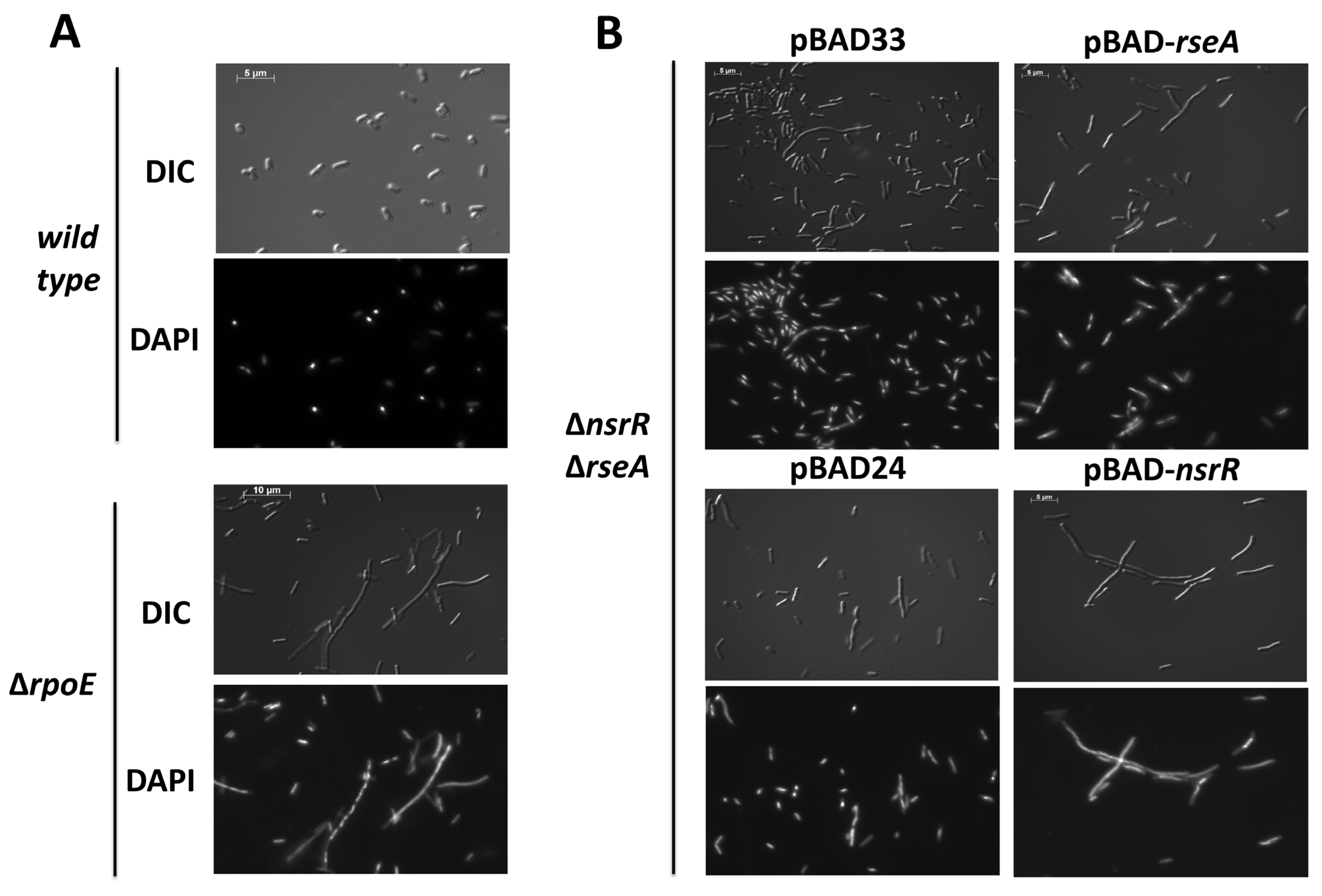
2.1. NsrR Overexpression Represses σE-Dependent Promoters and Disrupts Cell Division

As previously reported, overproduction of NsrR was sufficient to repress rybB-lacZ and rpoE270P2-lacZ fusion in the absence of rseA [15]. NsrR was expressed from an arabinose-inducible promoter, and the addition of arabinose was necessary to see repression (Figure 1A,B; 1A without arabinose and 1B with arabinose). The overexpression of nsrR in the presence of 0.02% arabinose resulted in a severe growth defect (Figure 1C); the overexpression of nsrR in the presence of 0.002% arabinose did not inhibit growth as rapidly (Figure 1C). These growth defects prompted us to hypothesize that NsrR impairs cell division or chromosome segregation. A previous report demonstrated aberrant cellular morphology phenotypes upon inhibition of σE activity because of RseA overexpression [37]. We hypothesized that we would observe aberrant cellular morphology because of NsrR overexpression, due to the NsrR repression of rpoE. Therefore, we examined the cellular morphology upon the overexpression of NsrR, using RseA overexpression and the absence of rpoE as positive controls. For our positive control, wild-type and ∆rpoE cells were grown to the mid-exponential phase (OD600 = 0.5), and aliquots of the cultures were then harvested and analyzed by differential interference contrast and fluorescence microscopy using DAPI staining. The absence of rpoE led to severe cellular filamentation in a significant proportion of the population of cells (Figure 2A). For the analysis of the NsrR overexpression, we transformed a ∆nsrRrseA double mutant with a plasmid containing an arabinose-inducible allele of rseA or nsrR. We then grew the cells in LB supplemented with ampicillin or chloramphenicol and 0.002% arabinose to an OD600 of 0.7–1.0. We then harvested aliquots of these cultures and analyzed them by differential interference contrast and fluorescence microscopy using DAPI staining (Figure 2B). Consistent with previous reports [37], overexpression of RseA leads to the formation of membrane blebs (Figure 2B). In addition, there was also some cellular filamentation in a proportion of the cells. The overexpression of nsrR led to a severe cellular filamentation that was seen in many of the cells (Figure 2B). Taken together, this suggests that nsrR overexpression results in cellular morphology defects that are very similar to those seen upon removing or inactivating rpoE.

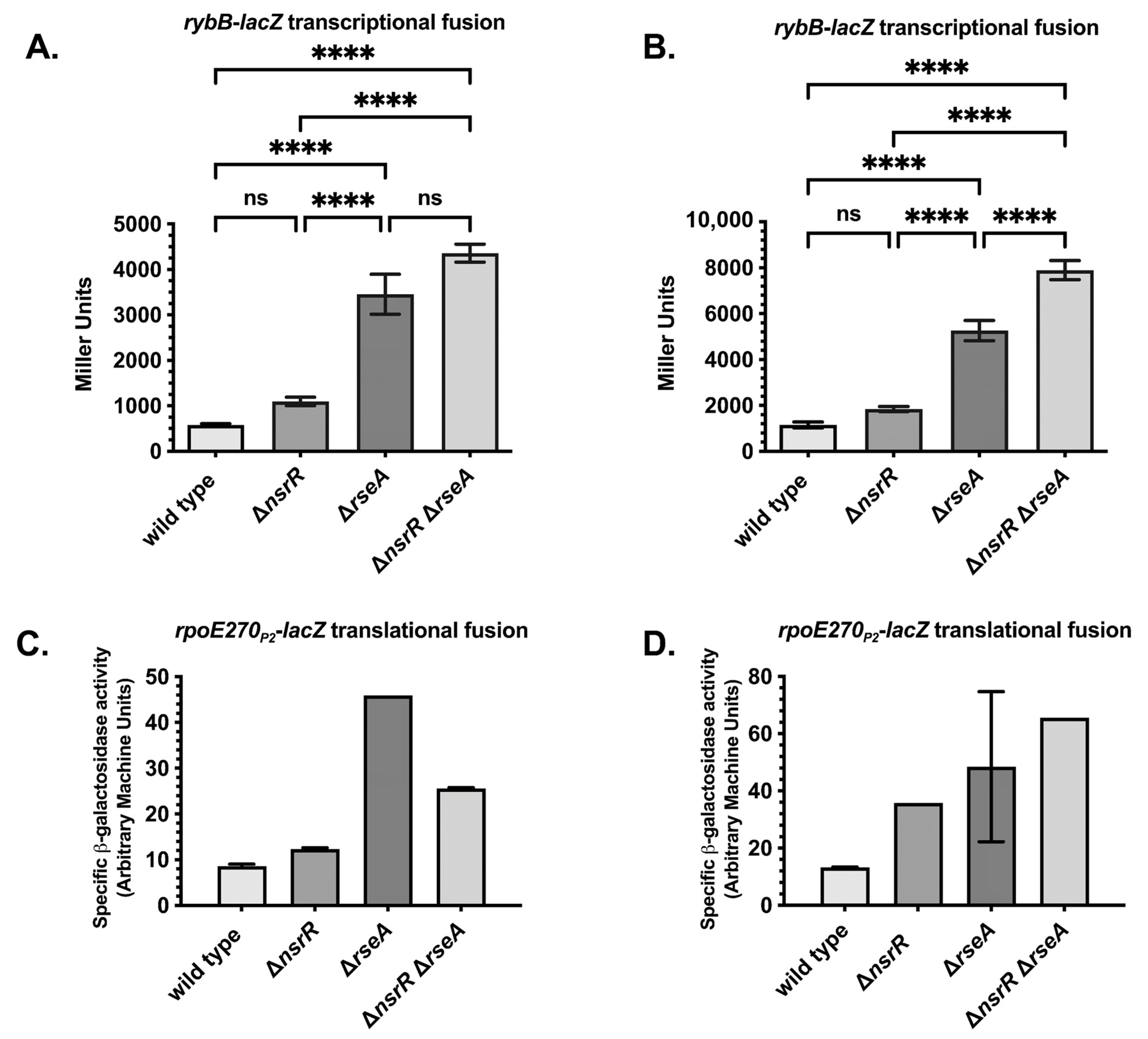
2.2. NsrR and RseA Independently Repress rybB and rpoE, Supporting a Parallel Regulatory Model

To assess the physiological significance of the effects of nsrR on rybB and rpoE, we tested the effect of ΔnsrR on the expression of rybB and rpoE. We transduced ΔnsrR::tet mutations into strains carrying rybB-lacZ transcriptional fusions and rpoE270P2-lacZ translational fusions, in wild-type or ΔrseA genetic backgrounds, and measured their activity at various stages of growth. As expected, ΔrseA significantly increased the expression levels of both fusions, both in the exponential (Figure 3A,C) and stationary phases of growth (Figure 3B,D). The absence of nsrR resulted in an approximate 2-fold increase in rybB-lacZ expression in rseA+ cells, which was statistically significant, in both the exponential and stationary phases of growth (Figure 3A,B). The statistically significant increase in the expression of rybB-lacZ in rseA mutants is much greater at approximately 7-fold in the exponential phase and 4-fold in the stationary phase (Figure 3A,B). In cells deleted for rseA, ΔnsrR further increased the activity of rybB-lacZ (Figure 3A,B), confirming and extending our previous finding that NsrR repression is independent of RseA. However, this increase was not statistically significant in exponential-phase cultures. The ΔnsrR mutant strain of rpoE270P2-lacZ also had a statistically significant increase in expression versus the wild-type strain (Figure 3C,D). While this increase was less than 2-fold in the exponential phase, there was a 2–3-fold increase in the stationary phase (Figure 3C,D). In contrast with the rybB-lacZ fusion strain, the ΔnsrR ΔrseA double mutant did not exhibit an increase in activity in comparison to the ΔrseA mutant in exponential phase for reasons that are not clear. In stationary phase, there is a slight additive increase that is not likely to be statistically significant (Figure 3C,D). Overall, these results confirm that the multicopy repression previously seen reflects a physiologically relevant role of NsrR in the regulation of the σE response.

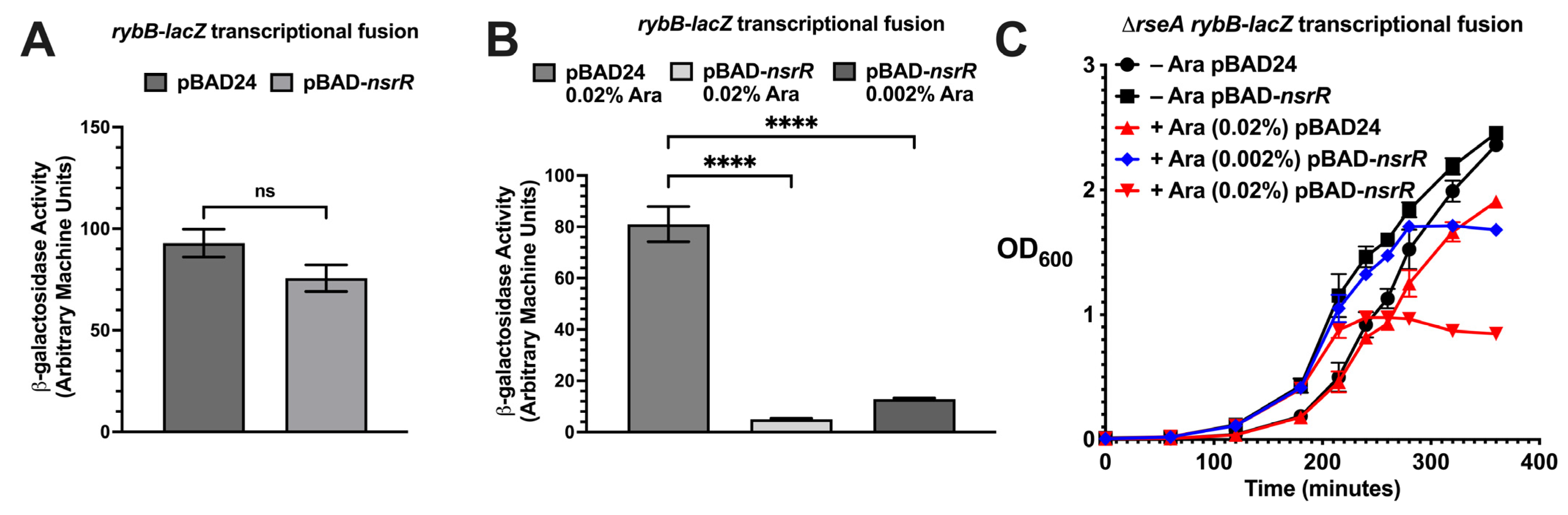
2.3. NsrR Interferes with RpoE-Dependent Promoter Activation in an Inducible Expression System

To test this hypothesis, we measured the ability of NsrR to repress rpoE or rybB promoters in strains in which the sole source of rpoE and nsrR was from compatible plasmids with different inducible promoters (on the pTrc99A-rpoE and pBAD33-nsrR plasmids, respectively). To execute the experiment, we grew the cultures to an OD600 of 0.5 and then added IPTG or IPTG and arabinose and isolated samples every 15 min after induction for 120 min (Figure 4). For both the rpoE270P2-lacZ translational and rybB-lacZ transcriptional fusions, the fusions were active in the absence of IPTG and increased over time, suggesting that there is leaky induction of rpoE from the plasmid-borne IPTG-inducible allele of rpoE (Figure 4A–D, black line). Upon the addition of IPTG, the activities of both the rpoE270P2-lacZ translational and rybB-lacZ transcriptional fusions increased over time and were significantly greater than the activities in the absence of IPTG, confirming the σE-dependent activation of rpoE270P2-lacZ and rybB-lacZ, as expected (Figure 4A–D, blue line). This induction was unaffected by the induction of the pBAD33 vector with arabinose (Figure 4A,C, red line). However, the induction of NsrR decreased the activities of both the rpoE270P2-lacZ and rybB-lacZ fusions, in comparison to the activities of the fusions in the presence of IPTG alone (compare red to blue lines in Figure 4B,D). Therefore, overexpression of NsrR can inhibit the σE induction of rybB and rpoEP2 promoters and is not simply acting at the level of RpoE synthesis. This would presumably occur through the direct interaction of NsrR with either the σE-dependent rybB and rpoEP2 promoters through interaction with the σE protein itself in a manner analogous to RseA.

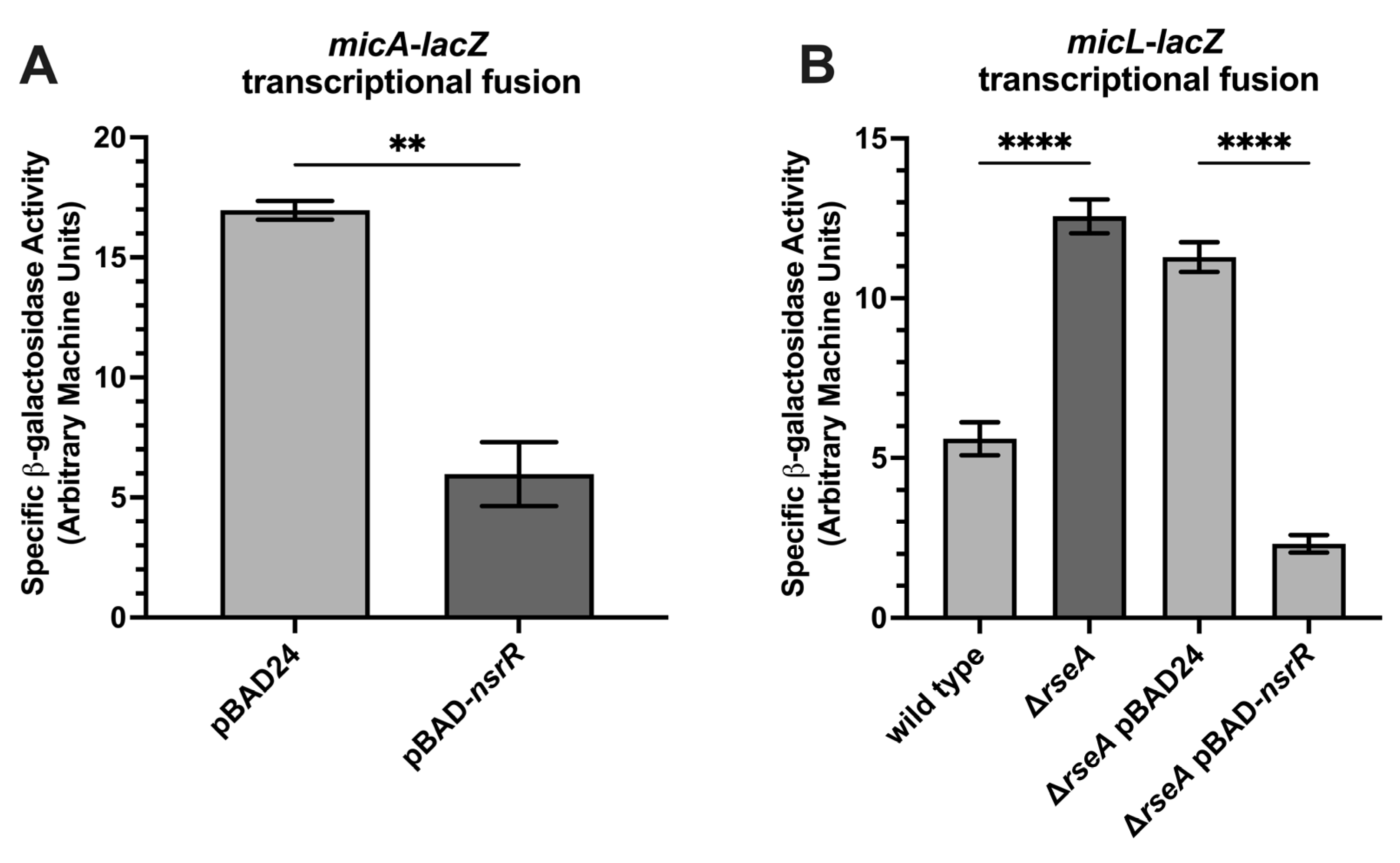
2.4. NsrR Represses Additional σE-Regulated sRNAs

To determine whether NsrR represses additional σE-regulated small RNAs beyond rybB, we examined micA and micL expression using lacZ transcriptional reporter fusions. The micA-lacZ strain harboring either pBAD24 or pBAD24-nsrR was grown to the late exponential phase of growth in LB supplemented with 0.02% arabinose. Overexpression of nsrR significantly reduced the micA-lacZ activity (Figure 5A). Similarly, nsrR overexpression repressed the micL-lacZ expression in a ΔrseA background (Figure 5B), suggesting that this repression occurs independently of RseA-mediated sequestration. These findings extend NsrR’s regulatory reach within the σE regulon and raise the possibility that, like RseA, NsrR may function in a parallel pathway to modulate σE activity. This observation prompted us to compare repression kinetics of NsrR and RseA on the rybB promoter.

2.5. NsrR Does Not Exhibit Robust Binding to σE Target Promoters In Vitro

To test whether NsrR directly represses rpoE or rybB through promoter binding, we performed electrophoretic mobility shift assays (EMSAs), DNase I footprinting, and in vitro transcription assays using purified NsrR and radiolabeled promoter fragments. However, none of these approaches revealed consistent or robust DNA-binding activity under standard conditions. These negative results suggest that NsrR may not repress these promoters via classical DNA binding or that such interactions are weak, transient, or context dependent—potentially requiring stress signals or co-factors absent in vitro. These findings are consistent with previous ChIP-seq studies that did not identify rpoE or rybB as direct NsrR targets [38]. To address this possibility, we plan to apply recently developed chromatin-profiling methods with significantly enhanced sensitivity over the previous technical standard (ChIP-seq), under defined stress conditions, as described in the discussion, to evaluate the potential for condition-dependent promoter occupancy.

2.6. NsrR Phenocopies RseA Repression Kinetics at the rybB Promoter

To test the relative repression kinetics of RseA and NsrR on rybB transcription (as a representative of the σE regulon with one of the strongest promoters), we activated the rybB-lacZ transcriptional fusion followed by the rapid induction of NsrR and RseA—measuring β-galactosidase activity every 15 min for 105 min. The rationale behind this experiment is that if NsrR directly represses σE-dependent promoters, the kinetics of NsrR repression of rybB will be similar or identical to RseA repression. If NsrR acts indirectly (represses or activates the synthesis of another regulator, for instance), we would expect NsrR repression to be significantly slower than repression by the RseA anti-sigma factor, reflecting the time to either deplete or accumulate the direct regulator. To tightly control nsrR and rseA levels for this assay, we used a rybB-lacZ transcriptional fusion strain with a ΔnsrR ΔrseA genetic background transformed with vectors or with plasmids carrying arabinose-inducible alleles of nsrR or rseA. We then grew these strains and the vector controls in LB supplemented with 0.2% glucose to an OD600 of 0.5 (exponential—Figure 6A) or 1.0 (stationary—Figure 5B) and shifted the cells to LB supplemented with 0.2% arabinose. We analyzed culture aliquots for specific β-galactosidase activity at 15 min intervals, following arabinose induction, up to 120 min. For the cultures shifted in exponential phase, the specific β-galactosidase activity of the rybB-lacZ transcriptional fusion strain containing the pBAD24 or pBAD33 vector controls increased over time, consistent with the activity of σE-dependent promoters. The activity in the strain containing pBAD33-rseA did not increase, and slightly decreased, consistent with the known direct negative regulation of σE activity by RseA. The strain containing pBAD24-nsrR was very similar in behavior to the RseA-expressing strain. These kinetics suggest that NsrR directly regulates σE-dependent transcription. Taken together, these observations prompted us to investigate a potential NsrR-RpoE protein–protein interaction mechanism.

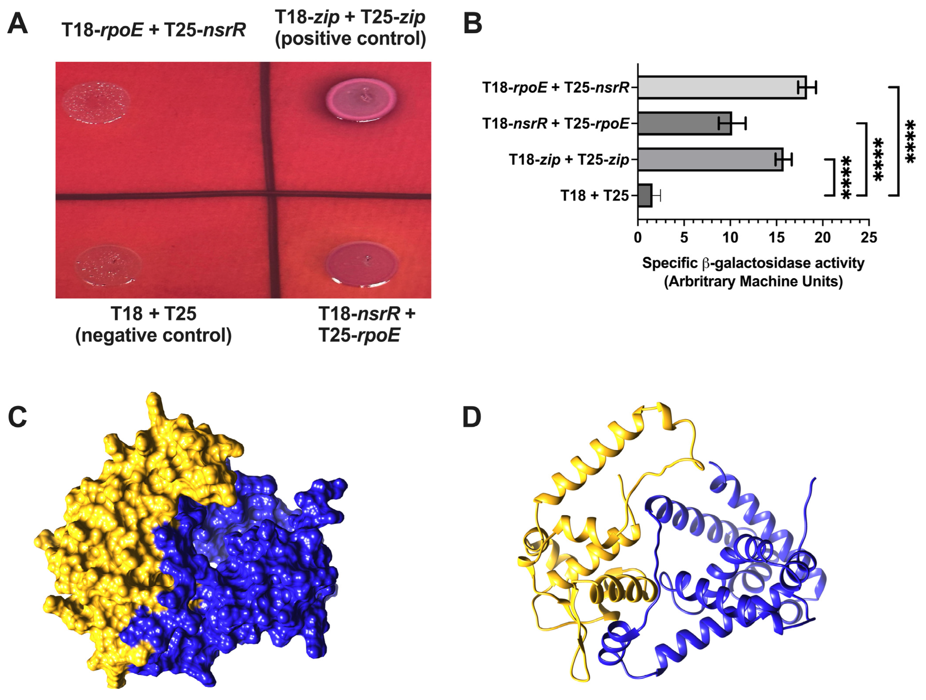
2.7. NsrR–RpoE Interaction Confirmed by Bacterial Adenylate Cyclase Two-Hybrid (BACTH) and Explored via AlphaFold3 Multimer Modeling

Given the rapid and parallel repression kinetics of NsrR and RseA at the rybB promoter, we hypothesized that NsrR may regulate σE activity post-translationally through a direct interaction with RpoE, analogous to the canonical anti-sigma factor (RseA). We employed the BACTH system to test for interactions and used AlphaFold3 multimer modeling to visualize potential interaction surfaces. The BACTH system detects protein–protein interactions based on cAMP-dependent transcriptional activation. Reciprocal fusions of NsrR and RpoE to the T18 and T25 adenylate cyclase fragments produced detectable colony color on MacConkey maltose agar comparable to that of the leucine zipper positive control (Figure 7A). Quantitative β-galactosidase assays confirmed a significant interaction between NsrR and RpoE relative to vector-only controls (Figure 7B). To investigate the molecular nature of this interaction, we used AlphaFold3 multimer modeling, which offers improved capabilities for predicting multi-subunit complexes and flexible protein interfaces, to model the NsrR–RpoE interaction [36]. Direct modeling of the NsrR–RpoE heterodimer yielded low confidence scores (pTM 0.48, ipTM 0.17; Figure S1A), suggesting model uncertainty. However, consistent with prior reports that NsrR functions as a dimer [39], AlphaFold3 confidently predicted the NsrR homodimer (pTM 0.83, ipTM 0.82; Figure 7C). We then modeled the interaction between the NsrR dimer and RpoE. Among five predicted models (Figure 7D and Supplemental Figure S1B–E), the NsrR dimer structure was stable, but the RpoE orientation varied, likely due to the presence of a flexible linker region (residues 92–116). When RpoE is a part of the RNA polymerase holoenzyme (e.g., in complex with RpoA, RpoB, RpoC, and RpoZ; PDB: 6JBQ) [40], this linker is extended, allowing its σ70 domains to contact promoter DNA. In contrast, when RpoE is bound to RseA (PDB: 1OR7—Supplemental Figure S1G) [40], the linker adopts a helical structure, bringing its N- and C-terminal domains into close proximity. These differences highlight the challenge of predicting flexible domain orientations with current algorithms. While AlphaFold3 offers valuable structural insight, experimental validation through mutagenesis or high-resolution structural methods will be necessary to confirm the specific mode of the NsrR–RpoE interaction.

3. Discussion

3.1. NsrR Is a Dual Regulator of Envelope and Nitrosative Stress Responses

Our study reveals an expanded role for the nitric oxide (NO)-sensing transcriptional regulator (NsrR) in Escherichia coli, extending beyond its canonical repression of NO detoxification and iron–sulfur cluster maintenance genes. We demonstrate that NsrR modulates the σE (RpoE) envelope stress response by repressing σE-regulated targets, including rybB, micA, micL, and rpoE itself. These effects occur independently of RseA, the membrane-bound anti-sigma factor that typically sequesters σE under non-stress conditions, suggesting that NsrR functions through a parallel regulatory mechanism.
Rather than constraining σE under basal conditions—when it is already sequestered—NsrR may act after the initial activation of σE, functioning as a secondary brake that dampens prolonged or excessive envelope stress signaling. This dual role positions NsrR as a redox-sensitive checkpoint that coordinates nitrosative and envelope stress responses. By modulating σE output following activation, NsrR helps to maintain the balance between outer membrane protein (OMP) synthesis and envelope remodeling during cell elongation and division—an essential equilibrium for bacterial viability.

3.2. RybB Repression as a Model for the NsrR-Dependent Modulation of σE

We used the small RNA rybB, one of the most prominent σE targets, as a model for dissecting the NsrR-dependent modulation of the envelope stress response. NsrR overexpression strongly repressed rybB-lacZ activity and phenocopied σE inactivation, producing filamentous morphology and growth defects. Conversely, ΔnsrR resulted in elevated expression of both rybB and rpoE, with additive effects in a ΔrseA background. This suggests that NsrR and RseA exert independent yet convergent repression of σE activity.
Importantly, NsrR also repressed micA and micL, two additional σE-regulated sRNAs, highlighting a broader role in the post-transcriptional branch of the σE regulon. These sRNAs fine-tune OMP levels by promoting the degradation of outer membrane protein mRNAs. Thus, NsrR’s repression of these regulatory RNAs suggests it serves not only as a transcriptional repressor but also as a master regulator that constrains downstream RNA-based stress adaptations to limit energy expenditure and prevent deleterious remodeling of the cell envelope.

3.3. NsrR May Act as a Noncanonical Anti-σE Factor

Given the kinetic and phenotypic similarities between NsrR and RseA repression of σE-dependent promoter activity, we hypothesized that NsrR could directly modulate σE activity via protein–protein interactions. Supporting this model, bacterial two-hybrid assays revealed a direct interaction between NsrR and RpoE, further corroborated by multimer modeling (Figure 6), which predicted a plausible NsrR–RpoE interface. These data support a model in which NsrR may act as a noncanonical anti-σE factor, directly antagonizing the σE function through physical sequestration or inhibition.
Such dual-function regulators—combining transcriptional repression with direct sigma factor binding—are rare but increasingly recognized in bacterial stress networks. This model draws conceptual parallels to the Bacillus subtilis SigB system, where a complex cascade of sigma, anti-sigma, and anti-anti-sigma factors modulates stress responses [41,42]. In E. coli, a redox-sensitive interaction between NsrR and σE could serve as a novel regulatory checkpoint, integrating environmental NO signals with envelope stress signaling.

3.4. Physiological and Evolutionary Implications

The ability of NsrR to repress σE targets may reflect an adaptive mechanism that prioritizes NO detoxification and redox homeostasis over envelope repair during transient stress. In host environments, such as macrophages—where NO exposure and membrane perturbation often co-occur—this regulatory logic ensures that envelope stress responses are only activated when both redox and envelope stress thresholds are surpassed. Under non-lethal conditions, NsrR-mediated repression may help to avoid the futile or energetically costly induction of envelope repair pathways.
Upon the oxidation of NsrR’s [Fe–S] cluster, de-repression of the σE regulon would allow the appropriate induction of protective and reparative functions. This model supports a broader framework in which NsrR functions not only as a sensor of NO but also as a temporal gatekeeper for multi-stress adaptation, fine-tuning bacterial survival strategies under compound environmental challenges.

3.5. Open Questions and Future Directions

These findings suggest that NsrR may regulate σE through both direct protein–protein interactions and indirect regulatory effects, extending its role beyond the nitric oxide response. While the precise mechanisms remain to be defined, our data support a model in which NsrR functions as a noncanonical anti-sigma factor. Future studies will be required to clarify the molecular interface, regulatory scope, and physiological triggers of this interaction.
Notably, it remains unclear whether envelope stress directly modulates NsrR expression or activity. Although NsrR is classically regulated by nitric oxide via the oxidation of its [Fe–S] cluster, envelope stress or altered σE activity may influence nsrR stability or activity. Supporting this, Nicoloff et al. (2017) [43] identified a point mutation in nsrR, in a screen for suppressors of high σE activity in a ΔrseA genetic background. While the functional consequence of this point mutation is unknown, it is possible that it could augment repression of σE synthesis or activity. Such a mechanism would position NsrR as both a redox-responsible regulator and a feedback inhibitor that retrains prolonged σE activity under envelope stress conditions.
Given the conservation of both NsrR and σE across Gram-negative bacteria, analogous cross-regulatory mechanisms may operate in pathogens such as Salmonella, Klebsiella, and Pseudomonas. Dissecting how NO-sensing regulators influence envelope integrity pathways could uncover novel strategies to disrupt bacterial stress tolerance, persistence, and virulence.

4. Materials and Methods

4.1. Media and Growth Conditions

E. coli strains were cultured in LB (Lennox, Richardson, TX, USA) medium or on LB agar plates with appropriate antibiotics and inducers as described. For plasmid selection and maintenance, antibiotics were added at the following final concentrations: kanamycin (25 μg/mL), chloramphenicol (25 μg/mL), tetracycline (25 μg/mL), and ampicillin (100 μg/mL). PBAD-regulated promoters were repressed by supplementing media with 0.2% glucose and induced with L-arabinose at final concentrations of 0.02% or 0.002%. IPTG was used at 10 or 100 μM for the induction of plasmid-encoded rpoE. Plasmids were introduced via TSS transformation or electroporation. P1vir phage transduction was used to transfer chromosomal mutations following standard protocols [44].

4.2. Strains, Plasmids, and Oligonucleotides

All the strains were derived from E. coli K-12 MG1655. Full lists of strains, plasmids, and primers are provided in Table 1, Table 2 and Table 3.

4.2.1. Chromosomal Deletion–Insertion Mutations of nsrR and rpoE

A ΔnsrR::tet mutation was generated by λ-Red recombineering [44,46,47,48]. A tetracycline resistance cassette flanked by 50 bp homology arms upstream and downstream of nsrR was amplified using primers KT237 and KT238 to create a linear allelic exchange substrate (AES) for recombination. The PCR product was electroporated into strain NM200 (mini-λ::cat) after the induction of λ-Red recombinase at 43.5 °C for 15 min [47]. Transformants (putative recombinants) were selected on LB–tetracycline plates and confirmed by PCR and sequencing. A ΔrpoE::kan mutation was generating in an identical manner with minor changes. Briefly, the AES was amplified using primers KT926 and KT927. And transformants (putative recombinants) were selected on LB-kanamycin plates.

4.2.2. Construction of Plasmid-Based Arabinose-Inducible nsrR and rseA Alleles

The nsrR gene was amplified from MG1655 genomic DNA, using primers KT191 and KT192, and digested with Eco RI and Pst I before ligation into pBAD24. Primers KT906 and KT907 were used for the PCR amplification of nsrR, which was digested with Sac I and Pst I before ligation into pBAD33. The rseA gene was amplified with primers KT95 and KT75 and digested with Sac I and Xba I and cloned similarly into pBAD33. The constructs were verified by restriction digestion and sequencing. The primers are listed in Table 3.

4.2.3. Construction of Bacterial Adenylate Cyclase Two-Hybrid Assay (BACTH) Plasmids

The nsrR and rpoE open reading frames were PCR-amplified using the gene-specific primers listed in Table 3 and cloned in-frame with either the T18 or T25 domain of adenylate cyclase. Cloning was performed using HiFi Assembly Master Mix (New England Biolabs, Ipswich, MA, USA) according to the manufacturer’s instructions. The pEB355 and pEB354 vectors were used to generate T18 and T25 fusion constructs, respectively. Specifically, T18-nsrR was constructed using primers KT2223 and KT2224, and T18-rpoE was constructed using KT2225 and KT2226. T25-nsrR and T25-rpoE were constructed with primer pairs KT2227/KT2228 and KT2229/KT2230, respectively. All the plasmid constructs were confirmed by restriction digestion and Sanger sequencing and were subsequently used to assess NsrR–RpoE protein–protein interactions in the BACTH system.

4.3. Bacterial Two-Hybrid (BACTH) Assay

Protein–protein interactions were assessed using the bacterial adenylate cyclase two-hybrid (BACTH) system [49,50]. Plasmid constructs encoding in-frame fusions of nsrR or rpoE to the T18 or T25 domains of adenylate cyclase were introduced to E. coli BTH101 by electroporation. The following plasmid pairs were tested: pEB355 and pEB354 (empty vector (negative control)), pEB355-zip and pEB354-zip (positive control), pEB355-nsrR + pEB354-rpoE, and pEB355-rpoE + pEB354-nsrR. These constructs generated reciprocal T18 and T25 fusions of the two proteins of interest. Electrocompetent BTH101 cells were prepared by washing log-phase cultures three times with ice-cold sterile water. Plasmid mixtures were electroporated using the Bio-Rad MicroPulser™, Bio Rad, Hercules, CA, USA (Ec1 “Bacteria” setting), and cells were recovered in LB for 1 h at 30 °C. Aliquots were plated on LB agar containing ampicillin and kanamycin for the selection of double transformants. Selected colonies were grown overnight in liquid LB supplemented with the same antibiotics and 100 μM IPTG to induce the expression of the fusion proteins. Cultures were spotted onto MacConkey maltose agar plates and incubated at 30 °C for 24 to 48 h. The colony color was used as a qualitative measure of interaction-dependent cAMP restoration and lac operon activation. Quantitative β-galactosidase assays were performed, as described below, on parallel cultures to confirm and measure the interaction strength.

4.4. Quantitative β-Galactosidase Assays

β-galactosidase assays were performed using a standard protocol [51] or in 96-well plates, as previously described [52]. For the standard protocol, the specific β-galactosidase activity was expressed as Miller units, as previously described [51]. Cultures were harvested at the exponential or stationary phase, permeabilized, and incubated with ONPG. Absorbance at 420 nm measured using a SpectraMax 250 plate reader (Marshall Scientific, Hampton, NH, USA). The specific β-galactosidase activity was expressed as arbitrary machine units and defined as the slope of OD420/OD600. The results represent the means of at least three biological replicates.

4.5. Microscopy Experiments

Cells were grown to the mid-log phase of growth, fixed, and stained with DAPI. The morphology was analyzed using differential interference contrast (DIC) and fluorescence microscopy. Images were acquired on a Axio Imager.M2 upright fluorescence microscope (Carl Zeiss Microscopy, LLC, White Plains, NY, USA), using 100× magnification, and processed using Zeiss Zen software version 3.12 (Carl Zeiss Microscopy, LLC, White Plains, NY, USA).

4.6. Protein–DNA Interaction Assays

Electrophoretic Mobility Shift Assays (EMSAs), DNase I footprinting, and in vitro transcription assays were performed using purified NsrR, as well as RpoE reconstituted with purified RNA polymerase holoenzyme and radiolabeled promoter fragments from rpoE and rybB. Assays were performed under standard binding conditions. No robust or specific binding was observed under any condition tested. The protocol details are available upon request.

4.7. Structural Modeling Using AlphaFold3

Protein–protein interaction modeling between NsrR and RpoE was performed using AlphaFold3. Amino acid sequences of full-length E. coli NsrR and RpoE were used as input to the AlphaFold server with default options. Top5 models and experimental structures 6JBQ and 1OR7 in PDB were download and examined in UCSF ChimeraX [53] to generate the illustrations.

4.8. Statistical Analysis

All the statistical analyses were performed using GraphPad Prism 10. One-way ANOVA was used to assess group differences. Bartlett’s or Brown–Forsythe tests were applied to evaluate assumptions of equal variance, depending on the dataset. When comparing two groups, unpaired t-tests were used. Unless otherwise noted, the data represent the mean of at least three independent biological replicates. Bars and connected scatter points display group means; error bars represent the standard error of the mean (SEM). Specific statistical tests and significance thresholds are noted in individual figure legends.

Supplementary Materials

The following supporting information can be downloaded at https://www.mdpi.com/article/10.3390/ijms26136318/s1.

Author Contributions

Conceptualization, K.M.T.; methodology, K.M.T., D.L. and C.-H.T.; validation, K.M.T., D.L. and C.-H.T.; formal analysis, J.I.A., D.L., C.-H.T. and K.M.T.; investigation, J.I.A., J.N. and K.M.T.; data curation, K.M.T., D.L. and C.-H.T.; writing—original draft, J.I.A., J.N. and D.L.; writing—review and editing, J.I.A., J.N., C.-H.T. and K.M.T.; visualization, J.I.A., J.N., C.-H.T. and K.M.T. All authors have read and agreed to the published version of the manuscript.

Funding

This work was supported by award numbers SC2GM105419 and R35GM152163 from the National Institute of General Medical Sciences and 1904089 from the National Science Foundation (NSF) to K.M.T; and TL1R001431 for graduate student support to J.I.A and J.N.

Institutional Review Board Statement

Not applicable—no animals or human subjects or samples were used in this study.

Informed Consent Statement

Not applicable.

Data Availability Statement

The data are contained within the article.

Conflicts of Interest

The authors declare no conflicts of interest.

References

  1. Raina, S.; Missiakas, D.; Georgopoulos, C. The rpoE gene encoding the sigma E (sigma 24) heat shock sigma factor of Escherichia coli. EMBO J. 1995, 14, 1043–1055. [Google Scholar] [CrossRef] [PubMed]
  2. Mescas, J.; Rouviere, P.E.; Erickson, J.W.; Donohue, T.J.; Gross, C.A. The activity of sigma E, an Escherichia coli heat-inducible sigma-factor, is modulated by experssion of out membrane proteins. Genes Dev. 1993, 7, 2618–2628. [Google Scholar]
  3. Erickson, J.W.; A Gross, C. Identification of the sigma E subunit of Escherichia coli RNA polymerase: A second alternate sigma factor involved in high-temperature gene expression. Genes Dev. 1989, 3, 1462–1471. [Google Scholar] [CrossRef]
  4. Missiakas, D.; Mayer, M.P.; Lemaire, M.; Georgopoulos, C.; Raina, S. Modulation of the Escherichia coli sE (RpoE) heat-shock transcription-factor activity by the RseA, RseB and RseC proteins. Mol. Microbiol. 1997, 24, 355–371. [Google Scholar] [CrossRef]
  5. De Las Penans, A.; Connolly, L.; Gross, C.A. The sE-mediated response to extracytoplasmic stress in Escherichia coli is transduced by RseA and RseB, two negative regulators of sE. Mol. Microbiol. 1997, 24, 373–385. [Google Scholar] [CrossRef]
  6. Alba, B.M.; Leeds, J.A.; Onufryk, C.; Lu, C.Z.; Gross, C.A. DegS and YaeL participate sequentially in the cleavage of RseA to activate the ςE-dependent extracytoplasmic stress response. Genes Dev. 2002, 16, 2156–2168. [Google Scholar] [CrossRef]
  7. Walsh, N.P.; Alba, B.M.; Bose, B.; A Gross, C.; Sauer, R.T. OMP Peptide Signals Initiate the Envelope-Stress Response by Activating DegS Protease via Relief of Inhibition Mediated by Its PDZ Domain. Cell 2003, 113, 61–71. [Google Scholar] [CrossRef]
  8. Snyder, W.B.; Davis, L.J.; Danese, P.N.; Cosma, C.L.; Silhavy, T.J. Overproduction of NlpE, a new outer membrane lipoprotein, suppresses the toxicity of periplasmic LacZ by activation of the Cpx signal transduction pathway. J. Bacteriol. 1995, 177, 4216–4223. [Google Scholar] [CrossRef]
  9. Campbell, E.A.; Tupy, J.L.; Gruber, T.M.; Wang, S.; Sharp, M.M.; Gross, C.A.; Darst, S.A. Crystal Structure of Escherichia coli σE with the Cytoplasmic Domain of Its Anti-σ RseA. Mol. Cell 2003, 11, 1067–1078. [Google Scholar] [CrossRef]
  10. Kanehara, K.; Ito, K.; Akiyama, Y. YaeL (EcfE) activates the sE pathway of stress response through a site-2 cleavage of anti-sE, RseA. Genes Dev. 2002, 16, 2147–2155. [Google Scholar] [CrossRef]
  11. Ades, S.E.; Connolly, L.E.; Alba, B.M.; Gross, C.A. The Escherichia coli sigma E-dependent extracytoplasmic stress response is controlled by the regulated proteolysis of an anti-sigma factor. Genes Dev. 1999, 13, 2449–2461. [Google Scholar] [CrossRef] [PubMed]
  12. A Rhodius, V.; Suh, W.C.; Nonaka, G.; West, J.; A Gross, C.; Eisen, J.A. Conserved and Variable Functions of the σE Stress Response in Related Genomes. PLoS Biol. 2005, 4, e2. [Google Scholar] [CrossRef] [PubMed]
  13. Johansen, J.; Eriksen, M.; Kallipolitis, B.; Valentin-Hansen, P. Down-regulation of Outer Membrane Proteins by Noncoding RNAs: Unraveling the cAMP–CRP- and σE-Dependent CyaR–ompX Regulatory Case. J. Mol. Biol. 2008, 383, 1–9. [Google Scholar] [CrossRef] [PubMed]
  14. Nonaka, G.; Blankschien, M.; Herman, C.; Gross, C.A.; Rhodius, V.A. Regulon and promoter analysis of the E. coli heat-shock factor, σ32, reveals a multifaceted cellular response to heat stress. Genes Dev. 2006, 20, 1776–1789. [Google Scholar] [CrossRef]
  15. Thompson, K.M.; Rhodius, V.A.; Gottesman, S. σ E Regulates and Is Regulated by a Small RNA in Escherichia coli. J. Bacteriol. 2007, 189, 4243–4256. [Google Scholar] [CrossRef]
  16. Gogol, E.B.; Rhodius, V.A.; Papenfort, K.; Vogel, J.; Gross, C.A. Small RNAs endow a transcriptional activator with essential repressor functions for single-tier control of a global stress regulon. Proc. Natl. Acad. Sci. USA 2011, 108, 12875–12880. [Google Scholar] [CrossRef]
  17. Udekwu, K.I.; Wagner, E.G. Sigma E controls biogenesis of the antisense RNA MicA. Nucleic Acids Res. 2007, 35, 1018–1037. [Google Scholar] [CrossRef]
  18. Coornaert, A.; Lu, A.; Mandin, P.; Springer, M.; Gottesman, S.; Guillier, M. MicA sRNA links the PhoP regulon to cell envelope stress. Mol. Microbiol. 2010, 76, 467–479. [Google Scholar] [CrossRef]
  19. Guo, M.S.; Updegrove, T.B.; Gogol, E.B.; Shabalina, S.A.; Gross, C.A.; Storz, G. MicL, a new σE-dependent sRNA, combats envelope stress by repressing synthesis of Lpp, the major outer membrane lipoprotein. Genes Dev. 2014, 28, 1620–1634. [Google Scholar] [CrossRef]
  20. Papenfort, K.; Pfeiffer, V.; Mika, F.; Lucchini, S.; Hinton, J.C.; Vogel, J. SigmaE-dependent small RNAs of Salmonella respond to membrane stress by accelerating global omp mRNA decay. Mol. Microbiol. 2006, 62, 1674–1688. [Google Scholar] [CrossRef]
  21. Udekwu, K.I.; Darfeuille, F.; Vogel, J.; Reimegård, J.; Holmqvist, E.; Wagner, E.G.H. Hfq-dependent regulation of OmpA synthesis is mediated by an antisense RNA. Genes Dev. 2005, 19, 2355–2366. [Google Scholar] [CrossRef] [PubMed]
  22. Dartigalongue, C.; Missiakas, D.; Raina, S. Characterization of the Escherichia coli sigma E regulon. J. Biol. Chem. 2001, 276, 20866–20875. [Google Scholar] [CrossRef] [PubMed]
  23. Johansen, J.; Rasmussen, A.A.; Overgaard, M.; Valentin-Hansen, P. Conserved Small Non-coding RNAs that belong to the σE Regulon: Role in Down-regulation of Outer Membrane Proteins. J. Mol. Biol. 2006, 364, 1–8. [Google Scholar] [CrossRef] [PubMed]
  24. Guillier, M.; Gottesman, S.; Storz, G. Modulating the outer membrane with small RNAs. Genes Dev. 2006, 20, 2338–2348. [Google Scholar] [CrossRef]
  25. Klein, G.; Raina, S. Regulated Control of the Assembly and Diversity of LPS by Noncoding sRNAs. BioMed Res. Int. 2015, 2015, 153561. [Google Scholar] [CrossRef]
  26. Klein, G.; Kobylak, N.; Lindner, B.; Stupak, A.; Raina, S. Assembly of Lipopolysaccharide in Escherichia coli Requires the Essential LapB Heat Shock Protein. J. Biol. Chem. 2014, 289, 14829–14853. [Google Scholar] [CrossRef]
  27. Klein, G.; Raina, S. Small regulatory bacterial RNAs regulating the envelope stress response. Biochem. Soc. Trans. 2017, 45, 417–425. [Google Scholar] [CrossRef]
  28. Wassarman, K.M.; Repoila, F.; Rosenow, C.; Storz, G.; Gottesman, S. Identification of novel small RNAs using comparative genomics and microarrays. Genes Dev. 2001, 15, 1637–1651. [Google Scholar] [CrossRef]
  29. Gopalkrishnan, S.; Nicoloff, H.; Ades, S.E. Co-ordinated regulation of the extracytoplasmic stress factor, sigmaE, with other Escherichia coli sigma factors by (p)ppGpp and DksA may be achieved by specific regulation of individual holoenzymes. Mol. Microbiol. 2014, 93, 479–493. [Google Scholar] [CrossRef]
  30. Costanzo, A.; Nicoloff, H.; Barchinger, S.E.; Banta, A.B.; Gourse, R.L.; Ades, S.E. ppGpp and DksA likely regulate the activity of the extracytoplasmic stress factor σE in Escherichia coli by both direct and indirect mechanisms. Mol. Microbiol. 2007, 67, 619–632. [Google Scholar] [CrossRef]
  31. Vogel, J.; Papenfort, K. Small non-coding RNAs and the bacterial outer membrane. Curr. Opin. Microbiol. 2006, 9, 605–611. [Google Scholar] [CrossRef] [PubMed]
  32. Guillier, M.; Gottesman, S. Remodelling of the Escherichia coli outer membrane by two small regulatory RNAs. Mol. Microbiol. 2005, 59, 231–247. [Google Scholar] [CrossRef]
  33. Filenko, N.; Spiro, S.; Browning, D.F.; Squire, D.; Overton, T.W.; Cole, J.; Constantinidou, C. The NsrR Regulon of Escherichia coli K-12 Includes Genes Encoding the Hybrid Cluster Protein and the Periplasmic, Respiratory Nitrite Reductase. J. Bacteriol. 2007, 189, 4410–4417. [Google Scholar] [CrossRef]
  34. Bodenmiller, D.M.; Spiro, S. The yjeB (nsrR) gene of Escherichia coli encodes a nitric oxide-sensitive transcriptional regulator. J. Bacteriol. 2006, 188, 874–881. [Google Scholar] [CrossRef]
  35. Efromovich, S.; Grainger, D.; Bodenmiller, D.; Spiro, S. Genome-wide identification of binding sites for the nitric oxide-sensitive transcriptional regulator NsrR. Methods Enzymol. 2008, 437, 211–233. [Google Scholar] [CrossRef]
  36. Abramson, J.; Adler, J.; Dunger, J.; Evans, R.; Green, T.; Pritzel, A.; Ronneberger, O.; Willmore, L.; Ballard, A.J.; Bambrick, J.; et al. Accurate structure prediction of biomolecular interactions with AlphaFold 3. Nature 2024, 630, 493–500. [Google Scholar] [CrossRef]
  37. Hayden, J.D.; Ades, S.E.; Sandler, S. The Extracytoplasmic Stress Factor, σE, Is Required to Maintain Cell Envelope Integrity in Escherichia coli. PLoS ONE 2008, 3, e1573. [Google Scholar] [CrossRef]
  38. Partridge, J.D.; Bodenmiller, D.M.; Humphrys, M.S.; Spiro, S. NsrR targets in the Escherichia coli genome: New insights into DNA sequence requirements for binding and a role for NsrR in the regulation of motility. Mol. Microbiol. 2009, 73, 680–694. [Google Scholar] [CrossRef]
  39. Tucker, N.P.; Hicks, M.G.; Clarke, T.A.; Crack, J.C.; Chandra, G.; Le Brun, N.E.; Dixon, R.; Hutchings, M.I.; Yuan, A. The Transcriptional Repressor Protein NsrR Senses Nitric Oxide Directly via a [2Fe-2S] Cluster. PLoS ONE 2008, 3, e3623. [Google Scholar] [CrossRef]
  40. Fang, C.; Li, L.; Shen, L.; Shi, J.; Wang, S.; Feng, Y.; Zhang, Y. Structures and mechanism of transcription initiation by bacterial ECF factors. Nucleic Acids Res. 2019, 47, 7094–7104. [Google Scholar] [CrossRef]
  41. Panȳ-Farrȳ, J.; Jonas, B.; Hardwick, S.W.; Gronau, K.; Lewis, R.J.; Hecker, M.; Engelmann, S. Role of RsbU in Controlling SigB Activity in Staphylococcus aureus following Alkaline Stress. J. Bacteriol. 2009, 191, 2561–2573. [Google Scholar] [CrossRef] [PubMed]
  42. Hecker, M.; Pané-Farré, J.; Uwe, V. SigB-Dependent General Stress Response in Bacillus subtilis and Related Gram-Positive Bacteria. Annu. Rev. Microbiol. 2007, 61, 215–236. [Google Scholar] [CrossRef] [PubMed]
  43. Nicoloff, H.; Gopalkrishnan, S.; Ades, S.E.; de Boer, P.A.J. Appropriate Regulation of the σE-Dependent Envelope Stress Response Is Necessary To Maintain Cell Envelope Integrity and Stationary-Phase Survival in Escherichia coli. J. Bacteriol. 2017, 199, e00089-17. [Google Scholar] [CrossRef] [PubMed]
  44. Thomason, L.C.; Costantino, N.; Court, D.L. E. coli Genome Manipulation by P1 Transduction. Curr. Protoc. Mol. Biol. 2007, 79, 1.17.1–1.17.8. [Google Scholar] [CrossRef]
  45. Guzman, L.M.; Belin, D.; Carson, M.J.; Beckwith, J. Tight regulation, modulation, and high-level expression by vectors containing the arabinose PBAD promoter. J. Bacteriol. 1995, 177, 4121–4130. [Google Scholar] [CrossRef]
  46. Sharan, S.K.; Thomason, L.C.; Kuznetsov, S.G.; Court, D.L. Recombineering: A homologous recombination-based method of genetic engineering. Nat. Protoc. 2009, 4, 206–223. [Google Scholar] [CrossRef]
  47. Court, D.L.; Swaminathan, S.; Yu, D.; Wilson, H.; Baker, T.; Bubunenko, M.; Sawitzke, J.; Sharan, S.K. Mini-λ: A tractable system for chromosome and BAC engineering. Gene 2003, 315, 63–69. [Google Scholar] [CrossRef]
  48. Yu, D.; Ellis, H.M.; Lee, E.-C.; Jenkins, N.A.; Copeland, N.G.; Court, D.L. An efficient recombination system for chromosome engineering in Escherichia coli. Proc. Natl. Acad. Sci. USA 2000, 97, 5978–5983. [Google Scholar] [CrossRef]
  49. Battesti, A.; Bouveret, E. The bacterial two-hybrid system based on adenylate cyclase reconstitution in Escherichia coli. Methods 2012, 58, 325–334. [Google Scholar] [CrossRef]
  50. Battesti, A.; Bouveret, E. Improvement of bacterial two-hybrid vectors for detection of fusion proteins and transfer to pBAD-tandem affinity purification, calmodulin binding peptide, or 6-histidine tag vectors. Proteomics 2008, 8, 4768–4771. [Google Scholar] [CrossRef]
  51. Miller, J.H. A Short Course in Bacterial Genetics; Cold Spring Harbor Laboratory Press: Plainview, NY, USA, 1992. [Google Scholar]
  52. Thibodeau, S.A.; Fang, R.; Joung, J.K. High-throughput β-galactosidase assay for bacterial cell-based reporter systems. BioTechniques 2004, 36, 410–415. [Google Scholar] [CrossRef] [PubMed]
  53. Meng, E.C.; Goddard, T.D.; Pettersen, E.F.; Couch, G.S.; Pearson, Z.J.; Morris, J.H.; Ferrin, T.E. UCSF ChimeraX: Tools for structure building and analysis. Protein Sci. 2023, 32, e4792. [Google Scholar] [CrossRef]
Figure 1. The effect of NsrR overexpression on rybB expression and bacterial growth. (A) β-galactosidase activity of strain KMT12078 (ΔrseA rybB-lacZ) harboring either empty vector (pBAD24, KMT12090) or pBAD24-nsrR (KMT12091), grown in LB + ampicillin (50 μg/mL) with either 0.002% or 0.02% arabinose. Cultures were harvested at OD600 1.6–2.3. (B) Dose-dependent repression of rybB-lacZ by NsrR overexpression. KMT12078 carrying pBAD24 or pBAD24-nsrR was grown in LB + ampicillin with 0.002% or 0.02% arabinose. (C) Growth curves of KMT12078 harboring pBAD24 or pBAD24-nsrR in LB + ampicillin with indicated arabinose concentrations. OD600 was monitored over time. Significance: **** p < 0.0001, ns = not significant.
Ijms 26 06318 g001
Figure 2. The cellular morphology effects of NsrR overexpression and RpoE inactivation. Differential interference contrast (DIC) and DAPI-stained fluorescence microscopy images of (A) wild-type (KMT414) and ΔrpoE (KMT471) cells grown to OD600 ~1.0; (B) ΔnsrR ΔrseA (KMT473) cells transformed with vector control—pBAD33 or pBAD24, pBAD33-rseA, or pBAD24-nsrR, grown in LB + appropriate antibiotics and 0.02% arabinose to OD600 0.8–1.0 before imaging.
Ijms 26 06318 g002
Figure 3. The expression of rybB and rpoE in ΔnsrR, ΔrseA, and double mutants. (A,B) β-galactosidase activity of rybB-lacZ transcriptional fusion strains in exponential (A) and stationary (B) phases. Strains include wild-type (KMT12093), ΔrseA (KMT12094), ΔnsrR (KMT12099), and ΔnsrR ΔrseA (KMT12100) grown in LB at 37 °C. (C,D) β-galactosidase activity of rpoE270P2-lacZ translational fusion strains with the same genotypes (KMT14000, KMT14001, KMT14021, and KMT14022) and growth conditions, measured during exponential (C) and stationary (D) phases of growth. Bars represent the means of at least three independent biological replicates. Statistical analysis was performed using one-way ANOVA (GraphPad Prism 10). Significance: **** p < 0.0001, ns = not significant.
Ijms 26 06318 g003
Figure 4. Inducible expression of RpoE and repression by NsrR in fusion reporter assays. (A,B) Time-course analysis of rpoE270P2-lacZ translational fusion activity in strain KMT14022 (ΔnsrR ΔrseA). Cells contained either pBAD33 (vector control, (A)) (KMT14032) or pBAD33-nsrR (KMT14033) (B) along with pCL245 (IPTG-inducible rpoE on pTrc99A). Cultures were grown to OD600 = 0.5 and then induced with 100 µM IPTG or with IPTG plus 0.2% arabinose. β-galactosidase activity was measured every 15 min for 120 min post induction. (C,D) Time-course analysis of rybB-lacZ transcriptional fusion activity in strain KMT12131 (ΔnsrR ΔrseA) treated in the same manner as described above. Cultures contained either pBAD33 (a vector control), (C) (KMT12133), or pBAD33-nsrR (KMT12134) (D), along with pCL245. X/Y data points represent the means of at least three independent biological replicates. Error bars represent SEMs of at least three independent biological replicates.
Ijms 26 06318 g004
Figure 5. Reporter activity from micA and micL fusions in strains overexpressing NsrR. (A) β-galactosidase activity from a micA-lacZ transcriptional fusion strain containing either a vector control (pBAD24–KMT12294) or nsrR expressed from an arabinose-inducible plasmid (pBAD24-nsrR: KMT12295). Cultures were grown with or without 0.02% arabinose and harvested at the late exponential phase of growth. Statistical analysis was performed using an unpaired t-test. Significance: ** p < 0.0010. (B) β-galactosidase activity from a micL-lacZ transcriptional fusion strain in a wild type (JN100) ΔrseA mutant background (JN101) with an empty vector control-pBAD24 (JN102) or an nsrR overexpression plasmid (JN103), grown and harvested as in (A). Statistical analysis was performed using one-way ANOVA (GraphPad Prism 10). Significance: **** p < 0.0001.
Ijms 26 06318 g005
Figure 6. Time-course repression of rybB by RseA and NsrR. Strain KMT12100 (ΔrseA ΔnsrR rybB-lacZ) was transformed with arabinose-inducible plasmids expressing either nsrR (pBAD24-nsrR: KMT12122) or rseA (pBAD33-rseA: KMT12124), or with their respective vector controls (pBAD24 and pBAD33: KMT12121 and KMT12123, respectively). Overnight cultures were diluted in LB supplemented with either 0.2% glucose and ampicillin (for pBAD24 constructs) or chloramphenicol (for pBAD33 constructs) to repress expression from the arabinose inducible promoter. Cultures were grown to OD600 = 0.5 (A) the exponential phase of growth) or to OD600 = 1.0 (B) the stationary phase of growth), harvested by filtration, and resuspended in pre-warmed LB containing 0.02% arabinose to induce expression of nsrR or rseA. Aliquots were collected at 0 (pre-induction), 5, 10, 15, 20, 25, 30, and 45 min post induction for the measurement of rybB-lacZ activity. Statistical analysis was performed using one-way ANOVA (GraphPad Prism 10).
Ijms 26 06318 g006
Figure 7. Analysis of the NsrR–RpoE interaction using The Bacterial Adenylate Cyclase Two-Hybrid assay and AlphaFold3 structural modeling. (A) Bacterial adenylate cyclase two-hybrid (BACTH) assay showing colony color on MacConkey Maltose agar. NsrR and RpoE were fused to the T18 and T25 fragments of adenylate cyclase in both orientations. Plates include vector-only negative control and leucine zipper positive control. (B) Quantification of β-galactosidase activity from the BACTH assay. Constructs expressing T18–NsrR + T25–RpoE and the reciprocal T18–RpoE + T25–NsrR were compared to controls. Bars represent the means of at least three biological replicates; error bars indicate SEM. Statistical analysis was performed using one-way ANOVA (GraphPad Prism 10). Significance: **** p < 0.0001. (C) The ribbon diagram of the NsrR homodimer model. (D) The best AlphaFold3 model of the NsrR dimer and RpoE with pTM 0.53 and ipTM 0.39. NsrR dimer is in purple and coral and has the same orientation as in (C). RpoE is in rainbow coloring, where the N terminal is in blue and the C terminal is in red.
Ijms 26 06318 g007
Table 1. NsrR targets coregulated by RpoE or CpxR or related to envelope homeostasis.
σE RegulonCpxR RegulonEnvelope Homeostasis
rpoE 1spy 2mipA (yeaF2,6
rybB 1dsbA 2yfhB (pgpC2
mica 1 mepH (ydhO2
htrG (ygiM/ecfG2,3,4 ygiH (inner membrane protein) 2
hcp 2,3,5 cbrB 2
smpA 2,3
1—This work; 2—[38]; 3—RpoE regulon [12]; 4—Regulated by MicA [16]; 5—hcp is a major NsrR target [38]; 6—MipA acts as a multicopy suppressor of mutations that decrease rybB promoter activity [15].
Table 2. Bacterial Strains.
Strain or PlasmidGenotypeSource or Reference
Strains
MG1655Esherichia coli K12Thomspon et al. 2007 [15]
DJ480MG1655 ΔlacIZYAThomspon et al. 2007 [15]
JN100NM580 micL-lacZ WTNM580 x micL-lacZ PCR gBlock by Electroporation
JN101NM580 micL-lacZ ΔrseA::kanJN100 x P1 (ΔrseA::kan)
JN102NM580 micL-lacZ ΔrseA::kan pBAD24 JN101 + pBAD24 (TSS Transformation)
JN103NM580 micL-lacZ ΔrseA::kan pBAD-nsrR JN102 +pBAD-nsrR (TSS Transformation)
KMT243DJ480 mini-l::tet ΔrseA::kanKMT295 (induced) + ΔrseA::kan (PCR w/oligos KT76 and KT77) cassette via electroporation
KMT414MG1655 ΔlacZYAfrtfrt lacIQ Δara714 ΔParaE::frtfrtPCP18araENM534 obtained from Nadim Majdalani as a recipient for frtkanfrt-PLlacO-rpoE
KMT422DJ480 ΔnsrR::tetThis work, x ΔnsrR::tet PCR
KMT456E. coli BTH101 pEB354 (T25 linker) KanRT25 linker vector, EcoR I/Xho I, from Aureilia Battesti, Ph.D.
KMT457E. coli BTH101 pEB354 (T25-Zip) KanRPositive control for pEB354 (T25 linker) Aureilia Battesti, Ph.D.
KMT458E. coli BTH101 pEB355 (T18 linker) AmpRT18 linker vector, EcoR I/Xho I, from Aureilia Battesti, Ph.D.
KMT459E. coli BTH101 pEB355 (T18-Zip) AmpRPositive control for pEB355 (T18 linker) Aureilia Battesti, Ph.D.
KMT470MG1655 ΔlacZYAfrtfrt lacIQ Δara714 ΔParaE::frtfrtPCP18araE ΔnsrR::tetKMT414 x P1 (ΔnsrR::tet)
KMT471MG1655 ΔlacZYAfrtfrt lacIQ Δara714 ΔParaE::frtfrtPCP18araE ΔrpoE::catKMT414 x P1 (ΔrpoE::cat)
KMT473MG1655 ΔlacZYAfrtfrt lacIQ Δara714 ΔParaE::frtfrtPCP18araE ΔnsrR::tet ΔrseA::kanKMT470 x P1 (ΔrseA::kan)
KMT10003MC1061 [ΦlrpoH P3::lacZ] rpoE+ with suppressor of rpoE::WCmCAG41001-lab collection
KMT12000DJ480 rybB-lacZ long fusion (−69 to +22)Thomspon et al. 2007 [15]
KMT12069KMT12000 Δara714 leu::Tn10(tet)Thomspon et al. 2007 [15]
KMT12078KMT12069 ΔrseA::kanThomspon et al. 2007 [15]
KMT12090KMT12078 pBAD24Thomspon et al. 2007 [15]
KMT12091KMT12078 pBAD-nsrRThomspon et al. 2007 [15]
KMT12093KMT12000 Δlara714 leu+This work
KMT12094KMT12093 ΔrseA::kanThis work
KMT12099KMT12093 ΔnsrR::tet This work
KMT12100KMT12094 ΔnsrR::tet This work
KMT12121DJ480 rybB-lacZ [long fusion] Δara714 leu+ ΔrseA::kan ΔnsrR::tet pBAD2412100 pBAD24 (TSS transformation)
KMT12122DJ480 rybB-lacZ [long fusion] Δara714 leu+ ΔrseA::kan ΔnsrR::tet pBAD-nsrR12100 pBAD-nsrR (TSS transformation)
KMT12123DJ480 rybB-lacZ [long fusion] Δara714 leu+ ΔrseA::kan ΔnsrR::tet pBAD3312100 pBAD33 (TSS transformation)
KMT12124DJ480 rybB-lacZ [long fusion] Δara714 leu+ ΔrseA::kan ΔnsrR::tet pBAD33-rseA12100 pBAD33-rseA (TSS transformation)
KMT12131DJ480 rybB-lacZ [long fusion] Δara714 leu+ ΔnsrR::tet ΔrpoE::kanKMT12130 x ΔrpoE::kan PCR kanR at 37 °C on LB-kan after ara induction
KMT12132DJ480 rybB-lacZ [long fusion] Δara714 leu+ ΔnsrR::tet ΔrpoE::kan pBAD33 pTrc99AKMT12131 + pBAD33 + pTrc99A (Electroporation and AmpR, CmR)
KMT12133DJ480 rybB-lacZ [long fusion] Δara714 leu+ ΔnsrR::tet ΔrpoE::kan pBAD33 + pCL245 (pTrc99A-rpoE)KMT12131 + pBAD33 + pCL245 (pTrc99A-rpoE) (Electroporation and AmpR, CmR)
KMT12134DJ480 rybB-lacZ [long fusion] Δara714 leu+ ΔnsrR::tet ΔrpoE::kan pBAD33-nsrR + pCL245 (pTrc99A-rpoE)KMT12131 + pBAD33-nsrR + pCL245 (pTrc99A-rpoE) (Electroporation and AmpR, CmR)
KMT12218MG1655 lacIq micA-lacZNM580 x micA-lacZ PCR
KMT12294MG1655 lacIq micA-lacZ pBAD24KMT12218 + pBAD24 (TSS Transformation)
KMT12295MG1655 lacIq micA-lacZ pBAD24-nsrRKMT12218 + pBAD24-nsrR (TSS Transformation)
KMT14000DJ480 Δara714 leu+ rpoE270P2-lacZ translational fusionThomspon et al. 2007 [15]
KMT14001KMT14000 ΔrseA::kanThomspon et al. 2007 [15]
KMT14021KMT14000 ΔnsrR::tetThis work
KMT14022KMT14021 ΔrseA::kanThis work
KMT14029DJ480 Δara leu+ ΔnsrR::tet ΔrpoE::kan rpoE270P2-lacZ translational fusionKMT14021 x P1 (DJ480 ΔrpoE::kan)
KMT14031DJ480 Δara leu+ ΔnsrR::tet ΔrpoE::kan rpoE270P2-lacZ translational fusion pTrc99A pBAD33KMT14029 + pTrc99A & pBAD33 (double electroporation and selection on LB-Cm-Amp)
KMT14032DJ480 Δara leu+ ΔnsrR::tet ΔrpoE::kan rpoE270P2-lacZ translational fusion pCL245 (pTrc99A-rpoE), pBAD33KMT14029 + pCL245 (pTrc99A-rpoE) & pBAD33 (double electroporation and selection on LB-Cm-Amp)
KMT14033DJ480 Δara leu+ ΔnsrR::tet ΔrpoE::kan rpoE270P2-lacZ translational fusion pCL245 (pTrc99A-rpoE), pBAD33-nsrRKMT14029 + pCL245 (pTrc99A-rpoE) & pBAD33-nsrR (double electroporation and selection on LB-Cm-Amp)
NM200DJ480 mini-l::catNadim Majdalani, Ph.D.
NM300DJ480 mini-l::tet“                    ”
Plasmid
pBAD24araC+, ColE1 ori, bla, MCS, AmpRGuzman et al., 1995 [45]
pBAD33araC+, p15A ori, cat, MCS, CmR“                    ”
pBAD24-nsrRnsrR gene cloned into EcoR I/Pst I site of MCS in pBAD24, AmpR Thompson et al., 2007 [15]
pBAD33-rseArseA gene cloned into Sac I/Xba I of MCS in pBAD33, CmRThis work
pCL245rpoE gene cloned into pTrc99A, AmpRRhodius et al. 2006 [14]
pEB354 (T25) Bacterial Adenylate Cylase Two Hybrid Interaction Expression Vector, PLac, LacO site, CAP binding site, p15A ori, Adenylate Cyclase (cya) in T25 domain linker, (KanR)
pEB354-zip (T25-zip)zip domain cloned into the EcoR I/Xho I site of pEB354, (KanR)This work
pEB354-nsrR (T25-nsrR)nsrR gene cloned into the EcoR I/Xho I site of pEB354, (KanR)This work
pEB354-zip (T25-rpoE)rpoE gene domain cloned into the EcoR I/Xho I site of pEB354, (KanR)This work
pEB355 (T18) Bacterial Adenylate Cylase Two Hybrid Interaction Expression Vector, PLac, LacO site, CAP binding site, ColE1 ori, Adenylate Cyclase (cya) in T18 domain linker, bla (AmpR)
pEB355-zip (T8-zip)zip domain cloned into the EcoR I/Xho I site of pEB355, bla (AmpR)This work
pEB355-zip (T8-nsrR)nsrR gene cloned into the EcoR I/Xho I site of pEB355, bla (AmpR)This work
pEB355-zip (T8-rpoE)rpoE gene cloned into the EcoR I/Xho I site of pEB355, bla (AmpR)This work
pTrc99AlacIq ColE1 ori, bla MCS, IPTG inducible PtrcThompson et al., 2007 [15]
Table 3. Oligonucleotides.
Oligonucleotide ID Sequence 5′-3′ Purpose
KT75tttctagattactgcgattgcgttcctaaagtttgreverse primer for the amplification and cloning of rseA into pBAD33 or pBAD24 Xba I site
KT76atgcagaaagaacaactttccgctttaatggatggcgaaacgctggatagaaagccacgttgtgtctcaa ΔrseA::kan forward oligo for the amplification of recombineering substrate
KT77 ttactgcgattgcgttcctaaagtttgaattcctggcacctgtacagcgggcgctgaggtctgcctcgtg ΔrseA::kan reverse oligo for the amplification of recombineering substrate
KT95aagtccggagctcaggaggaattcaccatgcagaaagaacaactttccgctttaatforward primer for the amplification and cloning of rseA into pBAD33 or pTrc99A Sac I site
KT237 ttatcatcaatataaatgtattttttcccgatttcccttttgaggttgatctagacatcattaattccta forward primer for the amplification of ΔnsrR::tet recombineering substrate
KT238tcttgtgacatctcggttcctccgttgtcatctctgatgaagattttcggaagctaaatcttctttatcg reverse primer for the amplification of ΔnsrR::tet recombineering substrate
KT906gcgtgagctcgatttcccttttgaggttgatgtgcag forward primer for the amplification and cloning of nsrR into pBAD33 (21 nucleotides upstream of the ATG) Sac I site
KT907gcgtctgcagtcactccaccagcaataatttataaagreverse primer for the amplification and cloning of nsrR into pBAD33 Pst I site
KT926TGGCGTTTCGATAGCGCGTGGAAATTTGGTTTGGGGAGACTTTACCTCGGgcgctgaggtctgcctcgtgforward primer for ΔrpoE::kan recombineering substrate
KT927 AAGTTGTTCTTTCTGCATGCCTAATACCCTTATCCAGTATCCCGCTATCGaaagccacgttgtgtctcaareverse primer for the amplification of ΔrpoE::kan recombineering substrate
Disclaimer/Publisher’s Note: The statements, opinions and data contained in all publications are solely those of the individual author(s) and contributor(s) and not of MDPI and/or the editor(s). MDPI and/or the editor(s) disclaim responsibility for any injury to people or property resulting from any ideas, methods, instructions or products referred to in the content.

