Article

Nuclear Fraction Proteome Analyses During rAAV Production of AAV2-Plasmid-Transfected HEK-293 Cells

by
Susanne K. Golm
1,2,3,
Raimund Hoffrogge
1,3,4 and
Kristian M. Müller
1,2,*
1
Department of Biotechnology, Faculty of Technology, Bielefeld University, 33501 Bielefeld, Germany
2
Cellular and Molecular Biotechnology, Faculty of Technology, Bielefeld University, 33501 Bielefeld, Germany
3
Cell Culture Technology, Faculty of Technology, Bielefeld University, 33501 Bielefeld, Germany
4
Center for Biotechnology, Bielefeld University, 33501 Bielefeld, Germany
*
Author to whom correspondence should be addressed.
Int. J. Mol. Sci. 2025, 26(13), 6315; https://doi.org/10.3390/ijms26136315
Submission received: 8 April 2025 / Revised: 18 May 2025 / Accepted: 3 June 2025 / Published: 30 June 2025

Abstract

Recombinant adeno-associated virus (rAAV) is the leading vector for gene replacement therapy; however, the roles and regulation of host proteins in rAAV production remain incompletely understood. In this comparative proteomic analysis, we focused on proteins in the nucleus, the epicenter of DNA uptake, transcription, capsid assembly, and packaging. HEK-293 cells were analyzed under the following three conditions: (i) untransfected, (ii) mock-transfected with the ITR and an unrelated plasmid, and (iii) triple-transfected with rAAV2 production plasmids. Cells were harvested at 24 and 72 h post-transfection, and nuclear fractions were processed using filter-aided sample preparation (FASP) followed by nano-scale liquid chromatography–tandem mass spectrometry (nLC-Orbitrap MS/MS). Across all samples, we identified 3384 proteins, revealing significant regulatory changes associated with transfection and rAAV production. Transfection alone accounted for some of the most substantial proteomic shifts, while rAAV production induced diverse regulatory changes linked to cell cycle control, structure, and metabolism. STRING analysis of significantly regulated proteins also identified an enrichment of those associated with the Gene Ontology (GO) term ‘response to virus’. Additionally, we examined proteins with reported relation to adenoviral components. Our findings help to unravel the complexity of rAAV production, identify interesting targets for further investigation, and may contribute to improving rAAV yield.

1. Introduction

Adeno-associated viruses are the preferred carrier for gene replacement therapies. In 2012, the first approved gene therapy in Europe was Glybera, based on the AAV1 capsid [1]. Several approvals in, e.g., Europe and the USA followed, such as Luxturna (AAV2, 2017, [2]), Zolgensma (AAV9, 2019, [3]), Hemgenix (AAV5, 2022, [4]), Upstaza (AAV2, 2022, [5]), Roctavian (AAV5, 2023, [6]), Elevidys (AAVrh74, 2024, [7]), and Beqvez (AAVRh74var, 2024, [8]). Luxturna, Zolgensma, Upstaza, and Beqvez are produced in mammalian human embryonic kidney cells (HEK) derived from the 293 clone [9].
The triple transfection of HEK-293 serves as a classic rAAV production system. This cell line was immortalized with a fragment of the Adenovirus 5 genome, features a near-triploid karyotype with some polyploidy, and stably expresses the adenoviral proteins E1A and E1B [10]. Three required plasmids provide the basis for rAAV production: the therapeutic transgene cassette flanked by the necessary inverted terminal repeats (ITRs) of the AAV genome, as well as the non-structural AAV (Rep78, Rep68, Rep52, Rep48, AAP, and MAAP) and structural (Cap VP1, VP2, and VP3) proteins. Production has also been reported in yeast [11], Sf9 insect cells [12,13], and Chinese hamster ovary cells [14]. Furthermore, AAV capsids were produced in E. coli [15,16].
The production of rAAV-based therapeutics poses a number of challenges, such as extensive up- and downstream processes as well as limited yield, which results in high manufacturing costs. In this context, the question arises of how rAAV production affects the host cell and how host-cell regulation processes or modifications could be used to further increase productivity. Several transcriptomic and proteomic experiments have been carried out to investigate the impact of rAAV production on the host cells. For example, a proteomic approach was performed by Strasser et al., comparing untransfected and, for rAAV5 production, transfected FreeStyle 293-F cells. This study showed that rAAV production leads to altered expression of proteins involved in processes such as metabolism, proliferation and cell death, endocytosis, and lysosomal degradation [17]. Chung et al. examined a HEK-293-derived suspension cell line transfected for rAAV9 production with polyethylenimine (PEI) by RNA-Seq and identified several up-regulated pathways, including inflammatory and antiviral responses, as well as higher protein levels of interferon-stimulated cytokines and chemokines [18]. Another transcriptome study was conducted by Wang et al. and investigated two HEK-293 cell lines from different suppliers adapted to different media in order to identify significantly regulated metabolic pathways and examine cellular characteristics of the host cell, which may support rAAV production. They compared rAAV-producing cells with the parental cell lines and found significantly enriched and up-regulated host cell innate immune response signaling pathways (e.g., RIG-I-like receptor signaling pathway, Toll-like receptor signaling, cytosolic DNA sensing, and JAK-STAT signaling). This regulation was associated with enriched gene ontology terms for host cellular stress responses, including endoplasmic reticulum stress, autophagy, and apoptosis in virus production. In contrast, fatty acid metabolism and neutral amino acid transport were down-regulated in the late phase of viral production [19]. The impact of transfection on host cells and rAAV productivity is of great interest. Lu et al. performed a kinetic multi-omics analysis of rAAV production of transfected HEK cells [20]. Another kinetic study compared the different transfection systems of virus co-infection and multi-plasmid transfection by transcriptomic and proteomic analysis [21]. In a SWATH-MS proteomic approach, Patra et al. investigated three different transfection conditions (standard, sub-optimal, and optimal transfection) in HEKT cells [22]. The reviews of Wang et al. and Gurazada et al. provide a good overview of omics studies in this field [23,24].
We aimed to provide answers to the question of how rAAV production influences the host cell’s nuclear proteome. Since gene regulation and virus assembly are located in the nucleus [25,26], we hypothesize that limiting or supporting factors for production also reside in the nucleus and that the pre-selection for nuclear proteins helps to zoom in on important changes. We analyzed untransfected adherent HEK-293 cells and compared the nuclear proteome with that of mock-transfected cells or that of rAAV2 production-transfected cells at 24 h and 72 h post-transfection. Our label-free quantitative MS proteome analysis identified more than 3300 proteins, and we discuss the relation of selected proteins to AAV production. This study provides a valuable addition to the above-mentioned, existing proteomic analysis of the whole cell proteome approaches.

2. Results

The nucleus serves as a critical cellular compartment for rAAV production, as it provides the essential environment and machinery for viral DNA synthesis from transfected DNA and transcription, as well as capsid assembly and packaging of the viral DNA into the capsids. Therefore, we decoded with an nLC-ESI-MS approach the nuclear proteome of (i) untransfected (HEK sample), (ii) mock-transfected (MOCK sample), and (iii) AAV2-production-transfected (AAV sample) HEK-293 cells (Figure 1). Samples were generated in triplicate for robust statistical analysis of the results. HEK-293 cells were checked for successful transfection by fluorescence microscopy 24 h and 72 h post-transfection (ptf) based on mVenus, expressed by the plasmid pITR-mVenus, and then harvested. Nuclear fractions were generated by several steps of solubilization and precipitation, and samples were prepared for nLC-ESI MS/MS measurements by ‘filter aided sample preparation’ (FASP) based on a tryptic digestion and membrane adsorption. Raw data were analyzed for protein identification and label-free quantification with Proteome Discoverer Software (see Methods for details).
Figure 2 presents images of the HEK, MOCK, and AAV samples for both harvesting time points, 24 h and 72 h ptf. In the HEK control samples no fluorescence signal was detected, whereas in MOCK and AAV samples most cells were fluorescent, indicating successful transfection. As expected, fluorescence intensity increased from 24 h ptf to 72 h ptf. Here, we were interested in the confirmation of successful transfection. In general, fluorescence microscopy is also a powerful tool for the investigation of rAAV biology [27].

2.1. Protein Identification and Sample Variability

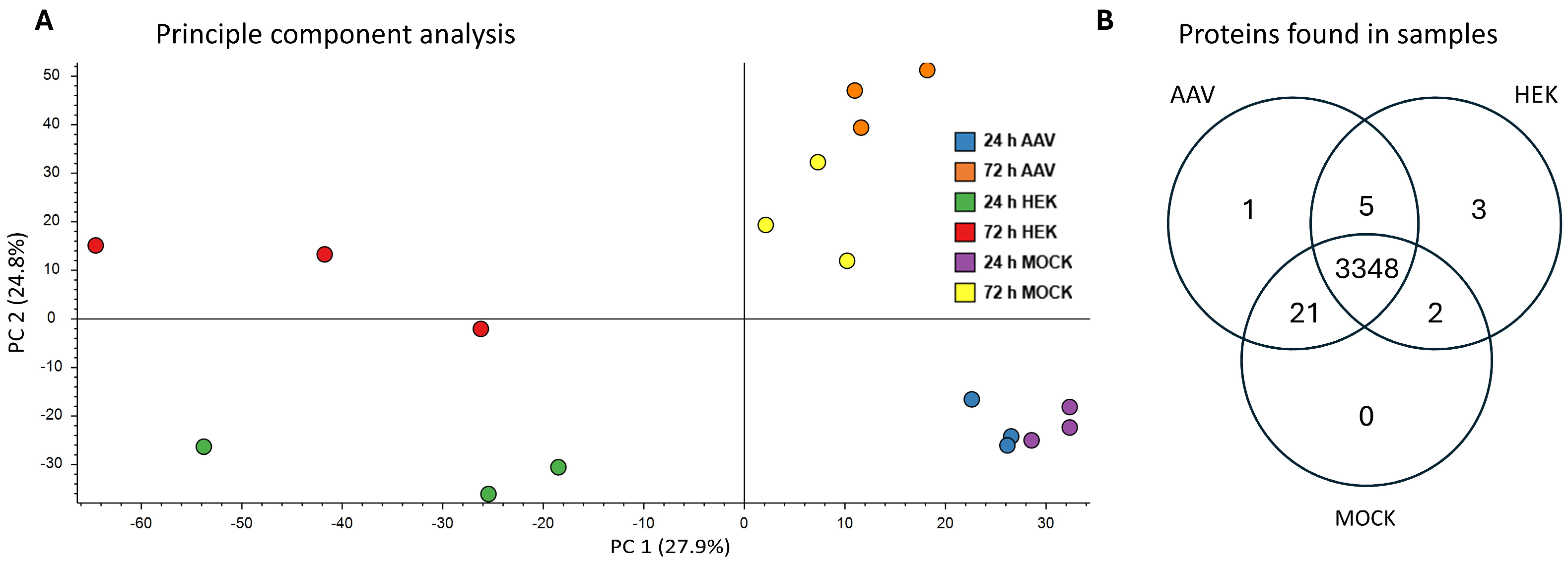
Raw nLC-ESI MS/MS data of the untransfected (HEK sample), mock-transfected (MOCK sample), and rAAV-production-transfected (AAV sample) HEK-293 cells, 24 h and 72 h post-transfection, were processed and then analyzed with Proteome Discoverer software. In total, 3384 human proteins were identified in all samples combined when filtering for proteins with at least one detected unique peptide. A principal component analysis (PCA) revealed a grouping of samples mostly based on their status of transfection and time of sampling. AAV-transfected and MOCK-transfected samples were distinct from untransfected samples by principal component 1, and samples taken at 24 h ptf were distinct from the 72 h ptf samples based on principal component 2 (Figure 3A). Additionally, distribution of identified proteins over all samples is visualized in a Venn diagram (Figure 3B). For an improved overview, 24 h and 72 h samples of each sample group (AAV, MOCK, HEK) were combined. As a reliable base for quantitative analysis, 3348 proteins are found in all sample groups altogether. Whereas one protein was only identified in AAV samples and seven in HEK samples with a confident identification. Furthermore, 21 proteins are present only in AAV and MOCK samples, five proteins in AAV and HEK samples, and two proteins in MOCK and HEK samples. In addition, the rAAV proteins Rep78, Rep52, VP1, and AAP were identified in AAV samples (proteins not included in Figure 3B).

2.2. Identification of Top Regulated HEK-293 Proteins

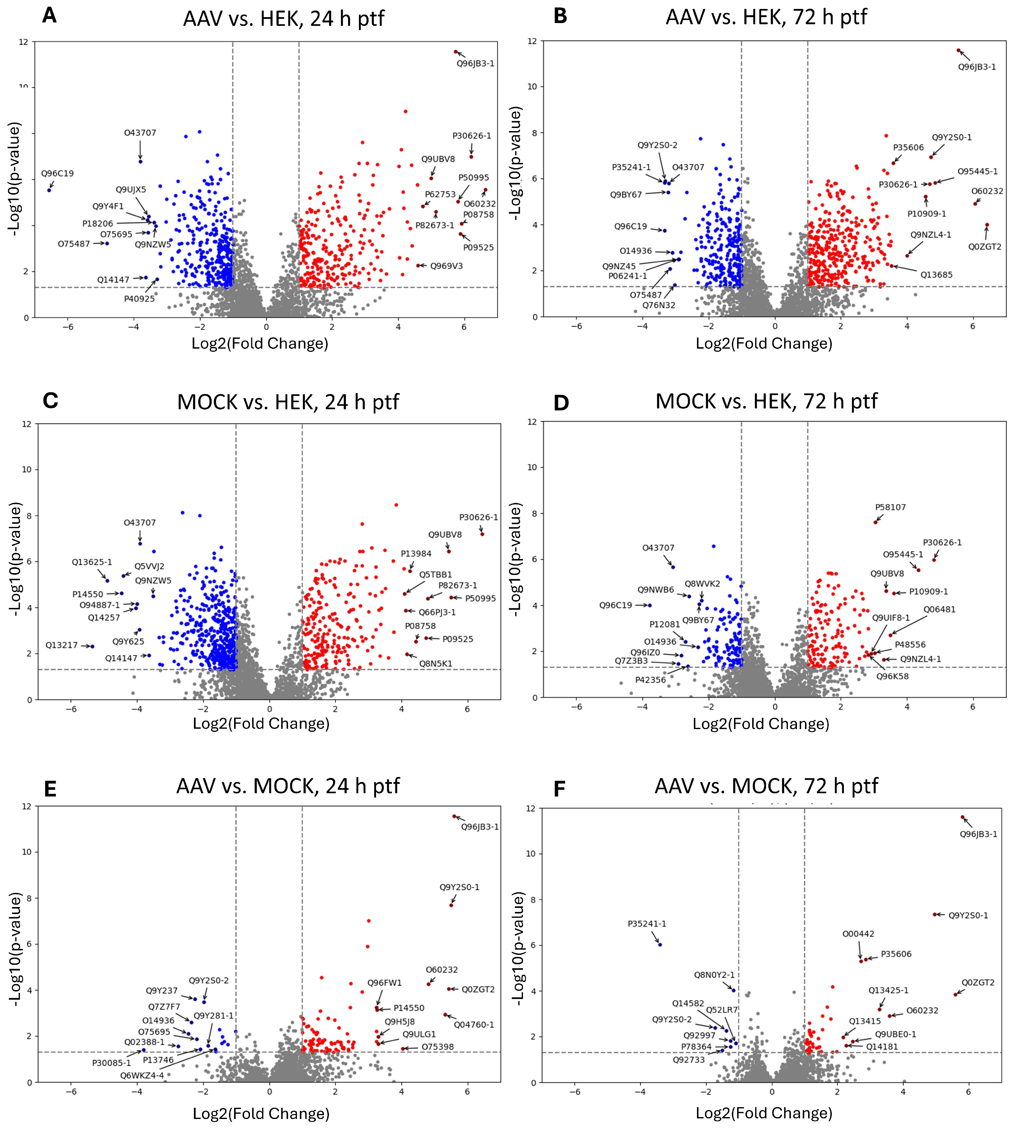
To identify significantly up- and down-regulated cellular proteins across treatments, abundance ratios (fold change) were calculated for different sample groups of the same harvest time point. Either rAAV-transfected samples or MOCK-transfected samples were compared to non-transfected HEK-293 samples. To identify significant events, proteins with at least two unique peptides, a p-value maximum of 0.05, and a minimum fold change of 2 were set as the threshold. The results were visualized by volcano plots for each comparison. The ten proteins with the strongest up-regulation and down-regulation are labeled with their UniProt accession number in each volcano plot (Figure 4).

2.3. Comparison of Top Regulated Proteins

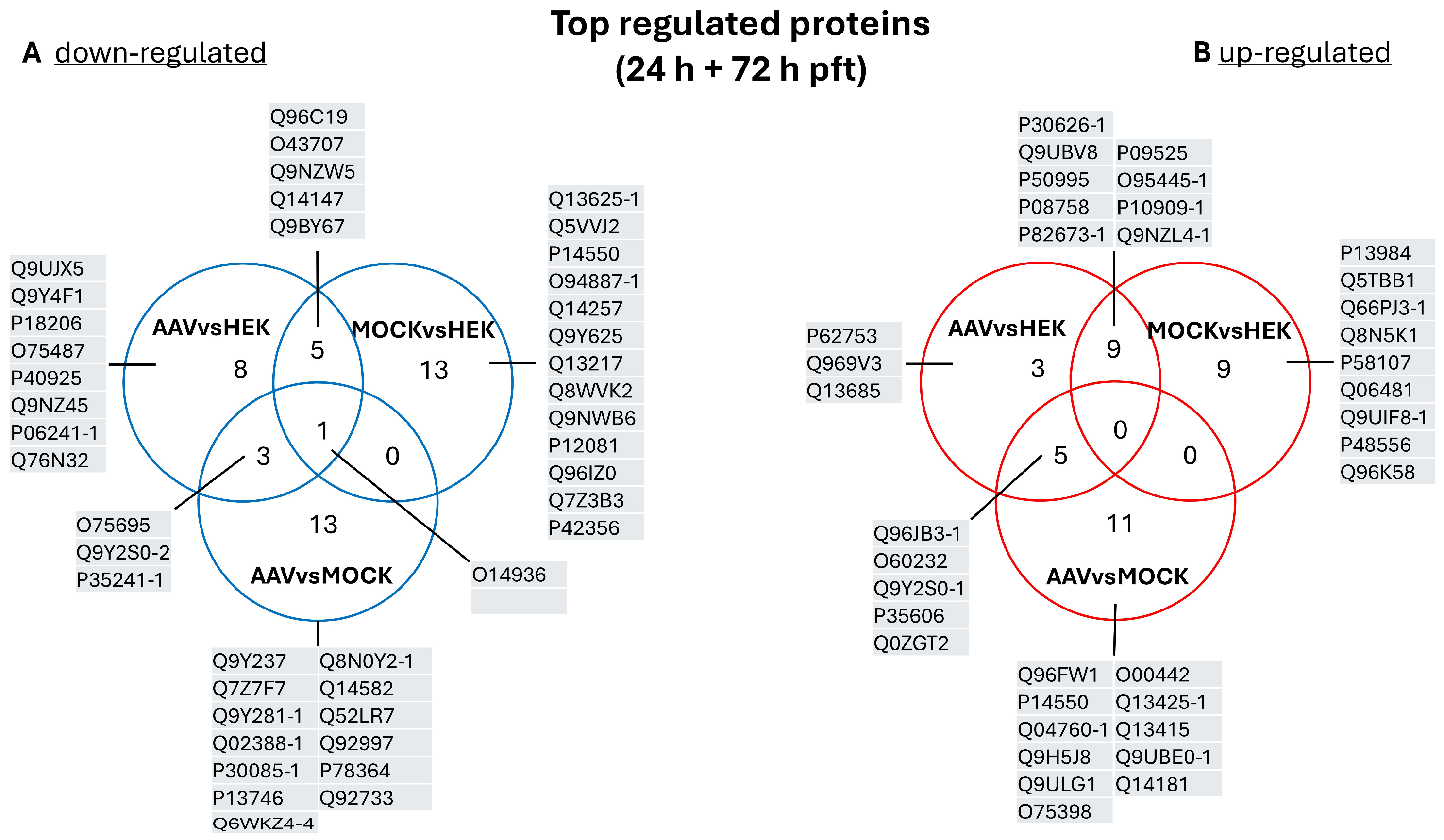
To provide a better overview, we merged the top ten regulated proteins of both comparison time points in a Venn diagram (labeled in Figure 4 and displayed in Figure 5) and then checked for overlaps.
The comparison of the top down-regulated proteins identified three shared proteins in AAV vs. HEK and AAV vs. MOCK, namely protein XRP2 (O75695), radixin (P35241-1), and isoform 2 of DNA-directed RNA polymerases I and III subunit RPAC2 (Q9Y2S0-2). One protein was down-regulated in all sample groups, namely peripheral plasma membrane protein CASK (O14936). Furthermore, this comparison revealed five proteins down-regulated in AAV vs. HEK and MOCK vs. HEK. Interestingly, five proteins were found for AAV vs. HEK in the top rank of down-regulated proteins. The comparison of AAV vs. MOCK revealed 13 exclusive down-regulated proteins. Also, 13 down-regulated proteins were only in the MOCK vs. HEK comparison. Among the top up-regulated proteins (24 h + 72 h ptf), five proteins were found in AAV vs. HEK and AAV vs. MOCK; nine in AAV vs. HEK and MOCK vs. HEK; three in AAV vs. HEK; eleven in AAV vs. MOCK; and nine just in the MOCK vs. HEK group.
Additionally, those groups of significant proteins are listed with protein name, UniProt accession, and fold change in Table 1 for down-regulated proteins and in Table 2 for up-regulated proteins. To enable a fast search of proteins of interest, tables are sorted alphabetically for the UniProt accession numbers for each comparison (AAV vs. HEK, AAV vs. MOCK, etc.).

2.4. Comparison of Significantly Regulated Proteins and Respective Functional Groups

The total number of significant up- and down-regulated proteins detected in the MOCK vs. HEK samples at 24 h was higher than that of regulated proteins in AAV vs. HEK samples. This observation suggests that the transfection itself had a high impact on the regulation of cellular proteins. In AAV-producing HEK-293 cells versus non-transfected HEK-293 cells, at 24 h ptf 235 proteins and at 72 h ptf 318 proteins were significantly up-regulated, while at 24 h 374 proteins and at 72 h 212 proteins were significantly down-regulated. In MOCK-transfected samples versus non-transfected samples, at 24 h ptf 215 and at 72 h ptf 132 proteins were up-regulated, and at 24 h ptf 425 and at 72 h ptf 116 proteins were down-regulated (Figure 6).
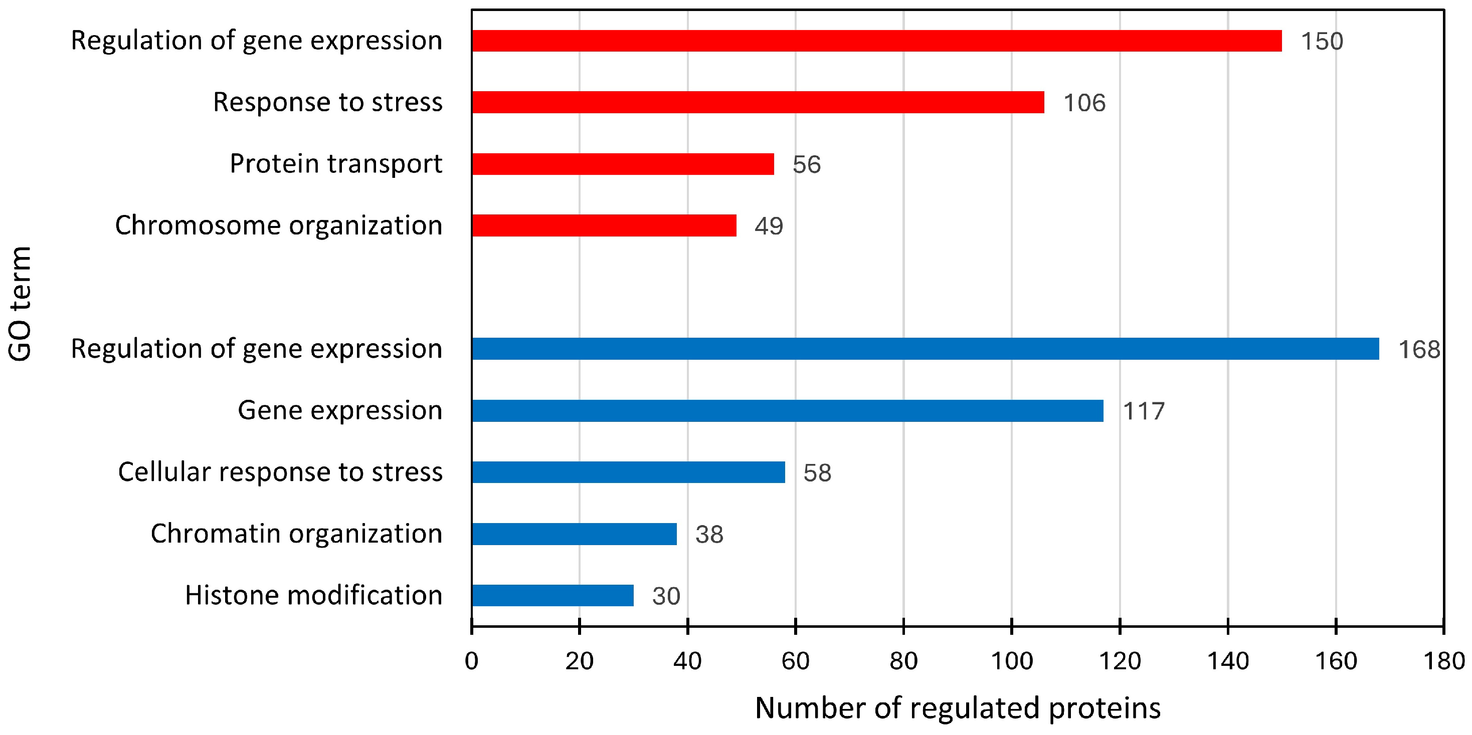
Using the STRING online tool, we further analyzed proteins regulated in rAAV-producing cells versus HEK-293 cells at both time points (24 h and 72 h ptf). We did not exclude proteins that were also regulated in the MOCK samples, as we were interested in obtaining a picture of the whole process. The proteins were put in context using gene ontology annotations assigned by the STRING tool. In the AAV vs. HEK samples, down-regulated proteins were ascribed to the following GO terms: histone modification (30 proteins), chromatin organization (38 proteins), cellular response to stress (58 proteins), and gene expression (168 proteins). Up-regulated proteins had the following GO terms: chromosome organization (49 proteins), protein transport (56 proteins), response to stress (106 proteins), and regulation of gene expression (150 proteins), as shown in Figure 7.

2.5. Up-Regulated Proteins in the Context of the GO Term ‘Response to Virus’

In the GO enrichment analyses, we specifically looked at the GO term ‘response to virus’ (GO:0009615) as a subgroup of the GO term ‘response to stress’. Ten proteins with this GO assignment were identified, and an overview of the regulations of these proteins is given in Table 3.

2.6. rAAV-Production Specific Response in HEK-293 Cells

In the literature, we found that cyclic GMP-AMP synthase (cGAS) may play a role in rAAV production [18]. In our dataset cGAS was not detected, but two proteins associated with cGAS were up-regulated in the AAV samples. In Table 4, these proteins are listed with their calculated abundance ratios.

2.7. Identified Proteins with Context to Adenoviral Proteins

Finally, we checked for known linkages of identified proteins to adenoviral proteins by literature research, and we identified six candidates. These proteins are listed in Table 5 with related fold changes for each of our comparisons.

3. Discussion

3.1. Protein Identification and Sample Variability

Out of the 3384 identified proteins, proteins detected only in one dataset (Figure 3B) are discussed here separately apart from the significantly regulated proteins (Section 4.2). Uniquely identified in the AAV sample (72 h ptf) was the ‘signal transducer and activator of transcription 3′ (STAT3, UniProt P40763). As a signal transducer and transcription activator, STAT3 has multiple functions reported in the context of cytokines and the associated JAKs’ functions in cell differentiation, cell division, cell growth, and immune response [28]. STAT3 plays a role in cell cycle regulation by inducing the expression of key genes for the progression from G1 to S phase [29].
The three proteins CREB-binding protein, protein phosphatase inhibitor 2, and EF-hand domain-containing protein D1 were found only in the HEK samples, which in turn implicates that these are down-regulated upon transfection.
The CREB-binding protein (Q92793-1, gene CREBBP) is a lysine acetyltransferase and modifies key factors involved in DNA replication and interacts with multiple transcription factors and thereby plays a role in transcription activation [30]. The CREBBP acetylates Ku70, and Ku70 acetylation is increased due to DNA damage by UV radiation. Furthermore, a cytoplasmic translocation of CREB-binding protein in this process was reported [31]. Human Ku70, a nuclear protein, serves as a cytosolic DNA sensor. Upon transfection with DNA or infection with DNA viruses, Ku70 translocates from the nucleus into the cytoplasm [32]. Ku70 is also known to interact with Rep proteins and accumulates in AAV replication centers [33,34]. In this study, Ku70 is increased in AAV samples 72 h ptf. The infection and production of AAVs can induce a DNA damage response [34,35,36].
Protein phosphatase inhibitor 2 (P41236, PPP1R2) influences protein phosphatase 1 [37] and is known to regulate Aurora A/Nek2a and the PP1/Aurora A complex [38]. In the AAV production analysis of Strasser et al., aurora kinase was found to be strongly down-regulated in transfected cells and consequently postulated to lead to a reduced cell proliferation [17]. Aurora kinase A is a mitotic serine/threonine kinase that plays an important role in the regulation of cell cycle progression [39] and was not found in our nuclear samples.
The EF-hand domain-containing protein D1 (Q9BUP0, EFHD1) is known to act as a calcium sensor for mitochondrial flash activation [40] and a calcium-dependent actin–bundling activity [41]. In our experiments, transfection was performed by calcium phosphate, and this may influence protein localization to the cytoplasmic fraction.

3.2. Comparison of Top Regulated Proteins

In the analysis of top regulated proteins, we focused on proteins that were regulated between the AAV 24 h vs. MOCK 24 h samples (with proteins also listed in the combined 24 h AAV vs. HEK), as this reflects the peak of the virus production. Four proteins were significantly up-regulated more than 32-fold (log2(fold change) > 5). These include the following: HIC2 (Q96JB3) with a fold change of 48.1; RPAC2 (Q9Y2S0-1, gene POLR1D), also mentioned above, with a fold change of 45.2; NEXN (Nexilin, Q0ZGT2 gene gene Nexilin) with a fold change of 43.2; and LGUL (Lactoylglutathione lyase/glyoxalase I, Q04760-1, gene GLO1) with a fold change of 40.0. These proteins are discussed in the following paragraphs.
HIC2 (hypermethylated in cancer 2 protein) was originally described as a transcription factor and tumor suppressor that localizes in nuclear dots [42]. A recent report indicates that HIC2 is repressed post-transcriptionally by let-7 miRNA, and this may be reduced by DICER depletion [43]. Overexpression of HIC2 in glioblastoma cells (LN229 and U251) induced G2/M cell cycle arrest by down-regulating CDK1 [44]. HIC2 carries a conserved GLDLSKK/R motif related to the PxDLSxK/R motif in adenoviral 2 and 5 E1A proteins, which mediates binding to C-terminal binding proteins [45].
The RPAC2 subunit isoform 1 (Q9Y2S0/P0DPB6), which is shared between RNA polymerase I and III, stabilizes the enzymes’ core and is encoded by the POLR1D gene [46]. PolI transcribes ribosomal RNAs, and the transcriptome of PolIII consists of short, highly structured noncoding RNAs (ncRNAs) [47]. POLR1D expression was increased in colorectal cancer samples and implicated in cancer occurrence, progression, and resistance [48,49]. Silencing of POLR1D in lung cancer cells SK-MES-1 and H2170 inhibited proliferation. In our case, the up-regulation of RPAC2 may be induced by the increased demand for ribosomes for the synthesis of the AAV capsid proteins and, to a lesser extent, also the gene of interest.
Nexilin, also called F-actin binding protein, was analyzed in the context of cardiomyopathy [50], has been used as a gene of interest in AAV-mediated gene replacement [51], and is a seemingly unexpected find in our experiment. However, since AAV2 transduction is dependent on actin and induces remodeling of the cytoskeleton [52], a crosstalk to cellular microfilaments might also happen during production and influence related proteins. Expression studies showed that nexilin colocalized with F-actin and that an N-terminal fragment tended to aggregate in the nucleus. Further, a Z-disc association may relate to a broader stress response [53].
Lactoylglutathione lyase, also named glyoxalase I (GLO1), is an important enzyme for the detoxification of methylglyoxal (2-oxopropanal), a natural reactive byproduct of metabolism. The enzyme is up-regulated in many tumors, and inhibitors have been developed. In clinical investigations, GLO1 expression correlates negatively with cancer survival, and a predominant nuclear staining has been reported [54]. Further, a correlation of expression with key cell cycle genes has been observed, and lentiviral-mediated overexpression decreased the number of Hep3B and Huh-7 cells in the G2 phase and increased the number of cells in the S phase [55].
Three proteins are also highly up-regulated in a second interesting group: AAV vs. HEK and MOCK vs. HEK samples at 24 ptf. These proteins are sorcin (P30626-1), peflin (Q9UBV8), and annexin A11 (P50995); all three are calcium-binding proteins.
Sorcin and peflin belong to the penta-EF-hand (PEF) family [56]. The up-regulation of these three proteins is potentially induced by the transfection reagent of this study. For transfection, the calcium phosphate method was used, and in this way the calcium concentration was increased. This may induce a cellular response by up-regulating these three proteins. Peflin and the apoptosis-linked gene 2 protein (ALG-2) can form a heterodimer, which dissociates in the presence of Ca2+ [57]. ALG-2 interacts with Annexin 11, and the latter can interact with Sorcin [58,59,60]. This shows that all three proteins are linked. Further on, Annexin 11 is also involved in the ER-to-Golgi transport [61]. Whereas peflin was reported to be a negative regulator of ER-to-Golgi transport [62].
In the AAV vs. HEK group, the protein ZNRD2 (zinc ribbon domain containing 2, O60232), also named Sjoegren syndrome/scleroderma autoantigen 1, p27, or SSSCA1, is one of the most up-regulated proteins (>100-fold AAV vs. HEK 24 h ptf). ZNRD2 is found in many tissues, overexpressed in cancers, and regulated by Myc (see below). ZNRD2 binds zinc and proteins linked to microtubules and mitosis, possesses a nuclear exit sequence, and has been found to be centromere associated [63]. The transfection might play a major role in its detection, with some contribution from AAV production.
Interestingly, the EF-hand domain-containing protein D2 (Q96C19) is highly down-regulated in the AAV vs. HEK and in the MOCK vs. HEK groups. This protein is a calcium-binding actin-binding protein [64] and has functions in rapid cytoskeletal rearrangements [65], calcium-dependent exocytosis [66], and calcium-independent exocytosis [67]. The homologous EF-hand domain-containing protein D1 is only identified in HEK samples (discussed before). These findings suggest a role for these proteins in calcium-dependent transfection. Furthermore, a regulation of these proteins by Zn2+ as well as Ca2+ was hypothesized [68].

3.3. Comparison of Significantly Regulated Proteins and Respective Functional Groups

Chung et al. identified DNA/chromatin organization and DNA metabolic processes in GO enrichment analysis of triple transfected suspension HEK-293 cell RNA-Seq analysis [18]. In our data, proteins of the GO terms ‘chromatin organization’ and ‘histone modification’ show lower expression levels in AAV samples than in HEK samples. On the other hand, proteins related to the GO term ‘chromosome organization’ are up-regulated. In this context it should be mentioned that Chung et al. focused on up-regulated RNAs and mainly identified up-regulation of histone subunits 6 h and 12 h ptf. Our data acquisition started at 24 h ptf and does not cover early production time points. Interestingly, Tworig et al. increased rAAV production with a histone deacetylase (HDAC) inhibitor treatment [69].
The rAAV production triggers antiviral and innate immune responses. For example, Chung et al. and Strasser et al. identified a ‘defense response’ in cells producing rAAVs [17,18]. This GO term is included in our analysis in the GO term response to stress (discussed below in the more specific subgroup `response to virus’).

3.4. Up-Regulated Proteins in the Context of the GO Term ‘Response to Virus’

As we were interested in proteins affected by the viral nature of rAAV production, ten identified proteins within the GO term ‘response to virus’ (GO:0009615) assignment are discussed below (see Table 3).

3.4.1. Clusterin (CLUS)

Clusterin (CLUS) is a multifunctional chaperonic glycoprotein associated with diverse cellular functions. A nucleolar localization of secretory clusterin (sCLU) was reported in response to different nucleolar stresses and the association with Cajal bodies post nucleolar stresses [70]. The functional knockdown of clusterin led to altered nuclear morphology and shrunken tubulin filaments. Nuclear morphology regulators nucleophosmin 1 (NPM1) and fibrillarin (FBL) were down-regulated in CLUS knockout cells. Kadam et al. postulated a possible role of clusterin in stabilizing these proteins [70]. NPM1 and FBL were not significantly regulated in our experiment. However, this does not exclude a role in rAAV production. Razin et al. described a virus-induced relocalization of nucleolar proteins to the nucleoplasm for further uses in viral replication compartments [71]. The nucleolar proteins nucleophosmin, fibrillarin, nucleolin (NCL), nucleolar transcription factor 1 (UBF), and nucleolar and coiled-body phosphoprotein 1 (NOLC1) were not regulated in our data. However, the nuclear protein POLR1A was down-regulated in AAV samples 24 h ptf, and treacle protein (TCOFI, isoform 4 here) was up-regulated in 24 h ptf samples of MOCK and AAV. Furthermore, Fraefel et al. observed for AAV infection that AAV DNA replication occurs in compartments, the formation of these compartments is dependent on AAV Rep, AAV ITR, and helper virus, and AAV replication compartments can associate with modified promyelocytic leukemia (PML) nuclear bodies (NBs) [72]. In adenovirus infection, Ad protein E4-orf3 binds and redistributes PML NBs to filamentous structures [73,74]. PML protein was not significantly up-regulated in our AAV and MOCK samples. Therefore, in contrast to AAV infection, nuclear organization may differ in rAAV production, also in a cell-type-specific manner.

3.4.2. Deoxynucleoside Triphosphate Triphosphohydrolase SAMHD1 (SAMHD1)

SAMHD1 is a triphosphohydrolase (dNTPase), which shows restrictive functions against RNA and DNA viruses. The role of SAMDH1 in cellular defense against HIV infection is the hydrolysis of dNTPs, whereby it inhibits HIV-1 reverse transcription and, with this, HIV infection. Furthermore, SAMDH1 is important for proliferation, cell cycle regulation, apoptosis, replication fork progression, and initiating immunity and DNA damage response. Phosphorylation of SAMHD1 is used by the cell to activate dNTP hydrolysis during the G2 phase of the cell. On the other hand, dephosphorylation of SAMDH during the S phase leads to higher dNTP levels [75]. Viruses such as the Epstein–Barr virus, the human herpesvirus 6/7, the human cytomegalovirus, and Kaposi sarcoma-associated herpesvirus seem to inactivate SAMHD1 by phosphorylation through viral kinases. Some viruses are known to use the ubiquitin-proteasome system to target SAMHD1 degradation by polyubiquitinylation of SAMHD1 by influencing the CRL4 or TRIM21 E3 ubiquitin ligase. Also, SUMOylation of SAMHD1 was shown to direct a dNTP-independent antiviral mechanism [76]. These findings in cellular response to other viruses give evidence that the mechanisms may also play an important role in rAAV production. Although the overall amount of SAMHD1 seemed to be increased in our approach, one has to take into account that the multiple phosphorylations lead to a distorted picture of the real activity of this factor. In this study, SAMHD1 was significantly up-regulated 72 h ptf (10-fold compared to untransfected HEK samples). This indicates that the AAV production has an impact on the SAMHD1 amount and that this protein acts as an antiviral factor during rAAV production in HEK-293 cells. As the SAMHD1 protein amount is increased, the AAV seems not to have mechanisms like other viruses that lead to a degradation of SAMHD.

3.4.3. Schlafen Family Member 11 (SLFN11)

Schlafen family member 11 (SLN11) is one of the most studied Schlafen members. It plays important roles in cancer treatment and virus restriction, e.g., in the replication of RNA viruses like HIV and the Flavivirus genus [77]. The degradation of SLN11 by the DNA virus cytomegalovirus was reported. This degradation is induced by the viral protein RL1, which recruits SLN11 to the CRL4 E3 ubiquitin ligase complex [78]. In our study, SLFN11 was significantly up-regulated in AAV samples at 24 h (8-fold) and 72 h (6-fold) ptf but also in MOCK samples at 24 h (4-fold) ptf. In AAV samples, proteins of DNA damage response were up-regulated, and SLFN11 is known to inhibit DNA replication in response to DNA damage and block cell death. SLFN11 is recruited to sites of DNA damage and stalled replication forks in response to replication stress sensed by DNA damage responses. SLFN11 interacts with replication protein A (RPA1) and replication helicase subunit MCM3 at replication foci and selectively blocks fork progression by chromatin opening in proximity of replication initiation sites [79,80,81]. Interestingly, Li et al. showed the lack of SLN11 did not improve AAV titers in AAV-infected cells [82].

3.4.4. Histone-Lysine N-Methyltransferase SETD2 (SETD2)

SETD2 is a very interesting protein based on our expression analysis, because it is only significantly regulated in AAV samples 72 h ptf. The histone methyltransferase SETD2 specifically trimethylates ‘Lys-36’ of histone H3 (H3K36me3) and methylates STAT1. The methylation of STAT1 increases interferon-alpha (IFNα)-mediated antiviral immunity. In hepatocytes the loss of SETD2 promotes HBV infection [83]. Zhang et al. reported inhibition of interferon pathway activation by SARS-CoV-2 protein NSP9 targeting SETD2 [84]. Therefore, the inhibition of SETD2 is a potentially interesting target for improving rAAV production yield. On the other hand, methylation of histone H3Lys36 is increased by splicing [85]. In rAAV-producing cells, splicing is essential for the rAAV production, since the mRNA for most AAV proteins is generated by alternative splicing.

3.4.5. 2′,5′-Phosphodiesterase 12 (PDE12)

2′,5′-Phosphodiesterase 12 (PDE12) is a mitochondrial protein and is known to remove mitochondrial RNA poly(A)tails and control mitochondrial translation [86]. PDE12 degrades the second messenger 2′,5′-oligoadenylate (2-5A) and thereby inhibits IFN-mediated antiviral defense by impeding RNase-L activation [87]. How expression of PDE12 is increased in HEK-293 cells producing rAAV needs to be investigated. Also, it would be interesting if activation of PDE12 expression before transfection would improve rAAV production.

3.4.6. ATP-Dependent RNA Helicase DDX3X (DDX3X)

DDX3X has functions in transcription, mRNA maturation, export, and translation [88]. Furthermore, roles in antiviral immunity [89,90] and also for replication of several RNA viruses like hepatitis C virus [91] and human immunodeficiency virus were reported [92]. In our experiment, DDX3X was significantly up-regulated in all AAV and MOCK samples. Up-regulation in AAV samples was slightly increased compared to MOCK samples. Inhibition of DDX3X may have a positive effect on AAV production and on transgene expression in HEK-293 cells.

3.4.7. Heat Shock Protein Beta-1 (HSPB1)

HSPB1 (Hsp27) inhibits cytochrome-C-mediated activation of caspase-3 and -9 and reduces apoptosome formation [93,94]. Additionally, HSPB1 increases cell survival via inhibition of Bax [95]. Interestingly, HSPB1 is more up-regulated in MOCK samples 72 h ptf than in AAV samples 72 h ptf. The mechanisms behind this observation remain unknown and need to be investigated.

3.4.8. Tripartite Motif-Containing Protein 6 (TRIM6)

The E3 ubiquitin ligase TRIM6 plays an important role in the activation of the type-I interferon signaling pathway [96] and promotes replication and assembly of Ebola virus [97,98]. The potential role of TRIM6 in rAAV production is an intriguing question that could advance our understanding of rAAV biology.

3.4.9. Zinc Finger CCHC Domain-Containing Protein 3 (ZCCHC3)

A further interesting candidate is the zinc finger CCHC domain-containing protein 3 (ZCCHC3). In this study, the ZCCHC3 protein is similarly up-regulated in rAAV-producing and MOCK-transfected cells 24 h post-transfection. This indicates no rAAV-specific or virus-specific cellular response. Of course, the transfection process itself is an important putative bottleneck for AAV production, next to viral defense mechanisms of the host cells. Therefore, ZCCHC3 may also be interesting for rAAV production and for general transient transfection approaches. Lian et al. identified ZCCHC3 as a positive regulator in dsRNA- and dsDNA-virus-mediated cellular antiviral immune response [99,100]. Chen et al. detected increased ZCCHC3 levels in influenza virus (RNA virus)-infected cells [101]. In this context ZCCHC3 may also be a promising target for increased rAAV particle production. ZCCHC3 is also further discussed in our next chapter.

3.4.10. Heat Shock Protein HSP 90-Alpha (HSP90AA1)

The heat shock protein HSP 90 is a chaperone with a broad range of reviewed functions in apoptosis, signaling pathways, cell cycle control, protein folding, and degradation [102,103]. Interestingly, HSP90 is reported to support the internalization, virus replication, and gene expression of DNA viruses, reviewed by Lubkowska et al. [104]. HSP90AA1 is significantly up-regulated in AAV samples 72 h ptf in comparison to HEK samples. Dietmair et al. also reported up-regulation of HSP90AA1 in a multi-omics analysis of recombinant protein-producing HEK-293 cells [105]. Therefore, HSP90 may also support processes in rAAV production.

3.5. rAAV-Production-Specific Response in HEK-293 Cells

Transcriptomic and proteomic studies on the production of rAAV by plasmid transfection in HEK-293 cells led to the identification of induction factors of specific antiviral and inflammatory responses [17,18]. The authors discuss that cyclic GMP-AMP synthase (cGAS) may play a role in rAAV production. Chung et al. investigated the kinetic transcriptional response of suspension HEK-293 cells to rAAV production following transient polyethylenimine plasmid transfection and detected low amounts of cGAS transcripts in transfected cells. In our dataset, cGAS was not detected, presumably because cGAS is a cytosolic protein. However, in our experiment, the following two proteins associated with cGAS were up-regulated in the AAV samples:
PPP6C is the catalytic subunit of protein phosphatase 6 (PP6). The phosphatase PPP6C is constitutively associated with cGAS in unstimulated cells [106]. PPP6C negatively regulates dsDNA-induced interferon regulatory factor 3 (IRF3) activation but does not affect NF-kappa-B activation [107]. PPP6C deficiency greatly inhibits herpes simplex virus 1 (HSV-1) and vesicular stomatitis virus (VSV) replication [107]. In the context of Kaposi’s sarcoma-associated herpesvirus (KSHV), PPP6C interacts with STING, and loss of PPP6C enhances STING phosphorylation. PPP6C negatively regulates the cGAS-STING pathway by removing STING phosphorylation [107]. In our data, PPP6C was significantly up-regulated in rAAV-producing cells 72 h ptf. Interestingly, PPP6C was not up-regulated in MOCK samples at any time point. These results indicate a specific host cell response to rAAV production.
The zinc finger CCHC domain-containing protein 3 (ZCCHC3) was reported to be a co-sensor of cGAS. ZCCHC3 enhances the binding of cGAS to dsDNA by directly binding to dsDNA [99]. Additionally, this protein was reported to promote the antiviral TRIM25-mediated RIG-I activation [100,108]. Therefore, ZCCHC3 may influence transfection and rAAV production.

3.6. Identified Proteins with Context to Adenoviral Proteins

During rAAV production in HEK-293 cells, adenoviral proteins are expressed from the helper plasmid. Among them is the E4orf4 protein, which has toxic effects on the host cells [109]. In the literature, two human proteins are described in the context of E4orf4. Kleinberger and Shenk showed that E4orf4 interacts with the protein PP2A by a direct association with the phosphatase Bα/B55 (PPP2R2A) regulatory subunit [110]. Ben-Israel et al. showed down-regulation of Myc proto-oncogene protein (MYC) by E4orf4 expression by PPP2R2A interaction [111]. PPP2R2A is up-regulated in AAV vs. HEK 72 h ptf and in AAV vs. MOCK 24 and 72 h ptf. MYC, as an important nuclear factor, was also identified in our data, but no significant regulation was observed. Our data indicate that in rAAV production, MYC expression is only marginally affected by E4orf4. Interestingly, the PP2A subunit PPP2R1B was not up-regulated in AAV and MOCK samples in comparison to HEK samples.
The up-regulated protein DDX6 binds and re-localizes with the adenoviral protein E4 11k (product of E4orf3) in cytoplasmic p-bodies [112]. DDX6 is known to have a nucleocytoplasmic localization in human cells [113]. Increased DDX6 nuclear localization in our rAAV-producing HEK-293 cells indicates a potential influence of E4 11k on DDX6 transport.
Cullin5 was up-regulated in rAAV-producing cells, but also in MOCK-transfected cells. This protein was reported to bind adenoviral proteins E1B55K and E4orf6. The complex leads to degradation of p53, but also of the adeno-associated proteins Rep and Cap [114]. Degradation of p53 promotes cell cycle and inhibits apoptosis, which presumably has a positive effect on rAAV production, whereby degradation of Rep and Cap proteins may reduce rAAV production. In our study, p53 was down-regulated in AAV samples 72 h ptf. Furthermore, the Mre11 complex is a substrate of the E4orf6/E1B55K complex and consists of Mre11, Rad50, and NBS1. This complex detects double-strand breaks and induces p53-dependent apoptosis [115]. Interestingly, Rad50 was down-regulated in AAV samples 24 h and 72 h ptf and in MOCK samples 24 h ptf. Also, Mre11 was down-regulated in MOCK and AAV samples 24 h ptf. Our data suggest a Cullin5-initiated host cell reaction to transfection but also to rAAV production. Recently, Radukic et al. showed stabilization of AAV ITRs in E. coli plasmids in the absence of the bacterial Mre11 homolog, called SbcC, suggesting a need for rAAV-producing cells to down-regulate the Mre11/Rad50 complex for faithful genome replication [116].

4. Materials and Methods

4.1. Cell Culture of HEK-293 Cells

HEK-293 cells (DSMZ no. ACC 305, Braunschweig, Germany) were cultured in Dulbecco’s Modified Eagle Medium supplemented with 10 % (v/v) fetal calf serum and 1 % (v/v) penicillin/streptomycin (all from Sigma Aldrich, Steinheim, Germany) at 37 °C and 5 % CO2 in T175 culture flasks (Sarstedt, Nümbrecht, Germany).

4.2. Transfection of HEK-293 Cells

For transfection, 9 × 10⁶ HEK-293 cells were seeded in 150 mm cell culture dishes and incubated overnight. A total amount of 45 μg DNA per 150 mm dish was transfected. One Bijou sample container was filled with 1 mL of 2× HBS (50 mM HEPES, 1.5 mM Na2HPO4, 280 mM NaCl, pH 7.05, sterile filtered), and the second one was filled with 1 mL of CaCl2 (0.3 M).
Three types of samples were generated, each in triplicate: First, HEK-293 cells without transfection (HEK Sample); second, HEK-293 cells transfected with plasmids pHelper (pZMB0088), pRepCap (pZMB0618), and pITR-mVenus (pZMB0522) for rAAV2 production (AAV Sample) in a molar ratio of 1:1:1; third, HEK-293 cells transfected with the pITR-mVenus and a second non-human-coding pUC19 plasmid (pZMB0169) (MOCK Sample). For MOCK transfection, cells were transfected with the same amount of the ITR plasmid as for AAV production, while the DNA amount of pHelper and pRepCap was replaced by the noncoding plasmid. The DNA was added to the CaCl2 solution and was transferred dropwise to the container with 2× HBS and vortexed three times for 3 s. Immediately, 2 mL of this solution was pipetted dropwise to the cells. After 24 h and 72 h of cultivation, cells of two 150 mm cell culture plates (Sarstedt) were harvested by scraping, washed three times with PBS by resuspension and pelleting (800× g, 5 min), and finally pelleted by centrifugation (3000× g, 10 min). The cell pellets were stored at −80 °C.

4.3. Fluorescence Microscopy

mVenus expression was imaged with an automated fluorescence microscope (DMI 6000B, Leica Microsystem, Wetzlar, Germany) 24 h and 72 h post-transfection.

4.4. Protein Fractionation into Nuclear Fraction

Nuclear proteins were isolated from cell pellets using different buffers. First, the pellet was resuspended by addition of 1.25 mL LB1 buffer (50 mM HEPES-KOH pH 7.5, 140 mM NaCl, 1 mM EDTA, 10% (v/v) glycerin, 0.5% (v/v) NP-40, 0.25% (v/v) Triton X-100, pH 7.5), incubation for 10 min at 4 °C on a tube roller, and centrifugation for 5 min at 2000× g and 4 °C. Second, the resulting pellet was resuspended in 1.25 mL LB2 buffer (10 mM Tris-HCl, 200 mM NaCl, 1 mM EDTA, 0.5 mM EGTA, pH 8.0) and centrifuged for 5 min at 2000× g and 4 °C. The pellet was resuspended in 225 μL LB3 buffer (10 mM Tris-HCl, 100 mM NaCl, 1 mM EDTA, 0.5 mM EGTA, 0.1% (v/v) sodium deoxycholate, 0.5% (v/v) N-lauroylsarcosine, pH 8.0). Next, ultrasonication was performed three times for 10 s (Branson S-250A sonifier, 40% amplitude, Thermo Fisher Scientific, Darmstadt, Germany) with samples cooled on ice. The sample was supplemented with 22.5 μL of 10% Triton-X 100, vortexed, and centrifuged for 10 min (at 4 °C, 13,300× g). The supernatant was used for further MS-sample generation. The protein amount was quantified with a bicinchoninic acid assay (BCA, UP40840A, Interchim, Montluçon, France) according to the manufacturer’s protocol with bovine serum albumin as a reference substance measured at 560 nm with an iMark microplate absorbance reader (Bio-Rad, Feldkirchen, Germany).

4.5. Filter-Aided Sample Preparation (FASP) for Mass Spectrometry

The filter-aided sample preparation (FASP) method was adapted after Wiśniewski et al. [117]. Moreover, 50 µg of protein was precipitated by a 9-fold acetone volume, vortexed, stored at −20 °C overnight, and centrifuged for 10 min at 13,300× g. After centrifugation, the supernatant was aspirated and the pellet was dried at room temperature for 10 min. The pellet was resuspended in 200 µL UA buffer (8 mM urea, 100 mM Tris-HCl, pH 9) and added to a 30 kDa cutoff centrifugation device (Nanosep with Omega Membrane, Fisher Scientific, Schwerte, Germany). Samples were centrifuged at 9600× g, respectively, at room temperature until the volume passed the filter completely. This step was repeated once. Reduction and alkylation were performed by adding 100 μL dithiothreitol (DTT) (50 mM DTT in UA buffer) to the filter device and incubating for 5 min at 60 °C and 600 rpm (MultiTherm shaker, Benchmark Scientific, NJ, Sayreville, USA). This step was followed by centrifugation at 9600× g at room temperature (until the volume passed the filter completely). Next, 100 µL of iodoacetamide (IAA) buffer (50 mM IAA in UA buffer) were added to the filter device, followed by incubation for 1 min at room temperature with 600 rpm and centrifugation (9600× g at room temperature until volume passed the filter completely). The filter device was washed three times by adding 150 μL of UA buffer. A tryptic digest (trypsin/LysC, Mass Spec Grade, Promega, Mannheim, Germany) with 10 ng/μL in DB buffer (50 mM Tris-HCl, pH 8.5, 50 µL) was performed overnight at 37 °C and 300 rpm shaking. On the next day, samples were centrifuged (9600× g at room temperature until volume passed the filter completely), and peptide-containing flowthrough was analyzed by absorption measurement (205 nm, NanoDrop, Thermo Fisher Scientific, Darmstadt, Germany) for peptide content. From each sample, 20 µg peptide was dried in a SpeedVac and then dissolved in 10 μL 2.5% v/v acetonitrile (ACN)/0.1% trifluoroacetic acid (TFA) in LC-MS grade water (Merck, Darmstadt, Germany). To remove particles, samples were centrifuged for 5 min at 13,000× g. Supernatant was used for Nano-Liquid Chromatography–Orbitrap Mass Spectrometry (nLC-Orbitrap MS/MS, Thermo Fisher Scientific, Dreieich, Germany) measurement.

4.6. Nano-Liquid Chromatography–Orbitrap Mass Spectrometry Measurement

In this study, 1 µg of peptide sample was loaded to the nano-HPLC system (UltiMate 3000 RSLC, Thermo Fisher, Dreieich, Germany) by autosampler. Desalting was performed with a C18 pre-column cartridge (Acclaim PepMap™ 100, 300 µm I.D. × 5 mm) and separated on a 25 cm C18 column (Acclaim™ PepMap™ 100, 2 µm particle size, 75 µm I.D. × 25 cm; all parts were purchased from Thermo Fisher Scientific, Dreieich, Germany). An effective gradient of 5–30% of solvent B (80% ACN, 0.1% formic acid (FA) in LC-MS grade water mixed with solvent A (0.1% FA in LC-MS grade water) in 120 min with a flow rate of 300 nL/min was performed. Online ESI-Orbitrap mass spectrometry measurements were carried out by a Q Exactive Plus instrument (Thermo Fisher Scientific, Dreieich, Germany), equipped with a dual MALDI-/ESI-source (Spectroglyph LLC, Kennewick, WA, USA) in data-dependent top 10 acquisition mode. The MS scan range was 350–2000 m/z with a resolution of 70,000, and the dynamic exclusion time of precursors for MS/MS was set to 60 s. Precursor ions were scanned with a resolution of 17,500 and fragmented with a normalized collision energy (CE) of 28.

4.7. Data Analysis: Protein Identification and Label-Free Quantification

Protein identification and quantification were performed with Proteome Discoverer versions 2.4 and 3.0 with precursor detection node (Thermo Fisher Scientific). The human protein database (Homo sapiens SwissProt DB (TaxID 9606), Version 25 October 2017) was used as a template for peptide spectrum matching. For the protein identification and label-free quantification (LFQ), mass tolerances of 10 ppm for precursor ions and 0.02 Da for deviation from the theoretical masses from MS/MS were allowed. The maximum number of missed cleavages for tryptic digest was set to two. In the validation steps, a false discovery rate (FDR) of 0.01 was selected for protein and peptide identification. Dynamic modifications of oxidation (M) and deamidation (N, Q) and fixed modification of carbamidomethylation (C) were set. LFQ values were normalized based on total peptide amount. Protein abundance was calculated based on the average of the top-three quantified peptides (top N set to 3) of normalized LFQ values. Fold changes (abundance ratios) were calculated based on the protein abundance setting larger values to a maximum of 100 fold change. Statistical quantitative analysis and principal component analysis (PCA) were performed with Proteome Discoverer software. The analysis of top regulated proteins (Section 4.2) is based on the evaluation with version PD 2.4 (according to data in Supplementary Materials: Table S1_PDv24), and all other analyses were performed with PD 3.0 (according to data in Supplementary Materials: Table S2_PDv30). The mass spectrometry proteomics raw data were deposited to the ProteomeXchange Consortium via the PRIDE [118] partner repository with the dataset identifier PXD062079.
For protein identification, only proteins with at least one unique peptide were allowed. For gene ontology enrichment analysis, the Search Tool of the Retrieval of Interacting Genes/Proteins (STRING, http://string-db.org/) was used for significantly regulated proteins. The following settings were used for analysis: Network: full STRING network; required score: medium confidence (0.4); FDR stringency: medium (5%); and minimum count in network: 2. Venn diagrams were generated with the online tool VENNY 2.1 (https://bioinfogp.cnb.csic.es/tools/venny/).
For analysis of top regulated proteins, we considered proteins as significantly regulated only if they held at least two unique peptides, a minimum fold change of 2 of abundance ratios (LFQ values), and a maximum p-value of 0.05. Volcano plots were generated with Python (Python.org, Python 3.12.4) based on log2-transformed fold changes and log10-transformed p-values generated by a two-sided t-test with significance values.

5. Conclusions

In this study, we compared untreated, mock-transfected, and, for rAAV production, transfected HEK-293 cells. Analyses of these data revealed interesting proteins, many of which are reported to play a role in the cell cycle, gene expression, and host cell response to viruses, or to be associated with adenoviral proteins. In the transfection process, three plasmids were added, expressing several proteins known to interfere with the cell cycle as well as several proteins resulting in the viral product. These complex effects on the nuclear host cell proteome are difficult to reconcile with one typical GO term or pathway. Our data underline the important roles of already published factors but also provide novel proteins in this context. These are promising targets for further investigation of their impact on rAAV production and on transfection in general. Depending on their regulation and known functions, approaches with small-molecule inhibitors, knockdown, knockout, or overexpression can be interesting to improve rAAV production. In this study, we utilized the GOI plasmid with an unrelated plasmid as a reference. Deciphering in more detail the isolated effect of the Helper or RepCap plasmid on the host cell by testing additional plasmid combinations would be of interest. Further on, an integrated look at the interplay of AAV production and host expression by including other transfection methods, further selected HEK-293 clones, RNAseq data, metabolic analyses, and proteomic studies will provide a more holistic view. Next to the nuclear fraction, these studies could include the cytoplasmic fraction and even specific organelles and the culture medium to gain a broader overview of regulated pathways and help to understand and optimize cellular processes for a reliable, high-titer viral-particle generation.

Supplementary Materials

The following supporting information can be downloaded at: https://www.mdpi.com/article/10.3390/ijms26136315/s1.

Author Contributions

Conceptualization, S.K.G., R.H. and K.M.M.; methodology, S.K.G.; software, S.K.G. and R.H.; validation, S.K.G., R.H. and K.M.M.; formal analysis, S.K.G.; resources, R.H. and K.M.M.; data curation, R.H.; writing—original draft preparation, S.K.G.; writing—review and editing, R.H. and K.M.M.; visualization, S.K.G.; supervision, R.H. and K.M.M.; project administration, K.M.M.; project funding was provided by Bielefeld University to R.H. and K.M.M. All authors have read and agreed to the published version of the manuscript.

Funding

This research received no external funding.

Institutional Review Board Statement

Not applicable.

Informed Consent Statement

Not applicable.

Data Availability Statement

The mass spectrometry proteomics raw data were deposited to the ProteomeXchange Consortium via the PRIDE partner repository with the dataset identifier PXD062079.

Acknowledgments

We thank Nadine Rodrigues De Carvalho for technical tips, Robert Heyer for discussions, Marco T. Radukic for manuscript proofreading, and Oliver Hertel for the figure created with BioRender.

Conflicts of Interest

The authors declare no conflicts of interest.

Abbreviations

The following abbreviations are used in this manuscript:
rAAVrecombinant adeno-associated virus
HEKHEK samples (untransfected)
MOCKMOCK samples (mock-transfected)
AAVAAV samples (triple-transfected with rAAV2 production plasmids)
ptfpost-transfection
ITRinverted terminal repeats
LFQlabel-free quantification
PCAprinciple component analysis
GOgene ontology

References

  1. Glybera. Available online: https://www.ema.europa.eu/en/medicines/human/EPAR/glybera (accessed on 8 July 2024).
  2. Luxturna. Available online: https://www.fda.gov/vaccines-blood-biologics/cellular-gene-therapy-products/luxturna (accessed on 11 July 2024).
  3. Zolgensma. Available online: https://www.fda.gov/vaccines-blood-biologics/zolgensma (accessed on 8 July 2024).
  4. Hemgenix. Available online: https://www.fda.gov/vaccines-blood-biologics/vaccines/hemgenix (accessed on 9 July 2024).
  5. Upstaza. Available online: https://www.ema.europa.eu/en/medicines/human/EPAR/upstaza (accessed on 15 January 2025).
  6. Roctavian. Available online: https://www.fda.gov/vaccines-blood-biologics/roctavian (accessed on 8 July 2024).
  7. Elevidys. Available online: https://www.fda.gov/vaccines-blood-biologics/tissue-tissue-products/elevidys (accessed on 9 July 2024).
  8. Beqvez. Available online: https://www.fda.gov/vaccines-blood-biologics/cellular-gene-therapy-products/beqvez (accessed on 11 July 2024).
  9. Russell, W.C.; Graham, F.L.; Smiley, J.; Nairn, R. Characteristics of a Human Cell Line Transformed by DNA from Human Adenovirus Type 5. J. Gen. Virol. 1977, 36, 59–72. [Google Scholar] [CrossRef]
  10. Berk, A.J. Recent Lessons in Gene Expression, Cell Cycle Control, and Cell Biology from Adenovirus. Oncogene 2005, 24, 7673–7685. [Google Scholar] [CrossRef] [PubMed]
  11. Aponte-Ubillus, J.J.; Barajas, D.; Sterling, H.; Aghajanirefah, A.; Bardliving, C.; Peltier, J.; Shamlou, P.; Roy, M.; Gold, D. Proteome Profiling and Vector Yield Optimization in a Recombinant Adeno-associated Virus-producing Yeast Model. Microbiologyopen 2020, 9, e1136. [Google Scholar] [CrossRef] [PubMed]
  12. Handyside, B.; Ismail, A.M.; Zhang, L.; Yates, B.; Xie, L.; Sihn, C.-R.; Murphy, R.; Bouwman, T.; Kim, C.K.; De Angelis, R.; et al. Vector Genome Loss and Epigenetic Modifications Mediate Decline in Transgene Expression of AAV5 Vectors Produced in Mammalian and Insect Cells. Mol. Ther. 2022, 30, 3570–3586. [Google Scholar] [CrossRef]
  13. Merten, O.-W. Development of Stable Packaging and Producer Cell Lines for the Production of AAV Vectors. Microorganisms 2024, 12, 384. [Google Scholar] [CrossRef]
  14. Cao, T.M.; Chen, D.; Barnard, G.C.; Shen, A. Recombinant Adeno-associated Virus Production Evaluation in Chinese Hamster Ovary Cells. Biotechnol. Bioeng. 2024, 121, 395–402. [Google Scholar] [CrossRef]
  15. Le, D.T.; Radukic, M.T.; Müller, K.M. Adeno-Associated Virus Capsid Protein Expression in Escherichia Coli and Chemically Defined Capsid Assembly. Sci. Rep. 2019, 9, 18631. [Google Scholar] [CrossRef] [PubMed]
  16. Le, D.T.; Radukic, M.T.; Teschner, K.; Becker, L.; Müller, K.M. Synthesis and Concomitant Assembly of Adeno-Associated Virus-like Particles in Escherichia Coli. ACS Synth. Biol. 2022, 11, 3601–3607. [Google Scholar] [CrossRef]
  17. Strasser, L.; Boi, S.; Guapo, F.; Donohue, N.; Barron, N.; Rainbow-Fletcher, A.; Bones, J. Proteomic Landscape of Adeno-Associated Virus (AAV)-Producing HEK293 Cells. Int. J. Mol. Sci. 2021, 22, 11499. [Google Scholar] [CrossRef]
  18. Chung, C.-H.; Murphy, C.M.; Wingate, V.P.; Pavlicek, J.W.; Nakashima, R.; Wei, W.; McCarty, D.; Rabinowitz, J.; Barton, E. Production of rAAV by Plasmid Transfection Induces Antiviral and Inflammatory Responses in Suspension HEK293 Cells. Mol. Ther.—Methods Clin. Dev. 2023, 28, 272–283. [Google Scholar] [CrossRef]
  19. Wang, Y.; Fu, Q.; Lee, Y.S.; Sha, S.; Yoon, S. Transcriptomic Features Reveal Molecular Signatures Associated with Recombinant Adeno-associated Virus Production in HEK293 Cells. Biotechnol. Prog. 2023, 39, e3346. [Google Scholar] [CrossRef] [PubMed]
  20. Lu, M.; Lee, Z.; Hu, W. Multi-omics Kinetic Analysis of Recombinant Adeno-associated Virus Production by Plasmid Transfection of HEK293 Cells. Biotechnol. Prog. 2024, 40, e3428. [Google Scholar] [CrossRef] [PubMed]
  21. Lin, Y.-C.; Lu, M.; Cai, W.; Hu, W.-S. Comparative Transcriptomic and Proteomic Kinetic Analysis of Adeno-Associated Virus Production Systems. Appl. Microbiol. Biotechnol. 2024, 108, 385. [Google Scholar] [CrossRef] [PubMed]
  22. Patra, A.T.; Tan, E.; Kok, Y.J.; Ng, S.K.; Bi, X. Temporal Insights into Molecular and Cellular Responses during rAAV Production in HEK293T Cells. Mol. Ther.—Methods Clin. Dev. 2024, 32, 101278. [Google Scholar] [CrossRef]
  23. Wang, Y.; Fu, Q.; Park, S.Y.; Lee, Y.S.; Park, S.-Y.; Lee, D.-Y.; Yoon, S. Decoding Cellular Mechanism of Recombinant Adeno-Associated Virus (rAAV) and Engineering Host-Cell Factories toward Intensified Viral Vector Manufacturing. Biotechnol. Adv. 2024, 71, 108322. [Google Scholar] [CrossRef]
  24. Gurazada, S.G.R.; Kennedy, H.M.; Braatz, R.D.; Mehrman, S.J.; Polson, S.W.; Rombel, I.T. HEK-Omics: The Promise of Omics to Optimize HEK293 for Recombinant Adeno-Associated Virus (rAAV) Gene Therapy Manufacturing. Biotechnol. Adv. 2025, 79, 108506. [Google Scholar] [CrossRef]
  25. Wistuba, A.; Kern, A.; Weger, S.; Grimm, D.; Kleinschmidt, J.A. Subcellular Compartmentalization of Adeno-Associated Virus Type 2 Assembly. J. Virol. 1997, 71, 1341–1352. [Google Scholar] [CrossRef]
  26. Maurer, A.C.; Weitzman, M.D. Adeno-Associated Virus Genome Interactions Important for Vector Production and Transduction. Hum. Gene Ther. 2020, 31, 499–511. [Google Scholar] [CrossRef]
  27. Golm, S.K.; Hübner, W.; Müller, K.M. Fluorescence Microscopy in Adeno-Associated Virus Research. Viruses 2023, 15, 1174. [Google Scholar] [CrossRef]
  28. Lv, Y.; Qi, J.; Babon, J.J.; Cao, L.; Fan, G.; Lang, J.; Zhang, J.; Mi, P.; Kobe, B.; Wang, F. The JAK-STAT Pathway: From Structural Biology to Cytokine Engineering. Signal Transduct. Target. Ther. 2024, 9, 221. [Google Scholar] [CrossRef]
  29. Saxena, N.K.; Vertino, P.M.; Anania, F.A.; Sharma, D. Leptin-Induced Growth Stimulation of Breast Cancer Cells Involves Recruitment of Histone Acetyltransferases and Mediator Complex to CYCLIN D1 Promoter via Activation of Stat3. J. Biol. Chem. 2007, 282, 13316–13325. [Google Scholar] [CrossRef] [PubMed]
  30. Vo, N.; Goodman, R.H. CREB-Binding Protein and P300 in Transcriptional Regulation. J. Biol. Chem. 2001, 276, 13505–13508. [Google Scholar] [CrossRef] [PubMed]
  31. Cohen, H.Y.; Lavu, S.; Bitterman, K.J.; Hekking, B.; Imahiyerobo, T.A.; Miller, C.; Frye, R.; Ploegh, H.; Kessler, B.M.; Sinclair, D.A. Acetylation of the C Terminus of Ku70 by CBP and PCAF Controls Bax-Mediated Apoptosis. Mol. Cell 2004, 13, 627–638. [Google Scholar] [CrossRef] [PubMed]
  32. Sui, H.; Chen, Q.; Imamichi, T. Cytoplasmic-translocated Ku70 Senses Intracellular DNA and Mediates Interferon-lambda1 Induction. Immunology 2021, 163, 323–337. [Google Scholar] [CrossRef]
  33. Nash, K.; Chen, W.; Salganik, M.; Muzyczka, N. Identification of Cellular Proteins That Interact with the Adeno-Associated Virus Rep Protein. J. Virol. 2009, 83, 454–469. [Google Scholar] [CrossRef]
  34. Schwartz, R.A.; Carson, C.T.; Schuberth, C.; Weitzman, M.D. Adeno-Associated Virus Replication Induces a DNA Damage Response Coordinated by DNA-Dependent Protein Kinase. J. Virol. 2009, 83, 6269–6278. [Google Scholar] [CrossRef]
  35. Collaco, R.F.; Bevington, J.M.; Bhrigu, V.; Kalman-Maltese, V.; Trempe, J.P. Adeno-Associated Virus and Adenovirus Coinfection Induces a Cellular DNA Damage and Repair Response via Redundant Phosphatidylinositol 3-like Kinase Pathways. Virology 2009, 392, 24–33. [Google Scholar] [CrossRef]
  36. Ning, K.; Kuz, C.A.; Cheng, F.; Feng, Z.; Yan, Z.; Qiu, J. Adeno-Associated Virus Monoinfection Induces a DNA Damage Response and DNA Repair That Contributes to Viral DNA Replication. mBio 2023, 14, e03528-22. [Google Scholar] [CrossRef]
  37. Bresch, A.-M.; Yerich, N.; Wang, R.; Sperry, A.O. The PP1 Regulator PPP1R2 Coordinately Regulates AURKA and PP1 to Control Centrosome Phosphorylation and Maintain Central Spindle Architecture. BMC Mol. Cell Biol. 2020, 21, 84. [Google Scholar] [CrossRef]
  38. Rebelo, S.; Santos, M.; Martins, F.; Da Cruz E Silva, E.F.; Da Cruz E Silva, O.A.B. Protein Phosphatase 1 Is a Key Player in Nuclear Events. Cell. Signal. 2015, 27, 2589–2598. [Google Scholar] [CrossRef]
  39. Willems, E.; Dedobbeleer, M.; Digregorio, M.; Lombard, A.; Lumapat, P.N.; Rogister, B. The Functional Diversity of Aurora Kinases: A Comprehensive Review. Cell Div. 2018, 13, 7. [Google Scholar] [CrossRef] [PubMed]
  40. Hou, T.; Jian, C.; Xu, J.; Huang, A.Y.; Xi, J.; Hu, K.; Wei, L.; Cheng, H.; Wang, X. Identification of EFHD1 as a Novel Ca2+ Sensor for Mitoflash Activation. Cell Calcium 2016, 59, 262–270. [Google Scholar] [CrossRef] [PubMed]
  41. Mun, S.A.; Park, J.; Park, K.R.; Lee, Y.; Kang, J.Y.; Park, T.; Jin, M.; Yang, J.; Jun, C.-D.; Eom, S.H. Structural and Biochemical Characterization of EFhd1/Swiprosin-2, an Actin-Binding Protein in Mitochondria. Front. Cell Dev. Biol. 2021, 8, 628222. [Google Scholar] [CrossRef] [PubMed]
  42. Deltour, S.; Pinte, S.; Guérardel, C.; Leprince, D. Characterization of HRG22, a Human Homologue of the Putative Tumor Suppressor Gene HIC1. Biochem. Biophys. Res. Commun. 2001, 287, 427–434. [Google Scholar] [CrossRef]
  43. Huang, P.; Peslak, S.A.; Shehu, V.; Keller, C.A.; Giardine, B.; Shi, J.; Hardison, R.C.; Blobel, G.A.; Khandros, E. Let-7 miRNAs Repress HIC2 to Regulate BCL11A Transcription and Hemoglobin Switching. Blood 2024, 143, 1980–1991. [Google Scholar] [CrossRef]
  44. Lu, P.; Shangguan, W.; Zhao, Q. HIC2 Promotes Cell Cycle Transitions by Upregulating CDK1 Expression in Glioblastoma. Asian J. Surg. 2023, 46, 4536–4538. [Google Scholar] [CrossRef]
  45. Deltour, S.; Pinte, S.; Guerardel, C.; Wasylyk, B.; Leprince, D. The Human Candidate Tumor Suppressor Gene HIC1 Recruits CtBP through a Degenerate GLDLSKK Motif. Mol. Cell. Biol. 2002, 22, 4890–4901. [Google Scholar] [CrossRef]
  46. Misiaszek, A.D.; Girbig, M.; Grötsch, H.; Baudin, F.; Murciano, B.; Lafita, A.; Müller, C.W. Cryo-EM Structures of Human RNA Polymerase I. Nat. Struct. Mol. Biol. 2021, 28, 997–1008. [Google Scholar] [CrossRef]
  47. Zhou, S.; Van Bortle, K. The Pol III Transcriptome: Basic Features, Recurrent Patterns, and Emerging Roles in Cancer. WIREs RNA 2023, 14, e1782. [Google Scholar] [CrossRef]
  48. Tian, Y.; Sun, F.; Zhong, Y.; Huang, W.; Wang, G.; Liu, C.; Xiao, Y.; Wu, J.; Mu, L. Expression and Clinical Significance of POLR1D in Colorectal Cancer. Oncology 2020, 98, 138–145. [Google Scholar] [CrossRef]
  49. Zhou, Q.; Perakis, S.O.; Ulz, P.; Mohan, S.; Riedl, J.M.; Talakic, E.; Lax, S.; Tötsch, M.; Hoefler, G.; Bauernhofer, T.; et al. Cell-Free DNA Analysis Reveals POLR1D-Mediated Resistance to Bevacizumab in Colorectal Cancer. Genome Med. 2020, 12, 20. [Google Scholar] [CrossRef] [PubMed]
  50. Lee, A.; Hakuno, F.; Northcott, P.; Pessin, J.E.; Adcock, M.R. Nexilin, a Cardiomyopathy-Associated F-Actin Binding Protein, Binds and Regulates IRS1 Signaling in Skeletal Muscle Cells. PLoS ONE 2013, 8, e55634. [Google Scholar] [CrossRef] [PubMed]
  51. Shao, Y.; Liu, C.; Liao, H.-K.; Zhang, R.; Yuan, B.; Yang, H.; Li, R.; Zhu, S.; Fang, X.; Rodriguez Esteban, C.; et al. In Vivo Rescue of Genetic Dilated Cardiomyopathy by Systemic Delivery of Nexilin. Genome Biol. 2024, 25, 135. [Google Scholar] [CrossRef] [PubMed]
  52. Nonnenmacher, M.; Weber, T. Adeno-Associated Virus 2 Infection Requires Endocytosis through the CLIC/GEEC Pathway. Cell Host Microbe 2011, 10, 563–576. [Google Scholar] [CrossRef]
  53. Wang, H.; Li, Z.; Wang, J.; Sun, K.; Cui, Q.; Song, L.; Zou, Y.; Wang, X.; Liu, X.; Hui, R.; et al. Mutations in NEXN, a Z-Disc Gene, Are Associated with Hypertrophic Cardiomyopathy. Am. J. Hum. Genet. 2010, 87, 687–693. [Google Scholar] [CrossRef]
  54. Kreycy, N.; Gotzian, C.; Fleming, T.; Flechtenmacher, C.; Grabe, N.; Plinkert, P.; Hess, J.; Zaoui, K. Glyoxalase 1 Expression Is Associated with an Unfavorable Prognosis of Oropharyngeal Squamous Cell Carcinoma. BMC Cancer 2017, 17, 382. [Google Scholar] [CrossRef]
  55. Zhang, Y.; Tang, X.; Liu, L.; Cai, D.; Gou, S.; Hao, S.; Li, Y.; Shen, J.; Chen, Y.; Zhao, Y.; et al. GLO1 Regulates Hepatocellular Carcinoma Proliferation and Migration through the Cell Cycle Pathway. BMC Cancer 2024, 24, 1297. [Google Scholar] [CrossRef]
  56. Maki, M.; Kitaura, Y.; Satoh, H.; Ohkouchi, S.; Shibata, H. Structures, Functions and Molecular Evolution of the Penta-EF-Hand Ca2+-Binding Proteins. Biochim. Biophys. Acta BBA—Proteins Proteom. 2002, 1600, 51–60. [Google Scholar] [CrossRef]
  57. Kitaura, Y.; Matsumoto, S.; Satoh, H.; Hitomi, K.; Maki, M. Peflin and ALG-2, Members of the Penta-EF-Hand Protein Family, Form a Heterodimer That Dissociates in a Ca2+-Dependent Manner. J. Biol. Chem. 2001, 276, 14053–14058. [Google Scholar] [CrossRef]
  58. Satoh, H.; Shibata, H.; Nakano, Y.; Kitaura, Y.; Maki, M. ALG-2 Interacts with the Amino-Terminal Domain of Annexin XI in a Ca2+-Dependent Manner. Biochem. Biophys. Res. Commun. 2002, 291, 1166–1172. [Google Scholar] [CrossRef]
  59. Hayes, M.J.; Rescher, U.; Gerke, V.; Moss, S.E. Annexin–Actin Interactions. Traffic 2004, 5, 571–576. [Google Scholar] [CrossRef] [PubMed]
  60. Maki, M.; Suzuki, H.; Shibata, H. Structure and Function of ALG-2, a Penta-EF-Hand Calcium-Dependent Adaptor Protein. Sci. China Life Sci. 2011, 54, 770–779. [Google Scholar] [CrossRef] [PubMed]
  61. Shibata, H.; Kanadome, T.; Sugiura, H.; Yokoyama, T.; Yamamuro, M.; Moss, S.E.; Maki, M. A New Role for Annexin A11 in the Early Secretory Pathway via Stabilizing Sec31A Protein at the Endoplasmic Reticulum Exit Sites (ERES). J. Biol. Chem. 2015, 290, 4981–4993. [Google Scholar] [CrossRef] [PubMed]
  62. Rayl, M.; Truitt, M.; Held, A.; Sargeant, J.; Thorsen, K.; Hay, J.C. Penta-EF-Hand Protein Peflin Is a Negative Regulator of ER-To-Golgi Transport. PLoS ONE 2016, 11, e0157227. [Google Scholar] [CrossRef]
  63. Perdreau-Dahl, H.; Progida, C.; Barfeld, S.J.; Guldsten, H.; Thiede, B.; Arntzen, M.; Bakke, O.; Mills, I.G.; Krauss, S.; Morth, J.P. Sjögren Syndrome/Scleroderma Autoantigen 1 Is a Direct Tankyrase Binding Partner in Cancer Cells. Commun. Biol. 2020, 3, 123. [Google Scholar] [CrossRef]
  64. Ferrer-Acosta, Y.N.; Rodriguez Cruz, E.; Del, C.; Vaquer, A.; E Vega, I. Functional and Structural Analysis of the Conserved EFhd2 Protein. Protein Pept. Lett. 2013, 20, 573–583. [Google Scholar] [CrossRef]
  65. Lehne, F.; Pokrant, T.; Parbin, S.; Salinas, G.; Großhans, J.; Rust, K.; Faix, J.; Bogdan, S. Calcium Bursts Allow Rapid Reorganization of EFhD2/Swip-1 Cross-Linked Actin Networks in Epithelial Wound Closure. Nat. Commun. 2022, 13, 2492. [Google Scholar] [CrossRef]
  66. Hornbruch-Freitag, C.; Griemert, B.; Buttgereit, D.; Renkawitz-Pohl, R. Drosophila Swiprosin-1/EFHD2 Accumulates at the Prefusion Complex Stage during Drosophila Myoblast Fusion. J. Cell Sci. 2011, 124, 3266–3278. [Google Scholar] [CrossRef]
  67. Lehne, F.; Bogdan, S. Swip-1 Promotes Exocytosis of Glue Granules in the Exocrine Drosophila Salivary Gland. J. Cell Sci. 2023, 136, jcs260366. [Google Scholar] [CrossRef]
  68. Mun, S.A.; Park, J.; Kang, J.Y.; Park, T.; Jin, M.; Yang, J.; Eom, S.H. Structural and Biochemical Insights into Zn2+ -Bound EF-Hand Proteins, EFhd1 and EFhd2. IUCrJ 2023, 10, 233–245. [Google Scholar] [CrossRef]
  69. Tworig, J.; Grafton, F.; Hörer, M.; Reid, C.A.; Mandegar, M.A. Transcriptomics-Informed Pharmacology Identifies Epigenetic and Cell Cycle Regulators as Enhancers of AAV Production. bioRxiv 2024. [Google Scholar] [CrossRef]
  70. Kadam, R.; Harish, M.; Dalvi, K.; Teni, T. Novel Nucleolar Localization of Clusterin and Its Associated Functions in Human Oral Cancers: An in Vitro and in Silico Analysis. Cell Biochem. Funct. 2021, 39, 380–391. [Google Scholar] [CrossRef] [PubMed]
  71. Razin, S.V.; Gavrilov, A.A.; Iarovaia, O.V. Modification of Nuclear Compartments and the 3D Genome in the Course of a Viral Infection. Acta Naturae 2020, 12, 34–46. [Google Scholar] [CrossRef] [PubMed]
  72. Fraefel, C.; Bittermann, A.G.; Büeler, H.; Heid, I.; Bächi, T.; Ackermann, M. Spatial and Temporal Organization of Adeno-Associated Virus DNA Replication in Live Cells. J. Virol. 2004, 78, 389–398. [Google Scholar] [CrossRef]
  73. Carvalho, T.; Seeler, J.S.; Ohman, K.; Jordan, P.; Pettersson, U.; Akusjärvi, G.; Carmo-Fonseca, M.; Dejean, A. Targeting of Adenovirus E1A and E4-ORF3 Proteins to Nuclear Matrix-Associated PML Bodies. J. Cell Biol. 1995, 131, 45–56. [Google Scholar] [CrossRef]
  74. Müller, S.; Dejean, A. Viral Immediate-Early Proteins Abrogate the Modification by SUMO-1 of PML and Sp100 Proteins, Correlating with Nuclear Body Disruption. J. Virol. 1999, 73, 5137–5143. [Google Scholar] [CrossRef]
  75. Zhang, K.; Lv, D.-W.; Li, R. Conserved Herpesvirus Protein Kinases Target SAMHD1 to Facilitate Virus Replication. Cell Rep. 2019, 28, 449–459.e5. [Google Scholar] [CrossRef]
  76. Chamontin, C.; Bossis, G.; Nisole, S.; Arhel, N.J.; Maarifi, G. Regulation of Viral Restriction by Post-Translational Modifications. Viruses 2021, 13, 2197. [Google Scholar] [CrossRef]
  77. Valdez, F.; Salvador, J.; Palermo, P.M.; Mohl, J.E.; Hanley, K.A.; Watts, D.; Llano, M. Schlafen 11 Restricts Flavivirus Replication. J. Virol. 2019, 93. [Google Scholar] [CrossRef]
  78. Nightingale, K.; Potts, M.; Hunter, L.M.; Fielding, C.A.; Zerbe, C.M.; Fletcher-Etherington, A.; Nobre, L.; Wang, E.C.Y.; Strang, B.L.; Houghton, J.W.; et al. Human Cytomegalovirus Protein RL1 Degrades the Antiviral Factor SLFN11 via Recruitment of the CRL4 E3 Ubiquitin Ligase Complex. Proc. Natl. Acad. Sci. USA 2022, 119, e2108173119. [Google Scholar] [CrossRef]
  79. Mu, Y.; Lou, J.; Srivastava, M.; Zhao, B.; Feng, X.; Liu, T.; Chen, J.; Huang, J. SLFN 11 Inhibits Checkpoint Maintenance and Homologous Recombination Repair. EMBO Rep. 2016, 17, 94–109. [Google Scholar] [CrossRef] [PubMed]
  80. Murai, J.; Tang, S.-W.; Leo, E.; Baechler, S.A.; Redon, C.E.; Zhang, H.; Al Abo, M.; Rajapakse, V.N.; Nakamura, E.; Jenkins, L.M.M.; et al. SLFN11 Blocks Stressed Replication Forks Independently of ATR. Mol. Cell 2018, 69, 371–384.e6. [Google Scholar] [CrossRef] [PubMed]
  81. Murai, J.; Zhang, H.; Pongor, L.; Tang, S.-W.; Jo, U.; Moribe, F.; Ma, Y.; Tomita, M.; Pommier, Y. Chromatin Remodeling and Immediate Early Gene Activation by SLFN11 in Response to Replication Stress. Cell Rep. 2020, 30, 4137–4151.e6. [Google Scholar] [CrossRef] [PubMed]
  82. Li, M.; Kao, E.; Gao, X.; Sandig, H.; Limmer, K.; Pavon-Eternod, M.; Jones, T.E.; Landry, S.; Pan, T.; Weitzman, M.D.; et al. Codon-Usage-Based Inhibition of HIV Protein Synthesis by Human Schlafen 11. Nature 2012, 491, 125–128. [Google Scholar] [CrossRef]
  83. Chen, K.; Liu, J.; Liu, S.; Xia, M.; Zhang, X.; Han, D.; Jiang, Y.; Wang, C.; Cao, X. Methyltransferase SETD2-Mediated Methylation of STAT1 Is Critical for Interferon Antiviral Activity. Cell 2017, 170, 492–506.e14. [Google Scholar] [CrossRef]
  84. Zhang, Y.; Shang, L.; Zhang, J.; Liu, Y.; Jin, C.; Zhao, Y.; Lei, X.; Wang, W.; Xiao, X.; Zhang, X.; et al. An Antibody-Based Proximity Labeling Map Reveals Mechanisms of SARS-CoV-2 Inhibition of Antiviral Immunity. Cell Chem. Biol. 2022, 29, 5–18.e6. [Google Scholar] [CrossRef]
  85. De Almeida, S.F.; Grosso, A.R.; Koch, F.; Fenouil, R.; Carvalho, S.; Andrade, J.; Levezinho, H.; Gut, M.; Eick, D.; Gut, I.; et al. Splicing Enhances Recruitment of Methyltransferase HYPB/Setd2 and Methylation of Histone H3 Lys36. Nat. Struct. Mol. Biol. 2011, 18, 977–983. [Google Scholar] [CrossRef]
  86. Rorbach, J.; Nicholls, T.J.J.; Minczuk, M. PDE12 Removes Mitochondrial RNA Poly(A) Tails and Controls Translation in Human Mitochondria. Nucleic Acids Res. 2011, 39, 7750–7763. [Google Scholar] [CrossRef]
  87. Wood, E.R.; Bledsoe, R.; Chai, J.; Daka, P.; Deng, H.; Ding, Y.; Harris-Gurley, S.; Kryn, L.H.; Nartey, E.; Nichols, J.; et al. The Role of Phosphodiesterase 12 (PDE12) as a Negative Regulator of the Innate Immune Response and the Discovery of Antiviral Inhibitors. J. Biol. Chem. 2015, 290, 19681–19696. [Google Scholar] [CrossRef]
  88. Khadivjam, B.; Stegen, C.; Hogue-Racine, M.-A.; El Bilali, N.; Döhner, K.; Sodeik, B.; Lippé, R. The ATP-Dependent RNA Helicase DDX3X Modulates Herpes Simplex Virus 1 Gene Expression. J. Virol. 2017, 91, e02411-16. [Google Scholar] [CrossRef]
  89. Atkinson, S.C.; Heaton, S.M.; Audsley, M.D.; Kleifeld, O.; Borg, N.A. TRIM25 and DEAD-Box RNA Helicase DDX3X Cooperate to Regulate RIG-I-Mediated Antiviral Immunity. Int. J. Mol. Sci. 2021, 22, 9094. [Google Scholar] [CrossRef] [PubMed]
  90. Niu, Q.; Cheng, Y.; Wang, H.; Yan, Y.; Sun, J. Chicken DDX3X Activates IFN-b via the chSTING-chIRF7-IFN-b Signaling Axis. Front. Immunol. 2018, 10, 822. [Google Scholar] [CrossRef] [PubMed]
  91. Pene, V.; Li, Q.; Sodroski, C.; Hsu, C.-S.; Liang, T.J. Dynamic Interaction of Stress Granules, DDX3X, and IKK-α Mediates Multiple Functions in Hepatitis C Virus Infection. J Virol 2015, 89, 5462–5477. [Google Scholar] [CrossRef] [PubMed]
  92. Yedavalli, V.S.R.K.; Zhang, N.; Cai, H.; Zhang, P.; Starost, M.F.; Hosmane, R.S.; Jeang, K.-T. Ring Expanded Nucleoside Analogues Inhibit RNA Helicase and Intracellular Human Immunodeficiency Virus Type 1 Replication. J. Med. Chem. 2008, 51, 5043–5051. [Google Scholar] [CrossRef]
  93. Bruey, J.-M.; Ducasse, C.; Bonniaud, P.; Ravagnan, L.; Susin, S.A.; Diaz-Latoud, C.; Gurbuxani, S.; Arrigo, A.-P.; Kroemer, G.; Solary, E.; et al. Hsp27 Negatively Regulates Cell Death by Interacting with Cytochrome c. Nat. Cell Biol. 2000, 2, 645–652. [Google Scholar] [CrossRef]
  94. Sakamoto, H.; Mashima, T.; Yamamoto, K.; Tsuruo, T. Modulation of Heat-Shock Protein 27 (Hsp27) Anti-Apoptotic Activity by Methylglyoxal Modification. J. Biol. Chem. 2002, 277, 45770–45775. [Google Scholar] [CrossRef]
  95. Havasi, A.; Li, Z.; Wang, Z.; Martin, J.L.; Botla, V.; Ruchalski, K.; Schwartz, J.H.; Borkan, S.C. Hsp27 Inhibits Bax Activation and Apoptosis via a Phosphatidylinositol 3-Kinase-Dependent Mechanism. J. Biol. Chem. 2008, 283, 12305–12313. [Google Scholar] [CrossRef]
  96. Giraldo, M.I.; Hage, A.; Van Tol, S.; Rajsbaum, R. TRIM Proteins in Host Defense and Viral Pathogenesis. Curr. Clin. Microbiol. Rep. 2020, 7, 101–114. [Google Scholar] [CrossRef]
  97. Bharaj, P.; Atkins, C.; Luthra, P.; Giraldo, M.I.; Dawes, B.E.; Miorin, L.; Johnson, J.R.; Krogan, N.J.; Basler, C.F.; Freiberg, A.N.; et al. The Host E3-Ubiquitin Ligase TRIM6 Ubiquitinates the Ebola Virus VP35 Protein and Promotes Virus Replication. J. Virol. 2017, 91, e00833-17. [Google Scholar] [CrossRef]
  98. Van Tol, S.; Kalveram, B.; Ilinykh, P.A.; Ronk, A.; Huang, K.; Aguilera-Aguirre, L.; Bharaj, P.; Hage, A.; Atkins, C.; Giraldo, M.I.; et al. Ubiquitination of Ebola Virus VP35 at Lysine 309 Regulates Viral Transcription and Assembly. PLoS Pathog. 2022, 18, e1010532. [Google Scholar] [CrossRef]
  99. Lian, H.; Wei, J.; Zang, R.; Ye, W.; Yang, Q.; Zhang, X.-N.; Chen, Y.-D.; Fu, Y.-Z.; Hu, M.-M.; Lei, C.-Q.; et al. ZCCHC3 Is a Co-Sensor of cGAS for dsDNA Recognition in Innate Immune Response. Nat. Commun. 2018, 9, 3349. [Google Scholar] [CrossRef] [PubMed]
  100. Lian, H.; Zang, R.; Wei, J.; Ye, W.; Hu, M.-M.; Chen, Y.-D.; Zhang, X.-N.; Guo, Y.; Lei, C.-Q.; Yang, Q.; et al. The Zinc-Finger Protein ZCCHC3 Binds RNA and Facilitates Viral RNA Sensing and Activation of the RIG-I-like Receptors. Immunity 2018, 49, 438–448.e5. [Google Scholar] [CrossRef] [PubMed]
  101. Chen, X.; Li, Z.; Wang, S.; Tong, G.; Chen, K.; Zhao, Y. Proteomic Analysis Reveals Zinc-Finger CCHC-Type Containing Protein 3 as a Factor Inhibiting Virus Infection by Promoting Innate Signaling. Virus Res. 2022, 319, 198876. [Google Scholar] [CrossRef] [PubMed]
  102. Hoter, A.; El-Sabban, M.E.; Naim, H.Y. The HSP90 Family: Structure, Regulation, Function, and Implications in Health and Disease. Int. J. Mol. Sci. 2018, 19, 2560. [Google Scholar] [CrossRef]
  103. Silbermann, L.-M.; Vermeer, B.; Schmid, S.; Tych, K. The Known Unknowns of the Hsp90 Chaperone. eLife 2024, 13, e102666. [Google Scholar] [CrossRef]
  104. Lubkowska, A.; Pluta, W.; Strońska, A.; Lalko, A. Role of Heat Shock Proteins (HSP70 and HSP90) in Viral Infection. Int. J. Mol. Sci. 2021, 22, 9366. [Google Scholar] [CrossRef]
  105. Dietmair, S.; Hodson, M.P.; Quek, L.-E.; Timmins, N.E.; Gray, P.; Nielsen, L.K. A Multi-Omics Analysis of Recombinant Protein Production in Hek293 Cells. PLoS ONE 2012, 7, e43394. [Google Scholar] [CrossRef]
  106. Li, M.; Shu, H.-B. Dephosphorylation of cGAS by PPP6C Impairs Its Substrate Binding Activity and Innate Antiviral Response. Protein Cell 2020, 11, 584–599. [Google Scholar] [CrossRef]
  107. Ni, G.; Ma, Z.; Wong, J.P.; Zhang, Z.; Cousins, E.; Major, M.B.; Damania, B. PPP6C Negatively Regulates STING-Dependent Innate Immune Responses. mBio 2020, 11, e01728-20. [Google Scholar] [CrossRef]
  108. Oshiumi, H. Recent Advances and Contradictions in the Study of the Individual Roles of Ubiquitin Ligases That Regulate RIG-I-Like Receptor-Mediated Antiviral Innate Immune Responses. Front. Immunol. 2020, 11, 1296. [Google Scholar] [CrossRef]
  109. Li, S.; Brignole, C.; Marcellus, R.; Thirlwell, S.; Binda, O.; McQuoid, M.J.; Ashby, D.; Chan, H.; Zhang, Z.; Miron, M.-J.; et al. The Adenovirus E4orf4 Protein Induces G2/M Arrest and Cell Death by Blocking Protein Phosphatase 2A Activity Regulated by the B55 Subunit. J. Virol. 2009, 83, 8340–8352. [Google Scholar] [CrossRef] [PubMed]
  110. Kleinberger, T.; Shenk, T. Adenovirus E4orf4 Protein Binds to Protein Phosphatase 2A, and the Complex Down Regulates ElA-Enhanced junB Transcription. J. Virol. 1993, 67, 7556–7560. [Google Scholar] [CrossRef] [PubMed]
  111. Ben-Israel, H.; Sharf, R.; Rechavi, G.; Kleinberger, T. Adenovirus E4orf4 Protein Downregulates MYC Expression through Interaction with the PP2A-B55 Subunit. J. Virol. 2008, 82, 9381–9388. [Google Scholar] [CrossRef] [PubMed]
  112. Greer, A.E.; Hearing, P.; Ketner, G. The Adenovirus E4 11 k Protein Binds and Relocalizes the Cytoplasmic P-Body Component Ddx6 to Aggresomes. Virology 2011, 417, 161–168. [Google Scholar] [CrossRef]
  113. Huang, J.-H.; Ku, W.-C.; Chen, Y.-C.; Chang, Y.-L.; Chu, C.-Y. Dual Mechanisms Regulate the Nucleocytoplasmic Localization of Human DDX6. Sci. Rep. 2017, 7, 42853. [Google Scholar] [CrossRef]
  114. Okumura, F.; Joo-Okumura, A.; Nakatsukasa, K.; Kamura, T. The Role of Cullin 5-Containing Ubiquitin Ligases. Cell Div. 2016, 11, 1. [Google Scholar] [CrossRef]
  115. Stracker, T.H.; Petrini, J.H.J. The MRE11 Complex: Starting from the Ends. Nat. Rev. Mol. Cell Biol. 2011, 12, 90–103. [Google Scholar] [CrossRef]
  116. Radukic, M.T.; Le, D.T.; Krassuski, T.; Borchert, P.; Leach, D.R.F.; Müller, K.M. Degradation and Stable Maintenance of AAV Inverted Terminal Repeats in E. coli. bioRxiv 2024. [Google Scholar] [CrossRef]
  117. Wiśniewski, J.R.; Zougman, A.; Nagaraj, N.; Mann, M. Universal Sample Preparation Method for Proteome Analysis. Nat. Methods 2009, 6, 359–362. [Google Scholar] [CrossRef]
  118. Perez-Riverol, Y.; Bandla, C.; Kundu, D.J.; Kamatchinathan, S.; Bai, J.; Hewapathirana, S.; John, N.S.; Prakash, A.; Walzer, M.; Wang, S.; et al. The PRIDE Database at 20 Years: 2025 Update. Nucleic Acids Res. 2025, 53, D543–D553. [Google Scholar] [CrossRef]
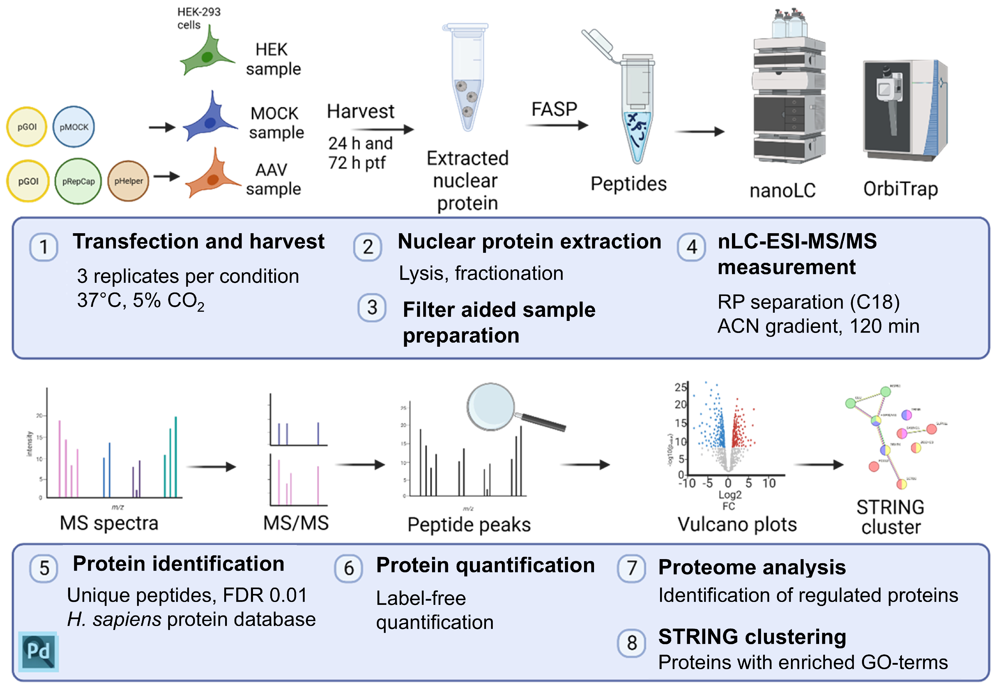
Figure 1. Schematic overview of the workflow of this study. (1) Untransfected (HEK), mock-transfected (MOCK), and rAAV-production-transfected (AAV) cells were harvested 24 h or 72 h after transfection (ptf) in biological triplicates. (2) Samples were prepared for MS/MS measurement by preparing nuclear protein fractions. (3) The nuclear proteins were further processed by the FASP method, including tryptic digest. (4) nLC-ESI-MS/MS measurement was performed with a 120 min gradient with acetonitrile (ACN) on a reverse phase (RP) separation (C18) column. (5, 6) Proteins were identified with Proteome Discoverer (PD) software and quantified in a label-free quantification approach. (7) Regulations of proteins were analyzed, and (8) enriched GO terms were investigated with STRING. Created with BioRender (https://biorender.com/).
Ijms 26 06315 g001
Figure 2. Bright field and fluorescence pictures of HEK-293 cells from HEK, MOCK, and AAV samples 24 h and 72 h post-transfection. After 24 h and 72 h, mVenus fluorescence was detected in MOCK and AAV samples, indicating successful transfection of the pITR-mVenus plasmid.
Ijms 26 06315 g002
Figure 3. (A) Principal component analysis (PCA) plot of the biological triplicates of all six sample groups, generated with Proteome Discoverer (PD) software. In this figure, 24 h ptf HEK control samples are in green; rAAV-transfected are in blue; and MOCK-transfected are in purple. Further, 72 h ptf HEK controls are in red; rAAV-transfected are in orange; and MOCK-transfected are in yellow. (B) Distribution of identified proteins for AAV, HEK, and MOCK samples at both time points combined.
Ijms 26 06315 g003
Figure 4. Analysis of significantly regulated proteins in transfected compared to untransfected HEK-293 cells. Samples were compared according to the time points of harvest, 24 h or 72 h post-transfection. Volcano plots were generated with a cut-off of <0.05 for the p-value and a log2(fold change) of >1, respectively, shown as dashed lines (blue color highlights significantly downregulated and red upregulated proteins). The 10 strongest regulated proteins (fold change) are labeled with UniProt accession numbers. Panels (AF) show the comparisons given in the respective panel titles (AAV vs. HEK, MOCK vs. HEK, AAV vs. MOCK) with 24 h ptf on the left and 72 h ptf on the right side.
Ijms 26 06315 g004
Figure 5. Venn diagram of top regulated proteins. The top ten regulated proteins were highlighted in the volcano plots (Figure 4), and the top regulated proteins of 24 h and 72 h ptf were merged for each comparison (AAV vs. HEK, MOCK vs. HEK, and AAV vs. MOCK). The number of regulated proteins can exceed ten when different proteins are regulated at different time points out of each comparison (see volcano plots, Figure 4). (A) Top down-regulated proteins and (B) up-regulated proteins. UniProt accessions numbers are listed for proteins of each group. A detailed list of detected proteins can be found in the Supplementary Materials.
Ijms 26 06315 g005
Figure 6. Venn diagram of (A) significantly down-regulated or (B) up-regulated proteins in each sample group (versus HEK-untransfected) and their overlaps.
Ijms 26 06315 g006
Figure 7. GO terms of down-regulated (blue) and up-regulated (red) proteins in AAV samples, identified by STRING analysis.
Ijms 26 06315 g007
Table 1. Top down-regulated proteins. Proteins are listed with name, UniProt accession number (sorted in ascending order per group), and fold changes at 24 h and 72 h ptf. At each time point, fold changes in significant top regulated proteins are displayed in bold.
GroupProteinUniProtFold Change
AAV/HEKMOCK/HEKAAV/MOCK
24 h72 h24 h72 h24 h72 h
AAV vs. HEKGlypican-4O754870.04 *0.11 *0.13 *0.190.260.59
Tyrosine-protein kinase FynP06241-10.11 *0.13 *0.13 *0.290.840.46
VinculinP182060.10 *0.39 *0.25 *0.450.390.87
Malate dehydrogenase, cytoplasmicP409250.10 *0.270.190.430.540.61
Centrosomal protein of 68 kDaQ76N323.170.12 *0.530.285.960.44
CDGSH iron-sulfur domain-containing protein 1Q9NZ450.690.13 *0.710.220.970.59
Anaphase-promoting complex subunit 4Q9UJX50.09 *0.580.14 *0.410.61.44
FERM, RhoGEF, and pleckstrin domain-containing protein 1Q9Y4F10.08 *0.390.22 *0.390.381.01
MOCK vs. HEKFERM, RhoGEF, and pleckstrin domain-containing protein 2O94887-10.210.420.06 *0.24 *3.431.78
Histidine-tRNA ligaseP120810.430.350.420.15 *1.012.26
Alcohol dehydrogenase [NADP(+)]P145500.431.410.05 *1.079.561.32
Phosphatidylinositol 4-kinase alphaP423561.30.570.290.16 *4.583.54
DnaJ homolog subfamily C member 3Q132170.171.750.03 *0.966.741.83
Apoptosis-stimulating of p53 protein 2Q13625-10.17 *0.360.03 *0.535.090.68
Reticulocalbin-2Q142570.16 *0.880.06 *0.25 *2.63.54
Histone H2A deubiquitinase MYSM1Q5VVJ20.060.260.05 *0.431.290.61
KAT8 regulatory NSL complex subunit 1Q7Z3B30.240.270.14 *0.13 *1.772.04
U4/U6.U5 small nuclear ribonucleoprotein 27 kDa proteinQ8WVK20.70.15 *0.43 *0.22 *1.630.7
PRKC apoptosis WT1 regulator proteinQ96IZ01.340.380.860.14 *1.552.7
Arginine- and glutamate-rich protein 1Q9NWB60.540.24 *0.500.17 *1.071.45
Glypican-6Q9Y6250.14 *0.40.07 *0.572.110.7
AAV vs. MOCKHLA class I histocompatibility antigen, A-11 alpha chainP137460.351.251.461.460.24 *0.85
UMP-CMP kinaseP30085-10.291.494.060.670.07 *2.24
Polyhomeotic-like protein 1P783640.30 *0.26 *0.570.630.520.42 *
Collagen alpha-1(VII) chainQ02388-10.310.92.121.380.15 *0.65
Max dimerization protein 4Q145820.12 *0.25 *0.35 *0.650.35 *0.38 *
Enhancer of polycomb homolog 2Q52LR70.29 *0.22 *0.34 *0.470.850.47 *
Rab11 family-interacting protein 1Q6WKZ4-40.361.941.131.940.32 *1.00
39S ribosomal protein L55, mitochondrialQ7Z7F72.032.6910.41 *1.770.20 *1.52
Zinc finger protein 444Q8N0Y2-10.760.45 *1.2310.620.45 *
Proline-rich protein PRCCQ927330.25 *0.19 *0.27 *0.540.930.35 *
segment polarity protein disheveled homolog DVL-3Q929972.610.694.32 *1.660.600.42 *
Peptidyl-prolyl cis-trans isomerase NIMA-interacting 4Q9Y2370.15 *0.820.720.80.21 *1.02
Cofilin-2Q9Y281-10.670.812.441.210.28 *0.67
AAV vs. HEK and MOCK vs. HEKAlpha-actinin-4O437070.07 *0.11 *0.07 *0.12 *1.080.92
Probable ATP-dependent RNA helicase DHX34Q141470.08 *0.070.08 *0.1310.52
EF-hand domain-containing protein D2Q96C190.01 *0.10 *0.010.07 *1.191.35
Cell adhesion molecule 1Q9BY670.14 *0.11 *0.16 *0.21 *0.920.52
MAGUK p55 subfamily member 6Q9NZW50.10 *0.21 *0.09 *0.25 *1.130.86
AAV vs. HEK and AAV vs. MOCKProtein XRP2O756950.09 *0.20 *0.390.430.22 *0.47
RadixinP35241-11.130.10 *1.131.051.000.09 *
Isoform 2 of DNA-directed RNA polymerases I and III subunit RPAC2Q9Y2S0-2
P0DPB5
0.25 *0.10 *0.980.33 *0.25 *0.30 *
AAV vs. MOCK and MOCK vs. HEK and AAV vs. HEKPeripheral plasma membrane protein CASKO149360.14 *0.12 *0.760.20 *0.18 *0.59
* significant fold change based on a t-test with a threshold of 0.05.
Table 2. Top up-regulated proteins. Proteins are listed with name, UniProt accession number (sorted in ascending order per group), and fold changes at 24 h and 72 h ptf. At each time point, fold changes in significant top regulated proteins are displayed in bold.
GroupProteinUniProtFold Change
AAV/HEKMOCK/HEKAAV/MOCK
24 h72 h24 h72 h24 h72 h
AAV vs. HEK40S ribosomal protein S6P6275326.9 *4.1 *5.98 *3.444.51.18
Angio-associated migratory cell proteinQ136851.511.6 *0.914.311.692.7
Nicalin, BOS complex subunit NCLNQ969V324.2 *1.86.481.663.731.11
MOCK vs. HEKGeneral transcription factor IIF subunit 2P1398423.93 *8.74 *19.1 *6.5 *1.261.35
26S proteasome non-ATPase regulatory subunit 8P485561.1111.46 *1.18.1 *0.981.41
EpiplakinP5810718.55 *10.31 *14.4 *8.2 *1.291.26
Amyloid-like protein 2Q064812.298.78 *5.2 *11.3 *0.440.78
Ribonuclease h2 subunit bQ5TBB117.89 *10.44 *17.2 *7.0 *1.041.5
ADP-ribosylation factor-like protein 6-interacting protein 4Q66PJ3-119.43 *1.1417.5 *1.21.110.94
CDGSH iron-sulfur domain-containing protein 2Q8N5K117.41.1917.8 *0.70.971.63
Zinc finger protein 668Q96K581.34.211.87.1 *0.710.59
Bromodomain adjacent to zinc finger domain protein 2BQ9UIF8-112.4910.62 *12.2 *7.5 *1.031.42
AAV vs. MOCKRNA 3′-terminal phosphate cyclaseO004420.012.84 *0.260.43 *0.016.5 *
Deformed epidermal autoregulatory factor 1 homologO753983.460.640.210.5716.3 *1.1
Alcohol dehydrogenase [NADP(+)]P145500.431.410.04 *1.079.6 *1.3
Lactoylglutathione lyaseQ04760-14.091.020.10.5840.0 *1.8
Origin recognition complex subunit 1Q134159.731.522.330.344.24.5 *
Beta-2-syntrophinQ13425-10.353.860.510.40.79.6 *
DNA polymerase alpha subunit BQ141810.313.050.010.64≥1004.8 *
Ubiquitin thioesterase otub1Q96FW13.92.130.410.669.5 *3.2
TATA box-binding protein-associated factor RNA polymerase I subunit DQ9H5J819.23 *0.781.970.749.8 *1.0
SUMO-activating enzyme subunit 1Q9UBE0-11.072.780.340.53.25.5 *
DNA helicase ino80Q9ULG14.211.380.430.969.9 *1.4
AAV vs. HEK and MOCK vs. HEKApolipoprotein MO95445-14.2 *28.8 *4.2 *20.3 *1.021.42
Annexin A5P0875860.0 *4.421.6 *6.42.770.68
Annexin A4P0952559.1 *4.826.8 *3.62.21.33
ClusterinP10909-1≥10023.7 *≥10012.2 *1.781.95
SorcinP30626-174.1 *25.9 *86.7 *28.1 *0.850.92
Annexin A11P5099555.9 *6.745.2 *6.21.231.08
28S ribosomal protein S35, mitochondrialP82673-135.4 *3.327.7 *2.91.271.14
Hsp70-binding protein 1Q9NZL4-11.716.0 *1.99.8 *0.931.63
PeflinQ9UBV831.8 *7.5 *43.2 *10.3 *0.730.72
AAV vs. HEK and AAV vs. MOCKZNRD2O60232≥100 *66.7 *3.545.5428.3 *12.0 *
Coatomer subunit betaP3560617.1 *12.0 *2.191.647.8 *7.3 *
NexilinQ0ZGT221.3 *85.8 *0.491.7743.2 *48.3 *
Hypermethylated in cancer 2 proteinQ96JB3-153.3 *47.2 *1.110.8448.1 *55.9 *
DNA-directed RNA polymerases I and III subunit RPAC2Q9Y2S0-1 P0DPB621.1 *26.5 *0.4670.85145.2 *31.1 *
* significant fold change based on a t-test with a threshold of 0.05.
Table 3. List of regulated proteins assigned to the GO term ‘response to virus’ with their UniProt accession number, name, abbreviation, gene symbol, and fold changes. Fold changes are colored by range: white < 2, light pink < 4, pink < 8, and red ≥ 8.
UniProt AccessionDescriptionProteinGene
Symbol
Fold Change
AAV vs. HEK
Fold Change MOCK vs. HEKFold Change AAV vs. MOCK
24 h72 h24 h72 h24 h72 h
P10909-1ClusterinCLUSCLU4.9 *20.9 **4.29.2 **1.22.3
Q9Y3Z3Deoxynucleoside triphosphate triphosphohydrolase SAMHD1SAMH1SAMHD14.2 *10.8 **1.04.0 *4.2 *2.7
Q7Z7L1Schlafen family member 11SLN11SLFN118.1 *6.2 **4.3 *2.51.92.5
Q9BYW2Histone-lysine N-methyltransferase SETD2SETD2SETD21.44.3 **0.61.52.22.9
Q6L8Q7-12′,5′-phosphodiesterase 12PDE12PDE12n/a ***3.2 *n/a1.3n/a2.4 *
O00571ATP-dependent RNA helicase DDX3XDDX3XDDX3X2.7 **2.6 **2.0 **2.2 **1.31.2
P04792Heat shock protein beta-1HSPB1HSPB10.92.5 *1.99.9 *0.91.2
Q9NUD5Zinc finger CCHC domain-containing protein 3ZCHC3ZCCHC34.8 **2.3 *5.9 **1.80.81.2
Q9C030Tripartite motif-containing protein 6TRIM6TRIM60.72.1 *0.81.80.91.2
P07900Heat shock protein HSP 90-alphaHSP90AA1HSP90AA10.62.0 **0.61.21.11.7 *
* significant fold change calculated in a t-test with a threshold of 0.05; ** significant fold changes, based on Benjamini-corrected p-values of the t-test with a threshold of 0.05; *** ‘n/a’ abbreviates ‘not available’.
Table 4. Regulated proteins with context to antiviral response in HEK-293 cells producing rAAV upon plasmid transfection identified by literature search. Proteins are listed with their UniProt accession number, name, abbreviation, gene symbol, and fold changes. Fold changes are colored by range: white < 2, light pink < 4, pink < 8.
UniProt AccessionDescriptionProteinGene
Symbol
Fold Change
AAV vs. HEK
Fold Change MOCK vs. HEKFold Change AAV vs. MOCK
24 h72 h24 h72 h24 h72 h
O00743Serine/threonine-protein phosphatase 6 catalytic subunitPPP6CPPP6C1.12.8 *0.81.11.32.6
Q9NUD5Zinc finger CCHC domain-containing protein 3ZCHC3ZCCHC34.8 **2.3 *5.9 **1.80.81.2
* significant fold change calculated in a t-test with a threshold of 0.05; ** significant fold changes, based on Benjamini-corrected p-values of the t-test with a threshold of 0.05.
Table 5. Regulated proteins with context to adenoviral proteins identified by literature search. Proteins are listed with UniProt accession number, name, abbreviation, gene symbol, and fold changes. Fold changes are colored by range: deep blue ≤ 0.2, blue ≤ 0.5, white < 2, light pink < 4, pink < 8.
UniProt AccessionDescriptionProteinGene
Symbol
Fold Change
AAV vs. HEK
Fold Change MOCK vs. HEKFold Change AAV vs. MOCK
24 h72 h24 h72 h24 h72 h
P63151Serine/threonine-protein phosphatase 2A 55 kDa regulatory subunit B alpha isoform2ABAPPP2R2A1.32.6 *0.61.32.2 *2
P30154Serine/threonine-protein phosphatase 2A 65 kDa regulatory subunit A beta isoform2AABPPP2R1B0.70.60.3 **0.62.3 *0.9
P01106Myc proto-oncogene proteinMYCMYC0.60.70.70.90.90.8
P26196Probable ATP-dependent RNA helicase DDX6DDX6DDX62.1 **1.9 **1.6 *1.51.31.3
P04637Cellular tumor antigen p53P53TP530.80.6 **0.90.80.90.7
Q92878DNA repair protein Rad50RAD50RAD500.4 *0.2 **0.20.61.70.3 *
Q93034Cullin-5CUL5CUL52.7 *5.4 **1.621.72.7
* significant fold change calculated in a t-test with a threshold of 0.05; ** significant fold changes, based on Benjamini-corrected p-values of the t-test with a threshold of 0.05.
Disclaimer/Publisher’s Note: The statements, opinions and data contained in all publications are solely those of the individual author(s) and contributor(s) and not of MDPI and/or the editor(s). MDPI and/or the editor(s) disclaim responsibility for any injury to people or property resulting from any ideas, methods, instructions or products referred to in the content.